503 Journal of Sport & Exercise Psychology, 2013, 35, 503-513 © 2013 Human Kinetics, Inc. Gavin Tempest and Gaynor Parfitt are with the Exercise for Health and Human Performance Group, Sansom Institute for Health Research, School of Health Sciences, University of South Australia, Adelaide, SA, Australia. Imagery Use and Affective Responses During Exercise: An Examination of Cerebral Hemodynamics Using Near-Infrared Spectroscopy Gavin Tempest and Gaynor Parfitt University of South Australia Imagery, as a cognitive strategy, can improve affective responses during moderate-intensity exercise. The effects of imagery at higher intensities of exercise have not been examined. Further, the effect of imagery use and activity in the frontal cortex during exercise is unknown. Using a crossover design (imagery and control), activity of the frontal cortex (reflected by changes in cerebral hemodynamics using near-infrared spectros- copy) and affective responses were measured during exercise at intensities 5% above the ventilatory threshold (VT) and the respiratory compensation point (RCP). Results indicated that imagery use influenced activity of the frontal cortex and was associated with a more positive affective response at intensities above VT, but not RCP to exhaustion (p < .05). These findings provide direct neurophysiological evidence of imagery use and activity in the frontal cortex during exercise at intensities above VT that positively impact affective responses. Keywords: frontal cortex, cognitive strategy, exercise intensity, physical activity, enjoyment How an individual feels (pleasure/displeasure) during exercise is an important factor for exercise adher- ence and participation (for a recent review, see Ekkekakis, Parfitt, & Petruzzello, 2011). This affective response, a construct underlying core affect (Russell, 1980), can be positive or negative and has been shown to significantly predict future exercise behavior in both adolescents (Schneider, Dunn, & Cooper, 2009) and adults (Williams et al., 2008; Williams, Dunsiger, Jennings, & Marcus, 2012). A causal link between affective responses, recog- nized as a possible adjunct measure of exercise intensity (Garber et al., 2011), and adherence to exercise has been fueled by evidence toward a dose–response relationship between the intensity of exercise (standardized to gas exchange thresholds; GETs) and affective responses (Ekkekakis, 2003). Research has established that affective responses are variable at exercise intensities around the ventilatory threshold (VT; the point of transition from aerobic to anaerobic metabolism), with some individuals reporting a positive and others a negative response (Ekkekakis, Hall, & Petruzzello, 2008; Rose & Parfitt, 2010). Above the VT, proximal to the respiratory compensation point (RCP; the point at which a physiological steady state can no longer be maintained and metabolic acidosis occurs and leads to exhaustion), affective responses are uniformly nega- tive (Ekkekakis, Hall, & Petruzzello, 2004; Parfitt, Rose, & Burgess, 2006; Rose & Parfitt, 2007; Welch, Hulley, Ferguson, & Beauchamp, 2007). Individuals experience physical discomfort and negative affective responses (feelings of displeasure) during exercise, especially following a period of seden- tary living (Petruzzello, Jones, & Tate, 1997). Cognitive or psychological strategies have the potential to promote positive affective responses (increase feelings of pleasure) during exercise (Blanchard, Rodgers, & Gauvin, 2004; Lind, Welch, & Ekkekakis, 2009; Salmon, Hanneman, & Harwood, 2010). Imagery is a cognitive strategy that involves re- creating experiences using information from all the senses—namely, sight, sound, smell, and touch—stored in the memory (Weinberg, 2008). During exercise, an individual might image themself doing a specific activ- ity (e.g., exercising on a bike) and focus on their senses (e.g., the sound of the wheel spinning, the feeling of their legs pumping) to achieve a specific goal for that activity (e.g., to complete a certain distance). Frequent exercis- ers have reported using imagery techniques more than less-frequent exercisers for cognitive and motivational purposes, such as for goal attainment and psychological management (Hall, 1995; Hausenblas, Hall, Rodgers, & Munroe, 1999). The use of imagery techniques during and follow- ing moderate-intensity exercise (50% heart rate reserve; HRR) has recently been shown to improve affective responses (Stanley & Cumming, 2010a; 2010b). Based upon Lang’s (1979) bioinformational theory of emotional JOURNAL OF SPORT EXERCISE PSYCHOLOGY Official Journal of NASPSPA www.JSEP-Journal.com ORIGINAL RESEARCH

Welcome message from author
This document is posted to help you gain knowledge. Please leave a comment to let me know what you think about it! Share it to your friends and learn new things together.
Transcript
503
Journal of Sport & Exercise Psychology, 2013, 35, 503-513 © 2013 Human Kinetics, Inc.
Gavin Tempest and Gaynor Parfitt are with the Exercise for Health and Human Performance Group, Sansom Institute for Health Research, School of Health Sciences, University of South Australia, Adelaide, SA, Australia.
Imagery Use and Affective Responses During Exercise: An Examination of Cerebral Hemodynamics
Using Near-Infrared Spectroscopy
Gavin Tempest and Gaynor ParfittUniversity of South Australia
Imagery, as a cognitive strategy, can improve affective responses during moderate-intensity exercise. The effects of imagery at higher intensities of exercise have not been examined. Further, the effect of imagery use and activity in the frontal cortex during exercise is unknown. Using a crossover design (imagery and control), activity of the frontal cortex (reflected by changes in cerebral hemodynamics using near-infrared spectros-copy) and affective responses were measured during exercise at intensities 5% above the ventilatory threshold (VT) and the respiratory compensation point (RCP). Results indicated that imagery use influenced activity of the frontal cortex and was associated with a more positive affective response at intensities above VT, but not RCP to exhaustion (p < .05). These findings provide direct neurophysiological evidence of imagery use and activity in the frontal cortex during exercise at intensities above VT that positively impact affective responses.
Keywords: frontal cortex, cognitive strategy, exercise intensity, physical activity, enjoyment
How an individual feels (pleasure/displeasure) during exercise is an important factor for exercise adher-ence and participation (for a recent review, see Ekkekakis, Parfitt, & Petruzzello, 2011). This affective response, a construct underlying core affect (Russell, 1980), can be positive or negative and has been shown to significantly predict future exercise behavior in both adolescents (Schneider, Dunn, & Cooper, 2009) and adults (Williams et al., 2008; Williams, Dunsiger, Jennings, & Marcus, 2012). A causal link between affective responses, recog-nized as a possible adjunct measure of exercise intensity (Garber et al., 2011), and adherence to exercise has been fueled by evidence toward a dose–response relationship between the intensity of exercise (standardized to gas exchange thresholds; GETs) and affective responses (Ekkekakis, 2003).
Research has established that affective responses are variable at exercise intensities around the ventilatory threshold (VT; the point of transition from aerobic to anaerobic metabolism), with some individuals reporting a positive and others a negative response (Ekkekakis, Hall, & Petruzzello, 2008; Rose & Parfitt, 2010). Above the VT, proximal to the respiratory compensation point (RCP; the point at which a physiological steady state can no longer be maintained and metabolic acidosis occurs and leads to exhaustion), affective responses are uniformly nega-
tive (Ekkekakis, Hall, & Petruzzello, 2004; Parfitt, Rose, & Burgess, 2006; Rose & Parfitt, 2007; Welch, Hulley, Ferguson, & Beauchamp, 2007).
Individuals experience physical discomfort and negative affective responses (feelings of displeasure) during exercise, especially following a period of seden-tary living (Petruzzello, Jones, & Tate, 1997). Cognitive or psychological strategies have the potential to promote positive affective responses (increase feelings of pleasure) during exercise (Blanchard, Rodgers, & Gauvin, 2004; Lind, Welch, & Ekkekakis, 2009; Salmon, Hanneman, & Harwood, 2010).
Imagery is a cognitive strategy that involves re-creating experiences using information from all the senses—namely, sight, sound, smell, and touch—stored in the memory (Weinberg, 2008). During exercise, an individual might image themself doing a specific activ-ity (e.g., exercising on a bike) and focus on their senses (e.g., the sound of the wheel spinning, the feeling of their legs pumping) to achieve a specific goal for that activity (e.g., to complete a certain distance). Frequent exercis-ers have reported using imagery techniques more than less-frequent exercisers for cognitive and motivational purposes, such as for goal attainment and psychological management (Hall, 1995; Hausenblas, Hall, Rodgers, & Munroe, 1999).
The use of imagery techniques during and follow-ing moderate-intensity exercise (50% heart rate reserve; HRR) has recently been shown to improve affective responses (Stanley & Cumming, 2010a; 2010b). Based upon Lang’s (1979) bioinformational theory of emotional
JOURNAL OF SPORT EXERCISE
PSYCHOLOGYOfficial Journal of NASPSPA
www.JSEP-Journal.comORIGINAL RESEARCH
504 Tempest and Parfitt
imagery, Stanley and Cumming (2010a) examined the effectiveness of technique-, enjoyment-, and energy-focused imagery scripts. Following exercise, affective responses were significantly more positive in participants who used enjoyment-focused scripts and reductions in exhaustion were marginally decreased in participants who used energy-focused scripts (Stanley & Cumming, 2010a). These results support the use of imagery as a suit-able cognitive strategy during exercise to help promote a more positive or pleasurable experience. However, the findings from Stanley and Cumming (2010a) are based upon moderate-intensity exercise, where affective responses are frequently reported to be mostly positive (Ekkekakis et al., 2011) and the exercise intensity (50% HRR) was not standardized to individual metabolic mark-ers, such as GETs.
How affective responses are regulated in the brain during exercise and the influence of such cognitive strate-gies have not yet been investigated. Evidence from brain-imaging studies has shown increased activity in different divisions of the frontal cortex (for example, dorsolateral and ventromedial areas), induced by attempts of active cognitive coping with a negative affective stimulus (Och-sner, Bunge, Gross, & Gabrieli, 2002; Ochsner & Gross, 2008; Phan et al., 2005). The frontal cortex is thought to provide top-down regulation of subcortical structures in the presence of emotional stimuli through active cognitive coping (appraisal or reappraisal) processes. Top-down regulation primarily activates bilateral areas of the fron-tal cortex. However, positive stimuli have been shown to induce activity in left-lateralized (Kim & Hamann, 2007; Ochsner et al., 2004) and negative stimuli in right-lateralized areas of the frontal cortex (Ochsner et al., 2004). Specifically, activity in the left dorsolateral frontal cortex has been shown to increase during reappraisal and is associated with reduced self-reported negative affect (Ochsner et al., 2002) and activity in the right dorsolateral frontal cortex is increased in attempts to inhibit emotional responses (Beauregard, Levesque, & Bourgouin, 2001). A cognitively driven strategy such as imagery should engage the frontal cortex during exercise at intensities that would otherwise induce a negative affective state (i.e., above VT), and result in a more positive (or less negative) affective response through a top-down regulatory process.
Near-infrared spectroscopy (NIRS), a noninvasive optical neuroimaging technique, is an effective tool for examining cerebral hemodynamics in areas on the surface of the frontal cortex and can be used during exercise to provide direct temporal (Huppert et al., 2006) and acceptable spatial resolution (approx. 1 cm; Rooks, Thom, McCully, & Dishman, 2010). NIRS provides a quantitative measure of changes in cerebral oxyhemoglo-bin (O2Hb), deoxyhemoglobin (HHb), and blood volume (total hemoglobin; tHb = O2Hb + HHb), which reflects functional neuronal activation (Bhambhani, Malik, & Mookerjee, 2007; Ekkekakis, 2009; Ferrari, Mottola, & Quaresima, 2004).
NIRS has been used to study parieto-occipital areas of the cortex and the affective perception of imagery
(Köchel et al., 2011) and in combination with electro-encephalographic (EEG) methods (Herrmann et al., 2008). This research has successfully replicated findings investigating visual imagery and affective perception using functional magnetic resonance imaging (fMRI) methods (Schienle, Schäfer, & Vaitl, 2008; Schienle, Schäfer, Pignanelli, & Vaitl, 2009). In recent fMRI work, participants used imagery (kinetic and visual) to imagine themselves walking. This resulted in activation in frontoparietal regions, specifically the right and left dorsolateral prefrontal cortices and the anterior insulae (Cremers, Dessoullieres, & Garraux, 2012; van der Meulen, Allali, Rieger, Assal, & Vuilleumier, 2012). No studies to our knowledge have used NIRS to examine areas of the frontal cortex and imagery use or affec-tive responses to exercise. Therefore, the current study investigates whether imagery use influences activity (reflected by changes in hemodynamics using NIRS) of the dorsolateral region of the frontal cortex (both right and left hemispheres) and affective responses to exercise at intensities (standardized to GETs) above VT (5% above VT) and at RCP to exhaustion.
The following three hypotheses were examined.
1. Affective responses are (a) more positive at exercise intensities above VT than at exercise intensities RCP to exhaustion and (b) more positive in the imagery than in the control condition.
2. Activity of the frontal cortex is greater in the imagery than in the control condition.
3. Activity of the frontal cortex is associated with affective responses to exercise and imagery use.
The study aimed to examine frontal cortex activity of both right- and left-lateralized areas of the frontal cortex during exercise (Hypotheses 2 and 3).
Methods
Participants
Participants (n = 14; age, M = 21.79, SD = 2.15 years; height, M = 173.93, SD = 9.73 cm; body mass, M = 73.22, SD =18.17 kg) were recruited from a student population. There were equal numbers of males and females and all participants reported partaking >3 hr of moderate physical activity per week. The volunteers read and signed an informed consent form that was approved by the University Ethics Committee and completed a preexercise testing health status questionnaire before participation in the study.
NIRS
Cerebral hemodynamics were measured using NIRS (NIRO 200 Hamamatsu Photonics, Hamamatsu, Japan). The emitter and detector were encased in a rubber holder with a separation distance of 4 cm. A differential path length factor of 5.93 for the adult forehead was used (van der Zee et al., 1992) to provide a measure of concentra-
Imagery Use and Affective Responses During Exercise 505
tion changes in micromolar (µM) units of O2Hb, HHb, and tHb. Tissue oxygenation index (TOI), percentage ratio of O2Hb to tHb indicating oxygen availability, was measured using spatial resolved spectroscopy based on the attenuation of light from two light-detector distances (Wolf, Ferrari, & Quaresima, 2007). The probes were placed over the left and right hemispheres (dorsolateral prefrontal cortex areas; midpoint between Fp1-F3 and Fp2-F4 respectively, of the international 10-20 system for EEG electrode placement) and secured to the skin using a double adhesive sticker. Elastic surgical tape and a dark bandage were placed over the holders around the forehead.
Affective Responses
Affective valence (pleasure/displeasure) was measured using the Feeling Scale (Hardy & Rejeski, 1989). The scale is a unidimensional 11-point scale ranging from –5 to +5 with verbal anchors at all odd integers and at the zero point (–5, very bad; –3, bad; –1, fairly bad; 0, neutral; 1, fairly good; 3, good; 5, very good). The sim-plicity and brevity of the Feeling Scale allows multiple assessments to be made during exercise (i.e., pre-, during, and postexercise) to capture any acute fluctuations in responses. Van Landuyt, Ekkekakis, Hall, & Petruzzello (2000) have previously shown Feeling Scale correlations ranging from 0.51 to 0.88 with the Valance scale of the Self-Assessment Manikin (Lang, 1980). The Feeling Scale corresponds to one of the two dimensions of the circumplex model of feeling states (Russell, 1980) and is recommended to measure basic affect (pleasure/displeasure; see Ekkekakis & Petruzzello, 1999). The Feeling Scale has been successfully used in studies with adolescents (Schneider et al., 2009; Sheppard & Parfitt, 2008), and both active (Ekkekakis et al., 2004, 2008) and sedentary (Lind et al., 2005; Parfitt et al., 2006) adults.
Imagery Script
The imagery script combined enjoyment- and energy-focused imagery in line with previous work by Stanley and Cumming (2010a, 2010b). The authors showed that enjoyment-focused imagery improved individual’s affective responses as it provided the necessary response propositions for a more positive feeling state during exercise at moderate intensities (below VT). Energy-focused imagery, despite having response propositions to increase energy levels thought to impact motivational and emotional responses, did not increase a more positive feeling state any more than imagery without affective con-tent (i.e., technique imagery). However, energy-focused imagery did appear to decrease feelings of physical exhaustion, which would be beneficial for improving feeling states, particularly at higher intensities of exercise (above VT). Therefore, the combination of enjoyment- and energy-focused imagery was used because it should positively influence affective responses during exercise at intensities above VT.
The imagery script was read to each participant by the experimenter (lasting approximately 5 min) as they sat comfortably in a quiet room outside the exercise laboratory. The script required participants to recall a previous exercise situation when they had found exercise enjoyable and when they had felt energized. The use of one’s own personal experience has been shown to be more effective that using a standardized or generic imagery script (Lang, 1979; Wilson, Smith, Burden, & Holmes, 2010). Participants were asked to re-create the feelings and to imagine their previous experience as vividly as possible (e.g., sounds, colors, smells, and people). After recalling how pleasurable and energized they felt, they were instructed to use these memories in the exercise they were about to perform.
The participants were then seated on the recumbent cycle ergometer in the exercise laboratory and the script was read to the participant by the experimenter once again. Excerpts of the imagery scripts were read to the participants every 2–3 min to keep the participants focused on feelings of enjoyment and energy during exercise. In the control condition, participants received no instructions or prompts during exercise and sat com-fortably in the quiet room before exercise in place of the imagery script. To check participants’ perceived ability to use imagery and to evaluate their ease of use and the effect of the imagery script during exercise, participants were asked to complete a postexercise imagery script evaluation (see Stanley & Cumming, 2010a). The evalu-ation uses a 7-point Likert scale to provide a rating of participant’s visual and kinetic abilities (how well the image of exercise was created and how strongly they felt could feel the action of the imagined exercise, respec-tively), clarity, emotion, energy, and enjoyment evoked from the imagery script. The evaluation scores were used as an indicator of compliance to and effectiveness of the imagery techniques. The relationship between postexercise imagery scores and cerebral hemodynamics in the imagery condition was examined (Hypothesis 3).
Procedures
This study employed a crossover design. Each participant completed a control and experimental (i.e., imagery) condition so that the effect of imagery could be com-pared between the two sessions. The study required the participants to visit the laboratory on three occasions. On the first visit, participants’ age, height, and weight were recorded. The procedures for the exercise tests were explained and a description of the Feeling Scale was provided. The participants then completed an incremental (20 W/min) exercise test to exhaustion on a recumbent cycle ergometer (Lode Angio BV, Groningen, the Netherlands). Expired gases were collected continu-ously using a computerized metabolic analysis system (Metaylzer 3B, Cortex Biophysik, Leipzig, Germany). Participants’ individual peak oxygen uptake (VO2peak), VT, and RCP were determined. The achievement of VO2peak was verified by (a) a peak or plateau in oxygen
506 Tempest and Parfitt
consumption (changes <2 mL·kg–1·min–1) with increas-ing workload and (b) a respiratory exchange ratio of at least 1.10.
The participants returned for a further two sessions: imagery and control (in a randomized, counterbalanced order). Following a 3-min warm-up (50 W), both sessions involved exercising for 10 min at a workload correspond-ing to 5% above VT (above VT; M = 108, SD = 21 W), followed by 10 min of passive recovery, and then exercise at RCP to exhaustion (M = 177, SD = 32 W). A 10-min duration of exercise at the above VT intensity was chosen to ensure all participants would complete the exercise. Participants reported their affective responses pre- and postexercise, and at 2-min intervals during exercise.
Data Reduction
The VO2peak was determined by the highest 30-s average of oxygen uptake (VO2 L/min). The VT was determined using the three-method procedure proposed by Gaskill et al. (2001). This involved agreement in the visual determi-nation of VT using (a) the ventilatory equivalent method, (b) the excess CO2 method, and (c) the modified V-slope method. The RCP was determined visually by agreement between plots of minute ventilation over CO2 production and ventilatory equivalent of CO2 over time (Beaver, Was-serman, & Whipp, 1986). The determination of VT and RCP were verified and checked by two investigators. The participants’ mean VO2 values (L/min) as percentages of VO2peak were 55.3% (SD = 2.6) at VT and 84.5% (SD = 2.7) at RCP. Corresponding workloads of 5% above VT and the RCP were determined for exercise in the two conditions (imagery and control).
A 30-s baseline measure of cerebral hemodynamics (O2Hb, HHb, and tHb) was recorded before exercise at each intensity (above VT and at RCP to exhaustion) to control for baseline differences within and between ses-sions. Baseline measures were subtracted from the data extracted during exercise. Therefore, O2Hb, HHb, and tHb represent the change in the hemodynamic response at selected points during exercise. The TOI is an absolute measure of oxygenation (%); therefore, adjustments for baseline measures were not required.
To standardize data points between participants and enable analysis of the Intensity (above VT and RCP to exhaustion) and Time (selected points during exercise above VT and RCP to exhaustion) factors, percentage duration of exercise was used. The NIRS variables were time aligned with the gas data and 20-s averages were extracted from the associated GETs: point of 5% above VT and at RCP, followed by 20, 40, 60, 80, and 100% duration of exercise. The duration of exercise at intensi-ties above VT was 10 min, and the mean from RCP to exhaustion was 9.35 min (SD = 5.47) in the imagery and 8.43 min (SD = 4.02) in the control condition. Affective responses were expressed as a percentage of the duration of exercise to include pre- and postexercise measures at intensities above VT and RCP to exhaustion: preexercise, GET, 20, 60, 100%, postexercise.
Statistical Analyses
The independent variable was the enjoyment- and energy-focused imagery script. The dependent variables were self-reported affective responses, changes in cerebral hemodynamics (measured using NIRS), and scores from the postexercise imagery evaluation.
To examine Hypothesis 1, a Condition (2; imagery and control) × Intensity (2; above VT and RCP to exhaustion) × Time (6; preexercise, GET, 20, 60, 100%, postexercise) analysis of variance (ANOVA) with repeated measures was conducted on the affective responses. To examine Hypothesis 2, separate Condition (2; imagery and control) × Hemisphere (2; right and left) × Time (6; GET, 20, 40, 60, 80, 100%) ANOVAs with repeated measures were conducted on each of the NIRS variables (O2Hb, HHb, tHb and TOI) at each of the exercise intensities (above VT and RCP to exhaustion). Finally, to examine Hypothesis 3, Pearson’s bivariate correlation analysis was used to exam-ine the relationships between cerebral hemodynamics, affective responses, and postexercise imagery evaluation scores (imagery condition only). Greenhouse–Geisser cor-rections were applied if the assumption of sphericity was not met, and significant main and interaction effects (p < .05) were followed up using repeated-measures contrasts. Effect sizes associated with F statistics were expressed as eta squared (η2) defined as η2 ≤ .01 = small; ≤ .06 = medium; and ≥ .14 = large (Cohen, 1988). All results are reported as means and standard deviations.
Results
Preliminary Analyses
To ensure the intensity of exercise (indicated by VO2 L/min) was the same between the two exercise sessions (imagery and control conditions), and that these were not different from the initial incremental exercise test, a Session × Intensity ANOVA with repeated measures was conducted. In this analysis, three intensities were compared: 5% above VT, RCP, and the end of exercise (exhaustion). The analysis showed that VO2 (L/min) at 5% above VT (M = 1.71, SD = 0.38), RCP (M = 2.49, SD = 0.56), and exhaustion (M = 2.93, SD = 0.65) were significantly different, F(1, 18) = 206.58, p < .001, η2 = .90. No significant main effect of Session, F(2, 26) = .18, p > .05, η2 = .00, or Session × Intensity interaction, F(4, 52) = .62, p > .05, η2 = .01, was recorded.
The postexercise imagery evaluation scores indicated participants’ visual (M = 5.82, SD = 1.07) and kinetic ability (M = 5.14, SD = 1.10), clarity (M = 5.64, SD = 0.84), emotion (M = 5.50, SD = 0.65), energy (M = 5.21, SD = 1.48), and enjoyment (M = 4.64, SD = 1.39) evoked from the script.
Affective Responses Preexercise, During, and Postexercise (Hypothesis 1)
Significant main effects of Condition, F(1, 13) = 29.06, p < .001, η2 = .04; Intensity, F(1, 13) = 61.88, p < .001, η2 = .31;
Imagery Use and Affective Responses During Exercise 507
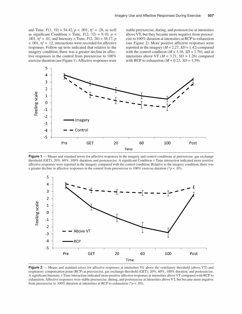
and Time, F(1, 19) = 54.42, p < .001, η2 = .28, as well as significant Condition × Time, F(2, 32) = 9.10, p < .001, η2 = .01, and Intensity × Time, F(2, 26) = 38.17, p < .001, η2 = .12, interactions were recorded for affective responses. Follow-up tests indicated that relative to the imagery condition, there was a greater decline in affec-tive responses in the control from preexercise to 100% exercise duration (see Figure 1). Affective responses were
Figure 1 — Means and standard errors for affective responses in the imagery and control conditions at preexercise, gas exchange threshold (GET), 20%, 60%, 100% duration, and postexercise. A significant Condition × Time interaction indicated more positive affective responses were reported in the imagery compared with the control condition. Relative to the imagery condition, there was a greater decline in affective responses in the control from preexercise to 100% exercise duration (*p < .05).
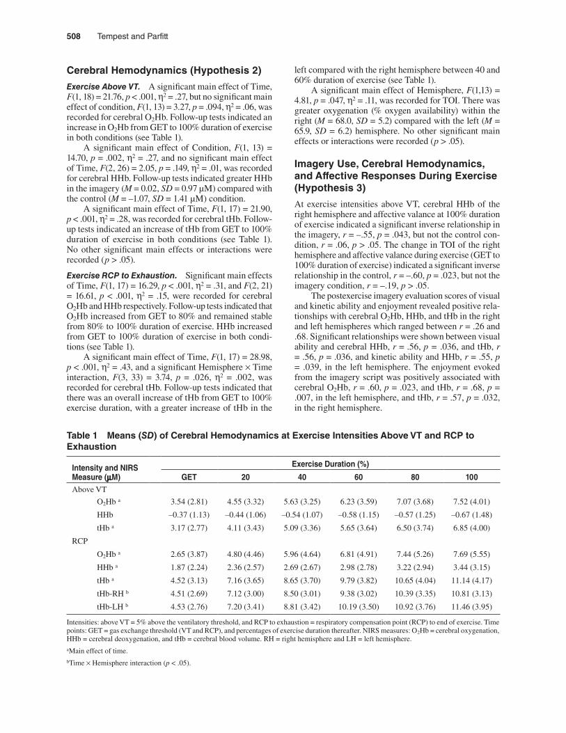
stable preexercise, during, and postexercise at intensities above VT, but they became more negative from preexer-cise to 100% duration at intensities at RCP to exhaustion (see Figure 2). More positive affective responses were reported in the imagery (M = 2.27, SD = 1.42) compared with the control condition (M = 1.16, SD = 1.76), and at intensities above VT (M = 3.21, SD = 1.26) compared with RCP to exhaustion (M = 0.23, SD = 1.59).
Figure 2 — Means and standard errors for affective responses at intensities 5% above the ventilatory threshold (above VT) and respiratory compensation point (RCP) at preexercise, gas exchange threshold (GET), 20%, 60%, 100% duration, and postexercise. A significant Intensity × Time interaction indicated more positive affective responses at intensities above VT compared with RCP to exhaustion. Affective responses were stable preexercise, during, and postexercise at intensities above VT, but became more negative from preexercise to 100% duration at intensities at RCP to exhaustion (*p < .05).
508 Tempest and Parfitt
Cerebral Hemodynamics (Hypothesis 2)
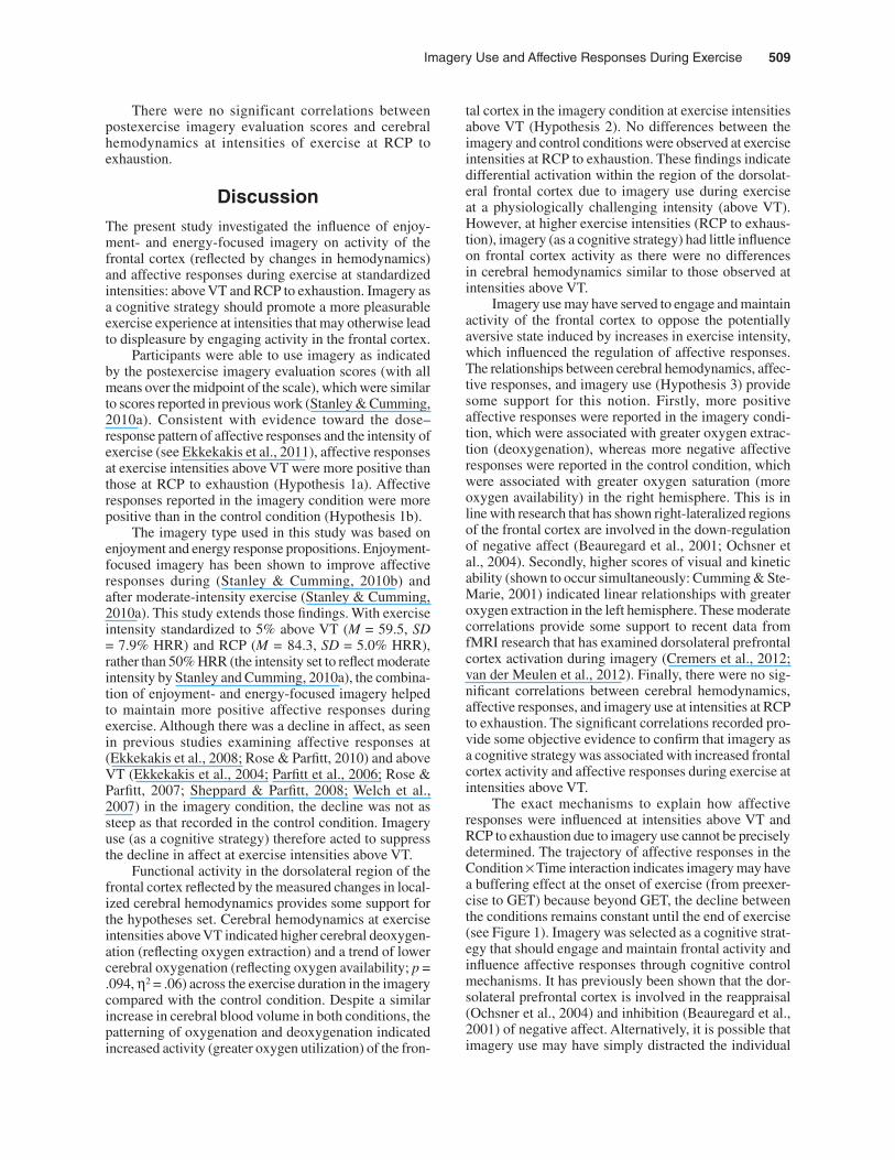
Exercise Above VT. A significant main effect of Time, F(1, 18) = 21.76, p < .001, η2 = .27, but no significant main effect of condition, F(1, 13) = 3.27, p = .094, η2 = .06, was recorded for cerebral O2Hb. Follow-up tests indicated an increase in O2Hb from GET to 100% duration of exercise in both conditions (see Table 1).
A significant main effect of Condition, F(1, 13) = 14.70, p = .002, η2 = .27, and no significant main effect of Time, F(2, 26) = 2.05, p = .149, η2 = .01, was recorded for cerebral HHb. Follow-up tests indicated greater HHb in the imagery (M = 0.02, SD = 0.97 µM) compared with the control (M = –1.07, SD = 1.41 µM) condition.
A significant main effect of Time, F(1, 17) = 21.90, p < .001, η2 = .28, was recorded for cerebral tHb. Follow-up tests indicated an increase of tHb from GET to 100% duration of exercise in both conditions (see Table 1). No other significant main effects or interactions were recorded (p > .05).
Exercise RCP to Exhaustion. Significant main effects of Time, F(1, 17) = 16.29, p < .001, η2 = .31, and F(2, 21) = 16.61, p < .001, η2 = .15, were recorded for cerebral O2Hb and HHb respectively. Follow-up tests indicated that O2Hb increased from GET to 80% and remained stable from 80% to 100% duration of exercise. HHb increased from GET to 100% duration of exercise in both condi-tions (see Table 1).
A significant main effect of Time, F(1, 17) = 28.98, p < .001, η2 = .43, and a significant Hemisphere × Time interaction, F(3, 33) = 3.74, p = .026, η2 = .002, was recorded for cerebral tHb. Follow-up tests indicated that there was an overall increase of tHb from GET to 100% exercise duration, with a greater increase of tHb in the
left compared with the right hemisphere between 40 and 60% duration of exercise (see Table 1).
A significant main effect of Hemisphere, F(1,13) = 4.81, p = .047, η2 = .11, was recorded for TOI. There was greater oxygenation (% oxygen availability) within the right (M = 68.0, SD = 5.2) compared with the left (M = 65.9, SD = 6.2) hemisphere. No other significant main effects or interactions were recorded (p > .05).
Imagery Use, Cerebral Hemodynamics, and Affective Responses During Exercise (Hypothesis 3)
At exercise intensities above VT, cerebral HHb of the right hemisphere and affective valance at 100% duration of exercise indicated a significant inverse relationship in the imagery, r = –.55, p = .043, but not the control con-dition, r = .06, p > .05. The change in TOI of the right hemisphere and affective valance during exercise (GET to 100% duration of exercise) indicated a significant inverse relationship in the control, r = –.60, p = .023, but not the imagery condition, r = –.19, p > .05.
The postexercise imagery evaluation scores of visual and kinetic ability and enjoyment revealed positive rela-tionships with cerebral O2Hb, HHb, and tHb in the right and left hemispheres which ranged between r = .26 and .68. Significant relationships were shown between visual ability and cerebral HHb, r = .56, p = .036, and tHb, r = .56, p = .036, and kinetic ability and HHb, r = .55, p = .039, in the left hemisphere. The enjoyment evoked from the imagery script was positively associated with cerebral O2Hb, r = .60, p = .023, and tHb, r = .68, p = .007, in the left hemisphere, and tHb, r = .57, p = .032, in the right hemisphere.
Table 1 Means (SD) of Cerebral Hemodynamics at Exercise Intensities Above VT and RCP to Exhaustion
Intensity and NIRSMeasure (μM)
Exercise Duration (%)
GET 20 40 60 80 100Above VT
O2Hb a 3.54 (2.81) 4.55 (3.32) 5.63 (3.25) 6.23 (3.59) 7.07 (3.68) 7.52 (4.01)
HHb –0.37 (1.13) –0.44 (1.06) –0.54 (1.07) –0.58 (1.15) –0.57 (1.25) –0.67 (1.48)
tHb a 3.17 (2.77) 4.11 (3.43) 5.09 (3.36) 5.65 (3.64) 6.50 (3.74) 6.85 (4.00)
RCP
O2Hb a 2.65 (3.87) 4.80 (4.46) 5.96 (4.64) 6.81 (4.91) 7.44 (5.26) 7.69 (5.55)
HHb a 1.87 (2.24) 2.36 (2.57) 2.69 (2.67) 2.98 (2.78) 3.22 (2.94) 3.44 (3.15)
tHb a 4.52 (3.13) 7.16 (3.65) 8.65 (3.70) 9.79 (3.82) 10.65 (4.04) 11.14 (4.17)
tHb-RH b 4.51 (2.69) 7.12 (3.00) 8.50 (3.01) 9.38 (3.02) 10.39 (3.35) 10.81 (3.13)
tHb-LH b 4.53 (2.76) 7.20 (3.41) 8.81 (3.42) 10.19 (3.50) 10.92 (3.76) 11.46 (3.95)
Intensities: above VT = 5% above the ventilatory threshold, and RCP to exhaustion = respiratory compensation point (RCP) to end of exercise. Time points: GET = gas exchange threshold (VT and RCP), and percentages of exercise duration thereafter. NIRS measures: O2Hb = cerebral oxygenation, HHb = cerebral deoxygenation, and tHb = cerebral blood volume. RH = right hemisphere and LH = left hemisphere.aMain effect of time.bTime × Hemisphere interaction (p < .05).
Imagery Use and Affective Responses During Exercise 509
There were no significant correlations between postexercise imagery evaluation scores and cerebral hemodynamics at intensities of exercise at RCP to exhaustion.
DiscussionThe present study investigated the influence of enjoy-ment- and energy-focused imagery on activity of the frontal cortex (reflected by changes in hemodynamics) and affective responses during exercise at standardized intensities: above VT and RCP to exhaustion. Imagery as a cognitive strategy should promote a more pleasurable exercise experience at intensities that may otherwise lead to displeasure by engaging activity in the frontal cortex.
Participants were able to use imagery as indicated by the postexercise imagery evaluation scores (with all means over the midpoint of the scale), which were similar to scores reported in previous work (Stanley & Cumming, 2010a). Consistent with evidence toward the dose–response pattern of affective responses and the intensity of exercise (see Ekkekakis et al., 2011), affective responses at exercise intensities above VT were more positive than those at RCP to exhaustion (Hypothesis 1a). Affective responses reported in the imagery condition were more positive than in the control condition (Hypothesis 1b).
The imagery type used in this study was based on enjoyment and energy response propositions. Enjoyment-focused imagery has been shown to improve affective responses during (Stanley & Cumming, 2010b) and after moderate-intensity exercise (Stanley & Cumming, 2010a). This study extends those findings. With exercise intensity standardized to 5% above VT (M = 59.5, SD = 7.9% HRR) and RCP (M = 84.3, SD = 5.0% HRR), rather than 50% HRR (the intensity set to reflect moderate intensity by Stanley and Cumming, 2010a), the combina-tion of enjoyment- and energy-focused imagery helped to maintain more positive affective responses during exercise. Although there was a decline in affect, as seen in previous studies examining affective responses at (Ekkekakis et al., 2008; Rose & Parfitt, 2010) and above VT (Ekkekakis et al., 2004; Parfitt et al., 2006; Rose & Parfitt, 2007; Sheppard & Parfitt, 2008; Welch et al., 2007) in the imagery condition, the decline was not as steep as that recorded in the control condition. Imagery use (as a cognitive strategy) therefore acted to suppress the decline in affect at exercise intensities above VT.
Functional activity in the dorsolateral region of the frontal cortex reflected by the measured changes in local-ized cerebral hemodynamics provides some support for the hypotheses set. Cerebral hemodynamics at exercise intensities above VT indicated higher cerebral deoxygen-ation (reflecting oxygen extraction) and a trend of lower cerebral oxygenation (reflecting oxygen availability; p = .094, η2 = .06) across the exercise duration in the imagery compared with the control condition. Despite a similar increase in cerebral blood volume in both conditions, the patterning of oxygenation and deoxygenation indicated increased activity (greater oxygen utilization) of the fron-
tal cortex in the imagery condition at exercise intensities above VT (Hypothesis 2). No differences between the imagery and control conditions were observed at exercise intensities at RCP to exhaustion. These findings indicate differential activation within the region of the dorsolat-eral frontal cortex due to imagery use during exercise at a physiologically challenging intensity (above VT). However, at higher exercise intensities (RCP to exhaus-tion), imagery (as a cognitive strategy) had little influence on frontal cortex activity as there were no differences in cerebral hemodynamics similar to those observed at intensities above VT.
Imagery use may have served to engage and maintain activity of the frontal cortex to oppose the potentially aversive state induced by increases in exercise intensity, which influenced the regulation of affective responses. The relationships between cerebral hemodynamics, affec-tive responses, and imagery use (Hypothesis 3) provide some support for this notion. Firstly, more positive affective responses were reported in the imagery condi-tion, which were associated with greater oxygen extrac-tion (deoxygenation), whereas more negative affective responses were reported in the control condition, which were associated with greater oxygen saturation (more oxygen availability) in the right hemisphere. This is in line with research that has shown right-lateralized regions of the frontal cortex are involved in the down-regulation of negative affect (Beauregard et al., 2001; Ochsner et al., 2004). Secondly, higher scores of visual and kinetic ability (shown to occur simultaneously: Cumming & Ste-Marie, 2001) indicated linear relationships with greater oxygen extraction in the left hemisphere. These moderate correlations provide some support to recent data from fMRI research that has examined dorsolateral prefrontal cortex activation during imagery (Cremers et al., 2012; van der Meulen et al., 2012). Finally, there were no sig-nificant correlations between cerebral hemodynamics, affective responses, and imagery use at intensities at RCP to exhaustion. The significant correlations recorded pro-vide some objective evidence to confirm that imagery as a cognitive strategy was associated with increased frontal cortex activity and affective responses during exercise at intensities above VT.
The exact mechanisms to explain how affective responses were influenced at intensities above VT and RCP to exhaustion due to imagery use cannot be precisely determined. The trajectory of affective responses in the Condition × Time interaction indicates imagery may have a buffering effect at the onset of exercise (from preexer-cise to GET) because beyond GET, the decline between the conditions remains constant until the end of exercise (see Figure 1). Imagery was selected as a cognitive strat-egy that should engage and maintain frontal activity and influence affective responses through cognitive control mechanisms. It has previously been shown that the dor-solateral prefrontal cortex is involved in the reappraisal (Ochsner et al., 2004) and inhibition (Beauregard et al., 2001) of negative affect. Alternatively, it is possible that imagery use may have simply distracted the individual
510 Tempest and Parfitt
through attentional processes from the aversive stimuli. In actively imaging, participant’s focus would be on the imagined feelings and away from the physical discomfort experienced during the actual exercise. These would be appropriate explanations for the intensities above VT. However, the mechanisms to explain the affective responses at exercise intensities at RCP to exhaustion are more speculative. The significant Condition × Time interaction for affective responses suggests that imagery had a small (η2 = .01) positive influence irrespective of intensity; however, the dorsolateral area showed no dif-ferential hemodynamic activity between the control and imagery conditions. Therefore, imagery was not able to engage the frontal cortex at this intensity. Any influence on affective responses is presumably attributed to other areas of the frontal cortex, such as the anterior cingulate and insula, which are highly involved in physiological and interoceptive awareness (Craig, 2005). In combination with ventrolateral and ventromedial frontal cortex, these areas may have influenced affective responses through a different (lower-order) regulatory mechanism at the higher exercise intensities.
Despite no differences in cerebral hemodynamics between the imagery and control conditions during exercise from RCP to exhaustion, there were changes in cerebral hemodynamics across the exercise duration. Cerebral oxygenation increased until 80% exercise duration and remained stable until exhaustion, whereas cerebral deoxygenation and blood volume increased from RCP to exhaustion. The rise in deoxygenation and the plateau of oxygenation above 80% duration indicated the onset of a hypofrontality response near exhaustion. Transient hypofrontality is a deregulation of prefrontal activation due to the reallocation of metabolic resources to subcortical and motor areas required to meet the inten-sified demands of the exercise stimulus (Dietrich, 2006).
Asymmetrical hemispheric differences were shown at exercise intensities at RCP to exhaustion. There was lower tissue saturation (less oxygen availability) in the left compared with right hemisphere from RCP to the end of exercise (exhaustion). It is speculated that the lower tissue saturation indicated final cognitive attempts to continue exercising at the challenging intensities. The increase in cerebral blood volume in the left hemisphere between 40 and 60% duration of exercise supports this suggestion (however, the effect size was negligible; η2 = .002). Greater tissue saturation (more oxygen availability) within the right hemisphere at exercise intensities at RCP to exhaustion reflected little active engagement of the dorsolateral region of the frontal cortex (as the oxygen was not being used); however, oxygen within the general area may have been required by surrounding structures (i.e., the cingulate and insular cortex) involved in process-ing the intensified interoceptive input (Woo, Kim, Kim, Petruzzello, & Hatfield, 2009).
The dual-mode model (DMM; Ekkekakis, 2003; Ekkekakis & Acevedo, 2006) can account for the cerebral hemodynamic changes in the frontal cortex and the influ-ence upon affective responses at these different intensi-
ties of exercise. The DMM is a framework developed to explain the dose–response pattern of affective responses and the intensity of exercise. The DMM suggests that the interplay between cognitive processes and interoceptive cues, which reflect two types of inputs to the amygdala (one from the frontal cortex and one from subcortical homeostatic afferents), determine affective responses. According to the DMM, the competition between the dual inputs is greatest at intensities around the VT, but above VT and proximal to the RCP, interoceptive cues become increasingly important as the maintenance of a physiological steady state is challenged. It is proposed that active involvement of the frontal cortex is required to suppress aversive stimuli (e.g., negative affect) presum-ably mediated by the amygdala driven by the intensified interoceptive input. Ekkekakis (2003) suggests that a cognitive strategy would be beneficial at exercise intensi-ties around VT to engage the frontal cortex and uphold cognitive dominance; therefore, a more positive affective response could be maintained. However, the intensified interoceptive input would be too strong at intensities at RCP to exhaustion. Although this study did not measure activity of the amygdala, increased activity in the right hemisphere and the more positive affective responses in the imagery condition suggest that imagery use may have served to down-regulate the potentially aversive state induced by exercise at intensities above VT. However, at intensities at RCP to exhaustion the intensified intero-ceptive input to subcortical areas is proposed to limit the capacity of the frontal cortex to remain actively engaged and prevents this down-regulation. Imagery appears to have some influence on affective responses at these intensities although no change in cerebral hemodynamics between the conditions was shown. The frontal cortex indicated some hypofrontality, but this occurred just before the end of the exercise (80% duration).
The enjoyment- and energy-focused imagery script used in this study required participants to think about previous exercise experiences. Participants within the current study were actively engaged in exercise and therefore all had experiences to recall, which may have improved their affective valance more than if they had been inactive. It could be the case that individuals with negative or minimal experiences to recall, a most likely scenario for sedentary and inactive populations, may not benefit as much from imagery use. However, if less active participants use imagery in the early stages of exercise participation, it could be argued to not only benefit these individuals by promoting a more positive feeling state (as indicated by Stanley and Cumming, 2010a), but also allow experience to be gained to fully maximize the effectiveness of imagery use during exercise in the later stages of exercise adoption (Cumming & Stanley, 2009).
The duration of exercise at the selected intensities (10 min above VT and RCP to exhaustion) do not truly reflect typical exercise that may be prescribed or under-taken during exercise adoption and maintenance. The intensities were chosen to examine a possible mechanistic notion that imagery as a cognitive strategy could maintain
Imagery Use and Affective Responses During Exercise 511
frontal cortex function and influence affective responses at intensities of exercise that are predominantly aversive. It is necessary for further research to examine the effec-tiveness of imagery in previously sedentary participants and whether imagery can be used in practice (e.g., in an exercise program structured to meet national guidelines) to help promote more positive affective responses to increase physical activity participation and adherence.
In summary, the current findings provide an insight into the role of the frontal cortex and the relationship between affective responses and the intensity of exercise. The use of enjoyment- and energy-focused imagery helped to engage activity in the dorsolateral region of the frontal cortex and maintain a more positive affec-tive response during exercise at intensities above VT in active participants within a laboratory-based setting. These findings suggest that individuals (active or not) may benefit from using enjoyment- and energy-focused imagery during exercise at intensities around the VT to improve feeling states during exercise, which may posi-tively influence their future exercise behavior.
Acknowledgments
The authors would like to thank Panteleimon Ekkekakis for advice on NIRS methodology and Matthew Butterfield and Toby Burgess-Smith for their assistance in participant recruit-ment and data collection.
ReferencesBeauregard, M., Levesque, J., & Bourgouin, P. (2001). Neural
Correlates of conscious self-regulation of emotion. The Journal of Neuroscience, 21, RC165. PubMed
Beaver. W.L., Wasserman, K., & Whipp, B.J. (1986). A new method for detecting anaerobic threshold by gas exchange. Journal of Applied Physiology, 60, 2020–2027. http://www.ncbi.nlm.nih.gov/pubmed/3087938
Bhambhani, Y., Malik, R., & Mookerjee, S. (2007). Cere-bral oxygenation declines at exercise intensities above the respiratory compensation threshold. Respiratory Physiology & Neurobiology, 156, 196–202. PubMed doi:10.1016/j.resp.2006.08.009
Blanchard, C.M., Rodgers, W.M., & Gauvin, L. (2004). The influence of exercise duration and cognitions during running on feeling states on an indoor running track envi-ronment. Psychology of Sport and Exercise, 5, 119–133. doi:10.1016/S1469-0292(03)00006-2
Cohen, J. (1988). Statistical power analysis for the behavioral sciences (2nd ed.). Hillsdale, NJ: Erlbaum.
Craig, A.D. (2005). Forebrain emotional asymmetry: a neu-roanatomical basis? Trends in Cognitive Sciences, 9, 566–571. PubMed doi:10.1016/j.tics.2005.10.005
Cremers, J., Dessoullieres, A., & Garraux, G. (2012). Hemi-spheric specialization during mental imagery of brisk walking. Human Brain Mapping, 33, 873–882. PubMed doi:10.1002/hbm.21255
Cumming, J., & Stanley, D.M. (2009). Are images of exercis-ing related to feeling states? Journal of Imagery Research
in Sport and Physical Activity, 4-1. doi:10.2202/1932-0191.1033.
Cumming, J., & Ste-Marie, D.M. (2001). The Cognitive and Motivational Effects of Imagery Training: A Matter of Perspective. The Sport Psychologist, 15, 276–288.
Dietrich, A. (2006). Transient hypofrontality as a mechanism for the psychological effects of exercise. Psychiatry Research, 145, 79–83. PubMed doi:10.1016/j.psychres.2005.07.033
Ekkekakis, P. (2003). Pleasure and displeasure from the body: Perspectives from exercise. Cognition and Emotion, 17, 213–239. doi:10.1080/02699930302292
Ekkekakis, P. (2009). Illuminating the black box: Investigat-ing prefrontal cortical dynamics during exercise with near-infrared spectroscopy. Journal of Sport & Exercise Psychology, 31, 505–553. PubMed
Ekkekakis, P., & Acevedo, E.O. (2006). Affective responses to acute exercise: Toward a psychobiological dose-response model. In E.O. Acevedo & P. Ekkekakis (Eds.), Psycho-biology of physical activity (pp. 91–109). Champaign, IL: Human Kinetics.
Ekkekakis, P., Hall, E.E., & Petruzzello, S.J. (2004). Practi-cal markers of the transition from aerobic to anaerobic metabolism during exercise: Rationale and a case for affect-based exercise prescription. Preventive Medicine, 38, 149–159. PubMed doi:10.1016/j.ypmed.2003.09.038
Ekkekakis, P., Hall, E.E., & Petruzzello, S.J. (2008). The rela-tionship between exercise intensity and affective responses demystified: To crack the forty-year-old nut, replace the forty-year-old nutcracker! Annals of Behavioral Medicine, 35, 136–149. PubMed doi:10.1007/s12160-008-9025-z
Ekkekakis, P., Parfitt, G., & Petruzzello, S.J. (2011). The plea-sure and displeasure people feel when they exercise at dif-ferent intensities: Decennial update and progress towards a tripartite rationale for exercise intensity prescription. Sports Medicine (Auckland, N.Z.), 41, 641–671. PubMed doi:10.2165/11590680-000000000-00000
Ekkekakis, P., & Petruzzello, S.J. (1999). Acute aerobic exer-cise and affect: Current status, problems, and prospects regarding dose-response. Sports Medicine (Auckland, N.Z.), 28, 337–374. PubMed doi:10.2165/00007256-199928050-00005
Ferrari, M., Mottola, L., & Quaresima, V. (2004). Principles, Techniques, and Limitations of Near Infrared Spec-troscopy. Canadian Journal of Applied Physiology, 29, 463–487. PubMed doi:10.1139/h04-031
Garber, C.E., Blissmer, B., Deschenes, M.R., Franklin, B.A., Lamonte, M.J., Lee, I., . . . Swain, D.P. (2011). American College of Sports Medicine position stand. Quantity and quality of exercise for developing and maintaining car-diorespiratory, musculoskeletal, and neuromotor fitness in apparently healthy adults: guidance for prescribing exercise’. Medicine and Science in Sports and Exercise, 43, 1334–1359. PubMed doi:10.1249/MSS.0b013e318213fefb
Gaskill, S.E., Ruby, B.C., Walker, A.J., Sanchez, O.A., Ser-fass, R.C., & Leon, A.S. (2001). Validity and reliability of combining three methods to determine the ventilatory threshold. Medicine and Science in Sports and Exer-cise, 33, 1841–1848. PubMed doi:10.1097/00005768-200111000-00007

512 Tempest and Parfitt
Hall, C. (1995). The motivational function of mental imagery for participation in sport and exercise. In J. Annett, B. Cripps, & H. Steinberg (Eds.), Exercise addiction: Motivation for participation in sport and exercise. Leicester, UK: British Psychological Society.
Hardy, C.J., & Rejeski, W.J. (1989). Not what, but how one feels: The measurement of affect during exercise. Journal of Sport & Exercise Psychology, 11, 304–317.
Hausenblas, H.A., Hall, C.R., Rodgers, W.M., & Munroe, K.J. (1999). Exercise imagery: its nature and measurement. Journal of Applied Sport and Exercise Psychology, 11, 304–317.
Herrmann, M.J., Huter, T., Plichta, M.M., Ehlis, A.C., Alpers, G.W., Muhlberger, A., & Fallgatter, A.J. (2008). Enhance-ment of activity of the primary visual cortex during processing of emotional stimuli as measured with event-related functional near infrared spectroscopy and event-related potentials. Human Brain Mapping, 29, 28–35 http://www.ncbi.nlm.nih.gov/pubmed/17315227. doi:10.1002/hbm.20368
Huppert, T.J., Hoge, R.D., Diamond, S.G., Franceschini, M.A., & Boas, D.A. (2006). A temporal comparison of BOLD, ASL, and NIRS hemodynamic responses to motor stimuli in adult humans. NeuroImage, 29, 368–382. PubMed doi:10.1016/j.neuroimage.2005.08.065
Kim, S.H., & Hamann, S. (2007). Neural correlates of posi-tive and negative emotion regulation. Journal of Cogni-tive Neuroscience, 19, 776–798. PubMed doi:10.1162/jocn.2007.19.5.776
Köchel, A., Plichta, M.M., Schäfer, A., Leutgeb, V., Scharmül-ler, W., Fallgatter, A.J., & Schienle, A. (2011). Affective perception and imagery: A NIRS study. International Journal of Psychophysiology, 80, 192–197. PubMed doi:10.1016/j.ijpsycho.2011.03.006
Lang, P.J. (1979). A bio-informational theory of emotional imagery. Psychophysiology, 16, 495–512. PubMed doi:10.1111/j.1469-8986.1979.tb01511.x
Lang, P.J. (1980). Behavioural treatment and bio-behavioural assessment: Computer applications. In J.B. Sodowski, J.H. Johnson, & T.A. Williams (Eds.), Technology in mental health care delivery systems (pp. 119–137). Norwood, NJ: Ablex.
Lind, E., Joens-Matre, R.R., & Ekkekakis, P. (2005). What intensity of physical activity do formerly sedentary middle-aged women select? Evidence of a coherent pat-tern from physiological, perceptual, and affective markers. Preventive Medicine, 40, 407–419. PubMed doi:10.1016/j.ypmed.2004.07.006
Lind, E., Welch, A.S., & Ekkekakis, P. (2009). Do “mind over muscle” strategies work? Examining the effects of attentional association and dissociation on exertional, affective, and physiological responses to exercise. Sports Medicine (Auckland, N.Z .), 39, 743–764. PubMed doi:10.2165/11315120-000000000-00000
Ochsner, K.N., Bunge, S.A., Gross, J.J., & Gabrieli, J.D.E. (2002). Rethinking feelings: An fMRI study of the cognitive regu-lation of emotion. Journal of Cognitive Neuroscience, 14, 1215–1229. PubMed doi:10.1162/089892902760807212
Ochsner, K.N., & Gross, J.J. (2008). Cognitive emotion regula-tion: Insights from social cognitive and affective neurosci-ence. Current Directions in Psychological Science, 17, 153–158. doi:10.1111/j.1467-8721.2008.00566.x
Ochsner, K.N., Ray, R.D., Robertson, E.R., Cooper, J.C., Chopra, S., Gabrieli, J.D.E., & Gross, J.J. (2004). For better or for worse: Neural Systems Supporting the Cognitive Down- and Up-regulation of Negative Emo-tion. NeuroImage, 23, 483–499. PubMed doi:10.1016/j.neuroimage.2004.06.030
Parfitt, G., Rose, E.A., & Burgess, W.M. (2006). The psy-chological and physiological responses of sedentary individuals to prescribed and preferred intensity exercise. British Journal of Health Psychology, 11, 39–53. PubMed doi:10.1348/135910705X43606
Petruzzello, S.J., Jones, A.C., & Tate, A.K. (1997). Affective responses to acute exercise: A test of opponent process theory. Journal of Sports, Medicine, and Physical Fitness, 37, 205–212. PubMed
Phan, K.L., Fitzgerald, D.A., Nathan, P.J., Moore, G.J., Uhde, T.W., & Tancer, M.E. (2005). Neural substrates for vol-untary suppression of negative affect: A functional mag-netic resonance imaging study. Biological Psychiatry, 57, 210–219. PubMed doi:10.1016/j.biopsych.2004.10.030
Rooks, C.R., Thom, N.J., McCully, K.K., & Dishman, R.K. (2010). Effects of incremental exercise on cerebral oxygen-ation measured by near-infrared spectroscopy: a systematic review. Progress in Neurobiology, 92, 134–150. PubMed doi:10.1016/j.pneurobio.2010.06.002
Rose, E.A., & Parfitt, G. (2007). A quantitative analysis and qualitative explanation of the individual differences in affective responses to prescribed and self-selected exercise intensities. Journal of Sport & Exercise Psychology, 29, 281–309. PubMed
Rose, E.A., & Parfitt, G. (2010). Exercise experience influ-ences affective and motivational outcomes of prescribed and self-selected intensity exercise. Scandinavian Journal of Medicine and Science in Sports. doi:10.1111/j.1600-0838.2010.01161.x
Russell, J.A. (1980). A circumplex model of affect. Journal of Personality and Social Psychology, 39, 1161–1178. doi:10.1037/h0077714
Salmon, P., Hanneman, S., & Harwood, B. (2010). Associative/Dissociative cognitive strategies in sustained physical activity: Literature review and proposal for a mindfulness-based conceptual model. The Sport Psychologist, 24, 127–156.
Schneider, M., Dunn, A., & Cooper, D. (2009). Affect, exercise and physical activity among healthy adolescents. Journal of Sport & Exercise Psychology, 31, 706–723. PubMed
Schienle, A., Schäfer, A., Pignanelli, R., & Vaitl, D. (2009). Worry tendencies predict brain activation during aversive imagery. Neuroscience Letters, 461, 289–292. PubMed doi:10.1016/j.neulet.2009.06.041
Schienle, A., Schäfer, A., & Vaitl, D. (2008). Individual dif-ferences in disgust imagery: a functional magnetic reso-nance imaging study. Neuroreport, 19, 527–530. PubMed doi:10.1097/WNR.0b013e3282f85e10
Imagery Use and Affective Responses During Exercise 513
Sheppard, K.E., & Parfitt, G. (2008). Patterning of physiologi-cal and affective responses during a graded exercise test in sedentary men and boys. Journal of Exercise Science and Fitness, 6, 121–129.
Stanley, D.M., & Cumming, J. (2010a). Not just how one feels, but what one images? The effect of imagery use on affec-tive responses to moderate exercise. International Journal of Sport and Exercise Psychology, 8, 343–359. doi:10.1080/1612197X.2010.9671957
Stanley, D.M., & Cumming, J. (2010b). Are we having fun yet? Testing the effects of imagery use on the affective and enjoyment responses to acute moderate exercise. Psychol-ogy of Sport and Exercise, 11, 582–590. doi:10.1016/j.psychsport.2010.06.010
van der Meulen, M., Allali, G., Rieger, S. W., Assal, F., & Vuilleumier, P. (2012). The influence of individual motor imagery ability on cerebral recruitment during gait imag-ery. Human Brain Mapping. doi:10.1002/hbm.22192
Van der Zee, P., Cope, M., Arridge, S.R., Essenpreis, M., Potter, L.A., Edwards, A.D., . . .. (1992). Experimentally measured optical pathlengths for the adult head, calf and forearm and the head of the newborn infant as a function of interoptode spacing. Advances in Experimental Medicine and Biology, 316, 143–153. PubMed doi:10.1007/978-1-4615-3404-4_17
Van Landuyt, L.M., Ekkekakis, P., Hall, E.E., & Petruzzello, S.J. (2000). Throwing the mountains into the lakes: On the perils of nomothetic conceptions of the exercise-affect relationship. Journal of Sport & Exercise Psychology, 22, 208–234.
Weinberg, R. (2008). Does imagery work? Effects on perfor-mance and mental skills. Journal of Imagery Research in Sport and Physical Activity, 3-1. doi:10.2202/1932-0191.1025
Welch, A.S., Hulley, A., Ferguson, C., & Beauchamp, M.R. (2007). Affective responses of inactive women to a maxi-mal incremental exercise test: A test of the Dual-Mode Model. Psychology of Sport and Exercise, 8, 401–423. doi:10.1016/j.psychsport.2006.09.002
Williams, D.M., Dunsiger, S., Ciccolo, J.T., Lewis, B.A., Albrecht, A.E., & Marcus, B.H. (2008). Acute affective response to a moderate-intensity exercise stimulus pre-dicts physical activity participation 6 and 12 months later. Psychology of Sport and Exercise, 9, 231–245. PubMed doi:10.1016/j.psychsport.2007.04.002
Williams, D.M., Dunsiger, S., Jennings, E.G., & Marcus, B.H. (2012). Does affective valance during and immediately following a 10-min walk predict concurrent and future physical activity? Annals of Behavioral Medicine, 44, 43–51. PubMed doi:10.1007/s12160-012-9362-9
Wilson, C., Smith, D., Burden, A., & Holmes, P. (2010). Par-ticipant-generated imagery scripts produce greater EMG activity and imagery ability. European Journal of Sport Science, 10, 417–425. doi:10.1080/17461391003770491
Wolf, M., Ferrari, M., & Quaresima, V. (2007). Progress of near-infrared spectroscopy and topography for brain and muscle clinical applications. Journal of Biomedical Optics, 12, 062104. PubMed doi:10.1117/1.2804899
Woo, M., Kim, S., Kim, J., Petruzzello, S.J., & Hatfield, B.D. (2009). Examining the exercise-affect dose-response rela-tionship: does duration influence frontal EEG asymmetry? International Journal of Psychophysiology, 72, 166–172. PubMed doi:10.1016/j.ijpsycho.2008.12.003
Manuscript submitted: October 29, 2012
Revision accepted: June 1, 2013
Related Documents