IL-1b Stimulates COX-2 Dependent PGE 2 Synthesis and CGRP Release in Rat Trigeminal Ganglia Cells Lars Neeb 1 , Peter Hellen 1 , Carsten Boehnke 1 , Jan Hoffmann 1 , Sigrid Schuh-Hofer 2 , Ulrich Dirnagl 1 , Uwe Reuter 1 * 1 Department of Neurology and Experimental Neurology, Charite ´ Universita ¨tsmedizin Berlin, Berlin, Germany, 2 Department of Neurology, Universita ¨tsklinikum Tu ¨ bingen, Tu ¨ bingen, Germany Abstract Objective: Pro-inflammatory cytokines like Interleukin-1 beta (IL-1b) have been implicated in the pathophysiology of migraine and inflammatory pain. The trigeminal ganglion and calcitonin gene-related peptide (CGRP) are crucial components in the pathophysiology of primary headaches. 5-HT1B/D receptor agonists, which reduce CGRP release, and cyclooxygenase (COX) inhibitors can abort trigeminally mediated pain. However, the cellular source of COX and the interplay between COX and CGRP within the trigeminal ganglion have not been clearly identified. Methods and Results: 1. We used primary cultured rat trigeminal ganglia cells to assess whether IL-1b can induce the expression of COX-2 and which cells express COX-2. Stimulation with IL-1b caused a dose and time dependent induction of COX-2 but not COX-1 mRNA. Immunohistochemistry revealed expression of COX-2 protein in neuronal and glial cells. 2. Functional significance was demonstrated by prostaglandin E2 (PGE 2 ) release 4 hours after stimulation with IL-1b, which could be aborted by a selective COX-2 (parecoxib) and a non-selective COX-inhibitor (indomethacin). 3. Induction of CGRP release, indicating functional neuronal activation, was seen 1 hour after PGE 2 and 24 hours after IL-1b stimulation. Immunohistochemistry showed trigeminal neurons as the source of CGRP. IL-1b induced CGRP release was blocked by parecoxib and indomethacin, but the 5-HT1B/D receptor agonist sumatriptan had no effect. Conclusion: We identified a COX-2 dependent pathway of cytokine induced CGRP release in trigeminal ganglia neurons that is not affected by 5-HT1B/D receptor activation. Activation of neuronal and glial cells in the trigeminal ganglion by IL-b leads to an elevated expression of COX-2 in these cells. Newly synthesized PGE 2 (by COX-2) in turn activates trigeminal neurons to release CGRP. These findings support a glia-neuron interaction in the trigeminal ganglion and demonstrate a sequential link between COX-2 and CGRP. The results could help to explain the mechanism of action of COX-2 inhibitors in migraine. Citation: Neeb L, Hellen P, Boehnke C, Hoffmann J, Schuh-Hofer S, et al. (2011) IL-1b Stimulates COX-2 Dependent PGE 2 Synthesis and CGRP Release in Rat Trigeminal Ganglia Cells. PLoS ONE 6(3): e17360. doi:10.1371/journal.pone.0017360 Editor: Stefan Bereswill, Charite ´-University Medicine Berlin, Germany Received December 29, 2010; Accepted January 28, 2011; Published March 4, 2011 Copyright: ß 2011 Neeb et al. This is an open-access article distributed under the terms of the Creative Commons Attribution License, which permits unrestricted use, distribution, and reproduction in any medium, provided the original author and source are credited. Funding: This work was supported by a grant from the Bundesministerium fu ¨ r Bildung und Forschung (BMBF 01EM 0515). The funders had no role in study design, data collection and analysis, decision to publish, or preparation of the manuscript. Competing Interests: The authors have declared that no competing interests exist. * E-mail: [email protected] Introduction Pro-inflammatory cytokines have been linked to inflammation and pain [1]. Interleukin-1b (IL-1b), interleukin-6 and tumor necrosis factor-a (TNFa) are known to induce hyperalgesia in rats [2–4]. Cytokines also seem to play an important role in pathophysiological mechanisms involved in migraine headache. Among others, IL-1b and TNFa levels were elevated in jugular vein blood during migraine attacks [5,6]. Plasma levels of IL-6 were also increased in patients with migraine compared to healthy controls [7]. Furthermore, enhanced expression of IL-1b was found in the meninges in an experimental animal model related to migraine [8]. The trigeminal system, neuropeptides and inflammatory mediators are key players in the pathophysiology of migraine. Activation of perivascular trigeminal nerves within meninges causes the release of calcitonin gene-related peptide (CGRP) and other peptides e.g. substance P [9,10]. This leads to a series of peripheral and central events such as vasodilatation, plasma protein extravasation [11] and neuronal activation [12]. CGRP is classified as the most important neuromediator in the pathophysiology of migraine and other primary headaches. It is believed not only to be involved in dilation of cerebral and dural blood vessels but also in release of inflammatory mediators from mast cells and transmission of nociceptive information [13]. In clinical studies, plasma levels of CGRP can be found to be elevated during migraine and cluster headache attacks [14,15]. Intravenous injection of CGRP induces a typical headache in migraineurs [16] and CGRP receptor antagonists (BIBN4096BS/MK-0974) can abort attacks [17,18]. On a cellular basis in an experimental cell culture model, stimulation of trigeminal ganglia neurons with potassium chloride, capsaicin or a cocktail of inflammatory mediators used to mimic neurogenic inflammation resulted in an elevated CGRP release in these cells. Stimulus induced CGRP release could be repressed by the 5-HT 1B/D agonist sumatriptan [19], which is used in acute migraine treatment, and furthermore by botulinum toxin type A [20] and topiramate [21], two substances proved to be effective in migraine prophylaxis. Stimulation with TNFa increased the PLoS ONE | www.plosone.org 1 March 2011 | Volume 6 | Issue 3 | e17360

Welcome message from author
This document is posted to help you gain knowledge. Please leave a comment to let me know what you think about it! Share it to your friends and learn new things together.
Transcript
IL-1b Stimulates COX-2 Dependent PGE2 Synthesis andCGRP Release in Rat Trigeminal Ganglia CellsLars Neeb1, Peter Hellen1, Carsten Boehnke1, Jan Hoffmann1, Sigrid Schuh-Hofer2, Ulrich Dirnagl1, Uwe
Reuter1*
1 Department of Neurology and Experimental Neurology, Charite Universitatsmedizin Berlin, Berlin, Germany, 2 Department of Neurology, Universitatsklinikum Tubingen,
Tubingen, Germany
Abstract
Objective: Pro-inflammatory cytokines like Interleukin-1 beta (IL-1b) have been implicated in the pathophysiology ofmigraine and inflammatory pain. The trigeminal ganglion and calcitonin gene-related peptide (CGRP) are crucialcomponents in the pathophysiology of primary headaches. 5-HT1B/D receptor agonists, which reduce CGRP release, andcyclooxygenase (COX) inhibitors can abort trigeminally mediated pain. However, the cellular source of COX and theinterplay between COX and CGRP within the trigeminal ganglion have not been clearly identified.
Methods and Results: 1. We used primary cultured rat trigeminal ganglia cells to assess whether IL-1b can induce theexpression of COX-2 and which cells express COX-2. Stimulation with IL-1b caused a dose and time dependent induction ofCOX-2 but not COX-1 mRNA. Immunohistochemistry revealed expression of COX-2 protein in neuronal and glial cells. 2.Functional significance was demonstrated by prostaglandin E2 (PGE2) release 4 hours after stimulation with IL-1b, whichcould be aborted by a selective COX-2 (parecoxib) and a non-selective COX-inhibitor (indomethacin). 3. Induction of CGRPrelease, indicating functional neuronal activation, was seen 1 hour after PGE2 and 24 hours after IL-1b stimulation.Immunohistochemistry showed trigeminal neurons as the source of CGRP. IL-1b induced CGRP release was blocked byparecoxib and indomethacin, but the 5-HT1B/D receptor agonist sumatriptan had no effect.
Conclusion: We identified a COX-2 dependent pathway of cytokine induced CGRP release in trigeminal ganglia neurons thatis not affected by 5-HT1B/D receptor activation. Activation of neuronal and glial cells in the trigeminal ganglion by IL-b leadsto an elevated expression of COX-2 in these cells. Newly synthesized PGE2 (by COX-2) in turn activates trigeminal neurons torelease CGRP. These findings support a glia-neuron interaction in the trigeminal ganglion and demonstrate a sequential linkbetween COX-2 and CGRP. The results could help to explain the mechanism of action of COX-2 inhibitors in migraine.
Citation: Neeb L, Hellen P, Boehnke C, Hoffmann J, Schuh-Hofer S, et al. (2011) IL-1b Stimulates COX-2 Dependent PGE2 Synthesis and CGRP Release in RatTrigeminal Ganglia Cells. PLoS ONE 6(3): e17360. doi:10.1371/journal.pone.0017360
Editor: Stefan Bereswill, Charite-University Medicine Berlin, Germany
Received December 29, 2010; Accepted January 28, 2011; Published March 4, 2011
Copyright: � 2011 Neeb et al. This is an open-access article distributed under the terms of the Creative Commons Attribution License, which permitsunrestricted use, distribution, and reproduction in any medium, provided the original author and source are credited.
Funding: This work was supported by a grant from the Bundesministerium fur Bildung und Forschung (BMBF 01EM 0515). The funders had no role in studydesign, data collection and analysis, decision to publish, or preparation of the manuscript.
Competing Interests: The authors have declared that no competing interests exist.
* E-mail: [email protected]
Introduction
Pro-inflammatory cytokines have been linked to inflammation
and pain [1]. Interleukin-1b (IL-1b), interleukin-6 and tumor
necrosis factor-a (TNFa) are known to induce hyperalgesia in rats
[2–4]. Cytokines also seem to play an important role in
pathophysiological mechanisms involved in migraine headache.
Among others, IL-1b and TNFa levels were elevated in jugular vein
blood during migraine attacks [5,6]. Plasma levels of IL-6 were also
increased in patients with migraine compared to healthy controls
[7]. Furthermore, enhanced expression of IL-1b was found in the
meninges in an experimental animal model related to migraine [8].
The trigeminal system, neuropeptides and inflammatory
mediators are key players in the pathophysiology of migraine.
Activation of perivascular trigeminal nerves within meninges
causes the release of calcitonin gene-related peptide (CGRP) and
other peptides e.g. substance P [9,10]. This leads to a series of
peripheral and central events such as vasodilatation, plasma
protein extravasation [11] and neuronal activation [12].
CGRP is classified as the most important neuromediator in the
pathophysiology of migraine and other primary headaches. It is
believed not only to be involved in dilation of cerebral and dural
blood vessels but also in release of inflammatory mediators from
mast cells and transmission of nociceptive information [13]. In
clinical studies, plasma levels of CGRP can be found to be elevated
during migraine and cluster headache attacks [14,15]. Intravenous
injection of CGRP induces a typical headache in migraineurs [16]
and CGRP receptor antagonists (BIBN4096BS/MK-0974) can
abort attacks [17,18].
On a cellular basis in an experimental cell culture model,
stimulation of trigeminal ganglia neurons with potassium chloride,
capsaicin or a cocktail of inflammatory mediators used to mimic
neurogenic inflammation resulted in an elevated CGRP release in
these cells. Stimulus induced CGRP release could be repressed by
the 5-HT1B/D agonist sumatriptan [19], which is used in acute
migraine treatment, and furthermore by botulinum toxin type A
[20] and topiramate [21], two substances proved to be effective in
migraine prophylaxis. Stimulation with TNFa increased the
PLoS ONE | www.plosone.org 1 March 2011 | Volume 6 | Issue 3 | e17360

synthesis and release of CGRP in trigeminal ganglia neurons [22]
indicating a link between cytokines and CGRP release.
In addition to CGRP, Cyclooxygenases (COX) are important
peripheral mediators of inflammation and pain. COX enzymes
are involved in migraine pathomechanisms as non-selective [23]
and selective COX-2 inhibitors [24,25] can abort attacks. The
constitutively expressed isoform COX-1 and the inducible enzyme
COX-2 both synthesize prostaglandins [26] which are involved in
neuronal sensitization phenomena induced by Interleukin 1b(IL-1b) [27]. However, the precise pathophysiological role of
COX and its reaction product prostaglandin E2 (PGE2) in
migraine remain unclear.
We investigated the expression of COX and its cellular sources
in cultured trigeminal ganglia cells (TGC) upon stimulation with
the cytokine IL-1b. We further assessed the effects of IL-1b on
CGRP release in vitro. Based on the efficacy of COX- inhibitors to
abort migraine we hypothesized that induced COX-2 expression
leads to PGE2 production in TGC which may have an effect on
CGRP release.
Materials and Methods
AnimalsWe used 3 days old male and female Sprague Dawley rats
(Charles River, Sulzheim, Germany). All animals were kept under
standard laboratory housing conditions with a 12-h light–dark
cycle and with an adult female Sprague Dawley rat (Charles River,
Sulzheim, Germany) with free access to food and water. For cell
culture procedures 3 day old rats were anaesthetized with an
isoflurane vaporizer (4%) and decapitated. All animal work was
carried out in accordance with the European Communities
Council Directive of 24 November 1986 (86/609/EEC) regarding
the care and use of animals for experimental procedures. The
sacrifice of the rats and extraction of their brains was reported to
and approved by the Landesamt fur Gesundheit und Soziales
Berlin (LaGeSo; T0322/96).
Cell cultureTrigeminal ganglia cell culture was established as previously
described [28] with minor modifications. In brief, trigeminal
ganglia were dissected from 3 day old male and female Sprague
Dawley rats (Charles River, Sulzheim, Germany). The cells were
incubated for 90 min at 37uC in 10 ml dissociation medium
(modified eagles medium; Biochrom, Berlin, Germany; with 10%
bovine serum, 10 mM HEPES, 44 mM glucose, 100 U penicillin
+ streptomycin, 2 mM glutamine, 100 IE insulin/l) containing
collagenase/dispase (final concentration 100 mg/ml) (Boehringer
Mannheim, Germany), rinsed twice with phosphate buffered
saline (PBS) 0.1 M and again incubated with trypsin/EDTA
(0.05%/0.02% w/v in PBS) for 30 min for dissociation. Subse-
quently, cells were rinsed twice with PBS and once with
dissociation medium, dissociated by Pasteur pipette and pelleted
by centrifugation at 21006 g for 2 min at 21uC. After suspension
in starter medium (Invitrogen, Karlsruhe, Germany) plus 1%
penicillin/streptomycin, 0,25% L-glutamine, 2% B27-supplement,
0,1% 25 mM glutamate, 2.5 mM calcium chloride and 100 ng/
ml NGF-b, cells were plated in 24 well plates and filled to 500 ml
with starter medium at a density of 0.561026 cells/cm2 (equates
approximately 2 ganglia/well). Cells used for immunohistochem-
istry were seeded on round glass cover slips previously inserted in
the well plates. Wells were pretreated by incubation with poly-l-
lysin (5% w/v in PBS) for 90 minutes at 4uC, then rinsed with
PBS, followed by incubation with coating medium (dissociation
medium with 1% w/v collagen G) for 90 minutes at 37uC in the
incubator. After that, the wells were rinsed twice with PBS and
filled with starter medium in which cells were seeded. Cytosine
arabinoside (final concentration 10 mM; Sigma Aldrich, Munich,
Germany) was added at day 1 and day 3 to minimize growth of
non-neuronal cells. Cultures were kept at 37uC and 5% CO2 and
fed with neurobasal medium + B27 medium every second day by
replacing 50% of the medium. Condition of cultures was assessed
by light microscopy, cell types by cell morphology and
immunohistochemistry (with antibodies against glial fibrillary
acidic protein (GFAP) for glial cells and b-tubulin III for neurons).
Cell damage was monitored by life-death assays. Stimulation
experiments and immunohistochemistry were performed on day 6.
Quantitative real-time RT-PCRCultured trigeminal ganglia cells were stimulated with recombi-
nant rat IL-1b (R&D Systems, Wiesbaden, Germany; 10 ng/ml,
diluted in PBS 0.1 M) or vehicle (0.1 M PBS). In experiments with
the IL-1 receptor antagonist (IL-1ra, R&D Systems, Wiesbaden,
Germany) cells were preincubated with 10 mg/ml IL-1ra 15 min
prior to stimulation with 10 ng/ml IL-1b. The supernatant was
removed carefully with a pipette at certain time points (1.5, 3 and
6 hours). Total cellular RNA of two equally treated wells was
extracted using Trizol reagent (Invitrogen, Karlsruhe, Germany)
according to the supplier’s manual. RNA preparation and cDNA
synthesis were performed as described previously by our laboratory
[29]. Real time reverse transcription quantitative polymerase chain
reaction (RT-PCR) was carried out on a LightCycler (Roche
Diagnostics GmbH, Mannheim, Germany) using the LC-Fast Start
DNA Master SYBR Green I kit as recommended by the
manufacturer and primers for CGRP, COX-1, COX-2 and
GAPDH (Promega, Mannheim, German). The DNA polymerase
was heat activated at 95uC followed by thermal cycling until
saturation. Table 1 lists the used primer sequences, cycling
conditions and primer efficacies. Specificity of all primers was
confirmed by sequencing of amplicons. Each essay was run in
duplicate. LightCycler Relative Quantification Software was used
for amplification, detection and determination of the crossing point.
The relative expression ratio for each time point of the measured
genes was calculated from the previously determined RT-PCR
efficiencies (E) of the primers and the crossing point deviation (DCP)
of the sample (treated with IL-1b or vehicle) versus the control
(0 hour time point) with normalization for RNA preparation and
reverse transcriptase reaction on the basis of its mRNA content of
the housekeeping gene Glycerinaldehyd-3-phosphat-Dehydroge-
nase [30].
Western blot analysisTrigeminal ganglia cells incubated 6 hours with IL-1b (10 ng/
ml) or vehicle were homogenized with an electric grinder on ice in
250 ml buffer containing 10 mM HEPES (pH 7.4), 0.42 M KCl,
5 mM MgCl2, 1 mM EDTA, 1 mM EGTA, 1 mM PMSF, 1 mM
DTT and a protease inhibitor cocktail (diluted 1:50 in PBS; Sigma
Aldrich, Munich, Germany). The specimen was centrifuged at
212006 g for 30 min at 4uC. Homogenization was performed as
described previously [31]. Whole cell lysates were separated on
tris-glycine gels (Invitrogen, Karlsruhe, Germany) and transferred
onto a nitrocellulose membrane. Blots were incubated overnight
with rabbit polyclonal anti COX-2 serum (1:1250; 160126;
Cayman Chemical, Ann Arbor, Michigan) and then probed with
a goat anti-rabbit horseradish peroxidase coupled secondary
antibody (1:7500; Amersham, Little Chalfont, Buckinghamshire,
UK). An enhanced chemoluminescence system (Luminol reagent,
Santa Cruz, California) was used for visualization. For loading
control, membranes were stripped and reprobed (2 hours) with a
COX-2 Dependent CGRP Release in TGC
PLoS ONE | www.plosone.org 2 March 2011 | Volume 6 | Issue 3 | e17360

mouse polyclonal anti b-actin antibody (1:5000; Sigma Aldrich,
Munich, Germany) followed by 2 hours incubation with goat anti-
mouse horseradish peroxidase coupled IgG (1:7500; Amersham,
Little Chalfont, Buckinghamshire, UK). For positive control
macrophage + IFNy/LPS cell lysate was used as provided by
the manufacturer (BDBiosciences, Heidelberg, Germany). Optical
density measurement for COX-2 was performed by dividing the
intensity of the COX-2 bands by the intensity of the house keeping
protein (b-Actin, 1:5000).
ImmunohistochemistryTrigeminal ganglia cell cultures (day 6) were incubated 6 or
24 hours with 10 ng/ml IL-1b or vehicle, subsequently rinsed with
PBS 0.1 M (pH 7.4) and fixed with methanol 100% for 15 min at
220u (n = 4). Cells were washed 3 times with PBS 0.1 M and
blocked with normal donkey serum 10% and 0.3% Triton X in
0.1 M PBS for 2 hours at 4uC. For COX-2/CGRP and b-tubulin
III/GFAP co-staining cells were incubated overnight at 4uC in
rabbit anti-COX-2 serum (diluted 1:300 in PBS; 160126; Cayman
Chemical, Ann Arbor, Michigan) or in rabbit anti-CGRP serum
(diluted 1:200 in PBS; C8198; Sigma Aldrich, Munich, Germany)
and in mouse anti-b-tubulin III serum (as a specific neuronal
marker) (diluted 1:600 in PBS; T5076; Sigma Aldrich, Munich,
Germany) or mouse anti glial fibrillary acidic protein (GFAP)
serum (as a glia marker) (diluted 1:300 in PBS; MAB360;
Chemicon) +3% normal donkey serum and 0.3% Triton X in
0.1 M PBS. The specimen was then washed 3610 min in PBS and
incubated for 90 min with Alexa Fluor 594 donkey anti-rabbit IgG
(diluted 1:600 in PBS; A-21207; Invitrogen, Karlsruhe, Germany)
and Alexa Fluor 488 donkey anti-mouse IgG (diluted 1:600 in
PBS; A-21202; Invitrogen, Karlsruhe, Germany) +3% normal
donkey serum and 0.3% Triton X in 0.1 M PBS at room
temperature.
Prior to use, all secondary antibodies were tested for non-
specific staining by omitting the primary antibodies in both IL-1band vehicle-treated cell cultures. Non-specific staining was not
observed with any of the secondary antibodies.
Cover slips were placed on slides, air-dried, mounted with a
glycerol-containing mounting medium (Mowiol, Calbiochem, Bad
Soden, Germany) and observed on a fluorescent microscope
(DMRA2, Leica GmbH, Wetzlar, Germany). Fluorescent images
were photographed by digital camera (DC300F, Leica GmbH,
Wetzlar, Germany) and captured with Leica DC Twain 5.1.10
(Leica GmbH, Wetzlar, Germany). Images were processed with
Photoshop 6.0 (Adobe, San Jose, CA, USA) to visualize co-
labelling by superimposing the digital images. Immunohistochem-
istry results of cell cultures were identical on day 2 and 6.
For co-staining of CGRP with COX-2 we used an Alexa Fluor
488 donkey anti-mouse IgG (A-21202; Invitrogen, Karlsruhe,
Germany) labeled rabbit anti-CGRP serum (diluted 1:200 in PBS;
C8198; Sigma Aldrich, Munich, Germany) and a rabbit anti-
COX-2 serum (diluted 1:300 in PBS) +3% normal donkey serum
and 0.3% Triton X in 0.1 M PBS. For labeling of the anti-CGRP
antibody we used the ApexTM Alexa Fluor 488 Antibody labeling
Kit according to the instructions of the manufacturer. Labeling
was performed to avoid cross binding with the use of the same
anti-CGRP and anti-COX-2 antibodies that were utilized in the
b-tubulin III/GFAP co-stainings before. Cells were incubated
overnight at 4uC and COX-2 antibodies were recognized by Alexa
Fluor 594 donkey anti-rabbit IgG (diluted 1:600 in PBS). All other
procedures (washing, incubation and mounting) were performed
as described above. Cover slips on slices were observed on a
fluorescent confocal microscope (Leica TCS SPE confocal
microscope; Leica Microsystems, Wetzlar, Germany). Images
were photographed by the system built in digital camera and
captured with Leica Confocal Software (Leica, Wetzlar, Ger-
many). Images were processed with ImageJ (National Institutes of
Health) to visualize co-labelling.
PGE2 determination by enzyme immunoassayFor PGE2 determination 6 day old cultured trigeminal ganglia
neurons were incubated with 10 ng/ml IL-1b or equal volume of
vehicle (PBS 0.1 M) for 30 min or 4 hours. For inhibition studies
cell cultures were preincubated for 15 min with sumatriptan
(10 mM), indomethacin (10 mM) or parecoxib (10 mM or 1 mM)
prior to stimulation with IL-1b (10 ng/ml). In control experi-
ments, equal volumes of vehicle (PBS 0.1 M) were added at the
corresponding time. Prior to stimulation 50 ml supernatant of each
well were removed to assess baseline content of PGE2. 30 min or
4 hours after stimulation the supernatants of two dishes were
pooled and 100 ml of the supernatant were removed for PGE2
determination. PGE2 release was determined using a specific
PGE2 enzyme immunoassay (Cayman Chemical, Ann Arbor,
Michigan, USA) according to the manufacturer’s instructions. The
baseline samples of the two corresponding wells were also pooled
and PGE2 content was determined. All samples were measured in
duplicates. PGE2 release was determined in pg/ml as absolute
increase over baseline values in the corresponding two wells.
CGRP determination by enzyme immunoassayAfter 6 days in culture the medium was gently removed and
replaced with fresh medium without NGF to exclude effects of
NGF on protein release. 1 hour later cells were stimulated with
IL-1b (10 ng/ml), PGE2 (100 nm or 10 mm) or equal volume of
Table 1. Sequences, cycling conditions and efficacies of all primers used in qRT-PCR.
Gene Primer (59-39) Den. Ann. Elo. Efficacy
CGRP forward AAG TTC TCC CCT TTC CTG GTreverse GAG ACC TTC AAC ACC CCA GCC
95uC15 s
66uC10 s
72uC15 s
1,918552
COX-1 forward TAA GTA CCA GGT GCT GGA TGGreverse GGT TTC CCC TAT AAG GAT GAG[59]
95uC15 s
58uC10 s
72uC 15 s 1,965760
COX-2 forward TGA TCG AAG ACT ACG TGC AAC ACreverse CAG CAA TCT GTC TGG TGA ATG AC
95uC15 s
63uC10 s
72uC10 s
1,702342
GAPDH forward AGA TTG GCA ATG CAT GCreverse CCT TCT TGA TGT CAT CAT ACT TGG
95uC15 s
63uC10 s
72uC10 s
1,816696
doi:10.1371/journal.pone.0017360.t001
COX-2 Dependent CGRP Release in TGC
PLoS ONE | www.plosone.org 3 March 2011 | Volume 6 | Issue 3 | e17360

vehicle (PBS 0.1 M). For inhibition studies cells were preincubated
45 min before stimulation with sumatriptan (10 mM or 100 mM),
indomethacin (10 mM) or parecoxib (10 mM). Prior to stimulation
50 ml supernatant of each well were removed to assess baseline
content of CGRP. After 1, 4, 10 or 24 hours the supernatants of
two dishes were pooled and 100 ml were removed for CGRP
determination using a specific CGRP enzyme immunoassay
(SPIbio, Montigny le Bretonneux, France) as recommended by
the manufacturer. For each experiment, one set of wells was
treated with 60 mM KCl to determine the responsiveness of the
cultures to depolarizing stimuli as described previously [19].
Cultures that exhibited a response less than 2-fold on CGRP
release after the depolarizing stimulus were not analyzed. CGRP
release was determined in pg/ml as absolute increase over baseline
values in the corresponding two wells. All samples were measured
in duplicates.
Statistical analysisFor PCR statistical analysis was performed using variance
analysis followed by Bonferroni correction. For PGE2 and CGRP
studies values were first tested for normal distribution (Kolmo-
gorov-Smirnov test) followed by an unpaired t-test to detect
statistically significant differences between two groups using SPSS
17 statistical software (SPSS, Chicago, IL, USA). Statistical
significance was assumed when p,0.05. Data are shown as mean
6 standard error of the mean (SEM).
Results
Characterization of trigeminal ganglia cell cultureTrigeminal ganglia are a heterogeneous tissue containing
neuronal cells, satellite cells and Schwann cells. Neurons were
identified by their typical pseudo-unipolar morphology of sensory
neurons and by staining with the neuronal marker b-tubulin III.
Under our conditions the cell culture obtained from rat trigeminal
ganglia contained approximately 10% b-tubulin III positive
neurons. The rest of the cell population consisted of astrocytes
staining positive for GFAP. Most of the sensory neurons were
surrounded by GFAP positive glial cells (satellite glial cells).
IL-1b induces COX-2 mRNAIncubation of cultured trigeminal ganglia cells with IL-1b (10 ng/
ml) led to a time-dependent expression of COX-2 mRNA (Fig. 1A).
COX-2 mRNA was significantly (,4.5 fold) increased 90 min after
incubation with IL-1b compared to vehicle (n = 4; p,0.05). COX-2
mRNA expression peaked after 3 hours (,7 fold increase; n = 5;
p,0.05) and declined after 6 hours but was still significantly (,3
fold; n = 4; p,0.05) increased compared to vehicle stimulation.
To determine whether IL-1b induces COX-2 mRNA specifi-
cally, COX-1 mRNA expression was analyzed at the 3 hours time
point. There was no difference between COX-1 mRNA
expression in IL-1b (10 ng/ml) (0.4960.02 SEM fold increase;
n = 3) and vehicle stimulated cells (0.3460.01 SEM fold increase;
n = 3; p.0.05).
A dose response for IL-1b (1 pg/ml; 100 pg/ml; 10 ng/ml;
100 ng/ml) induced COX-2 gene expression was established at
3 hours since COX-2 mRNA expression was maximal at this time
point. Increasing doses of IL-1b resulted in increased COX-2
mRNA levels (Fig. 1B). Stimulation with 1 pg/ml IL-1b showed
no difference compared to stimulation with vehicle (n = 4;
p.0.05). Higher doses of IL-1b (100 pg/ml; 10 ng/ml) led to a
significant increase of COX-2 mRNA expression. Further increase
of IL-1b doses (100 ng/ml) resulted in no further induction of
COX-2 mRNA (data not shown). As IL-1b 10 ng/ml caused a
reliable and strong COX-2 mRNA expression we used this dose
for all further experiments.
To confirm the specificity of IL-1b induced COX-2 mRNA the
IL-1 receptor antagonist (IL-1ra) (1 mg/ml) was added 15 min
Figure 1. Expression of COX-2 mRNA in trigeminal ganglia cellculture following IL-1b incubation. Induction of COX-2 mRNA wastime (A) and dose dependent (at the 3 hours time point; B). COX-2mRNA expression was maximal after 3 hours (A) and at the IL-1b doseof 10 ng/ml (B) (n = 4-5/group). Co-incubation with the IL-1ra led to asignificantly reduced COX-2 mRNA induction rate after 3 hoursindicating the specificity of the effect (C). Application of the IL-1ra +vehicle resulted in a minor COX-2 mRNA induction rate comparable tovehicle administration (n = 3). Values are expressed as mean 6 SEM.* p,0.05 compared to vehicle.doi:10.1371/journal.pone.0017360.g001
COX-2 Dependent CGRP Release in TGC
PLoS ONE | www.plosone.org 4 March 2011 | Volume 6 | Issue 3 | e17360

prior to IL-1b (10 ng/ml) to the supernatant. IL-1ra significantly
reduced COX-2 expression rate: IL-1b plus vehicle resulted in a 7-
fold COX-2 mRNA increase after 3 hours whereas co-adminis-
tration with IL-1ra led to a 3.5-fold increase (n = 4; p,0.05)
(Fig. 1C). Stimulation with IL-1b + IL-1ra was not significantly
different from vehicle + IL-1ra administration alone (p.0.05).
IL-1b induces COX-2 protein synthesis in neuronal andglial cells
To show that enhanced COX-2 transcription leads to increased
protein synthesis western blot analysis was performed after
6 hours. Immunoblot analysis of IL-1b stimulated trigeminal
ganglia cell cultures (n = 3) and the cell lysate (positive control)
revealed a single clear band after 6 hours at the size of
approximately 70 kDA corresponding to COX-2 protein
(Fig. 2A). In vehicle treated cultures (n = 3) a faint COX-2 band
could be detected. However, there was a striking difference in
signal intensity in all three experiments (optical density 0.15 6
0.04 SD for vehicle vs. 0.48 6 0.09 SD for IL-1b treated cells;
p,0.05).
Induced COX-2 protein expression in trigeminal ganglia cell
cultures could also be observed by immunohistochemistry with a
COX-2 antibody 6 hours after treatment with IL-1b (10 ng/ml)
(Fig. 2B; n = 4). Co-staining with mouse anti-b-tubulin III serum
(b-tub III) for the identification of neuronal cells or with mouse
anti-GFAP serum staining positive for glial cells revealed both
neuronal and glial cells as the cellular source for COX-2 protein
(Fig. 2B). Basal COX-2 expression could be observed in neuronal
and glial cells and induction of COX-2 expression was seen also in
both cell types. A strong induction of COX-2 was noted in
particular in large glial cells.
IL-1b induced PGE2 release is dependent on COX-2activity
To assess whether IL-1b induced COX-2 expression is func-
tionally significant, PGE2 release into the supernatant was
determined by Enzyme immunoassay (EIA). PGE2 release was
measured before and after maximal induction of COX-2 mRNA
(3 hours after stimulation with IL-1b). PGE2 content in the
supernatant was not significantly different 30 mins after stimula-
tion with IL-1b (486261 SEM pg/ml (IL-1b) vs. 3326105 SEM
pg/ml (vehicle); n = 4; p.0.05). In contrast, 4 hours after IL-1bstimulation PGE2 concentration in the supernatant of IL-1btreated cells was strongly elevated (18296640 SEM pg/ml) while
vehicle treatment was without effect (191681 pg/ml SEM; n = 4;
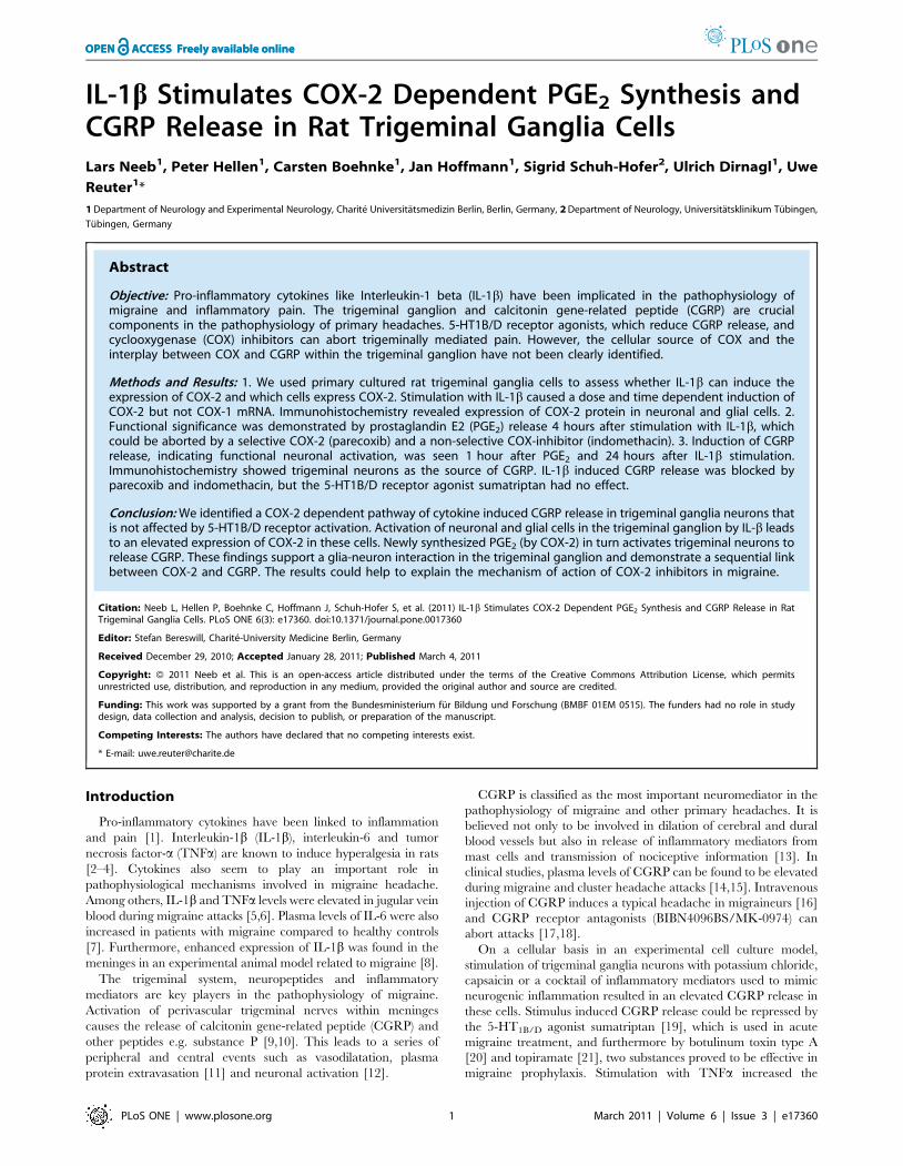
p,0.05 (Fig. 3A).
The non-selective COX inhibitor indomethacin (10 mM) and
the selective COX-2 inhibitor parecoxib (10 mM) administered to
the supernatant of TGC 15 min prior to IL-1b exposure
completely aborted PGE2 release after 4 hours (Fig. 3B). Statistical
significant difference (p,0.05) was achieved for all groups vs. IL-
1b + vehicle (n = 3–4/group). In contrast, sumatriptan (10 mM) did
neither affect IL-1b induced COX-2 mRNA synthesis nor PGE2
release (data not shown). 5-HT1B/D receptor expression in these
cells was detected by RT-PCR. Because selective and non-selective
COX-inhibitors block IL-1b induced PGE release we conclude
that PGE2 release from trigeminal ganglia cells is dependent on
COX-2 expression and function.
IL-1b induces delayed CGRP release in trigeminal ganglianeurons
TGN release CGRP upon stimulation with e.g. potassium
chloride, a cocktail of inflammatory agents, capsaicin (Durham,
Figure 2. Expression of COX-2 protein in trigeminal gangliacells after IL-1b stimulation. COX-2 protein in cell culture homog-enates was analyzed 6 hours after stimulation with IL-1b using Westernblot (n = 3). A representative image is shown in panel A. Cell lysate of IL-1b treated TGC and the positive control (IFNy/LPS treated macrophages)showed a clear band at 70 kDa corresponding to COX-2 protein. Vehiclestimulation resulted in a faint COX-2 expression. The expression of COX-2protein in cultured trigeminal ganglia cells exposed 6 hours to vehicle(10 ng/ml, upper panel B1-B3/B7-B9) or IL-1b (0.1 M PBS, lower panel B4-B9/B10-B12) is shown in fluorescent micrographs in panel B. Cells werestained with a mouse b-tubulin III antibody, indicative of neuronal cells(B1 and B4) or a mouse GFAP antibody, indicative of glial cells (B7 andB10), and a rabbit COX-2 antibody (B2, B5, B8, B11). The b-tubulin III andthe GFAP antibodies were recognized by an Alexa Fluor 488 labeledsecondary donkey anti-mouse antibody (green) and the COX-2 antibodywas recognized by an Alexa Fluor 594 labeled secondary donkey anti-rabbit antibody (red). Double stained cells appear orange in B3, B6, B9and B12. IL-1b caused a clear upregulation of COX-2 in neuronal and glialcells (lower panel) whereas a faint COX-2 expression could also beobserved in control experiments (upper panel). The strongest inductionof COX-2 was seen in bigger glial cells (40-100 mm) and neuronal cells.doi:10.1371/journal.pone.0017360.g002
COX-2 Dependent CGRP Release in TGC
PLoS ONE | www.plosone.org 5 March 2011 | Volume 6 | Issue 3 | e17360
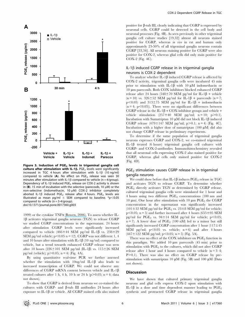
1999) or the cytokine TNFa (Bowen, 2006). To assess whether IL-
1b activates trigeminal ganglia neurons (TGN) to release CGRP
we studied CGRP concentrations in the supernatant. 24 hours
after stimulation CGRP levels were significantly increased
compared to vehicle (466644 SEM pg/ml IL-1b vs. 238629
SEM pg/ml vehicle; p,0.05 n = 12). CGRP was not different 1, 4
and 10 hours after stimulation with IL-1b (10 ng/ml) compared to
vehicle, but a trend towards enhanced CGRP release was seen
after 10 hours (2266101 SEM pg/ml (IL-1b) vs. 115626 SEM
pg/ml (vehicle); p.0.05; n = 4) (Fig. 4A).
By using quantitative real-time PCR we further assessed
whether the stimulation with 10ng/ml IL-1b also leads to
increased transcription of CGRP. We could not observe any
differences of CGRP mRNA content between vehicle and IL-1btreated cultures after 1 h, 4 h, 10 h or 24 h (p.0.05; n = 4; data
not shown).
To show that CGRP is derived from neurons we co-stained the
cultures with CGRP- and b-tub III antibodies 24 hours after
exposure to IL-1b or vehicle. All CGRP stained cells also stained
positive for b-tub III, clearly indicating that CGRP is expressed by
neuronal cells. CGRP could be detected in the cell body and
neuronal processes (Fig. 4B). As seen previously in other trigeminal
ganglia cell culture studies [19,32] almost all neurons stained
positive for CGRP, whereas in vivo in rat and human only
approximately 23-50% of all trigeminal ganglia neurons contain
CGRP [33,34]. All neurons staining positive for CGRP were also
positive for COX-2, whereas glial cells did only stain positive for
COX-2 (Fig. 4C).
IL-1b induced CGRP release in in trigeminal ganglianeurons is COX-2 dependent
To analyze whether IL-1b induced CGRP release is affected by
COX-2 activity, trigeminal ganglia cells were incubated 45 min
prior to stimulation with IL-1b with 10 mM indomethacin or
10 mm parecoxib. Both COX inhibitors blocked enhanced CGRP
release after 24 hours (548670 SEM pg/ml for IL-1b + vehicle
(n = 10) vs. 326652 SEM pg/ml for IL-1b + parecoxib (n = 7;
p,0.05) and 313675 SEM pg/ml for IL-1b + indomethacin
(n = 4; p,0.05)). There were no significant differences between
CGRP release in the IL-1b + COX-inhibitor groups and vehicle +vehicle stimulation (257640 SEM pg/ml; n = 10; p.0.1).
Incubation with Sumatriptan 10 mM did not block IL-1b induced
CGRP release (4796147 SEM pg/ml; p.0.1; n = 4) (Fig. 4C).
Incubation with a higher dose of sumatriptan (100 mM) did also
not change CGRP release in preliminary experiments.
To determine if the same population of trigeminal ganglia
neurons expresses CGRP and COX-2, we co-stained trigeminal
IL-1b treated (6 hours) trigeminal ganglia cell cultures with
CGRP- and COX-2-antibodies. Immunohistochemistry revealed
that all neuronal cells expressing COX-2 also stained positive for
CGRP, whereas glial cells only stained positive for COX-2
(Fig. 4D).
PGE2 stimulation causes CGRP release in in trigeminalganglia neurons
We could show before that IL-1b induces PGE2 release in TGC
and activates TGN to release CGRP. To determine whether
PGE2 directly activates TGN as determined by CGRP release,
cultured trigeminal ganglia cells were stimulated for 1 hour and
4 hours using two different PGE2 concentrations (100 nm and
10 mm). One hour after stimulation with 10 mm PGE2 the CGRP
concentration in the supernatant was significantly increased
(101632 SEM pg/ml for PGE2 vs. 2368 SEM pg/ml for vehicle;
p,0.05; n = 5) and further increased after 4 hours (655695 SEM
pg/ml for PGE2 vs. 94614 SEM pg/ml for vehicle; p,0.05;
n = 7). A lower dose of PGE2 (100 nM) led to a minor but still
significantly increased CGRP concentration after 1 hour (117645
SEM pg/ml; p,0.05 vs. vehicle; n = 6) and after 4 hours
(4476122 SEM pg/ml; p,0.05; n = 5) (Fig. 5A).
There was no effect of the COX inhibitors on PGE2 function in
this paradigm. We added 10 mm parecoxib (45 min) prior to
stimulation with PGE2 to the cultures, which did not alter CGRP
release after 1 hour and 4 hours compared to vehicle (n = 3–4;
P.0.1). There was also no effect on CGRP release by pre-
stimulation with sumatriptan 10 mM (Fig. 5B) and 100 mM (Data
not shown).
Discussion
We have shown that cultured primary trigeminal ganglia
neurons and glial cells express COX-2 upon stimulation with
IL-1b in a dose and time dependent manner leading to PGE2
synthesis and protracted CGRP release in trigeminal ganglia
Figure 3. Induction of PGE2 levels in trigeminal ganglia cellculture after stimulation with IL-1b. PGE2 levels were significantlyincreased in TGC 4 hours after stimulation with IL-1b (10 ng/ml)compared to vehicle (A). No effect on PGE2 release was seen 30minutes after stimulation with IL-1b compared to vehicle (n = 4/group).Dependency of IL-1b induced PGE2 release on COX-2 activity is shownin (B). 15 min of incubation with the selective (parecoxib, 10 mM) or thenon-selective (indomethacin, 10 mM) COX-2 inhibitor completelyaborted IL-1b induced PGE2 release after 4 hours. PGE2 levels areillustrated as mean pg/ml 6 SEM compared to baseline. *p,0.05compared to vehicle (n = 3-4/group).doi:10.1371/journal.pone.0017360.g003
COX-2 Dependent CGRP Release in TGC
PLoS ONE | www.plosone.org 6 March 2011 | Volume 6 | Issue 3 | e17360
neurons. IL-1b induced PGE2 and CGRP release was blocked by
selective and non-selective COX-2 inhibitors thereby demonstrat-
ing dependency on COX-2 enzyme activity. Neither PGE2 nor
CGRP release were mediated by activation of 5-HT1B/D receptors
as the pre-incubation with sumatriptan was without effect.
The trigeminal ganglion (TG) is known to play a key role in the
pathophysiology of migraine and other primary headaches. Cultured
sensory neurons derived from trigeminal or dorsal root ganglia have
been shown to express the characteristics of ‘‘differentiated pain
sensory’’ cells [28]. Cultured TG cells have been used to illustrate
molecular mechanisms related to migraine pathophysiology e.g.
5-HT1B/D controlled CGRP release [19–21,35,36].
For stimulation we used IL-1b, a pro-inflammatory cytokine of
significant importance in models of inflammatory pain [2,27,37]
and sensitization [38]. IL-1b has been shown to activate
nociceptors to induce pain hypersensitivity in a rat skin nerve
preparation [38]. In inflammatory pain IL-1b was identified as the
key mediator to induce COX-2 in spinal cord [27] and in cultured
dorsal root ganglia cells [39–42]. The observation that IL-1b is
elevated in plasma during a migraine attack points to an
involvement in migraine pathophysiology [5,6]. High levels of
chemokines could stimulate the activation of trigeminal nerves and
the release of vasoactive peptides, resulting in inflammation [43].
IL-1b induced COX-2 and PGE2 synthesis resulting inCGRP release
Stimulation of trigeminal ganglia cells with IL-1b led to a strong
induction of COX-2 mRNA and protein with subsequently PGE2
synthesis. Direct stimulation of TG cells with PGE2 resulted in
enhanced CGRP release in trigeminal ganglia neurons after one hour
and more pronounced after 4 hours. In an in vitro preparation of the
rat skull fluid-filled cavities electrical and chemical activation of
trigeminal afferents resulted in enhanced release of PGE2 and CGRP
from rat dura mater encephali [44] In a rat trigeminal ganglion cell
culture model Jenkins and co-workers identified the EP-2 receptor as
the key signaling mechanism for PGE2 induced CGRP release [45].
Enhancement of capsaicin induced CGRP release by PGE2 has been
observed in slices of the trigeminal nucleus caudalis, indicating a
prostaglandin induced neuronal sensitization [46].
While these observations may support a role of PGE2 in
neurogenic inflammation, it is uncertain which COX isoform
accounts for the PGE2 release. Since COX-1 is constitutively
Figure 4. IL-1b induced CGRP release in TGN is dependent on COX-2 activity but not on 5-HT1B/D receptor activation. A: IL-1b (10 ng/ml) but not vehicle stimulation for 1, 4, 10 (n = 4) or 24 hours (n = 12) resulted in significantly enhanced CGRP levels in the supernatant of culturedtrigeminal ganglia cells at the 24 hrs time point (* p,0.05 vs. vehicle). Earlier time points did not show a significant difference between groups. CGRPrelease is shown as mean pg/ml 6 SEM compared to baseline. Panel B shows a representative fluorescent photomicrograph of trigeminal gangliacells exposed 24 hours to IL-1b (10 ng/ml). Cells were stained with a mouse b-tubulin III antibody, which specifically recognizes neuronal cells (1) anda rabbit CGRP antibody (2). b-tubulin III staining was visualized with an Alexa 488 donkey anti-mouse IgG antibody (green, panel 1). CGRP IgG wasrecognized by Alexa 594 donkey anti-rabbit antibody (red, panel 2). All CGRP expressing cells stained positive for b-tubulin III (orange, panel 3). C: Forinhibition experiments TGC were exposed to either parecoxib (10 mM), sumatriptan ( mM), indomethacin (10 mM) or vehicle 45 min prior to 24 hstimulation with IL-1b (10 ng/ml). Incubation with parecoxib (n = 7) and indomethacin (n = 4) blocked CGRP release in the supernatant significantlycompared to IL-1b + vehicle (n = 10). Sumatriptan had no effect in the same paradigm (n = 4). CGRP release is shown as mean pg/ml 6 SEM comparedto baseline. * p,0.05 compared to IL-1b + vehicle. D: Immunofluorescence staining shows that CGRP is co-expressed with COX-2 in trigeminalganglia neurons (n = 3). Cultured trigeminal ganglia cells were exposed to IL-1b (10 ng/ml) for 6 hrs. Cells were stained with an Alexa Fluor 488donkey anti-mouse IgG labeled rabbit anti-CGRP serum (green, panel 1) and with a rabbit anti-COX-2 antibody, which was recognized by an AlexaFluor 594 labeled secondary donkey anti-rabbit antibody (red, panel 2). Double stained cells appear orange (panel 3). All CGRP synthesizing cells alsostained positive for COX-2. Additionally COX-2 was expressed in glial cells not staining positive for CGRP.doi:10.1371/journal.pone.0017360.g004
COX-2 Dependent CGRP Release in TGC
PLoS ONE | www.plosone.org 7 March 2011 | Volume 6 | Issue 3 | e17360

expressed in many cells types (e.g. dural macrophages, fibroblasts)
and PGE2 release occurs immediately after electrical and chemical
meningeal stimulation [44] COX-1 may account for this response.
In our model, immediate PGE2 release did not occur after
stimulation with IL-1b. Additionally, COX-1 mRNA remained
unchanged 3 hours after stimulation with IL-1b. In contrast, IL-
1b induced COX-2 mRNA expression after 3 hours and PGE2
release after 4 hours. PGE2 release could be blocked by the
selective COX-2 inhibitor parecoxib. These findings provide
evidence for a COX-2 mediated pathway.
Glia-neuron interactionWe found neuronal and glial cells as a source of COX-2 as
demonstrated by immunohistochemistry. In particular, a strong
stimulus dependent induction of COX-2 by IL-1b was seen in large
glial cells. Stimulation of cultured trigeminal cells with PGE2 and
IL-1b led to CGRP release exclusively in trigeminal ganglia neurons
(cell body and neuronal processes). Immunohistochemistry did not
reveal any CGRP expression in glial cells which is in line with the
findings of others in rat and human trigeminal ganglia [34].
Our findings support a glia-neuron interaction within the
trigeminal ganglion. We hypothesize that IL-1b activates glial cells
and neurons in the trigeminal ganglion, which leads to the
expression of COX-2 in these cells. In turn the COX-2 reaction
product PGE2 activates trigeminal neurons to release CGRP.
Glia-neuron interaction plays an important role for the normal
function of the brain as well in the pathophysiology of many CNS
diseases [47]. Over the last years the importance of CNS glia for
neuronal function in pain processing has been demonstrated in
various experimental pain states [48]. The physiological function of
Figure 5. CGRP release in the supernatant of culturedtrigeminal ganglia cells after stimulation with PGE2. A: PGE2
stimulation (100 nM and 10 mM) increased CGRP levels within thesupernatant time and dose dependently after 1 hour (n = 5/group) andmore pronounced after 4 hours compared to vehicle (n = 7/group).B: Incubation with 10 mM parecoxib and sumatriptan (10 mM and100 mM [not shown]) did not alter PGE2 release compared to incubationwith vehicle (n = 3-4). CGRP is shown as mean pg/ml 6 SEM comparedto baseline. * p,0.05 compared to vehicle. # p.0.1 compared to PGE +vehicle.doi:10.1371/journal.pone.0017360.g005
Figure 6. COX-2 dependent induction of CGRP release intrigeminal ganglia neurons. Stimulation with IL-1b leads to thesynthesis of COX-2 in trigeminal neurons and glial cells followed byPGE2 release. PGE2 in turn activatesTGN to release CGRP. PGE2 andCGRP release can be blocked by selective (parecoxib) or non-selective(indomethacin) COX-2 inhibitors. The attenuation of CGRP and PGE2
release could contribute to the effect of COX-inhibitors to revokesensitization and to abort pain.doi:10.1371/journal.pone.0017360.g006
COX-2 Dependent CGRP Release in TGC
PLoS ONE | www.plosone.org 8 March 2011 | Volume 6 | Issue 3 | e17360

the glial cells within the trigeminal ganglion is not well understood. In
the trigeminal ganglia cell bodies of neurons are surrounded by
satellite glial cells that can modulate their function and enhance their
excitability [49]. In a recently published work IL-1b induced COX-2
expression and PGE2 synthesis in cultured trigeminal satellite cells.
Stimulation of TGN with conditioned media from these activated
satellite cells led to sensitization of TGN resulting in increased CGRP
release after stimulation with capsaicin [50]. Activation of satellite
glial cells in the trigeminal ganglion modulates the excitability of TG
neurons via IL-1b following inflammation associated with hyperal-
gesia in rats [51]. The involvement of neuron-glia signaling via gap
junctions and release of nitric oxide and pro-inflammatory cytokines
from the trigeminal ganglia satellite glial cells has been demonstrated
recently in experimental models related to migraine pathophysiology
[52–55]. Interestingly, CGRP receptors are expressed on trigeminal
neurons and glial cells. CGRP released by trigeminal ganglia neurons
has been shown to function in a paracrine manner to activate
trigeminal satellite glial cells to release various cytokines including IL-
1b and nitric oxide, a molecule known to be involved in migraine
pathophysiology [52,54]. Additionally CGRP possesses autocrine
signaling function properties to increase mRNA levels of CGRP in
cultured trigeminal neurons [35]. Our finding that induction of
COX-2 expression by IL-1b in trigeminal glial and neuronal cells
leads to direct induction of CGRP release from trigeminal neurons
supports the notion of an important cross talk between neurons and
glial cells in the trigeminal ganglion in processes involved in
trigeminally mediated headaches.
IL-1b induced CGRP release in trigeminal ganglia neuronsIn our model exposure of trigeminal ganglia cell culture to IL-1b
led directly to delayed CGRP release from TGN 24 hours after
stimulation with an earlier non-significant trend after 10 hours. This
latency was not expected as IL-1b induced PGE2 release was
significant after 4 hours and PGE2 caused CGRP release after
1 hour. Delayed (24 hours) IL-1b induced CGRP release was
observed previously in dorsal root ganglia neurons. Blocking
experiments demonstrated that IL-1b might activate proteinkinase
C that in turn initiates c-Jun-N-terminal kinase mitogen activated
protein kinase followed by activation of nuclear factor-kappaB,
which finally induces alpha-CGRP gene expression and neuropep-
tide release from these sensory neurons [56]. In our hands IL-1b did
not lead to an induction of CGRP mRNA synthesis in cultured
trigeminal neurons as demonstrated by quantitative RT-PCR. We
speculate that a certain threshold of PGE2 concentration in the
supernatant is necessary to induce gradual CGRP expression, which
may take several hours to archive. Direct stimulation of TG with
10 mm PGE2 did also not lead to the induction of CGRP gene
expression. In line, in another rat model of isolated trigeminal
ganglion the infusion of the NO donor glyceroltrinitrate induced the
release of CGRP but did not change mRNA levels of CGRP [57].
Therefore, elevated CGRP release in the trigeminal ganglion in our
and other models seems to be dependent on enhanced secretion
rather than synthesis due to gene expression.
Non-selective (indomethacin) and selective (parecoxib) COX-2
inhibitors aborted L-1b induced CGRP release. In contrast,
neither IL-1b nor PGE2 induced CGRP release could be blocked
by sumatriptan, indicating a release mechanism independent of 5-
HT1B/D receptor activation. A COX-2 dependent CGRP release
could also be demonstrated in the dura mater in an isolated
preparation of fluid-filled rat skull cavities. The COX-2 inhibitor
S-flurbiprofen inhibited inflammatory mediator (bradykinin,
histamine and serotonin) induced CGRP and PGE2 release while
the 5-HT1B/D receptor agonist naratriptan was without effect [58].
Our findings demonstrate a direct link between COX-2 activity
and CGRP release in TGN. In a slightly different cell culture
model of trigeminal ganglia cells potassium chloride and
inflammatory cocktail induced rapid CGRP release that could
be blocked by sumatriptan [19]. The results of our study point to
an alternative pathway of cytokine induced CGRP release in TGN
regulated by COX-2 and not affected by sumatriptan.
ConclusionIn summary, our results demonstrate that primary trigeminal
ganglia cells are able to synthesize COX-2 and PGE2 upon
stimulation with the cytokine IL-1b in a functionally significant
mode resulting in delayed CGRP release in trigeminal ganglia
neurons. CGRP release is mediated by a COX-2 dependent
pathway and independent of 5-HT1B/D receptor activation (Fig. 6).
COX-2 expression in trigeminal ganglion cells could contribute
to the development of pain in trigeminal mediated headaches.
Non-selective [23] and selective COX-2 inhibitors [24,25] abort
migraine attacks. The demonstrated attenuation of CGRP release
by COX-2 inhibition could at least in part explain the mechanism
of COX inhibitors in migraine therapy.
Acknowledgments
We would like to thank Sonja Blumenau for excellent technical assistance
and Francisco Fernandez Klett for support with confocal imaging.
Author Contributions
Conceived and designed the experiments: LN UR. Performed the
experiments: LN CB PH JH SSH. Analyzed the data: LN UR PH.
Contributed reagents/materials/analysis tools: UR UD. Wrote the paper:
LN UR.
References
1. Sommer C, Kress M (2004) Recent findings on how proinflammatory cytokines
cause pain: peripheral mechanisms in inflammatory and neuropathic hyperal-
gesia. Neurosci Lett 361: 184–187.
2. Cunha JM, Cunha FQ, Poole S, Ferreira SH (2000) Cytokine-mediated
inflammatory hyperalgesia limited by interleukin-1 receptor antagonist.
Br J Pharmacol 130: 1418–1424.
3. Cunha FQ, Poole S, Lorenzetti BB, Ferreira SH (1992) The pivotal role of
tumour necrosis factor alpha in the development of inflammatory hyperalgesia.
Br J Pharmacol 107: 660–664.
4. Oka T, Oka K, Hosoi M, Hori T (1995) Intracerebroventricular injection of
interleukin-6 induces thermal hyperalgesia in rats. Brain Res 692: 123–128.
5. Perini F, D’Andrea G, Galloni E, Pignatelli F, Billo G, et al. (2005) Plasma
cytokine levels in migraineurs and controls. Headache 45: 926–931.
6. Sarchielli P, Alberti A, Baldi A, Coppola F, Rossi C, et al. (2006)
Proinflammatory cytokines, adhesion molecules, and lymphocyte integrin
expression in the internal jugular blood of migraine patients without aura
assessed ictally. Headache 46: 200–207.
7. Kocer A, Memisogullari R, Domac FM, Ilhan A, Kocer E, et al. (2009) IL-6
levels in migraine patients receiving topiramate. Pain Pract 9: 375–379.
8. Reuter U, Bolay H, Jansen-Olesen I, Chiarugi A, Sanchez del RM, et al. (2001)
Delayed inflammation in rat meninges: implications for migraine pathophysi-
ology. Brain 124: 2490–2502.
9. Uddman R, Edvinsson L, Ekman R, Kingman T, McCulloch J (1985)
Innervation of the feline cerebral vasculature by nerve fibers containing
calcitonin gene-related peptide: trigeminal origin and co-existence with
substance P. Neurosci Lett 62: 131–136.
10. Edvinsson L, Hara H, Uddman R (1989) Retrograde tracing of nerve fibers to
the rat middle cerebral artery with true blue: colocalization with different
peptides. J Cereb Blood Flow Metab 9: 212–218.
11. Waeber C, Moskowitz MA (2005) Migraine as an inflammatory disorder.
Neurology 64: S9–15.
12. Storer RJ, Akerman S, Goadsby PJ (2004) Calcitonin gene-related peptide
(CGRP) modulates nociceptive trigeminovascular transmission in the cat.
Br J Pharmacol 142: 1171–1181.
COX-2 Dependent CGRP Release in TGC
PLoS ONE | www.plosone.org 9 March 2011 | Volume 6 | Issue 3 | e17360

13. Durham PL (2006) Calcitonin gene-related peptide (CGRP) and migraine.
Headache 46(Suppl 1): S3–S8.14. Juhasz G, Zsombok T, Modos EA, Olajos S, Jakab B, et al. (2003) NO-induced
migraine attack: strong increase in plasma calcitonin gene-related peptide
(CGRP) concentration and negative correlation with platelet serotonin release.Pain 106: 461–470.
15. Fanciullacci M, Alessandri M, Figini M, Geppetti P, Michelacci S (1995)Increase in plasma calcitonin gene-related peptide from the extracerebral
circulation during nitroglycerin-induced cluster headache attack. Pain 60:
119–123.16. Lassen LH, Haderslev PA, Jacobsen VB, Iversen HK, Sperling B, et al. (2002)
CGRP may play a causative role in migraine. Cephalalgia 22: 54–61.17. Olesen J, Diener HC, Husstedt IW, Goadsby PJ, Hall D, et al. (2004) Calcitonin
gene-related peptide receptor antagonist BIBN 4096 BS for the acute treatmentof migraine. N Engl J Med 350: 1104–1110.
18. Ho TW, Ferrari MD, Dodick DW, Galet V, Kost J, et al. (2008) Efficacy and
tolerability of MK-0974 (telcagepant), a new oral antagonist of calcitonin gene-related peptide receptor, compared with zolmitriptan for acute migraine: a
randomised, placebo-controlled, parallel-treatment trial. Lancet 372:2115–2123.
19. Durham PL, Russo AF (1999) Regulation of calcitonin gene-related peptide
secretion by a serotonergic antimigraine drug. J Neurosci 19: 3423–3429.20. Durham PL, Cady R, Cady R (2004) Regulation of calcitonin gene-related
peptide secretion from trigeminal nerve cells by botulinum toxin type A:implications for migraine therapy. Headache 44: 35–42.
21. Durham PL, Niemann C, Cady R (2006) Repression of stimulated calcitoningene-related peptide secretion by topiramate. Headache 46: 1291–1295.
22. Bowen EJ, Schmidt TW, Firm CS, Russo AF, Durham PL (2006) Tumor
necrosis factor-alpha stimulation of calcitonin gene-related peptide expressionand secretion from rat trigeminal ganglion neurons. J Neurochem 96: 65–77.
23. Rasmussen MK, Binzer M (2001) Non-steroidal anti-inflammatory drugs in thetreatment of migraine. Curr Med Res Opin 17(Suppl 1): s26–s29.
24. Goebel H, Heinze A, Heinze-Kuhn K (2003) Parecoxib i.v. in der Behandlung
der aktuen Migraneattacke als Ersatzmedikaiton bei Ineffeketivitat vonTriptanen. Aktuelle Neurologie Supl S1: 776.
25. Silberstein S, Tepper S, Brandes J, Diamond M, Goldstein J, et al. (2004)Randomized, placebo-controlled trial of rofecoxib in the acute treatment of
migraine. Neurology 62: 1552–1557.26. Simmons DL, Botting RM, Hla T (2004) Cyclooxygenase isozymes: the biology
of prostaglandin synthesis and inhibition. Pharmacol Rev 56: 387–437.
27. Samad TA, Moore KA, Sapirstein A, Billet S, Allchorne A, et al. (2001)Interleukin-1beta-mediated induction of Cox-2 in the CNS contributes to
inflammatory pain hypersensitivity. Nature 410: 471–475.28. Baccaglini PI, Hogan PG (1983) Some rat sensory neurons in culture express
characteristics of differentiated pain sensory cells. Proc Natl Acad Sci U S A 80:
594–598.29. Ruscher K, Freyer D, Karsch M, Isaev N, Megow D, et al. (2002) Erythropoietin
is a paracrine mediator of ischemic tolerance in the brain: evidence from an invitro model. J Neurosci 22: 10291–10301.
30. Pfaffl MW (2001) A new mathematical model for relative quantification in real-time RT-PCR. Nucleic Acids Res 29: e45.
31. Matsushita K, Wu Y, Qiu J, Lang-Lazdunski L, Hirt L, et al. (2000) Fas receptor
and neuronal cell death after spinal cord ischemia. J Neurosci 20: 6879–6887.32. Kuris A, Xu CB, Zhou MF, Tajti J, Uddman R, et al. (2007) Enhanced
expression of CGRP in rat trigeminal ganglion neurons during cell and organculture. Brain Res 1173: 6–13.
33. O’Connor TP, van der Kooy D (1988) Enrichment of a vasoactive neuropeptide
(calcitonin gene related peptide) in the trigeminal sensory projection to theintracranial arteries. J Neurosci 8: 2468–2476.
34. Eftekhari S, Salvatore CA, Calamari A, Kane SA, Tajti J, et al. (2010)Differential distribution of calcitonin gene-related peptide and its receptor
components in the human trigeminal ganglion. Neuroscience 169: 683–696.
35. Zhang Z, Winborn CS, Marquez dP, Russo AF (2007) Sensitization of calcitoningene-related peptide receptors by receptor activity-modifying protein-1 in the
trigeminal ganglion. J Neurosci 27: 2693–2703.36. Jansen-Olesen I, Zhou M, Zinck T, Xu CB, Edvinsson L (2005) Expression of
inducible nitric oxide synthase in trigeminal ganglion cells during culture. BasicClin Pharmacol Toxicol 97: 355–363.
37. Safieh-Garabedian B, Poole S, Allchorne A, Winter J, Woolf CJ (1995)
Contribution of interleukin-1 beta to the inflammation-induced increase in nerve
growth factor levels and inflammatory hyperalgesia. Br J Pharmacol 115:
1265–1275.
38. Binshtok AM, Wang H, Zimmermann K, Amaya F, Vardeh D, et al. (2008)Nociceptors are interleukin-1beta sensors. J Neurosci 28: 14062–14073.
39. Inoue A, Ikoma K, Morioka N, Kumagai K, Hashimoto T, et al. (1999)
Interleukin-1beta induces substance P release from primary afferent neuronsthrough the cyclooxygenase-2 system. J Neurochem 73: 2206–2213.
40. Igwe OJ, Murray JN, Moolwaney AS (2001) Interleukin 1-induced cyclooxy-
genase and nitric oxide synthase gene expression in the rat dorsal root ganglia ismodulated by antioxidants. Neuroscience 105: 971–985.
41. Morioka N, Inoue A, Hanada T, Kumagai K, Takeda K, et al. (2002) Nitric
oxide synergistically potentiates interleukin-1 beta-induced increase of cycloox-ygenase-2 mRNA levels, resulting in the facilitation of substance P release from
primary afferent neurons: involvement of cGMP-independent mechanisms.
Neuropharmacology 43: 868–876.
42. Fehrenbacher JC, Burkey TH, Nicol GD, Vasko MR (2005) Tumor necrosis
factor alpha and interleukin-1beta stimulate the expression of cyclooxygenase II
but do not alter prostaglandin E2 receptor mRNA levels in cultured dorsal rootganglia cells. Pain 113: 113–122.
43. Bruno PP, Carpino F, Carpino G, Zicari A (2007) An overview on immune
system and migraine. Eur Rev Med Pharmacol Sci 11: 245–248.
44. Ebersberger A, Averbeck B, Messlinger K, Reeh PW (1999) Release of substance
P, calcitonin gene-related peptide and prostaglandin E2 from rat dura mater
encephali following electrical and chemical stimulation in vitro. Neuroscience89: 901–907.
45. Jenkins DW, Feniuk W, Humphrey PP (2001) Characterization of theprostanoid receptor types involved in mediating calcitonin gene-related peptide
release from cultured rat trigeminal neurones. Br J Pharmacol 134: 1296–1302.
46. Jenkins DW, Langmead CJ, Parsons AA, Strijbos PJ (2004) Regulation ofcalcitonin gene-related peptide release from rat trigeminal nucleus caudalis slices
in vitro. Neurosci Lett 366: 241–244.
47. Benarroch EE (2005) Neuron-astrocyte interactions: partnership for normalfunction and disease in the central nervous system. Mayo Clin Proc 80:
1326–1338.
48. McMahon SB, Malcangio M (2009) Current challenges in glia-pain biology.Neuron 64: 46–54.
49. Hanani M (2005) Satellite glial cells in sensory ganglia: from form to function.
Brain Res Brain Res Rev 48: 457–476.
50. Capuano A, De CA, Lisi L, Tringali G, Navarra P, et al. (2009)Proinflammatory-activated trigeminal satellite cells promote neuronal sensitiza-
tion: relevance for migraine pathology. Mol Pain 5: 43.
51. Takeda M, Tanimoto T, Kadoi J, Nasu M, Takahashi M, et al. (2007) Enhancedexcitability of nociceptive trigeminal ganglion neurons by satellite glial cytokine
following peripheral inflammation. Pain 129: 155–166.
52. Thalakoti S, Patil VV, Damodaram S, Vause CV, Langford LE, et al. (2007)Neuron-glia signaling in trigeminal ganglion: implications for migraine
pathology. Headache 47: 1008–1023.
53. Ceruti S, Fumagalli M, Villa G, Verderio C, Abbracchio MP (2008)Purinoceptor-mediated calcium signaling in primary neuron-glia trigeminal
cultures. Cell Calcium 43: 576–590.
54. Li J, Vause CV, Durham PL (2008) Calcitonin gene-related peptide stimulationof nitric oxide synthesis and release from trigeminal ganglion glial cells. Brain
Res 1196: 22–32.
55. Damodaram S, Thalakoti S, Freeman SE, Garrett FG, Durham PL (2009)Tonabersat inhibits trigeminal ganglion neuronal-satellite glial cell signaling.
Headache 49: 5–20.
56. Hou L, Li W, Wang X (2003) Mechanism of interleukin-1 beta-inducedcalcitonin gene-related peptide production from dorsal root ganglion neurons of
neonatal rats. J Neurosci Res 73: 188–197.
57. Eberhardt M, Neeb L, Vogel EM, Tiegs G, Reuter U, et al. (2009)Glyceroltrinitrate facilitates stimulated CGRP release but not gene expression
of CGRP or its receptor components in rat trigeminal ganglia. Neuropeptides43: 483–489.
58. Zimmermann K, Reeh PW, Averbeck B (2003) S+ -flurbiprofen but not 5-HT1
agonists suppress basal and stimulated CGRP and PGE2 release from isolatedrat dura mater. Pain 103: 313–320.
59. Ivanov AI, Pero RS, Scheck AC, Romanovsky AA (2002) Prostaglandin E(2)-
synthesizing enzymes in fever: differential transcriptional regulation. Am J PhysiolRegul Integr Comp Physiol 283: R1104–R1117.
COX-2 Dependent CGRP Release in TGC
PLoS ONE | www.plosone.org 10 March 2011 | Volume 6 | Issue 3 | e17360
Related Documents