IL-1b/HMGB1 Complexes Promote The PGE 2 Biosynthesis Pathway in Synovial Fibroblasts P. Leclerc* 1 , H. W€ ah€ amaa† 1 , H. Idborg*, P. J. Jakobsson*, H. E. Harris* & M. Korotkova*‡ *Rheumatology Research Unit, Department of Medicine, Karolinska Institutet, Stockholm, Sweden; †Pediatric Rheumatology Unit, Department of Woman and Child Health, Karolinska Institutet, Stockholm, Sweden; and ‡Actar AB, Solna, Sweden Received 18 December 2012; Accepted in revised form 15 February 2013 Correspondence to: Dr. H. Wähämaa, Rheumatology Research Laboratorium Solna, CMM L8:04, 171 76 Stockholm, Sweden. E-mail: [email protected] 1 These authors contributed equally to this work. Abstract PGE 2 is a potent lipid mediator of pain and oedema found elevated in RA. Microsomal prostaglandin E synthase-1 (mPGES-1) is a terminal enzyme of the PGE 2 pathway inducible by proinflammatory cytokines. mPGES-1 is markedly upregulated in RA synovial tissue despite antirheumatic treatments, suggesting that multiple inflammatory stimuli contribute to its induction. High-mobility group box chromosomal protein 1 (HMGB1) is known to induce inflammation both by direct interaction with TLR4 and by enhancement of other proinflam- matory molecules signalling, through complex formation. The high expression of extracellular HMGB1 within the inflamed synovium, implies its pro-arthrito- genic role in RA. We aimed to investigate the effects of IL-1b/HMGB1 complexes on mPGES-1 and other enzymes of the PGE 2 pathway in synovial fibroblasts (SFs) from patients with arthritis. Furthermore, we studied the effect of COX-2 inhibition and IL-1RI antagonism on prostanoid and cytokine production by SFs. Stimulation of SFs with HMGB1 in complex with suboptimal amounts of IL-1b significantly increased mPGES-1 and COX-2 expressions as well as PGE 2 production, as compared to treatment with HMGB1 or IL-1b alone. Furthermore, NS-398 reduced the production of IL-6 and IL-8, thus indicating that IL-1b/HMGB1 complexes modulate cytokine production in part through prostanoid synthesis. Treatment with IL-1RA completely abolished the induced PGE 2 and cytokine production, suggesting an effect mediated through IL-1RI. IL-1b/HMGB1 complexes promote the induction of mPGES-1, COX-2 and PGE 2 in SF. The amplification of the PGE 2 biosynthesis pathway by HMGB1 might constitute an important pathogenic mechanism perpetuating inflammatory and destructive activities in rheumatoid arthritis. Introduction Rheumatoid arthritis (RA) is a chronic inflammatory disease characterized by synovial inflammation as well as bone and cartilage destruction, leading to loss of joint function. In RA, the inflamed synovium produces elevated levels of PGE 2 , a powerful proinflammatory lipid mediator that triggers pain and oedema. PGE 2 production/biosyn- thesis in inflammation is mainly regulated by the concerted activities of three enzymes: microsomal prostaglandin E synthase-1 (mPGES-1), cyclooxygenase-2 (COX-2) and 15- hydroxyprostaglandin dehydrogenase (15-PGDH). The former two enzymes are involved in the synthesis of PGE 2 and are inducible by a range of proinflammatory stimuli such as IL-1b, TNF-a and IL-6 [1, 2], whereas 15- PGDH is an enzyme degrading PGE 2 . The role of the inducible PGE 2 pathway in RA has proven to be central as COX-2 selective inhibitors are an effective treatment for RA-associated pain and inflammation [3]. mPGES-1 acts downstream from the cyclooxygenase enzymes and is responsible for the conversion of PGH 2 into PGE 2 [4]. mPGES-1 is strongly upregulated in the RA synovium, making it a potential therapeutic target [5]. Moreover, the expression of mPGES-1 in RA synovial tissue is not properly targeted by current antirheumatic treatments. In fact, TNF-a targeted therapy, B cell depletion therapy and methotrexate treatment all fail to inhibit the expression of mPGES-1 in the RA synovium, suggesting that multiple mechanisms are involved in the induction of mPGES-1 expression in the RA synovium [6–8]. High-mobility group box chromosomal protein 1 (HMGB1) is an alarmin possessing distinct functions intra- and extracellularly. In the cell nucleus, HMGB1 acts as a DNA-binding protein, participating Ó 2013 The Authors. 350 Scandinavian Journal of Immunology © 2013 Blackwell Publishing Ltd. EXPERIMENTAL IMMUNOLOGY doi: 10.1111/sji.12041 ..................................................................................................................................................................

Welcome message from author
This document is posted to help you gain knowledge. Please leave a comment to let me know what you think about it! Share it to your friends and learn new things together.
Transcript

IL-1b/HMGB1 Complexes Promote The PGE2Biosynthesis Pathway in Synovial Fibroblasts
P. Leclerc*1, H. W€ah€amaa†1, H. Idborg*, P. J. Jakobsson*, H. E. Harris* & M. Korotkova*‡
*Rheumatology Research Unit, Department of
Medicine, Karolinska Institutet, Stockholm,
Sweden; †Pediatric Rheumatology Unit,
Department of Woman and Child Health,
Karolinska Institutet, Stockholm, Sweden; and
‡Actar AB, Solna, Sweden
Received 18 December 2012; Accepted inrevised form 15 February 2013
Correspondence to: Dr. H. Wähämaa,Rheumatology Research Laboratorium
Solna, CMM L8:04, 171 76 Stockholm, Sweden.
E-mail: [email protected]
1These authors contributed equally to this work.
Abstract
PGE2 is a potent lipid mediator of pain and oedema found elevated in RA.Microsomal prostaglandin E synthase-1 (mPGES-1) is a terminal enzyme of thePGE2 pathway inducible by proinflammatory cytokines. mPGES-1 is markedlyupregulated in RA synovial tissue despite antirheumatic treatments, suggestingthat multiple inflammatory stimuli contribute to its induction. High-mobilitygroup box chromosomal protein 1 (HMGB1) is known to induce inflammationboth by direct interaction with TLR4 and by enhancement of other proinflam-matory molecules signalling, through complex formation. The high expression ofextracellular HMGB1 within the inflamed synovium, implies its pro-arthrito-genic role in RA. We aimed to investigate the effects of IL-1b/HMGB1complexes on mPGES-1 and other enzymes of the PGE2 pathway in synovialfibroblasts (SFs) from patients with arthritis. Furthermore, we studied the effectof COX-2 inhibition and IL-1RI antagonism on prostanoid and cytokineproduction by SFs. Stimulation of SFs with HMGB1 in complex withsuboptimal amounts of IL-1b significantly increased mPGES-1 and COX-2expressions as well as PGE2 production, as compared to treatment with HMGB1or IL-1b alone. Furthermore, NS-398 reduced the production of IL-6 and IL-8,thus indicating that IL-1b/HMGB1 complexes modulate cytokine production inpart through prostanoid synthesis. Treatment with IL-1RA completely abolishedthe induced PGE2 and cytokine production, suggesting an effect mediatedthrough IL-1RI. IL-1b/HMGB1 complexes promote the induction of mPGES-1,COX-2 and PGE2 in SF. The amplification of the PGE2 biosynthesis pathway byHMGB1 might constitute an important pathogenic mechanism perpetuatinginflammatory and destructive activities in rheumatoid arthritis.
Introduction
Rheumatoid arthritis (RA) is a chronic inflammatorydisease characterized by synovial inflammation as well asbone and cartilage destruction, leading to loss of jointfunction. In RA, the inflamed synovium produces elevatedlevels of PGE2, a powerful proinflammatory lipid mediatorthat triggers pain and oedema. PGE2 production/biosyn-thesis in inflammation is mainly regulated by the concertedactivities of three enzymes: microsomal prostaglandin Esynthase-1 (mPGES-1), cyclooxygenase-2 (COX-2) and 15-hydroxyprostaglandin dehydrogenase (15-PGDH). Theformer two enzymes are involved in the synthesis ofPGE2 and are inducible by a range of proinflammatorystimuli such as IL-1b, TNF-a and IL-6 [1, 2], whereas 15-PGDH is an enzyme degrading PGE2. The role of theinducible PGE2 pathway in RA has proven to be central as
COX-2 selective inhibitors are an effective treatment forRA-associated pain and inflammation [3]. mPGES-1 actsdownstream from the cyclooxygenase enzymes and isresponsible for the conversion of PGH2 into PGE2 [4].mPGES-1 is strongly upregulated in the RA synovium,making it a potential therapeutic target [5]. Moreover, theexpression of mPGES-1 in RA synovial tissue is notproperly targeted by current antirheumatic treatments. Infact, TNF-a targeted therapy, B cell depletion therapy andmethotrexate treatment all fail to inhibit the expression ofmPGES-1 in the RA synovium, suggesting that multiplemechanisms are involved in the induction of mPGES-1expression in the RA synovium [6–8].
High-mobility group box chromosomal protein 1(HMGB1) is an alarmin possessing distinct functionsintra- and extracellularly. In the cell nucleus,HMGB1 acts as a DNA-binding protein, participating
� 2013 The Authors.
350 Scandinavian Journal of Immunology © 2013 Blackwell Publishing Ltd.
E X P E R IM EN T A L IMMUNO LOG Y doi: 10.1111/sji.12041..................................................................................................................................................................

in transcription, recombination and DNA repair. Whenreleased to the extracellular space, HMGB1 acts as animportant mediator of inflammation, promoting bothacute inflammation and subsequent tissue repair [9]. Weand other research groups have demonstrated that theinflammation-promoting effects of HMGB1 are in partmediated through its binding to TLR2 and TLR4 andrequire the HMGB1 molecule to be in a specific redoxstatus [10, 11]. However, regardless of its oxidationstatus, HMGB1 is also able to potently promote andenhance inflammation by complex formation with otherproinflammatory molecules such as IL-1a/b, LPS, CpG-DNA, the TLR1/TLR2-agonist Pam3CSK4, nucleosomesand CXCL12 [12–17]. In rodent models of experimentallyinduced arthritis, HMGB1 is an essential mediator ofinflammation and joint destruction as HMGB1-blockingtherapies ameliorate both the inflammatory and tissuedestructive disease course in collagen-induced arthritisand the spontaneous arthritis developing in DNAseII xIFNR1 deficient mice [18–22]. In RA, extracellularHMGB1 is found within the inflamed synovial tissueand in the synovial fluid, implying its proarthritogenicrole in RA [23]. Moreover, we have recently demonstratedthat HMGB1 in complex with suboptimal concentrationsof IL-1a, IL-1b or LPS was able to induce inflammatorycytokine production from RA synovial fibroblasts (RASF)[24]. Interestingly, like mPGES-1, HMGB1 levels in theRA synovium are reduced by intra-articular treatmentwith glucocorticoids, but remain unaltered in patients onanti-TNF therapy [25, 26], suggesting that HMGB1might be a potential inducer of mPGES-1 in inflamedtissue.
The involvement of IL-1 in RA has previously beenshown by clinical improvement in patients on IL-1receptor antagonist (IL-1Ra) therapy, reducing both clin-ical signs of inflammation and joint erosion [27]. IL-1b canstimulate synovial fibroblasts to release mediators promot-ing inflammation and cartilage/bone degradation [1, 24].In this study, we aimed to investigate the effects of IL-1b/HMGB1 complexes on mPGES-1 and other enzymes of thePGE2 pathway in SFs from patients with inflammatoryarthritis. Furthermore, we studied the effect of COX-2inhibition and IL-1RI antagonism on prostanoid andcytokine production by SFs.
Materials and methods
Preparation of rHMGB1 from E. Coli
Recombinant rat HMGB1 (rHMGB1), with a 99%identity to human HMGB1 [28] and containing acalmodulin-binding protein tag, was expressed in E. colistrain BL21 (for sequence see [29]). Protein was purified bysequential ion exchange chromatography (MonoS 5/50 GLcolumn, GE Healthcare, Chalfont St. Giles, UK) and
calmodulin affinity chromatography (Calmodulin sepharose4B, GE Healthcare, Uppsala, Sweden). Endotoxin wasremoved by filtration through Acodisc Units with MustangE Membranes (0.25 lm, Pall Life Sciences, East Hills, NY,USA), yielding endotoxin levels below 0.03 EU/lgprotein, as measured by the Limulus assay. Preparationsof HMGB1 in 20 mM 3-(N-Morpholino) propanesulphonicacid (MOPS), 400-mM NaCl, 20-mM EGTA, 10-mM
dithiothreitol at pH 8.0 were stored at �80°C until dayof use. The HMGB1 used in the studies did not inducecytokine production per se.
Preparation of IL-1b/HMGB1 solutions
r/tHMGB1 and IL-1b were mixed in PBS. A 50X IL-1b/HMGB1 solution was prepared in a ratio allowing theindicated final concentrations in cell cultures after dilution.Solutions were incubated at 4°C for 16 h before addition tocell cultures. IL-1b/HMGB1 complex formation haspreviously been demonstrated [30].
Cell cultures
Synovial fibroblasts were obtained from ten patients: twofrom RA patients were purchased from Asterand (Detroit,MI, USA), and eight were propagated from synovial tissuesisolated from four RA and four juvenile idiopathic arthritispatients undergoing joint replacement surgery as previ-ously described [31]. This study was approved by theInstitutional Ethical Committee (Solna, Stockholm, Swe-den ethical number 2009/1262-31/3) and is in compliancewith all ethical standards and patients′ consent accordingto the Declaration of Helsinki. Briefly, synovial tissueswere minced, and explants were maintained in DMEMsupplemented with 10% heat-inactivated FCS (PAALaboratories, Linz, Austria), 100 U/ml penicillin,100 lg/ml streptomycin and HEPES (Life Technologies,Paisely, Scotland, UK) (complete DMEM) in a tissueculture incubator at 37°C with 5% CO2 content. Tissueexplants and non-adherent cells were discarded after1–2 weeks of culture. Adherent cells were trypsinizedwith trypsin-EDTA (Gibco, Scotland, UK) at 80%confluence and used for experiments at passages 3–8.Synovial fibroblasts grown to confluence were trypsinizedwith trypsin-EDTA and washed with complete DMEM.Cell viability was assessed using trypan blue (Merck,Darmstadt, Germany) in every experimental set up and was95–100%.
Cells were plated in 96-well plates at 4000 cells/well,6-well plates at 160 000 cells/well or 10 cm Nunclondishes at 800 000 cells/well and allowed to rest for15–17 h in a tissue culture incubator at 37°C with 5%CO2 content. Medium was discarded, and cells werewashed twice with OPTIMEM (Gibco, Scotland, UK)supplemented with 100 U/ml penicillin, 100 lg/ml
� 2013 The Authors.
Scandinavian Journal of Immunology © 2013 Blackwell Publishing Ltd.
P. Leclerc et al. HMGB1-Complexes Induce PGE2 in Synovial Fibroblasts 351..................................................................................................................................................................

streptomycin. Cell stimulations were carried in OPTI-MEM. The cells were stimulated for 4–72 h with100 ng/ml rHMGB1 or calf thymus-extracted (t)HMGB1(kind gift from Dr. Michael Bustin, NIH) alone or incomplex with 0.05 (10 donors) or sometimes with 0.5(4 donors) ng/ml rIL-1b (R&D systems, Minneapolis,MN, USA) as indicated. The latter two concentrations ofrIL-1b were referred to as IL-1blow and were used inparallel because the threshold of response of SFs to IL-1 balone was found to vary among experiments. The highestconcentration of IL-1 blow not eliciting a response fromSF was used as a negative control to IL-1blow/HMGB1complexes. 5 ng/ml rIL-1b (IL-1bhigh) was used as apositive control. In certain experiments, cells werepretreated for 1–2 h with 5 lg/ml IL-1 receptor antag-onist (IL-1Ra/anakinra). NS-398 (Sigma-Aldrich,St.Louis, MO, USA), the COX-2 inhibitor, or vehiclealone (0.5% DMSO), was added at the concentration of0.1 lM, simultaneously with inflammatory stimuli. Su-pernatants were collected at the indicated time points andstored at �70°C until analysis.
Prostanoid analysis
PGE2 levels were measured by enzyme immunoassay(Cayman Chemicals, Ann Harbor, MI, USA) and normal-ized for cell viability using a MTT-based in vitro toxicologyassay kit (Sigma- Aldrich). Prostaglandin profiling wasperformed by liquid chromatography coupled to tandemmass spectrometry (LC-MS/MS) on a Waters 2795 HPLC(Waters Corporation, Milford, MA, USA) coupled to atriple quadrupole mass spectrometer (Acquity TQ Detec-tor, Waters Corporation). Cell culture supernatants wereanalysed for PGE2, PGD2, PGF2a, TXB2 and 6-keto-PGF1a. After addition of deuterated isomers of all analytesto 50 ll of sample, prostanoids were extracted on an OasisHLB Extraction Plate (Waters Corporation). Extractedmaterial was evaporated and reconstituted in 50 ll samplesolvent (H2O, 7% acetonitrile (ACN), 0.05% formic acid(FA)), and 40 ll were injected. Separation of the ana-lytes was achieved on a Synergi Hydro-RP column(100 mm x 2 mm i.d., 2.5 lm particle size and 100 �Apore size, Phenomenex, CA, USA) during a 45 minstepwise linear gradient using Milli-Q H2O as mobilephase A and ACN, 0.05% FA as mobile phase B.Concentration of mobile phase B was increased from10% to 25% during 9 min in step 1, then to 45% during22 min in step 2 and furthermore to 70% during 5 min instep 3, followed by a wash step at 90% mobile phase B andre-equilibration at 10% mobile phase B. The analytes weredetected in multiple reaction monitoring (MRM) mode,recording the transitions of m/z 351.2 ? m/z 271.2 forPGE2 and PGD2, m/z 353.2 ? m/z 309.1 for PGF2a, m/z369.2 ? m/z 169.1 for TXB2 and m/z 369.3 ? m/z 163.2for 6-keto PGF1a. Analysis of the MRM data was carried
out with MassLynx software, version 4.1, using an internalstandard calibration curve of all analytes for quantification.When prostanoid concentrations were below the limit ofquantification (LOQ), they were given an arbitrary valueequal to the lowest quantifiable standard for statisticalpurpose.
Western blot
Cells lysis was performed on ice (30 min) using tissueprotein extraction reagent (T-PER) (Thermo Scientific,Rockford, IL, USA) supplemented with 1X completeprotease inhibitor cocktail (Roche Diagnostics GmbH,Mannheim, Germany). Gel electrophoresis was carried onthe NuPAGE� Novex� Bis-Tris gel system (InvitrogenAB, Lidingo, Sweden), and proteins were transferred to apolyvinylidene difluoride membrane using a Trans-Blot SDsemi-dry transfer cell (Bio-Rad Laboratories AB, Uppsala,Sweden). After saturating 25 min with 5% milk in PBS(0.1% Tween 20), the membranes were incubated withprimary (overnight, 4°C) and secondary (1 h, roomtemperature) antibodies. Membranes were washed threetimes 10 min in PBS (0.1% Tween 20) after incubationswith each antibody. The bands were detected by enhancedchemiluminescence (ECL). Densitometry analysis of WBband intensities was performed using the image labsoftware version 3.0 on the molecular imager gel docXR+ system (Bio-Rad Laboratories, Sweden). Primaryantibodies: Polyclonal anti-mPGES-1, anti-mPGES-2 anti-cPGES, anti-COX-1 and anti-15-PGDH and monoclonalanti-COX-2 antibodies were purchased from Caymanchemicals, MI, USA. The anti-GAPDH antibodies werefrom Abcam. Secondary antibodies: the HRP-coupled anti-rabbit and anti-mouse antibodies were from GE healthcare,Sweden.
Cytometric bead array for detection of cytokine production
Proinflammatory cytokine/chemokine production (IL-6,IL-8, RANTES, MCP-1, TNF-a, IL-12, IL-10, IFN-c,IFN-a and IP-10) was determined using Inflammatory orFlex bead flow CBA (B&D Biosciences, Pharmingen, SanDiego, CA, USA) and analysed according to the manufac-turer’s instructions.
Statistics
Data are expressed as the mean � SEM. One-way analysesof variance (ANOVA) followed by the Tukey–Kramer testfor multiple comparisons were used to compare thetreatment groups. P values less than 0.05 were consideredsignificantly. For Figure 2, statistics were performed onmean area under the curve values for each treatment group.All statistics were performed using Prism (GraphPadSoftware, version 4).
Scandinavian Journal of Immunology, 2013, 77, 350–360
352 HMGB1-Complexes Induce PGE2 in Synovial Fibroblasts P. Leclerc et al...................................................................................................................................................................

Results
IL-1b and HMGB1 act synergistically to trigger PGE2production in synovial fibroblasts from arthritic patients
To elucidate whether HMGB1 could contribute to theinflammatory process seen in arthritis via the induction ofPGE2 synthesis, we investigated the effect of IL-1blow/HMGB1 complexes on synovial fibroblasts.
SFs were cultured for 24 hours in the presence ofthymus-extracted or recombinant HMGB1 (100 ng/ml)and low-concentration IL-1b (IL-1blow, 0.05 or 0.5 ng/ml)alone or in combination with HMGB1. Thymus-extractedor recombinant HMGB1 alone did not induce PGE2production by SFs, while a slight increase could be detectedin response to low concentrations of IL-1b (Fig. 1A,B).When IL-1blow was combined with either recombinant orthymus-extracted HMGB1, PGE2 levels were significantlyincreased (P < 0.001) and reached the PGE2 levels inducedby high-concentration (5 ng/ml) IL-1b (IL-1bhigh)(Fig. 1A,B). IL-1blow/HMGB1 complexes could also exerta synergistic effect on IL-6 and IL-8 production (Fig. 1C,D). The data are expressed as per cent of the responseobtained with IL-1blow/HMGB1 complexes to account forinterpatient variability. However, the PGE2 and cytokineresponses in absolute units (for three representativepatients) can be viewed in Figure S1.
The kinetics of IL-1blow/HMGB1-induced PGE2 and cytokine/
chemokine production in SFs
It has earlier been established that IL-1b stimulationinduces production of PGE2 and proinflammatory cyto-kines from synovial fibroblasts [32]. Therefore, our nextaim was to investigate whether the kinetics of PGE2 orcytokine production/profile induced in SFs were influencedby the action of IL-1blow/HMGB1 complexes.
Fibroblasts were incubated for 4-72 h with rHMGB1and IL-1blow alone or in combination, or with IL-1bhigh.HMGB1 alone or the suboptimal IL-1blow concentrationdid not induce any PGE2 or cytokine/chemokine produc-tion when compared to unstimulated cells (Fig. 2A–E). Inresponse to the IL-1blow/rHMGB1 treatment, PGE2 wasshown to be elevated already after 12 h, with plateaulevels between 12 and 24 h after which it slowly declined(Fig. 2A). A shorter incubation time revealed elevationwas initiated between 4 and 8 h post-stimulation(Fig. 2B). In response to the IL-1bhigh treatment, PGE2levels were enhanced at 12-24 h and slowly decreasedafter 24 h. Next, we studied the kinetics of cytokineproduction from synovial fibroblasts. IL-1blow/rHMGB1-induced IL-8, IL-6, MCP-1 and RANTES were detectableafter 12 h of stimulation and peaked at 48–72 h. Asimilar profile was observed with the IL-1bhigh-inducedcytokine/chemokine production (Fig. 2C–F). Neither
Uns
timul
ated
rHM
GB1
IL-1
β (lo
w)
IL-1
β (lo
w)/
rHM
GB1
IL-1
β (h
igh)
Uns
timul
ated
tHM
GB1
IL-1
β (lo
w)
IL-1
β (lo
w)/
tHM
GB1
IL-1
β (h
igh)
125
100
75
50
25
0
125A B
C D
100
75
50
25
0
PGE2
(% IL
-1β
(low
)/rH
MG
B1
PGE2
(% IL
-1β
(low
)/tH
MG
B10.001 0.001
Uns
timul
ated
rHM
GB1
IL-1
β (lo
w)
IL-1
β (lo
w)/
rHM
GB1
125
100
75
50
25
0
IL-6
(% IL
-1β
(low
)/rH
MG
B1 0.001
Uns
timul
ated
rHM
GB1
IL-1
β (lo
w)
IL-1
β (lo
w)/
rHM
GB1
125
100
75
50
25
0
IL-8
(% IL
-1β
(low
)/rH
MG
B1 0.001
Figure 1 Low-concentration IL-1ß (IL-
1ßlow) in combination with rat recombinant
(r) or calf thymus-extracted (t)HMGB1
induces PGE2 and cytokine production. SFs
were cultured in the presence of IL-1ß and
HMGB1 separately or in complex. PGE2
production and cytokine release were
measured in supernatants after 24 h of
stimulation. (A) PGE2 production obtained
when using rHMGB1 (n = 12) (B) PGE2
production obtained when using tHMGB1
(n = 4). (C) IL-6 release (rHMGB1) (n = 3) D)
IL-8 release (rHMGB1) (n = 3). (A–D) resultsare expressed as per cent of the IL-1ßlow/r/
tHMGB1 response to account for interpatient
variability (mean � SEM). PGE2 was
measured by EIA and cytokines by CBA.
P values were calculated by parametric
ANOVA (Tukey–Kramer post hoc test).
� 2013 The Authors.
Scandinavian Journal of Immunology © 2013 Blackwell Publishing Ltd.
P. Leclerc et al. HMGB1-Complexes Induce PGE2 in Synovial Fibroblasts 353..................................................................................................................................................................

IL-1blow/rHMGB1 nor IL-1bhigh alone induced TNF-a,IL-12, IL-10, IFN-c, IFN-a or IP-10 (IL-1b) productionby synovial fibroblasts.
Thus, the IL-1blow/HMGB1 complexes induced PGE2and the cytokines/chemokines (IL-8, IL-6, MCP-1 andRANTES) with similar time kinetics as the IL-1bhighstimulation alone. Nevertheless, IL-1bhigh gave a moresustained induction of IL-6, IL-8 and MCP-1 after 48–72 hof stimulation.
IL-1blow/HMGB1 induces the expression of the COX-2/
mPGES-1 axis
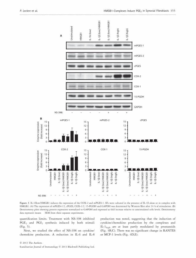
Next, we investigated whether IL-1blow/HMGB1 com-plexes modulated the expression of enzymes of the PGE2pathway in the same fashion as the IL-1bhigh stimulationalone. SFs were cultured for 24 h in the presence ofrHMGB1 and IL-1blow alone or in complex or IL-1bhighalone. Cells were harvested and the expression of PGE2pathway enzymes was investigated by Western blot.Unstimulated SFs showed low expression levels for thevarious enzymes analysed (Fig. 3A). When rHMGB1 andIL-1blow were administered alone, all enzyme expressionremained unchanged. When combined, however, the twostimuli triggered a noticeable upregulation in COX-2 andmPGES-1 expression, as can be visualized by a densi-tometry analysis (Fig. 3B). This explains the increase inPGE2 depicted in Fig. 1(A,B). No change could berecorded in the expression of mPGES-2, cPGES, COX-1
or 15-PGDH. The same pattern was observed inIL-1bhigh-stimulated SFs (Fig. 3A), thus suggesting thatHMGB1 acts as an enhancer of IL-1b signalling pathway.No consistent effect on enzyme expression could bedetected when COX-2 inhibitor NS-398 100 nM wasadded to IL-1blow/HMGB1 and IL-1bhigh–stimulatedcells. PGE2 synthesis was completely inhibited byNS-398 (data not shown).
Specific blockage of COX-2 enzymatic activity inhibits
IL-1blow/HMGB1-induced PGE2 synthesis and modulates
cytokine production from SFs
We also investigated whether prostaglandin productionwas differentially modulated in the presence of HMGB1by using the selective COX-2 inhibitor NS-398 and bystudying the prostanoid profile elicited by IL-1blow/rHMGB1 complexes in SFs stimulated for 24 h. UsingLC-MS/MS, the prostanoids PGE2, PGI2; PGD2, TxB2
and PGF2a were analysed in cell supernatants. The PGE2production triggered by IL-1blow/rHMGB1 and IL-1bhighwas significantly inhibited by the presence of 0.1 lMNS-398 (Fig. 4A). When we analysed the prostanoidprofile of SFs treated with rHMGB1 or IL-1blow, nomodulation of prostanoid production could be detected.IL-1blow/rHMGB1 and IL-1bhigh, however, gave riseto similar prostanoid profiles with PGE2 and PGI2(6-keto PGF1a) clearly upregulated. The other primaryprostanoids (PGD2, TxB2, PGF2a) remained under
400
300
200
100
0
400
300
200
100
00 12 48 72603624 0 12 48 72603624
0 12 24 48 726036
250200150100
500
0
125A B
C D
E F
100755025
04 16 2420128
PGE2
(% IL
-1β
(low
)/H
MG
B1)
PGE2
(% IL
-1β
(low
)/H
MG
B1)
IL-8
(% IL
-1β
(low
)/H
MG
B1)
IL-6
(% IL
-1β
(low
)/H
MG
B1)
0 12 48 72603624 0 12 48 72603624
400
300
200
100
0
200
150
100
50
0
MCP
-1(%
IL-1
β (lo
w)/
HM
GB1
)
RAN
TES
(% IL
-1β
(low
)/H
MG
B1)
Time (h) Time (h)
Time (h)Time (h)
Time (h) Time (h)
*****
***
***
***
***
**
***
***
****
unstim.HMGB1IL-1β (low)IL-1β (low)/HMGB1
unstim.HMGB1IL-1β (low)IL-1β (low)/HMGB1IL-1β (high)
Figure 2 The PGE2 production induced by
the IL-1blow/HMGB1 complexes precedes
cytokine release. SFs were cultured in the
presence of IL-1b alone or in complex with
HMGB1. PGE2 (A-B) and cytokine/chemokine
(C-F) production was measured in supernatants
after 4–72 h of stimulation. Results are
expressed as per cent of IL-1blow/HMGB1 (at
24 h for PGE2 and at 48 h for cytokines/
chemokines) to account for interpatient
variability. Data are expressed as means �SEM from three separate experiments, and P
values were calculated by parametric ANOVA.
The Tukey–Kramer post hoc test was used to
compare the HMGB1/IL-1blow and IL-1bhightreatment groups to the IL-1blow treatment
group.**P < 0.01, ***P < 0.001
Scandinavian Journal of Immunology, 2013, 77, 350–360
354 HMGB1-Complexes Induce PGE2 in Synovial Fibroblasts P. Leclerc et al...................................................................................................................................................................
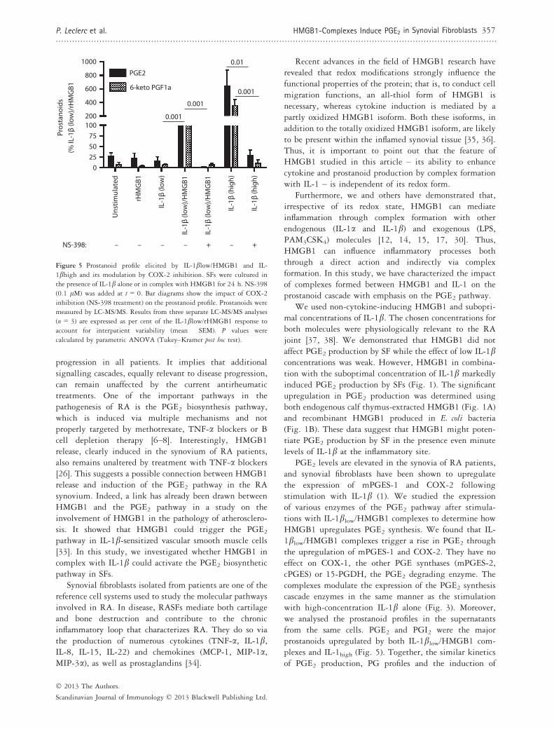
quantification limits. Treatment with NS-398 inhibitedPGE2 and PGI2 synthesis induced by both stimuli(Fig. 5).
Next, we studied the effect of NS-398 on cytokine/chemokine production. A reduction in IL-6 and IL-8
production was noted, suggesting that the induction ofcytokine/chemokine production by the complexes andIL-1high are at least partly modulated by prostanoids(Fig. 4B,C). There was no significant change in RANTESor MCP-1 levels (Fig. 4D,E).
mPGES-1
COX-2
Enzy
me
expr
essi
on(fo
ld in
crea
se)
0
3
6
9
12
Uns
timul
ated
IL-1
β (lo
w)/
HM
GB1
IL-1
β (h
igh)
mPGES-1
COX-2
15-PGDH
GAPDH
Uns
timul
ated
HM
GB1
COX-1IL
-1b
(low
)
IL-1
β (lo
w)/
HM
GB1
IL-1
β (h
igh)
15
0
3
6
9
12
15 COX-1
Enzy
me
expr
essi
on(fo
ld in
crea
se)
0
3
6
9
12
15
0
3
6
9
12
15 15-PGDH
HM
GB1
IL-1
β (lo
w)
IL-1
β (h
igh)
IL-1
β (lo
w)/
HM
GB1
+– – – – + –
Uns
timul
ated
IL-1
β (lo
w)/
HM
GB1
IL-1
β (h
igh)
HM
GB1
IL-1
β (lo
w)
IL-1
β (h
igh)
IL-1
β (lo
w)/
HM
GB1
NS-398: +– – – – + –
IL-1
β (lo
w)/
HM
GB1
IL-1
β (h
igh)
NS-398:
A
B
+–– – – + –
mPGES-2
cPGES
mPGES-2
0
3
6
9
12
15cPGES
Uns
timul
ated
IL-1
β (lo
w)/
HM
GB1
IL-1
β (h
igh)
HM
GB1
IL-1
β (lo
w)
IL-1
β (h
igh)
IL-1
β (lo
w)/
HM
GB1
+– – – – + –
0
3
6
9
12
15
Figure 3 IL-1ßlow/HMGB1 induces the expression of the COX-2 and mPGES-1. SFs were cultured in the presence of IL-1ß alone or in complex with
HMGB1. (A) The expression of mPGES-1/2, cPGES, COX-1/2, 15-PGDH and GAPDH was determined by Western Blot after 24 h of stimulation. (B)
Densitometry plots showing protein expression normalized to GAPDH and expressed as fold increase relative to unstimulated cells levels. Densitometry
data represent means � SEM from three separate experiments.
� 2013 The Authors.
Scandinavian Journal of Immunology © 2013 Blackwell Publishing Ltd.
P. Leclerc et al. HMGB1-Complexes Induce PGE2 in Synovial Fibroblasts 355..................................................................................................................................................................

IL-1blow/HMGB1 utilizes IL-1RI signalling for the induction of
PGE2 production
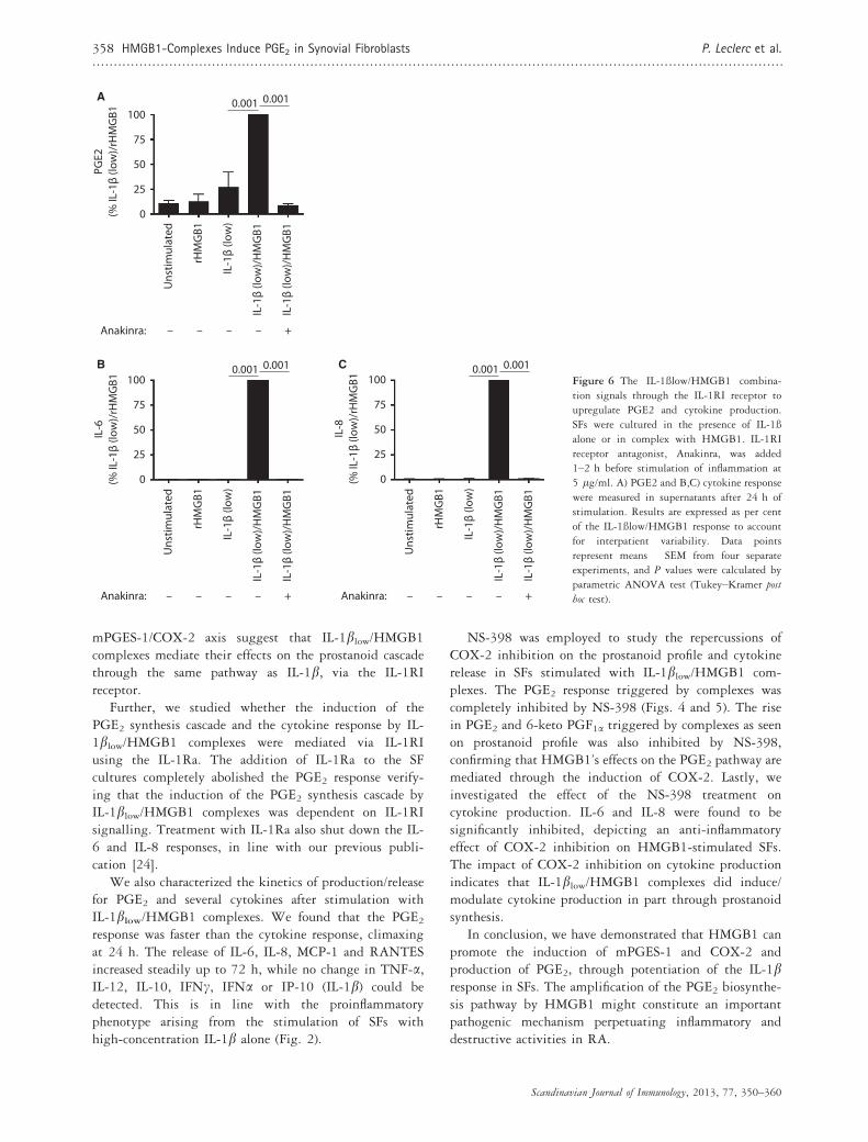
Our next aim was to investigate whether the PGE2production induced by IL-1blow/HMGB1 was mediatedthrough IL-1RI. After 24 h of stimulation, a significantincrease in PGE2 was detected in IL-1blow/rHMGB1-stimulated cells. Most importantly, when anakinra (5 lg/ml) was added 1–2 h prior to stimulation, it maintainedPGE2 at unstimulated levels. (Fig. 6A) These resultsindicate that IL-1blow/HMGB1 complexes signal exclusively
through IL-1RI to increase PGE2 synthesis (Fig. 6A) Thesame patternwas recorded for the production of cytokines IL-6 and IL-8, supporting our previous findings (Fig. 6B)[24].
Discussion
RA therapy has gone through great advances with thecoming of biologics. These drugs are directed towardsspecific inflammatory processes targeting cytokines orimmune cells. Still, none of those treatments, alone or incombination with DMARDs can stop, halt or reverse RA
Uns
timul
ated
rHM
GB1
IL-1
β (lo
w)
IL-1
β (lo
w)/
HM
GB1
IL-1
β (lo
w)/
HM
GB1
IL-1
β (h
igh)
IL-1
β (h
igh)
NS-398: – – – – +– +
100755025
0
PGE2
(% IL
-1β
(low
)/rH
MG
B1
500750
1000
0.001
0.001A B
C
E
D
Uns
timul
ated
rHM
GB1
IL-1
β (lo
w)
IL-1
β (lo
w)/
HM
GB1
IL-1
β (lo
w)/
HM
GB1
IL-1
β (h
igh)
IL-1
β (h
igh)
NS-398: – – – – +– +
100755025
0
IL-6
(% IL
-1β
(low
)/rH
MG
B1
100300
500
0.001
0.001700
Uns
timul
ated
rHM
GB1
IL-1
β (lo
w)
IL-1
β (lo
w)/
HM
GB1
IL-1
β (lo
w)/
HM
GB1
IL-1
β (h
igh)
IL-1
β (h
igh)
NS-398: – – – – +– +
100755025
0
IL-8
(% IL
-1β
(low
)/rH
MG
B1
200400600
0.05
0.001
Uns
timul
ated
rHM
GB1
IL-1
β (lo
w)
IL-1
β (lo
w)/
HM
GB1
IL-1
β (lo
w)/
HM
GB1
IL-1
β (h
igh)
IL-1
β (h
igh)
100
0
RAN
TES
(% IL
-1β
(low
)/rH
MG
B1
200
300
NS-398: – – – – +– +
Uns
timul
ated
rHM
GB1
IL-1
β (lo
w)
IL-1
β (lo
w)/
HM
GB1
IL-1
β (lo
w)/
HM
GB1
IL-1
β (h
igh)
IL-1
β (h
igh)
NS-398: – – – – +– +
100
0
MCP
-1(%
IL-1
β (lo
w)/
rHM
GB1
200
300
Figure 4 Specific blockage of COX-2
enzymatic activity inhibits IL-1blow/HMGB1- induced PGE2 synthesis and
reduces cytokine production from RASFs.
Specific blockage of COX-2 enzymatic
activity inhibits IL-1blow/rHMGB1-induced
PGE2 and reduces cytokine production from
SFs. SFs were cultured in the presence of IL-1balone or in complex with HMGB1 for 24 h.
NS-398 was added at t = 0. Bar diagrams
show the impact of COX-2 inhibition
(NS-398 treatment) on (A) PGE2 and (B–E)cytokine production by SFs. Results are from
three separate experiments and expressed as
percent of the IL-1blow/rHMGB1 response
to account for interpatient variability
(mean � SEM). PGE2 was measured by
enzyme immunoassay and cytokines by CBA.
P values were calculated by parametric ANOVA
(Tukey-Kramer post hoc test).
Scandinavian Journal of Immunology, 2013, 77, 350–360
356 HMGB1-Complexes Induce PGE2 in Synovial Fibroblasts P. Leclerc et al...................................................................................................................................................................
progression in all patients. It implies that additionalsignalling cascades, equally relevant to disease progression,can remain unaffected by the current antirheumatictreatments. One of the important pathways in thepathogenesis of RA is the PGE2 biosynthesis pathway,which is induced via multiple mechanisms and notproperly targeted by methotrexate, TNF-a blockers or Bcell depletion therapy [6–8]. Interestingly, HMGB1release, clearly induced in the synovium of RA patients,also remains unaltered by treatment with TNF-a blockers[26]. This suggests a possible connection between HMGB1release and induction of the PGE2 pathway in the RAsynovium. Indeed, a link has already been drawn betweenHMGB1 and the PGE2 pathway in a study on theinvolvement of HMGB1 in the pathology of atherosclero-sis. It showed that HMGB1 could trigger the PGE2pathway in IL-1b-sensitized vascular smooth muscle cells[33]. In this study, we investigated whether HMGB1 incomplex with IL-1b could activate the PGE2 biosyntheticpathway in SFs.
Synovial fibroblasts isolated from patients are one of thereference cell systems used to study the molecular pathwaysinvolved in RA. In disease, RASFs mediate both cartilageand bone destruction and contribute to the chronicinflammatory loop that characterizes RA. They do so viathe production of numerous cytokines (TNF-a, IL-1b,IL-8, IL-15, IL-22) and chemokines (MCP-1, MIP-1a,MIP-3a), as well as prostaglandins [34].
Recent advances in the field of HMGB1 research haverevealed that redox modifications strongly influence thefunctional properties of the protein; that is, to conduct cellmigration functions, an all-thiol form of HMGB1 isnecessary, whereas cytokine induction is mediated by apartly oxidized HMGB1 isoform. Both these isoforms, inaddition to the totally oxidized HMGB1 isoform, are likelyto be present within the inflamed synovial tissue [35, 36].Thus, it is important to point out that the feature ofHMGB1 studied in this article – its ability to enhancecytokine and prostanoid production by complex formationwith IL-1 – is independent of its redox form.
Furthermore, we and others have demonstrated that,irrespective of its redox state, HMGB1 can mediateinflammation through complex formation with otherendogenous (IL-1a and IL-1b) and exogenous (LPS,PAM3CSK4) molecules [12, 14, 15, 17, 30]. Thus,HMGB1 can influence inflammatory processes boththrough a direct action and indirectly via complexformation. In this study, we have characterized the impactof complexes formed between HMGB1 and IL-1 on theprostanoid cascade with emphasis on the PGE2 pathway.
We used non-cytokine-inducing HMGB1 and subopti-mal concentrations of IL-1b. The chosen concentrations forboth molecules were physiologically relevant to the RAjoint [37, 38]. We demonstrated that HMGB1 did notaffect PGE2 production by SF while the effect of low IL-1bconcentrations was weak. However, HMGB1 in combina-tion with the suboptimal concentration of IL-1b markedlyinduced PGE2 production by SFs (Fig. 1). The significantupregulation in PGE2 production was determined usingboth endogenous calf thymus-extracted HMGB1 (Fig. 1A)and recombinant HMGB1 produced in E. coli bacteria(Fig. 1B). These data suggest that HMGB1 might poten-tiate PGE2 production by SF in the presence even minutelevels of IL-1b at the inflammatory site.
PGE2 levels are elevated in the synovia of RA patients,and synovial fibroblasts have been shown to upregulatethe expression of mPGES-1 and COX-2 followingstimulation with IL-1b (1). We studied the expressionof various enzymes of the PGE2 pathway after stimula-tions with IL-1blow/HMGB1 complexes to determine howHMGB1 upregulates PGE2 synthesis. We found that IL-1blow/HMGB1 complexes trigger a rise in PGE2 throughthe upregulation of mPGES-1 and COX-2. They have noeffect on COX-1, the other PGE synthases (mPGES-2,cPGES) or 15-PGDH, the PGE2 degrading enzyme. Thecomplexes modulate the expression of the PGE2 synthesiscascade enzymes in the same manner as the stimulationwith high-concentration IL-1b alone (Fig. 3). Moreover,we analysed the prostanoid profiles in the supernatantsfrom the same cells. PGE2 and PGI2 were the majorprostanoids upregulated by both IL-1blow/HMGB1 com-plexes and IL-1high (Fig. 5). Together, the similar kineticsof PGE2 production, PG profiles and the induction of
Uns
timul
ated
rHM
GB1
IL-1
β (lo
w)
IL-1
β (lo
w)/
HM
GB1
IL-1
β (lo
w)/
HM
GB1
IL-1
β (h
igh)
IL-1
β (h
igh)
NS-398: – – – – +– +
100755025
0
200
400
1000
800
600
Pros
tano
ids
(% IL
-1β
(low
)/rH
MG
B1PGE2
6-keto PGF1a
0.001
0.001
0.001
0.01
Figure 5 Prostanoid profile elicited by IL-1blow/HMGB1 and IL-
1bhigh and its modulation by COX-2 inhibition. SFs were cultured in
the presence of IL-1b alone or in complex with HMGB1 for 24 h. NS-398
(0.1 lM) was added at t = 0. Bar diagrams show the impact of COX-2
inhibition (NS-398 treatment) on the prostanoid profile. Prostanoids were
measured by LC-MS/MS. Results from three separate LC-MS/MS analyses
(n = 3) are expressed as per cent of the IL-1blow/rHMGB1 response to
account for interpatient variability (mean � SEM). P values were
calculated by parametric ANOVA (Tukey–Kramer post hoc test).
� 2013 The Authors.
Scandinavian Journal of Immunology © 2013 Blackwell Publishing Ltd.
P. Leclerc et al. HMGB1-Complexes Induce PGE2 in Synovial Fibroblasts 357..................................................................................................................................................................
mPGES-1/COX-2 axis suggest that IL-1blow/HMGB1complexes mediate their effects on the prostanoid cascadethrough the same pathway as IL-1b, via the IL-1RIreceptor.
Further, we studied whether the induction of thePGE2 synthesis cascade and the cytokine response by IL-1blow/HMGB1 complexes were mediated via IL-1RIusing the IL-1Ra. The addition of IL-1Ra to the SFcultures completely abolished the PGE2 response verify-ing that the induction of the PGE2 synthesis cascade byIL-1blow/HMGB1 complexes was dependent on IL-1RIsignalling. Treatment with IL-1Ra also shut down the IL-6 and IL-8 responses, in line with our previous publi-cation [24].
We also characterized the kinetics of production/releasefor PGE2 and several cytokines after stimulation withIL-1blow/HMGB1 complexes. We found that the PGE2response was faster than the cytokine response, climaxingat 24 h. The release of IL-6, IL-8, MCP-1 and RANTESincreased steadily up to 72 h, while no change in TNF-a,IL-12, IL-10, IFNc, IFNa or IP-10 (IL-1b) could bedetected. This is in line with the proinflammatoryphenotype arising from the stimulation of SFs withhigh-concentration IL-1b alone (Fig. 2).
NS-398 was employed to study the repercussions ofCOX-2 inhibition on the prostanoid profile and cytokinerelease in SFs stimulated with IL-1blow/HMGB1 com-plexes. The PGE2 response triggered by complexes wascompletely inhibited by NS-398 (Figs. 4 and 5). The risein PGE2 and 6-keto PGF1a triggered by complexes as seenon prostanoid profile was also inhibited by NS-398,confirming that HMGB1’s effects on the PGE2 pathway aremediated through the induction of COX-2. Lastly, weinvestigated the effect of the NS-398 treatment oncytokine production. IL-6 and IL-8 were found to besignificantly inhibited, depicting an anti-inflammatoryeffect of COX-2 inhibition on HMGB1-stimulated SFs.The impact of COX-2 inhibition on cytokine productionindicates that IL-1blow/HMGB1 complexes did induce/modulate cytokine production in part through prostanoidsynthesis.
In conclusion, we have demonstrated that HMGB1 canpromote the induction of mPGES-1 and COX-2 andproduction of PGE2, through potentiation of the IL-1bresponse in SFs. The amplification of the PGE2 biosynthe-sis pathway by HMGB1 might constitute an importantpathogenic mechanism perpetuating inflammatory anddestructive activities in RA.
Uns
timul
ated
rHM
GB1
IL-1
β (lo
w)
IL-1
β (lo
w)/
HM
GB1
100
75
50
25
0
PGE2
(% IL
-1β
(low
)/rH
MG
B10.001
IL-1
β (lo
w)/
HM
GB1
0.001
Anakinra: – – +
Uns
timul
ated
rHM
GB1
IL-1
β (lo
w)
IL-1
β (lo
w)/
HM
GB1
100
75
50
25
0
IL-6
(% IL
-1β
(low
)/rH
MG
B1
0.001
IL-1
β (lo
w)/
HM
GB1
0.001
Anakinra: – – +
Uns
timul
ated
rHM
GB1
IL-1
β (lo
w)
IL-1
β (lo
w)/
HM
GB1
100
75
50
25
0
IL-8
(% IL
-1β
(low
)/rH
MG
B10.001
IL-1
β (lo
w)/
HM
GB1
0.001
Anakinra: –
– –
– – – – – +
A
B CFigure 6 The IL-1ßlow/HMGB1 combina-
tion signals through the IL-1RI receptor to
upregulate PGE2 and cytokine production.
SFs were cultured in the presence of IL-1ß
alone or in complex with HMGB1. IL-1RI
receptor antagonist, Anakinra, was added
1–2 h before stimulation of inflammation at
5 lg/ml. A) PGE2 and B,C) cytokine response
were measured in supernatants after 24 h of
stimulation. Results are expressed as per cent
of the IL-1ßlow/HMGB1 response to account
for interpatient variability. Data points
represent means � SEM from four separate
experiments, and P values were calculated by
parametric ANOVA test (Tukey–Kramer post
hoc test).
Scandinavian Journal of Immunology, 2013, 77, 350–360
358 HMGB1-Complexes Induce PGE2 in Synovial Fibroblasts P. Leclerc et al...................................................................................................................................................................

Therefore, targeting HMBG1 or mPGES-1 couldcomplement the current therapies in the treatment ofrheumatoid arthritis.
Acknowledgment
The work presented was supported by grants from TheSwedish Medical Research Council, the Swedish Rheu-matism Association, King Gustaf V 80 year Foundation,the Karolinska Institutet Foundation, the �Ake WibergFoundation, Stiftelsen Allm€anna Barnhuset, the SwedishAssociation against Rheumatism von Kantzow founda-tion, the Loo and Hans Ostermans Foundation, the Axeland Eva Wallstr€oms Foundation, VINNOVA, the Apo-tekare Hedbergs foundation and the Sigurd and ElsaGoljes minnesfond. We also received support from theregional agreement on medical training and clinicalresearch (ALF) between the Stockholm Country Counciland Karolinska Institutet. Special thanks to the orthope-dic surgeon unit at Karolinska Sjukhuset, MarianneEngstr€om and Yvonne Sundstr€om for providing us withsynovial tissue and to Joan Raouf for cell cultureassistance.
References
1 Stichtenoth DO, Thoren S, Bian H, Peters-Golden M, Jakobsson PJ,
Crofford LJ. Microsomal prostaglandin E synthase is regulated by
proinflammatory cytokines and glucocorticoids in primary rheuma-
toid synovial cells. J Immunol. 2001;167:469–74.2 Thoren S, Jakobsson PJ. Coordinate up- and down-regulation of
glutathione-dependent prostaglandin E synthase and cyclooxygenase-
2 in A549 cells. Inhibition by NS-398 and leukotriene C4. Eur J
Biochem 2000;267:6428–34.3 Simon LS, Weaver AL, Graham DY et al. Anti-inflammatory and
upper gastrointestinal effects of celecoxib in rheumatoid arthritis: a
randomized controlled trial. JAMA 1999;282:1921–8.4 Jakobsson PJ, Thoren S, Morgenstern R, Samuelsson B. Identification
of human prostaglandin E synthase: a microsomal, glutathione-
dependent, inducible enzyme, constituting a potential novel drug
target. Proc Natl Acad Sci USA 1999;96:7220–5.5 Westman M, Korotkova M, af Klint E et al. Expression of microsomal
prostaglandin E synthase 1 in rheumatoid arthritis synovium.
Arthritis Rheum 2004;50:1774–80.6 Korotkova M, Westman M, Gheorghe KR et al. Effects of antirheu-
matic treatments on the prostaglandin E2 biosynthetic pathway.
Arthritis Rheum 2005;52:3439–47.7 Gheorghe KR, Thurlings RM, Westman M et al. Prostaglandin E2
synthesizing enzymes in rheumatoid arthritis B cells and the effects of
B cell depleting therapy on enzyme expression. PLoS ONE 2011;6:
e16378.
8 Gheorghe KR, Sadique S, Leclerc P et al. Limited effect of
anti-rheumatic treatment on 15-prostaglandin dehydrogenase in
rheumatoid arthritis synovial tissue. Arthritis Res Ther. 2012;14:
R121.
9 Castiglioni A, Canti V, Rovere-Querini P, Manfredi AA. High-
mobility group box 1 (HMGB1) as a master regulator of innate
immunity. Cell Tissue Res 2011;343:189–99.10 Yang H, Hreggvidsdottir HS, Palmblad K et al. A critical cysteine is
required for HMGB1 binding to Toll-like receptor 4 and activation of
macrophage cytokine release. Proc Natl Acad Sci USA2010;107:11942–7.
11 Kazama H, Ricci JE, Herndon JM, Hoppe G, Green DR, Ferguson
TA. Induction of immunological tolerance by apoptotic cells requires
caspase-dependent oxidation of high-mobility group box-1 protein.
Immunity 2008;29:21–32.12 Ivanov S, Dragoi AM, Wang X et al. A novel role for HMGB1 in
TLR9-mediated inflammatory responses to CpG-DNA. Blood2007;110:1970–81.
13 Urbonaviciute V, Furnrohr BG, Meister S et al. Induction of
inflammatory and immune responses by HMGB1-nucleosome com-
plexes: implications for the pathogenesis of SLE. J Exp Med2008;205:3007–18.
14 Youn JH, Kwak MS, Wu J et al. Identification of lipopolysaccharide-
binding peptide regionswithinHMGB1 and their effects on subclinical
endotoxemia in a mouse model. Eur J Immunol 2011;41:2753–62.15 Tian J, Avalos AM, Mao SY et al. Toll-like receptor 9-dependent
activation by DNA-containing immune complexes is mediated by
HMGB1 and RAGE. Nat Immunol 2007;8:487–96.16 Schiraldi M, Raucci A, Munoz LM et al. HMGB1 promotes
recruitment of inflammatory cells to damaged tissues by forming a
complex with CXCL12 and signaling via CXCR4. J Exp Med
2012;209:551–63.17 Hreggvidsdottir HS, Ostberg T, Wahamaa H et al. The alarmin
HMGB1 acts in synergy with endogenous and exogenous dan-
ger signals to promote inflammation. J Leukoc Biol 2009;86:
655–62.18 Hamada T, Torikai M, Kuwazuru A et al. Extracellular high mobility
group box chromosomal protein 1 is a coupling factor for hypoxia and
inflammation in arthritis. Arthritis Rheum 2008;58:2675–85.19 Kokkola R, Li J, Sundberg E et al. Successful treatment of collagen-
induced arthritis in mice and rats by targeting extracellular high
mobility group box chromosomal protein 1 activity. Arthritis Rheum
2003;48:2052–8.20 Pisetsky DS, Erlandsson-Harris H, Andersson U. High-mobility
group box protein 1 (HMGB1): an alarmin mediating the pathogen-
esis of rheumatic disease. Arthritis Res Ther. 2008;10:209.
21 SchierbeckH,LundbackP,PalmbladK et al.Monoclonal anti-HMGB1
(high mobility group box chromosomal protein 1) antibody protection
in two experimental arthritis models.Mol Med 2011;17:1039–44.22 Ostberg T, Kawane K, Nagata S et al. Protective targeting of high
mobility group box chromosomal protein 1 in a spontaneous arthritis
model. Arthritis Rheum 2010 Oct;62:2963–72.23 Kokkola R, Sundberg E, Ulfgren AK et al. High mobility group box
chromosomal protein 1: a novel proinflammatory mediator in
synovitis. Arthritis Rheum 2002;46:2598–603.24 Wahamaa H, Schierbeck H, Hreggvidsdottir HS et al. High mobility
group box protein 1 in complex with lipopolysaccharide or IL-1
promotes an increased inflammatory phenotype in synovial fibroblasts.
Arthritis Res Ther. 2011;13:R136.
25 af Klint E, Grundtman C, Engstrom M et al. Intraarticular gluco-
corticoid treatment reduces inflammation in synovial cell infiltrations
more efficiently than in synovial blood vessels. Arthritis Rheum.2005;52:3880–9.
26 Sundberg E, Grundtman C, Af Klint E et al. Systemic TNF blockade
does not modulate synovial expression of the pro-inflammatory
mediator HMGB1 in rheumatoid arthritis patients–a prospective
clinical study. Arthritis Res Ther. 2008;10:R33.
27 Bresnihan B, Alvaro-Gracia JM, Cobby M et al. Treatment of
rheumatoid arthritis with recombinant human interleukin-1 receptor
antagonist. Arthritis Rheum 1998;41:2196–204.28 Paonessa G, Frank R, Cortese R. Nucleotide sequence of rat liver
HMG1 cDNA. Nucleic Acids Res 1987;15:9077.
29 Wang H, Bloom O, Zhang M et al. HMG-1 as a late mediator of
endotoxin lethality in mice. Science 1999;285:248–51.
� 2013 The Authors.
Scandinavian Journal of Immunology © 2013 Blackwell Publishing Ltd.
P. Leclerc et al. HMGB1-Complexes Induce PGE2 in Synovial Fibroblasts 359..................................................................................................................................................................
30 Sha Y, Zmijewski J, Xu Z, Abraham E. HMGB1 develops enhanced
proinflammatory activity by binding to cytokines. J Immunol.
2008;180:2531–7.31 Glennas A, Thorsrud AK, Rugstad HE, Jellum E. Mapping of
proteins from cultured fibroblasts of synovial and subcutaneous origin
by high resolution two-dimensional polyacrylamide gel electropho-
resis. Ann Rheum Dis 1985;44:302–6.32 Angel J, Berenbaum F, Le Denmat C, Nevalainen T, Masliah J,
Fournier C. Interleukin-1-induced prostaglandin E2 biosynthesis in
human synovial cells involves the activation of cytosolic phospholi-
pase A2 and cyclooxygenase-2. Eur J Biochem 1994;226:125–31.33 Jaulmes A, Thierry S, Janvier B, Raymondjean M, Marechal V.
Activation of sPLA2-IIA and PGE2 production by high mobility
group protein B1 in vascular smooth muscle cells sensitized by IL-1
beta. Faseb J. 2006;20:1727–9.34 Huber LC, Distler O, Tarner I, Gay RE, Gay S, Pap T. Synovial
fibroblasts: key players in rheumatoid arthritis. Rheumatology (Oxford)
2006;45:669–75.35 Venereau E, Casalgrandi M, Schiraldi M et al. Mutually exclusive
redox forms of HMGB1 promote cell recruitment or proinflammatory
cytokine release. J Exp Med 2012;209:1519–28.36 Yang H, Lundback P, Ottosson L et al. Redox modification of
cysteine residues regulates the cytokine activity of high mobility
group box-1 (HMGB1). Mol Med 2012;18:250–9.
37 Taniguchi N, Kawahara K, Yone K et al. High mobility group box
chromosomal protein 1 plays a role in the pathogenesis of rheumatoid
arthritis as a novel cytokine. Arthritis Rheum 2003;48:971–81.38 Kahle P, Saal JG, Schaudt K, Zacher J, Fritz P, Pawelec G.
Determination of cytokines in synovial fluids: correlation with
diagnosis and histomorphological characteristics of synovial tissue.
Ann Rheum Dis 1992;51:731–4.
Supporting Information
Additional supporting information may be found in theonline version of this article:
Figure S1. PGE2 and cytokine response to IL-1blow/HMGB1 complexes in three representative patients todepict inter-patient variability. SFs were cultured in thepresence of IL-1b and HMGB1 separately or in complex.PGE2 production and cytokine release were measured insupernatants after 24 h of stimulation. Mediator produc-tion obtained when using rHMGB1 (A) PGE2 B) IL-6 C)IL-8 D) MCP-1 E) RANTES. PGE2 was measures by EIAand cytokines by CBA. Data is expressed in pg/ml.
Scandinavian Journal of Immunology, 2013, 77, 350–360
360 HMGB1-Complexes Induce PGE2 in Synovial Fibroblasts P. Leclerc et al...................................................................................................................................................................
Related Documents