Identification of a Conserved Anti-Apoptotic Protein That Modulates the Mitochondrial Apoptosis Pathway Yu Zhang 1. , Elisabet Johansson 2. , Marian L. Miller 2 , Reiner U. Ja ¨ nicke 3 , Donald J. Ferguson 4 , David Plas 5 , Jarek Meller 6 , Marshall W. Anderson 7 * 1 School of Pharmacy, University of Cincinnati, Cincinnati, Ohio, United States of America, 2 Department of Environmental Health, College of Medicine, University of Cincinnati, Cincinnati, Ohio, United States of America, 3 Laboratory of Molecular Radiooncology, Clinic and Policlinic for Radiation Therapy and Radiooncology, Clinical Center of the University of Du ¨ sseldorf, Du ¨ sseldorf, Germany, 4 Department of Microbiology, Miami University, Oxford, Ohio, United States of America, 5 Department of Cancer and Cell Biology, College of Medicine, University of Cincinnati, Cincinnati, Ohio, United States of America, 6 Division of Biomedical Informatics, Departments of Environmental Health and Biomedical Engineering, University of Cincinnati, Children’s Hospital Medical Center, Cincinnati, Ohio, United States of America, 7 Department of Medicine, Cancer Center, Medical College of Wisconsin, Milwaukee, Wisconsin, United States of America Abstract Here we identified an evolutionarily highly conserved and ubiquitously expressed protein (C9orf82) that shows structural similarities to the death effector domain of apoptosis-related proteins. RNAi knockdown of C9orf82 induced apoptosis in A- 549 and MCF7/casp3-10b lung and breast carcinoma cells, respectively, but not in cells lacking caspase-3, caspase-10 or both. Apoptosis was associated with activated caspases-3, -8, -9 and -10, and inactivation of caspases 10 or 3 was sufficient to block apoptosis in this pathway. Apoptosis upon knockdown of C9orf82 was associated with increased caspase-10 expression and activation, which was required for the generation of an 11 kDa tBid fragment and activation of Caspase-9. These data suggest that C9orf82 functions as an anti-apoptotic protein that modulates a caspase-10 dependent mitochondrial caspase-3/9 feedback amplification loop. We designate this ubiquitously expressed and evolutionarily conserved anti-apoptotic protein Conserved Anti-Apoptotic Protein (CAAP). We also demonstrated that treatment of MCF7/ casp3-10b cells with staurosporine and etoposides induced apoptosis and knockdown of CAAP expression. This implies that the CAAP protein could be a target for chemotherapeutic agents. Citation: Zhang Y, Johansson E, Miller ML, Ja ¨nicke RU, Ferguson DJ, et al. (2011) Identification of a Conserved Anti-Apoptotic Protein That Modulates the Mitochondrial Apoptosis Pathway. PLoS ONE 6(9): e25284. doi:10.1371/journal.pone.0025284 Editor: Andrea C. LeBlanc, McGill University, Canada Received April 18, 2011; Accepted August 31, 2011; Published September 30, 2011 Copyright: ß 2011 Zhang et al. This is an open-access article distributed under the terms of the Creative Commons Attribution License, which permits unrestricted use, distribution, and reproduction in any medium, provided the original author and source are credited. Funding: This work was supported by the University of Cincinnati College of Medicine and by a grant from the National Institute of Environmental Health Sciences (NIEHS) to the Center for Environmental Genetics (P30 ES06096). The funders had no role in study design, data collection and analysis, decision to publish, or preparation of the manuscript. Competing Interests: The authors have declared that no competing interests exist. * E-mail: [email protected] . These authors contributed equally to this work. Introduction CAAP (C9orf82) is an unannotated gene residing between 26,830,685 and 26,882,725 bp on Chr 9p harboring two alternative transcriptional start sites and six exons with a total length of 2143 bp, as well as a 39 UTR of 1047 bp (Ensembl). We originally detected this gene by a 59 RACE analysis when searching this region for genes related to tumorigenesis [1]. Our preliminary studies indicated several characteristics of CAAP that merited further investigation. First of all, it exhibited a high degree of evolutionary conservation, and was expressed at some level in every human tissue examined from panels of both normal (adult and fetal) and tumor tissues. Moreover, using bioinformatics approaches, one of its putative domains was predicted to share structural similarity with the death effector domain (DED). DED and the related death domain (DD) are found in a large superfamily of proteins that regulate apoptosis [2]. Therefore, we further characterized CAAP as a potential agent in apoptosis- related signaling. Apoptotic signaling pathways are induced by activation of caspases which then cleave key protein substrates resulting in cell death [3]. Based on their structure, caspases can be divided into two classes. Caspases-2, -8, -9, and -10 contain long amino- terminal prodomains and normally function as initiators of apoptotic pathways, whereas caspases-3, -6, and -7 have only short prodomains and function as effectors of cell death [4–8]. The activation of the initiator caspase-9 in the intrinsic mitochondrial apoptosis pathway involves BH3 proteins of the Bcl-2 family that function as monitors of cellular damage. In response to cellular damage, these proteins promote activation of the pro-apoptotic activities of Bax and Bak, inducing the release of cytochrome c, and subsequent formation of the apoptosome, which is a multi- subunit caspase scaffold that activates the caspase-9-dependent apoptotic pathway [9–11]. In the death receptor-mediated apoptosis pathway, a protein complex recruiting the Fas-associated protein with a death domain (FADD), and procaspase-8 and/or - 10 is called the death-inducing signaling complex (DISC) [12]. The procaspases-8 and -10 in the DISC are activated by oligomerization followed by proteolytic self-processing enabling them to activate downstream effector caspases including caspase-3 [4]. Recent studies of the mitochondrial apoptosis pathway demonstrate that caspase-8 and -10 can also be activated PLoS ONE | www.plosone.org 1 September 2011 | Volume 6 | Issue 9 | e25284

Welcome message from author
This document is posted to help you gain knowledge. Please leave a comment to let me know what you think about it! Share it to your friends and learn new things together.
Transcript
Identification of a Conserved Anti-Apoptotic ProteinThat Modulates the Mitochondrial Apoptosis PathwayYu Zhang1., Elisabet Johansson2., Marian L. Miller2, Reiner U. Janicke3, Donald J. Ferguson4, David
Plas5, Jarek Meller6, Marshall W. Anderson7*
1 School of Pharmacy, University of Cincinnati, Cincinnati, Ohio, United States of America, 2 Department of Environmental Health, College of Medicine, University of
Cincinnati, Cincinnati, Ohio, United States of America, 3 Laboratory of Molecular Radiooncology, Clinic and Policlinic for Radiation Therapy and Radiooncology, Clinical
Center of the University of Dusseldorf, Dusseldorf, Germany, 4 Department of Microbiology, Miami University, Oxford, Ohio, United States of America, 5 Department of
Cancer and Cell Biology, College of Medicine, University of Cincinnati, Cincinnati, Ohio, United States of America, 6 Division of Biomedical Informatics, Departments of
Environmental Health and Biomedical Engineering, University of Cincinnati, Children’s Hospital Medical Center, Cincinnati, Ohio, United States of America, 7 Department
of Medicine, Cancer Center, Medical College of Wisconsin, Milwaukee, Wisconsin, United States of America
Abstract
Here we identified an evolutionarily highly conserved and ubiquitously expressed protein (C9orf82) that shows structuralsimilarities to the death effector domain of apoptosis-related proteins. RNAi knockdown of C9orf82 induced apoptosis in A-549 and MCF7/casp3-10b lung and breast carcinoma cells, respectively, but not in cells lacking caspase-3, caspase-10 orboth. Apoptosis was associated with activated caspases-3, -8, -9 and -10, and inactivation of caspases 10 or 3 was sufficientto block apoptosis in this pathway. Apoptosis upon knockdown of C9orf82 was associated with increased caspase-10expression and activation, which was required for the generation of an 11 kDa tBid fragment and activation of Caspase-9.These data suggest that C9orf82 functions as an anti-apoptotic protein that modulates a caspase-10 dependentmitochondrial caspase-3/9 feedback amplification loop. We designate this ubiquitously expressed and evolutionarilyconserved anti-apoptotic protein Conserved Anti-Apoptotic Protein (CAAP). We also demonstrated that treatment of MCF7/casp3-10b cells with staurosporine and etoposides induced apoptosis and knockdown of CAAP expression. This implies thatthe CAAP protein could be a target for chemotherapeutic agents.
Citation: Zhang Y, Johansson E, Miller ML, Janicke RU, Ferguson DJ, et al. (2011) Identification of a Conserved Anti-Apoptotic Protein That Modulates theMitochondrial Apoptosis Pathway. PLoS ONE 6(9): e25284. doi:10.1371/journal.pone.0025284
Editor: Andrea C. LeBlanc, McGill University, Canada
Received April 18, 2011; Accepted August 31, 2011; Published September 30, 2011
Copyright: � 2011 Zhang et al. This is an open-access article distributed under the terms of the Creative Commons Attribution License, which permitsunrestricted use, distribution, and reproduction in any medium, provided the original author and source are credited.
Funding: This work was supported by the University of Cincinnati College of Medicine and by a grant from the National Institute of Environmental HealthSciences (NIEHS) to the Center for Environmental Genetics (P30 ES06096). The funders had no role in study design, data collection and analysis, decision topublish, or preparation of the manuscript.
Competing Interests: The authors have declared that no competing interests exist.
* E-mail: [email protected]
. These authors contributed equally to this work.
Introduction
CAAP (C9orf82) is an unannotated gene residing between
26,830,685 and 26,882,725 bp on Chr 9p harboring two
alternative transcriptional start sites and six exons with a total
length of 2143 bp, as well as a 39 UTR of 1047 bp (Ensembl). We
originally detected this gene by a 59 RACE analysis when
searching this region for genes related to tumorigenesis [1]. Our
preliminary studies indicated several characteristics of CAAP that
merited further investigation. First of all, it exhibited a high degree
of evolutionary conservation, and was expressed at some level in
every human tissue examined from panels of both normal (adult
and fetal) and tumor tissues. Moreover, using bioinformatics
approaches, one of its putative domains was predicted to share
structural similarity with the death effector domain (DED). DED
and the related death domain (DD) are found in a large
superfamily of proteins that regulate apoptosis [2]. Therefore,
we further characterized CAAP as a potential agent in apoptosis-
related signaling.
Apoptotic signaling pathways are induced by activation of
caspases which then cleave key protein substrates resulting in cell
death [3]. Based on their structure, caspases can be divided into
two classes. Caspases-2, -8, -9, and -10 contain long amino-
terminal prodomains and normally function as initiators of
apoptotic pathways, whereas caspases-3, -6, and -7 have only
short prodomains and function as effectors of cell death [4–8]. The
activation of the initiator caspase-9 in the intrinsic mitochondrial
apoptosis pathway involves BH3 proteins of the Bcl-2 family that
function as monitors of cellular damage. In response to cellular
damage, these proteins promote activation of the pro-apoptotic
activities of Bax and Bak, inducing the release of cytochrome c,
and subsequent formation of the apoptosome, which is a multi-
subunit caspase scaffold that activates the caspase-9-dependent
apoptotic pathway [9–11]. In the death receptor-mediated
apoptosis pathway, a protein complex recruiting the Fas-associated
protein with a death domain (FADD), and procaspase-8 and/or -
10 is called the death-inducing signaling complex (DISC) [12].
The procaspases-8 and -10 in the DISC are activated by
oligomerization followed by proteolytic self-processing enabling
them to activate downstream effector caspases including caspase-3
[4]. Recent studies of the mitochondrial apoptosis pathway
demonstrate that caspase-8 and -10 can also be activated
PLoS ONE | www.plosone.org 1 September 2011 | Volume 6 | Issue 9 | e25284
downstream of the mitochondria by caspase-3, indicating the
existence of so-called amplification loops where caspase-8 or -10
activate caspase-9 and -3 [13–18].
In this context, it should be noted that activated caspase-8 and -
10 can also proteolytically activate pro-apoptotic Bcl-2 family
member Bid generating tBid [17,18]. tBid causes the release of
mitochondrial cytochrome c resulting in the activation of caspase-
9 which can further enhance caspase-3 activity to complete the
apoptotic process [19–21].
To determine whether and to what extent CAAP is involved in
the regulation of apoptosis, we examined caspase activation and
apoptosis signaling in the presence and absence of CAAP in
several tumor models. Our study revealed that CAAP exerts a
prominent anti-apoptotic function that critically depends on the
presence of caspases-3 and -10. In addition, we demonstrated that
treatment of MCF-7/casp3-10b cells with staurosporine and
etoposide triggered knockdown of the CAAP expression concur-
rent with the induction of apoptosis. These data suggest that
CAAP may be a target site for chemotherapy since it does not
require siRNA to knockdown the expression of this anti-apoptotic
protein.
Materials and Methods
Cell line and cell cultureThe human lung carcinoma cell line A-549 was obtained from
ATCC (Manassas, Virginia) grown at 37uC under 5% CO2 in
RPMI-1640 medium supplemented with 10% FBS and antibiotics.
MCF-7 breast carcinoma cells were maintained in RPMI-1640
supplemented with 10% fetal bovine serum and antibiotics. MCF-
7/casp3 cells stably expressing caspase-3, MCF-7/casp-10b stably
expressing caspase-10b and MCF-7/casp3-10b stably expressing
both caspase-3 and caspase-10b were maintained in the same
medium supplemented with either 400 mg of neomycin/ml (Fisher
Scientific) [MCF-7/casp3], 200 mg of hygromycin/ml (Santa Cruz
Biotechnology) [MCF-7/casp-10b] or a combination of both
antibiotics [MCF-7/casp3-10b].
Plasmid DNAMyc-FADD-DN (dominant-negative form of FADD) was from
Keyclone Technologies. Cincinnati. OH.
siRNA Preparation and TransfectionsiRNA probes were synthesized using SilencerH siRNA
Construction Kit from Ambion (Austin, Texas). The siRNA
Target Finder (http://www.ambion.com/techlib/misc/ siRNA_
finder.html) was used to select target sequences, and sequences
selected were analyzed using BLAST (http://www.ncbi.nlm.nih.
gov/BLAST/) to minimize off-target knockdown effects. Two
target sequences denoted as si48 and si67 were selected in CAAP:
si48: 59-AACCTGAAGGTTTGGAATTAA-39
si67: 59-AAGTGTGAATGAGATTCTAGG-39
The non-targeting siRNA negative control (Ncontrol) (D-
007206-13-20) and the sicasp-8 were purchased from Dharmacon
Research Inc:
sicasp-8: 59AA-GGGUCAUGCUCUAUCAGAU-dTdT-39
Transfection of A549 cells or MCF-7, MCF-7/casp3, MCF-7/
casp10, and MCF-7/casp3-10b cell lines was performed according
to the procedure used by Sharp et al (2005) [22]. Cells were
transfected at a confluency of 85–90%. Prior to transfection,
regular medium was replaced with low serum (4% FBS) medium.
The cells were transfected with siRNA using Lipofectamine (LF
2000) at a ratio of 1:3 siRNA (mg): Lipofectamine (ml). The final
concentration of siRNAs was 43 or 65 nM for A-549 cells and
22 nM for MCF cells. The cells were incubated with the siRNA-
Lipofectamine 2000 complex overnight. For A549 cells, 24 hrs
after transfection the low serum medium was replaced with normal
medium (10% FBS) without antibiotics and 48 hr after transfec-
tion floating and adherent cells were harvested and prepared for
analysis of apoptosis. Floating and adherent MCF cells were
harvested 24 hours after transfection, and prepared for analysis of
apoptosis.
Reverse transcription and PCRFor RT-PCR, total RNA was isolated using TRIzolH reagent
from Invitrogen (Carlsbad, California) according to manufactur-
er’s instructions. First-strand cDNA was synthesized by reverse
transcription of 1 mg total RNA from cell lines for 60 minutes at
42uC using random primers and 20 units AMV reverse
transcriptase in a total volume of 20 ml.
Analysis of CAAP expressionFor screening CAAP mRNA expression in normal, fetal, and
tumor tissues, MTC Multiple Tissue cDNA panels were obtained
from Clontech (Mountain View, California). The primers used
for amplification of the main splice variant (CAAP) were
DS93SPLJ1/2F (59-CTCCTTGCAGCAGGAAACTA-39) which
spans the boundary between exon 1 and exon 2, and DS93E2R2
(59-ATGACACAGTCAGGTCCAGT-39) which is located in
exon 2. For amplification of the alternative splice variant
(CAAP-ASV1) the primers used were DS93ASVF2 (59-GT-
CCGTCCTTGAGGTTCATGT-39) and DS93E2R2. PCR was
performed in 10 ml volumes containing 0.5 mM of each primer,
1.5 mM magnesium chloride, 200 mM of each dNTP, and 0.25
units Platinum Taq DNA polymerase (Invitrogen). Standard PCR
analysis was performed. Twenty PCR cycles were used for each
tissue. A human control tissue (Clontech) was analyzed in each of
the panels and results shown in the last column of each panel.
Quantitative PCR to assay suppressed CAAP expressionFor the determination of a successful knockdown of CAAP
mRNA expression in A549 cells after transfection with si48, si67,
and Ncontrol siRNAs, total RNA was isolated and first-strand
cDNA was synthesized by reverse transcription of 1 mg total RNA
from cell lines for 60 minutes at 42uC using random primers and
20 units AMV reverse transcriptase in a total volume of 20 ml. For
amplification of CAAP the primers used were DS93SPLJ1/2F and
DS93E2R2 as defined above. As a control, GAPDH was amplified
using the primers 59-CAGGAGGCATTGCTGATGAT-39 and
59-AGCGAGATCCCTCCAAAATC-39. Quantitative PCR was
carried out in 25 ml reactions containing 12.5 ml iQ SYBR
GREEN supermix (BioRad, Hercules, CA), 0.2 mM of each
primer and 10 ng of cDNA. The reactions were run using the
Realplex Mastercycler (Eppendorf) and the PCR program
included initial denaturation at 94uC for 10 minutes and then
50 cycles of 30 s at 94uC, 30 s at 56uC, and 45 s at 71uC. Cycle
number and percent knockdown of CAAP mRNA expression by
siRNAs were analyzed by using Eppendorf Mastercycler ep
Realplex software.
Apoptosis AssaysTo quantify apoptosis for both the adherent and floating cells,
we used the M30 FACS assay and a morphometric assay,
respectively. Media from plates were collected 24 hrs after
transfection of MCF cells and 48 hrs after transfection of A549
cells. After cell clumping was dispersed by pipeting, floating cells
were counted in aliquots of the media to determine the total
CAAP Regulates Caspase-10 in Apoptosis
PLoS ONE | www.plosone.org 2 September 2011 | Volume 6 | Issue 9 | e25284

number of floating cells. The media was then centrifuged at
1200 rpm for 5 minutes to obtain a pellet of floating cells. The
cells were resuspended in a small quantity of phosphate buffered,
isoosmolar, 2.5% gluteraldehyde/2% paraformaldehyde fluid and
placed in a conical Beem capsule and centrifuged at 1000 g for
5 minutes. Cells were fixed overnight, post fixed in 1% osmium
tetroxide for 2 hr, dehydrated through graded ethanols, propylene
oxide and embedded in Spurr’s resin. One micron thick sections
were made in at least two levels within the cell pellet, and were
stained with toluidine blue for light microscopy. Cells were
counted at 12506. The mean number of cells counted per group
was approximately one thousand, except for Ncontrol treatment
where all cells were counted. The fraction of floating cells which
were apoptotic was determined from these data.
Immediately after media was removed as described above,
adherent cells were washed with cold PBS and trypsinized. After
cell clumps were dispersed by pipetting, trypsinized cells were
counted in the PBS solution to determine the total number of
adherent cells. Cells were washed in PBS and fixed in ice-cold
methanol for 30 min at 220uC. Fixed cells were stained with M30
CytoDEATH antibody according to manufacturer’s instructions.
The antibody M30 CytoDEATH (Roche Applied Sciences)
recognizes a specific caspase cleavage site within cytokeratin 18
that is not detectable in the native keratin. Stained cells were
resuspended in PBS to 26105 cells/ml. After removal of clumping
by pipetting, FACS analysis was run on a BD FACS ARIA
instrument. At least 104 cells were analyzed for each sample. The
fraction of adherent cells which were apoptotic was determined
from the M30 FACS assay data.
Since we counted the total # of floating cells and adherent cells,
the % of floating and adherent cells was determined, and thus,
Percent of apoptosis was calculated as following:
Percent of apoptosis = % of floating cells6fraction of floating
cells that are apoptotic+% of adherent cells6fraction of adherent
cells that are apoptotic. The percentage of floating cells that were
apoptotic was always greater than 95% and thus we used this
number to calculate apoptosis.
Caspase activity measurementThe day before transfection 3.06105 A-549 cells or 4.06105
MCF cells were plated in 60 mm dishes. After 24 hrs A549 cells
were transfected with either Ncontrol 65 nM or si67-65 nM
siRNA, MCF cells were transfected with either Ncontrol 22 nM or
si67-22 nM siRNA and 16 or 24 hrs after transfection floating and
adherent cells were combined and prepared for caspase activity
measurements. Caspase activities were measured by using the
caspase colorimetric assay kit from BioVision by following
manufacturer’s instructions. Briefly, cell lysates (50–100 mg protein
in 50 ml lysis buffer) were incubated with 200 mM of colorimetric
caspase-3 substrate DEVD-pNA, caspase-8 substrate IETD-pNA,
caspase-9 substrate LEHD-pNA and caspase-10 substrate AEVD-
pNA in 50 ml of 26 reaction buffer containing 10 nM DTT at
37uC for 2 hrs, and samples read at 405 mm in a microtiter plate
reader.
Effect of FADD-DN on apoptosis induced by CAAPknockdown or TRAIL
The day before transfection, 36105 A-549 cells were plated in
60 mm dishes. The next day empty vector or FADD-DN were
transfected into cells using LF 2000. After 24 hrs proteins were
isolated from the cells as described below.
A-549 cells (36105) were plated in 60 mm dishes and after
24 hrs cells were transfected with empty vector or FADD-DN.
After 24 hrs, cells were transfected with si67 (65 nM) or treated
with TRAIL (35 ng/ml). Floating and adherent cells were
harvested 24 hrs after transfection, and prepared for analysis of
apoptosis.
Western Blot AnalysisCells were washed with cold PBS and lysed with RIPA buffer in
the presence of protease inhibitors, sonicated for 15 seconds, and
then centrifuged at 12,0006g at 4uC for 20 min. 35 mg of cellular
protein (quantitated by using the BCA protein assay from Pierce)
was loaded onto 4–12% NuPAGE Bis-Tris gradient gels
(invitrogen), electrophoresed and electrotransferred onto PVDF
membrane according to manufacturer’s instruction. Membranes
were then blocked with 5% milk TBS-T for 1 hr at room
temperature. Proteins were detected with the following antibodies:
rabbit anti-caspase 3 pAb ( BioMol), mouse anti-caspase 8 mAb,
rabbit anti-b-actin pAb and rabbit anti-caspase-9 pAb (Cell
Signaling Technology), mouse anti-caspase-10 (MBL med and
BioLab), mouse anti-Human FADD mAb (BD Transduction
Laboratories), rabbit anti-CAAP pAb (Quality Controlled Bio-
chemicals, Hopkinton, MA), rabbit anti-Bid pAb, and mouse anti-
PARP mAb ( BD Phararmingen). Membranes were incubated
with primary antibody overnight at 4uC and followed by
incubation with HRP-conjugated secondary antibodies (Cell
Signaling Technology). Immunoreaction bands were visualized
by using enhanced chemiluminescence (Perkin Elmer).
Treatment of MCF-7/casp3-10b cells with chemicalagents
MCF-7/casp3-10b cells were treated with either staurosporine
(STS) or etoposide. STS treatment: MCF-7/casp3-10b cells were
treated at dose of 1.0 mM for 4, 6, and 8 hrs and then western blot
analysis was performed to examine CAAP expression and cleavage
of caspase-3 and -9. Etoposide treatment: MCF-7/casp3-10b cells
were treated at dose of 100 mM and 500 mM for 24 hrs and then
harvested for western blot analysis of CAAP expression and
cleavage of PARP and procaspase-10 expression.
Results
Genomic organization and evolutionary profile of CAAPCAAP is located on chromosome 9p, band 21.2, and consists of
six exons spanning over 51.5 kilobases (Fig. 1A). 59-RACE
experiments indicated that CAAP has an alternative splice variant
(CAAP-ASV1) with an extended first exon at a canonical splice
donor 154 bp downstream of the splice donor in the prototypic
first exon (Fig. 1A). This splicing event was documented in dbEST,
and 15 of the 140 mRNA and EST sequences listed in the
Unigene cluster for CAAP contained the additional part of the first
exon. CAAP-ASV1 contains a stop codon at the end of the
extended first exon, which results in a truncated protein of 133
amino acids in length. Target locations of siRNA sequences are
indicated by arrows (Fig. 1A).
The 59-end and promoter regions of the gene are GC rich and
do not contain a TATA-box near the presumed transcription start
site. The promoter region of CAAP contains several transcription
factor binding sites including NF-KB, cMyb, SP1, and ETS-1. A
search of the TRANSFAC database showed that the promoter
region contains a 10-bp sequence (GGAAGTGACG: dashed red
underline) (Fig. 1B) that is also present in the promoter of the
retinoblastoma gene, where it constitutes a cis-acting element
susceptible to negative regulation by the tumor suppressor p53
[23]. Partly overlapping with the p53 control element is a motif
(GCCGGAAGT: dashed blue underline) for binding of the Ets1
oncogene (Fig. 1B).
CAAP Regulates Caspase-10 in Apoptosis
PLoS ONE | www.plosone.org 3 September 2011 | Volume 6 | Issue 9 | e25284

Figure 1. Genomic organization and evolutionary profile of CAAP. (A) Unfilled bars represent coding sequence, and grey bars untranslatedregions. The hatched bar shows the extension of the first exon that creates an alternative transcript. The arrowhead indicates the stop codon inCAAP-ASV1. Target locations of siRNA sequences are indicated by arrows. (B) The nucleotide sequence of the promoter region of CAAP harbors
CAAP Regulates Caspase-10 in Apoptosis
PLoS ONE | www.plosone.org 4 September 2011 | Volume 6 | Issue 9 | e25284

Multiple alignments show that CAAP contains four regions that
are highly conserved among mammalian species including
chimpanzee, dog, mouse, and rat as well as in more distant
species like Xenopus laevis (Fig. 1C). These four regions are: 1) a
short region at the N-terminal end including amino acid (aa)
residues 1 to 29 that is rich in basic residues; 2) a helical domain
between residues 94 to 169 that is predicted to share structural
similarity (with very low sequence homology) with the DED; 3) a
region adjacent to the putative DED between residues 170 to 247
that contains two predicted helices; and 4) a C-terminal domain
between residues 292 to 361 which is highly conserved in all
mammalian species examined as well as in Xenopus, and that is
predicted to also contain two well-defined helices. The four
distinct conserved domains are shown as shaded boxes in Fig. 1D.
On the other hand, the predicted translation product of CAAP-
ASV1 only contains the orthologous region A, as the region of the
protein resulting from the extension of the first exon is not
conserved among mammalian species. The function of this splice
variant will not be examined in this study.
Structural analyses of CAAPSequence alignment, secondary structure prediction, and fold
recognition methods were used to characterize CAAP and identify
its putative homologs. Simple BLAST alignments did not result in
significant matches into annotated protein families. Subsequent
Psi-BLAST iterations revealed only weak sequence similarity to
multiple (mostly helical) protein families. To map putative
domains in CAAP and to enhance subsequent fold recognition
searches, we used PsiPRED and SABLE to derive consensus
secondary structure, as well as solvent accessibility predictions.
According to these predictions, CAAP contains two putative
globular regions, one located approximately between residues 90
and 250 and another between residues 290 and 360, comprising
several helices with multiple sites predicted to be fully buried in the
hydrophobic core of these putative globular domains (Fig. 2A).
Although PsiPRED predictions are largely consistent with those
obtained by SABLE (which are shown in Fig. 2A), there are several
additional helical segments predicted by PsiPRED between
residues 90 and 130, which are highlighted in Fig. 2B. On the
other hand, the N-terminus domain which coincides with the first
exon is consistently predicted to be largely unstructured with
several putative secondary structure elements. It should be noted
that despite the presence of some hydrophobic fragments, CAAP is
overall strongly hydrophilic and predicted to be a soluble protein.
Several fold recognition methods were used to help identify
structural homologs by threading the primary sequence of CAAP
through databases of known protein structures or evolutionary
profiles of protein families. The well benchmarked FFAS server
[24] identified a death effector domain (DED) as the best match
for CAAP in the PFAM database [25]. The observed and
predicted helical segments were qualitatively consistent, support-
ing the prediction of a 6 alpha-helical bundle between amino acids
94 and 169 (Fig. 2B). In addition, several functionally important
charged as well as hydrophobic sites observed in DED of FADD
(human and mouse) were conserved in CAAP, as highlighted in
Fig. 2C. This indicates that not only structural, but also functional
similarity could exist between the central domain of CAAP and
other DED proteins.
Ubiquitous Expression of CAAP in tissues and cell linesExpression of CAAP was tested by PCR on commercially
available primary tissue cDNA panels. It was found to be
expressed in all human tissues examined (Fig. S1). In normal
tissue, expression appeared to be higher in pancreas, spleen, testis,
kidney, and liver, whereas brain and leukocytes exhibited only a
low expression of CAAP. We also screened a variety of primary
tumors and all were found to express CAAP. Moreover, CAAP
was also expressed in all fetal tissues examined, with highest
expression in fetal lung. An almost identical expression pattern was
also found for the splice variant CAAP-ASV1 (Fig. S1). These data
may reflect relative expression differences and not quantitative
expression of CAAP in these tissues. In addition, as determined by
RT-PCR, one immortalized normal human bronchioepthelial cell
line as well as fourteen lung tumor cell lines were also found to be
CAAP positive (data not shown), indicating that CAAP is a
ubiquitously expressed protein.
Knockdown of CAAP results in caspase activation andapoptosis
To examine the role of CAAP in apoptosis signaling, we
silenced expression of the CAAP gene in A-549 lung carcinoma
cells by two independent siRNA duplexes (si67 and si48).
Compared to a non-targeting siRNA pool (Ncontrol), both
siRNAs produced an efficient knockdown of CAAP mRNA
expression at both concentrations (43 nM and 65 nM) employed
(Fig. 3A). Following transfection of A-549 cells with si48, si67 or
siNcontrol, floating (Fig. S2) and adherent cells were harvested to
analyze apoptosis induction as described in the Methods section.
Percent of apoptosis after treatment with si48 was 27% and 35%
at 43 nM and 65 nM dosages respectively, and treatment with the
si67 siRNA induced apoptosis values of 28% and 33% with the
two dosages employed, respectively (Fig. 3B). Thus, compared to
the siNcontrol treatment, there is an obvious statistical difference
in the extent of apoptosis in cells treated with si48 or si67 RNAi,
p,0.001. The CAAP protein was also knocked down after siRNA
treatment as shown in the panel under the apoptosis data (Fig. 3B).
Based on these results, we used only the si67-65 nM probe for
most of the remaining studies.
Cell lysates from A-549 cells transfected with the si67-65 nM or
Ncontrol RNAi were also used to determine the activation of
caspases-3, -8, -9 and -10. At 24 hrs after transfection, there was a
7.1-fold increase in caspase-3 activity for the substrate DEVD-
pNA, a 2.6-fold increase in caspase-10 activity for the substrate
AEVD-pNA, and a 1.8-fold increase in both caspases-8 and -9
activities for the IETD-pNA and LEHD-pNA substrate respec-
tively (Fig. 3C). Similar results were obtained 16 hrs post
transfection (Fig. 3C), suggesting that these caspases are involved
in apoptosis induced by knockdown of CAAP expression.
Next, several fmk-peptide caspase inhibitors were tested to
determine which caspases were required for apoptosis induced by
several transcription factor binding sites (underlined). Bent arrows show transcriptional start sites according to ENSEMBLE. See the text that describesthe sequence with dashed red (and blue) underline. Evolutionary profile of CAAP. (C) Multiple sequence alignments were obtained using theClustalW server: asterisks indicate conserved (within the orthologs considered here) positions, whereas colons indicate positions conserved in all butone species included in the figure: amino acid residues are colored according to their physicochemical properties, as defined at http://www.ebi.ac.uk/Tools/clustalw/color_frame.html. (D) Schematic representation of the overall structure of CAAP with putative domains shown as shaded boxes: the Nterminal conserved basic domain, Box A; DED domain, Box DED; central conserved domain adjacent to DED, Box B; and a conserved C terminusdomain, Box C. Two variable and relatively unstructured domains are represented by white boxes.doi:10.1371/journal.pone.0025284.g001
CAAP Regulates Caspase-10 in Apoptosis
PLoS ONE | www.plosone.org 5 September 2011 | Volume 6 | Issue 9 | e25284

knockdown of CAAP expression (Fig. 3D). Both, the pan-caspase
inhibitor Z-VAD-FMK as well as the caspase-10 inhibitory
peptide AEVD-FMK inhibited apoptosis by approximately 80%,
whereas IETD-FMK that preferentially targets caspase-8, blocked
cell death by only 19%. Fig. 3E shows that the DEVD-FMK
caspase-3 inhibitor blocked cell death by 50%. These data suggest
Figure 2. Overall structure of CAAP. (A) SABLE (http://sable.cchmc.org) was used to predict the structure of CAAP. The amino acid sequence,predicted secondary structures (with helices shown as red braids, beta strands as green arrows, and loops as blue lines) (row A), the confidence ofsecondary structure predictions at each position (row B, the higher the bar the more confident the prediction), predicted solvent accessibilities (rowC, black boxes corresponding to fully buried positions, and shades of grey representing the degree of solvent exposure), and the confidence bars forthe solvent accessibility prediction (row D, the higher the bar the more confident the prediction), are shown in the respective rows in the figure.Alignment of the central CAAP domain. (B) FFAS alignment of the central CAAP domain and a DED family representative Q8UVG5 (alsoindicated in blue in Fig. 2C). The SABLE and PsiPRED (http://bioinf.cs.ucl.ac.uk/psipred/) predicted helices (the latter highlighted using red font foramino acid residues) within the central CAAP domain are shown as red braids. As can be seen from the Figure, they are qualitatively consistent withthose of the DED matching domain shown for comparison below. However, due to low homology, FFAS likely misaligns some of the helical regions,as indicated by arrows between predicted and observed helices (the latter also shown in the context of multiple alignment in Fig. 2C); Alignment ofthe DED domains (C) Multiple alignment of DED domains from several members of the DD superfamily, superimposed with the approximateposition of canonical six helices of DD [2], which are shown as gray boxes. Note that CAAP sequence, which is shown on top (using FFAS alignmentinto Q8UVG5), is consistent with the range of substitutions observed within the family, and also that it appears to be most consistent with its FADDmembers (human and mouse), as indicated in red to highlight several conserved motifs. Note also, that several functionally relevant motifs, such asFLAEH (residues 115–119) or KxL (residues 157–159), appear to be misaligned in the automatically generated FFAS alignment.doi:10.1371/journal.pone.0025284.g002
CAAP Regulates Caspase-10 in Apoptosis
PLoS ONE | www.plosone.org 6 September 2011 | Volume 6 | Issue 9 | e25284

that apoptosis induced by the knockdown of CAAP depends
mainly on caspase-10 and caspase-3.
Since proteolytic activation of caspases is an important step in
caspase-dependent apoptotic cell death, we examined processing
of caspases-8, -9, and -10 and PARP, and a substrate of caspase-3,
by Western blot analyses. Cleavage of the 116 kDa PARP proteins
to the 85 kDa fragment was detected in A549 cells after
knockdown of the CAAP protein with si67 and si48 RNAi at
both dosages (Fig. 4A), supporting our hypothesis that knockdown
of CAAP induces a caspase-dependent apoptosis program. This
result is also consistent with the observed seven fold increase of
caspase-3 activity as demonstrated in Fig. 3C.
We also observed processing of the 55 kDa procaspase-8 into
the intermediate 43/41 kDa fragments, and the 47 kDa procas-
pase-9 was cleaved into its intermediate 37/35 kDa fragments
(Fig. 4A). Although processing of these initiator caspases was
relatively weak, these events were reproducibly observed with both
doses of the CAAP siRNAs si67 and si48 and are also consistent
with the fold increases of their activities (Fig. 3C). Interestingly, the
expression level of procaspase-10 was significantly increased after
CAAP knockdown (Fig. 4A). To verify this surprising observation,
we compared the expression levels of procaspase-10 after silencing
of CAAP expression at different time points. The results presented
in Fig. 4B clearly demonstrate that at all time points examined the
levels of procaspase-10 significantly increase following CAAP
knockdown, but remain unchanged in the presence of the
Ncontrol siRNA. These data suggest that CAAP might be
involved in the regulation of procaspase-10 expression.
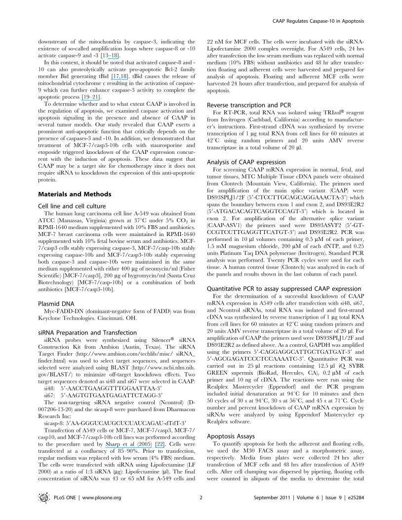
Apoptosis induced by CAAP does not depend upondeath receptor pathway
To determine whether apoptosis induced by RNAi knockdown
of CAAP requires the death receptor pathway, we transiently
expressed a C-terminal deleted dominant-negative mutant of
FADD (Fig. 5A). As expected, expression of FADD-DN protected
A-549 cells from apoptosis induced by the death receptor ligand
TRAIL, but not from apoptosis induced by RNAi knockdown of
the CAAP gene (Fig. 5B). LF 2000 was used for transfection. The
efficiency was not known since the vector does not contain a
fluorescence tag. However, the data in Fig. 5B suggest an efficient
transfection, as the response to TRAIL in all cells resembled vector
control upon expression of FADD-DN.
Figure 3. Knockdown of CAAP induces caspase-dependent apoptosis in A549 cells. (A) The expression levels of CAAP in lung cancer A-549cells were knocked down by the siRNA’s, si67 and si48 relative to a non-targeting control siRNA at the indicated concentrations. (the higher the barthe more confident the prediction). (B) Determination of apoptosis in A-549 following transfection with the two CAAP siRNAs si67 and si48 comparedto the apoptosis in the non-targeting control siRNA at the indicated concentrations. (C) Determination of the fold increase in caspase-3, -8, -9 and -10activities following CAAP knockdown by si67 (65 nM) compared to the Ncontrol siRNA (65 nM). (D) Determination of apoptosis in A549 cells 24 hoursfollowing CAAP knockdown by si67 in the absence or presence of 75 mM of IETD-FMK (caspase-8 inhibitor), AEVD-FMK (caspase-10 inhibitor), or Z-VAD-FMK, a pan-caspase inhibitor. (E) Determination of apoptosis in A549 cells 24 hours following CAAP knockdown by si67 in the absence orpresence of DEVD-FMK (caspase-3 inhibitor).doi:10.1371/journal.pone.0025284.g003
CAAP Regulates Caspase-10 in Apoptosis
PLoS ONE | www.plosone.org 7 September 2011 | Volume 6 | Issue 9 | e25284

Apoptosis induced by the knockdown of CAAP criticallydepends upon the simultaneous presence of caspase-3and -10
To investigate the role of caspase-10 in the apoptosis pathway
induced by knockdown of CAAP, we utilized MCF-7 breast
cancer cells that express neither caspase-3 nor caspase-10 [26,27].
In addition, three derivative cell lines that stably express either
caspase-3 (MCF-7/casp3), caspase-10b (MCF-7/casp-10b), or
both caspases (MCF-7/casp- 3-10b) were used.
Interestingly, although expression of CAAP was efficiently
knocked down in all MCF-7 clones (Fig. 6A), significant apoptosis
was only observed in MCF-7/casp3-10b (Fig. 6B) cells. Here, 60%
of the cells were found to be dead, whereas in the other three cell
lines only 8 to 13% of the cells appeared apoptotic, indicating that
both caspase-3 and –10 are required for apoptosis induced by
knockdown of CAAP. Consistently, a significant increase in
caspase activities could only be detected in MCF-7/casp3-10b
cells, but not in the parental MCF-7 cells or in the derivative line
expressing caspase-10b (Fig. 6C). On the other hand, MCF-7/
casp3 cells displayed some caspase-3 activity that, however, was
clearly less than in MCF-7/casp3-10b cells and hence, not
sufficient to induce apoptosis. An even clearer picture emerged
when we analyzed caspase processing in these cells in the presence
and absence of CAAP. As judged by the appearance of cleavage
fragments (caspase-3, -8, and -9) or by the loss of the pro-form
(caspase-10), all four caspases investigated were processed
following knockdown of CAAP demonstrating a caspase-depen-
dent apoptotic pathway (Fig. 6D). Processing intensities of caspases
-3, -8 and -10 appeared to be interdependent (Fig. 6D). For
instance, processing of caspase-8 was dramatically accelerated in
the presence of either caspase-3 or caspase-10 compared to the
parent cells and was further enhanced in MCF-7/casp3-10b cells
that express both caspases (Fig. 6D). This interdependency was
even more pronounced for caspase-3 and -10 that were both only
marginally processed when expressed individually. In MCF-7/
casp3-10b cells, however, processing of both caspases increased
dramatically (Fig. 6D), suggesting that caspase-3 and -10 require
each other for an efficient activation process. Relative to parental
cells, processing of caspase-9 was observed in all of the MCF-7 cell
lines, but was the strongest in the MCF-7/casp3-10b cells. This
suggests that both caspase-3 and -10 are also required for the
processing of caspase-9 (Fig. 6C and 6D). It is noted that in
Fig. 6D, the amount of procaspase-9 is significantly reduced in the
si67 lane compared to Ncontrol lane. Together, these data are not
only consistent with the caspase activation profiles obtained
following knockdown of CAAP (Fig. 6C), but also with the degree
of apoptosis induction that was the highest in MCF-7/casp3-10b
cells (Fig. 6B).
Finally, we analyzed cleavage of Bid into tBid that can be
mediated by several caspases including caspase-3, -8 and -10 [28].
Consistent with the varying levels of processed and activated
capase-8 following depletion of CAAP (see Fig. 6D), cleavage of
Bid into an approximately 13 kDa fragment was detected
accordingly in all four MCF-7 lines (Figure 7A). However, the
levels of tBid further increased upon caspase-10 expression in
MCF-7 cells demonstrating that caspase-10 significantly contrib-
utes to this event. Most interestingly, whereas a larger 13 kD
fragment of Bid was detected in each of the four MCF-7 cell lines,
a faster migrating 11 kD tBid fragment was only generated in
Caspase-3-10b cells which is the only cell line exhibiting significant
apoptosis after knockdown of CAAP expression. Moreover, both
tBid fragments were also detected following CAAP knockdown in
caspase-10-expressing A-549 cells indicating that these events play
Figure 4. Knockdown of CAAP induces caspase processing,cleavage of PARP and results in increased expression ofcaspase-10. (A) Determination of caspase processing and PARPcleavage by Western blot analyses of floating and adherent A-549 cells48 hours post transfection with si67 and si48 at dosages of 65 nM and43 nM. The p85 cleavage fragment of PARP after treatment with thesiRNAs and the activation of procaspase 9, as shown by the 37/35fragments, and caspase-8 as shown by the 43/41 fragments, areconsistent with the fold increase of Caspase-3, -8, and -9 activitiesshown in Fig. 3C and discussed in the text. Comparing the expressionlevels of procaspase 10 in the mock and Ncontrols to the levels in thesi67 and si48 lanes implies that the expression of procaspase 10 isincreased after RNAi knockdown of CAAP. A representative experimentout of three is shown. (B) Western blot analyses for the status of CAAPand procaspase-10 in floating and adherent A-549 cells 16 h, 24 h or48 h post transfection with either Ncontrol or si48 or si67 siRNA.Probing the membrane with a ß-actin antibody served as a loadingcontrol. Procaspase-10 protein levels were reproducibly higher follow-ing knockdown of CAAP.doi:10.1371/journal.pone.0025284.g004
CAAP Regulates Caspase-10 in Apoptosis
PLoS ONE | www.plosone.org 8 September 2011 | Volume 6 | Issue 9 | e25284
an important role in apoptosis induced by knockdown of CAAP
expression (Fig. 7B).
Chemotherapeutic Agents Suppress CAAP ExpressionAlthough CAAP knockdown was sufficient to initiate apoptosis,
the response of CAAP to cell-extrinsic apoptotic stimuli was not
known. Staurosporine (STS) is a well known chemical agent that
induces apoptosis in numerous cancer cell lines [29]. We treated
the MCF-7/casp3-10b cells with a low dose (1.0 mM) of STS
which resulted in the knockdown of CAAP expression and
induction of apoptosis based on the strong cleavage of caspase-3
and -10b for each of the three time points of treatment (Fig. 8A).
Although STS is not a well known chemotherapeutic agent, there
are numerous ongoing clinical trials using STS derivatives in
combination with other drugs [30–35]. Etoposide, a widely used
chemotherapeutic agent [14], was tested for effects on CAAP
protein expression. As shown in Fig. 8B, the high dose of etoposide
(500 mM) completely abrogated CAAP protein expression and
induced apoptosis based on PARP cleavage and the processing of
procaspase-10. It should be noted that the lower dose of etoposide
(100 mM) had a small effect on the cleavage of PARP and
processing of procaspase-10 expression. Altogether, the data
suggest that CAAP is a newly discovered element of the apoptosis
control pathway that includes caspases -10 and -3. Future studies
will focus on determining the mechanisms and functions of CAAP
regulation on apoptosis, and the cellular response to cytotoxic
chemotherapeutics.
Discussion
CAAP is expressed to varying degrees at the mRNA and protein
level in every normal (adult and fetal) and cancer tissues of human
origin examined and exhibit an evolutionarily preserved structure
with several well-conserved domains. In particular, the central
region of CAAP is predicted by bioinformatics analyses to share
structural similarity with the death effector domain implying a role
in apoptosis signaling. Indeed, siRNA-mediated knockdown of
CAAP expression induces apoptosis in different cancer cell lines
(A-549, MCF-7/casp3-10b) that proceeds independently of the
caspase-8-dependent death receptor pathway, suggesting an
involvement of mitochondria. This is consistent with the cleavage
of Bid into tBid that was most pronounced in MCF-7/casp3/10b
cells that among the various MCF-7 lines proved to be the most
sensitive line toward apoptosis induction by knockdown of CAAP.
In addition, together with A-549 cells, MCF-7/casp3/10b cells
were the only cells in which the knockdown of CAAP resulted not
only in apoptosis, but also in the generation of an 11 kDa tBid
fragment. Although caspase-10 was shown, at least in vitro, to be
responsible for this cleavage [27], its physiological impact on
apoptosis signaling is unknown. Remarkably, although apoptosis
induced by the knockdown of CAAP results in the processing and
activation of caspassses-3, -8, -9 and -10, it requires the
simultaneous activation of caspase-3 and caspase-10 to induce
apoptosis. MCF-7 cells lacking caspase-10, but expressing
endogenous caspase -8 and -9 do not succumb to apoptosis, even
following exogenous expression of caspase-3. Only the additional
presence of caspase-10 rendered MCF-7/casp3-10b cells sensitive
toward apoptosis induction by the knockdown of CAAP. However,
to fulfill this role, caspase-3 is required for an efficient activation of
caspase-10 indicating the necessity for an interdependent caspase
activation network.
Apoptosis induced by the knockdown of CAAP proceeds
independently of the death receptor pathways (Fig. 5B), but requires
both caspase-3 and -10. The lack of caspase-10 and/or caspase-3
significantly prevented apoptosis not only in parental MCF-7 cells,
but also in the MCF-7/casp3 and MCF-7/casp-10b derivatives.
The prominent role of caspase-10 is also documented by our results
showing that CAAP negatively regulates either directly or indirectly
the expression of procaspase-10. Thereby, it appears possible that
the knockdown of CAAP results in apoptosis induction merely by a
facilitated oligomerization of caspase-10 due to an increased
caspase-10 pool. Also, caspases-8 and -9 are activated after
Figure 5. Apoptosis induced by CAAP knockdown proceeds independently of death receptor pathway. (A) Western blot analysis forexpression of dominant-negative FADD (FADD-DN) in A-549 cells that were transfected with an empty vector or the FADD-DN construct. b-actinexpression was determined as a control for loading. (B) Determination of apoptosis of A-549 cells that were transfected with empty vector or FADD-DN. After 24 hrs cells were transfected with si67 or treated with TRAIL (35 ng/ml). The percentage of apoptotic cells was determined after anadditional 24 hrs. Apoptosis values for the si67-65 nM transfected cells or the TRAIL treated cells are expressed relative to the amount of apoptosisinduced in the vector- or FADD-DN transfected cells. These data demonstrate that FADD-DN overexpression blocks TRAIL induced apoptosis but notapoptosis induced by CAAP knockdown.doi:10.1371/journal.pone.0025284.g005
CAAP Regulates Caspase-10 in Apoptosis
PLoS ONE | www.plosone.org 9 September 2011 | Volume 6 | Issue 9 | e25284

knockdown of CAAP expression in all four MCF-7 clones and in A-
549 cells and, this implies that CAAP may also regulate the
activation of both caspase-8 and -10. Thus, we propose that CAAP
is an anti-apoptotic protein which negatively regulates an apoptosis
pathway that probably proceeds via activation of mitochondria as
apoptosis correlated with cleavage of Bid (Fig. 7).
The requirement for caspase-3 in the activation of caspase-10
has been previously observed [14]. Our data suggest a similar
feedback amplification loop, in which caspase-10 and -8 activate
caspase-9 and thus caspase-3. The lack of caspase-3 in MCF-7/
casp-10b cells reduces the activity of caspase-10b by 83%, and that
of caspase-8 by 75% which implies that activation of these caspases
Figure 6. Caspase-3 and -10 are required for apoptosis induction by the knockdown of CAAP. (A) A western blot demonstrates thatCAAP protein expression is reduced in all four of the MCF-7 cell lines after transfection of si67. (B) Bar graph shows the total percent of apoptosisinduced by knockdown of CAAP as calculated according to the description in the Methods section. Each bar equals mean apoptosis 6 SEM, from fourexperiments. Asterisk denotes a statistically significant difference between MCF-7/casp3-10b cells and the other MCF-7 cell lines which implies thatcaspase-3 and -10 are required for apoptosis induction by knockdown of CAAP (P,0.0001 according to Student’s t-test). (C) Determination ofcaspase-3, -8, -9 and-10 activities in the four MCF-7 cell lines 24 hours post transfection with si67 at 22 nM compared to Ncontrol at 22 nM. Each barequals mean fold increase 6 SEM from three experiments. (D) Western blot analyses for the determination of caspase processing in the four MCF-7lines. Twenty-four hrs after transfection, both floating and adherent cells were harvested for Western blot analysis. We compared each of the threederivative cells with the parent MCF-7 cells. Note that procaspase-3 is at 32 kDa and the band below is unknown. Cleavage fragments were onlydetected in lanes in which CAAP expression was knocked down by the si67 probe. One representative experiment out of three is shown.doi:10.1371/journal.pone.0025284.g006
CAAP Regulates Caspase-10 in Apoptosis
PLoS ONE | www.plosone.org 10 September 2011 | Volume 6 | Issue 9 | e25284

are responsive to caspase-3. Likewise, the lack of caspase-10 also
reduced the activation of caspase-9 by 65% (compare caspase-9
activity in MCF-7/casp3-10b cells to its activity in MCF-7/casp3)
(Fig. 6C), implying that full activation of caspase-9 requires
caspase-10 and -8. Based on these data, we propose that the
knockdown of CAAP induces a caspase3/9 feedback amplification
loop (Fig. 9). Soo-Jung Park [4] and Ho-June Lee [36] also
identified novel caspase-10 dependent apoptotic pathways.
However, these pathways were not related to a feedback
amplification loop (Fig. 9).
Chemotherapeutic agents also induce the mitochondrial death
pathway involving an amplification loop. Filomenko et al (2006)
[14] treated U937 or HeLa cells with etoposide which induced a
similar caspase 3/9 amplification feedback loop. Haefen et al
(2003) [16] treated BJAB cells with paclitaxel which induced a 3/8
amplification feedback loop that is also similar to the CAAP
regulated amplification loop, except that this 3/8 loop does not
include caspase-10, whereas CAAP controls a loop including both
caspase-8 and -10 as amplifying executioners. In addition, an in
vivo analysis of programmed cell death of dorsal root ganglia
neurons in mice demonstrated that the apoptotic pathway
proceeded via a 3/9 feedback amplification loop [37]. Our
identification of an ubiquitously expressed and evolutionary
conserved CAAP gene as a negative modulator of the 3/9
feedback amplification loop requiring caspase-10 thus represents a
novel pathway that might result in the development of new
therapeutic strategies.
Due to numerous p53-binding sites in its promoter region,
procaspase-10 expression can be induced in a p53-dependent
manner in response to DNA damage caused by chemotherapeutic
agents [36]. One consequence of increased procaspase-10 is to
contribute to p53-induced apoptosis following irreversible cellular
damage. CAAP might also have a role in p53-induced apoptosis as
it negatively modulates procaspase-10 expression and function.
Interestingly, the promoter region of CAAP contains a sequence
that is also present in the promoter of the retinoblastoma gene,
where it constitutes a cis-acting element susceptible to negative
regulation by the tumor suppressor p53 [23]. Thus, it is
conceivable that p53-induced apoptosis requires prior repression
of CAAP transcription.
In summary, our results demonstrate that CAAP is a
ubiquitously expressed and evolutionarily conserved protein
exhibiting a potent anti-apoptotic function. It appears that CAAP
interferes with the activation of caspase-10, that in turn regulates
the generation of an 11 kDa tBid fragment and a caspase-3/9
feedback amplification loop required for an efficient activation of
the mitochondrial death pathway. Although our data suggest that
CAAP restrains a caspase-3/9 feedback amplification loop that is
caspase-10 dependent and utilized by some chemotherapeutic
agents, the direct mechanism by which CAAP controls the
expression of caspase-10 is unknown. In addition, we demonstrat-
ed that CAAP protein levels are drastically reduced in response to
STS or etoposide, concurrent with the activation of apoptosis.
These results suggest that CAAP may be a target for chemother-
apy site since it does not require siRNA to knockdown the
expression of this anti-apoptotic protein. Further studies will be
required to determine the molecular mechanisms and components
contributing to the anti-apoptotic functions of CAAP.
Figure 7. Western blot analysis of Cleavage of Bid after knockdown of CAAP in the MCF-7 cell lines and A-549 cells. (A) The upperpanels show Bid cleavage was observed in each of the MCF-7 cell lines after knockdown of CAAP by transfection with si67 at 22 nM, but not in theNcontrol lanes. In addition to the 13 kDa fragment, observed in each of the MCF-7 lines after knockdown of CAAP, an 11 kDa tBid fragment wasdetected in the MCF-7/casp3-10b cells. This is consistent with the observation that only in the MCF-7/casp3-10b cell line was significant apoptosisinduced by knockdown of CAAP. (B) Although cleavage of Bid was observed in the treatment with both si67 and si48 transfection at both doses, thecleavage bands in the A-549 cells were weaker than in the MCF-7 cell lines.doi:10.1371/journal.pone.0025284.g007
CAAP Regulates Caspase-10 in Apoptosis
PLoS ONE | www.plosone.org 11 September 2011 | Volume 6 | Issue 9 | e25284

Figure 8. Knockdown of CAAP expression after treatment of cells with chemotherapeutic agents. (A) MCF-7/casp3-10b cells weretreated with starurosporine (STS) for 4, 6, or 8 hrs and then examined for CAAP expression and cleavage of procaspase-3 and-9 by Western blotanalysis. (B) MCF-7/casp3-10a cells were treated with etoposide at dose of 100 mM and 500 mM for 24 hrs and then harvested for western blotanalysis of CAAP expression and cleavage of PARP and procaspase-10 expression.doi:10.1371/journal.pone.0025284.g008
CAAP Regulates Caspase-10 in Apoptosis
PLoS ONE | www.plosone.org 12 September 2011 | Volume 6 | Issue 9 | e25284

Supporting Information
Figure S1 Expression of CAAP and the main splicevariant as assayed by RT-PCR. Twenty PCR cycles were
performed for each tissue. A human control cDNA (Clontech)
was used in each of the 4 panels and is shown in the last Colum in
each panel. Note that the expression of the control is the same in
each panel. Normal tissue panel 1: heart, brain, placenta, lung,
liver, kidney, pancreas. Normal tissue panel 2: spleen, thymus,
prostate, testis, ovary, small intestine, colon, leukocytes. Tumor
tissue panel: breast carcinoma GI-101, lung carcinoma LX-1,
colon adenocarcinoma CX-1, lung carcinoma GI-117, prostatic
adenocarcinoma PC3, colon adenocarcinoma GI-112, ovarian
carcinoma GI-102, pancreatic adenocarcinoma GI-103. Fetal
tissue panel: brain, lung, liver, kidney, heart, spleen, thymus,
skeletal muscle. The last two lanes in each panel are human
control cDNA (Clontech), and H2O control.
(TIF)
Figure S2 Histological apoptotic phenotype of floatingcells. Light microscopy of 1 micron thick toluidine blue stained
sections show that floating cells are preponderantly apoptotic after
treatment with si67 and si48 at dosages of 43 nM or 65 nM.
Apoptotic phenotype was found in all groups in which DNA was
peripheral in the nucleus and cytoplasm was blebbing. A non-
apoptotic cell is shown for comparison. Bar = 25 microns in all
images.
(TIF)
Acknowledgments
We thank Megan Raczon and Xiao Xuan Hu for preparation of the
manuscript.
Author Contributions
Conceived and designed the experiments: MWA DP. Performed the
experiments: YZ EJ. Analyzed the data: YZ EJ MLM RUJ DJF DP JM
MWA. Wrote the paper: MWA DP RUJ.
References
1. Wiest JS, Franklin WA, Otstot JT, Forbey K, Varella-Garcia M, et al. (1977)
Identification of a novel region of homozygous deletion on chromosome 9p in
squamous cell carcinoma of the lung: the location of a putative tumor suppressor
gene. Cancer Res 57: 1–6.
2. Park HH, Lo YC, Lin SC, Wang L, Yang JK, et al. (2007) The death domain
superfamily in intracellular signaling of apoptosis and inflammation. Annu Rev
Immunol 25: 561–86.
3. Fishcer U, Janicke RU, Schulze-Osthoff K (2003) Many cuts to ruin: a
comprehensive update of caspase substrates. Cell Death Differ 10: 76–100.
4. Park SJ, Wu CH, Gordon JD, Zhong X, Emami A, et al. (2004) Taxol induces
caspase-10-dependent apoptosis. J Bio Chem 279: 51057–67.
5. Thornberry NA, Lazebnik Y (1998) Caspases: enemies within. Science 281: 1312–16.
6. Salvesen GS, Dixit VM (1997) Caspases: intracellular signaling by proteolysis.
Cell 91: 443.
Figure 9. Schematic representation of Caspase-3/9 feedback loop of amplification which is induced by knockdown of CAAPexpression in A-549 and MCF-7/casp3-10b cells.doi:10.1371/journal.pone.0025284.g009
CAAP Regulates Caspase-10 in Apoptosis
PLoS ONE | www.plosone.org 13 September 2011 | Volume 6 | Issue 9 | e25284
7. Green D, Kroemer G (1998) The central executioners of apoptosis: caspases or
mitochondria? Trends Cell Biol 8: 267–71.8. Yang X, Chang HY, Baltimore D (1998) Autoproteolytic activation of pro-
caspases by oligomerization. Mol Cell 1: 319–25.
9. Mikhailov V, Mikhailova M, Degenhardt K, Venkatachalam MA, White E,et al. (2003) Association of Bax and Bak homo-oligomers in mitochondria. Bax
requirement for Bak reorganization and cytochrome c release. J Biol Chem 278:536.
10. Letai A, Bassik MC, Walensky LD, Sorcinelli MD, Weiler S, et al. (2002)
Distinct BH3 domains either sensitize or activate mitochondrial apoptosis,serving as prototype cancer therapeutics. Cancer Cell 2: 183–92.
11. Hill JM, Morisawa G, Kim T, Huang T, Wei Y, et al. (2004) Identification of anexpanded binding surface on the FADD death domain responsible for
interaction with CD95/Fas. J Biol Chem 279: 1474–81.12. Peter ME, Krammer PH (2003) The CD95 (APO-1/Fas) DISC and beyond.
Cell Death and Differ 10: 26–35.
13. Engels IH, Stepczynska A, Stroh C, Lauber K, Berg C, et al. (2000) Caspase-8/FLICE functions as an executioner caspase in anticancer drug-induced
apoptosis. Oncogene 19: 4563–73.14. Filomenko R, Prevotat L, Rebe C, Cortier M, Jeannin JF, et al. (2006) Caspase
10 involvement in cycotoxic drug-induced apoptosis of tumor cells. Oncogene
25: 7635–45.15. Wieder T, Essmann F, Prokop A, Schmelz K, Schulze-Osthoff K, et al. (2001)
Activation of caspase-8 in drug-induced apoptosis of B-lymphoid cells isindependent of CD95/Fas receptor-ligand interaction and occurs downstream of
caspase-3. Blood 97: 1378–87.16. von Haefen C, Wieder T, Essmann F, Schulze-Osthoff K, Dorken B, et al.
(2003) Paclitaxel-induced apoptosis in BJAB cells proceeds via a death receptor-
independent, caspases-3/-8-driven mitochondrial amplification loop. Oncogene22: 2236–47.
17. Li H, Zhu H, Xu C, Yuan J (1998) Cleavage of BID by caspase 8 mediates themitochondrial damage in the Fas pathway of apoptosis. Cell 94: 491–501.
18. Luo X, Budihardjo I, Zou H, Slaughter C, Wang X (1998) Bid, a Bcl2
interacting protein, mediates cytochrome c release from mitochondria inresponse to activation of cell surface death receptors. Cell 94: 481–90.
19. Green DR (2000) Apoptotic pathways: paper wraps stone blunts scissors. Cell102: 1–4.
20. Cain K, Brown DG, Langlais C, Cohen GM (1999) Caspase activation involvesthe formation of the aposome, a large (,700 kDa) caspase-activating complex.
J Biol Chem 274: 22686–92.
21. Cowling V, Downward J (2002) Caspase-6 is the direct activator of caspase-8 inthe cytochrome c-induced apoptosis pathway: absolute requirement for removal
of caspase-6 prodomain. Cell Death and Differentiation 9: 1046–56.22. Sharp DA, Lawrence DA, Ashkenzi A (2005) Selective knockdown of the long
variant of cellular FLICE inhibitory protein augments death receptor-mediated
caspase-8 activation and apoptosis. J Biol Chem 280: 19401–9.
23. Shiio Y, Yamamoto T, Yamaguchi N (1992) Negative regulation of Rb
expression by the p53 gene product. Proc Natl Acad Sci USA 89: 5206–10.24. Jaroszewski L, Rychlewski L, Li Z, Li W, Godzik A (2005) FFAS03: a server for
profile-profile sequence alignments. Nucleic Acids Res 33: W284–8.
25. Finn RD, Mistry J, Schuster-Bockler B, Griffiths-Jones S, Hollich V, et al. (2006)Pfam: clans, web tools and services. Nucleic Acids Res 34: D247–51.
26. Janicke RU, Ng P, Sprengart ML, Porter AG (1998) Caspase-3 is required foralpha-fodrin cleavage but dispensable for cleavage of other death substrates in
apoptosis. J Biol Chem 273: 15540–5.
27. Engels IH, Totzke G, Fischer U, Schulze-Osthoff K, Janicke RU (2005)Caspase-10 sensitizes breast carcinoma cells to TRAIL-induced but not tumor
necrosis factor-induced apoptosis in a caspase-3-dependent manner. Mol CellBiol 25: 2808–18.
28. Fischer U, Stroh C, Schulze-Osthoff K (2006) Unique and overlapping substratespecificities of caspase-8 and caspase-10. Oncogene 25: 152–9.
29. Manns J, Daubrawa M, Driessen S, Paasch F, Hoffmann N, et al. (2011)
Triggering of a novel intrinsic apoptosis pathway by the kinase inhibitorstaurosporine: activation of caspase-9 in the absences of Apaf-1. The FASEB
Journal fj: 10-177527.30. Kummar S, Gutierrez ME, Gardner ER, Figg WD, Melillo G, et al. (2010) A
phase I trial of UCN-01 and prednisone in patients with refractory solid tumors
and lymphomas. Cancer Chemother Pharmacol 65: 383–389.31. Jimeno A, Rudek MA, Purcell T, Laheru DA, Messersmith WA, et al. (2008)
Phase I and pharmacokinetic study of UCN-01 in combination with irinotecanin patients with solid tumors. Cancer Chemother Pharmacol 61(3): 423–33.
32. Sampath D, Cortes J, Estrov Z, Du M, Shi Z, et al. (2006) Pharmacodynamics ofcytarabine alone and in combination with 7-hydroxystaurosporine (UCN-01) in
AML blasts in vitro and during a clinical trial. Blood 107(6): 2517–24.
33. Hotte SJ, Oza A, Winquist EW, Moore M, Chen EX, et al. (2006) Phase I trialof UCN-01 in combination with topotecan in patients with advanced solid
cancers: a Princess Margaret Hospital Phase II Consortium study. Annals ofOncology 17(2): 334–40.
34. Kortmansky J, Shah MA, Kaubisch A, Weyerbacher A, Yi S, et al. (2005) Phase
I trial of the cyclin-dependent kinase inhibitor and protein kinase C inhibitor 7-hydroxystaurosporine in combination with Fluorouracil in patients with
advanced solid tumors. Journal of Clinical Oncology 23(9): 1875–84.35. Monnerat C, Henriksson R, Le Chevalier T, Novello S, Berthaud P, et al. (2004)
Phase I study of PKC412 (N-benzoyl-staurosporine), a novel oral protein kinaseC inhibitor, combined with gemcitabine and cisplatin in patients with non-small-
cell lung cancer. Annals of Oncology 15(2): 316–23.
36. Lee HJ, Pyo JO, Oh Y, Kim HJ, Hong SH, et al. (2007) YK.AK2 activates anovel apoptotic pathway through formation of a complex with FADD and
caspase-10. Nature Cell Biol 11: 1303–10.37. Rikhof B, Corn PG, El-Deiry WS (2003) Caspase 10 levels are increased
following DNA damage in a p53-dependent manner. Canc Bio Therapy 2:
707–12.
CAAP Regulates Caspase-10 in Apoptosis
PLoS ONE | www.plosone.org 14 September 2011 | Volume 6 | Issue 9 | e25284
Related Documents