1 Hypoxia in Surgical Patients Alexis Riddell, LAT3 General Surgery, West of Scotland Deanery, UK. Andrew J Jackson ST5 General & Vascular Surgery, West of Scotland Deanery, UK. David R Ball Consultant Anaesthetist, NHS Dumfries & Galloway, UK. Jacob S Dreyer Consultant General Surgeon, NHS Dumfries & Galloway, UK. Introduction This article discusses basic physiology of oxygen delivery, pathophysiology and mechanisms of hypoxia, the most common causes of hypoxia in surgical patients and principles of management. The aim is not to provide a detailed overview, but structure to enable early recognition, diagnosis and treatment. Hypoxia is impaired tissue oxygenation. It is one of the most common post-operative complications but often not recognised because it is not looked for, e.g. post-operative confusion can often be secondary to hypoxia. Patients who are critically ill usually have increased oxygen demands; oxygen delivery is therefore fundamental to managing sick patients. Physiology of Oxygen Transport Oxygen Delivery = oxygen content x cardiac output, where Oxygen content = (Hb x 1.34 x SaO2) + (0.0032 x PaO2). Fully saturated haemoglobin carries 1.34 ml oxygen/gram of Hb, but the constant can vary slightly. The maximum amount of oxygen that can dissolve in blood is 0.0032 ml/dl/mmHg PaO2. At Hb=15 and SaO2=98 blood carries 198ml O2/litre, of which 195 ml is carried as oxygenated haemoblobin. Cardiac output depends on stroke volume and heart rate (CO = SV x HR); stroke volume is dependent on cardiac preload, contractility and afterload. Heart rate increases early with hypoxia. Peripheral perfusion and tissue oxygen delivery depend on cardiac output and peripheral resistance (BP = CO x PR).

Welcome message from author
This document is posted to help you gain knowledge. Please leave a comment to let me know what you think about it! Share it to your friends and learn new things together.
Transcript
1
Hypoxia in Surgical Patients
Alexis Riddell, LAT3 General Surgery, West of Scotland Deanery, UK.
Andrew J Jackson ST5 General & Vascular Surgery, West of Scotland Deanery, UK.
David R Ball Consultant Anaesthetist, NHS Dumfries & Galloway, UK.
Jacob S Dreyer Consultant General Surgeon, NHS Dumfries & Galloway, UK.
Introduction
This article discusses basic physiology of oxygen delivery, pathophysiology and
mechanisms of hypoxia, the most common causes of hypoxia in surgical patients and
principles of management. The aim is not to provide a detailed overview, but structure to
enable early recognition, diagnosis and treatment.
Hypoxia is impaired tissue oxygenation. It is one of the most common post-operative
complications but often not recognised because it is not looked for, e.g. post-operative
confusion can often be secondary to hypoxia. Patients who are critically ill usually have
increased oxygen demands; oxygen delivery is therefore fundamental to managing sick
patients.
Physiology of Oxygen Transport
Oxygen Delivery = oxygen content x cardiac output, where
Oxygen content = (Hb x 1.34 x SaO2) + (0.0032 x PaO2). Fully saturated haemoglobin
carries 1.34 ml oxygen/gram of Hb, but the constant can vary slightly. The maximum
amount of oxygen that can dissolve in blood is 0.0032 ml/dl/mmHg PaO2. At Hb=15 and
SaO2=98 blood carries 198ml O2/litre, of which 195 ml is carried as oxygenated
haemoblobin.
Cardiac output depends on stroke volume and heart rate (CO = SV x HR); stroke volume is
dependent on cardiac preload, contractility and afterload. Heart rate increases early with
hypoxia. Peripheral perfusion and tissue oxygen delivery depend on cardiac output and
peripheral resistance (BP = CO x PR).
2
Oxygen delivery therefore depends on:
• A patent and open Airway (see February 2012 review).
• Effective Ventilation (see March 2012 review):
– Central drive, volume, rate, Functional Residual Capacity (FRC).
• Oxygen availability:
– Percentage oxygen in inspired air (FIO2), oxygen pressure in the air and
alveoli (pAO2), pulmonary capillaries (paO2).
• Oxygen transport:
– Haemoglobin level (Hb), Cardiac output, Peripheral resistance. Each
haemoglobin molecule can bind four oxygen molecules; binding of each
molecule facilitates binding of the next until Hb is fully saturated, i.e. the
affinity for the 4th oxygen molecule is much higher than for the 1st. This is the
biochemical basis for the sigmoid shape of the Oxygen-Haemoglobin
dissociation curve.
• Tissue factors:
– Oxygen release, Diffusion, Utilisation. Oxygen release is enhanced by shifting
the oxygen-haemoglobin curve to the right by a lower pH and higher
temperature in active tissue (e.g. contracting muscles) and by higher levels of
2,3-DPG (raised by exercise, higher altitude).
Pathophysiology of Hypoxia
The following factors need to be maintained to prevent tissue hypoxia:1,2
1. Patent airway
2. Effective ventilation
3. Effective gas interchange
4. Arterial oxygen saturation (SaO2) and pressure (PaO2)
5. Effective systemic and capillary circulation
6. Haemoglobin concentration and integrity
7. Effective oxygen release from Hb
8. Unhindered extracellular diffusion
9. Normal oxygen use by cells.
3
Physiologically hypoxia is usually classified into four groups:1,2
(a) Hypoxic hypoxia, when the pressure of oxygen in arterial blood (PaO2) is reduced,
accompanied by decreased SaO2 of haemoglobin. This is caused by inefficient gas
exchange (when PAO2 would be maintained) or decreased PAO2, e.g. at high altitude or
suffocation due to airway obstruction or breathing in a closed space with loss of oxygen.
(b) Anaemic hypoxia, due to decreased oxygen carrying capacity in blood, e.g. due to loss
of red blood cells (RBCs), inadequate Hb within RBCs or carbon monoxide (CO) poisoning.
Oxygen binding sites on Hb have higher affinity for CO than O2 which prevents oxygenation
and patients do not show clinical symptoms and signs of hypoxia. In sickle cell anaemia the
O2-Hb dissociation curve shifts to the right so that oxygen is released in the tissues more
easily, compensating for a Hb of 6-8 g/l.
(c) Circulatory hypoxia, also known as stagnant or ischemic hypoxia, when too little
oxygenated blood is delivered to the tissues. This can be localised, e.g. with acute arterial
insufficiency, or general, e.g. with circulatory shock or cardiac failure.
(d) Histotoxic hypoxia, which means that oxygen delivery is normal but tissues cannot use
O2 due to toxins affecting cellular respiration, e.g. with cyanide poisoning. Methylene blue
can be used in cyanide poisoning to bind cyanide molecules but this forms met-
haemoglobin (where iron is reduced to Fe3+), which has a much lower affinity for O2,
limiting oxygen delivery.
In critical care the following mechanisms provide a practical mnemonic to think of
potential causes of hypoxia:
1. ↓pAO2
Alveolar PO2 can drop significantly at altitude. In practice this is of importance when
transferring patients with e.g. chest injuries, acute blood loss, shock or anaemia in
unpressurised aircraft at high altitude. This is relevant in regions already at high
altitude or crossing mountain ranges e.g. in parts of Ethiopia, the central great lakes
states and South Africa.
2. ∆FiO2
Patients on ventilators have their FiO2 controlled artificially.
4
3. ↓V
Decreased ventilation will primarily cause CO2 accumulation. In normal patients this
will increase the ventilation rate via chemoreceptors, but patients who are on artificial
ventilation or are centrally depressed (e.g. due to opiate overdose) cannot mount this
response. Hypoxia occurs late and can rapidly progress to cardiac arrest.
4. ∆V/∆Q
This means that there is discrepancy between ventilated alveoli and alveolar
capillary perfusion, and there are two categories:
(a) Shunt: when alveoli are perfused but not ventilated (e.g. atelectasis, pneumonia),
or oxygen diffusion is limited (e.g. in ARDS), right to left shunting is increased, i.e.
more non-oxygenated blood reaches the systemic circulation. This is a significant
cause of hypoxia in critically ill patients.
(b) Ventilation-perfusion mismatch: when alveoli are ventilated but not perfused,
e.g. with pulmonary embolism (PE).
5. ↓CO
Hypovolaemic, cardiogenic or obstructive circulatory shock or congestive cardiac
failure (CCF) can cause significant enough hypoxia to cause rapid death. Patients on
artificial ventilation who are in CCF will usually not come off the ventilator unless
cardiac function is supported (e.g. with inotropes).
Assessment of the hypoxic post-operative patient
Clinical Assessment
Hypoxia is the inability to effectively oxygenate the tissues and is a threat to life.
This may result from pathology of the airway, breathing or circulation.
Prompt responses are crucial, allowing accurate diagnosis and effective treatment.
A five-step, structured, sequential set of responses to hypoxia is:
1 Review: The primary assessment is a rapid, targeted clinical examination of airway,
breathing, circulation and disability. This is conducted in correct order, with immediate
management of a life threatening problem when discovered.
The most important skills at this stage are the use of the trained human senses to “look,
listen and feel”, gathering important clinical information, informing diagnosis and acting
5
when necessary. Monitoring devices are useful when available, providing additional
information. This information is only helpful to the patient when it is accurate, timely and
used to guide effective treatment. Cutaneous pulse oximetry gives real-time data on the
oxygenation of haemoglobin (saturation) and peripheral pulse rate. These signs may be lost
with severe vasoconstriction, as in severe shock. Arterial blood gas analysis gives
information on arterial oxygen tension (which is not “saturation”), carbon dioxide tension
and blood acid-base status. Chest radiography and electrocardiography may provide
additional data, but may detract from immediate, time critical interventions.
When critical hypoxia is revealed by the primary assessment, the next response,
Resuscitation, is started without delay. Otherwise, the review phase may continue with the
secondary assessment, aimed at gathering more information. A patient history is taken with
attention to the acute and chronic aspects of the patient‟s condition, with identification of
specific symptoms and risks, such as asthma, smoking, heart disease etc. Review of
current and past medication (including missed doses) is necessary. A clinical examination
is done to identify signs of organ and system dysfunction.
Chart review is crucial. Changes in vital signs can inform diagnosis and treatment.
There is a further, tertiary assessment, but this is done upon completion of patient
treatment. This is a review of the clinical process, identifying strengths and weaknesses,
aimed at improving future care. This can be done in an educational setting and should be
conducted in a supportive, positive way.
2 Resuscitation: This is started when the review phase shows an immediate threat to life.
The resuscitation is also sequential and structured, aimed at restoration and maintenance
of oxygen to the tissues, especially the vital organs. The stepwise approach is part of the
Basic and Advanced Life support guidelines. Patients with hypoxia need oxygen.
Resuscitation responses to common hypoxic problems are outlined later in this review.
3 Request HELP. Management of postoperative hypoxia can be complex and demanding.
The chance of a successful outcome is increased when skilled help is sought and available
to help manage the situation. A team approach requires good clinical leadership, situational
awareness with effective task allocation. A key factor is concise, effective communication.
6
An “SBAR” communication format is helpful. A clinical record should be kept: when possible
a team member can be given this task.
4 Reassess the situation regularly. The clinical picture will most likely change both with
time and treatment and this is only apparent with reassessment. New information may be
elicited or become available from other sources, which may alter further management.
5 Resource: your situation. Identify what is needed to improve the chance of a good
outcome. This may involve acquisition of drugs, equipment or people to help with any of
steps listed above.
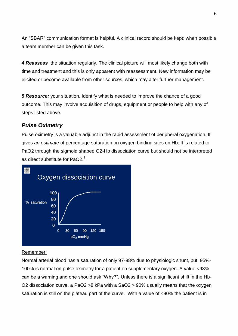
Pulse Oximetry
Pulse oximetry is a valuable adjunct in the rapid assessment of peripheral oxygenation. It
gives an estimate of percentage saturation on oxygen binding sites on Hb. It is related to
PaO2 through the sigmoid shaped O2-Hb dissociation curve but should not be interpreted
as direct substitute for PaO2.3
Oxygen dissociation curve
0
20
40
60
80
100
0 30 60 90 120 150
pO2 mmHg
% saturation
Remember:
Normal arterial blood has a saturation of only 97-98% due to physiologic shunt, but 95%-
100% is normal on pulse oximetry for a patient on supplementary oxygen. A value <93%
can be a warning and one should ask “Why?”. Unless there is a significant shift in the Hb-
O2 dissociation curve, a PaO2 >8 kPa with a SaO2 > 90% usually means that the oxygen
saturation is still on the plateau part of the curve. With a value of <90% the patient is in
7
serious trouble because the paO2-SaO2 ratio is now on the steep part of the curve and
saturation will drop rapidly with a minor decrease in PaO2.
Double check that you distinguish the SaO2 from the pulse rate when looking at the
monitor.
Error readings in pulse oximetry can occur due to:
• Low cardiac output
• Vasoconstriction
• SaO2 <70%
• Poor positioning
• Movement
• Hypothermia (often in trauma patients)
• Abnormal Hb (COHb, MetHb)
• Hyperthermic limb
• Dirty probe
• Black, blue or green nail polish
• External light
Arterial blood gas analysis (ABGs)
ABG analysis can be useful in the diagnosis and management of critical illness and injury,
but waiting for results should not delay immediate management of potential hypoxia. The
following account is a traditional interpretation. Another analysis, Stewart‟s “Strong Ion
Difference” approach is an alternative.
The ABG analyser measures:
Hydrogen ion concentration, reported as either hydrogen ion concentration [H+] or
pH (-log10[H+] ) . A lower pH value is more acidotic
Oxygen tension (PaO2), reported in kilopascals (kPa) or mmHg.
Carbon dioxide tension (PaCO2) (kPa or mmHg)
Other values such as bicarbonate [HC03-] expressed in mmol l-1 and Base Excess/Deficit
(BE/D), are calculated. Base Deficit is the amount of base that would be needed to correct
the pH of the sample to 7.4. Base excess is the amount of acid needed to correct to pH 7.4.
8
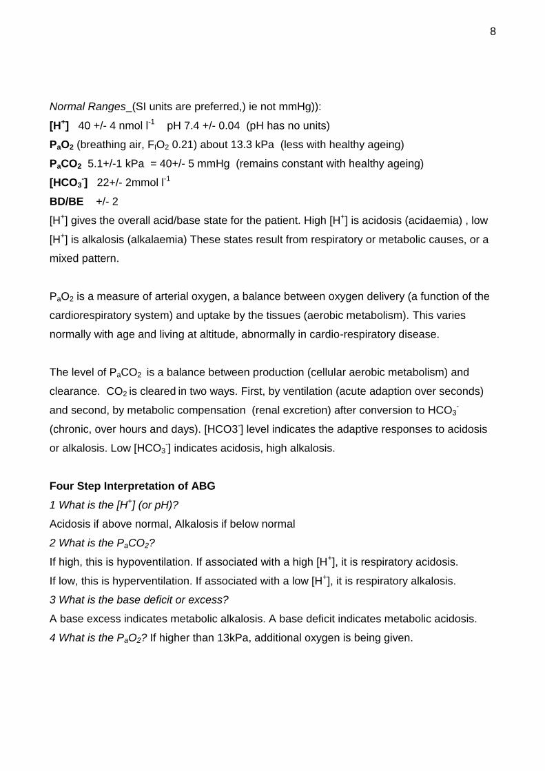
Normal Ranges (SI units are preferred,) ie not mmHg)):
[H+] 40 +/- 4 nmol l-1 pH 7.4 +/- 0.04 (pH has no units)
PaO2 (breathing air, FIO2 0.21) about 13.3 kPa (less with healthy ageing)
PaCO2 5.1+/-1 kPa = 40+/- 5 mmHg (remains constant with healthy ageing)
[HCO3-] 22+/- 2mmol l-1
BD/BE +/- 2
[H+] gives the overall acid/base state for the patient. High [H+] is acidosis (acidaemia) , low
[H+] is alkalosis (alkalaemia) These states result from respiratory or metabolic causes, or a
mixed pattern.
PaO2 is a measure of arterial oxygen, a balance between oxygen delivery (a function of the
cardiorespiratory system) and uptake by the tissues (aerobic metabolism). This varies
normally with age and living at altitude, abnormally in cardio-respiratory disease.
The level of PaCO2 is a balance between production (cellular aerobic metabolism) and
clearance. CO2 is cleared in two ways. First, by ventilation (acute adaption over seconds)
and second, by metabolic compensation (renal excretion) after conversion to HCO3-
(chronic, over hours and days). [HCO3-] level indicates the adaptive responses to acidosis
or alkalosis. Low [HCO3-] indicates acidosis, high alkalosis.
Four Step Interpretation of ABG
1 What is the [H+] (or pH)?
Acidosis if above normal, Alkalosis if below normal
2 What is the PaCO2?
If high, this is hypoventilation. If associated with a high [H+], it is respiratory acidosis.
If low, this is hyperventilation. If associated with a low [H+], it is respiratory alkalosis.
3 What is the base deficit or excess?
A base excess indicates metabolic alkalosis. A base deficit indicates metabolic acidosis.
4 What is the PaO2? If higher than 13kPa, additional oxygen is being given.

9
Chest X-Ray
Chest X-Ray is of no value in diagnosing hypoxia and should not delay immediate
treatment, but can help with the diagnosis of specific conditions that cause hypoxia,
especially of lung parenchymal disease and pleural or bony abnormalities after trauma (See
section below and March 2012 review on chest trauma).
Causes of hypoxia in post-operative patients:
Patients at risk of hypoxia
• Pre-op hypoxia
– Smokers, COPD
• Reduced FRC
– Elderly, Obesity, Diabetes, General Anaesthetic
• Surgical pathology
– Restricted ventilation, SIRS
• Post-op Sedation
• Hypothermia
• Fluid overload
Common causes of post-operative hypoxia
• Pulmonary oedema
• Bronchopneumonia
• Lobar pneumonia
• Pre-existent COPD
• Atelectasis with hypoventilation
• Pulmonary embolism
• ARDS
Mechanisms of becoming hypoxic
Hypoxia can occur via interference of physiological mechanisms of oxygenation at different
levels, as discussed above. To keep things practical the causes of post-operative hypoxia
are discussed in the following categories:
1. Lack of Alveolar Ventilation.
10
Adequate oxygen levels are prevented from entering the alveoli to facilitate
gas exchange.
2. Lack of Alveolar Perfusion.
Inadequate levels of blood are supplied to the lung to facilitate gas exchange.
3. Decreased alveolar diffusion.
Adequate levels of both blood and oxygen are available in the alveoli and
pulmonary circulation but alveolar pathology prevents gas exchange
occurring.
Lack of Alveolar Ventilation
Alveolar ventilation represents the volume of gas available to the alveolar surface area per
unit time. Lack of alveolar ventilation can therefore occur due to numerous mechanisms in
the post-operative surgical patient, all of which should be considered during the
assessment. 4
Upper Airway Obstruction
Airway obstruction must always be excluded as part of ABC assessment of the patient.
Immediate clearance of an identifiable upper airway obstruction is necessary if identified. 5
Clinical Features: Features of upper airway obstruction are noted during the initial airway
assessment. The airway may be noted to be blocked by a bolus. A patient with reduced
consciousness may not be supporting his/her own airway and the soft tissues of the
oropharynx impair airway efficiency.
Very rarely anaphylaxis will present post-operatively, with upper airway swelling and
obstruction. In such a scenario the patient will be acutely unwell with additional
bronchospasm and cardiovascular instability.
Management: Any identifiable obstruction should be immediately cleared if possible. Post-
operative patients who become unwell with reduced level of consciousness may not be able
to support their own airway effectively. A Guedel airway should then be inserted
immediately. Further emergency assessment should be performed and the patient may
require intubation and ventilation. 6
11
If anaphylaxis is suspected adrenaline should be administered immediately along with
airway support, supplemental oxygen, intravenous hydrocortisone and intravenous fluid
therapy. 7
Respiratory Depression: Opiates and Carbon Dioxide Narcosis
An often encountered complication in post-operative patients that can present as hypoxia is
reduced ventilation secondary to reduced respiratory drive. The commonest causes to
consider are impaired consciousness due to drug toxicity (most commonly opiates) and
impaired consciousness due to carbon dioxide narcosis.
Opiate toxicity occurs readily in post-operative patients who often have significant analgesic
requirements. An opiate „overdose‟ need not occur with high drug doses. Patients with renal
impairment do not excrete opiates effectively and accumulation can occur. The elderly are
often also susceptible to such effects. In toxic concentrations respiratory depression occurs,
with hypoventilation. 8
Carbon Dioxide narcosis occurs in patients with pre-existing Chronic Obstructive Pulmonary
Disease (COPD). Respiration in some patients with COPD is dependent upon „hypoxic
drive‟, rather than hypercapnia. 9 If a patient is given high concentrations of oxygen, their
respiratory stimulus is lost and respiratory depression occurs. The patient can quickly
develop carbon dioxide retention, respiratory acidosis and reduced consciousness, causing
hypoventilation and associated hypoxia.
Clinical features: The patient will present with signs of hypoxia, and reduced level of
consciousness (GCS). A reduced respiratory rate will be noticed. In opiate toxicity pin-point
pupils will be seen. In carbon dioxide narcosis a bounding pulse is often noted and a
history of COPD. An arterial blood gas sample can be taken to measure carbon dioxide
levels.
Management: Initial management is to secure the patients airway and offer respiratory
support as necessary. Patients with CO2 narcosis will require non-invasive ventilation
(CPAP, BiPAP) if conscious or intubation and ventilation for respiratory support to facilitate
„blowing off‟ excess CO2.
In patients with suspected opiate toxicity management should be similar but naloxone, an
opioid antagonist, should be administered immediately. If the patient recovers quickly

12
always remember that the half life of naloxone is less than morphine, so the effect may
wear off before the patient has metabolised enough morphine to prevent recurrent
respiratory depression. Further doses may be required. 10
Atelectasis and Lobar Collapse
Atelectasis is a frequently encountered post-operative complication causing hypoxia. It
usually occurs in the first 48 hours following surgery. In abdominal and thoracic surgery the
normal mechanisms by which mucus is cleared is impaired by pain, inhibiting deep
breathing and coughing. Mucus retention occurs with resorption of alveolar air, leading to
alveolar collapse. This normally occurs in the basal lobes. Secondary infection can then
occur. 11
Clinical Features: Patients with atelectasis may report dyspnoea and cough with signs of
hypoxia, tachypnoea and have reduced bibasal air entry. Normally this is following
abdominal or thoracic surgery. There may be an associated fever, though recent evidence
questions the link between fever and atelectasis. 12
Management: Management of atelectasis includes assessment of degree of respiratory
support required. In general supplemental oxygen therapy will be sufficient, but ventilation
is required in extreme cases in a deteriorating patient.
An assessment of the patient‟s analgesic requirements should be performed and this
optimised to ensure they can breathe and cough without inhibition.
Chest physiotherapy is required to ensure clearance of mucus and secretions. It is also
helpful in preventing secondary infection. 13
Antibiotic treatment is not initially required unless secondary infection is suspected.
Pneumothorax
Pneumothorax can occur in post-operative patients either spontaneously or as an iatrogenic
complication of a procedure such as insertion of central venous catheter. For detailed
assessment and management details see the March 2012 review on chest trauma.
13
Bronchospasm
Bronchospasm is a sudden constriction of bronchiolar muscle. It is stimulated by histamine
release and degranulation of mast cells and basophils. Bronchospasm inhibits air entry and
exit into the alveoli. It can occur in post-operative patients with pre-existing pulmonary
conditions such as asthma or chronic obstructive pulmonary disease with contributing
airway hyper-reactivity.14 It is also a feature of anaphylaxis.
Clinical Features: The main clinical feature of bronchospasm is wheeze on auscultation of
the chest. Often a history of COPD or asthma will be identified.
Management: Nebulised bronchodilators should be administered to the patient. Nebulised
salbutamol or ipratropium bromide are highly effective. 15
Very rarely an acute asthma attack will become severe and life-threatening. Under these
circumstances immediate input from intensive care is required as the patient may require
intubation and ventilation.
Once the patient is stabilised, the patient‟s prescription chart should be reviewed. Non-
steroidal anti-inflammatory drugs are frequently administered as analgesia post-operatively
and may have been the provoking factor for bronchospam. A new prescription of aspirin
could also contribute. 16 These should be discontinued.
Lack of Alveolar Perfusion/ Ventilation Perfusion mismatch
Pulmonary Embolus
Pulmonary embolus causes obstruction to the pulmonary vascular tree by an embolus,
usually from a deep vein thrombosis of the pelvic or large leg veins. Inadequate pulmonary
perfusion to adequately ventilated areas of the lung occurs, impairing gas exchange and
causing hypoxia. Immobility, cancer and surgery are significant risk factors. 17
The embolus is not always thrombus; air embolism can occur following insertion of central
venous catheters and rarely fat embolism can occur in patients who have sustained long
bone fractures.
14
Clinical Features: Pulmonary embolus can present in a variety of fashions. Small,
subclinical emboli can occur without any symptoms. In general, the larger the embolus and
larger the ventilation/perfusion mismatch, the more profound the symptoms.
A massive pulmonary embolus classically presents with a collapsed, haemodynamically
unstable patient who may have just visited the toilet (the straining dislodging the distal
thrombus). Smaller emboli can present with dyspnoea, pleuritic chest pain and
haemoptysis with signs of hypoxia. Other clinically useful signs are: 18
Tachycardia
Tachypnoea
Signs of DVT
Low grade fever
New onset arrhythmia
Investigations of use in identifying pulmonary embolus include an ECG which may
demonstrate sinus tachycardia, atrial fibrillation, signs of right heart strain or the classic
S1Q3T3 pattern. None of these are specific for pulmonary embolus however. 19
A CT Pulmonary Angiogram or Ventilation Perfusion scan are diagnostic if the patient is
stable. In the unstable patient with diagnostic doubt, a portable bedside echocardiogram
can be performed. Evidence of right heart strain and signs of increased pulmonary artery
pressure are indirect signs of pulmonary embolus.20
D-dimer testing is not useful in post-operative patients with pulmonary embolus as surgery
increases serum levels.21
If fat embolism is suspected urine can be sent for microscopy, where fat cells can be
identified.
Management: Management of pulmonary embolus is dependent on the patient‟s clinical
condition. If the patient is collapsed immediate resuscitation is necessary, with airway
support, 100% supplemental oxygen and parenteral fluid therapy. If the patient can be
stabilised, or in a non-collapsed patient, investigations to confirm the diagnosis should be
performed.

15
Heparinisation under such circumstances is essential. A continuous infusion of
unfractionated heparin is currently recommended for massive pulmonary embolus, while
low molecular weight heparin is used in other cases. Adequate physiological support should
be provided. 22
Thrombolysis has also been recommended in collapsed patients with massive pulmonary
embolus. Absolute contraindications to this are active gastrointestinal or intracranial
bleeding. Recent surgery is a relative contraindication. The risk of bleeding must be
balanced against the risk of cardiovascular instability caused by pulmonary embolus. 22
Long term management in an established diagnosis of pulmonary embolus is
anticoagulation therapy with warfarin for a period of 6 months.
The management of fat embolus is supportive therapy. Anticoagulants and thrombolysis
have no role.
Decreased Alveolar Diffusion
Pneumonia
Pneumonia is a disorder marked by inflammation of the lungs, most commonly caused by
bacteria in post-operative patients. Lung inflammation prevents adequate gas exchange,
despite adequate ventilation and perfusion. Progression can lead to segmental bronchial
collapse, reducing oxygenation further by preventing alveolar ventilation.
Colonisation of the lung with pathogenic bacteria due to aspiration of contaminated
secretions, combined with relative immunosuppresion due to surgery make this a common
postoperative complication.
Distinction between hospital acquired and nosocomial pneumonia is essential in guiding
antimicrobial therapy. Hospital acquired pneumonia is defined as pneumonia which occurs
after 48 hours in hospital. 23
Clinical Features: A working diagnosis of postoperative pneumonia can be made in the
presence of 3 or more of the following features without any other obvious cause: cough,
sputum production, dyspnoea, chest pain, temperature >38 degrees Celsius and
tachycardia.24
16
Management : Initial management is supportive, with administration of oxygen and
intravenous fluid therapy. Physiotherapy is essential to allow the patient to clear secretions
from the lung.25
Antimicrobial treatment is based upon local sensitivities. Typically postoperative pneumonia
is poly-microbial, the majority being caused by gram negative aerobes. The most commonly
isolated organisms are Pseudomonas aeruginosa, Enterobacter species, Klebsiella
pneumoniae, Acinetobacter species, Serratia and Citrobacter species. In the absence of
positive culture penicillin and an aminoglycoside should provide adequate cover. 23
The postoperative patient with pneumonia should be frequently reassessed to ensure that
no further deterioration occurs. If the patient‟s condition worsens despite full supportive
management and he/she appears to be tiring (especially with worsening tachypnoea), input
from intensive care should be sought as the patient may require ventilatory support.
Pulmonary Oedema
Pulmonary oedema is accumulation of fluid in the lung parenchyma. It prevents effective
diffusion of gas between the alveoli and pulmonary circulation, leading to hypoxia.
Pulmonary oedema can occur readily in postoperative patients. It is generally cardiogenic,
as a result of :
Acute deterioration in cardiac function:
o Myocardial infarction, acute coronary syndrome or arrhythmia.
Fluid Overload.
o Excessive parenteral fluid therapy can cause left ventricular dilatation
and cardiac failure.
o With renal failure this can occur more readily.
o Patients with pre-existing cardiac disease are more susceptible to this
complication.
Clinical Features: The patient may appear to be in respiratory distress with dyspnoea,
tachypnoea and signs of hypoxia. They may also have tachycardia, and a narrow pulse
pressure. The patient may be hypotensive. An elevated Jugular Venous Pulse may be

17
noted, or an elevated central venous pressure if monitoring is in place. On auscultation of
the chest inspiratory crackles will be heard. 26
Fluid charts should be assessed for a discrepancy between urine output and volume of
parenteral fluid administered. This may give a clue that the patient's deterioration is due to
pulmonary oedema.
In acute deterioration, myocardial infarction should be strongly considered as a possible
provoking cause . An ECG is essential, along with cardiac enzymes.
A chest x-ray may demonstrate features of interstitial oedema, Kerley B lines, a bats-wing
appearance and upper zone diversion. 27
Management: Initial management requires supportive therapy, sitting the patient upright
and administering 100% oxygen.
When the diagnosis is confidently established, frusemide 40 to 80mg can be given
intravenously, and diuresis and clinical response monitored.
Intravenous nitrates can also be given if the systolic blood pressure is greater than
90mmHg. These cause peripheral vasodilation and reduce cardiac pre-load, improving
function. An isosorbide dinitrate infusion at 2-10mg/h titrated to blood pressure should be
given. 28 Low dose intravenous morphine (given as intermittent 1-2 mg IV boluses) will
decrease pulmonary venous pressure and buy time until other therapies take effect.
The patient should then be reassessed. If there is no clinical improvement, a further bolus
of frusemide should be given and either non-invasive ventilation (CPAP or BiPAP) or
intubation and ventilation considered. 29
ARDS
Adult Respiratory Distress Syndrome (ARDS) is a condition characterised by inflammation
of the lung parenchyma causing impaired gas diffusion. It normally presents as a sequel to
severe systemic inflammatory response syndrome (SIRS), when release of pro-
inflammatory cytokines causes macrophage activation and neutrophil recruitment in the
lung. This causes local microvascular disturbance and parenchymal inflammation and
oedema. 30
18
The inflamed lungs become „stiff‟, reducing ventilator capacity and compounding the effect
on respiratory function. 31
ARDS should not be diagnosed in a post-operative patient who has had uncomplicated
elective surgery, but other parenchymal causes of hypoxia should be considered. It is more
likely in patients with profound sepsis (who may have had surgery to correct the cause),
extensive trauma or massive blood transfusion.
Clinical Features: ARDS occurs within 24 to 48 hours of major injury, sepsis or non-
infective SIRS (e.g. pancreatitis). The post-operative patient with ARDS will generally show
signs of clinical deterioration with tachycardia, tachypnoea, hypotension and hypoxia or
increasing oxygen requirements.
ARDS is characterised by:32
Acute onset and rapid deterioration
Bilateral infiltrates on Chest X-Ray sparing costophrenic angles
Pulmonary artery wedge pressure <18mmHg (If Swan-Ganz catheter in situ)
Clinical evidence of left ventricular dysfunction
A PaO2: FiO2 ratio of < 200mmHg (the gradient between inspired oxygen level
and that detected in blood)
The absence of cardiac disease
Management: ARDS causes severe respiratory problems. Whilst supportive management
with 100% supplemental oxygen and cardiovascular support may temporise the condition,
management with intubation and ventilation is almost inevitable. Mechanical ventilation
allows for stabilisation of the patients respiratory condition while the systemic cause of
ARDS is treated and reversed. Until the systemic cause is addressed, ARDS is likely to
persist. 33
There are little specific treatments for ARDS, although steroids, nitric oxide and surfactant
therapy have been investigated in small studies. 32

19
Summary
Mild to moderate hypoxia is a common surgical complication, often under-diagnosed.
Severe life-threatening hypoxia is fortunately rare but needs rapid action to prevent death.
A physiological approach guides rapid diagnosis of the pathophysiological cause of hypoxia
and of support of oxygen delivery until a specific diagnosis of causitive disease process can
be made. Decision for intervention is based mainly on clinical assessment, with
discretionary interpretation of chest X-rays, blood gases and pulse oximetry.
References
(1) Ganong WF. Review of Medical Physiology 12th ed; Lange; 1985: 542-71.
(2) Sherwood L. Human Physiology 5th ed; Thomson Brooks/Cole; 2004: 484-97.
(3) Kamat V. Pulse Oximetry. Indian J Anaesth 2002; 46(4): 261-8.
(4) Lotz P, Heise U, Schaffer J, Wollinsky KH. The effect of intraoperative PEEP ventilation
and postoperative CPAP breathing on postoperative lung function following upper
abdominal surgery. Anaesthetist 1984;33(4):177-188.
(5) Kirkpatrick AW, Ball CG, D'Amours SK, Zygun D. Acute resuscitation of the unstable
adult trauma patient: bedside diagnosis and therapy. Can J Surg 2008;51(1):57-69.
(6) Robertson GE. Advanced Life Support Guidelines. Br J Anaesthes 1997;79:172-177.
(7) Working Group of the Resuscitation Council. Emergency treatment of anaphylactic
reactions. 2008; Available at: http://www.resus.org.uk/pages/reaction.pdf.
(8) Clarke SFJ, Dargan PL, Jones AL. Naloxone in opiod poisoning: walking the tightrope.
Emerg Med J 2005;22:612-616.
(9) Lee KD. Oxygen tests and the determination of hypoxic respiratory drive in patients with
chronic lung disease. Proc R Soc Med 1975;68(4):240-242.
(10) Martin WR. Naloxone. Ann Intern Med 1976;85:765-768.
(11) Woodring JH, Reed JC. Types and mechanisms of pulmonary atelectasis. J Thoracic
Imaging 1996;11:92-108.
(12) Mavros MN, Velmahos GC, Falagas ME. Atelectasis as a cause of postoperative fever:
where is the clinical evidence? Chest 2011;140(2):418-424.
(13) Schindler MB. Treatment of atelectasis. Where is the evidence? Critical Care
2005;9:341-342.

20
(14) Hansen EF, Vestbo J. Bronchodilator reversibility in COPD: the roguish but harmless
little brother of airway hyperresponsiveness?. Eur Resp J 2005;26:6-7.
(15) The British Thoracic Society. British Guideline on the Management of Asthma. 2011;
Available at: http://www.brit-
thoracic.org.uk/Portals/0/Guidelines/AsthmaGuidelines/qrg101%202011.pdf.
(16) Babu KS, Salvi SS. Aspirin and asthma. Chest 2000;118(5):1470-1476.
(17) White RH. The epidemiology of venous thromboembolism. Circulation 2003;107(23):4-
8.
(18) Robinson GV. Pulmonary embolism in hospital practice. BMJ 2006;332:156-160.
(19) Chan TC, Vilke GM.
Electrocardiographic manifestations: pulmonary embolism. J Emerg Med 2003;21(3):263-
270.
(20) Goldhaber SZ. Echocardiography in the management of pulmonary embolism. Ann Int
Med 2002;136(9):691-700.
(21) Dindo D, Breitenstein S, Hahnloser D, Seifert B, Yakarisik S, Asmis LM, et al. Kinetics
of D-Dimer fafter general surgery. Blood Coagul Fibrinolysis 2009;20(5):347-352.
(22) The British Thoracic Society. BTS Guidelines for the Management of Suspected Acute
Pulmonary Embolism. Thorax 2003;58:470-484.
(23) Kollef MH. Prevention of Postoperative Pneumonia. Hospital Physician 2007;64:47-60.
(24) Celli BR, Rodriguez KS, Snider GL. A controlled trial of intermittent positive pressure
breathing, incentive spirometry, and deep breathing exercises in preventing pulmonary
complications after abdominal surgery. Am Rev Resp Dis 1984;130:12-15.
(25) Smith LC, Ellis ER. Is retained mucus a risk factor for the development of
postoperative pneumonia? - Implications for the physiotherapist. Physiotherapy Theory and
Practice 2000;16(2):69-80.
(26) American College of Emergency Physicians Subcommittee on Acute Heart Failure
Syndromes. Critical issues in the evaluation and management of adult patients presenting
to the emergency department with acute heart failure syndromes. Ann Emerg Med
2007;49:627-669.
(27) Radiology Masterclass. Chest X-Ray Abnormalities: Cardiomegaly and Heart Failure.
2007; Available at:
http://radiologymasterclass.co.uk/tutorials/chest/chest_pathology/chest_pathology_page8.ht
ml.
21
(28) Heart Failure Association. Guidelines for the diagnosis and treatment of acute and
chronic heart failure. Eur Heart J 2008;29(19):2388-2442.
(29) Vitali FM, Saconato H, Ladeira MT. Non-invasive positive pressure ventilation (CPAP
or bilevel NPPV) for cardiogenic pulmonary edema. Cochrane Database Syst Rev
2008;3:CD005351.
(30) Hill AG, Hill GL. Metabolic response to severe injury. Br J Surg 1998;85:884-890.
(31) Bernard G, Artigas A, Brigham K, Carlet J, Falke K, lamy M, et al. The American
Consensus Conference on ARDS. Definitions, mechanisms, relevant outcomes, and clinical
trial coordination. Am J Respir Crit Care Med 1994;149(3):818-824.
(32) Ware L, Matthay M. The Acute Respiratory Distress Syndrome. N Engl J Med
2000;342(18):1334-1349.
(33) MacIntyre N. Mechanical Ventilation Strategies for Lung Protection. Semin Resp Crit
Care Med 2000;21(3):215-222.
Related Documents