Hypersensitivity Hypersensitivity pneumonitis pneumonitis w.pongsak

Welcome message from author
This document is posted to help you gain knowledge. Please leave a comment to let me know what you think about it! Share it to your friends and learn new things together.
Transcript

Hypersensitivity Hypersensitivity
pneumonitispneumonitis
w.pongsak

scopescope
� Epidemiology
� Clinical manifestration
� Diagnosis
� management
epidemiologyepidemiology
� ILD 30/100000
� HP less than 2% of incident cases
� Definition of dis
� Definite diagnosis
� Classification of resp. tract dis
� Geographic variable
� In general =>prevlence 0.5-3% of exposure
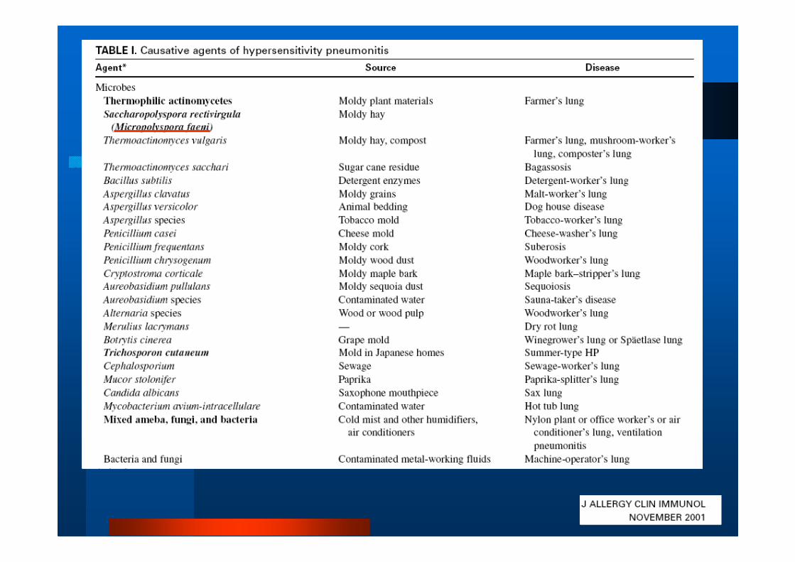

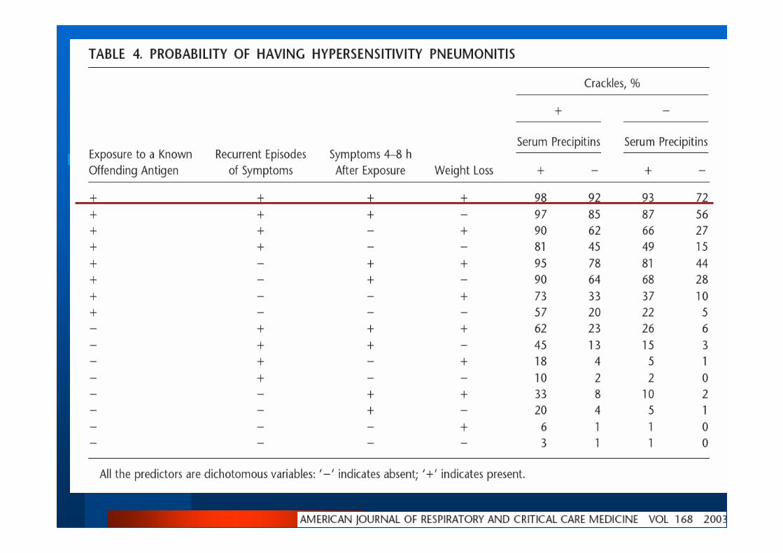
Diagnostic criteriaDiagnostic criteria
� Several diagnostic criteria have been published
� The most widely used => Richersonet al
� Criteria => not validate
unknown diagnostic accuracy


Diagnostic methodDiagnostic method
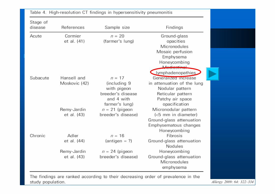
�CXR
- in acute HP => fine ground-glass appearance
nodular, straited patchy opacity
- in subacute HP => spare lung base,linear
shadow,small nodule
- in chronic HP => loss lung volume,Reticular
infiltration,interstitial fibrosis,predominant upper
& middle lung zone
- 20% normal CXR Pleural effeusion & hilar
adenopathy are rare


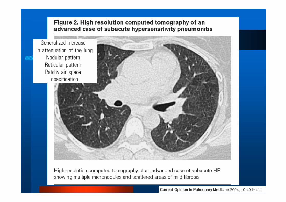
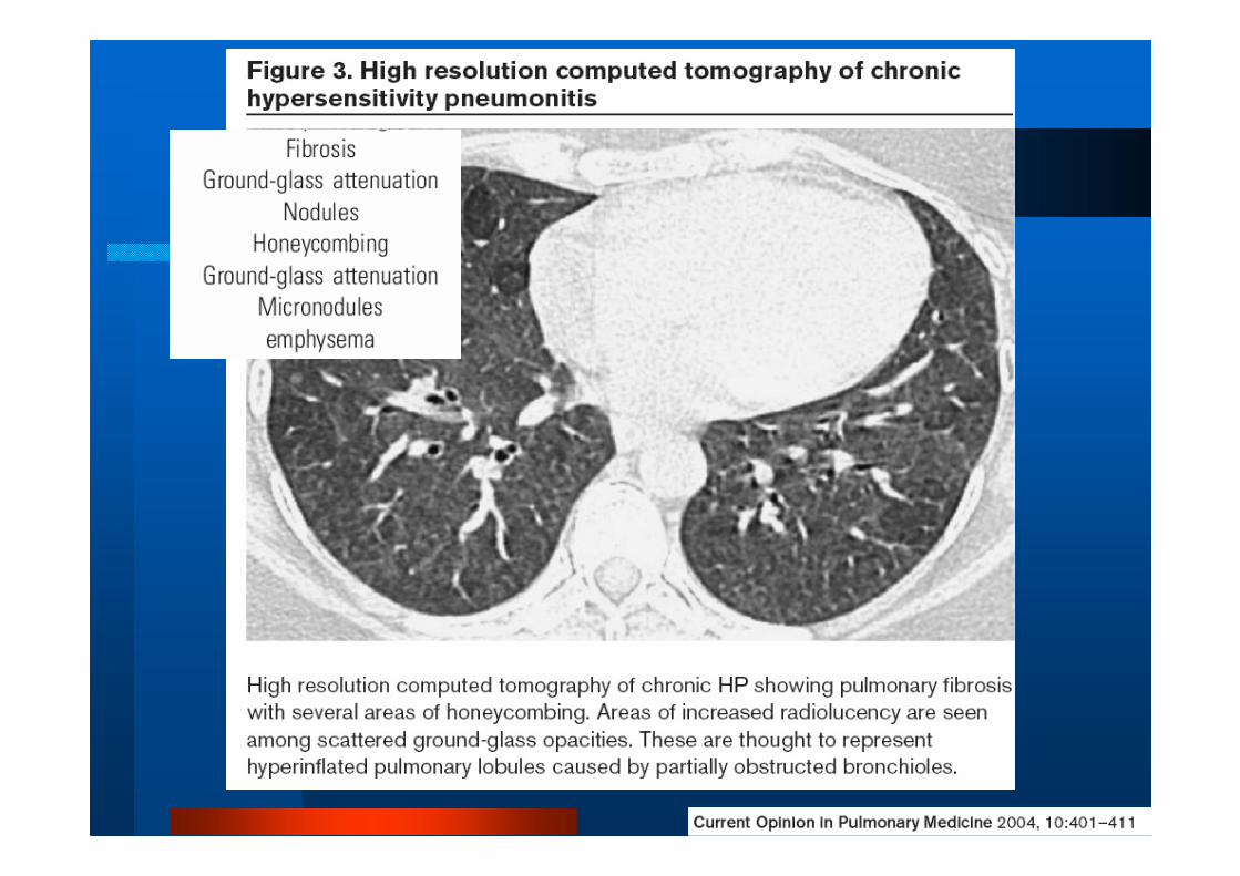
� CT scan
pattern not specific but suggestive HP
ground glass appearance!
Poorly defined ,centrilobularmicronodule ,mosaic pattern and expiratory airtrap. increase propability of HP


� PFTs
- guild to therapy
- not useful for differentiating HP from other
ILD
- acute HP =>restrictive pattern with low DLCO
- chronic pattern can be restrictive ( Farmer
lung show obstructive defect)
- ABG wide A-a gradient, hypoxemia in some
case
- 22% normal DLCO at the time Dx

� Specific antibodies
- not always present in HP
- 1-15% +ve sAbs develop HP
- use for supportive evidence
- +ve sAbs is sig. predictor of HP
- not all antigen are commercial available
- ELISA is prefer

� Inhalation challenge
- lack of standardization
- further study was need

�BAL
- important role for Dx HP
- normal lymph number => rule out HP
- predominant CD8+, CD4+/CD8+ < 1
- what dis. that CD4+, CD4+/CD8+ > 1 ?
Sarcoidosis (ratio >4
100% PPV for DDx)

Keyword of cell in BALKeyword of cell in BAL
in acute phase CD4 predominant and increase CD4/CD8 ratio and then follow
by predominant CD8+ Tcell and decrease CD4/CD8 ratio in chronic phase
Is that true ?Is that true ?Depend on - dose and type of inhaled antigen
- stage of disease
- other nonspecific stimulation

n=25 n=30n=8


LungLung biopsybiopsy
� Acute
- PMN,Eo infiltrate in alveolar space
- DAD
- Ig and complement deposition in vss.
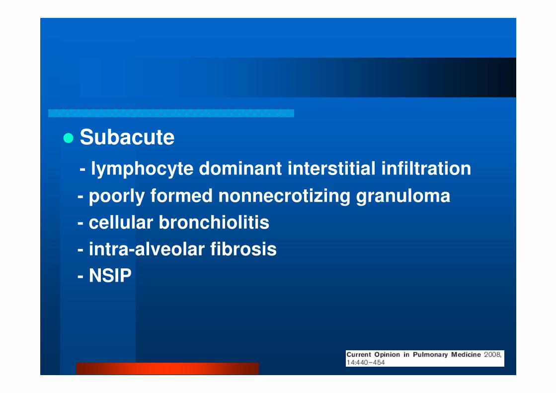
� Subacute
- lymphocyte dominant interstitial infiltration
- poorly formed nonnecrotizing granuloma
- cellular bronchiolitis
- intra-alveolar fibrosis
- NSIP


NonNon--caseatingcaseating granulomagranuloma differ from those found in differ from those found in
sarcoidosissarcoidosis by appearing by appearing
--smallersmaller
--Less well definedLess well defined
--higher predominance of lymphocytes higher predominance of lymphocytes
--located in alveolar walls inlocated in alveolar walls in centrilobularcentrilobular distribution distribution
rather than in bronchial wall, rather than in bronchial wall, subpleuralsubpleural perivascularperivascular
areaarea
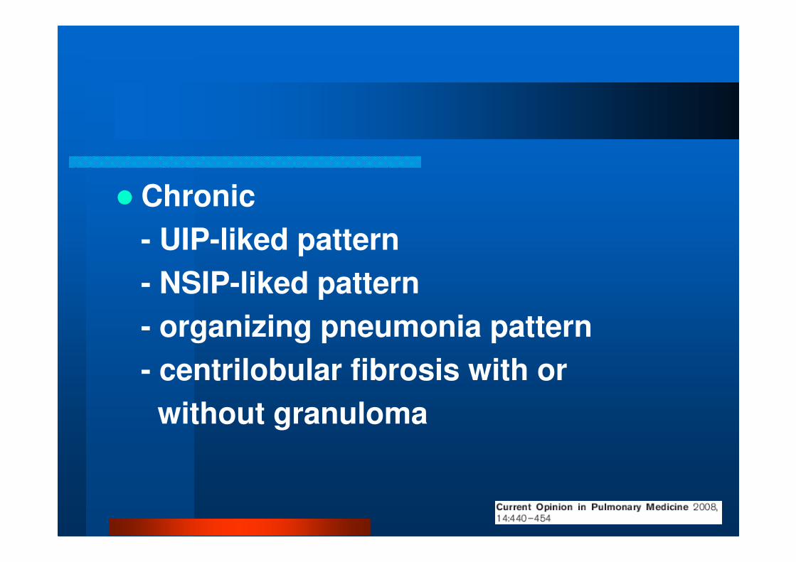
� Chronic
- UIP-liked pattern
- NSIP-liked pattern
- organizing pneumonia pattern
- centrilobular fibrosis with or
without granuloma


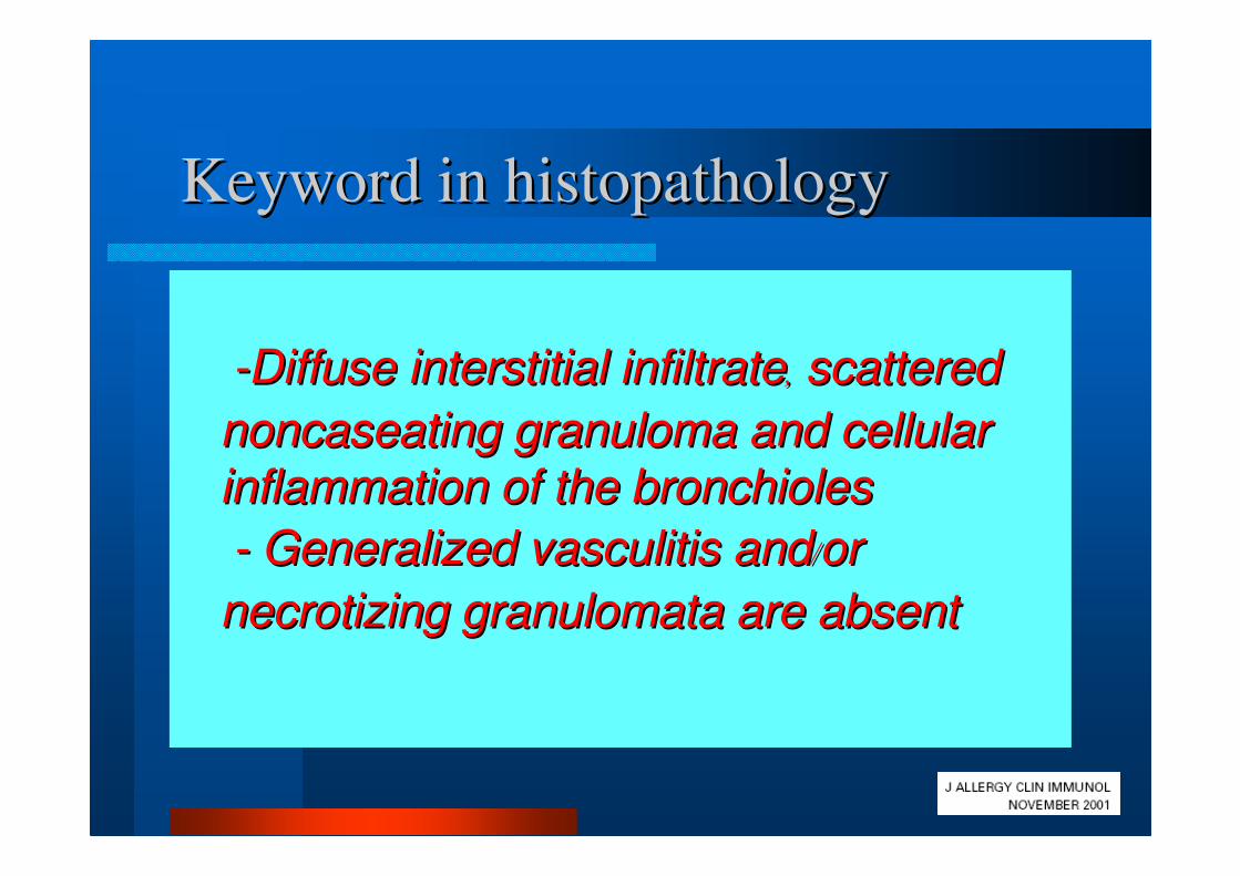
Keyword in histopathologyKeyword in histopathology
--DDiffuseiffuse interstitialinterstitial infiltrateinfiltrate,, scatteredscattered
noncaseatingnoncaseating granulomgranuloma aa andnd cellularcellular
inflammationinflammation ofof thethe bronchiolesbronchioles
-- GeneralizedGeneralized vasculitisvasculitis andand//oror
necrotizingnecrotizing granulomatagranulomata areare absentabsent


Significant predictor of HPSignificant predictor of HP

Classification of HPClassification of HP
� Acute - influenza-like symptom begin 2-9 hrs after exposure
- peak typically 6-24 hrs
- cough,dyspnea are common but not universal
- spontaneous resolve in 2-5 dys
- recurrent symptom when expose to causative agent
- PE => crackle

� Subacute
- gradually onset over several days to weeks
- marked dyspnea and cough may progress to
severe dyspnea and cyanosis,leading to
urgent hospitalization
- Mild symptoms
- Extend over 10-14 days
- Usually reversible
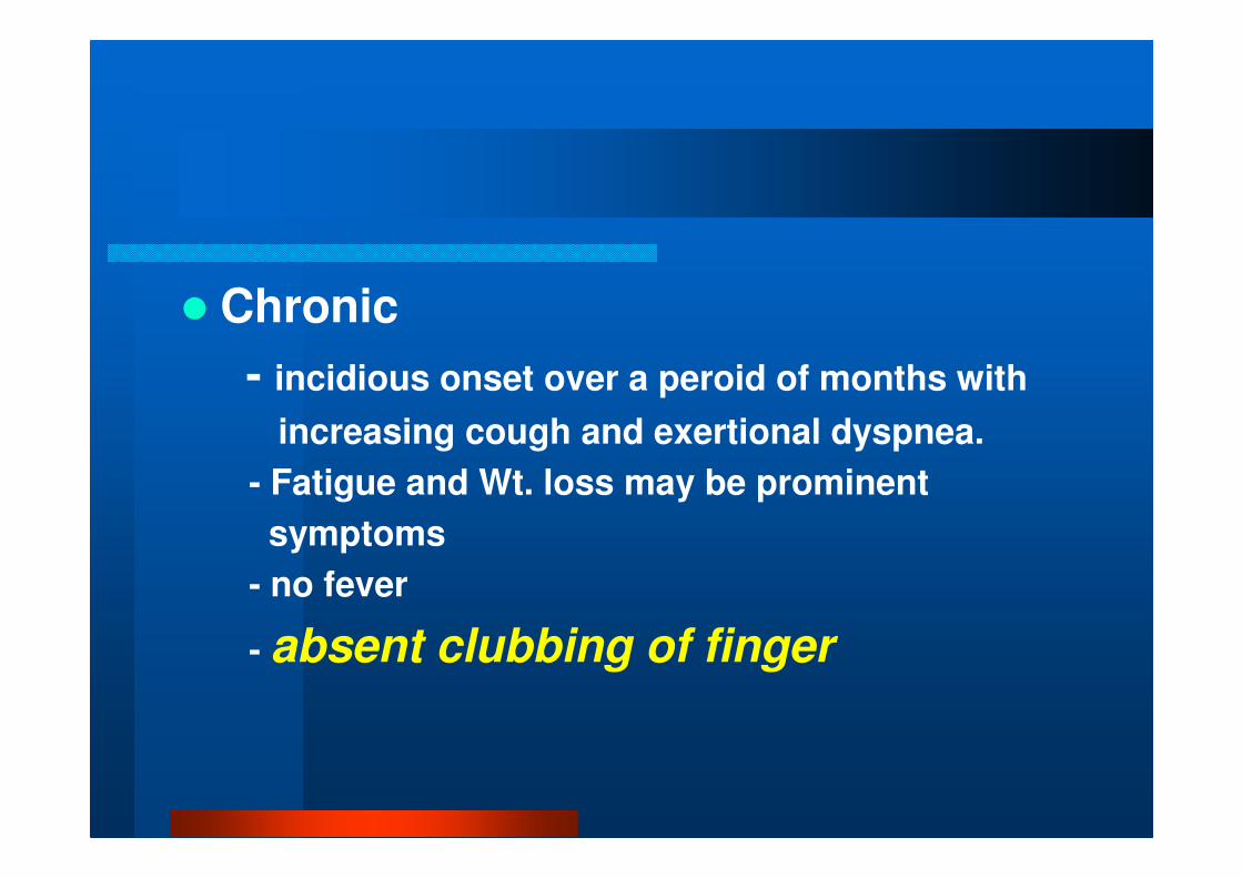
� Chronic
- incidious onset over a peroid of months with
increasing cough and exertional dyspnea.
- Fatigue and Wt. loss may be prominent
symptoms
- no fever
- absent clubbing of finger

Differential diagnosis

pathophysiologypathophysiology
� Immune complex mediated reaction
� Cell mediated reaction=> granulomaformation
Hypersensitivity reaction
type III and IV reaction
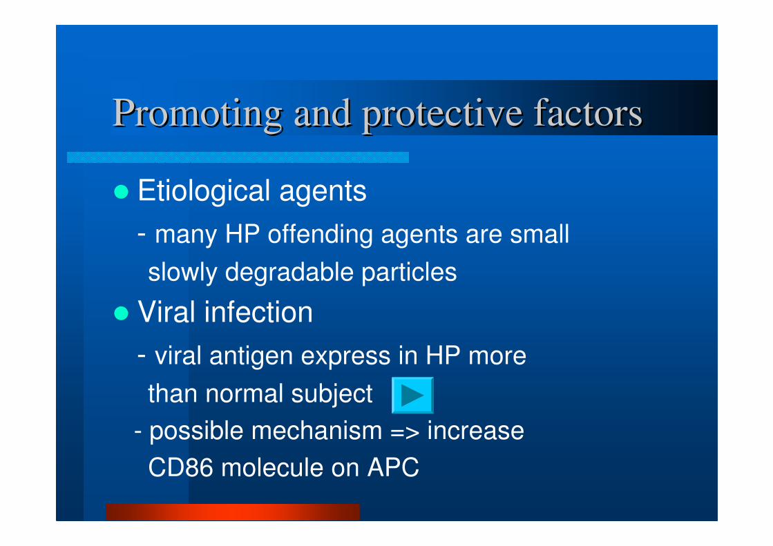
Promoting and protective factorsPromoting and protective factors
� Etiological agents
- many HP offending agents are small
slowly degradable particles
� Viral infection
- viral antigen express in HP more
than normal subject
- possible mechanism => increase
CD86 molecule on APC

R= o.7
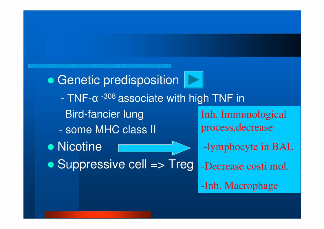
� Genetic predisposition
- TNF-α -308 associate with high TNF in
Bird-fancier lung
- some MHC class II
� Nicotine
� Suppressive cell => Treg
Inh. Immunological
process,decrease
-lymphocyte in BAL
-Decrease costi mol.
-Inh. Macrophage
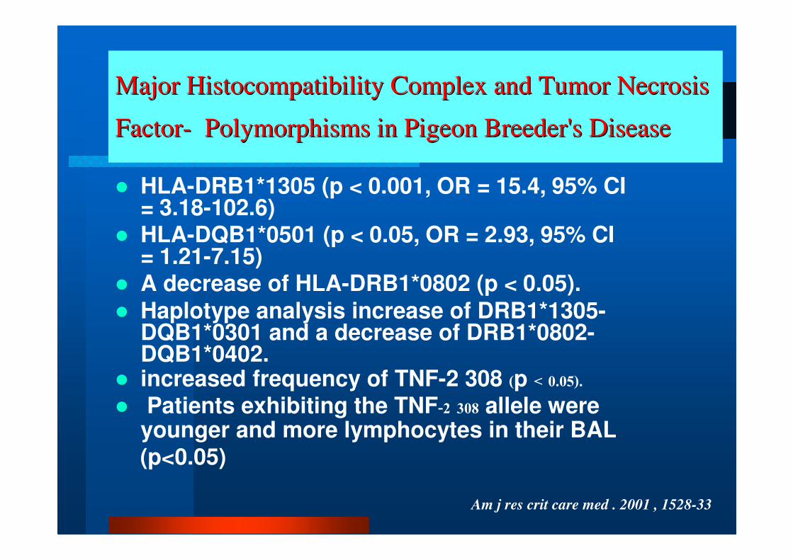
Major Major HistocompatibilityHistocompatibility Complex and Tumor Necrosis Complex and Tumor Necrosis
FactorFactor-- Polymorphisms in Pigeon Breeder's DiseasePolymorphisms in Pigeon Breeder's Disease
� HLA-DRB1*1305 (p < 0.001, OR = 15.4, 95% CI = 3.18-102.6)
� HLA-DQB1*0501 (p < 0.05, OR = 2.93, 95% CI = 1.21-7.15)
� A decrease of HLA-DRB1*0802 (p < 0.05).
� Haplotype analysis increase of DRB1*1305-DQB1*0301 and a decrease of DRB1*0802-DQB1*0402.
� increased frequency of TNF-2 308 (p < 0.05).� Patients exhibiting the TNF-2 308 allele were
younger and more lymphocytes in their BAL(p<0.05)
Am j res crit care med . 2001 , 1528-33

immunopathogenesisimmunopathogenesis

immunopathogenesisimmunopathogenesis
� Proliferation of CD8+ T cell
� production of antibody by proliferation of plasma cell
stimulated by TH1 cell
� Both pathways begin after inhaled antigen-carrying
particles are ingestd by Macrophage
� 3 phase of HP overlap in immunopathogenesis
� Greater production of TNF-α(TNF A2 alle)
� CD8+ Tcell in lung have increase usage of Vβregions of T cell receptor gene

� Acute phase
- soluble Ag bind to IgG Ab=>immune complex
- initiate complement cascade=>C5
- macrophage activation
- PMN, T cell, Monocyte recruitment
- MIP-1α (chemotactic factor for MǾ,monocyte,Tcell)& IL-8
- IFN-γ(develop granuloma),IL-1,TNF-α,IL-12(TH1)
- IL-6( from activated MǾ induce B cell, CD8+ T cell)
- CD80/86,CD28
- early phase Th1 and later CD8+
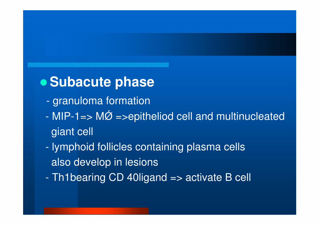
�Subacute phase
- granuloma formation
- MIP-1=> MǾ =>epitheliod cell and multinucleated
giant cell
- lymphoid follicles containing plasma cells
also develop in lesions
- Th1bearing CD 40ligand => activate B cell
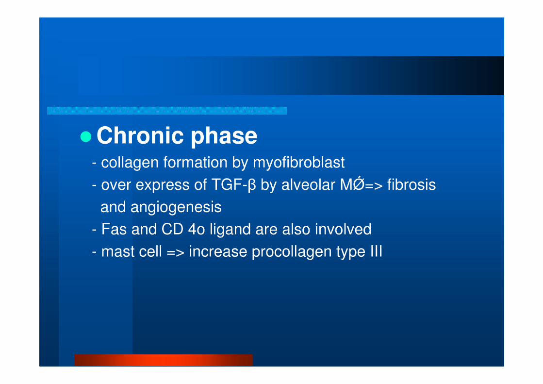
�Chronic phase- collagen formation by myofibroblast
- over express of TGF-β by alveolar MǾ=> fibrosis
and angiogenesis
- Fas and CD 4o ligand are also involved
- mast cell => increase procollagen type III

Middleton :6thedition


prognosis


managementmanagement
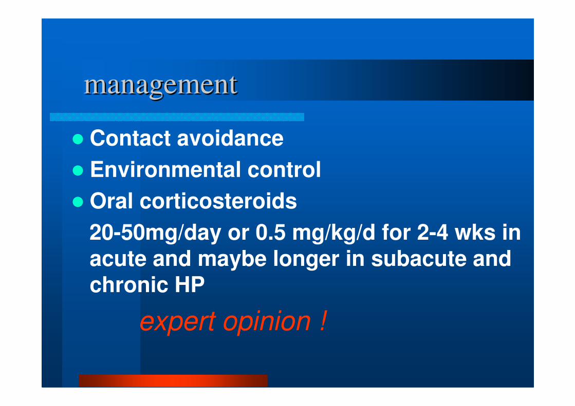
� Contact avoidance
� Environmental control
� Oral corticosteroids
20-50mg/day or 0.5 mg/kg/d for 2-4 wks in acute and maybe longer in subacute and chronic HP
expert opinion !


Steroid improve lung function
more rapid than placebo but no
influence in long term!

quizquiz
1. All of the following agents have been shown to cause both occupational asthma and hypersensitivity pneumonitis EXCEPT:
A. Toluene diisocyanateB. Trimellitic anhydrideC. Micropolyspora faeniD. Bacillus subtilisE. Diphenylmethane diisocyanate

2. Which of the following groups of
symptoms are common in the chronic form of Hypersensitivity Pneumonitis?
A. Progressive dyspnea, cough, fever B. Malaise, weakness, fever
C. Cough, malaise, anorexiaD. Cough, weakness, myalgias

3.The immunologic basis of hypersensitivity pneumonitis appears to be:
A. Type 3 (immune complex)B. Type 1 (IgE)C. Type 4 (Cell mediated)D. Combination of Type 3 and Type 4E. Combination of Type 1 and Type 3
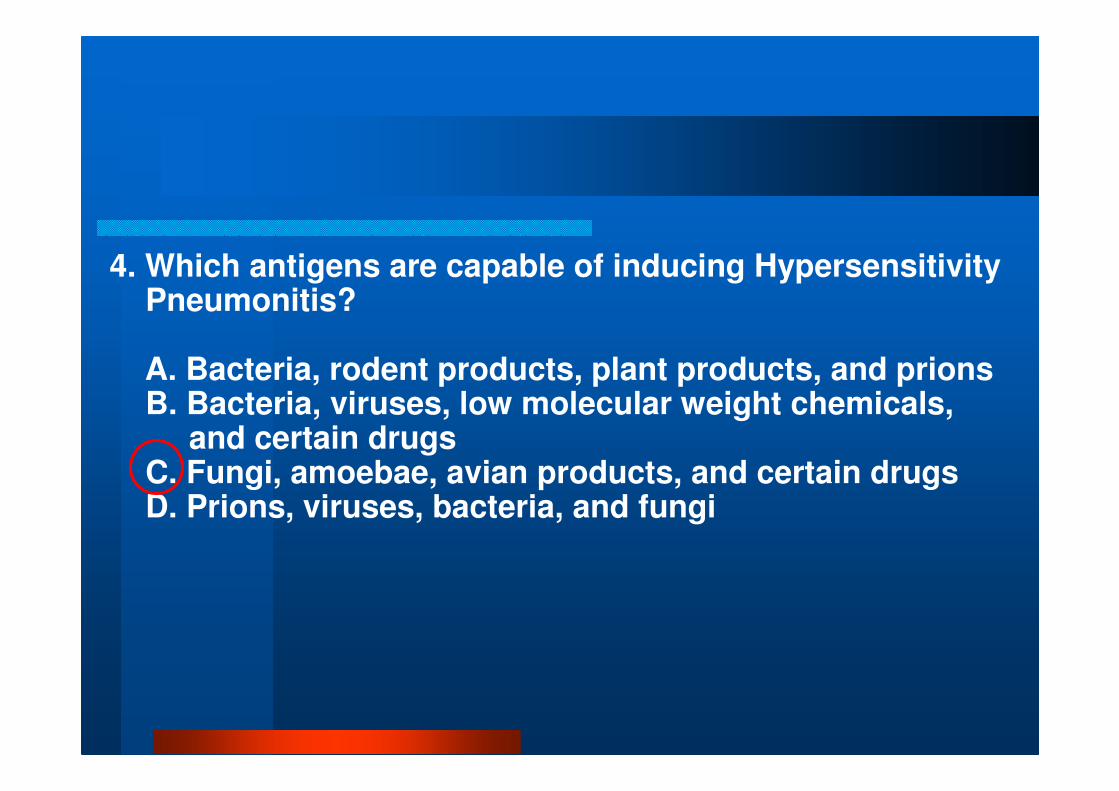
4. Which antigens are capable of inducing Hypersensitivity Pneumonitis?
A. Bacteria, rodent products, plant products, and prionsB. Bacteria, viruses, low molecular weight chemicals,
and certain drugsC. Fungi, amoebae, avian products, and certain drugsD. Prions, viruses, bacteria, and fungi

5.Which of the following best represents CD4 and CD8 lymphocyte numbers found in bronchoalveolar lavage samples of patients with Hypersensitivity Pneumonitis vs. normal controls?
A. Increased CD4, increased CD8, decreased CD4/CD8 ratio B. Decreased CD4, decreased CD8, decreased CD4/CD8 ratioC. Decreased CD4, increased CD8, decreased CD4/CD8 ratioD. Decreased CD4, decreased CD8, increased CD4/CD8 ratio

6. Which of the following is a major criterion for the diagnosis of Hypersensitivity Pneumonitis?A. Bibasilar dry ralesB. Decreased diffusing capacityC. Arterial hypoxemiaD. Lung larvage fluid lymphocytosis
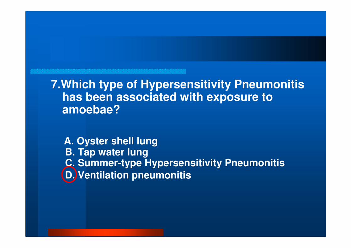
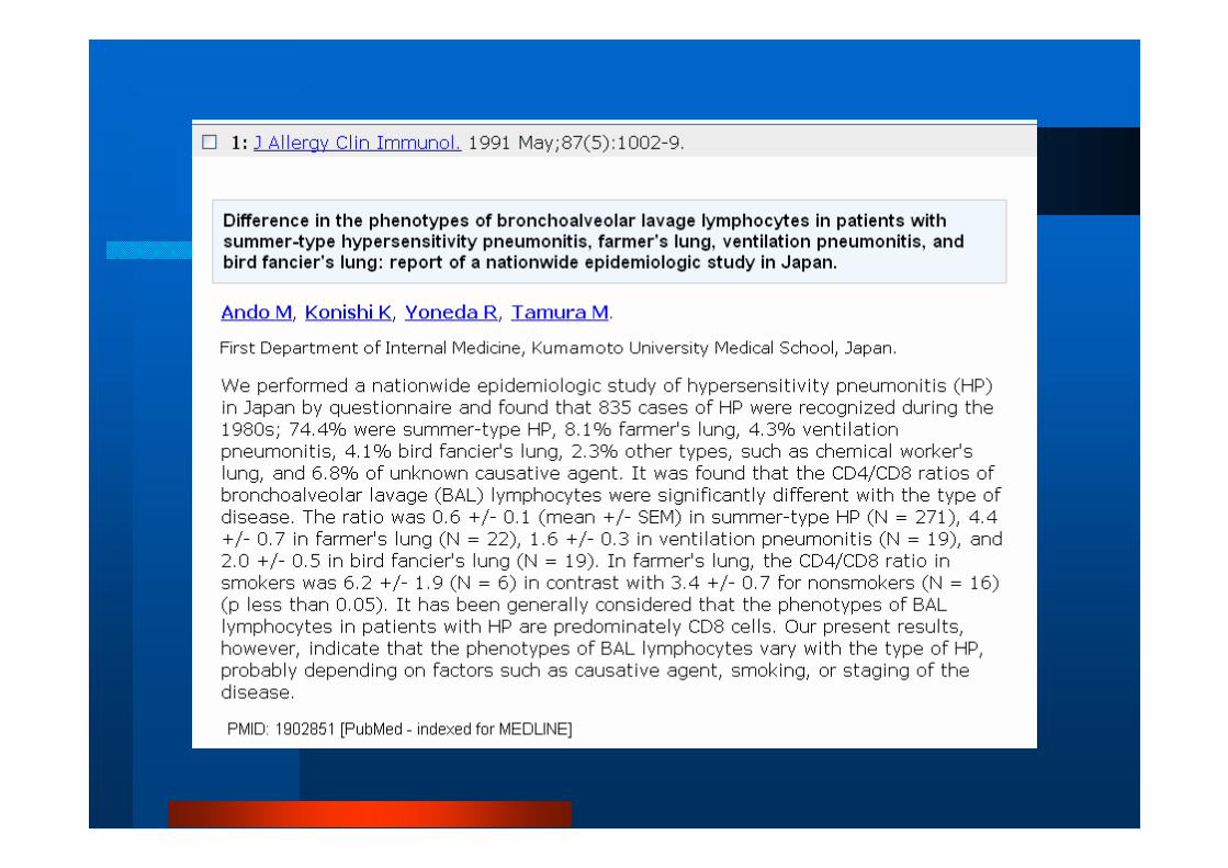
7.Which type of Hypersensitivity Pneumonitishas been associated with exposure to amoebae?
A. Oyster shell lungB. Tap water lung C. Summer-type Hypersensitivity Pneumonitis
D. Ventilation pneumonitis

8.Which of the following is associated
with Farmer’s lung?A. HistoplasmosisB. Cryptococcus C. Thermophilic actinomycetesD. Aspergillus fumigatus

9. Which of the following scenarios is most indicative of sarcoidosis?
A. Restrictive pattern on PFT, increased ACE, increased T suppressor cells in BAL
B. Obstructive pattern on PFT, decreased ACE, increase in T helper cells in BAL
C. Restrictive pattern on PFT, increased ACE, increase in T helper cells in BAL
D. Obstructive pattern on PFT, increased ACE, increase in T suppressor cells in BAL

10.The most common form of Hypersensitivity Pneumonitis in the pediatric population is related to the inhalation of which of the following?
A. MedicationsB. Insect proteinsC. Avian proteinsD. Rodent urinary proteins

conclusionconclusion
� Difficult to determine prevalence and
incidence
� Classification
� Diagnosis
� Characteristic imaging and pathology
� Pathophysiology
� Immunology
� Treatment

Thank you for your Thank you for your
attention!attention!

1.126

1.126

Related Documents