Gene Expression Profiles Distinguish Idiopathic Pulmonary Fibrosis from Hypersensitivity Pneumonitis Moises Selman, Annie Pardo, Lourdes Barrera, Andrea Estrada, Susan R. Watson, Keith Wilson, Natasha Aziz, Naftali Kaminski*, and Albert Zlotnik* Instituto Nacional de Enfermedades Respiratorias, Mexico City; Facultad de Ciencias, Universidad Nacional Auto ´noma de Me ´xico, Mexico City, Mexico; Eos Biotechnology, South San Francisco, California; and Simmons Center for Interstitial Lung Disease, Pulmonary, Allergy, and Critical Care Medicine, University of Pittsburgh Medical Center, Pittsburgh, Pennsylvania Rationale: Many of the interstitial lung diseases represent a diagnos- tic and therapeutic challenge because their clinical and even histo- logic features are often nonspecific. Likewise, the transcriptional signatures of most of them are unknown. Objective: To compare the gene expression patterns from patients with idiopathic pulmonary fibrosis (IPF) hypersensitivity pneumoni- tis (HP), and nonspecific interstitial pneumonia (NSIP) using custom oligonucleotide microarrays. Methods: We profiled lung biopsies from 15 patients with IPF, 12 with HP, and eight with NSIP. Labeled complementary ribonucleic acid was hybridized to a custom Affymetrix oligonucleotide DNA microarray using standard Affymetrix protocols. The custom array, Hu03, contained 59,619 probe sets representing an estimated 46,000 gene clusters. Results: We identified statistically significant gene expression signa- tures that characterize HP and IPF. The HP gene expression signa- ture was enriched for genes that are functionally associated with inflammation, T-cell activation, and immune responses, whereas the IPF signature was characterized by the expression of tissue remodeling, epithelial, and myofibroblast genes. We then com- pared these gene expression signatures to classify NSIP, a histologic pattern that is often difficult to differentiate consistently from HP and IPF. Two cases exhibited an IPF-like gene expression, another one could be more properly classified as HP, whereas others did not resemble HP or IPF, suggesting that they may represent idiopathic NSIP. Conclusions: Our results underscore the value of gene expression signatures to classify the interstitial lung diseases and to understand pathogenic mechanisms, and suggest new ways to improve the diagnosis and treatment of patients with these diseases. Keywords: global transcription analysis; interstitial lung diseases; lung fibrosis; microarrays; nonspecific interstitial pneumonia Pulmonary fibrosis is the final common outcome of a diverse group of interstitial lung diseases (ILDs). A number of these disorders have known etiologies, arising from exposure to or- ganic or inorganic particles or drugs, or they may have an associa- tion to collagen-vascular diseases (1). However, approximately 40 to 50% of these disorders are of unknown etiology and are (Received in original form April 25, 2005; accepted in final form September 15, 2005) *These authors contributed equally to this study. Supported in part by National Institutes of Health grant HL 073745-01, and by a generous donation from the Simmons family (N.K.), and by DGAPA, UNAM IN215003 (A.P.). Correspondence and requests for reprints should be addressed to Albert Zlotnik, Ph.D., Neurocrine Biosciences, 12790 El Camino Real, San Diego, CA 92130. E-mail: [email protected] This article has an online supplement, which is accessible from this issue’s table of contents at www.atsjournals.org Am J Respir Crit Care Med Vol 173. pp 188–198, 2006 Originally Published in Press as DOI: 10.1164/rccm.200504-644OC on September 15, 2005 Internet address: www.atsjournals.org classified as idiopathic interstitial pneumonias (IIPs) (2). The most common IIP is idiopathic pulmonary fibrosis (IPF), which represents a chronic and usually fatal lung disorder morphologi- cally characterized by a usual interstitial pneumonia (UIP) pat- tern (2,3). Nonspecific interstitial pneumonia (NSIP) is a more recently recognized morphologic pattern characterized by vary- ing degrees of inflammation and fibrosis. These changes are temporally uniform, suggesting that they have occurred over a single, relatively narrow time span (2,3). Importantly, NSIP can be difficult to differentiate consistently from hypersensitivity pneumonitis (HP) and UIP, and significant interobserver vari- ability exists even among expert pathologists (4). HP is a complex syndrome with variable clinical presentation and natural history, and represents a diagnostic challenge for clinicians and, in the chronic form, even for experienced patholo- gists. The diagnosis of HP requires a high index of suspicion and a combination of clinical, environmental, radiologic, physiologic, and pathologic findings (5, 6). Chronic HP often develops into fibrosis with destruction of the lung parenchyma, a process that mimics IPF (7). It is widely believed that lung fibrosis is triggered by an injury that results in inflammation, which, if unresolved, is followed by fibroblast proliferation/activation and extracellular matrix accu- mulation. However, recent evidence suggests that this sequence is likely relevant for most ILDs, including HP, but probably not for IPF, which seems to represent primarily an epithelial/ fibroblastic disorder (8,9). To gain a better understanding of the molecular mechanisms that underlie the lung phenotypes in IPF, HP, and NSIP, we sought to identify and compare their gene expression “signatures” using genomewide, custom oligonucleotide microarrays. We hy- pothesized that these diseases exhibit different gene expression signatures that should be useful to distinguish these disorders and also provide insights regarding the divergent pathways leading to lung fibrosis. We also expected that specific signatures could lead to the identification of new biomarkers to aid in the diagnosis and classification of these diseases. Our results indicate that IPF can be distinguished from HP by the expression of tissue remodeling and epithelial and myofi- broblast genes, whereas HP is characterized by the expression of inflammatory genes representing T-cell activation and immune responses. Using these expression signatures, we were able to reclassify some of the NSIP cases as being closer to IPF or HP, whereas others were likely to represent idiopathic NSIP. Our results highlight the mechanistic differences between ILD sub- types and underscore the potential that transcriptional profiling has for the differential diagnosis of ILD. METHODS Fifteen patients with IPF, 12 with HP, and eight with NSIP-like pattern were included in this study. The study received approval from the local ethics committee, and informed consent was obtained from all subjects. Diagnosis of IPF and HP was performed as described elsewhere and

Welcome message from author
This document is posted to help you gain knowledge. Please leave a comment to let me know what you think about it! Share it to your friends and learn new things together.
Transcript

Gene Expression Profiles Distinguish IdiopathicPulmonary Fibrosis from Hypersensitivity PneumonitisMoises Selman, Annie Pardo, Lourdes Barrera, Andrea Estrada, Susan R. Watson, Keith Wilson, Natasha Aziz,Naftali Kaminski*, and Albert Zlotnik*
Instituto Nacional de Enfermedades Respiratorias, Mexico City; Facultad de Ciencias, Universidad Nacional Autonoma de Mexico, Mexico City,Mexico; Eos Biotechnology, South San Francisco, California; and Simmons Center for Interstitial Lung Disease, Pulmonary, Allergy, andCritical Care Medicine, University of Pittsburgh Medical Center, Pittsburgh, Pennsylvania
Rationale: Many of the interstitial lung diseases represent a diagnos-tic and therapeutic challenge because their clinical and even histo-logic features are often nonspecific. Likewise, the transcriptionalsignatures of most of them are unknown.Objective: To compare the gene expression patterns from patientswith idiopathic pulmonary fibrosis (IPF) hypersensitivity pneumoni-tis (HP), and nonspecific interstitial pneumonia (NSIP) using customoligonucleotide microarrays.Methods: We profiled lung biopsies from 15 patients with IPF, 12with HP, and eight with NSIP. Labeled complementary ribonucleicacid was hybridized to a custom Affymetrix oligonucleotide DNAmicroarray using standard Affymetrix protocols. The custom array,Hu03, contained 59,619 probe sets representing an estimated46,000 gene clusters.Results: We identified statistically significant gene expression signa-tures that characterize HP and IPF. The HP gene expression signa-ture was enriched for genes that are functionally associated withinflammation, T-cell activation, and immune responses, whereasthe IPF signature was characterized by the expression of tissueremodeling, epithelial, and myofibroblast genes. We then com-pared these gene expression signatures to classify NSIP, a histologicpattern that is often difficult to differentiate consistently from HPand IPF. Two cases exhibited an IPF-like gene expression, anotherone could be more properly classified as HP, whereas others did notresemble HP or IPF, suggesting that they may represent idiopathicNSIP.Conclusions: Our results underscore the value of gene expressionsignatures to classify the interstitial lung diseases and to understandpathogenic mechanisms, and suggest new ways to improve thediagnosis and treatment of patients with these diseases.
Keywords: global transcription analysis; interstitial lung diseases; lungfibrosis; microarrays; nonspecific interstitial pneumonia
Pulmonary fibrosis is the final common outcome of a diversegroup of interstitial lung diseases (ILDs). A number of thesedisorders have known etiologies, arising from exposure to or-ganic or inorganic particles or drugs, or they may have an associa-tion to collagen-vascular diseases (1). However, approximately40 to 50% of these disorders are of unknown etiology and are
(Received in original form April 25, 2005; accepted in final form September 15, 2005)
*These authors contributed equally to this study.
Supported in part by National Institutes of Health grant HL 073745-01, and bya generous donation from the Simmons family (N.K.), and by DGAPA, UNAMIN215003 (A.P.).
Correspondence and requests for reprints should be addressed to Albert Zlotnik,Ph.D., Neurocrine Biosciences, 12790 El Camino Real, San Diego, CA 92130.E-mail: [email protected]
This article has an online supplement, which is accessible from this issue’s tableof contents at www.atsjournals.org
Am J Respir Crit Care Med Vol 173. pp 188–198, 2006Originally Published in Press as DOI: 10.1164/rccm.200504-644OC on September 15, 2005Internet address: www.atsjournals.org
classified as idiopathic interstitial pneumonias (IIPs) (2). Themost common IIP is idiopathic pulmonary fibrosis (IPF), whichrepresents a chronic and usually fatal lung disorder morphologi-cally characterized by a usual interstitial pneumonia (UIP) pat-tern (2,3). Nonspecific interstitial pneumonia (NSIP) is a morerecently recognized morphologic pattern characterized by vary-ing degrees of inflammation and fibrosis. These changes aretemporally uniform, suggesting that they have occurred over asingle, relatively narrow time span (2,3). Importantly, NSIP canbe difficult to differentiate consistently from hypersensitivitypneumonitis (HP) and UIP, and significant interobserver vari-ability exists even among expert pathologists (4).
HP is a complex syndrome with variable clinical presentationand natural history, and represents a diagnostic challenge forclinicians and, in the chronic form, even for experienced patholo-gists. The diagnosis of HP requires a high index of suspicion anda combination of clinical, environmental, radiologic, physiologic,and pathologic findings (5, 6). Chronic HP often develops intofibrosis with destruction of the lung parenchyma, a process thatmimics IPF (7).
It is widely believed that lung fibrosis is triggered by an injurythat results in inflammation, which, if unresolved, is followed byfibroblast proliferation/activation and extracellular matrix accu-mulation. However, recent evidence suggests that this sequenceis likely relevant for most ILDs, including HP, but probablynot for IPF, which seems to represent primarily an epithelial/fibroblastic disorder (8,9).
To gain a better understanding of the molecular mechanismsthat underlie the lung phenotypes in IPF, HP, and NSIP, wesought to identify and compare their gene expression “signatures”using genomewide, custom oligonucleotide microarrays. We hy-pothesized that these diseases exhibit different gene expressionsignatures that should be useful to distinguish these disorders andalso provide insights regarding the divergent pathways leading tolung fibrosis. We also expected that specific signatures could leadto the identification of new biomarkers to aid in the diagnosis andclassification of these diseases.
Our results indicate that IPF can be distinguished from HPby the expression of tissue remodeling and epithelial and myofi-broblast genes, whereas HP is characterized by the expression ofinflammatory genes representing T-cell activation and immuneresponses. Using these expression signatures, we were able toreclassify some of the NSIP cases as being closer to IPF or HP,whereas others were likely to represent idiopathic NSIP. Ourresults highlight the mechanistic differences between ILD sub-types and underscore the potential that transcriptional profilinghas for the differential diagnosis of ILD.
METHODS
Fifteen patients with IPF, 12 with HP, and eight with NSIP-like patternwere included in this study. The study received approval from the localethics committee, and informed consent was obtained from all subjects.Diagnosis of IPF and HP was performed as described elsewhere and

Selman, Pardo, Barrera, et al.: Microarray Analysis in IPF and HP 189
confirmed by morphology (3, 10, 11). Diagnosis of NSIP was suspectedin patients with an interstitial pneumonia that did not meet the clinical/radiologic criteria for other IIPs and was based primarily on histology(2, 3). Control samples included nonpathologic lung specimens obtainedfrom trauma victims. None of the patients had been treated with cortico-steroids/immunosuppressive drugs at the time of biopsy. Additionaldetails are provided in the online supplement. One fragment of thelung samples was frozen in liquid nitrogen for RNA extraction andsubsequent expression analysis by DNA microarrays.
RNA Extraction and DNA Microarray Hybridization
RNA extracted from lung tissue was converted into biotinylated cRNAand hybridized to a custom Affymetrix (Santa Clara, CA) oligonucleo-tide microarray (Hu03 containing 59,619 probe sets representing anestimated 46,000 gene clusters [see online supplement] using standardAffymetrix protocols). The generation of the gene expression data wasperformed as described (12).
Statistical Methods
Statistical analyses were performed using ScoreGenes software package(Jerusalem, Israel; available from http://www.cs.huji.ac.il/labs/compbio/scoregenes/) (13–15). The most informative genes were defined as thosethat pass the false-discovery rate of 5% (16) for t test or for thresholdnumber of misclassifications (TNoM) method. To further assess thepredictive power of the datasets, we used the Leave-One-Out Cross-Validation (LOOCV) statistical method. This method simulates re-moval of a single sample every trial and trains on the rest. The procedureis repeated until each sample is left out once and the number of correctand incorrect predictions is counted. For each p value, a classificationscore was calculated for every sample in a specific class (Reference 15and the online supplement).
Functional Annotations
Enrichment of functional annotations was performed using National In-stitutes of Health DAVID and EASE online (http://apps1.niaid.nih.gov/david/) (17, 18). Statistical significance was determined using Fisher’sexact score and EASE score (17). Additional details on statistical methodsand functional annotations are provided in the online supplement.
Bronchoalveolar Lavage
Bronchoalveolar lavage (BAL) was performed as described (10). Cellswere used for differential cell counts and fluorescence-activated cellsorter (FACS) analyses. Insulinlike growth factor binding protein 4(IGFBP-4) was measured by ELISA following the instructions of themanufacturer (Diagnostic Systems Laboratories, Inc., Webster, TX).Results are expressed in micrograms of IGFBP-4/mg protein. Addi-tional details are provided in the online supplement.
Immunohistochemistry
Tissue sections were treated as previously described (10). The followingantibodies were used: Human anti–IGFBP-4 and N-cadherin polyclonalantibodies (Santa Cruz Biotech, Santa Cruz, CA), and human matrixmetalloproteinase 1 (MMP-1) monoclonal antibody (Oncogene, SanDiego, CA). The primary antibody was replaced by nonimmune serumfor negative controls. Additional details are provided in the onlinesupplement.
Flow Cytometric Analyses of Surface CD3, CD4, CD8, andCD69 on BAL Lymphocytes
BAL cells (1 � 106) were labeled with fluorescein isothiocyanate–conjugated anti-CD3, phycoerythrin-conjugated anti-CD4, PerCp anti-CD8, and fluorescein isothiocyanate anti-CD69 (Becton Dickinson, SanJose, CA). Flow cytometry was performed using a FACScan (BectonDickinson) and analyzed with CellQuest software (Becton Dickinson).Additional details are provided in the online supplement.
RESULTS
Baseline Characteristics of Patients with IPF, HP, or NSIP
Demographic data, pulmonary function test results, and BALdifferential cell counts are summarized in Table 1. All patients
exhibited clinical, radiologic, and functional evidence of ILD,with variable degrees of dyspnea, decreased lung capacities, andhypoxemia at rest that worsened during exercise. In general, thepatients with IPF were older, predominantly male, and morelikely to have been cigarette smokers than the patients with HPor NSIP. By contrast, all patients with HP were female. Thissex prevalence has been previously described in avian antigen-induced HP in Mexico (19). In the HP group, differential cellcounts in BAL fluid were characterized by marked lymphocyto-sis, whereas in IPF, most BAL inflammatory cells were macro-phages with a moderate increase in neutrophils and eosinophils.
Gene Expression Patterns Distinguish IPF and HP
IPF is a disease of unknown etiology, and by far the most severeILD with very poor prognosis. HP is an ILD of known etiology,although its pathogenic mechanisms are not completely under-stood. Although considered a more benign ILD, some patientswith chronic HP also evolve to end-stage lung fibrosis and dieof the progressive destruction of the lung parenchyma. We there-fore compared the gene expression patterns of these diseases,and asked whether we could find genes specifically expressed ineach disease. A well-acknowledged challenge with microarraydata is the asymmetry between the number of samples and thenumber of parameters measured. This asymmetry may lead tospurious p values regardless of which scoring system is used.
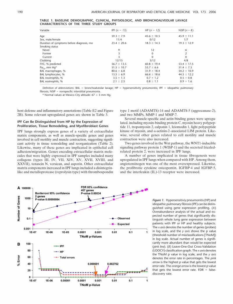
To address this challenge, we performed overabundance anal-ysis, which revealed that the actual number of genes for everyp value was substantially higher than would be expected underthe null hypothesis that the separation of the samples is random(Figure 1A). Using the nonparametric TNoM scoring method (seeMethods here and in the online supplement), we identified twogenes that had TNoM score p values that passed Bonferroni correc-tion and 407 genes that passed a significance threshold controllingthe false-discovery rate at 5% as previously described (16). Toidentify the genes that best distinguished IPF from HP, we per-formed LOOCV for every TNoM score p value (Figure 1B). Mini-mal error rates were observed at TNoM score p values of 0.002762and 0.000691 (Figure 1B) where classification errors were 1 and 0for misclassifying HP as IPF and IPF as HP, respectively, stronglysuggesting that the signature distinguishing these two diseases issignificant. Unsupervised clustering also distinguished the dis-eases, as did Student’s t test (data not shown).
The Gene Expression Signatures That Characterize HP or IPFAre Composed of Distinctly Different Functional Groups
To better understand the mechanistic nature of the genes thatdistinguish IPF from HP, we looked for Gene Ontology func-tional annotations that were enriched in genes that were higherin IPF lungs or in HP lungs using the National Institutes ofHealth DAVID and EASE online (http://apps1.niaid.nih.gov/david/) (17, 18). This analysis was only performed on transcriptsthat could be found in LocusLink (http://www.ncbi.nlm.nih.gov/projects/LocusLink/). Most of the genes that were significantlyincreased in IPF were enriched for genes involved in develop-ment, extracellular matrix structure and turnover, and cellulargrowth and differentiation. The 354 upregulated genes that in-clude the complete list of statistically significant annotations arelisted in Table E1 on the online supplement. Some relevantupregulated genes are shown in Table 2. Furthermore, in addi-tion to the statistically significant p values, many of these geneswere associated with the relevant annotations (Figure 2A), sug-gesting that this represents a biologically meaningful phenome-non. The functional profile was completely different in HP lungs.The 595 genes that were overexpressed in HP compared with IPFand had locus-link information were enriched with a variety of
190 AMERICAN JOURNAL OF RESPIRATORY AND CRITICAL CARE MEDICINE VOL 173 2006
TABLE 1. BASELINE DEMOGRAPHIC, CLINICAL, PHYSIOLOGIC, AND BRONCHOALVEOLAR LAVAGECHARACTERISTICS OF THE THREE STUDY GROUPS
Variable IPF (n � 15) HP (n � 12) NSIP (n � 8 )
Age 59.3 � 7.9 45.6 � 10.5 45.9 � 11.1Sex, male/female 11/4 0/12 1/7Duration of symptoms before diagnosis, mo 23.4 � 20.6 18.3 � 14.3 19.3 � 12.9Smoking status
Never 9 12 6Former 3 0 2Current 3 0 0
Clubbing 12/15 3/12 4/8FVC, % predicted 56.7 � 13.5 60.8 � 19.4 53.4 � 17.5PaO2
, mm Hg* 51.5 � 10.7 53.7 � 6.6 51.4 � 7.3BAL macrophages, % 80.6 � 6.8 31.9 � 18.4 54.2 � 10.9BAL lymphocytes, % 13.5 � 6.9 66.6 � 18.6 44.5 � 12.2BAL neutrophils, % 3.5 � 1.3 0.7 � 1.2 0.5 � 0.8BAL eosinophils, % 2.1 � 2.5 0.8 � 1.1 0.9 � 1.6
Definition of abbreviations: BAL � bronchoalveolar lavage; HP � hypersensitivity pneumonitis; IPF � idiopathic pulmonaryfibrosis; NSIP � nonspecific interstitial pneumonia.
* Normal values at Mexico City altitude: 67 � 3 mm Hg.
host defense and inflammatory annotations (Table E2 and Figure2B). Some relevant upregulated genes are shown in Table 3.
IPF Can Be Distinguished from HP by the Expression ofProliferation, Tissue Remodeling, and Myofibroblast Genes
IPF lungs strongly express genes of a variety of extracellularmatrix components, as well as muscle-specific genes and genesinvolved in cell motility and muscle contraction, suggesting signifi-cant activity in tissue remodeling and reorganization (Table 2).Likewise, many of these genes are implicated in epithelial celladhesion/migration. Genes encoding extracellular matrix mole-cules that were highly expressed in IPF samples included manycollagens (types III, IV, VII, XIV, XV, XVII, XVIII, andXXVII), tenascin N, versican, and asporin. Other extracellularmatrix components increased in IPF lungs included a disintegrin-like and metalloprotease (reprolysin type) with thrombospondin
Figure 1. Hypersensitiviy pneumonitis (HP) andidiopathic pulmonary fibrosis (IPF) can be distin-guished using gene expression profiling. (A )Overabundance analysis of the actual and ex-pected number of genes that significantly dis-tinguish whole lung gene expression betweenpatients with IPF or HP and healthy subjects.The x axis denotes the number of genes (probes)in log scale, and the y axis shows the p value(threshold number of misclassifications [TNoM])in log scale. Actual number of genes is signifi-cantly more abundant than would be expected(pink line). (B ) Leave-One-Out Cross-Validation(LOOCV) classification graph. The x axis denotesthe TNoM p value in log scale, and the y axisdenotes the error rate in percentages. The pinkarrow is the highest p value that gets the lowesterror rate. The orange arrow is the lowest p valuethat gets the lowest error rate. FDR � false-discovery rate.
type 1 motif (ADAMTS)-14 and ADAMTS-5 (aggrecanase-2),and two MMPs, MMP-1 and MMP-7.
Several muscle-specific and actin-binding genes were upregu-lated, including myosin-binding protein C, myosin heavy polypep-tide 11, tropomyosin 2, calponin 1, leiomodin 1, light polypeptidekinase of myosin, and �-actinin-2–associated LIM protein. Like-wise, several other genes related to cell motility and musclecontraction were also increased.
Two genes involved in the Wnt pathway, the WNT1-induciblesignaling pathway protein 1 (WISP-1) and the secreted frizzled-related protein 2, were increased in IPF lungs.
A number of genes implicated in tissue fibrogenesis wereupregulated in IPF lungs when compared with HP. Among them,angiotensinogen was one of the most overexpressed. Likewise,the profibrotic cytokine osteopontin, IGFBP-4 and IGFBP-5,and the interleukin (IL)-13 receptor were increased.

Selman, Pardo, Barrera, et al.: Microarray Analysis in IPF and HP 191
TABLE 2. UPREGULATED GENES IN IDIOPATHIC PULMONARY FIBROSIS
Gene Name Locus-Link No. TNoM p Value t Test p Value
NM_017680: asporin (LRR class 1; ASPN), mRNA 54829 4.0382E-05 0.00028842NM_033285: tumor protein p53 inducible nuclear protein 1 (TP53INP1), mRNA 94241 0.00033652 2.5438E-06NM_000029: angiotensinogen (serine [or cysteine] proteinase; AGT), mRNA 183 0.00033652 3.6249E-06NM_000861: histamine receptor H1 (HRH1), mRNA 3269 0.00033652 1.6201E-05Hs.31386: secreted frizzled-related protein 2 6423 0.00033652 0.00020623NM_001792: cadherin 2, type 1, N-cadherin (neuronal) (CDH2), mRNA 1000 0.00033652 0.00026976NM_005214: cytotoxic T-lymphocyte–associated protein 4 (CTLA4), mRNA 1493 0.00033652 0.00039581NM_000577: interleukin-1 receptor antagonist (IL1RN), transcript variant 3, mRNA 3557 0.00033652 0.00194308NM_033326: SRY (sex-determining region Y)-box 6 (SOX6), mRNA 55553 0.00033652 0.0019824NM_004425: extracellular matrix protein 1 (ECM1), transcript variant 1, mRNA 1893 0.00033652 0.00223442NM_001847: collagen, type IV, �6 (COL4A6), transcript variant A, mRNA 1288 0.00033652 0.00342796NM_006475: osteoblast-specific factor 2 (fasciclin I-like; OSF-2), mRNA 10631 0.00033652 0.00359433NM_130445: collagen, type XVIII, �1 (COL18A1), transcript variant 2, mRNA 80781 0.00033652 0.00457581NM_002423: matrix metalloproteinase 7 (matrilysin, uterine; MMP7), mRNA 4316 0.00033652 0.00486756NM_002991: chemokine (C-C motif) ligand 24 (CCL24), mRNA 6369 0.00033652 0.0099309NM_000582: secreted phosphoprotein 1 (osteopontin, SPP1), mRNA 6696 0.00201911 2.6178E-05Hs.156369: tenascin N 63923 0.00201911 7.8339E-05NM_005268: gap junction protein, �5 (connexin 31.1; GJB5), mRNA 2709 0.00201911 8.237E-05NM_000494: collagen, type XVII, �1 (COL17A1), transcript variant long, mRNA 1308 0.00201911 8.792E-05NM_032888: collagen, type XXVII, �1 (COL27A1), mRNA 85301 0.00201911 0.00012678Hs.9914: follistatin 10468 0.00201911 0.0001307NM_022097: hepatocellular carcinoma antigen gene 520 (LOC63928), mRNA 63928 0.00201911 0.00014831NM_001723: bullous pemphigoid antigen 1 (BPAG1), transcript variant 1e, mRNA 667 0.00201911 0.00020654NM_006142: stratifin (SFN), mRNA 2810 0.00201911 0.00030087NM_152999: six transmembrane epithelial antigen of prostate 2 (STEAP2), mRNA 261729 0.00201911 0.00039041Hs.306692: epithelial membrane protein 1 2012 0.00201911 0.00042533NM_014799: hephaestin (HEPH), transcript variant 2, mRNA 9843 0.00201911 0.00051385Hs.512555: collagen, type XIV, �1 (undulin) 7373 0.00201911 0.00058874Hs.398100: mucin 6, gastric 4588 0.00201911 0.00087085NM_003289: tropomyosin 2 (�; TPM2), mRNA 7169 0.00201911 0.00090799NM_002546: tumor necrosis factor receptor superfamily, member 11b
(osteoprotegerin; TNFRSF11B), mRNA 4982 0.00201911 0.00106604NM_002785: pregnancy-specific �1-glycoprotein 11 (PSG11), mRNA 5680 0.00201911 0.00148639NM_005786: serologically defined colon cancer antigen 33 (SDCCAG33), mRNA 10194 0.00201911 0.00134524NM_002421: matrix metalloproteinase 1 (interstitial collagenase; MMP1), mRNA 4312 0.00201911 0.00180712NM_014476: �-actinin-2–associated LIM protein (ALP), mRNA 27295 0.00201911 0.00185632NM_153756: fibronectin type III domain containing 5 (FNDC5), mRNA 252995 0.00201911 0.00225902NM_004385: chondroitin sulfate proteoglycan 2 (versican; CSPG2), mRNA 1462 0.00201911 0.00284683NM_001552: insulinlike growth factor binding protein 4 (IGFBP4), mRNA 3487 0.00201911 0.003664Hs.443625: collagen, type III, �1 1281 0.00201911 0.00486324NM_000599: insulinlike growth factor binding protein 5 (IGFBP5), mRNA 3488 0.00201911 0.00619535NM_001855: collagen, type XV, �1 (COL15A1), mRNA 1306 0.00201911 0.00778896NM_004363: carcinoembryonic antigen-related cell adhesion molecule 5 (CEACAM5), mRNA 1048 0.00201911 0.00834501Hs.169849: myosin binding protein C, slow type 4604 0.00928793 0.00042598NM_003882: WNT1 inducible signaling pathway protein 1 (WISP1), transcript variant 1, mRNA 8840 0.00928793 0.0004304Hs.172028: a disintegrin and metalloproteinase domain 10 102 0.00928793 0.00046155Hs.58324: a disintegrin-like and metalloprotease
(reprolysin type) with thrombospondin type 1 motif, 5 (aggrecanase-2) 11096 0.00928793 0.00105437Hs.56255: keratin 6 irs4 121391 0.00928793 0.00155258Hs.443567: transitional epithelia response protein 29914 0.00928793 0.00190983NM_001793: cadherin 3, type 1, P-cadherin (placental; CDH3), mRNA 1001 0.00928793 0.00217983Hs.352156: a disintegrin-like and metalloprotease
(reprolysin type) with thrombospondin type 1 motif, 14 140766 0.00928793 0.00277865NM_015444: Ras-induced senescence 1 (RIS1), mRNA 25907 0.00928793 0.00397939Hs.28005: transforming growth factor, � receptor I (activin A receptor type II–like kinase, 53 kD) 7046 0.00928793 0.00403024NM_002273: keratin 8 (KRT8), mRNA 3856 0.00928793 0.00463512NM_007159: sarcolemma-associated protein (SLMAP), mRNA 7871 0.00928793 0.00467059NM_053025: myosin, light polypeptide kinase (MYLK), transcript variant 1, mRNA 4638 0.00928793 0.00584138NM_000640: interleukin-13 receptor, �2 (IL13RA2), mRNA 3598 0.00928793 0.00666183NM_012134: leiomodin 1 (smooth muscle; LMOD1), mRNA 25802 0.00928793 0.00847851NM_000094: collagen, type VII, �1 (COL7A1), mRNA 1294 0.00928793 0.00937536NM_001202: bone morphogenetic protein 4 (BMP4), transcript variant 1, mRNA 652 0.00928793 0.00953431
Definition of abbreviation: TNoM � threshold number of misclassifications.
Several epithelial-related genes were upregulated in IPFlungs. These include keratins 6 and 8, mucin 6, stratifin, whichregulates cell growth and is also a tumor suppressor gene, andextracellular matrix protein 1, a gene located in chromosome1q21 (close to the epidermal differentiation complex), which is
believed to play an important role in epithelial cell differentia-tion (20). N-cadherin, a member of a large family of calcium-dependent cell adhesion molecules, was also increased. Takentogether, these data indicate that IPF involves profoundchanges within epithelial cells of the lung and in the connective

192 AMERICAN JOURNAL OF RESPIRATORY AND CRITICAL CARE MEDICINE VOL 173 2006
Figure 2. The distribution of dif-ferentially expressed genes amongfunctional categories. (A) Genesthat had locus-link informationand were significantly increased inIPF compared with HP. (B) Genesthat were increased in HP com-pared with IPF. Note the large per-centage of genes that belong tothe distinct functional annota-tions. All enrichments are statisti-cally significant (Fisher’s exactscore p value � 0.05).
tissue, which together mediate morphologic restructuring of thelung.
Localization of IGFBP-4, N-Cadherin, and MMP-1Immunoreactive Proteins in IPF Lungs
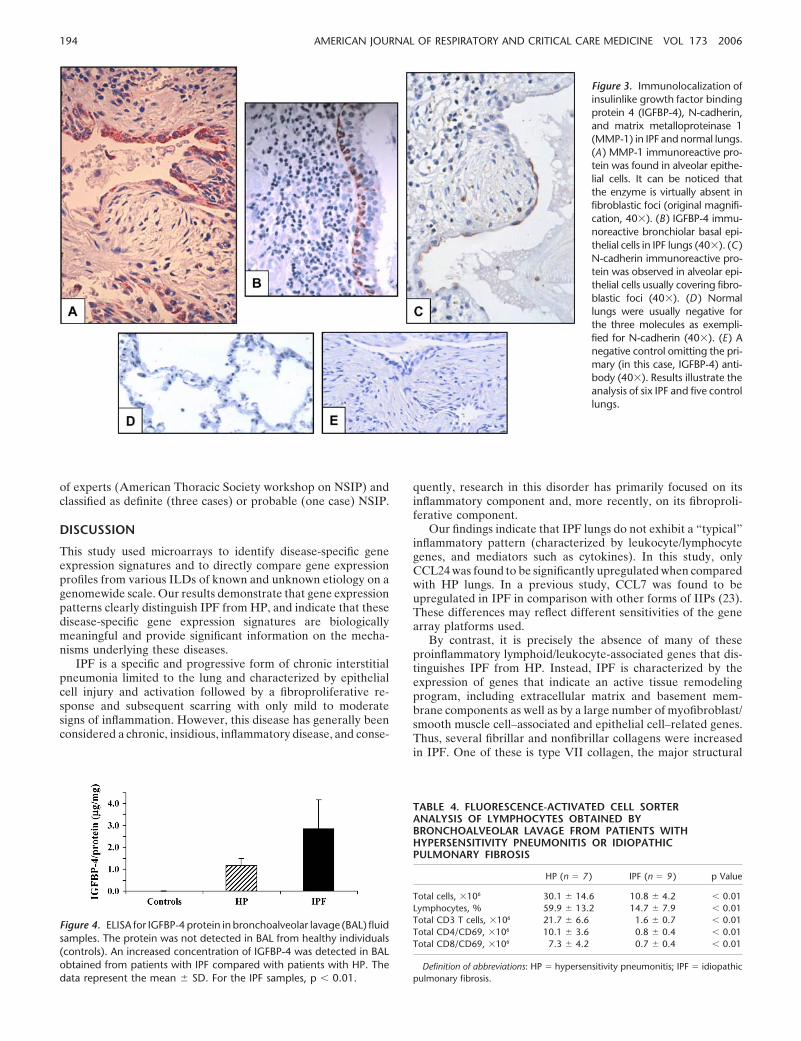
To establish whether the information obtained using gene ex-pression profiling reflects protein expression, we selected threerepresentative genes found elevated in the IPF lungs (Table 2).The cellular sources of N-cadherin, IGFBP-4, and MMP-1 wereexamined by immunohistochemistry on tissue sections of bothIPF and control lungs. As shown in Figure 3, MMP-1 was de-tected in hyperplastic type 2 pneumocytes, whereas IGFBP-4was found expressed by alveolar and bronchiolar (basal) epithe-lial cells. N-cadherin was detected in flattened alveolar epithelialcells covering fibroblast foci. MMP-1 was also revealed in macro-phages in HP lungs (not shown). These findings strongly suggestthat the gene expression levels correlate with protein expressionin the tissues studied and support the notion that epithelial cellsare highly active in IPF.
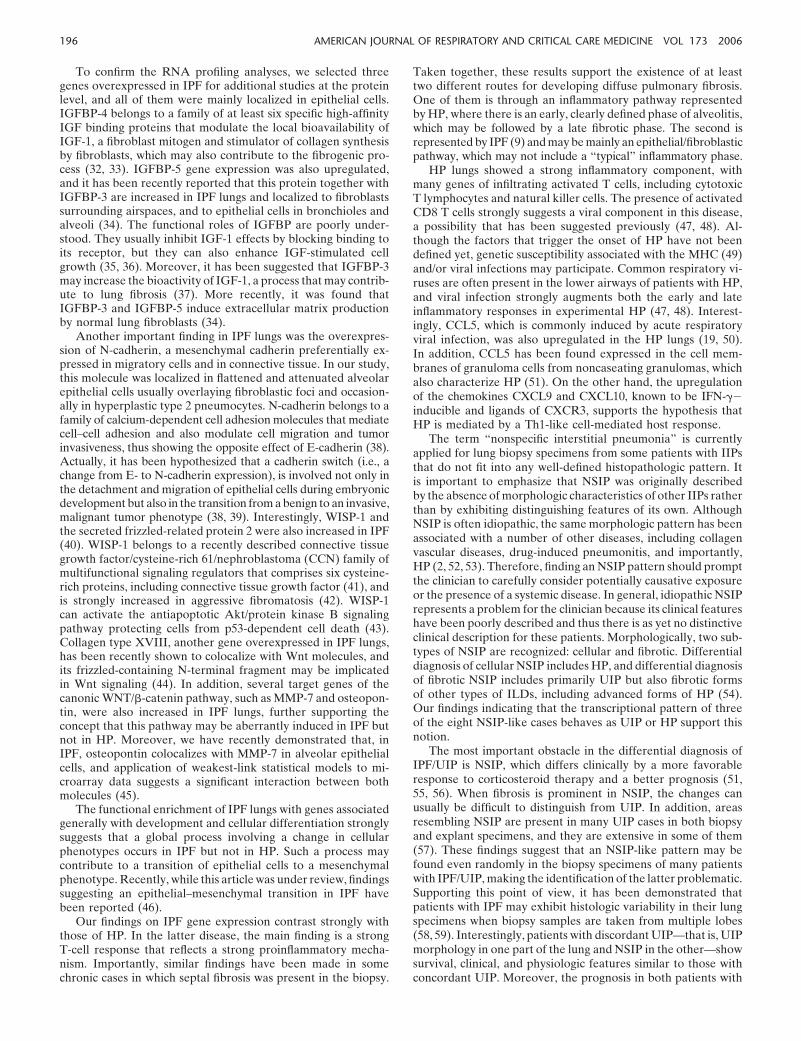
To explore this further, we quantified IGFBP-4 in BAL fluidsof patients with IPF or HP as well as suitable control subjects.As shown in Figure 4, IGFBP-4 protein was undetectable inBAL from control individuals, and was significantly elevated inBAL from patients with IPF compared with patients with HP(2.84 � 1.35 vs. 1.17 � 0.33 �g/mg protein; p � 0.01).
HP Is Characterized by the Upregulation of Genes Related toT-Cell Activation and Immune Responses
In HP lungs, microarray analyses identified a variety of genestypically associated with inflammation, T-cell activation, andimmune responses (Table 3). Genes related to T-lymphocyteactivation included Src-like adaptor 2, CD2, components of theT-cell receptor complex (CD3-D, and CDR-E), and the � chainof CD8. Likewise, major histocompatibility complex (MHC)class II transactivator, the master regulator of MHC class II ex-pression, and several genes encoding MHC class I and II mole-cules were upregulated.
Several chemokines were increased in HP, including CXCL9and CXCL10. These recruit activated T and natural killer cells
through the chemokine receptor CXCR3 and have been associ-ated with T-helper type 1 (Th1) immune responses (21). CXCR4and CCR5 and their ligands CCL5 and CCL4 were also upregu-lated, suggesting that the recruiting/homing program for lunglymphocytes involves multiple chemokines. Another increasedgene was CD84, which is associated with follicular T-helper cells,a third major subset of nonpolarized effector T cells that provideshelp to B cells (22).
Also, several genes involved in host defense were overex-pressed, including granzyme A, which reflects the presence ofactivated CD8 T lymphocytes (see below). Other immune genesincreased in HP included several members of the tumor necrosisfactor superfamily, IL-16 and several cytokine receptors, includ-ing IL-2R, IL-6R, IL-10R, IL-17R, and IL-21R.
FACS Analysis of BAL Cells Confirmed the Presence ofActivated T Cells in HP but Not in IPF
Patients with HP showed a significant increase in total T lympho-cytes as well as in the number of activated CD4� and CD8� Tcells in the BAL when compared with BAL cells from patientswith IPF (Table 4).
HP and IPF Gene Expression Signatures Identify NSIPDiagnostic Subgroups
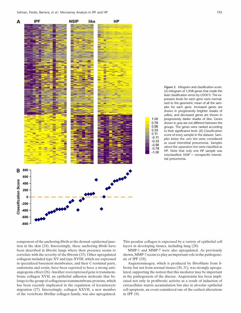
We then analyzed the patients with NSIP in the context of themolecular signatures that distinguish IPF from HP. To avoidoverfitting, we opted to use the larger set of genes (1,058 genes;TNoM p value, 0.002762; Figure 1B) with the minimal error ratefor classification purposes. We used the 12 HP cases and the 15UIP samples as the training set. The NSIP-like samples wereclassified according to them. Figure 5A shows the Infogram ofthese 1,058 genes. The test samples are in the middle, whereasIPF lungs are on the left and HP on the right. Samples wereranked by their classification score. Samples in the test set closestto IPF classify as IPF, whereas samples closest to HP classify asHP. The classification graph reveals that, in the training set, allIPF samples were correctly classified as IPF, whereas one HPsample was classified as IPF. In the test set, one NSIP samplewas classified as HP (Figure 5B). This patient had a history of

Selman, Pardo, Barrera, et al.: Microarray Analysis in IPF and HP 193
TABLE 3. UPREGULATED GENES IN HYPERSENSITIVITY PNEUMONITIS
Gene Name Locus-Link No. TNoM p Value t Test p Value
NM_005516: major histocompatibility complex, class I, E (HLA-E), mRNA 3133 3.1063E-06 8.581E-08NM_002985: chemokine (C-C motif) ligand 5 (CCL5), mRNA 6352 3.1063E-06 1.1854E-07Hs.351874: major histocompatibility complex, class II, DO� 3111 3.1063E-06 2.0498E-07Hs.368409: major histocompatibility complex, class II, DP �1 3115 3.1063E-06 2.6687E-06NM_014387: linker for activation of T cells (LAT), mRNA 27040 3.1063E-06 2.9103E-06NM_000732: CD3D antigen, polypeptide (TiT3 complex; CD3D), mRNA 915 4.0382E-05 1.1558E-06NM_002209: integrin, �L (antigen CD11A [p180], lymphocyte function–associated antigen 1;
� polypeptide; ITGAL), mRNA 3683 4.0382E-05 1.3539E-06NM_002198: interferon regulatory factor 1 (IRF1), mRNA 3659 4.0382E-05 2.2803E-06NM_005601: natural killer cell group 7 sequence (NKG7), mRNA 4818 4.0382E-05 2.5394E-06NM_001767: CD2 antigen (p50), sheep red blood cell receptor (CD2), mRNA 914 4.0382E-05 3.4159E-06NM_001558: interleukin-10 receptor, � (IL10RA), mRNA 3587 4.0382E-05 1.0847E-05NM_006120: major histocompatibility complex, class II, DM� (HLA-DMA), mRNA 3108 4.0382E-05 1.141E-05NM_002118: major histocompatibility complex, class II, DM� (HLA-DMB), mRNA 3109 4.0382E-05 1.3627E-05NM_000246: MHC class II transactivator (MHC2TA), mRNA 4261 4.0382E-05 2.1963E-05NM_001109: a disintegrin and metalloproteinase domain 8 (ADAM8), mRNA 101 4.0382E-05 3.0077E-05NM_000565: interleukin-6 receptor (IL6R), transcript variant 1, mRNA 3570 4.0382E-05 3.5004E-05NM_004355: CD74 antigen (invariant polypeptide of major histocompatibility complex,
class II antigen-associated; CD74) 972 4.0382E-05 4.5692E-05NM_000206: interleukin-2 receptor, (severe combined immunodeficiency; IL2RG), mRNA 3561 4.0382E-05 5.3729E-05NM_001778: CD48 antigen (B-cell membrane protein; CD48), mRNA 962 4.0382E-05 0.00019398NM_002339: lymphocyte-specific protein 1 (LSP1), mRNA 4046 4.0382E-05 0.0004061NM_006144: granzyme A (granzyme 1, cytotoxic T-lymphocyte-associated serine esterase 3; GZMA), mRNA 3001 0.00033652 3.5408E-06NM_003874: CD84 antigen (leukocyte antigen; CD84), mRNA 8832 0.00033652 2.1163E-05Hs.181244: major histocompatibility complex, class I, A 3105 0.00033652 2.3716E-05NM_000265: neutrophil cytosolic factor 1 (47 kD, chronic granulomatous disease, autosomal 1; NCF1), mRNA 4687 0.00033652 2.7125E-05NM_001768: CD8 antigen, � polypeptide (p32) (CD8A), transcript variant 1, mRNA 925 0.00033652 3.6982E-05NM_004355: CD74 antigen (invariant polypeptide of major histocompatibility complex,
class II antigen-associated; CD74) 972 0.00033652 3.7304E-05Hs.387679: major histocompatibility complex, class II, DQ �1 3117 0.00033652 7.4311E-05Hs.387787: killer cell lectinlike receptor subfamily K, member 1 22914 0.00033652 0.00010199NM_003019: surfactant, pulmonary-associated protein D (SFTPD), mRNA 6441 0.00033652 0.00015899NM_016326: chemokine-like factor (CKLF), transcript variant 3, mRNA 51192 0.00033652 0.00027442NM_001242: tumor necrosis factor receptor superfamily, member 7 (TNFRSF7), mRNA 939 0.00033652 0.00038993NM_012292: minor histocompatibility antigen HA-1 (HA-1), mRNA 23526 0.00033652 0.00046328NM_000878: interleukin-2 receptor, � (IL2RB), mRNA 3560 0.00033652 0.00531621NM_003879: CASP8 and FADD-like apoptosis regulator (CFLAR), mRNA 8837 0.00033652 0.00648208NM_002984: chemokine (C-C motif) ligand 4 (CCL4), mRNA 6351 0.00201911 6.3196E-06NM_002416: chemokine (C-X-C motif) ligand 9 (CXCL9), mRNA 4283 0.00201911 5.4339E-05NM_004221: natural killer cell transcript 4 (NK4), mRNA 9235 0.00201911 9.8112E-05NM_021181: SLAM family member 7 (SLAMF7), mRNA 57823 0.00201911 0.00016428NM_000063: complement component 2 (C2), mRNA 717 0.00201911 0.00024703NM_172369: complement component 1, q subcomponent, gamma polypeptide (C1QG), RNA 714 0.00201911 0.00040263NM_003467: chemokine (C-X-C motif) receptor 4 (CXCR4), mRNA 7852 0.00201911 0.00043211NM_001079: �-chain (TCR)–associated protein kinase 70 kD (ZAP70), mRNA 7535 0.00201911 0.00061774NM_020125: SLAM family member 8 (SLAMF8), mRNA 56833 0.00201911 0.00062281NM_001565: chemokine (C-X-C motif) ligand 10 (CXCL10), mRNA 3627 0.00201911 0.00109636NM_004513: interleukin 16 (lymphocyte chemoattractant factor; IL16), transcript variant 1, RNA 3603 0.00201911 0.00188205NM_005781: activated Cdc42-associated kinase 1 (ACK1), mRNA 10188 0.00201911 0.00213368NM_007261: leukocyte membrane antigen (CMRF-35H), mRNA 11314 0.00201911 0.00214496NM_153461: interleukin-17 receptor C (IL17RC), transcript variant 1, mRNA 84818 0.00201911 0.0026955NM_000733: CD3E antigen, ε polypeptide (TiT3 complex; CD3E), mRNA 916 0.00201911 0.00294939NM_021798: interleukin-21 receptor (IL21R), transcript variant 1, mRNA 50615 0.00201911 0.003582NM_001953: endothelial cell growth factor 1 (platelet-derived; ECGF1), mRNA 1890 0.00201911 0.00438273NM_000579: chemokine (C-C motif) receptor 5 (CCR5), mRNA 1234 0.00928793 9.6163E-05NM_019111: major histocompatibility complex, class II, DR � (HLA-DRA), mRNA 3122 0.00928793 0.00036044NM_003820: tumor necrosis factor receptor superfamily, member 14 (herpesvirus entry mediator; TNFRSF14), mRNA 8764 0.00928793 0.00041457Hs.409934: major histocompatibility complex, class II, DQ �1 3119 0.00928793 0.00055793NM_006926: surfactant, pulmonary-associated protein A2 (SFTPA2), mRNA 6436 0.00928793 0.0007712NM_015259: inducible T-cell costimulator ligand (ICOSL), mRNA 23308 0.00928793 0.00281981NM_001774: CD37 antigen (CD37), mRNA 951 0.00928793 0.00288764NM_001784: CD97 antigen (CD97), transcript variant 2, mRNA 976 0.00928793 0.00541578NM_014312: cortical thymocyte receptor (X. laevis CTX) like (CTXL), mRNA 23584 0.00928793 0.00621325NM_003810: tumor necrosis factor (ligand) superfamily, member 10 (TNFSF10), mRNA 8743 0.00928793 0.00811714
For definition of abbreviation, see Table 2.
previous exposure to home birds, a hobby common in Mexico(where the samples were collected), but without cause–effectrelationship, positive precipitins, or apparent exposure to otherorganic particles. Two other NSIP samples were classified as
IPF, supporting the notion that fibrotic NSIP may be undistin-guishable from UIP. The other five NSIP samples did not classifyas either IPF or HP, suggesting that they indeed represent trueidiopathic NSIP. Four of these cases were evaluated by a panel
194 AMERICAN JOURNAL OF RESPIRATORY AND CRITICAL CARE MEDICINE VOL 173 2006
Figure 3. Immunolocalization ofinsulinlike growth factor bindingprotein 4 (IGFBP-4), N-cadherin,and matrix metalloproteinase 1(MMP-1) in IPF and normal lungs.(A ) MMP-1 immunoreactive pro-tein was found in alveolar epithe-lial cells. It can be noticed thatthe enzyme is virtually absent infibroblastic foci (original magnifi-cation, 40�). (B ) IGFBP-4 immu-noreactive bronchiolar basal epi-thelial cells in IPF lungs (40�). (C )N-cadherin immunoreactive pro-tein was observed in alveolar epi-thelial cells usually covering fibro-blastic foci (40�). (D ) Normallungs were usually negative forthe three molecules as exempli-fied for N-cadherin (40�). (E ) Anegative control omitting the pri-mary (in this case, IGFBP-4) anti-body (40�). Results illustrate theanalysis of six IPF and five controllungs.
of experts (American Thoracic Society workshop on NSIP) andclassified as definite (three cases) or probable (one case) NSIP.
DISCUSSION
This study used microarrays to identify disease-specific geneexpression signatures and to directly compare gene expressionprofiles from various ILDs of known and unknown etiology on agenomewide scale. Our results demonstrate that gene expressionpatterns clearly distinguish IPF from HP, and indicate that thesedisease-specific gene expression signatures are biologicallymeaningful and provide significant information on the mecha-nisms underlying these diseases.
IPF is a specific and progressive form of chronic interstitialpneumonia limited to the lung and characterized by epithelialcell injury and activation followed by a fibroproliferative re-sponse and subsequent scarring with only mild to moderatesigns of inflammation. However, this disease has generally beenconsidered a chronic, insidious, inflammatory disease, and conse-
Figure 4. ELISA for IGFBP-4 protein in bronchoalveolar lavage (BAL) fluidsamples. The protein was not detected in BAL from healthy individuals(controls). An increased concentration of IGFBP-4 was detected in BALobtained from patients with IPF compared with patients with HP. Thedata represent the mean � SD. For the IPF samples, p � 0.01.
quently, research in this disorder has primarily focused on itsinflammatory component and, more recently, on its fibroproli-ferative component.
Our findings indicate that IPF lungs do not exhibit a “typical”inflammatory pattern (characterized by leukocyte/lymphocytegenes, and mediators such as cytokines). In this study, onlyCCL24 was found to be significantly upregulated when comparedwith HP lungs. In a previous study, CCL7 was found to beupregulated in IPF in comparison with other forms of IIPs (23).These differences may reflect different sensitivities of the genearray platforms used.
By contrast, it is precisely the absence of many of theseproinflammatory lymphoid/leukocyte-associated genes that dis-tinguishes IPF from HP. Instead, IPF is characterized by theexpression of genes that indicate an active tissue remodelingprogram, including extracellular matrix and basement mem-brane components as well as by a large number of myofibroblast/smooth muscle cell–associated and epithelial cell–related genes.Thus, several fibrillar and nonfibrillar collagens were increasedin IPF. One of these is type VII collagen, the major structural
TABLE 4. FLUORESCENCE-ACTIVATED CELL SORTERANALYSIS OF LYMPHOCYTES OBTAINED BYBRONCHOALVEOLAR LAVAGE FROM PATIENTS WITHHYPERSENSITIVITY PNEUMONITIS OR IDIOPATHICPULMONARY FIBROSIS
HP (n � 7 ) IPF (n � 9 ) p Value
Total cells, �106 30.1 � 14.6 10.8 � 4.2 � 0.01Lymphocytes, % 59.9 � 13.2 14.7 � 7.9 � 0.01Total CD3 T cells, �106 21.7 � 6.6 1.6 � 0.7 � 0.01Total CD4/CD69, �106 10.1 � 3.6 0.8 � 0.4 � 0.01Total CD8/CD69, �106 7.3 � 4.2 0.7 � 0.4 � 0.01
Definition of abbreviations: HP � hypersensitivity pneumonitis; IPF � idiopathicpulmonary fibrosis.
Selman, Pardo, Barrera, et al.: Microarray Analysis in IPF and HP 195
Figure 5. Infogram and classification score.(A) Infogram of 1,058 genes that made theleast classification errors by LOOCV. The ex-pression levels for each gene were normal-ized to the geometric mean of all the sam-ples for each gene. Increased genes areshown in progressively brighter shades ofyellow, and decreased genes are shown inprogressively darker shades of blue. Genesshown in gray are not different between thegroups. The genes were ranked accordingto their significance level. (B ) Classificationscore of every sample in the dataset. Sam-ples below the zero line were consideredas usual interstitial pneumonia. Samplesabove the separation line were classified asHP. Note that only one HP sample wasmisclassified. NSIP � nonspecific intersti-tial pneumonia.
component of the anchoring fibrils at the dermal–epidermal junc-tion in the skin (24). Interestingly, these anchoring fibrils havebeen described in fibrotic lungs where their presence seems tocorrelate with the severity of the fibrosis (25). Other upregulatedcollagens included type XV and type XVIII, which are expressedin specialized basement membranes, and their C-terminal parts,endostatin and restin, have been reported to have a strong anti-angiogenic effect (26). Another overexpressed gene is transmem-brane collagen XVII, an epithelial adhesion molecule that be-longs to the group of collagenous transmembrane proteins, whichhas been recently implicated in the regulation of keratinocytemigration (27). Interestingly, collagen XXVII, a new memberof the vertebrate fibrillar collagen family, was also upregulated.
This peculiar collagen is expressed by a variety of epithelial celllayers in developing tissues, including lung (28).
MMP-1 and MMP-7 were also upregulated. As previouslyshown, MMP-7 seems to play an important role in the pathogene-sis of IPF (29).
Angiotensinogen, which is produced by fibroblasts from fi-brotic but not from normal tissues (30, 31), was strongly upregu-lated, supporting the notion that this mediator may be importantin the pathogenesis of the disease. Angiotensin has been impli-cated not only in profibrotic activity as a result of induction ofextracellular matrix accumulation but also in alveolar epithelialcell apoptosis, an event considered one of the earliest alterationsin IPF (9).
196 AMERICAN JOURNAL OF RESPIRATORY AND CRITICAL CARE MEDICINE VOL 173 2006
To confirm the RNA profiling analyses, we selected threegenes overexpressed in IPF for additional studies at the proteinlevel, and all of them were mainly localized in epithelial cells.IGFBP-4 belongs to a family of at least six specific high-affinityIGF binding proteins that modulate the local bioavailability ofIGF-1, a fibroblast mitogen and stimulator of collagen synthesisby fibroblasts, which may also contribute to the fibrogenic pro-cess (32, 33). IGFBP-5 gene expression was also upregulated,and it has been recently reported that this protein together withIGFBP-3 are increased in IPF lungs and localized to fibroblastssurrounding airspaces, and to epithelial cells in bronchioles andalveoli (34). The functional roles of IGFBP are poorly under-stood. They usually inhibit IGF-1 effects by blocking binding toits receptor, but they can also enhance IGF-stimulated cellgrowth (35, 36). Moreover, it has been suggested that IGFBP-3may increase the bioactivity of IGF-1, a process that may contrib-ute to lung fibrosis (37). More recently, it was found thatIGFBP-3 and IGFBP-5 induce extracellular matrix productionby normal lung fibroblasts (34).
Another important finding in IPF lungs was the overexpres-sion of N-cadherin, a mesenchymal cadherin preferentially ex-pressed in migratory cells and in connective tissue. In our study,this molecule was localized in flattened and attenuated alveolarepithelial cells usually overlaying fibroblastic foci and occasion-ally in hyperplastic type 2 pneumocytes. N-cadherin belongs to afamily of calcium-dependent cell adhesion molecules that mediatecell–cell adhesion and also modulate cell migration and tumorinvasiveness, thus showing the opposite effect of E-cadherin (38).Actually, it has been hypothesized that a cadherin switch (i.e., achange from E- to N-cadherin expression), is involved not only inthe detachment and migration of epithelial cells during embryonicdevelopment but also in the transition from a benign to an invasive,malignant tumor phenotype (38, 39). Interestingly, WISP-1 andthe secreted frizzled-related protein 2 were also increased in IPF(40). WISP-1 belongs to a recently described connective tissuegrowth factor/cysteine-rich 61/nephroblastoma (CCN) family ofmultifunctional signaling regulators that comprises six cysteine-rich proteins, including connective tissue growth factor (41), andis strongly increased in aggressive fibromatosis (42). WISP-1can activate the antiapoptotic Akt/protein kinase B signalingpathway protecting cells from p53-dependent cell death (43).Collagen type XVIII, another gene overexpressed in IPF lungs,has been recently shown to colocalize with Wnt molecules, andits frizzled-containing N-terminal fragment may be implicatedin Wnt signaling (44). In addition, several target genes of thecanonic WNT/�-catenin pathway, such as MMP-7 and osteopon-tin, were also increased in IPF lungs, further supporting theconcept that this pathway may be aberrantly induced in IPF butnot in HP. Moreover, we have recently demonstrated that, inIPF, osteopontin colocalizes with MMP-7 in alveolar epithelialcells, and application of weakest-link statistical models to mi-croarray data suggests a significant interaction between bothmolecules (45).
The functional enrichment of IPF lungs with genes associatedgenerally with development and cellular differentiation stronglysuggests that a global process involving a change in cellularphenotypes occurs in IPF but not in HP. Such a process maycontribute to a transition of epithelial cells to a mesenchymalphenotype. Recently, while this article was under review, findingssuggesting an epithelial–mesenchymal transition in IPF havebeen reported (46).
Our findings on IPF gene expression contrast strongly withthose of HP. In the latter disease, the main finding is a strongT-cell response that reflects a strong proinflammatory mecha-nism. Importantly, similar findings have been made in somechronic cases in which septal fibrosis was present in the biopsy.
Taken together, these results support the existence of at leasttwo different routes for developing diffuse pulmonary fibrosis.One of them is through an inflammatory pathway representedby HP, where there is an early, clearly defined phase of alveolitis,which may be followed by a late fibrotic phase. The second isrepresented by IPF (9) and may be mainly an epithelial/fibroblasticpathway, which may not include a “typical” inflammatory phase.
HP lungs showed a strong inflammatory component, withmany genes of infiltrating activated T cells, including cytotoxicT lymphocytes and natural killer cells. The presence of activatedCD8 T cells strongly suggests a viral component in this disease,a possibility that has been suggested previously (47, 48). Al-though the factors that trigger the onset of HP have not beendefined yet, genetic susceptibility associated with the MHC (49)and/or viral infections may participate. Common respiratory vi-ruses are often present in the lower airways of patients with HP,and viral infection strongly augments both the early and lateinflammatory responses in experimental HP (47, 48). Interest-ingly, CCL5, which is commonly induced by acute respiratoryviral infection, was also upregulated in the HP lungs (19, 50).In addition, CCL5 has been found expressed in the cell mem-branes of granuloma cells from noncaseating granulomas, whichalso characterize HP (51). On the other hand, the upregulationof the chemokines CXCL9 and CXCL10, known to be IFN-�inducible and ligands of CXCR3, supports the hypothesis thatHP is mediated by a Th1-like cell-mediated host response.
The term “nonspecific interstitial pneumonia” is currentlyapplied for lung biopsy specimens from some patients with IIPsthat do not fit into any well-defined histopathologic pattern. Itis important to emphasize that NSIP was originally describedby the absence of morphologic characteristics of other IIPs ratherthan by exhibiting distinguishing features of its own. AlthoughNSIP is often idiopathic, the same morphologic pattern has beenassociated with a number of other diseases, including collagenvascular diseases, drug-induced pneumonitis, and importantly,HP (2, 52, 53). Therefore, finding an NSIP pattern should promptthe clinician to carefully consider potentially causative exposureor the presence of a systemic disease. In general, idiopathic NSIPrepresents a problem for the clinician because its clinical featureshave been poorly described and thus there is as yet no distinctiveclinical description for these patients. Morphologically, two sub-types of NSIP are recognized: cellular and fibrotic. Differentialdiagnosis of cellular NSIP includes HP, and differential diagnosisof fibrotic NSIP includes primarily UIP but also fibrotic formsof other types of ILDs, including advanced forms of HP (54).Our findings indicating that the transcriptional pattern of threeof the eight NSIP-like cases behaves as UIP or HP support thisnotion.
The most important obstacle in the differential diagnosis ofIPF/UIP is NSIP, which differs clinically by a more favorableresponse to corticosteroid therapy and a better prognosis (51,55, 56). When fibrosis is prominent in NSIP, the changes canusually be difficult to distinguish from UIP. In addition, areasresembling NSIP are present in many UIP cases in both biopsyand explant specimens, and they are extensive in some of them(57). These findings suggest that an NSIP-like pattern may befound even randomly in the biopsy specimens of many patientswith IPF/UIP, making the identification of the latter problematic.Supporting this point of view, it has been demonstrated thatpatients with IPF may exhibit histologic variability in their lungspecimens when biopsy samples are taken from multiple lobes(58, 59). Interestingly, patients with discordant UIP—that is, UIPmorphology in one part of the lung and NSIP in the other—showsurvival, clinical, and physiologic features similar to those withconcordant UIP. Moreover, the prognosis in both patients with

Selman, Pardo, Barrera, et al.: Microarray Analysis in IPF and HP 197
concordant and discordant UIP is significantly worse than thatof the patients with concordant NSIP (NSIP in multiple biopsies).
Although our study includes the largest number of humansamples reported in microarray analysis of IPF (or any othernonmalignant lung disease), the sample size still prevents gener-alization of our results. However, given the statistical magnitudeof the differences between IPF and HP, the magnitude andstatistical significance of the difference in functional groups en-riched within each disease gene expression signature, and thebiological plausibility of the results, it is unlikely that largernumbers of samples will change our conclusions significantly.Although, the results of this study should be tested on otherindependent datasets, the identification of statistically signifi-cant, biologically meaningful, disease-specific gene expressionsignatures suggests that we could use these signatures to predictwhether patients would respond to immunosuppressive therapyor to future specific antifibrotic therapies. Currently, it is recom-mended (with little scientific support) that a therapeutic trialwith corticosteroids and some immunosuppressive drugs wouldbe administered to any patient with IPF/UIP and definitely topatients with the other more traditional inflammatory ILDs. Ourdata support the view that gene expression signatures can be abetter predictor of patient responsiveness to antiinflammatorytherapy, thus preventing the side effects of an unindicated thera-peutic trial from other patients. In addition, when specific antifi-brotic therapies may be ready to be tested in clinical trials inthe near future, the best way to select patients may be to usetheir tissue functional gene expression signatures.
In summary, genomewide gene expression profiling of indi-vidual IPF, HP, and NSIP lungs has allowed the identificationof genetic signatures for these diseases. This may speed up theunderstanding of the molecular mechanisms underlying thesedisorders and may also potentially aid in their differential diag-nosis. Advances in such diagnoses could potentially be invaluableto, first, distinguish HP from IPF, and second, to provide a moreaccurate diagnosis in cases currently diagnosed as NSIP.
Conflict of Interest Statement : M.S. does not have a financial relationship with acommercial entity that has an interest in the subject of this manuscript. A.P. doesnot have a financial relationship with a commercial entity that has an interest inthe subject of this manuscript. L.B. does not have a financial relationship with acommercial entity that has an interest in the subject of this manuscript. A.E. doesnot have a financial relationship with a commercial entity that has an interest inthe subject of this manuscript. S.R.W. was an employee of Eos Biotechnologyfrom November 1998–February 2003. She has filed patents on the lung fibrosismicroarray data, and owns shares in Protein Design Labs. K.W. is currently em-ployed by and has stock and stock options in Protein Design Labs, which is theparent company of Eos Biotechnology, an original sponsor of the research. N.A.was an employee of Eos Biotechnology from January 2000–March 2003. She hasfiled patents on the lung fibrosis microarray data, and owns shares in ProteinDesign Labs. N.K. does not have a financial relationship with a commercial entitythat has an interest in the subject of this manuscript. A.Z. was an employee ofEos Biotechnology from September 2000–March 2003. He owns shares in ProteinDesign Labs, a company that acquired Eos Biotechnology. A provisional patentapplication was filed by Eos Biotechnology on some data contained in this article.He is currently employed by Neurocrine Biosciences, which has no financial inter-ests in the contents of this article.
Acknowledgment : The authors thank Jennifer Kane for technical support.
References
1. Schwarz ML, King TE, Raghu G. Approach to the evaluation and diagno-sis of interstitial lung disease. In: King TE, Schwarz ML, editors.Interstitial lung disease. Ontario, BC, Canada: B.C. Decker; 2003.pp. 1–30.
2. American Thoracic Society/European Respiratory Society. AmericanThoracic Society/European Respiratory Society international multidis-ciplinary classification of the idiopathic interstitial pneumonias. Am JRespir Crit Care Med 2002;165:277–304.
3. Katzenstein ALA, Myers JL. Idiopathic pulmonary fibrosis: clinical rele-vance of pathologic classification. Am J Respir Crit Care Med 1998;157:1301–1315.
4. Nicholson AG, Colby TV, du Bois RM, Hansell DM, Wells AU. Theprognostic significance of the histologic pattern of interstitial pneumo-nia in patients presenting with the clinical entity of cryptogenic fibros-ing alveolitis. Am J Respir Crit Care Med 2000;162:2213–2217.
5. Patel AM, Ryu JH, Reed CE. Hypersensitivity pneumonitis: current con-cepts and future questions. J Allergy Clin Immunol 2001;108:661–670.
6. Ramırez-Venegas A, Sansores RH, Perez Padilla R, Carrillo G, SelmanM. Utility of a provocation test for diagnosis of chronic pigeon breedersdisease. Am J Respir Crit Care Med 1998;158:862–869.
7. Perez-Padilla R, Salas J, Chapela R, Sanchez M, Carrillo G, Perez R,Sansores R, Gaxiola M, Selman M. Mortality in Mexican patients withchronic pigeon breeders lung compared to those with usual interstitialpneumonia. Am Rev Respir Dis 1993;148:49–53.
8. Selman M, King TE, Pardo A. Idiopathic pulmonary fibrosis: prevailingand evolving hypothesis about its pathogenesis and implications fortherapy. Ann Intern Med 2001;134:134–136.
9. Pardo A, Selman M. Molecular mechanisms of pulmonary fibrosis. FrontBiosci 2002;7:d1743–d1761.
10. Pardo A, Barrios R, Gaxiola M, Segura-Valdez L, Carrillo G, EstradaA, Mejia M, Selman M. Increase of lung neutrophils in hypersensitivitypneumonitis is associated with lung fibrosis. Am J Respir Crit CareMed 2000;161:1698–1704.
11. American Thoracic Society. Idiopathic pulmonary fibrosis: diagnosis andtreatment. International consensus statement. American ThoracicSociety (ATS) and the European Respiratory Society (ERS). Am JRespir Crit Care Med 2000;161:646–664.
12. Henshall SM, Afar DE, Hiller J, Horvath LG, Quinn DI, Rasiah KK,Gish K, Willhite D, Kench JG, Gardiner-Garden M, et al. Survivalanalysis of genome-wide gene expression profiles of prostate cancersidentifies new prognostic targets of disease relapse. Cancer Res 2003;63:4196–4203.
13. Kaminski N, Friedman N. Practical approaches to analyzing results ofmicroarray experiments. Am J Respir Cell Mol Biol 2002;27:125–132.
14. Achiron A, Gurevich M, Friedman N, Kaminski N, Mandel M. Bloodtranscriptional signatures of multiple sclerosis: unique gene expressionof disease activity. Ann Neurol 2004;55:410–417.
15. Ben-Dor A, Bruhn L, Friedman N, Nachman I, Schummer M, YakhiniZ. Tissue classification with gene expression profiles. J Comput Biol2000;7:559–583.
16. Benjamini Y, Hochberg Y. Controlling the false discovery rate: a practicaland powerful approach to multiple testing. J R Stat Soc B 1995;57:289–300.
17. Hosack DA, Dennis G Jr, Sherman BT, Lane HC, Lempicki RA. Identi-fying biological themes within lists of genes with EASE. Genome Biol2003;4:R70.
18. Dennis G Jr, Sherman BT, Hosack DA, Yang J, Gao W, Lane HC,Lempicki RA. DAVID: Database for Annotation, Visualization, andIntegrated Discovery. Genome Biol 2003;4:3.
19. Selman M. Hypersensitivity pneumonitis: a multifaceted deceiving disor-der. Clin Chest Med 2004;25:531–547.
20. Smits P, Poumay Y, Karperien M, Tylzanowski P, Wauters J, Huyle-broeck D, Ponec M, Merregaert J. Differentiation-dependent alterna-tive splicing and expression of the extracellular matrix protein 1 genein human keratinocytes. J Invest Dermatol 2000;114:718–724.
21. Luther SA, Cyster JG. Chemokines as regulators of T cell differentiation.Nat Immunol 2001;2:102–107.
22. Chtanova T, Tangye SG, Newton R, Frank N, Hodge MR, Rolph MS,Mackay CR. T follicular helper cells express a distinctive transcrip-tional profile, reflecting their role as non-Th1/Th2 effector cells thatprovide help for B cells. J Immunol 2004;173:68–78.
23. Choi ES, Jakubzick C, Carpenter KJ, Kunkel SL, Evanoff H, MartinezFJ, Flaherty KR, Toews GB, Colby TV, Kazerooni EA, et al. Enhancedmonocyte chemoattractant protein-3/CC chemokine ligand-7 in usualinterstitial pneumonia. Am J Respir Crit Care Med 2004;170:508–515.
24. Rattenholl A, Pappano WN, Koch M, Keene DR, Kadler KE, SasakiT, Timpl R, Burgeson RE, Greenspan DS, Bruckner-Tuderman L.Proteinases of the bone morphogenetic protein-1 family convert pro-collagen VII to mature anchoring fibril collagen. J Biol Chem 2002;277:26372–26378.
25. Kawanami O, Ferrans VJ, Roberts WC, Crystal RG, Fulmer JD. Anchor-ing fibrils: a new connective tissue structure in fibrotic lung disease.Am J Pathol 1987;92:389–410.
26. Tomono Y, Natio I, Ando K, Yonezawa T, Sado Y, Hirakawa S, Arata J,Okigaki T, Ninomiya Y. Epitope-defined,onoclonal antibodies againstmultiplexin collagens demonstrate that type XV and XVIII collagensare expressed in specialized basement membranes. Cell Struct Funct2002;27:9–20.

198 AMERICAN JOURNAL OF RESPIRATORY AND CRITICAL CARE MEDICINE VOL 173 2006
27. Tasanen K, Tunggal L, Chometon G, Bruckner-Tuderman L, AumailleyM. Keratinocytes from patients lacking collagen XVII display a migra-tory phenotype. Am J Pathol 2004;164:2027–2038.
28. Boot-Handford RP, Tuckwell DS, Plumb DA, Rock CF, Poulsom R. Anovel and highly conserved collagen (pro(alpha)1(XXVII)) with aunique expression pattern and unusual molecular characteristics estab-lishes a new clade within the vertebrate fibrillar collagen family. J BiolChem 2003;278:31067–31077.
29. Zuo F, Kaminski N, Eugui E, Allard J, Yakhini Z, Ben-Dor A, LolliniL, Morris D, Kim Y, DeLustro B, et al. Gene expression analysisreveals matrilysin as a key regulator of pulmonary fibrosis in mice andhumans. Proc Natl Acad Sci USA 2002;99:6292–6297.
30. Wang R, Ramos C, Joshi I, Zagariya A, Pardo A, Selman M, UhalBD. Human lung myofibroblast-derived inducers of alveolar epithelialapoptosis identified as angiotensin peptides. Am J Physiol 1999;277:L1158–L1164.
31. Kawaguchi Y, Takagi K, Hara M, Fukasawa C, Sugiura T, Nishimagi E,Harigai M, Kamatani N. Angiotensin II in the lesional skin of systemicsclerosis patients contributes to tissue fibrosis via angiotensin II type1 receptors. Arthritis Rheum 2004;50:216–226.
32. Bonner JC, Brody AR. Cytokine-binding proteins in the lung. Am JPhysiol 1995;268:L869–L878.
33. Goldstein RH, Poliks CF. Stimulation of collagen formation by insulinand insulin-like growth factor I in cultures by human lung fibroblasts.Endocrinology 1989;124:964–970.
34. Pilewski JM, Liu L, Henry AC, Knauer AV, Feghali-Bostwick CA. Insu-lin-like growth factor binding proteins 3 and 5 are overexpressed inidiopathic pulmonary fibrosis and contribute to extracellular matrixdeposition. Am J Pathol 2005;166:399–407.
35. Jones JI, Clemmons DR. Insulin-like growth factors and their bindingproteins: biological actions. Endocr Rev 1995;16:3–34.
36. Hamon GA, Hunt TK, Spencer EM. In vivo effects of systemic insuline-like growth factor-I alone and complexed with insulin-like growthfactor binding protein-3 on corticosteroid suppressed wounds. GrowthRegul 1993;3:53–56.
37. Aston C, Jagirdar J, Lee TC, Hur T, Hintz RL, Rom WN. Enhancedinsulin-like growth factor molecules in idiopathic pulmonary fibrosis.Am J Respir Crit Care Med 1995;151:1597–1603.
38. Christofori G. Changing neighbours, changing behaviour: cell adhesionmolecule-mediated signalling during tumour progression. EMBO J2003;22:2318–2323.
39. Hazan RB, Phillips GR, Qiao RF, Norton L, Aaronson SA. Exogenousexpression of N-cadherin in breast cancer cells induces cell migration,invasion, and metastasis. J Cell Biol 2000;148:779–790.
40. Hsieh JC. Specificity of WNT-receptor interactions. Front Biosci2004;9:1333–1338.
41. Perbal B. CCN proteins: multifunctional signalling regulators. Lancet2004;3:363:62–64.
42. Skubitz KM, Skubitz AP. Gene expression in aggressive fibromatosis.J Lab Clin Med 2004;143:89–98.
43. Su F, Overholtzer M, Besser D, Levine AJ. WISP-1 attenuates p53-mediated apoptosis in response to DNA damage through activationof the Akt kinase. Genes Dev 2002;16:46–57.
44. Elamaa H, Snellman A, Rehn M, Autio-Harmainen H, Pihlajaniemi T.Characterization of the human type XVIII collagen gene and proteo-lytic processing and tissue location of the variant containing a frizzledmotif. Matrix Biol 2003;22:427–442.
45. Pardo A, Gibson K, Cisneros J, Richards TJ, Yang Y, Becerril C, YousemS, Herrera I, Ruiz V, Selman M, et al. Up-regulation and profibroticrole of osteopontin in human idiopathic pulmonary fibrosis. PLoSMed 2005;2:e251.
46. Willis BC, Liebler JM, Luby-Phelps K, Nicholson AG, Crandall ED, duBois RM, Borok Z. Induction of epithelial-mesenchymal transition inalveolar epithelial cells by transforming growth factor-beta1: potentialrole in idiopathic pulmonary fibrosis. Am J Pathol 2005;166:1321–1332.
47. Dakhama A, Hegele RG, Laflamme G, Israel-Assayag E, Cormier Y.Common respiratory viruses in lower airways of patients with acutehypersensitivity pneumonitis. Am J Respir Crit Care Med 1999;159:1316–1322.
48. Gudmundsson G, Monick MM, Hunninghake GW. Viral infection modu-lates expression of hypersensitivity pneumonitis. J Immunol 1999;162:7397–7401.
49. Camarena A, Juarez A, Mejia M, Estrada A, Carrillo G, Falfan R,Zuniga J, Navarro C, Granados J, Selman M. Major histocompatibilitycomplex and TNF-� gene polymorphisms in pigeon breeder’s disease.Am J Respir Crit Care Med 2001;163:1528–1533.
50. Glass WG, Rosemberg HF, Murphy PM. Chemokine regulation of in-flammation during acute viral infection. Curr Opin Allergy Clin Immu-nol 2003;3:467–473.
51. Oki M, Ohtani H, Kinouchi Y, Sato E, Nakamura S, Matsumoto T,Nagura H, Yoshie O, Shimosegawa T. Accumulation of CCR5� Tcells around RANTES� granulomas in Crohn’s disease: a pivotal siteof Th1-shifted immune response? Lab Invest 2005;85:137–145.
52. Travis WD, Matsui K, Moss J, Ferrans VJ. Idiopathic nonspecific intersti-tial pneumonia: prognostic significance of cellular and fibrosingpatterns: survival comparison with usual interstitial pneumonia anddesquamative interstitial pneumonia. Am J Surg Pathol 2000;24:19–33.
53. Arakawa H, Yamada H, Kurihara Y, Nakajima Y, Takeda A, FukushimaY, Fujioka M. Nonspecific interstitial pneumonia associated with poly-myositis and dermatomyositis: serial high-resolution CT findings andfunctional correlation. Chest 2003;123:1096–1103.
54. Katzenstein ALA, Fiorelli RF. Nonspecific interstitial pneumonia/fibrosis: histologic features and clinical significance. Am J Surg Pathol1994;18:136–147.
55. Bjoraker JA, Ryu JH, Edwin MK, Myers JL, Tazelaar HD, SchroederDR, Offord KP. Prognostic significance of histopathologic subsets inidiopathic pulmonary fibrosis. Am J Respir Crit Care Med 1998;157:199–203.
56. Daniil ZD, Gilchrist FC, Nicholson AG, Hansell DM, Harris J, ColbyTV, du Bois RM. A histologic pattern of nonspecific interstitial pneu-monia is associated with a better prognosis than usual interstitial pneu-monia in patients with cryptogenic fibrosing alveolitis. Am J RespirCrit Care Med 1999;160:899–905.
57. Katzenstein AL, Zisman DA, Litzky LA, Nguyen BT, Kotloff RM. Usualinterstitial pneumonia: histologic study of biopsy and explant speci-mens. Am J Surg Pathol 2002;26:1567–1577.
58. Flaherty KR, Travis WD, Colby TV, Toews GB, Kazerooni EA, GrossBH, Jain A, Strawderman RL, Flint A, Lynch JP, et al. Histopathologicvariability in usual and nonspecific interstitial pneumonias. Am JRespir Crit Care Med 2001;164:1722–1727.
59. Monaghan H, Wells AU, Colby TV, du Bois RM, Hansell DM, NicholsonAG. Prognostic implications of histologic patterns in multiple surgicallung biopsies from patients with idiopathic interstitial pneumonias.Chest 2004;125:522–526.
Related Documents