Functional Diversity of T-Cell Subpopulations in Subacute and Chronic Hypersensitivity Pneumonitis Lourdes Barrera 1 *, Felipe Mendoza 1 , Joaquı ´n Zun ˜iga 1 , Andrea Estrada 1 , Ana C. Zamora 2 , Emma I. Melendro 3 , Remedios Ramı ´rez 4 , Annie Pardo 4 , and Moise ´s Selman 1 1 Instituto Nacional de Enfermedades Respiratorias, Mexico City, Mexico; 2 Hospital Universitario ‘‘Dr. Jose ´ E. Gonza ´lez,’’ Monterrey, Nuevo Leo ´n, Me ´xico; 3 Facultad de Medicina, Universidad Nacional Auto ´noma de Me ´xico, Mexico City, Mexico; and 4 Facultad de Ciencias, Universidad Nacional Auto ´noma de Me ´xico, Mexico City, Mexico Rationale: Hypersensitivity pneumonitis (HP) exhibits a diverse out- come. Patients with acute/subacute HP usually improve, whereas patients with chronic disease often progress to fibrosis. However, the mechanisms underlying this difference are unknown. Objectives: To examine the T-cell profile from patients with subacute HP and chronic HP. Methods: T cells were obtained by bronchoalveolar lavage from 25 patients with subacute HP, 30 patients with chronic HP, and 8 control subjects. T-cell phenotype and functional profile were evaluated by flow cytometry, cytometric bead array, and immunohistochemistry. Measurements and Main Results: Patients with chronic HP showed higher CD4 1 :CD8 1 ratio (median, 3.05; range, 0.3–15; subacute HP: median, 1.3; range, 0.1–10; control: median, 1.3; range, 0.7–2.0; P , 0.01), and a decrease of gdT cells (median, 2.0; range, 0.5–3.4; subacute HP: median, 10; range, 4.8–17; control: median, 15; range, 5–19; P , 0.01). Patients with chronic HP exhibited an increase in the terminally differentiated memory CD4 1 and CD8 1 T-cell subsets compared with patients with subacute HP (P , 0.05). However, memory cells from chronic HP showed lower IFN-g production and decreased cytotoxic activity by CD8 1 T lymphocytes. Chronic HP displayed a Th2-like phenotype with increased CXCR4 expression (median, 6%; range, 1.7–36, vs. control subjects: median, 0.7%; range, 0.2–1.4; and subacute HP: median, 2.2%; range, 0.1–5.3; P , 0.01), and decreased CXCR3 expression (median, 4.3%; range, 1.4– 25%, vs. subacute HP: median, 37%; range, 4.9–78%; P , 0.01). Likewise, supernatants from antigen-specific–stimulated cells from chronic HP produced higher levels of IL-4 (80 6 63 pg/ml vs. 25 6 7 pg/ml; P , 0.01), and lower levels of IFN-g (3,818 6 1671 pg/ml vs. 100 6 61 pg/ml; P , 0.01) compared with subacute HP. Conclusions: Our findings indicate that patients with chronic HP lose effector T-cell function and exhibit skewing toward Th2 activity, which may be implicated in the fibrotic response that characterizes this clinical form. Keywords: allergic alveolitis; cytotoxic; hypersensitivity pneumonitis; T cells; Th1/Th2 cells Hypersensitivity pneumonitis (HP) is a complex syndrome of varying intensity and clinical presentation that results from an immunologically induced lung inflammation in response to a wide variety of inhaled antigens (1). The clinical presentation is heterogeneous and, thus, HP may present as an acute, subacute, or chronic form, depending on, among several factors, the intensity of inhaled antigen and repetitiveness of exposure (1, 2). Importantly, patients with acute or subacute HP usually respond to treatment, whereas patients with chronic disease often progress to an irreversible destruction of the lung, either by fibrosis or emphysematous changes (1, 3, 4). HP is characterized by a remarkable T-cell alveolitis. How- ever, characterization of lymphocyte phenotypes, primarily CD4 1 /CD8 1 T-cell subsets, has given inconsistent results. Some authors have reported an increase in CD8 1 T cells with a decrease in the CD4 1 :CD8 1 ratio, whereas others have shown that both subpopulations accumulate without changes in that ratio, and still others have reported a clear predominance of CD4 1 T cells (5–9). Several reasons may account for this variability, including the type of inhaled antigen and the clinical presentation (1, 9). Studies of other T-cell phenotypes in human HP (i.e., Th1 vs. Th2), and of memory/effector T cells, are scanty, whereas other putative regulatory or effector cells, like natural killer (NK) T (NKT) cells, among others, have not been studied (10). Likewise, immunological memory in the context of recent descriptions based on different homing and effector capabilities of T-cell sub- populations (11–14) has not been examined in this disease. Importantly, possible differences between subacute and chronic disease have not been studied, and we hypothesized that the exhaustion/skewing of T cells in chronic HP is the principal reason for the inability of the host to eliminate the persisting antigen. Supporting this notion, a specific mechanism of T-cell exhaustion in chronic lymphocyte choriomeningitis virus (LCMV), which ends in a limited proliferative potential and inability to produce cytokines, was recently described (15). It is well known that naive T helper cells can differentiate to at least two different memory cells during the immune response: Th1 cells, which secrete IFN-g and tumor necrosis factor-a; and Th2 cells, which secrete IL-4 and IL-13 (16). Interestingly, a growing body of evidence suggests that polarized T cells (i.e., Th1 AT A GLANCE COMMENTARY Scientific Knowledge on the Subject The mechanisms by which some patients with hypersensi- tivity pneumonitis progress to fibrosis are yet unclear. What This Study Adds to the Field This study demonstrates that patients with chronic hyper- sensitivity have different phenotypic/functional lung T-cell subsets compared with patients with subacute disease, likely associated with the fibrotic response. (Received in original form January 17, 2007; accepted in final form October 9, 2007) Supported by grant IN215003 from La Direccio ´ n General de Asuntos del Personal Acade ´mico, Universidad Nacional Auto ´ noma de Me ´xico (UNAM), and by UNAM grant SDI.PTID.05.6. *This work was submitted in partial Fulfillment of the requirements to obtain the Ph.D. degree for L.B. at Biological Sciences, UNAM. Correspondence and requests for reprints should be addressed to Moise ´s Selman, M.D., Instituto Nacional de Enfermedades Respiratorias, Tlalpan 4502, CP 14080, Mexico DF, Mexico. E-mail: [email protected] This article has an online supplement, which is accessible from this issue’s table of contents at www.atsjournals.org Am J Respir Crit Care Med Vol 177. pp 44–55, 2008 Originally Published in Press as DOI: 10.1164/rccm.200701-093OC on October 18, 2007 Internet address: www.atsjournals.org

Welcome message from author
This document is posted to help you gain knowledge. Please leave a comment to let me know what you think about it! Share it to your friends and learn new things together.
Transcript

Functional Diversity of T-Cell Subpopulations inSubacute and Chronic Hypersensitivity Pneumonitis
Lourdes Barrera1*, Felipe Mendoza1, Joaquın Zuniga1, Andrea Estrada1, Ana C. Zamora2, Emma I. Melendro3,Remedios Ramırez4, Annie Pardo4, and Moises Selman1
1Instituto Nacional de Enfermedades Respiratorias, Mexico City, Mexico; 2Hospital Universitario ‘‘Dr. Jose E. Gonzalez,’’ Monterrey,
Nuevo Leon, Mexico; 3Facultad de Medicina, Universidad Nacional Autonoma de Mexico, Mexico City, Mexico; and 4Facultad de Ciencias,
Universidad Nacional Autonoma de Mexico, Mexico City, Mexico
Rationale: Hypersensitivity pneumonitis (HP) exhibits a diverse out-come. Patients with acute/subacute HP usually improve, whereaspatients with chronic disease often progress to fibrosis. However,the mechanisms underlying this difference are unknown.Objectives: To examine the T-cell profile from patients with subacuteHP and chronic HP.Methods: T cells were obtained by bronchoalveolar lavage from 25patients with subacuteHP, 30 patients withchronic HP, and 8 controlsubjects. T-cell phenotype and functional profile were evaluated byflow cytometry, cytometric bead array, and immunohistochemistry.Measurements and Main Results: Patients with chronic HP showedhigher CD41:CD81 ratio (median, 3.05; range, 0.3–15; subacute HP:median, 1.3; range, 0.1–10; control: median, 1.3; range, 0.7–2.0; P ,
0.01), and a decrease of gdT cells (median, 2.0; range, 0.5–3.4;subacute HP: median, 10; range, 4.8–17; control: median, 15; range,5–19; P , 0.01). Patients with chronic HP exhibited an increase in theterminally differentiated memory CD41 and CD81 T-cell subsetscompared with patients with subacute HP (P , 0.05). However,memory cells from chronic HP showed lower IFN-g production anddecreased cytotoxic activity by CD81 T lymphocytes. Chronic HPdisplayed a Th2-like phenotype with increased CXCR4 expression(median, 6%; range, 1.7–36, vs. control subjects: median, 0.7%;range, 0.2–1.4; and subacute HP: median, 2.2%; range, 0.1–5.3; P ,
0.01), and decreased CXCR3 expression (median, 4.3%; range, 1.4–25%, vs. subacute HP: median, 37%; range, 4.9–78%; P , 0.01).Likewise, supernatants from antigen-specific–stimulated cells fromchronic HP produced higher levels of IL-4 (80 6 63 pg/ml vs. 25 6 7pg/ml; P , 0.01), and lower levels of IFN-g (3,818 6 1671 pg/ml vs.100 6 61 pg/ml; P , 0.01) compared with subacute HP.Conclusions: Our findings indicate that patients with chronic HP loseeffector T-cell function and exhibit skewing toward Th2 activity,which may be implicated in the fibrotic response that characterizesthis clinical form.
Keywords: allergic alveolitis; cytotoxic; hypersensitivity pneumonitis;
T cells; Th1/Th2 cells
Hypersensitivity pneumonitis (HP) is a complex syndrome ofvarying intensity and clinical presentation that results from animmunologically induced lung inflammation in response to a
wide variety of inhaled antigens (1). The clinical presentation isheterogeneous and, thus, HP may present as an acute, subacute,or chronic form, depending on, among several factors, theintensity of inhaled antigen and repetitiveness of exposure (1,2). Importantly, patients with acute or subacute HP usuallyrespond to treatment, whereas patients with chronic diseaseoften progress to an irreversible destruction of the lung, eitherby fibrosis or emphysematous changes (1, 3, 4).
HP is characterized by a remarkable T-cell alveolitis. How-ever, characterization of lymphocyte phenotypes, primarilyCD41/CD81 T-cell subsets, has given inconsistent results. Someauthors have reported an increase in CD81 T cells with adecrease in the CD41:CD81 ratio, whereas others have shownthat both subpopulations accumulate without changes in thatratio, and still others have reported a clear predominance ofCD41 T cells (5–9). Several reasons may account for thisvariability, including the type of inhaled antigen and the clinicalpresentation (1, 9).
Studies of other T-cell phenotypes in human HP (i.e., Th1 vs.Th2), and of memory/effector T cells, are scanty, whereas otherputative regulatory or effector cells, like natural killer (NK) T(NKT) cells, among others, have not been studied (10). Likewise,immunological memory in the context of recent descriptionsbased on different homing and effector capabilities of T-cell sub-populations (11–14) has not been examined in this disease.
Importantly, possible differences between subacute andchronic disease have not been studied, and we hypothesizedthat the exhaustion/skewing of T cells in chronic HP is theprincipal reason for the inability of the host to eliminate thepersisting antigen. Supporting this notion, a specific mechanismof T-cell exhaustion in chronic lymphocyte choriomeningitisvirus (LCMV), which ends in a limited proliferative potentialand inability to produce cytokines, was recently described (15).
It is well known that naive T helper cells can differentiate to atleast two different memory cells during the immune response:Th1 cells, which secrete IFN-g and tumor necrosis factor-a; andTh2 cells, which secrete IL-4 and IL-13 (16). Interestingly, agrowing body of evidence suggests that polarized T cells (i.e., Th1
AT A GLANCE COMMENTARY
Scientific Knowledge on the Subject
The mechanisms by which some patients with hypersensi-tivity pneumonitis progress to fibrosis are yet unclear.
What This Study Adds to the Field
This study demonstrates that patients with chronic hyper-sensitivity have different phenotypic/functional lung T-cellsubsets compared with patients with subacute disease,likely associated with the fibrotic response.
(Received in original form January 17, 2007; accepted in final form October 9, 2007)
Supported by grant IN215003 from La Direccion General de Asuntos del Personal
Academico, Universidad Nacional Autonoma de Mexico (UNAM), and by UNAM
grant SDI.PTID.05.6.
*This work was submitted in partial Fulfillment of the requirements to obtain the
Ph.D. degree for L.B. at Biological Sciences, UNAM.
Correspondence and requests for reprints should be addressed to Moises Selman,
M.D., Instituto Nacional de Enfermedades Respiratorias, Tlalpan 4502, CP 14080,
Mexico DF, Mexico. E-mail: [email protected]
This article has an online supplement, which is accessible from this issue’s table of
contents at www.atsjournals.org
Am J Respir Crit Care Med Vol 177. pp 44–55, 2008
Originally Published in Press as DOI: 10.1164/rccm.200701-093OC on October 18, 2007
Internet address: www.atsjournals.org

or Th2) may regulate the fibrotic response, which may accountfor patients with chronic HP progressing to fibrosis (17).
In order to better characterize the T-cell subpopulationspresent in subacute and chronic HP lungs, we analyzed thevariety and functional activity of distinct T-lymphocyte sub-populations from cells obtained by bronchoalveolar lavage(BAL). Our results show that patients with chronic diseasehave a higher CD41:CD81 ratio with a Th2-like response,decreased numbers of gd T cells, and a functional impairmentof the local CD81 memory T-cell subset.
METHODS
Additional details are provided in online supplement. A total of 55patients with HP were included in this study. Patients were arbitrarilyclassified as having subacute HP (25 patients; 42.5 6 11.4 yr [mean 6
SD]) and chronic HP (30 patients; 50.3 6 8.1 yr). None of the patientshad been treated with corticosteroids or immunosuppressive drug atthe time of the study. Eight healthy subjects (37.6 6 6.5 yr) werestudied as control subjects. The protocol was approved by the ethicscommittee of the Instituto Nacional de Enfermedades Respiratorias.Diagnosis of HP was obtained as previously described (18, 19). Sub-acute HP was defined as: (1) less than 6 months of symptoms beforediagnosis; (2) high-resolution computed tomography showing poorlydefined nodules and ground glass attenuation; (3) biopsy (15 patients)showing an inflammatory infiltrate without fibrosis. Chronic HP wasdefined as: (1) more than 24 months of symptoms before diagnosis; (2)high-resolution computed tomography displaying in addition to nod-ules and ground glass, irregular linear opacities, lobar volume loss, andoccasionally cystic lesions; (3) biopsy (22 patients) showing more than20% extent of fibrotic infiltrate. A pathologist and a radiologist,blinded to the clinical data, scored the lesions.
BAL
BAL was performed as previously described (18). Cell aliquots wereresuspended in 10% dimethyl sulfoxide and 90% fetal bovine serumand kept in liquid nitrogen until use. Cells were thawed as previouslydescribed (20), and only cryopreserved samples showing 95% orgreater viability were used.
Determination of T-Lymphocyte Surface Phenotypes
BAL cells were defrosted, washed, resuspended in 50 ml of stainingbuffer, and incubated with monoclonal antibodies to determine CD41,CD81, memory T cells, gd T cells, NKT, T helper, and T cytotoxicsubpopulations (BD Pharmingen, San Diego, CA). iNKT were exam-ined with anti-Va24-JaQ TCR chain (BD Pharmingen). Cells werefixed in 1% paraformaldehyde for cytometry analysis (21).
Intracellular Cytokine Staining
BAL cells (2 3 105/ml) were stimulated for 6 hours with either: (1)phorbol myristate acetate (PMA; 10 ng/ml) and ionomycin (1 mM) toevaluate surface CD107a/b and intracellular perforin; or (2) pigeonserum (100 mg/ml) to determine IFN-g, IL-4, and IL-10.
Determination of Cytokines/Chemokines
IL-2, IL-10, IL-4, and IFN-g were measured by cytometric bead array(22). IFN-g–inducible protein 10 (IP-10/CXCL10), monokine inducedby IFN-g (MIG/CXCL9), thymus- and activation-regulated chemokine(TARC/CCL17), regulated upon activation, normal T-cell expressedand secreted (RANTES)/CCL5, and transforming growth factor-bwere measured by ELISA using commercial kits (ImmunoassayQuantikine; R&D Systems, Minneapolis, MN).
Functional Cytotoxic Assay for CD107a/b Expression
Peripheral blood mononuclear cells (PBMCs) and BAL cells from fivepatients with subacute and five patients with chronic HP patients werestimulated with PMA and ionomycin. Conjugated antibody to CD107a/bwas added to the culture before stimulation. The cultures were
incubated for 5 hours in the presence of monesin and brefeldin-A(BD Biosciences) (23).
Flow Cytometry
Samples from all assays were analyzed in a FACS Aria flow cytometer(Becton Dickinson, San Jose, CA) by using FACS Diva software.Typically, 10,000 events from double-positive CD31CD41 or CD31CD81
gates for surface staining cells were acquired. For single-cell cytokineproduction, 40,000 events were acquired from CD31CD41 andCD31CD81 gates.
Immunohistochemistry
Tissue sections from patients with idiopathic pulmonary fibrosis andcontrol subjects were treated as described previously (18, 19). Humananti–IP-10 and MIG polyclonal antibodies (Santa Cruz Biotechnology,Santa Cruz, CA) were applied and incubated at 48C overnight. A sec-ondary biotinylated anti-immunoglobulin followed by horseradishperoxidase–conjugated streptavidin (BioGenex, San Ramon, CA) wasused according to manufacturer’s instructions. 3-Amino-9-ethyl-carbazole(BioGenex) in acetate buffer containing 0.05% H2O2 was used assubstrate. The sections were counterstained with hematoxylin. Theprimary antibody was replaced by non–immune serum for negativecontrol slides.
Statistical Analysis
Results were expressed as mean (6SD), and differences among groupswere analyzed through analysis of variance followed by Tukey’s test.Data lacking normal distribution were reported as median and range,and differences were evaluated through the nonparametric Kruskal-Wallis test, followed by Dunn’s multiple comparison test. Statisticalsignificance was set at P less than 0.05 bimarginally.
RESULTS
Baseline Characteristics of Both HP Subgroups
Demographic data, pulmonary function tests, BAL differentialcell counts, and outcome are summarized in Table 1. All pa-tients exhibited clinical, radiological, and functional evidence ofinterstitial lung disease, with variable degrees of dyspnea, de-creased lung capacities, and hypoxemia at rest that worsenedwith exercise. Patients with chronic HP were older, presentedmore severe hypoxemia and more frequently with digital club-
TABLE 1. DEMOGRAPHIC, CLINICAL, PHYSIOLOGIC, ANDBRONCHOALVEOLAR LAVAGE CHARACTERISTICS OF SUBACUTEAND CHRONIC HYPERSENSITIVITY PNEUMONITIS
Subacute HP Chronic HP
Characteristic (n 5 25) (n 5 30) P Value
Age, yr 42.1 6 10.9 50.3 6 8.1 ,0.01
Gender, female/male 22/3 28/2
Time of symptoms before diagnosis, mo 3.8 6 1.6 46.4 6 16.6 ,0.01
Smoking 3/25 5/30
Clubbing 3/25 14/30 ,0.01
FVC % predicted 61.9 6 18.7 59.6 6 17.9
FEV1 % predicted 64.5 6 17.4 61.8 6 16.0
FEV1/FVC % 92.2 6 6.9 86.6 6 15.6
PaO253.2 6 8.2 48.8 6 8.9 , 0.05
BAL macrophages, % 26.9 6 12.9 41.5 6 17.2 , 0.01
BAL lymphocytes, % 71.2 6 13.1 56.7 6 17.3 , 0.01
BAL neutrophils, % 0.9 6 1.2 1.0 6 1.8
BAL eosinophils, % 0.9 6 2.0 0.6 6 1.1
Follow-up at 2 yr
Healed/improved 16 5
Stable 6 13
Worsened 3 8
Died 0 4
Definition of abbreviations: BAL 5 bronchoalveolar lavage; HP 5 hypersensitivity
pneumonitis.
Data expressed as mean 6 SD.
Barrera, Mendoza, Zuniga, et al.: Functional Diversity of T Cells in HP 45

bing than the patients with subacute HP. Both subgroups werecharacterized by marked BAL lymphocytosis, but significantlyhigher levels were observed in the subacute group (71.2 6 13.1vs. 56.7 6 17.3; P , 0.01).
Follow-up
All patients received three boluses of methylprednisolone (1 g/d)and then were treated with (1) prednisone alone (0.5 mg/kg/d),reducing to a maintenance average daily dose of 10 mg, or(2) inhaled corticosteroids (beclomethasone, 1,000 mg twicea day). There were no differences between the types of initialtreatment among both groups (data not shown). Patients werefollowed for 2 years and outcome was classified as resolved(disappearance of symptoms and normalization of the respira-tory function tests), improved (.10% increase in FVC plus .4mm Hg increase in PaO2
), stable, worse (.10% decrease in FVCplus .4 mm Hg decrease in PaO2
), or dead of disease. After2 years of follow-up, most patients with subacute HP hadimproved or were stable, whereas 40% of the patients withchronic HP worsened or died from the disease (Table 1).
CD41:CD81 Ratio
CD41:CD81 ratio was analyzed in 25 patients with subacute HP,30 patients with chronic HP, and 8 healthy control subjects. Thisratio was widely variable, ranging from 0.1 to 10.0 in subacute
cases (median, 1.3), from 0.3 to 15.0 in chronic cases (median,3.05), and from 0.7 to 2.0 in control subjects (median, 1.3) (Figure1). However, a significant increase in BAL CD41:CD81 ratio wasobserved in patients with chronic HP compared with patients withsubacute HP and control subjects (P , 0.01). Interestingly, thethree patients with subacute HP who worsened showed higherCD41:CD81 ratio (4.1, 6.0, and 10, respectively). The change inCD41:CD81 ratio in patients with chronic HP was due to theincrease in absolute number of CD41 T cell per milliliter. Nodifferences were found in CD41:CD81 ratio among biopsied andnonbiopsied patients (subacute: biopsied [n 5 15; 1.7 6 1.6];nonbiopsied [n 5 10; 2.3 6 2.8; P 5 0.5]; chronic: biopsied [n 5 22;4.3 6 4.2] nonbiopsied: [n 5 8; 4.6 6 3.8; P 5 0.8]).
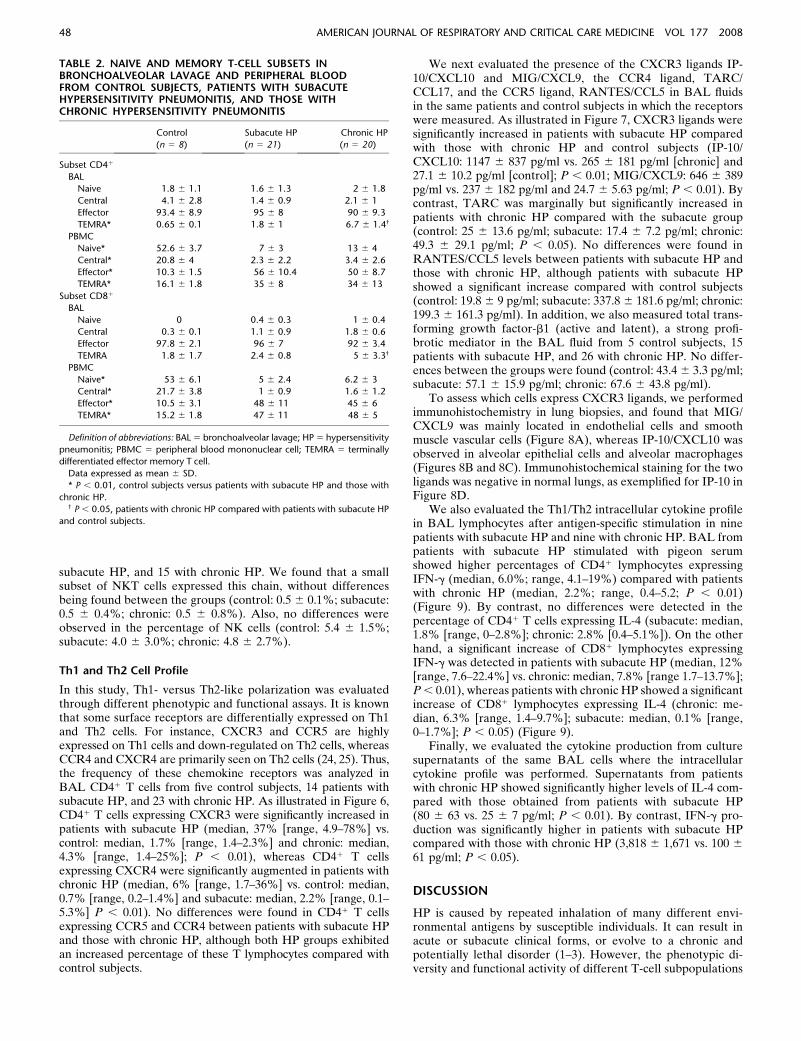
Lung-specific Memory CD4 and CD8 T Cells
To characterize memory cells, the surface markers, CD45RA,CD62L, CD4, CD8, and CD3, were studied in T lymphocytesfrom BAL and PBMCs from 8 control subjects, 21 patients withsubacute HP, and 20 with chronic HP. Based on their pheno-type, three distinct populations of CD41 T cells and CD81 Tcells were detected: a central memory population (CD45RA2
CD62Lhigh) and two effector memory populations; namely, theCD45RA2 CD62Llow and the terminally differentiated effectormemory T cells (TEMRA) CD45RA1 CD62Llow. As illustratedin Figure 2, patients with chronic HP showed a significant
Figure 1. CD41:CD81 ratio from healthy
control subjects, patients with subacute hy-
persensitivity pneumonitis (HP), and those
with chronic HP. (A) Representative flowcytometry analyses of bronchoalveolar la-
vage T lymphocytes showing the percentage
of CD4- and CD8-positive cells. (B)
CD41:CD81 ratio from control subjectsand subacute and chronic groups. Lines
represent median values. *P , 0.01.
46 AMERICAN JOURNAL OF RESPIRATORY AND CRITICAL CARE MEDICINE VOL 177 2008

increase in BAL TEMRA CD41 and CD81 compared with thesubacute HP group (CD41: 6.7 6 1.4% vs. 1.8 6 1%; P , 0.05,CD81: 5 6 3.3% vs. 2.4 6 0.8%; P , 0.05). Most of the PBMCfrom both patients with subacute HP and those with chronic HPwere effector and TEMRA memory cells, without significantdifferences between them; however, these last populations weresignificantly increased when compared with the control cells(Table 2).
Functional Analysis of Memory CD41 and CD81 T Cells
For functional analyses, PBMC and BAL cells from five pa-tients with subacute HP and five with chronic HP werestimulated with PMA (10 ng/ml) and ionomycin (1 mM) for6 hours. As shown in Figures 3A and 3B, patients with subacuteHP had a significantly higher percentage of BAL CD41 andCD81 effector and TEMRA memory cells producing IFN-gafter stimulation compared with patients with chronic HP. Incontrast, memory cell subpopulations obtained from PBMCexpressed low and similar levels IFN-g in both patients withsubacute HP and those with chronic HP (data not shown).
We next evaluated the cytotoxic activity of memory CD81 Tcells from PBMC and BAL from patients with subacute HP andthose with chronic HP. For this purpose, we analyzed simulta-neously the loss of intracellular perforin and the surface ex-pression of CD107a/b (22). Our results showed that only BAL
cells from patients with subacute HP exhibited a correlationbetween the increased expression of surface CD107a/b and lossof intracellular perforin, indicating functional cytotoxic activityof CD81 T cells (Figure 4). However, the major difference wasrelated to the ability to express CD107a/b. By contrast, therewere not significant differences in the loss of perforin. Nodifferences in PBMC memory CD81 T cells were detected.
gd T Lymphocytes, NK, and NKT Cells
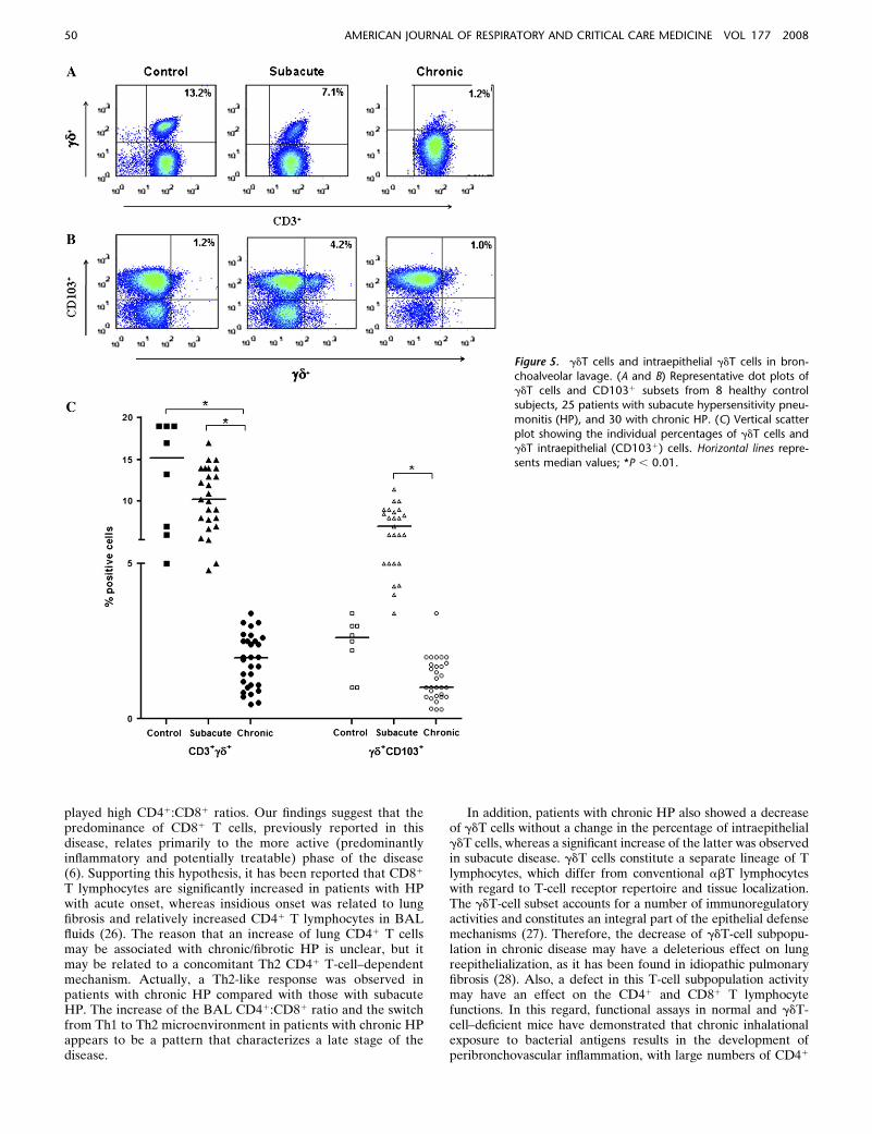
As shown in Figure 5, patients with chronic HP (n 5 20) hada significant decrease in the percentage of BAL gd1 T cellpercentage compared with patients with subacute HP (n 5 21)and control subjects (n 5 8) (median, 2.0% [range, 0.5–3.4%]vs. subacute: median, 10% [range, 4.8–17%] and control sub-jects: median, 15% [range, 5–19]; P , 0.01]. By contrast,intraepithelial gd1 T cells (CD1031) were significantly in-creased in patients with subacute HP (P , 0.01) when com-pared with control subjects and patients with chronic HP.
The proportion of conventional NKT cells (CD161CD561)was significantly decreased in patients with subacute HP andthose with chronic HP (control [n 5 8]: 13.1 6 3.0%; subacute[n 5 21]: 2 6 2.6%; chronic [n 5 20]: 7.3 6 4.1%; P , 0.01 andP , 0.05, respectively). Additionally, we determined the subsetof NKT cells that expressed the invariant Va24 TCR chain(VaiNKT cells) in BAL from 3 control subjects, 5 patients with
Figure 2. Phenotypic profile of memory CD41 and CD81 T cell subsets using multicolor flow cytometry. Bronchoalveolar lavage (BAL) (A) andperipheral blood mononuclear cells (PBMCs) (B) from 8 control subjects, 21 patients with subacute HP, and 20 with chronic HP were stained with
directly conjugated monoclonal antibodies against CD3, CD4, CD8, CD45RA, and CD62L in a five-color experiment. Cells were first identified based
on side scatter and CD3 properties, followed by gating on CD41 or CD81 cells. Differential gating on CD41 or CD81 T cells based on CD62L and
CD45RA basal expression revealed four populations: naive T cells (CD45RA1CD62L1), central memory T cells (CD45RA2CD62L1), effector T cells(CD45RA2CD62L2), and terminally differentiated effector memory T cells (TEMRA) (CD45RA1CD62L2). At least 105 events were analyzed.
Barrera, Mendoza, Zuniga, et al.: Functional Diversity of T Cells in HP 47
subacute HP, and 15 with chronic HP. We found that a smallsubset of NKT cells expressed this chain, without differencesbeing found between the groups (control: 0.5 6 0.1%; subacute:0.5 6 0.4%; chronic: 0.5 6 0.8%). Also, no differences wereobserved in the percentage of NK cells (control: 5.4 6 1.5%;subacute: 4.0 6 3.0%; chronic: 4.8 6 2.7%).
Th1 and Th2 Cell Profile
In this study, Th1- versus Th2-like polarization was evaluatedthrough different phenotypic and functional assays. It is knownthat some surface receptors are differentially expressed on Th1and Th2 cells. For instance, CXCR3 and CCR5 are highlyexpressed on Th1 cells and down-regulated on Th2 cells, whereasCCR4 and CXCR4 are primarily seen on Th2 cells (24, 25). Thus,the frequency of these chemokine receptors was analyzed inBAL CD41 T cells from five control subjects, 14 patients withsubacute HP, and 23 with chronic HP. As illustrated in Figure 6,CD41 T cells expressing CXCR3 were significantly increased inpatients with subacute HP (median, 37% [range, 4.9–78%] vs.control: median, 1.7% [range, 1.4–2.3%] and chronic: median,4.3% [range, 1.4–25%]; P , 0.01), whereas CD41 T cellsexpressing CXCR4 were significantly augmented in patients withchronic HP (median, 6% [range, 1.7–36%] vs. control: median,0.7% [range, 0.2–1.4%] and subacute: median, 2.2% [range, 0.1–5.3%] P , 0.01). No differences were found in CD41 T cellsexpressing CCR5 and CCR4 between patients with subacute HPand those with chronic HP, although both HP groups exhibitedan increased percentage of these T lymphocytes compared withcontrol subjects.
We next evaluated the presence of the CXCR3 ligands IP-10/CXCL10 and MIG/CXCL9, the CCR4 ligand, TARC/CCL17, and the CCR5 ligand, RANTES/CCL5 in BAL fluidsin the same patients and control subjects in which the receptorswere measured. As illustrated in Figure 7, CXCR3 ligands weresignificantly increased in patients with subacute HP comparedwith those with chronic HP and control subjects (IP-10/CXCL10: 1147 6 837 pg/ml vs. 265 6 181 pg/ml [chronic] and27.1 6 10.2 pg/ml [control]; P , 0.01; MIG/CXCL9: 646 6 389pg/ml vs. 237 6 182 pg/ml and 24.7 6 5.63 pg/ml; P , 0.01). Bycontrast, TARC was marginally but significantly increased inpatients with chronic HP compared with the subacute group(control: 25 6 13.6 pg/ml; subacute: 17.4 6 7.2 pg/ml; chronic:49.3 6 29.1 pg/ml; P , 0.05). No differences were found inRANTES/CCL5 levels between patients with subacute HP andthose with chronic HP, although patients with subacute HPshowed a significant increase compared with control subjects(control: 19.8 6 9 pg/ml; subacute: 337.8 6 181.6 pg/ml; chronic:199.3 6 161.3 pg/ml). In addition, we also measured total trans-forming growth factor-b1 (active and latent), a strong profi-brotic mediator in the BAL fluid from 5 control subjects, 15patients with subacute HP, and 26 with chronic HP. No differ-ences between the groups were found (control: 43.4 6 3.3 pg/ml;subacute: 57.1 6 15.9 pg/ml; chronic: 67.6 6 43.8 pg/ml).
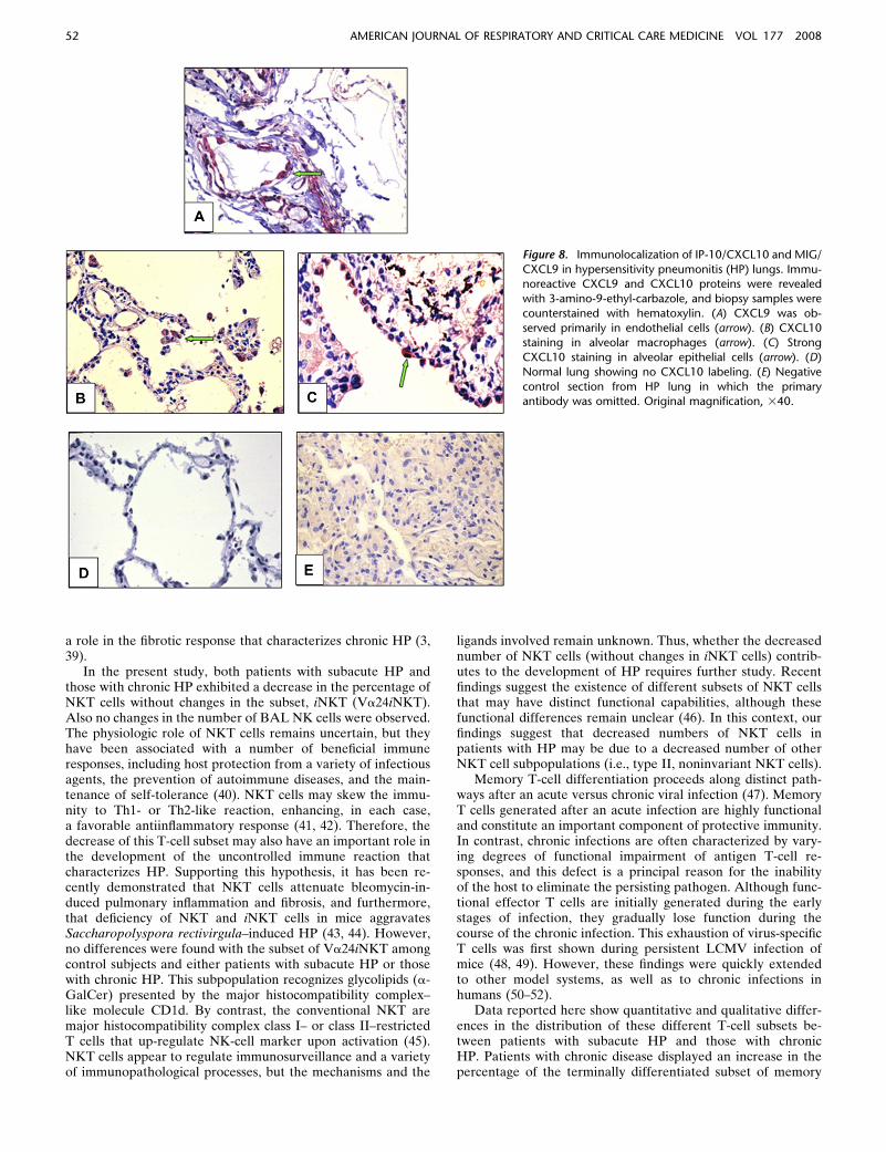
To assess which cells express CXCR3 ligands, we performedimmunohistochemistry in lung biopsies, and found that MIG/CXCL9 was mainly located in endothelial cells and smoothmuscle vascular cells (Figure 8A), whereas IP-10/CXCL10 wasobserved in alveolar epithelial cells and alveolar macrophages(Figures 8B and 8C). Immunohistochemical staining for the twoligands was negative in normal lungs, as exemplified for IP-10 inFigure 8D.
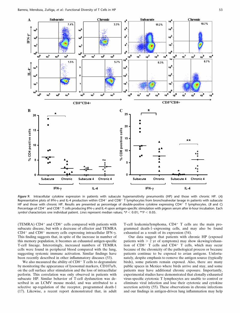
We also evaluated the Th1/Th2 intracellular cytokine profilein BAL lymphocytes after antigen-specific stimulation in ninepatients with subacute HP and nine with chronic HP. BAL frompatients with subacute HP stimulated with pigeon serumshowed higher percentages of CD41 lymphocytes expressingIFN-g (median, 6.0%; range, 4.1–19%) compared with patientswith chronic HP (median, 2.2%; range, 0.4–5.2; P , 0.01)(Figure 9). By contrast, no differences were detected in thepercentage of CD41 T cells expressing IL-4 (subacute: median,1.8% [range, 0–2.8%]; chronic: 2.8% [0.4–5.1%]). On the otherhand, a significant increase of CD81 lymphocytes expressingIFN-g was detected in patients with subacute HP (median, 12%[range, 7.6–22.4%] vs. chronic: median, 7.8% [range 1.7–13.7%];P , 0.01), whereas patients with chronic HP showed a significantincrease of CD81 lymphocytes expressing IL-4 (chronic: me-dian, 6.3% [range, 1.4–9.7%]; subacute: median, 0.1% [range,0–1.7%]; P , 0.05) (Figure 9).
Finally, we evaluated the cytokine production from culturesupernatants of the same BAL cells where the intracellularcytokine profile was performed. Supernatants from patientswith chronic HP showed significantly higher levels of IL-4 com-pared with those obtained from patients with subacute HP(80 6 63 vs. 25 6 7 pg/ml; P , 0.01). By contrast, IFN-g pro-duction was significantly higher in patients with subacute HPcompared with those with chronic HP (3,818 6 1,671 vs. 100 6
61 pg/ml; P , 0.05).
DISCUSSION
HP is caused by repeated inhalation of many different envi-ronmental antigens by susceptible individuals. It can result inacute or subacute clinical forms, or evolve to a chronic andpotentially lethal disorder (1–3). However, the phenotypic di-versity and functional activity of different T-cell subpopulations
TABLE 2. NAIVE AND MEMORY T-CELL SUBSETS INBRONCHOALVEOLAR LAVAGE AND PERIPHERAL BLOODFROM CONTROL SUBJECTS, PATIENTS WITH SUBACUTEHYPERSENSITIVITY PNEUMONITIS, AND THOSE WITHCHRONIC HYPERSENSITIVITY PNEUMONITIS
Control Subacute HP Chronic HP
(n 5 8) (n 5 21) (n 5 20)
Subset CD41
BAL
Naive 1.8 6 1.1 1.6 6 1.3 2 6 1.8
Central 4.1 6 2.8 1.4 6 0.9 2.1 6 1
Effector 93.4 6 8.9 95 6 8 90 6 9.3
TEMRA* 0.65 6 0.1 1.8 6 1 6.7 6 1.4†
PBMC
Naive* 52.6 6 3.7 7 6 3 13 6 4
Central* 20.8 6 4 2.3 6 2.2 3.4 6 2.6
Effector* 10.3 6 1.5 56 6 10.4 50 6 8.7
TEMRA* 16.1 6 1.8 35 6 8 34 6 13
Subset CD81
BAL
Naive 0 0.4 6 0.3 1 6 0.4
Central 0.3 6 0.1 1.1 6 0.9 1.8 6 0.6
Effector 97.8 6 2.1 96 6 7 92 6 3.4
TEMRA 1.8 6 1.7 2.4 6 0.8 5 6 3.3†
PBMC
Naive* 53 6 6.1 5 6 2.4 6.2 6 3
Central* 21.7 6 3.8 1 6 0.9 1.6 6 1.2
Effector* 10.5 6 3.1 48 6 11 45 6 6
TEMRA* 15.2 6 1.8 47 6 11 48 6 5
Definition of abbreviations: BAL 5 bronchoalveolar lavage; HP 5 hypersensitivity
pneumonitis; PBMC 5 peripheral blood mononuclear cell; TEMRA 5 terminally
differentiated effector memory T cell.
Data expressed as mean 6 SD.
* P , 0.01, control subjects versus patients with subacute HP and those with
chronic HP.† P , 0.05, patients with chronic HP compared with patients with subacute HP
and control subjects.
48 AMERICAN JOURNAL OF RESPIRATORY AND CRITICAL CARE MEDICINE VOL 177 2008

in patients with diverse clinical forms of the disease have notbeen characterized.
The results from this study reveal a number of likelypathogenic-associated differences in the T-cell subsets among
patients with subacute and chronic HP. Patients with chronicHP exhibited lower BAL lymphocytosis, with an increase of theCD41:CD81 ratio compared with those with subacute disease.Furthermore, patients with subacute HP who worsened dis-
Figure 3. Functional as-
sessment of effector andTEMRA bronchoalveolar la-
vage (BAL) T cells. (A) The
effector and TEMRA subsets
were gated and analyzedfor IFN-g expression after
6 hours stimulation with
phorbol myristate acetate/ionomycin. IFN-g–positive
cells are expressed as a per-
centage of CD41 or CD81
effector and TEMRA BAL Tcells. Error bars represent
1 SD of five experiments. (B)
A representative plot illus-
trating the increase of IFN-g–positive cells in patients
with subacute HP. *P ,
0.001; **P , 0.01; ***P ,
0.05.
Figure 4. Perforin release and its correlation with
surface expression of CD107 from bronchoalveolar
lavage (BAL) (A) and peripheral blood mononuclearcells (PBMCs) (B). T cells from patients with sub-
acute hypersensitivity pneumonitis (HP) and those
with chronic HP were stimulated with phorbol
myristate acetate/ionomycin in vitro for up to 5hours in the presence of anti-CD107a/b APC and
brefeldin/monesin. At time points designated, ali-
quots were removed, washed, and permeabilized,followed by staining for perforin, CD3, and CD8.
Accumulative data of four independent experi-
ments are shown. Error bars represent 1 SD. Solid
diamonds, solid line: CD107a/b1; solid squares,dotted line: perforin1.
Barrera, Mendoza, Zuniga, et al.: Functional Diversity of T Cells in HP 49
played high CD41:CD81 ratios. Our findings suggest that thepredominance of CD81 T cells, previously reported in thisdisease, relates primarily to the more active (predominantlyinflammatory and potentially treatable) phase of the disease(6). Supporting this hypothesis, it has been reported that CD81
T lymphocytes are significantly increased in patients with HPwith acute onset, whereas insidious onset was related to lungfibrosis and relatively increased CD41 T lymphocytes in BALfluids (26). The reason that an increase of lung CD41 T cellsmay be associated with chronic/fibrotic HP is unclear, but itmay be related to a concomitant Th2 CD41 T-cell–dependentmechanism. Actually, a Th2-like response was observed inpatients with chronic HP compared with those with subacuteHP. The increase of the BAL CD41:CD81 ratio and the switchfrom Th1 to Th2 microenvironment in patients with chronic HPappears to be a pattern that characterizes a late stage of thedisease.
In addition, patients with chronic HP also showed a decreaseof gdT cells without a change in the percentage of intraepithelialgdT cells, whereas a significant increase of the latter was observedin subacute disease. gdT cells constitute a separate lineage of Tlymphocytes, which differ from conventional abT lymphocyteswith regard to T-cell receptor repertoire and tissue localization.The gdT-cell subset accounts for a number of immunoregulatoryactivities and constitutes an integral part of the epithelial defensemechanisms (27). Therefore, the decrease of gdT-cell subpopu-lation in chronic disease may have a deleterious effect on lungreepithelialization, as it has been found in idiopathic pulmonaryfibrosis (28). Also, a defect in this T-cell subpopulation activitymay have an effect on the CD41 and CD81 T lymphocytefunctions. In this regard, functional assays in normal and gdT-cell–deficient mice have demonstrated that chronic inhalationalexposure to bacterial antigens results in the development ofperibronchovascular inflammation, with large numbers of CD41
Figure 5. gdT cells and intraepithelial gdT cells in bron-
choalveolar lavage. (A and B) Representative dot plots ofgdT cells and CD1031 subsets from 8 healthy control
subjects, 25 patients with subacute hypersensitivity pneu-
monitis (HP), and 30 with chronic HP. (C) Vertical scatter
plot showing the individual percentages of gdT cells andgdT intraepithelial (CD1031) cells. Horizontal lines repre-
sents median values; *P , 0.01.
50 AMERICAN JOURNAL OF RESPIRATORY AND CRITICAL CARE MEDICINE VOL 177 2008

and CD81 T cells (29). Moreover, gdT-cell–deficient mice treatedwith live Bacillus subtilis showed an accelerated collagen de-position, suggesting that these cells play a protective antifibroticrole (29).
Recent experimental and clinical evidence suggest thata Th1-type cytokine network plays an important role in HP(10, 30, 31). Likewise, it has been suggested that Th1 cellactivity may decline while Th2 activity increases (Th1/Th2switch hypothesis) in HP lungs evolving to fibrosis (1, 32). Itis well known that the Th2 cytokines, IL-4 and IL-13, enhancethe fibrotic process by induction of fibroblast proliferation andcollagen production (33, 34), whereas IFN-g, a Th1 cytokine,inhibits these processes (17). In this context, it has been recentlydemonstrated that mice overexpressing GATA binding protein3 (GATA-3), a transcription factor that encourages Th2 res-ponses (35–37), develop a more severe bleomycin-inducedpulmonary fibrosis concomitant with a significant decrease ofIFN-g in the lungs (38). With this hypothesis in mind, weevaluated the predominance of Th1 or Th2 profiles in subacuteand chronic HP using several approaches. Our results showedthat patients with subacute disease have a Th1-like response,whereas patients with chronic HP express predominantly a Th2-like phenotype. Thus, BAL lymphocytes from patients withsubacute HP, stimulated in vitro with avian antigens, expressedand released significantly higher levels of Th1 cytokines (IFN-g)and lower levels of Th2 cytokines (IL-4) compared with thosewith chronic disease. Likewise, we also found a significantincrease of lymphocytes expressing CXCR3 on T cells, as wellas the levels of the CXCR3 ligands, IP-10/CXCL10 and MIG/CXCL9, in BAL supernatants of patients with subacute HP.These chemotactic ligands might contribute to the directionalmigration of activated T cells and accumulation of cytotoxic Tlymphocytes in the lung in early stages of the disease. Bycontrast, T cells expressing CXCR4 (Th2) and TARC wereincreased in patients with chronic HP. Taken together, thesefindings raise the possibility that a Th1-to-Th2 switch may play
Figure 6. Expression of CXCR3,
CCR5, CXCR4, and CCR4 in bron-
choalveolar lavage (BAL) T cells
from healthy control subjects,patients with subacute hypersen-
sivity pneumonitis (HP), and those
with chronic HP. BAL T cells were
incubated with the specific mono-clonal antibodies and fixed in 1%
paraformaldehyde for analysis in
six-color flow cytometry. Horizon-
tal lines represent median values;*P , 0.01; **P , 0.05.
Figure 7. Bronchoalveolar lavage (BAL) concentrations of IFN-g–
inducible protein (IP)-10/CXCL10, monokine induced by IFN-g (MIG)/
CXCL9, RANTES (regulated upon activation, normal T-cell expressedand secreted)/CCL5, and thymus- and activation-regulated chemokine
(TARC)/CCL17 in healthy control subjects, patients with subacute
hypersensitivity pneumonitis (HP), and those with chronic HP. BAL
levels of chemokines were measured by ELISA as described in METHODS.Data represent means and SD; *P , 0.01; **P , 0.05.
Barrera, Mendoza, Zuniga, et al.: Functional Diversity of T Cells in HP 51
a role in the fibrotic response that characterizes chronic HP (3,39).
In the present study, both patients with subacute HP andthose with chronic HP exhibited a decrease in the percentage ofNKT cells without changes in the subset, iNKT (Va24iNKT).Also no changes in the number of BAL NK cells were observed.The physiologic role of NKT cells remains uncertain, but theyhave been associated with a number of beneficial immuneresponses, including host protection from a variety of infectiousagents, the prevention of autoimmune diseases, and the main-tenance of self-tolerance (40). NKT cells may skew the immu-nity to Th1- or Th2-like reaction, enhancing, in each case,a favorable antiinflammatory response (41, 42). Therefore, thedecrease of this T-cell subset may also have an important role inthe development of the uncontrolled immune reaction thatcharacterizes HP. Supporting this hypothesis, it has been re-cently demonstrated that NKT cells attenuate bleomycin-in-duced pulmonary inflammation and fibrosis, and furthermore,that deficiency of NKT and iNKT cells in mice aggravatesSaccharopolyspora rectivirgula–induced HP (43, 44). However,no differences were found with the subset of Va24iNKT amongcontrol subjects and either patients with subacute HP or thosewith chronic HP. This subpopulation recognizes glycolipids (a-GalCer) presented by the major histocompatibility complex–like molecule CD1d. By contrast, the conventional NKT aremajor histocompatibility complex class I– or class II–restrictedT cells that up-regulate NK-cell marker upon activation (45).NKT cells appear to regulate immunosurveillance and a varietyof immunopathological processes, but the mechanisms and the
ligands involved remain unknown. Thus, whether the decreasednumber of NKT cells (without changes in iNKT cells) contrib-utes to the development of HP requires further study. Recentfindings suggest the existence of different subsets of NKT cellsthat may have distinct functional capabilities, although thesefunctional differences remain unclear (46). In this context, ourfindings suggest that decreased numbers of NKT cells inpatients with HP may be due to a decreased number of otherNKT cell subpopulations (i.e., type II, noninvariant NKT cells).
Memory T-cell differentiation proceeds along distinct path-ways after an acute versus chronic viral infection (47). MemoryT cells generated after an acute infection are highly functionaland constitute an important component of protective immunity.In contrast, chronic infections are often characterized by vary-ing degrees of functional impairment of antigen T-cell re-sponses, and this defect is a principal reason for the inabilityof the host to eliminate the persisting pathogen. Although func-tional effector T cells are initially generated during the earlystages of infection, they gradually lose function during thecourse of the chronic infection. This exhaustion of virus-specificT cells was first shown during persistent LCMV infection ofmice (48, 49). However, these findings were quickly extendedto other model systems, as well as to chronic infections inhumans (50–52).
Data reported here show quantitative and qualitative differ-ences in the distribution of these different T-cell subsets be-tween patients with subacute HP and those with chronicHP. Patients with chronic disease displayed an increase in thepercentage of the terminally differentiated subset of memory
Figure 8. Immunolocalization of IP-10/CXCL10 and MIG/
CXCL9 in hypersensitivity pneumonitis (HP) lungs. Immu-noreactive CXCL9 and CXCL10 proteins were revealed
with 3-amino-9-ethyl-carbazole, and biopsy samples were
counterstained with hematoxylin. (A) CXCL9 was ob-
served primarily in endothelial cells (arrow). (B) CXCL10staining in alveolar macrophages (arrow). (C) Strong
CXCL10 staining in alveolar epithelial cells (arrow). (D)
Normal lung showing no CXCL10 labeling. (E) Negative
control section from HP lung in which the primaryantibody was omitted. Original magnification, 340.
52 AMERICAN JOURNAL OF RESPIRATORY AND CRITICAL CARE MEDICINE VOL 177 2008
(TEMRA) CD41 and CD81 cells compared with patients withsubacute disease, but with a decrease of effector and TEMRACD41 and CD81 memory cells expressing intracellular IFN-g.This finding suggests that, in spite of the increase in number ofthis memory population, it becomes an exhausted antigen-specificT-cell lineage. Interestingly, increased numbers of TEMRAcells were found in peripheral blood compared with the lung,suggesting systemic immune activation. Similar findings havebeen recently described in other inflammatory diseases (53).
We also measured the ability of CD81 T cells to degranulateby monitoring the appearance of lysosomal markers, CD107a/b,on the cell surface after stimulation and the loss of intracellularperforin. This correlation was only observed in patients withsubacute HP. Similar behavior of T-cell dysfunction was de-scribed in an LCMV mouse model, and was attributed to aselective up-regulation of the receptor, programmed death-1(17). Likewise, a recent report demonstrated that, in adult
T-cell leukemia/lymphoma, CD41 T cells are the main pro-grammed death-1–expressing cells, and may also be foundexhausted as a result of its expression (54).
Our data suggest that patients with chronic HP (exposedpatients with . 2 yr of symptoms) may show skewing/exhaus-tion of CD81 T cells and CD41 T cells, which may occurbecause of the chronicity of the pathological process or becausepatients continue to be exposed to avian antigens. Unfortu-nately, despite emphasis to remove the antigen source (typicallybirds), some patients remain exposed. Also, there are manypublic spaces in Mexico where birds arrive and stay, and somepatients may have additional chronic exposure. Importantly,experimental studies have demonstrated that clonally exhaustedvirus-specific cytotoxic T lymphocytes are unable to control oreliminate viral infection and lose their cytotoxic and cytokinesecretion activity (55). These observations in chronic infectionsand our findings in antigen-driven lung inflammation may help
Figure 9. Intracellular cytokine expression in patients with subacute hypersensitivity pneumonitis (HP) and those with chronic HP. (A)Representative plots of IFN-g and IL-4 production within CD41 and CD81 T lymphocytes from bronchoalveolar lavage in patients with subacute
HP and those with chronic HP. Results are presented as percentage of double-positive cytokine expressing CD41 T lymphocytes. (B and C)
Percentage of CD41 and CD81 T cells producing IFN-g and IL-4 upon antigen-specific stimulation with pigeon serum after 6-hour incubation. Each
symbol characterizes one individual patient. Lines represent median values; *P , 0.01; **P , 0.05.
Barrera, Mendoza, Zuniga, et al.: Functional Diversity of T Cells in HP 53
us to understand the dynamics between the T-cell cytotoxic pop-ulations and chronic antigen exposure in human lung disease.
In summary, the results of this study indicate that patientswith chronic HP have different phenotypic and functional BALT-cell subsets compared with patients with subacute disease.These differences include an increase in CD41:CD81 ratio,a decrease of gdT cells, a skewing toward Th2 as opposed toTh1, and exhaustion of effector CD81 and CD41 T cells. Thesefindings may provide insight in the development of new ther-apeutic strategies, such as the inhibition of GATA-3 transcrip-tion factor to modify the Th1/Th2 cell balance to enhance aTh1-like lung microenvironment.
Conflict of Interest Statement: None of the authors has a financial relationshipwith a commercial entity that has an interest in the subject of this manuscript.
References
1. Selman M. Hypersensitivity pneumonitis: a multifaceted deceiving
disorder. Clin Chest Med 2004;25:531–547.2. Lacasse Y, Selman M, Costabel U, Dalphin JC, Ando M, Morell F,
Erkinjuntti-Pekkanen R, Muller N, Colby TV, Schuyler M, et al. HPStudy Group. Clinical diagnosis of hypersensitivity pneumonitis. AmJ Respir Crit Care Med 2003;168:952–958.
3. Perez-Padilla R, Salas J, Chapela R, Sanchez M, Carrillo G, Perez R,
Sansores R, Gaxiola M, Selman M. Mortality in Mexican patientswith chronic pigeon breeder’s lung compared with those with usualinterstitial pneumonia. Am Rev Respir Dis 1993;148:49–53.
4. Malinen A, Erkinjuntti-Pekkanen R, Partanen P, Rytkonen H, Vanni-
nen R. Long-term sequelae of Farmer’s lung disease in HRCT: a 14-year follow-up study of 88 patients and 83 matched control farmers.Eur Radiol 2003;13:2212–2221.
5. Semenzato G. Immunology of interstitial lung diseases: cellular events
taking place in the lung of sarcoidosis, hypersensitivity pneumonitisand HIV infection. Eur Respir J 1991;4:94–102.
6. Welker L, Jorres R, Costabel U, Magnussen H. Predictive value of BAL
cell differentials in the diagnosis of interstitial lung diseases. EurRespir J 2004;24:1000–1006.
7. Suda T, Sato A, Ida M, Gemma H, Hayakawa H, Chida K. Hypersen-
sitivity pneumonitis associated with home ultrasonic humidifiers.Chest 1995;107:711–717.
8. Dai H, Guzman J, Bauer P, Costabel U. Elevated levels of soluble TNF
receptors in bronchoalveolar lavage fluid in extrinsic allergic alveo-litis. Clin Exp Allergy 1999;29:1209–1213.
9. Ando M, Konishi K, Yoneda R, Tamura M. Difference in the phenotypes
of bronchoalveolar lavage lymphocytes in patients with summer-typehypersensitivity pneumonitis, farmer’s lung, ventilation pneumonitis,and bird fancier’s lung: report of a nationwide epidemiologic study inJapan. J Allergy Clin Immunol 1991;87:1002–1009.
10. Yamasaki H, Ando M, Brazer W, Center D, Cruikshank W. Polarized
type 1 cytokine profile in bronchoalveolar lavage T cells of patientswith hypersensitivity pneumonitis. J Immunol 1999;163:3516–3523.
11. Sallusto F, Lanzavechia A. Central memory and effector memory T cell
subsets: function, generation and maintenance. Annu Rev Immunol2004;22:745–763.
12. Zaph C, Uzonna J, Beverley S, Scott P. Central memory T cells mediate
long-term immunity to Leishmania major in the absence of persistentparasites. Nat Med 2004;10:1104–1110.
13. Baron V, Bouneaud C, Cumano A, Lim A, Arstila T, Kourilsky P,
Ferradini L, Pannetier C. The repertoires of circulating human CD81
central and effector memory T cell subsets are largely distinct.Immunity 2003;18:193–204.
14. Wherry E, Teichgraber V, Becker T, Masopust D, Kaech S, Antia R,
Von Adrian UH, Ahmed R. Lineage relationship and protectiveimmunity of memory CD8 T cell subsets. Nat Immunol 2003;4:225–234.
15. Barber D, Wherry EJ, Masopoust D, Baogong Z, Allison JP, Sharpe
AH, Freeman GJ, Ahmed R. Restoring function in exhausted CD8 Tcells during chronic viral infection. Nature 2006;439:682–687.
16. Murphy KM, Reiner SL. The lineage decisions of helper T cells. Nat Rev
Immunol 2002;2:933–944.17. Wynn T. Fibrotic disease and the T(H)1/T(H)2 paradigm. Nat Rev
Immunol 2004;4:583–594.
18. Pardo A, Barrios R, Gaxiola M, Segura-Valdez L, Carrillo G, Estrada A,
Mejia M, Selman M. Increase of lung neutrophils in hypersensitivitypneumonitis is associated with lung fibrosis. Am J Respir Crit CareMed 2000;161:1698–1704.
19. Selman M, Pardo A, Barrera L, Estrada A, Watson S, Wilson K, Aziz N,
Kaminski N, Zlotnik A. Gene expression profiles distinguish idio-pathic pulmonary fibrosis from hypersensitivity pneumonitis. Am JRespir Crit Care Med 2006;173:188–198.
20. Disis ML, dela Rosa C, Goodell V, Kuan LY, Chang J, Kuus-Reichel K,
Clay TM, Lyerly HK, Bhatia S, Ghanekar SA, et al. Maximizing theretention of antigen specific lymphocyte function after cryopreserva-tion. J Immunol Methods 2006;308:13–18.
21. Navarro C, Mendoza F, Barrera L, Segura-Valdez L, Gaxiola M,
Paramo I, Selman M. Up-regulation of L-selectin and E-selectin inhypersensitivity pneumonitis. Chest 2002;121:354–360.
22. Chen R, Lowe L, Wilson J, Crowther E, Tzeggai K, Bishop J, Varro R.
Simultaneous quantification of six human cytokines in a single sampleusing microparticle-based flow cytometric technology. Clin Chem 1999;9:1693–1697.
23. Betts M, Brenchley J, Price D, De Rosa S, Douek D, Roederer M, Koup
R. Sensitive and viable identification of antigen-specific CD81T cellsby a flow cytometric assay for degranulation. J Immunol Methods2003;281:65–78.
24. King M, Ismail A, Davis L, Karp D. Oxidative stress promotes
polarization of human T cell differentiation toward a T helper 2phenotype. J Immunol 2006;176:2765–2772.
25. Bonecchi R, Bianchi G, Bordignon PP, D’Ambrosio D, Lang R, Borsatti
A, Sozzani S, Allavena P, Gray PA, Mantovani A, et al. Differentialexpression of chemokine receptors and chemotactic responsiveness oftype 1 T helper cells (Th1s) and Th2s. J Exp Med 1998;187:129–134.
26. . Murayama J, Yoshizawa Y, Ohtsuka M, Hasegawa S. Lung fibrosis in
hypersensitivity pneumonitis. Association with CD41 but not CD81
cell dominant alveolitis and insidious onset. Chest 1993;104:38–43.27. Girardi M. Immunosurveillance and immunoregulation by gammadelta
T cells. J Invest Dermatol 2006;126:25–31.28. Selman M, Pardo A. Role of epithelial cells in idiopathic pulmonary
fibrosis: from innocent targets to serial killers. Proc Am Thorac Soc2006;3:364–372.
29. Simonian P, Roark C, Diaz Del Valle F, Palmer B, Douglas I, Ikuta K,
Born W, O’Brien R, Fontenot A. Regulatory role of gd T cells in therecruitment of CD41 and CD81 T cells to lung and subsequentpulmonary fibrosis. J Immunol 2006;177:4436–4443.
30. Schuyler M, Gott K, Cherne A, Edwards B. Th1 CD41 cells adoptively
transfer experimental hypersensitivity pneumonitis. Cell Immunol 1997;177:169–175.
31. Gudmundsson G, Hunninghake G. Interferon-g is necessary for the ex-
pression of hypersensitivity pneumonitis. J Clin Invest 1997;99:2386–2390.
32. Pardo A, Selman M. Molecular mechanisms of pulmonary fibrosis. Front
Biosci 2002;7:1743–1761.33. Rennard S. Th2 cytokine regulation of type I collagen gel contraction
mediated by human lung mesenchymal cells. Am J Physiol 2002;282:L1049–L1056.
34. Sempowski G, Derdak S, Phipps R. Interleukin-4 and interferon g
discordantly regulate collagen biosynthesis by functionally distinctlung fibroblast subsets. J Cell Physiol 1996;167:290–296.
35. Zhang D, Cohn L, Ray P, Bottomly K, Ray A. Transcription factor
GATA-3 is differentially expressed in murine Th1 and Th2 cells andcontrols Th2-specific expression of the interleukin-5 gene. J BiolChem 1997;272:21597–21603.
36. Zheng D, Flavell R. The transcription factor GATA-3 is necessary and
sufficient for Th2 cytokine gene expression in CD4 T cells. Cell1997;89:587–596.
37. Usui T, Nishikomori R, Kitani A, Strober W. GATA-3 suppresses Th1
development by downregulation of Stat4 and not through effects onIL-12Rb2 chain or T-bet. Immunity 2003;18:415–428.
38. Kimura T, Ishii Y, Yoh K, Morishima Y, Iizuka T, Kiwamoto T,
Matsuno Y, Homma S, Nomura A, Sakamoto T, et al. Overexpressionof the transcription factor GATA-3 enhances the development ofpulmonary fibrosis. Am J Pathol 2006;169:96–104.
39. Vourlekis J, Schwarz M, Cherniack R, Curran-Everett D, Cool R, Tuder
M, King T Jr, Brown K. The effect of pulmonary fibrosis on survivalin patients with hypersensitivity pneumonitis. Am J Med 2004;116:662–668.
40. Kronenberg M. Toward an understanding of NKT cell biology: progress
and paradoxes. Annu Rev Immunol 2005;23:877–900.
54 AMERICAN JOURNAL OF RESPIRATORY AND CRITICAL CARE MEDICINE VOL 177 2008

41. Matsuda H, Suda T, Sato J, Nagata T, Koide Y, Chida K, Nakamura H.a-Galactosylceramide, a ligand of natural killer T cells, inhibits aller-gic airway inflammation. Am J Respir Cell Mol Biol 2005;33:22–31.
42. Sharif S, Arreaza G, Zucker P, Mi Q, Sondhi J, Naidenko O, KronenbergM, Koezuka Y, Delovitch T, Gombert J, et al. Activation of naturalkiller T cells by a-galactosylceramide treatment prevents the onsetand recurrence of autoimmune type 1 diabetes. Nat Med 2001;7:1057–1062.
43. Kim J, Kim Y, Kim S, Chung J, Park W, Chung D. Natural killer T(NKT) cells attenuate bleomycin-induced pulmonary fibrosis by pro-ducing interferon-g. Am J Pathol 2001;167:1231–1241.
44. Hwang S, Kim S, Park W, Chung D. IL-4-secreting NKT cells preventhypersensitivity pneumonitis by suppressing IFN-g producing neu-trophils. J Immunol 2006;177:5258–5268.
45. Kim CH, Butcher EC, Johnston B. Distinct subsets of human Va24-invariant NKT cells: cytokine responses and chemokine receptorexpression. Trends Immunol 2002;23:516–519.
46. Seino K, Taniguchi M. Functionally distinct NKT cell subsets andsubtypes. J Exp Med 2005;202:1623–1626.
47. Klenerman P, Hill A. T cells and viral persistence: lessons from diverseinfections. Nat Immunol 2005;6:873–879.
48. Zajac A. Viral immune evasion due to persistence of activated T cellswithout effector function. J Exp Med 1998;188:2205–2213.
49. Gallimore A. Induction and exhaustion of lymphocytic choriomeningitisvirus–specific cytotoxic T lymphocytes visualized using soluble tetra-meric major histocompatibility complex class-1 peptide complexes.J Exp Med 1998;187:1383–1393.
50. Pantaleo G, Koup RA. Correlates of immune protection in HIV-1infection: what we know, what we don’t know, what we should know.Nat Med 2004;10:806–810.
51. Letvin N, Walker B. Immunopathogenesis and immunotherapy in AIDSvirus infections. Nat Med 2003;9:861–866.
52. Rehermann B, Nascimbeni M. Immunology of hepatitis B and hepatitisC virus infection. Nat Rev Immunol 2005;5:215–229.
53. Haegele KF, Stueckle CA, Malin JP, Sindern E. Increase of CD81 T-effector memory cells in peripheral blood of patients with relapsing-remitting multiple sclerosis compared to healthy controls. J Neuro-immunol 2007;183:168–174.
54. Shimauchi T, Kabashima K, Nakashima D, Sugita K, Yamada Y,Hino R, Tokura Y. Augmented expression of programmeddeath-1 in both neoplastic and non-neoplastic CD41 T-cells inadult T-cell leukemia/lymphoma. Int J Cancer 2007;121:2585–2590.
55. Zhou S, Ou R, Huang L, Price GE, Moskophidis D. Differential tissue-specific regulation of antiviral CD81 T-cell immune responses duringchronic viral infection. J Virol 2004;78:3578–3600.
Barrera, Mendoza, Zuniga, et al.: Functional Diversity of T Cells in HP 55
Related Documents