Open Research Online The Open University’s repository of research publications and other research outputs Hyperosmotic stress induces axl activation and cleav- age in cerebral endothelial cells Journal Article How to cite: Wilhelm, Imola; Nagyoszi, P´ eter; Farkas, Attila E.; Couraud, Pierre-Olivier; Romero, Ignacio A.; Wek- sler, Babette; Fazakas, Csilla; Dung, Ngo Thi Khue; Bottka, S´ andor; Bauer, Hannelore; Bauer, Hans-Christian and Krizbai, Istv´ an A. (2008). Hyperosmotic stress induces axl activation and cleavage in cerebral endothelial cells. Journal of Neurochemistry, 107(1), pp. 116–126. For guidance on citations see FAQs . c [not recorded] Version: [not recorded] Link(s) to article on publisher’s website: http://dx.doi.org/doi:10.1111/j.1471-4159.2008.05590.x Copyright and Moral Rights for the articles on this site are retained by the individual authors and/or other copy- right owners. For more information on Open Research Online’s data policy on reuse of materials please consult the policies page. oro.open.ac.uk

Welcome message from author
This document is posted to help you gain knowledge. Please leave a comment to let me know what you think about it! Share it to your friends and learn new things together.
Transcript
Open Research OnlineThe Open University’s repository of research publicationsand other research outputs
Hyperosmotic stress induces axl activation and cleav-age in cerebral endothelial cells
Journal ArticleHow to cite:
Wilhelm, Imola; Nagyoszi, Peter; Farkas, Attila E.; Couraud, Pierre-Olivier; Romero, Ignacio A.; Wek-sler, Babette; Fazakas, Csilla; Dung, Ngo Thi Khue; Bottka, Sandor; Bauer, Hannelore; Bauer, Hans-Christianand Krizbai, Istvan A. (2008). Hyperosmotic stress induces axl activation and cleavage in cerebral endothelialcells. Journal of Neurochemistry, 107(1), pp. 116–126.
For guidance on citations see FAQs.
c© [not recorded]
Version: [not recorded]
Link(s) to article on publisher’s website:http://dx.doi.org/doi:10.1111/j.1471-4159.2008.05590.x
Copyright and Moral Rights for the articles on this site are retained by the individual authors and/or other copy-right owners. For more information on Open Research Online’s data policy on reuse of materials please consultthe policies page.
oro.open.ac.uk
This is an Accepted Work that has been peer-reviewed and approved for publication in the Journal of Neurochemistry, but has yet to undergo copy-editing and proof correction. See http://www.blackwell-synergy.com/loi/jnc for details. Please cite this article as a “Postprint”; doi: 10.1111/j.1471-4159.2008.05590.x
Submitted 18-3-08. Revised 11-6-08. Accepted 15-7-08.
HYPEROSMOTIC STRESS INDUCES AXL ACTIVATION AND CLEAVAGE IN
CEREBRAL ENDOTHELIAL CELLS
Imola Wilhelm1, Péter Nagyőszi1, Attila E. Farkas1, Pierre-Olivier Couraud2, Ignacio A.
Romero3, Babette Weksler2, Csilla Fazakas1, Ngo Thi Khue Dung1, Sándor Bottka4, Hannelore
Bauer5, Hans-Christian Bauer5, and István A. Krizbai1*
1Institute of Biophysics, Biological Research Center, Szeged, Hungary,
2INSERM U567, Institut Cochin, Paris, France,
3The Open University, Milton Keynes, UK,
4Institute of Plant Biology, Biological Research Center, Szeged, Hungary,
5ABT, Dept. Org. Biology, University of Salzburg, Salzburg, Austria
*Corresponding author:
István A. Krizbai
Institute of Biophysics, Biological Research Center,
Temesvári krt. 62,
6726 Szeged, Hungary
Tel: 36-62-599602
Fax: 36-62-433133
E-mail: [email protected]
2
Abbreviations:
BBB, blood-brain barrier
CECs, cerebral endothelial cells
DMNQ, 2,3-dimethyl-1,4-naphthoquinone
ERK, extracellular signal-regulated kinase
FAK, focal adhesion kinase
GAPDH, glutaraldehyde-3-phosphate dehydrogenase
Gas 6, growth arrest-specific protein 6
Ma, mannitol
PDTC, pyrrolidine dithiocarbamate
PI3K, phosphatidyl inositol 3-kinase
PMA, phorbol myristyl acetate
PKC, protein kinase C
3
ABSTRACT
Due to the relative impermeability of the blood-brain barrier many drugs are unable to reach
the CNS in therapeutically relevant concentration. One method to deliver drugs to the CNS is the
osmotic opening of the blood-brain barrier using mannitol. Hyperosmotic mannitol induces a
strong phosphorylation on tyrosine residues in a broad spectrum of proteins in cerebral
endothelial cells, the principal components of the blood-brain barrier. Previously we have shown
that among targets of tyrosine phosphorylation are β-catenin, extracellular signal-regulated kinase
1/2 and the non-receptor tyrosine kinase Src. The aim of this study was to identify new signaling
pathways activated by hypertonicity in cerebral endothelial cells. Using an antibody array and
immunoprecipitation we identified the receptor tyrosine kinase Axl to become tyrosine
phosphorylated in response to hyperosmotic mannitol. Besides activation, Axl was also cleaved
in response to osmotic stress. Degradation of Axl proved to be metalloproteinase- and
proteasome-dependent and resulted in 50-55 kDa C-terminal products which remained
phosphorylated even after degradation. Specific knockdown of Axl increased the rate of
apoptosis in hyperosmotic mannitol-treated cells, therefore we assume that activation of Axl may
be a protective mechanism against hypertonicity-induced apoptosis. Our results identify Axl as
an important element of osmotic stress-induced signaling.
Keywords: blood-brain barrier, cerebral endothelial cells, mannitol, hyperosmosis, Axl
Running title: Hyperosmotic phosphorylation and cleavage of Axl
4
INTRODUCTION
Cerebral endothelial cells (CECs) are the principal components of the blood-brain barrier
(BBB). They form a continuous monolayer and are interconnected by tight junctions which
impair the free movement of different chemical compounds from one side to the other side of the
barrier. The cerebral microvasculature is ensheated by astrocytic endfeet which play an essential
role in maintaining BBB phenotype (for review see: Abbott et al 2006). Low permeability of the
endothelial cell monolayer protects the CNS from potentially toxic substances and contributes
significantly to the homeostasis of the brain. Regulation of permeability is under complex
control. In these regulatory processes signaling molecules, like protein kinases and phosphatases,
cyclic nucleotides, calcium, etc. play important roles (Deli et al 1993; Hurst et al 1998; Lohmann
et al 2004; Krizbai and Deli 2003).
From clinical point of view, due to the relative impermeability of the barrier many drugs are
unable to reach the CNS in therapeutically relevant concentration, making the BBB one of the
major impediments in the treatment of CNS disorders. A large number of strategies have been
developed in order to circumvent this problem. One of the successfully used methods to deliver
drugs — especially antitumoral agents — to the CNS is the osmotic opening of the BBB using
mannitol (Kroll et al 1998; Neuwelt et al 1980; Neuwelt et al 1991). This causes a rapid opening
(within minutes) of the BBB which is reversible. The barrier function starts to recover
approximately 1 h after treatment, but complete recovery is achieved only after 6-8 h (Rapoport
et al 1980; Siegal et al 2000).
Despite successful clinical trials the molecular mechanisms underlying the effect of
hyperosmotic mannitol are largely unknown. Early studies were not able to detect any changes in
the structure of the interendothelial tight junctions of brain capillary endothelial cells (Farrell et
5
al 1984), however, later Lossinsky et al (1995) have demonstrated that a single intracarotid
injection of hypertonic 1.8 M L (+) arabinose widened interendothelial spaces. Peroxidase tracer
was observed within vesiculo-tubular profiles, and occasionally within widened interendothelial
junctional clefts. Furthermore, intraperitoneal infusion of hyperosmotic mannitol induced
increased expression of AQP4 in astrocytes, at sites where the blood-brain barrier was disrupted
(Arima et al 2003).
Hyperosmotic stress initiates a variety of compensatory and adaptive responses (for review
see: Maurice et al 2007). In response to hypertonicity cells can initiate a rapid reorganization of
the actin cytoskeleton. Di Ciano et al. have shown that hypertonic conditions induce
submembranous de novo F-actin assembly and promote association of cortactin with the actin-
related protein 2/3 (Arp2/3) complex (Di Ciano et al 2002). Further research has revealed
activation of Rho, Rac and Cdc42 as well (Di Ciano-Oliveira et al 2003; Lewis et al 2002). Our
atomic force microscopic studies have revealed a significant decrease in cell height and elasticity
accompanied by the appearance of cytoplasmic protrusions (Balint et al 2007).
There is increasing evidence that hypertonicity induces a concerted action of signaling
mechanisms. Protein phosphorylation plays an important role in the cellular adaptation to
hyperosmotic conditions and cellular shrinkage. It has been shown that one target of
phosphorylation is cortactin, an actin-binding protein which is phosphorylated in a Fyn-
dependent manner in Chinese hamster ovary cells (Kapus et al 1999). Another target protein of
hyperosmotic stress-stimulated phosphorylation is focal adhesion kinase (FAK) which becomes
phosphorylated on Tyr-397 and Tyr-577 via a Src-independent pathway in fibroblats and
epithelial cells (Lunn et al 2004). However, less is known about endothelial, especially cerebral
endothelial cells. In our previous study we have shown that β-catenin becomes tyrosine
6
phosphorylated in response to mannitol in CECs and this phosphorylation is Src kinase-
dependent (Farkas et al 2005).
The aim of this study was to identify new signaling pathways regulated by hyperosmosis in
CECs.
MATERIAL AND METHODS
Materials
All chemicals, if not otherwise stated, were purchased from Sigma.
The following inhibitors were used: genistein, PP1, Y27632 (Tocris), GM6001, calpeptin,
bisindolylmaleinimide, MG132, zVAD (Calbiochem), DMNQ, pyrrolidine dithiocarbamate
(PDTC), sodium vanadate, verapamil (Sigma), leupeptin, E64, pepstatin, pefabloc (Roche),
U0126 (Cell Signaling), wortmannin (Alomone).
Protein G Sepharose was purchased from GE Healthcare. We have used the following
antibodies: anti-Axl (C20) (Santa Cruz), anti-Akt, anti-phospho-Akt (Ser473), anti-cleaved
caspase-3 (Asp175) (Cell Signaling), anti-phosphotyrosine, anti-β-actin (Sigma), HRP-anti-goat
IgG (Santa Cruz), HRP-anti-mouse IgG (Sigma), HRP-anti-rabbit IgG (Cell Signaling), Cy3-anti-
goat IgG and -anti-rabbit IgG (Jackson).
Cell culture and treatments
The human cerebral endothelial cell line hCMEC/D3 was maintained as described
previously (Weksler et al 2005). Briefly, the cells were grown on rat tail collagen coated plates or
glass coverslips in EBM-2 medium (Cambrex) supplemented with EGM-2 Bullet Kit (Cambrex)
and 2.5% fetal bovine serum (FBS, Sigma).
7
Confluent monolayers of hCMEC/D3 cells were treated in serum free culture medium with
10-20% mannitol (0.55-1.1 M, corresponding to an additional 550-1100 mOsmol/l compared to
the 300 mOsmol/l control) and various other substances were added concurrently with mannitol
as indicated. Genistein treatment was performed with an 1 h preincubation.
Antibody array
For the detection of phosphorylated proteins we used an antibody array (Hypromatrix) and
followed the manufacturer’s recommendations during the whole procedure. Briefly, cells were
lysed in a buffer containing 20 mM Tris, 150 mM NaCl, 1 mM EDTA, 1% Triton X-100, 1 mM
sodium vanadate, 10 mM NaF, 1 mM Pefabloc. After centrifugation (10,000 x g, 10 min, 4°C)
the cell homogenate was incubated with the antibody matrix. Following washing steps, the
membrane was incubated with HRP-labeled anti-phosphotyrosine antibody (Hypromatrix) and
the reaction was visualized using ECL Plus (GE Healthcare).
Western-blot
Cells were washed with phosphate buffered saline (PBS) and scraped into ice-cold lysis
buffer (20 mM Tris, 150 mM NaCl, 1 mM EDTA, 1% Triton X-100, 1% sodium deoxycholate,
0.1% sodium dodecyl sulphate, 1 mM sodium vanadate, 10 mM NaF, 1 mM Pefabloc) and
incubated on ice for 30 min. For determination of water- and detergent soluble proteins the
following buffers were used: 20 mM Tris, 150 mM NaCl, 1 mM EDTA, 1 mM sodium vanadate,
10 mM NaF, 1 mM Pefabloc for water-soluble proteins, and the same buffer containing in
addition 1% TritonX-100 for detergent-soluble proteins. Lysates were clarified by centrifugation
at 10,000 x g for 10 min on 4°C and protein concentration was determined with the BCA method
(Pierce). Proteins were electrophoresed and blotted onto PVDF (Pall) or nitrocellulose
8
(Schleicher-Schuell) membranes. The immunoreaction was visualized using ECL Plus
chemiluminescence detection kit.
Immunoprecipitation
For immunoprecipitation experiments cells were homogenized in lysis buffer as described
above. Briefly, lysates were centrifuged at 10,000 x g for 10 min in a microfuge and the
supernatant was subjected to immunoprecipitation. After preclearing with protein G Sepharose,
supernatants were incubated with 2-5 μg primary antibody (anti-phosphotyrosine or -Axl) at 4°C
for 4 h. The formed immunocomplexes were precipitated by incubating the samples overnight
with protein G Sepharose beads. The precipitates were washed 4 times with lysis buffer, boiled in
sample buffer, and subjected to electrophoresis and immunoblotting.
Specific knockdown of Axl by RNA interference
StealthTM siRNA duplex oligoribonucleotides were designed using Invitrogen BLOCK-
iTTM RNAi designer and were purchased from Invitrogen. The sequences used were as follows:
sense, 5'-CAGGAACTGCATGCTGAATGAGAA-3'; antisense, 5'-
TTCTCATTCAGCATGCAGTTCCTGG-3'. As control non-targeting RNA we have used the
following scrambled oligonucleotides: sense, 5’-GACGTAGAGAGAGTTCCGACATACA-3’
and antisense 5’-TGTATGTCGGAACTCTCTCTACGTC-3’. Briefly, cells were plated at 50%
confluency. Transfection of oligonucleotides was then performed in OptiMEM medium
containing 10 nM RNA and LipofectamineTM RNAiMAX reagent (Invitrogen) following the
manufacturer's instructions. After 6 h the medium was changed to regular culture medium. In
order to increase the efficiency, a second transfection was performed the following day. Cells
were analyzed 24 h after the second transfection when they achieved confluency.
9
Immunofluorescence
Cells were fixed using a mixture of ice cold ethanol : acetic acid (95:5) for 10 min. After
blocking with 3% bovine serum albumin for 30 min, coverslips were incubated with primary
antibody. The staining was visualized using Cy3-conjugated secondary antibody. Coverslips were
mounted in anti-fading embedding medium (Biomeda) and the distribution of the signal was
studied using a Nikon Eclipse TE2000U photomicroscope with epifluorescent capabilities
connected to a digital camera (Spot RT KE).
Real-time PCR
For real-time PCR analysis we isolated total RNA from treated and untreated cells using
TRIzol reagent (Invitrogen) following the manufacturer’s recommendations. RNA was
transcribed into cDNA using a reverse transcription kit (Fermentas). The amplification was
performed on a BioRad iQ5 instrument using FastStart SYBR Green Mix (Roche) under the
following conditions: 30 cycles of 95°C for 15 s, 55°C for 30 s, 72°C for 30 s.
The following primer pairs were used for amplification: GAS6: 5'-
TGCTGTCATGAAAATCGCGG-3' and 5'-CATGTAGTCCAGGCTGTAGA-3', Axl: 5'-
GGTGGCTGTGAAGACGATGA-3' and 5'-CTCAGATACTCCATGCCACT-3' (Sun et al
2002), GAPDH (glutaraldehyde-3-phosphate dehydrogenase) (control): 5'-
GTGAAGGTCGGTGTCAACG-3' and 5'-GTGAAGACGCCAGTAGACTC-3'. Determination
of threshold cycle and quantitation was performed using the software of the instrument.
10
Two-dimensional electrophoresis coupled with Western-blot
Control and mannitol treated cells were washed with Tris buffered sorbitol (10 mM Tris, 25
mM sorbitol, pH=7.0) and scraped into sample-rehydration buffer (7 M urea, 2 M thiourea, 2%
CHAPS, 1% DTT, 0.2 mM vanadate, 1 mM Pefabloc, 1 mM NaF, 0.2% ampholite, traces of
bromophenol blue) and incubated at room temperature for 1 h. Debris was removed by
centrifugation at 16,000 x g for 20 min on 17°C. The two-dimensional electrophoresis was
carried out on a Protean IEF Cell using immobilized pH gradient strips (7 cm, pH 4-7 BioRad
Laboratories Inc.) and a Mini-Protean 3 cell according to the manufacturer’s instructions.
Western-blots were performed on the second dimension gels.
Determination of apoptosis
Cells cultured on glass coverslips were transfected with Axl siRNA construct (as described
previously), followed by treatment with 20% mannitol for 3 h. In order to control the efficiency
of silencing, parallel Western-blot analysis was performed using anti-Axl antibody (as described
previously). Immunofluorescence staining was done using anti-cleaved caspase-3 antibody
(according to the manufacturer’s instructions). 1,000 cells/coverslip were analyzed under
fluorescence microscope and labelled cells were counted. In another set of experiments nuclear
morphology was analysed using Hoechst 33342 staining (0.6 µg/ml). Nuclei containing
condensed chromatin at the nuclear envelope or fragmented chromatin were considered apoptotic
(Bresgen et al 2003).
11
RESULTS
Phosphorylation of Axl
In our previous studies we have shown that hyperosmotic mannitol induces a strong
phosphorylation on tyrosine residues in CECs (Farkas et al 2005). To further identify target
proteins of tyrosine phosphorylation an antibody array screening was applied using a matrix
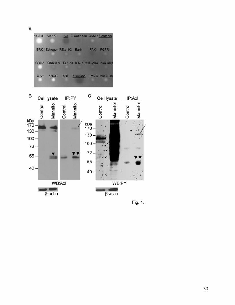
loaded with antibodies directed mainly against signaling molecules. Using this method we found
β-catenin, the MAPkinase extracellular signal-regulated kinase (ERK)1, p130Cas, focal adhesion
kinase (FAK) and the receptor tyrosine kinase Axl to become phosphorylated (Fig. 1A). In our
previous studies we have already shown that β-catenin and ERK are targets of mannitol induced
phosphorylation in CECs. Literature data are available confirming the phosphorylation of
p130Cas and FAK in response to hyperosmosis (Ueno et al 2001; Lunn et al 2004).
To prove that Axl can indeed be phosphorylated under hypertonic conditions we performed
immunoprecipitation studies using anti-phosphotyrosine antibody (Fig. 1B) and anti-Axl
antibody as well (Fig. 1C). Both approaches confirmed the tyrosine phosphorylation of Axl after
treatment of the cells with 20% mannitol for 30 min.
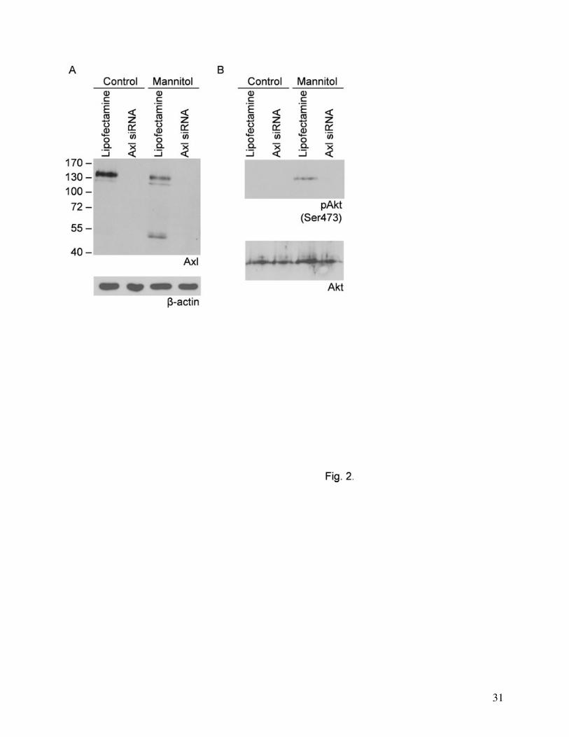
Activation of Akt
To test the activation of the possible downstream elements of Axl signaling we investigated
the phosphorylation of Akt (protein kinase B). RNA interference was used to specifically knock
down Axl and the result of silencing was controlled using Western-blot with anti-Axl antibody
(Fig. 2A). An almost complete silencing of Axl protein could be achieved. We found that
mannitol was able to induce the phosphorylation (on Ser473 residue) and thus the activation of
12
Akt (Fig. 2B). This phenomenon was prevented by Axl silencing, showing that mannitol induced
Akt activation occurs through the receptor tyrosine kinase Axl.
Degradation of Axl
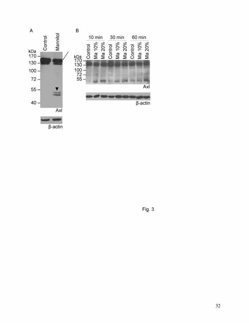
We have observed that besides phosphorylation, treatment of CECs with 20% mannitol
induced the appearance of a double proteolytic band with an apparent molecular weight of 50-55
kDa (arrowheads, Fig. 1B, 3A) accompanied by a decrease in the intensity of the full size Axl
band (Fig. 1B, 3A). Since our antibody recognizes the C-terminal of the protein, we conclude that
this degradation band corresponds to the intracellular domain of Axl. Degradation of Axl proved
to be time- and concentration-dependent and was clearly detectable after 10 min of mannitol
treatment (Fig. 3B).
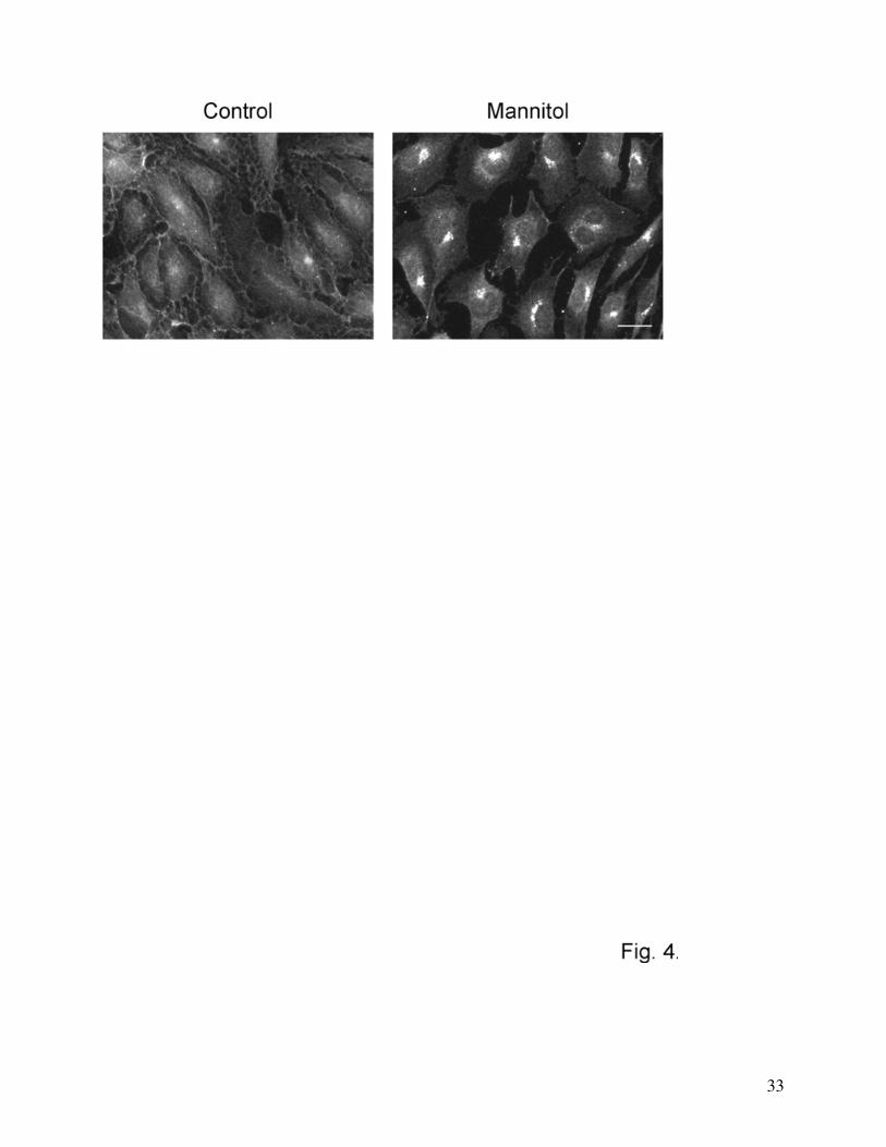
Changes in the localization of Axl in response to mannitol treatment
Results obtained with Western-blot analysis were supported by immunofluorescence studies
as well. Under control conditions Axl staining was diffuse, whereas treatment of CECs with 20%
mannitol led to the appearance of a conspicuous perinuclear staining (Fig. 4).
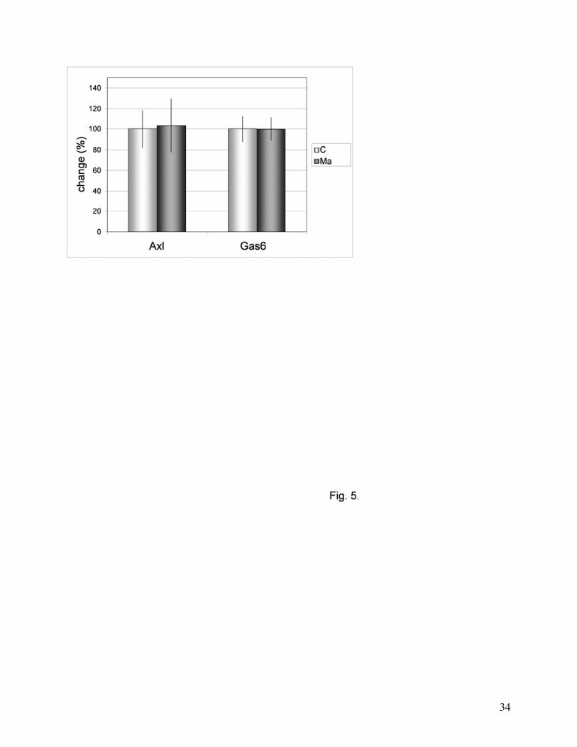
Regulation of Axl and Gas6 at mRNA level
To test if mannitol is able to regulate Axl at transcriptional level, we performed real-time
PCR experiments. We could not observe changes in the expression of either Axl or its ligand
Gas6 in response to mannitol treatment (Fig. 5).
13
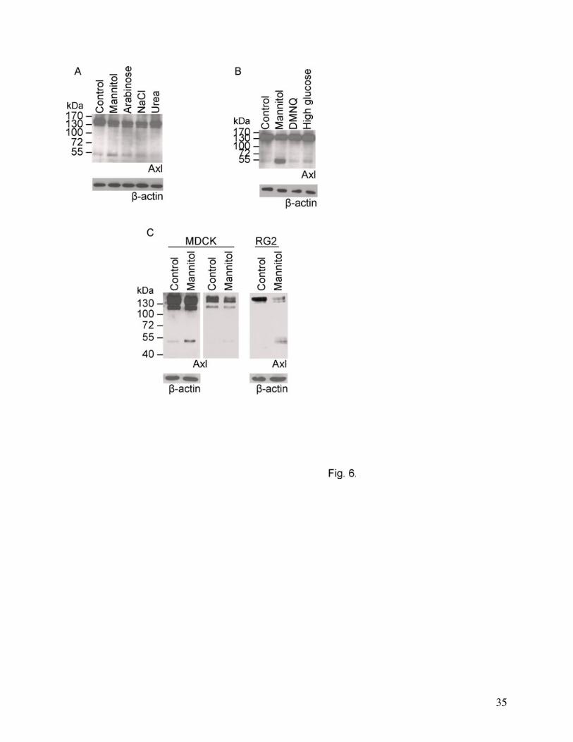
Effect of different stress factors on Axl degradation
We have tested the effect of other osmotic agents (arabinose, sodium chloride and urea) in
similar osmotic concentrations to 1.1 M mannitol (additional 1100 mOsmol/l). When osmotic
stress was caused by poorly cell permeable agents (mannitol, NaCl or arabinose) we observed the
appearance of the cleavage products with a parallel decrease in the main Axl band (Fig. 6A).
However, the cell permeable urea did not induce Axl cleavage (Fig. 6A), suggesting that cellular
shrinkage is probably responsible for Axl degradation. Other stress factors like oxidative stress
(DMNQ treatment), calcium or glucose deprivation (not shown) or high glucose concentration (8
g/l) did not cause alterations in Axl levels (Fig. 6B). Degradation of Axl in response to
hypertonic stress could be detected not only in CECs but in other cell types like Madine Darby
Canine Kidney (MDCK) or the rat glioma cell line RG2 as well (Fig. 6C).
Mechanism of Axl cleavage
To identify the mechanisms by which Axl degradation is regulated we have used a series of
inhibitors of different signaling pathways and protease inhibitors. Inhibition of nuclear factor-
kappa B (NFκB, using PDTC), Rho-kinase (using Y27632), calcium channel (using verapamil),
protein kinase C (PKC, using bisindolylmaleimide), phosphatidyl inositol 3-kinase (PI3K, using
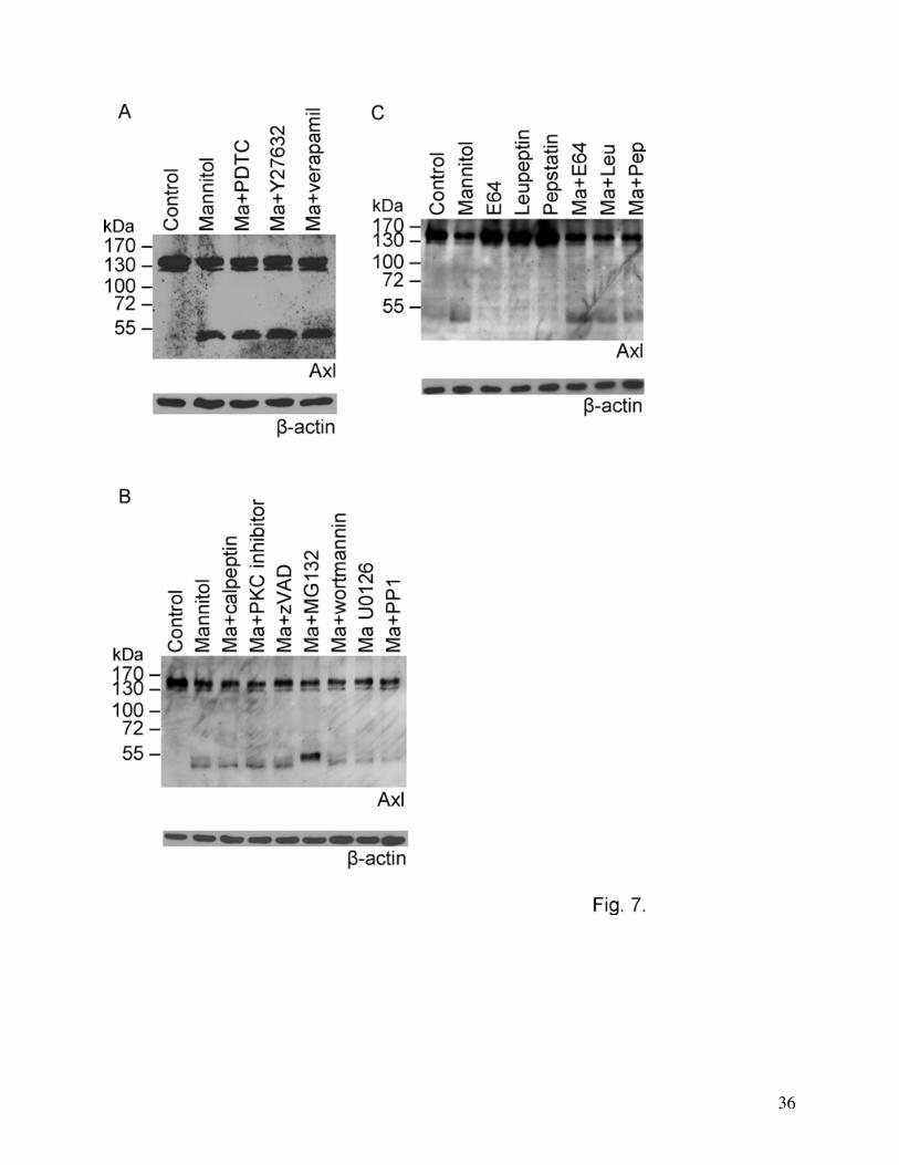
wortmannin), ERK 1/2 (using U0126) or Src kinase (using PP1) did not affect the degradation of
Axl. Caspase inhibition (using zVAD), inhibition of calpain (using calpeptin), and the use of
cysteine, serine or aspartic protease inhibitors (E64, leupeptin, pepstatin) were also ineffective in
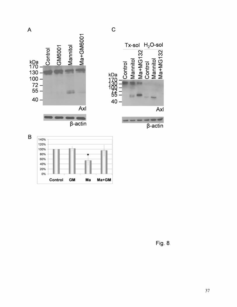
reducing the degradation of Axl (Fig. 7A, B and C). On the other hand, the matrix
metalloproteinase inhibitor GM6001 was able to almost completely inhibit the degradation of Axl
induced by mannitol (Fig. 8A and B).
14
We investigated the solubility of the different Axl bands. Only the lower band of the
degradation products proved to be soluble in detergent-free buffer. Both the whole protein and
the upper degradation band were water-insoluble and detergent (i.e. TritonX-100) soluble (Fig.
8C). The proteasome inhibitor MG132 induced the disappearance of the lower (water-soluble)
degradation fragment parallelly with the intensification of the upper (detergent-soluble) cleavage
product, however, no change in the amount of the 140 kDa band was seen (Fig. 7B and 8C).
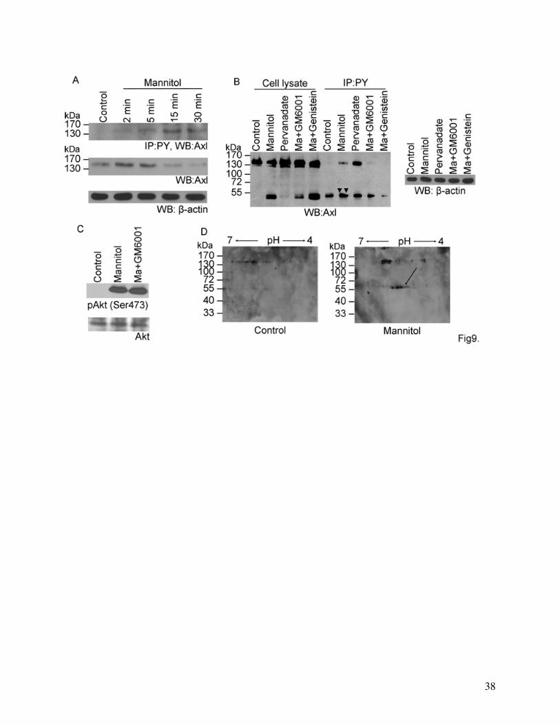
Phosphorylation and degradation of Axl
To study the possible relationship between the phosphorylation and degradation of Axl we
investigated the time course of Axl phosphorylation and degradation. Axl becomes
phosphorylated after 2 min and reaches its maximum at 15 min whereas the degradation starts
between 5-15 min (Fig. 9A). Pervanadate, a strong phosphatase inhibitor induced the
phosphorylation of Axl but did not cause its degradation indicating that the cleavage of Axl is not
induced by tyrosine phosphorylation (Fig. 9B). This was also supported by the fact that the
tyrosine kinase inhibitor genistein, which inhibited the phosphorylation of Axl, did not inhibit its
degradation (Fig. 9B). Furthermore, the metalloproteinase inhibitor GM6001 which inhibited the
degradation of Axl did not affect activation of Akt, a downstream element of Axl signaling (Fig.
9C).
Our results obtained from phosphotyrosine immunoprecipitation studies suggest that the
degradation products of Axl are phosphorylated in mannitol treated cells (double arrowheads on
Fig. 9B and 1B and C). The tyrosine kinase inhibitor genistein reduced the mannitol-induced
phosphorylation of the Axl degradation products to control levels (Fig. 9B). Presence of multiple
spots at the level of the 55 kDa degradation products on Axl Western-blots performed on two-
dimensional gels may also indicate differentially phosphorylated forms of the protein (Fig. 9D).
15
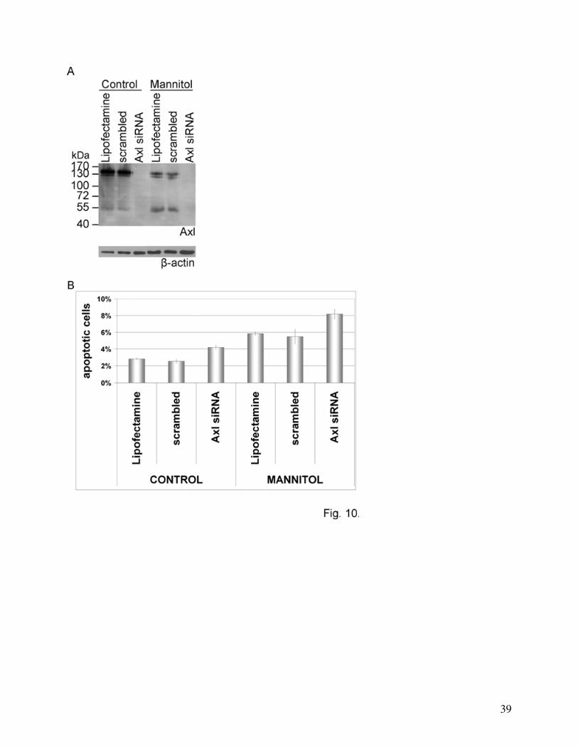
Role of Axl in mannitol-induced apoptosis
To gain insight into the role of mannitol-induced Axl activation we have counted the
number of apoptotic cells after mannitol treatment in control and Axl-silenced cells. The
knockdown was almost complete in Axl siRNA-transfected cells, however, the scrambled
sequence did not affect Axl expression (Fig. 10A). The rate of apoptosis was determined based
on cleaved caspase-3 staining. 30 min mannitol treatment did not induce significant changes in
the number of apoptotic cells. However, after 3 h mannitol treatment a more than 100% increase
was detectable: from 2.83±0.10% in Lipofectamine treated and 2.55±0.21% in scrambled RNA-
transfected cells to 5.83±0.21% and 5.50±0.85%, respectively (Fig. 10B). Axl silencing induced a
more than 50% increase in the number of apoptotic cells in control conditions, and a more than
40% increase in mannitol treated cells (Fig. 10B). Similar results were obtained when apoptosis
was assessed based on nuclear morphology (not shown).
DISCUSSION
Experiments of the present study were designed to identify signaling mechanisms activated
by hyperosmotic stress in CECs. In our previous studies (Farkas et al 2005) we have shown that
mannitol induces a strong phosphorylation on tyrosine residues in a broad spectrum of proteins
ranging between 50-200 kDa. We have further shown that among targets of tyrosine
phosphorylation are β-catenin and the non-receptor tyrosine kinase Src. Here by using a
screening approach based on an antibody array we could identify three additional proteins to
become tyrosine phosphorylated in response to hyperosmotic stress: p130Cas (p130 Crk-
associated substrate), FAK (focal adhesion kinase) and Axl. Moreover, we could confirm our
16
earlier finding that β-catenin and ERK1 are tyrosine phosphorylated in CECs in response to
hyperosmotic mannitol treatment.
p130Cas is an Src substrate signaling molecule localized to focal adhesions (Harte et al
1996) and is involved in several processes including motility, adhesion, proliferation and survival
(for review see: Defilippi et al 2006). p130Cas has already been shown to become tyrosine
phosphorylated in adipocytes in response to osmotic stress (Ueno et al 2001). FAK which is in
close interaction with p130Cas (Polte et al 1995) has also been shown to be tyrosine
phosphorylated under hyperosmotic conditions in fibroblasts and endothelial cells of non-cerebral
origin (Lunn et al 2004; Malek et al 1998).
Axl (also called ARK, UFO and Tyro7) is a member of a family of receptor tyrosine
kinases that includes Mer and Sky, having the vitamin K-dependent Gas 6 (growth arrest-specific
protein 6) as its natural ligand (for review see: Hafizi and Dahlback 2006a, 2006b). This family
of receptor tyrosine kinases is characterized by two immunoglobulin-like and two fibronectin
type III domains in the N-terminal region and a tyrosine kinase domain in the intracellular C-
terminal region. Axl has two alternatively spliced forms (O'Bryan et al 1995) that either contain
or lack 9 amino acids carboxyl-terminal to the fibronectin domains in the extracellular part of the
protein. Axl is emerging as a regulator of a large number of cellular functions and has been
shown to be involved in the regulation of different aspects of endothelial function as well. It has
been demonstrated that activation of Axl can rescue endothelial cells from apoptosis
(D'Arcangelo et al 2006; D'Arcangelo et al 2002; Hasanbasic et al 2004) and Axl is involved in
cell migration and vascular remodeling (Korshunov et al 2007; Korshunov et al 2006) and
angiogenesis (Gallicchio et al 2005; Holland et al 2005) as well.
Activation of Axl results in autocatalytic tyrosine phosphorylation, recruitment of different
signaling molecules (Grb2 and the p85 subunit of phosphatidylinositol-3 kinase) (Weinger et al
17
2008) and activation of downstream elements like Akt. By demonstrating that Akt is
phosphorylated on Ser473 residue only in cells expressing Axl we could identify a new signaling
pathway activated by osmotic stress in CECs.
Furthermore we have shown that Axl is cleaved in response to hyperosmosis, the C-
terminal products having an apparent molecular weight of about 50-55 kDa. Axl has been shown
to be posttranslationally regulated by proteolysis (O'Bryan et al 1995) resulting in the generation
of soluble Axl (Costa et al 1996). Our results show that the mannitol-induced cleavage of Axl is
metalloproteinase-dependent which is in line with previous reports demonstrating the role of
disintegrin-like metalloproteinase ADAM 10 in the cleavage of Axl (Budagian et al 2005). The
role of Axl cleavage and the function of soluble Axl is still far from being completely
understood. Soluble Axl has been detected in mouse serum and is constitutively released by
murine primary and transformed cells (Budagian et al 2005). It may play role in the regulation of
the bioavailabilty of the Axl ligand Gas6 by binding and thus inactivating it. Activation of PKC
has also been shown to trigger cleavage of Axl and the proteolytic cleavage site was found to
localize to a 14-amino acid region (VKEPSTPAFSWPWW) between the second fibronectin and
the transmembrane domain (O'Bryan et al 1995). However, in CECs the PKC inhibitor
bisindolylmaleimide was able to inhibit only PMA (phorbol myristyl acetate)-, but not
hyperosmosis-induced Axl degradation (data not shown) suggesting that there is a difference
between hyperosmosis- and PKC-induced Axl degradation.
It is well known that Axl is expressed at high levels in gliomas, mediating both tumor
growth and angiogenesis (Vajkoczy et al 2006). Specific targeting of Axl may become a
promising target of therapy of these tumors. Our results have shown that hyperosmotic mannitol-
induced cleavage of Axl was not restricted to cerebral endothelial, or tight junction-expressing
(endothelial and epithelial) cells, but glioma cells as well. Interestingly, when NaCl or arabinose
18
were used instead of mannitol in the same osmotic concentration Axl cleavage occured in the
same way, however, the cell permeable urea did not induce Axl degradation. Therefore, we
assume that cellular shrinkage might be responsible for this phenomenon.
We found that induction of tyrosine phosphorylation of Axl did not lead to its degradation,
and inhibition of tyrosine phosphorylation did not influence the mannitol-induced cleavage of
Axl. These findings suggest that there is no direct relationship between phosphorylation and
cleavage of Axl. Using an antibody recognizing the C-terminal region of Axl we have observed
that the degradation products containing the catalitically active intracellular domain of Axl
remain phosphorylated. Mainly the upper (TritonX-100 soluble) degradation band proved to be
phosphorylated, however, the two degradation bands seem to be different cleavage products and
not differently phosphorylated forms of the same cleavage products. This is supported by the fact
that treatment with genistein which inhibited the phosphorylation of Axl, did not inhibit the
appearance of two cleavage products (Fig. 9B). Moreover, the appearance of the lower
degradation band was prevented by the proteasome inhibitor MG132 (Fig. 7B and 8C) supporting
that in hyperosmotic mannitol-treated cells the metalloproteinase-mediated cleavage was
followed by a proteasomal cleavage, resulting in a water-soluble degradation product.
In order to address the functional implications of Axl activation, we have performed
knockdown experiments and examined hyperosmotic mannitol-induced apoptosis. It is well
documented that hypertonicity induces apoptosis in different cell types (for review see: Bortner
and Cidlowski 2007). On the other hand, activation of the Axl-Akt pathway is involved in cell
survival and anti-apoptotic mechanisms (for review see: Hafizi and Dahlback 2006b). We have
found an approximately 90-100% increase in apoptosis after treatment with hyperosmotic
mannitol. The relatively low absolute levels of apoptosis are apparently in contrast with previous
observations by Malek et al (1998), who have shown that treatment of bovine aortic endothelial
19
cells with 600 mOsmol/l mannitol for 3 h induced an increase in the rate of apoptosis from 1.2%
to 42%. However, microvascular endothelial cells may react differently to apoptotic stimuli than
macrovascular endothelial cells. It has been shown that hyperglycaemic conditions significantly
increased apoptosis in macrovascular endothelial cells, while increasing viability and inhibiting
apoptosis in microvascular endothelial cells (Duffy et al 2006). In our previous study we have
shown that prolonged oxidative stress even under hypoglycaemic conditions was able to induce
an apoptosis of about 7-20% with a great interspecies variability (Bresgen et al 2003). We
observed that Axl silencing increased the rate of apoptosis in hyperosmotic mannitol-treated
cells, therefore we assume that activation of Axl may be a protective mechanism against
mannitol-induced apoptosis.
Further studies are needed to clarify if Axl is involved in the hyperosmotic mannitol-
induced BBB opening. A direct relationship between Axl signaling and proteins of the tight
junction has not been shown so far. However, it is well known that a large number of signaling
pathways are able to regulate junctional functions and tyrosine phosphorylation may play an
important role in this process.
Taken together, our results identify Axl as an important element of hyperosmosis-induced
signaling in cerebral endothelial cells, and suggest that Axl could play an important role in the
adaptive response of cells to hyperosmotic stress.
Acknowledgements
This work was supported by NKTH XTTP SRT1 and GVOP-2004-0052/3.
20
REFERENCES
Abbott N. J., Rönnbäck L., Hansson E. (2006) Astrocyte-endothelial interactions at the blood-
brain barrier. Nat. Rev. Neurosci. 7, 41-53.
Arima H., Yamamoto N., Sobue K., Umenishi F., Tada T., Katsuya H., Asai K. (2003)
Hyperosmolar mannitol simulates expression of aquaporins 4 and 9 through a p38
mitogen-activated protein kinase-dependent pathway in rat astrocytes. J. Biol. Chem. 278,
44525-34.
Balint Z., Krizbai I. A., Wilhelm I., Farkas A. E., Parducz A., Szegletes Z., Varo G. (2007)
Changes induced by hyperosmotic mannitol in cerebral endothelial cells: an atomic force
microscopic study. Eur. Biophys. J. 36, 113-20.
Bortner C. D., Cidlowski J. A. (2007) Cell shrinkage and monovalent cation fluxes: role in
apoptosis. Arch. Biochem. Biophys. 462, 176-88.
Bresgen N., Karlhuber G., Krizbai I., Bauer H., Bauer H. C., Eckl P. M. (2003) Oxidative stress
in cultured cerebral endothelial cells induces chromosomal aberrations, micronuclei, and
apoptosis. J. Neurosci. Res. 72, 327-33.
Budagian V., Bulanova E., Orinska Z. et al. (2005) Soluble Axl is generated by ADAM10-
dependent cleavage and associates with Gas6 in mouse serum. Mol. Cell. Biol. 25, 9324-
39.
Burg M. B., Ferraris J. D., Dmitrieva N. I. (2007) Cellular response to hyperosmotic stresses.
Physiol. Rev. 87, 1441-74.
Costa M., Bellosta P., Basilico C. (1996) Cleavage and release of a soluble form of the receptor
tyrosine kinase ARK in vitro and in vivo. J. Cell. Physiol. 168, 737-44.
21
D'Arcangelo D., Ambrosino V., Giannuzzo M., Gaetano C., Capogrossi M. C. (2006) Axl
receptor activation mediates laminar shear stress anti-apoptotic effects in human
endothelial cells. Cardiovasc. Res. 71, 754-63.
D'Arcangelo D., Gaetano C., Capogrossi M. C. (2002) Acidification prevents endothelial cell
apoptosis by Axl activation. Circ. Res. 91, e4-e12.
Defilippi P., Di Stefano P., Cabodi S. (2006) p130Cas: a versatile scaffold in signaling networks.
Trends. Cell. Biol. 16, 257-63.
Deli M. A., Joo F., Krizbai I., Lengyel I., Nunzi M. G., Wolff J. R. (1993) Calcium/calmodulin-
stimulated protein kinase II is present in primary cultures of cerebral endothelial cells. J.
Neurochem. 60, 1960-3.
Di Ciano-Oliveira C., Sirokmany G., Szaszi K., Arthur W. T., Masszi A., Peterson M., Rotstein
O. D., Kapus A. (2003) Hyperosmotic stress activates Rho: differential involvement in
Rho kinase-dependent MLC phosphorylation and NKCC activation. Am. J. Physiol. Cell.
Physiol. 285, C555-66.
Di Ciano C., Nie Z., Szaszi K., Lewis A., Uruno T., Zhan X., Rotstein O. D., Mak A., Kapus A.
(2002) Osmotic stress-induced remodeling of the cortical cytoskeleton. Am. J. Physiol.
Cell. Physiol. 283, C850-65.
Duffy A., Liew A., O'Sullivan J., Avalos G., Samali A., O'Brien T.D. (2006) Distinct effects of
high-glucose conditions on endothelial cells of macrovascular and microvascular origins.
Endothelium 13, 9-16.
Farkas A., Szatmari E., Orbok A. et al. (2005) Hyperosmotic mannitol induces Src kinase-
dependent phosphorylation of beta-catenin in cerebral endothelial cells. J. Neurosci. Res.
80, 855-61.
22
Farrell C. L., Shivers R. R. (1984) Capillary junctions of the rat are not affected by osmotic
opening of the blood-brain barrier. Acta. Neuropathol. (Berl.) 63, 179-89.
Gallicchio M., Mitola S., Valdembri D., Fantozzi R., Varnum B., Avanzi G. C., Bussolino F.
(2005) Inhibition of vascular endothelial growth factor receptor 2-mediated endothelial
cell activation by Axl tyrosine kinase receptor. Blood 105, 1970-6.
Hafizi S., Dahlback B. (2006a) Gas6 and protein S. Vitamin K-dependent ligands for the Axl
receptor tyrosine kinase subfamily. FEBS. J. 273, 5231-44.
Hafizi S., Dahlback B. (2006b) Signalling and functional diversity within the Axl subfamily of
receptor tyrosine kinases. Cytokine Growth Factor Rev. 17, 295-304.
Harte M. T., Hildebrand J. D., Burnham M. R., Bouton A. H., Parsons J. T. (1996) p130Cas, a
substrate associated with v-Src and v-Crk, localizes to focal adhesions and binds to focal
adhesion kinase. J. Biol. Chem. 271, 13649-55.
Hasanbasic I., Cuerquis J., Varnum B., Blostein M. D. (2004) Intracellular signaling pathways
involved in Gas6-Axl-mediated survival of endothelial cells. Am. J. Physiol. Heart. Circ.
Physiol. 287, H1207-13.
Holland S. J., Powell M. J., Franci C., et al (2005) Multiple roles for the receptor tyrosine kinase
axl in tumor formation. Cancer. Res. 65, 9294-303.
Hurst R. D., Clark J. B. (1998) Alterations in transendothelial electrical resistance by vasoactive
agonists and cyclic AMP in a blood-brain barrier model system. Neurochem. Res. 23,
149-54.
Kapus A., Szaszi K., Sun J., Rizoli S., Rotstein O. D. (1999) Cell shrinkage regulates Src kinases
and induces tyrosine phosphorylation of cortactin, independent of the osmotic regulation
of Na+/H+ exchangers. J. Biol. Chem. 274, 8093-102.
23
Korshunov V. A., Daul M., Massett M. P., Berk B. C. (2007) Axl Mediates Vascular Remodeling
Induced by Deoxycorticosterone Acetate Salt Hypertension. Hypertension 50, 1057-62.
Korshunov V. A., Mohan A. M., Georger M. A., Berk B. C. (2006) Axl, a receptor tyrosine
kinase, mediates flow-induced vascular remodeling. Circ. Res. 98, 1446-52.
Kroll R. A., Neuwelt E. A. (1998) Outwitting the blood-brain barrier for therapeutic purposes:
osmotic opening and other means. Neurosurgery 42, 1083-99.
Krizbai I. A., Deli M. A. (2003) Signalling pathways regulating the tight junction permeability in
the blood-brain barrier. Cell. Mol. Biol. (Noisy-le-grand) 49, 23-31.
Lewis A., Di Ciano C., Rotstein O. D., Kapus A. (2002) Osmotic stress activates Rac and Cdc42
in neutrophils: role in hypertonicity-induced actin polymerization. Am. J. Physiol. Cell.
Physiol. 282, C271-9.
Lohmann C., Krischke M., Wegener J., Galla H. J. (2004) Tyrosine phosphatase inhibition
induces loss of blood-brain barrier integrity by matrix metalloproteinase-dependent and -
independent pathways. Brain. Res. 995, 184-96.
Lossinsky A. S., Vorbrodt A. W., Wisniewski H. M. (1995) Scanning and transmission electron
microscopic studies of microvascular pathology in the osmotically impaired blood-brain
barrier. J. Neurocytol. 24, 795-806.
Lunn J. A., Rozengurt E. (2004) Hyperosmotic stress induces rapid focal adhesion kinase
phosphorylation at tyrosines 397 and 577. Role of Src family kinases and Rho family
GTPases. J. Biol. Chem. 279, 45266-78.
Malek A. M., Goss G. G., Jiang L., Izumo S., Alper S. L. (1998) Mannitol at clinical
concentrations activates multiple signaling pathways and induces apoptosis in endothelial
cells. Stroke 29, 2631-40.
24
Neuwelt E. A., Frenkel E. P., Diehl J., Vu L. H., Rapoport S., Hill S. (1980) Reversible osmotic
blood-brain barrier disruption in humans: implications for the chemotherapy of malignant
brain tumors. Neurosurgery 7, 44-52.
Neuwelt E. A., Goldman D. L., Dahlborg S. A., Crossen J., Ramsey F., Roman-Goldstein S.,
Braziel R., Dana B. (1991) Primary CNS lymphoma treated with osmotic blood-brain
barrier disruption: prolonged survival and preservation of cognitive function. J. Clin.
Oncol. 9, 1580-90.
O'Bryan J. P., Fridell Y. W., Koski R., Varnum B., Liu E. T. (1995) The transforming receptor
tyrosine kinase, Axl, is post-translationally regulated by proteolytic cleavage. J. Biol.
Chem. 270, 551-7.
Polte T. R., Hanks S. K. (1995) Interaction between focal adhesion kinase and Crk-associated
tyrosine kinase substrate p130Cas. Proc. Natl Acad. Sci. USA 92, 10678-82.
Rapoport S. I., Fredericks W. R., Ohno K., Pettigrew K. D. (1980) Quantitative aspects of
reversible osmotic opening of the blood-brain barrier. Am. J. Physiol. 238, R421-31.
Siegal T., Rubinstein R., Bokstein F., Schwartz A., Lossos A., Shalom E., Chisin R., Gomori J.
M. (2000) In vivo assessment of the window of barrier opening after osmotic blood-brain
barrier disruption in humans. J. Neurosurg. 92, 599-605.
Sun W. S., Misao R., Iwagaki S., Fujimoto J., Tamaya T. (2002) Coexpression of growth arrest-
specific gene 6 and receptor tyrosine kinases, Axl and Sky, in human uterine
endometrium and ovarian endometriosis. Mol. Hum. Reprod. 8, 552-8.
Ueno E., Haruta T., Uno T., et al (2001) Potential role of Gab1 and phospholipase C-gamma in
osmotic shock-induced glucose uptake in 3T3-L1 adipocytes. Horm. Metab. Res. 33, 402-
6.
25
Vajkoczy P., Knyazev P., Kunkel A., et al (2006) Dominant-negative inhibition of the Axl
receptor tyrosine kinase suppresses brain tumor cell growth and invasion and prolongs
survival. Proc. Natl Acad. Sci. USA 103, 5799-804.
Weinger J. G., Gohari P., Yan Y., Backer J. M., Varnum B., Shafit-Zagardo B. (2008) In brain,
Axl recruits Grb2 and the p85 regulatory subunit of PI3 kinase; in vitro mutagenesis
defines the requisite binding sites for downstream Akt activation. J. Neurochem. In press.
Weksler B. B., Subileau E. A., Perriere N., et al (2005) Blood-brain barrier-specific properties of
a human adult brain endothelial cell line. FASEB. J. 19, 1872-4.
26
LEGEND TO FIGURES
Fig. 1. Tyrosine phosphorylation of Axl induced by mannitol.
(A): Detection of tyrosine phosphorylated proteins using antibody array. CECs were treated
with 20% mannitol for 30 min. The antibody array was incubated with the cell homogenate and
stained with anti-phosphotyrosine antibody. Identity of the proteins of interest was determined
from the position on the membrane.
(B, C): Detection of phospho-Axl using immunoprecipitation. CECs were treated with 20%
mannitol for 30 min. Immunoprecipitation was performed using anti-phosphotyrosine antibody
and blots were stained with anti-Axl antibody (B) or the samples were immunoprecipitated using
anti-Axl antibody and Western-blot was performed with anti-phosphotyrosine antibody (C).
Representative blots of three independent experiments are shown. Arrows indicate the
phosphorylated 140 kDa Axl protein. Arrowheads indicate cleavage products. β-actin was used as
loading control.
Fig. 2. Activation of Akt in response to hyperosmotic mannitol in cerebral endothelial cells.
Cells were transfected with Axl siRNA construct or Lipofectamine alone and treated with 20%
mannitol for 30 min.
(A): Axl and its degradation products disappear after silencing.
(B): Activation of Axl in response to hyperosmotic mannitol induces phosphorylation of
Akt. Blots were stained with anti-phospho-Akt (upper blot) or anti-Akt (lower blot) antibodies.
Representative blots of two independent experiments are shown.
27
Fig. 3. Effect of mannitol on the cleavage of Axl. CECs were treated with 20% mannitol for
30 min and blots were stained with anti-Axl antibody. Arrows indicate the location of the whole
Axl protein, arrowheads show the degradation products of Axl (A). One representative of ten
independent experiments is presented.
(B): Time and concentration dependence of Axl degradation. CECs were treated with 10 or
20% mannitol for 10, 30 or 60 min and blots were stained with anti-Axl antibody.
Fig. 4. Immunofluorescence staining of Axl in CECs. CECs were cultured on glass
coverslips, treated with 20% mannitol for 30 min, fixed and stained with anti-Axl antibody. One
representative image of three independent experiments is shown. Scale bar = 20 μm.
Fig. 5. Expression of Axl and Gas6 mRNA in CECs. Real-time PCR was performed to
assess the expression of Axl and Gas6 in response to mannitol. The housekeeping gene GAPDH
was used as internal control. Data represent percentage values compared to control (mean ±
SEM) calculated from four independent experiments. C = control, Ma = mannitol.
Fig. 6. Specificity of hyperosmotic stress-induced Axl degradation.
(A): Effect of different osmotic agents on the degradation of Axl. Cells were treated with
1.1 M mannitol, arabinose, urea or 0.55 M NaCl, respectively for 30 min.
(B): Effect of different stress factors on the degradation of Axl. Cells were treated with:
20% mannitol, 10 μM DMNQ or 8 g/l glucose, respectively.
(C): Degradation of Axl in MDCK and RG2 cells in response to mannitol treatment. The
first two panels show the same blot with different exposure times to visualize changes in Axl and
its degradation products. β-actin was used as loading control.
28
Fig. 7. Mechanism of Axl degradation. Cells were treated with 20% mannitol for 30 min in
the presence or absence of different inhibitors: NFκB inhibitor (10 μM PDTC), Rho-kinase
inhibitor (10 μM Y27632), L-type calcium channel blocker (10 μM verapamil) (A), calpain
inhibitor (10 μM calpeptin), PKC inhibitor (1 μM bisindolylmaleimide), caspase inhibitor (25 μM
zVAD), proteasome inhibitor (50 μM MG132), PI3kinase inhibitor (1 μM wortmannin), ERK 1/2
inhibitor (10 μM U0126) and Src inhibitor (10 μM PP1) (B) or protease inhibitors (100 μM E64,
50 μM leupeptin, 10 μM pepstatin) (C). β-actin was used as loading control.
Fig. 8. Role of metalloproteinases and of the proteasome in Axl degradation.
(A): The metalloprotease inhibitor GM6001 was able to inhibit Axl degradation. One
representative of five independent experiments is shown.
(B): Densitometric analysis of Axl Western-blots. Data represent percentage values of the
140 kDa Axl band intensity compared to control (mean ± SD) calculated from five independent
experiments. *: p<0.001 compared to control using t-test.
(C): Role of the proteasome. Protein samples were prepared from CECs treated with 20%
mannitol and 50 μM MG132 either in TritonX-100 containing buffer or detergent-free buffer.
One representative of three independent experiments is presented. β-actin was used as loading
control.
Fig. 9. Relationship between degradation and phosphorylation of Axl.
(A): Time course of Axl phosphorylation (A, upper blot) and Axl degradation (A, lower
blot). Cells were treated with 20% mannitol for the indicated times. Immunoprecipitation was
29
performed using anti-phosphotyrosine antibodies. Axl Western-blot was performed from the
immunoprecipitated samples (A, upper blot) and the cell lysates (A, lower blot).
(B): Phosphorylation of the degradation products. Cells were treated for 30 min with 20%
mannitol or 50 μM pervanadate or 20% mannitol and 10 μM GM6001 or 20% mannitol and 100
μM genistein, respectively. Immunoprecipitation was performed using anti-phosphotyrosine
antibody, followed by Axl Western-blot. Double arrowheads indicate the phosphorylated
cleavage products of mannitol-treated samples. β-actin was used as loading control.
(C): GM6001 does not influence phosphorylation of Akt. Cells were treated with 20%
mannitol for 30 min in the presence or absence of 10 μM GM6001 and blots were stained with
anti-phospho-Akt (upper blot) or Akt (lower blot) antibodies.
(D): Investigation of Axl using two-dimensional electrophoresis combined with Western-
blot. Cells were treated for 30 min with 20% mannitol. Proteins of cell lysates were separated in
two dimensions based on their isoelectric point and molecular weight, followed by Western-blot
analysis using anti-Axl antibody.
Fig. 10. Role of Axl in mannitol-induced apoptosis.
(A): Silencing of Axl.
(B): Investigation of apoptosis after Axl silencing and mannitol treatment. Cells were
cultured on glass coverslips and were transfected with Axl siRNA, scrambled RNA construct or
treated with Lipofectamine alone, followed by treatment with 20% mannitol for 3 h. The rate of
apoptosis was determined based on cleaved caspase-3 immunostaining. Values represent the
mean ± SD calculated from two independent experiments. p<0.05 among all groups except
Lipofectamine and scrambled, using ANOVA and LSD post hoc test.
30
31
32
33
34
35
36
37
38
39
Related Documents