Human Biochemistry Option B B1 Energy in Food (SL/HL) Food contains energy in the form of carbohydrates, fats and proteins. The amount of energy in a food is called the calorific value (measured in kcal per 100g). Food calorimetry involves the calculating the amount of energy in a food by its enthalpy of combustion. ( 1kcal = 4.18KJ) Calorific values are obtained using a bomb calorimeter. Find details about how this works. E.g. If 1.57g of a food is burnt completely in oxygen, 100 cm 3 of water is raised by a temperature of 5 0 . Calculate the enthalpy of combustion of the food per 100 grams, and then work out its calorific value. H = m C T = 0.1 x 4.18 x 5

Welcome message from author
This document is posted to help you gain knowledge. Please leave a comment to let me know what you think about it! Share it to your friends and learn new things together.
Transcript

Human Biochemistry
Option B
B1 Energy in Food (SL/HL)
Food contains energy in the form of carbohydrates, fats and proteins. The amount of energy in a food is called the calorific value (measured in kcal per 100g).
Food calorimetry involves the calculating the amount of energy in a food by its enthalpy of combustion. ( 1kcal = 4.18KJ)
Calorific values are obtained using a bomb calorimeter. Find details about how this works.
E.g. If 1.57g of a food is burnt completely in oxygen, 100 cm3 of water is raised by a temperature of 50. Calculate the enthalpy of combustion of the food per 100 grams, and then work out its calorific value.
H = m C T
= 0.1 x 4.18 x 5
= 2.09 kJ
= 2.09 / 4.18
= 0.50 kcal per 1.56g.
= 31.8 kcal per 100g.

E.g. Calculate the calorific value of a brand of chocolate biscuits if a 10g sample when burnt completely causes a temperature rise of 370 C for 25cm3 sample of water.B2 Proteins (SL/HL) Amino Acids
Proteins are polymers formed from 2-amino acid monomers.
An amino acid molecule contains 2 functional groups that are different.
It contains both a carboxylic acid and an amine functional group.
When both of these groups are attached to the same carbon atom, they are called 2 amino acids or amino acids.
E.g. general formula (Where R is an alkyl group)
Amino acids contain an asymmetric (chiral) carbon atom.
These molecules are therefore optically active and can exist as optical isomers.
Draw the optical isomers (enantiomers) below.

B2 Condensation Polymerisation of Amino Acids (SL/HL)
There are about 20 naturally occurring amino acids containing different R (alkyl) groups (see data booklet).
When amino acids react together, a condensation reaction occurs and water is eliminated.
The bond formed between the molecules is called a peptide bond.
The resulting molecule can continue to react giving a polypeptide condensation polymer.
E.g. Where R1, R2 and R3 are different alkyl groups.
Polypeptides are the basic building blocks of proteins.
B2 Protein Structure (SL/HL)
a) Primary Structure
The primary structure of a protein is the sequence or order of the different amino acids that make it up.
E.g. A tripeptide contains three amino acids joined together (R1 R2 and R3) but they can be in several different orders.
NH2-R1-R2-R3-COOH
NH2-R2-R1-R3-COOH

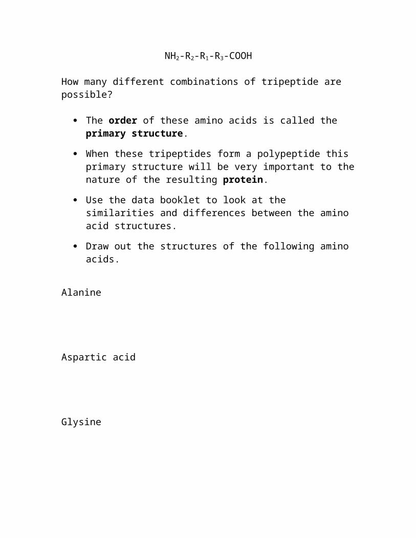
How many different combinations of tripeptide are possible?
The order of these amino acids is called the primary structure.
When these tripeptides form a polypeptide this primary structure will be very important to the nature of the resulting protein.
Use the data booklet to look at the similarities and differences between the amino acid structures.
Draw out the structures of the following amino acids.
Alanine
Aspartic acid
Glysine
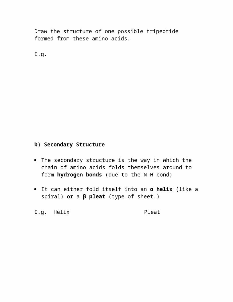
Draw the structure of one possible tripeptide formed from these amino acids.
E.g.
b) Secondary Structure
The secondary structure is the way in which the chain of amino acids folds themselves around to form hydrogen bonds (due to the N-H bond)
It can either fold itself into an α helix (like a spiral) or a β pleat (type of sheet.)
E.g. Helix Pleat
Label the H-bonds in these molecules.
c) Tertiary Structure
Further interactions between these spirals or sheets can lead to larger 3 dimensional structures.
Interactions between the R side chains that cause these structures may be:
1. Van De Waals forces between hydrocarbon groups.2. Hydrogen bonds between OH or NH groups.3. Ionic attraction between oxygen anions and nitrogen
cations.4. Disulphide bridges. This is when two sulphur atoms
on the side chains form a bond.5. Peptide bonds. Two separate chains form a peptide
bond between them. (COOH with NH2)
d) Quarternary Structure
This occurs when more than 2 spirals or sheets wrap around each other.
An example is haemoglobin that involves the interaction of 2 α helix and 2 β pleated sheets.
Use the following link to study the structure of proteins in more detail.http://www.chemsoc.org/networks/learnnet/cfb/proteins.htm
Uses of Proteins
1. Structural proteins. E.g. Collagen.2. Enzymes: Biological catalysts. E.g. Amylase.3. Hormones: Chemical messengers. E.g. Insulin.4. Antibodies: Immunoproteins.5. Transport proteins: E.g. Hemoglobin.6. Source of energy.
B2 Analysis of Proteins (SL/HL)
The structure of a protein can be analysed by 2 methods to determine the amino acids present.

In each method the protein must first be hydrolysed using a strong acid to break the peptide bonds and release the amino acids.
1. Paper Chromatography
This will determine the types of amino acids that make up the protein (it’s primary structure).
If a spot of the sample is introduced to some chromatography paper, and then a suitable solvent is used, it will rise up the paper (by capillary action) and dissolve the amino acids.
As the solvent continues to rise, the different amino acids will have different solubilities (depending on the nature of the R-side chains) and so will rise at different rates.
If the paper is sprayed with an organic dye (ninhydrin), the different amino acids will be seen at varying heights,
By measuring these heights and comparing them to the height of the solvent the retention factor (Rf) value for the each acid can be obtained.
Rf = distance travelled by sample Distance travelled by solvent.
The Rf values can be compared to known values to determine the identity of the amino acid.
If more than one amino acid has the same Rf value or separation is incomplete then another chromatogram must be produced with a different solvent. (Rotate chromatogram by 900)
2. Electrophoresis
Each amino acid has a particular pH due to the nature of the acidic or basic groups on the R-side chain. This is called the isoelectric point.

The structure of an amino acid will therefore be affected by changes in pH.
In acid conditions, when the pH is lower than the isoelectric point, the amino acid will act as a base causing the NH2 group on the acid to become protonated (-NH3
+), giving the amino acid a positive charge.
In alkali conditions, at high pH’s higher than the isoelectric point, the amino acid will act as an acid causing its COOH group to lose its proton (COO-) and become negatively charged.
At a pH that is exactly the same as the isoelectric point, the amino acid acts as both an acid and base and so has both a positive and negative charge. At this point it is said to be isoelectronic and is called a zwitterion.
These differences in acidity causing varying charges to form on the amino acids allow us to separate them according to the magnitude (size) of the charge.
E.g. A protein sample is hydrolysed with acid to break the peptide bonds and release the separate amino acid molecules.
Amino acid pH of isoelectric pointGlutamic acid 3.2Phenylanaline 5.5Serine 5.7Histidine 7.6Arginine 10.8
The amino acid mixture is applied in the centre of some chromatography paper soaked in a buffer (gel) that has a pH in the middle range of the sample (about 5.7 for this sample).
Then an electric potential difference (voltage) is applied at opposite ends of the chromatography paper.
The acids that have an isoelectric point with a pH that is more acidic than the buffer (glu and phen) will be negatively charged (due to

COO-) and will be attracted towards the positive plate. Glu will be attracted at a faster rate as is has a lower isoelectric pH.
Acids having an isoelectric pH that is the same as the pH of the buffer will contain both positive and negative charges and so will not move (Serine)
Acids with an isoelectric pH greater than the buffer will be protonated and positive (due to –NH3
+) and so will be attracted towards the negative plate. (Hist and Arg)
When separation is complete the gel is sprayed with an organic dye to see the amino acids.
The distance that the amino acid has travelled from its start position can be compared to known values/standard solutions and used to identify it.
Draw out the structures of each of the amino acid ions on the following piece of chromatography paper (pH buffer = 5.7)
Amino acid mixtureapplied here
B3 Carbohydrates (SL/HL) a) Monosaccharides
The simplest carbohydrates are called monosaccharides.

They contain a carbonyl functional group (C=O) and at least 2 alcohol groups (-OH).
They have the general formula CH2O with fructose C5H10O5 and glucose C6H12O6 among the simplest.
Both fructose and glucose can form as either a straight chain or a cyclic (ring) molecule.
The straight chain isomer is optically active having many asymmetric (chiral) carbon atoms.
Straight Chain Structure:
Fructose:
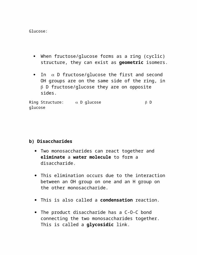
Glucose:
When fructose/glucose forms as a ring (cyclic) structure, they can exist as geometric isomers.
In D fructose/glucose the first and second OH groups are on the same side of the ring, in D fructose/glucose they are on opposite sides.
Ring Structure: D glucose D glucose
b) Disaccharides
Two monosaccharides can react together and eliminate a water molecule to form a disaccharide.
This elimination occurs due to the interaction between an OH group on one and an H group on the other monosaccharide.
This is also called a condensation reaction.
The product disaccharide has a C-O-C bond connecting the two monosaccharides together. This is called a glycosidic link.
Sucrose, lactose and maltose are examples of disaccharides. Find out which monosaccharides are react to form each of these.
Copy out the structure of sucrose from the data booklet/text book:Label the glycoside bond.
From which two monosaccharides does sucrose form?
c) Polysaccharides
When a number of monosaccharides condense together, they form a polymer structure called a polysaccharide.

Starch and cellulose are important types of polysaccharide. Which monosaccharide monomer polymerises to form starch and cellulose?
What is the structural difference between starch and cellulose polymers?
Why are most animals (including humans) able to digest/break down starch, but not cellulose?
Starch is a mixture of two isomers. The straight chain isomer is called amylose and is water soluble.
The branched chain isomer is called amylopectin and is insoluble.
In what way do the structures of amylase and amylopectin differ? Draw the structures below.
In the body, starch is initially broken down into glucose. It is then converted into another polysaccharide called Glycogen. This is the main storage carbohydrate. Its structure is very similar to amylopectin but the carbon chain is even more frequently branched.
Glycogen is stored in the liver and muscles. During prolonged exercise, when the immediate supply of glucose in the blood has been used up, the body restores the supply of glucose by breaking down glycogen. The branched structure helps in this.
Use the following link to study the structure of carbohydrates in more detail: http://www.chemsoc.org/networks/learnnet/cfb/carbohydrates.htmB3 Uses of Polysaccharides in the Body.
1. To provide energy. All types of carbohydrates are broken down to carbon dioxide and water during respiration. This is a

very exothermic reaction and releases lots of energy. E.g. Glucose.
2. To store energy. Polysaccharides (starch) are particularly good stores of carbohydrate molecules. They are often stored within the liver and released when energy is required. E.g. Glycogen.
3. As building blocks to make other important biological molecules.
B3 Dietry Fibre (SL/HL)
Dietry fibre is an essential component in a healthy diet.
It consists mainly of plant material such as cellulose, hemicellulose, lignin and pectin.
These materials cannot be digested by enzymes in the digestive tract, but may be digested by microflora in the gut.
A diet that is high in dietry fibre has been linked to the reduction of the following conditions; irritable bowel syndrome, constipation, obesity, Crohn’s disease, hemorroids and diabetes mellitus.
B4 Lipids (SL/HL)
Three types of lipids found in the human body include triglycerides (e.g. fats and oils), phospholipid (e.g. lecithin) and steroids (e.g. cholesterol)
Structure of Triglycerides Lipids are made by the condensation reaction between 3 carboxylic
(fatty) acid molecules and prop-1,2,3-triol (glycerol).
The 3 fatty acids involved may be the same or different.
The resulting lipid consists of 3 ester (glyceride) groups and is therefore called a triesters (or triglycerides).
E.g. Describe the condensation reaction of stearic acid CH3(CH2)16COOH, lauric acid CH3(CH2)10COOH and oleic acid CH3(CH2)7CH=CH(CH2)7COOH with glycerol.
Each of the ester groups are connected to a long chain non polar hydrocarbon group. This may be saturated or unsaturated (containing C=C bonds). Label the saturated or unsaturated carbon chains.
Structure of Phospholipids
These have the basic same structure as the triglyceride lipids.

The difference is that the 3rd ester group is replaced by a phosphate group (PO4
2-)
Draw out another triglyceride molecule. Use the data book to find the structure of octanoic acid and palmatic acid and use these to form 2 of your ester linkages. Add a phosphate group to the final position instead of an ester formed from a fatty acid.
One of the negatively charged oxygen atoms is usually attached to another hydrocarbon group forming a series of different phospholipids with different properties.
If an amine is the substituted group, this is called lecithin.
The fatty acid part of the molecule is hydrophobic but the phosphate group is hydrophilic.
Use the following link to study the structure of lipids in more detail: http://www.chemsoc.org/networks/learnnet/cfb/lipids.htm
Structure of Steroids
Using the data booklet, draw out the structure of cholesterol.

The central core of this molecule, consisting of four fused rings, is shared by all steroid lipids.
Cholesterol is one example of a steroid lipid. It is an essential component in the synthesis of cell membranes and is the precursor to our sex hormones and Vitamin D (see later).
Humans make about 2 g of cholesterol per day (mainly in the liver), and that makes up about 85% of blood cholesterol. Only about 15% comes from dietary sources (food).
Lipoproteins are clusters of proteins and lipids all tangled up together. These act as a means of transporting the insoluble lipids, including cholesterol, around in the blood.
There are two main categories of lipoproteins distinguished by how compact/dense they are. LDL or low density lipoprotein is sometimes termed “bad cholesterol,” being associated with deposition of cholesterol on artery walls, leading to heart disease and sometimes high blood pressure.
HDL or high density lipoprotein is termed ‘good cholesterol’. Medical experts think that HDL tends to carry cholesterol away from the arteries and back to the liver, where it's passed from the body. Some experts believe that that HDL removes excess cholesterol from artery walls, thus slowing its build up.
B4 Saturated and Unsaturated Fatty Acids
Saturated fatty acid molecules contain no C=C bonds.

E.g. palmitic acid CH3(CH2)14COOH
They tend to have higher melting points. This is because the hydrocarbon chain has a large surface area (sp3 hybridised) for the molecules to interact with each other and the strength of the Van De Waals forces is greater.
Unsaturated fatty acid molecules contain at least one C=C bond.
E.g. Linoleic acid CH3(CH2)4CH=CHCH2CH=CH(CH2)7COOH
If they contain a lot more C=C bonds they are called polyunsaturates.
Unsaturated fatty acids have lower melting points.
The presence of the C=C bonds leads to a change in the carbon bond angle from all of them 109.50 tetrahedral sp3 to some of them 1200 triganol sp2.
This means that the chains are unable to fit together as well and so the Van De Waal foces are weaker.
Most natural fatty acids are a mixture of saturated, unsaturated and polyunsaturated. They are classified according to the predominant type.
Determining the Amount of Unsaturation in fats/oils
To determine the number of unsaturated bonds within a molecule it be tested by its addition reaction with iodine.

Equation:
One mole of iodine reacts with one mole of C=C bonds.
More conveniently, the number of grams of iodine required to saturate 100g of a fat or oil are calculated. This is called the iodine number.
E.g 1. A triglyceride molecule of molar mass 300 contains one C=C bond per molecule. Calculate its iodine number.
E.g 2. A triglyceride of molar mass 250 has an iodine number of 305. Calculate the number of C=C bonds it contains per molecule.
Answers: 1. 84.6 2. Three C=C bonds
B4 Hydrolysis of Lipids During Digestion
Digestion of fats takes place in the small intestine.
The pancreas makes several enzymes, one of which is called lipase.

This enzyme works best in neutral conditions and so the pancreas also releases sodium hydrogen carbonate to neutralise the acidic conditions from the stomach.
The lipase then catalyses the hydrolysis of the lipids into fatty acids and glycerol.
E.g.
This hydrolysis reaction can be thought of as the opposite of a condensation reaction. Water is added into the molecule causing it to break apart.
B4 Energy Value of Fats Compared to Carbohydrates
Respiration:
C6H12O6 + 6 O2 6CO2 + 6H2O + ENERGY
Breaking of bonds requires less energy than the formation of the new (stronger) bonds. There is therefore a net release of energy causing an exothermic reaction.
The breaking down of fat molecules involves more bond breaking/forming than carbohydrate (glycogen) molecules.
The respiration of fats therefore results in a greater amount of energy release than the respiration of carbohydrates.
Fats are more compact and stored easier in the body; also more oxygen is needed to break down fats. This is why the body only breaks down fats when all the stored carbohydrate (glycogen) has been used up.
Uses of Fats in the Body
1. Stores of energy. Fats will be oxidised during respiration if there are insufficient carbohydrates.
2. Thermal insulation to keep warm.

3. To make cell membranes.4. Steroid hormones5. Reduce risk of heart disease: Omega-3 polyunsaturated fatty
acids.6. Lower levels of LDL cholesterol: polyunsaturated fats.
Negative effects
1. Obesity.2. Heart disease: Saturated fats formed from fatty acids such as lauric
(C12), myristic (C14) and palmatic (C16) cause increased levels of LDL cholesterol. Trans fatty acids (geometric isomers) also contribute to this as their structure means that they can also be deposited on arteries.
B5 Micronutients and Macronutrients (SL/HL) Micronutrients are substances required in very small amounts (mg or
µg) that mainly act as co factors of enzymes. Less than 0.005% of body mass.

E.g. Vitamins and trace elements such as Fe, Cu, F, Zn, I Se, Mn, Mo, Cr, Co and B.
Macronutrients are substances required in relatively large amounts. They are used for processes such as muscle contraction, nerve action and bone formation. More than 0.005% of body mass.
E.g. Proteins, fats, carbohydrates and minerals such as Na, Mg, K, Ca, P, S and Cl.
B5 Vitamins Vitamins protect the body from disease. Only vitamin D can be
synthesized in the body, the others must be obtained from a healthy diet.
Vitamins can be fat soluble or water soluble.
Fat soluble vitamins have long non-polar hydrocarbon chains. E.g. Vitamins A, D, E and K.
Water soluble vitamins contain a number of very polar O-H or N-H bonds that can hydrogen bond with water molecules. E.g. Vitamins B and C.
Heat can break down vitamins. Over cooking of food will therefore destroy most vitamins present.
Vitamins containing C=C or O-H bonds will be oxidised quite easily. Cooling in a fridge reduces the rate of this.
Food containing these types of vitamins will last longer since the vitamin will be oxidised before the food itself.
Vitamin A (Retinol)
This is a fat soluble vitamin due to it having a long non-polar hydrocarbon chain.
It is found in green vegetables and fruit (also carrots).

It is much more resistant to the high temperatures of cooking.
It is responsible for combining with a protein in our eyes that is responsible for maintaining good sight.
Use the data booklet to draw the structure of retinol. Label/circle the main functional group present.
A lack of it can lead to night blindness or a severe form of conjuctivitus (called xeropthalmia) that the most common cause of blindness.
Retinal is an essential component in the retina of the eye. It is produced by oxidising vitamin A (retinol) using light. What is the difference in the structure of retinal and retinol?
Vitamin C (Ascorbic Acid)
This is a water soluble vitamin due to its high number of O-H bonds that can hydrogen bond with water.
It is found in fresh fruit and vegetables.

Vitamin C is broken down by high temperature and so cooking reduces the amount considerably.
Use the data booklet to draw out the structure of ascorbic acid. Label/circle the main functional groups present.
It has been proved to be responsible for healing wounds and stopping infection. It is also involved in the synthesis of the protein collagen found in cartilage, tendons and ligaments.
Lack of vitamin C can cause a disease called ‘scurvy’ involving boils, lesions, rotten gums and swollen legs.
Vitamin D (Calciferol)
This is a fat soluble vitamin.
It can be found in fish, eggs and can also be made on the surface of the skin by absorption of uv light from the sun.

Use the data booklet to draw out the structure of calciferol. Label/circle the main functional groups present.
Note the similarity between calciferol and the lipid steroid structure of cholesterol.
Vitamin D is formed by the action of UV light in sunlight on cholesterol molecules that have “risen” to near the surface of the skin.
Vitamin D’s most important function is in the formation of bone structure.
A lack of this vitamin can lead to bone softening, and a malformation of the bone structure called ‘rickets’.
B5 Effects of Nutrient Deficiency (SL/HL)
Micronutrient Effect
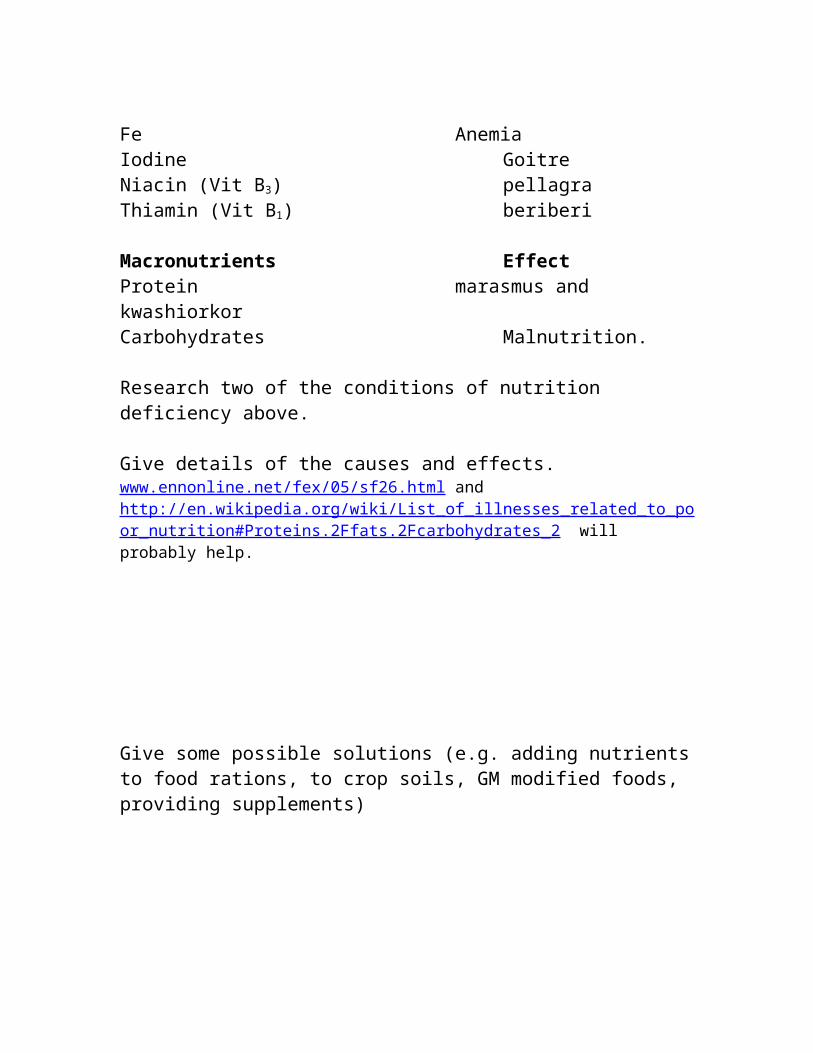
Fe AnemiaIodine GoitreNiacin (Vit B3) pellagraThiamin (Vit B1) beriberi

Macronutrients EffectProtein marasmus and kwashiorkorCarbohydrates Malnutrition.
Research two of the conditions of nutrition deficiency above.
Give details of the causes and effects.www.ennonline.net/fex/05/sf26.html and http://en.wikipedia.org/wiki/List_of_illnesses_related_to_poor_nutrition#Proteins.2Ffats.2Fcarbohydrates_2 will probably help.
Give some possible solutions (e.g. adding nutrients to food rations, to crop soils, GM modified foods, providing supplements)
To what extent will these solutions be successful in different circumstances?
B6 Hormones (SL/HL)
Hormones are chemical messengers. They bring about specific effects in the behaviour and development of animals.
Hormones do not belong to one particular chemical group. Some are amines, nitrogen-containing molecules, others are protein and
polypeptide in origin. A few are steroids that are derived from fats and lipids.
They are secreted directly into the blood by endocrine glands. The pituitary gland, found at the base of the brain, influences the activity of the other endocrine glands. It is controlled by the information it receives from the brain. This gland produces a large number of hormones that influence the activity of other glands
Adrenaline (Epinephrine)
This hormone is produced by two small glands near the kidneys.
It is a stimulant released in times of excitement that is responsible for expansion of the airways, increasing the heartbeat and releasing sugar (glucose) into the bloodstream.
Look at the structure of an adrenaline molecule in the data booklet.
Draw out the molecule and label some of the principal functional groups.
Insulin
This hormone is formed in the pancreas and is responsible for regulating the blood sugar levels.
Diabetics have a low level of insulin which means that their blood is not able to transfer glucose to the tissues and cells very efficiently.

This can result in always feeling tired, poor circulation and in severe cases can lead to the person going into a coma (hyperglycaemia).
It is treated by taking daily insulin injections.
The level needs to be monitored carefully as too much insulin can cause the transfer of glucose from the blood to the cells to be too effective. This decreases the blood sugar level and can cause dizziness and fainting (hypoglycaemia).
Thyroxine
Produced by the thyroid gland in the back of the neck.
This hormone controls the body’s metabolism. (e.g. breaking down carbohydrates)
This molecule contains iodine atoms.
A lack of iodine in the diet can lead to low levels of this hormone. This may lead to tiredness and weight gain.
An overactive thyroid gland produces larger amounts of thyroxine hormone and has the opposite effect such as anxiety and weight loss.
Use the data booklet to draw out the structure of thyroxine. Label the functional groups.
Aldosterone
This hormone controls the concentration of salt (sodium ions) in our blood.
When the body loses sodium ions, it loses water by osmosis, blood volume decreases and this causes blood pressure to decrease.

This causes the adrenal glands (near the kidney) to produce the hormone aldosterone.
Aldosterone increases the re absorption of salt from the intestine and kidneys, leading to greater retention of water and the blood pressure increases back to normal.
A diet high in salt (in many western countries) has the opposite effect. The body retains more water causing increased blood pressure.
ADH (anti-duretic hormone)
This hormone controls the concentration of water in the blood.
When the body has lost water, receptors stimulate the thirst centre in the brain and also stimulate the pituitary gland to release anti-duretic hormone (ADH).
ADH sends chemical messages to the kidneys to reabsorb water so that much more concentrated urine is produced.
Conversely, when fluid intake is very high, less ADH is produced and more water is released from the blood by the kidneys making more dilute urine.
Sex Hormones
Sex hormones are based on the lipid steroid structure.
They are synthesized from cholesterol.
Steroid sex hormones include the female oestrogens and progestins (mainly estradiol and progesterone) and the male androgens, (mainly testosterone).
Use the data book to draw the structure of these sex hormones. Label the main functional groups (note the similarity with structure of cholesterol).
The male and female sex hormones are very similar in structure, but contain slightly different functional groups. What are the main differences?
Testosterone is made in the testes (small amounts are also made in the overies). It is responsible for the male sexual characteristics. It is an anabolic steroid. This means it is also responsible for muscle and bone growth.
Progesterone and estradiol are produced in the ovaries. They are responsible for sexual development, the menstrual and reproductive cycles in females.
Anabolic Steroids
These have a very similar structure to testosterone that is responsible for muscle growth.
They were designed for patients whose muscles had wasted during pre-longed illness.

Some athletes use them to enhance performance but they can be detected in urine samples. Side effects include impotence, sterility, liver damage and heart disease.
Oral Contraceptives (Birth control)
During ovulation, if the egg is fertilised with a sperm and implants into the wall of the uterus it causes a large increase in the levels of female sex hormones.
During pregnancy these high levels of hormone are one of the reasons that the female stops ovulating.
The ‘pill’ involves a synthetic mix of progesterone and oestrogen that ‘mimics’ these hormones keeping the constantly levels high.
This prevents ovulation and therefore pregnancy.
The ‘mini pill’ involves a lower dose of a synthesized chemical equivalent of progesterone only.
This doesn’t prevent ovulation, but changes the composition of the cervical mucus, thus preventing sperm from entering the uterus and fertilizing the eggs.
B7 Enzymes (HL ONLY)
Characteristics of Enzymes
Enzymes are protein molecules.
They are particularly efficient biological catalysts. They can increase the rate of reaction by more than a million times!

They work by providing an alternative pathway for a reaction mechanism that has a lower activation energy.
Draw an enthalpy level diagram describing a catalysed and uncatalysed reaction. Label the enthalpy change and activation energy.
Unlike inorganic catalysts, enzymes are much more specific about the type of reaction that they catalyse.
The surface of the enzyme has an active site of a specific shape.
This shape depends upon the nature of the tertiary and quaternary structure.
It will only accept a reactant of a particular shape that fits into this site.
This makes enzymes very specific to the type of molecules that they will bond with.
One theory suggests that enzymes have a slightly flexible active site that is able to alter its shape slightly to make a stronger connection with the reactant molecule.
This is called the induced fit theory.
The active site of the enzyme is able to form a reversible complex with the substrate molecule.

The substrate absorbs onto the surface of the enzyme, forming a bond with the active site to give an enzyme-substrate complex.
The bonds formed in the complex weaken those in the substrate allowing them to break more easily. This lowers the activation energy of reaction.
The enzyme then releases the product molecules from the active site.
Draw diagrams to help explain the various stages of the induced fit theory. Label the active site and the enzyme-substrate complex.
B7 Rate of Enzyme Catalysed Reactions (HL ONLY). The rate of reaction is proportional to the concentration of the
reactant.
This is a first order reaction so that if the concentration of the reactant doubles, then the rate of reaction doubles.
However, this is only true at low concentrations. At higher concentrations, the rate of reaction can no longer continue to double as the concentration doubles.
This is because the number of active sites is fixed (limiting).

At a particular concentration of reactant, 30% of them may be occupied. If the conc. is doubled, then 60% will be occupied. If it is doubled again, the enzymes do not have sufficient active sites left to double the rate (it is saturated).
The rate of an enzyme catalysed reaction therefore reaches a maximum when all the sites are occupied. This is called Vmax.
Rate
[substrate]
When the rate of reaction is half its maximum value (½ Vmax), the concentration of the reactant is known as the Michaelis-Menton constant (Km).
This can be obtained easily from a graph of the rate against concentration. It indicates how well an enzyme functions at lower concentrations of reactant.
The lower the value of (Km), the more efficient the catalyst.B7 Competitive and Non-Competitive Inhibition (HL ONLY) If an enzyme is too active within the body, this may lead to health
problems.
One way to reduce the effectiveness of an enzyme is to use a substance that inhibits its action.
This is called an inhibitor.
There are 2 types of inhibitor that work in slightly different ways.
Competitive inhibitors are designed to have the same shape as the substrate molecules, but they are unable to react.
They therefore compete (with the substrate molecules) to occupy the active site of the enzymes and in so prevent the substrate from doing this.
However, as the concentration of the substrate increases, the effectiveness of a competitive inhibitor is reduced since it has more competition to reach and occupy the active sites first.
Rate
[Substrate]
Competitive inhibitors have the same Vmax, but Km is larger than the original.
Competitive inhibitors are therefore most effective at low substrate concentrations.
The second type of inhibitors is called non-competitive.
They work by bonding with the enzyme but not on its active site.
This bonding causes the enzyme to change the shape of its active site so that it is unable to bond with other reactants.
Non-competitive inhibitors are not affected by changes in concentration of the reactant, since they are not in competition with it.
Rate
[substrate]
Non-competitive inhibitors have a lower Vmax but the same Km as the original.
Non-competitive inhibitors are most effective at higher substrate concentrations.
B7 Factors That Affect Enzyme Activity (HL ONLY)
1. Temperature
An increase in temperature will increase the rate of a reaction involving enzymes as more of the particles will have the minimum energy required to react.
However, if the temperature is increased above about 45 degrees, then the enzyme will stop working.

This is because the temperature affects the weak bonds that hold the tertiary structure together.
The enzyme is said to be denatured.
Sketch a graph of temperature against rate for an enzyme catalysed reaction.
2. pH
Different pH values will also affect the bonds that hold the tertiary structure together.
This makes enzymes very sensitive to changes in pH.
3. Heavy Metal Ions.
The heavy metal ions that are often caused by pollution can poison an enzyme by reacting with and replacing the functional groups in the active site, making it ineffective.
Use the following link to study the characteristics of enzymes in more detail.http://www.chemsoc.org/networks/learnnet/cfb/enzymes.htmB8 Nucleic Acids (HL ONLY)
Structure of DNA and RNA
Two types of nucleic acids found in almost all cells.
Ribonucleic acid (RNA) and deoxyribonucleic acid (DNA).
These are both macromolecules (very large structures) with very high molar masses.

The repeating units in these structures consist of 3 groups. These units are called nucleotides.
One part of the nucleotide is a phosphate group containing phosphorus and oxygen atoms.
Another part is an organic base group containing N atoms.
The final part is a (pentose) sugar group that contains OH groups.
PHOSHATE SUGAR BASE
This 3 group nucleotide unit reacts with other nucleotide units to form a polymer.
This is a condensation reaction. It occurs between the phosphate group of one nucleotide and the sugar group of the next.
This condensation reaction eliminates water and gives the nucleic acid (DNA or RNA)
The bond that forms between the units is called a phosphodiester bond.
Differences Between DNA and RNA
DNA and RNA contain a different sugar group in their structure. (Deoxyribose for DNA compared to ribose for RNA).
DNA and RNA consist of 4 possible bases.
Three of the bases are the same for (A, C, G), but the fourth base is different. (T for DNA compared to U for RNA).
RNA forms a single helix structure.

In DNA the two polymers wind around each other to form a double helix.
The interactions between the two polymer helix are in the form of hydrogen bonds formed between the N-H groups in the base side chains.
The formation of these hydrogen bonds is very specific, for example adenine (A) will only hydrogen bond with thymine (T) and cystosine (C) will only H bond with guanine (G).
This means that the order of the bases is very important to the structure of the DNA.
Use the data booklet to find the structure of these bases. Draw the structure and show the hydrogen bonding expected between each.
A and T C and G
Protein Synthesis: Transcription and Translation.
A long DNA double helix polymer makes up a chromosome. Chromosomes come in pairs and exist in the nucleus of cells. Humans’ cells contain 46 chromosomes.
This is where all the genetic information of the organism is stored. An individual inherits some of this genetic information from their parents.
DNA can be thought of as a permanent reference library. It does not leave the nucleus and so cannot be used directly for protein synthesis. Instead the genetic code in a gene is transferred (transcribed) onto a smaller RNA molecule (messenger mRNA) that passes out of the

nucleus and acts as a template for protein synthesis (translation) in the cytoplasm.
An enzyme causes part of the double helix structure of a DNA molecule to unzip, breaking the H bonds between the bases.
The enzyme moves along the unzipped section attaching nucleotides that match the base sequence.
This single polymer copy is the messenger mRNA. It passes out of the nucleus acting as a mobile copy of the gene. This process is called transcription.
In the cytoplasm, the base sequence on the mRNA is used to synthesize a polypeptide. This process is called translation because the information on the gene (delivered by the mRNA) is translated into a particular polypeptide that then makes a particular protein.
A gene is a region within a DNA molecule. The base sequence in a gene is read in blocks of three, called a triplet or codon.
Each codon codes for the synthesis of a particular amino acid. The sequence of codons therefore builds up the sequence of amino acids in the polypeptide (primary structure).
E.g. Base sequence codon on mRNA Amino acid synthesizedGGA GlycineAAG lysineUCG serineUGA STOP
There are 4 different bases, therefore there are 64 different triplet codes (GGG, GGC, GGA, etc.).
Since there are only 20 possible amino acids, more than one triplet code can synthesize the same amino acid.
Each gene can synthesize one polypeptide. A number of polypeptides interact to produce a protein.
Summary
The DNA of a gene codes for the production of messenger RNA which codes for the production of a polypeptide.
Use the following link to study DNA in more detail.http://www.chemsoc.org/networks/learnnet/cfb/nucleicacids.htm
DNA Profiling
Each individual (except identical twins) contains different DNA sequences of bases.
If a sample of cells is obtained (blood, hair, saliva, semen etc.) is obtained it allows the identity of a person to be determined. This is often used in court cases as forensic evidence or in paternity cases.
The DNA must be extracted from the sample and purified.
Then the DNA strands must be broken down into smaller fragments using a restriction enzyme.
It is then separated in a similar way to electrophoresis and sprayed with a radioactive type of dye (31P).
The DNA profiles obtained will contain dark bands unique to an individual. These can be compared to known suspects (or to possible fathers in paternity cases) to allow identification.
TOK: In 1953, Crick and Watson worked out the structure of DNA. (They also devised a hypothesis for the role of mRNA). This has allowed scientists to understand more fully the chemistry involved in the ‘molecular basis of life’. What is the significance of these discoveries?
B9 Respiration (HL ONLY)
Aerobic Respiration
This describes the cell processes that require oxygen to release energy from organic molecules, usually provided in food.
In humans these molecules are mainly glucose, but if the need arises, fatty acids and amino acids can also be used.
C6H12O6 + 6O2 6CO2 + 6H2O + ENERGY
Since the product bonds are much stronger than the reactant bonds, this is a highly exothermic reaction.
It is also a redox reaction. Label the oxidation states of the elements involved.

Which elements are oxidized and reduced during this process? What transfer of electrons occurs?
In reality this process is controlled by splitting it into a variety of steps, so that the energy is released when required and in controlled amounts.
Initially the six carbons in the glucose molecule are split into two molecules each of three carbon atoms (called pyruvate C3H3O3).
C6H12O6 3 C3H3O3 + 6H+ + 3e-
The pyruvate then undergoes a complex set of reactions that eventually result in combing with oxygen and releasing energy (stored in the form of a molecule called adenosine triphosphate ATP ).
ATP slowly releases the energy when and where it is required.
In many ways, the driving force behind these processes is the electron transfer involved in the redox reactions.
Anaerobic Respiration
This describes the cell processes that DO NOT require oxygen to release energy.
In anaerobic respiration, glucose is broken down into pyruvate. This reaction releases only a fraction of the energy (as ATP molecules) compared to aerobic respiration.
Since no oxygen is present, the pyruvate cannot be broken down any further to release more energy.
In animals, the pyruvate is instead reduced to lactate (C3H6O3), the build up of which in the muscles causes fatigue. When oxygen becomes available again, it breaks down the lactate (in the liver). This is called the oxygen debt.
C3H3O3 + 3H+ + 3e- C3H6O3

In microorganisms (such as yeast), pyruvate is reduced into ethanol and carbon dioxide. This process is called fermentation.
C3H3O3 + 3H+ + 3e- C2H5OH + CO2
B9 Copper ions in Electron Transport (HL ONLY)
During respiration, the body breaks down glucose by oxidising it to produce energy.
This oxidation is catalysed by an enzyme called cytochrome.
Cytochrome contains copper ions in a complex bonded to four nitrogen atoms.
The ability of the copper ion to change its oxidation state (by losing or gaining electrons) in this complex catalysis the oxidation of the glucose.
The copper ions are reduced from +2 to +1 by gaining electrons. This causes a complex set of reactions but fundamentally it causes the carbon in the glucose to be oxidised (initially into pyruvate and eventually into carbon dioxide).

3 Cu2+ + 3 e- 3 Cu+
C6H12O6 3 C3H3O3 + 6H+ + 3e-
During the reduction of oxygen into water, the copper ions are re-oxidised back into the +2 state.
2Cu+ 2Cu2+ + 2e-
½ O2 + 2e- O2-
Since this process involves the copper ions gaining electrons and then losing them, this process is described as electron transport.
B9 Iron ions in Oxygen Transport (HL ONLY) Haemoglobin in the blood carries oxygen around the body
required for respiration.
Haemoglobin molecules are large biological protein molecules with an iron +2 ion complex in its centre, bonded to four nitrogen atoms that are each part of polypeptide chain. This is called the haem ring.
The nitrogen atoms in these groups act as ligands by donating electrons into the empty orbitals of the iron ion.
At the lungs, there is a high oxygen concentration and the haemoglobin molecules are able to bond with oxygen molecules (due to their non bonding electrons).
This is done by an extra coordinate bond forming between the oxygen and the iron ion, so that the oxygen becomes a fifth ligand.

When reaching cells that require oxygen, the iron ion breaks its bond with the oxygen molecule and releases.
HHb + O2 H+ + HbO2-
The iron is able to function in this way due to its ability to form a reversible complex with oxygen molecules.
Carbon monoxide and cyanide ions are very poisonous as they form non-reversible ligands with the iron ion in the haemoglobin molecule, preventing it from carrying oxygen.
Chemistry @ Shatin College, Hong Kong.
Related Documents