HUDSON EMERGENCY MEDICAL SERVICE EMS Protocols 2 0 1 6 Basic & Advanced Life Support Revision: 40.6

Welcome message from author
This document is posted to help you gain knowledge. Please leave a comment to let me know what you think about it! Share it to your friends and learn new things together.
Transcript

HUDSON EMERGENCY MEDICAL SERVICE EMS Protocols
2
0
1 6
Basic & Advanced Life Support
Revision: 40.6

HEMS Protocols
Revision: 40.6 Return to: 0-2 Table of Contents
PREFACE This EMS Protocol and procedure manual was established to provide an opportunity for optimal patient care coupled with multiple levels of EMS providers functioning within this city. This agency’s medical director is the sole physician authorized to provide Medical Control Authority to personnel within this agency. Each person functioning under the auspices of this agency is required to have the specific approval and authorization of the Medical Director. Errors in pre-hospital care are generally errors of omission. The EMS provider will be proactive in the implementation of these Protocols, and should not withhold or delay any indicated intervention. Providers should remember to “FIRST DO NO HARM”. GUIDELINES AND PROTOCOL This document contains both general guidelines and specific EMS Protocols for use by our EMS providers. This document will be made available to any interested EMS Medical Director. Volunteer or career, emergency medicine demands a strong commitment to the profession. It is the responsibility of each EMS provider to remain current in the lifelong process of EMS education. EMS providers are heavily encouraged to attend any available continuing education opportunities. Pre-Hospital and Emergency medicine continues to evolve at a rapid pace. Accordingly, this document is subject to revision as new information becomes available and consistent with currently acceptable medical practices.

HEMS Protocols
0-3 Return to: Revision: 40.6 Table of Contents
CONTINUOUS QUALITY IMPROVEMENT To maximize the quality of care in EMS, it is necessary to continually review all EMS activity in order to identify areas of excellence and topics for improvement. This approach allows for continuous improvement and optimal care. CQI is defined as a proactive process of systems evaluation to assess current performance and mold methodologies and practice patterns to further improve overall performance. Components of CQI include: active communications, documentation, case presentations, protocol review and refinement, medical direction involvement, medical community involvement, continuing education, and reassessment of expected goals and outcomes. Participation in the CQI process is mandatory in order to function within this system. The primary focus of CQI is on “system performance”. Specifically CQI focuses on the bigger picture of our system, including protocols, guidelines, equipment, training and standard operating procedures, rather than on the individual care provider, or patient. The EMS Medical Director may request additional documentation, for the purpose of gathering information about a particular call, event or procedure in question. Failure to cooperate with the CQI or quality assurance process may result in withdrawal of Medical Direction authorization.
DISCLAIMER Every attempt has been made to reflect sound medical guidelines and protocols based on currently accepted standards of care for out of hospital emergency medicine. It is the reader’s responsibility to stay informed of any new changes or recommendations made at the State or service level, and adopted by this agency. Despite our best efforts, these guidelines may contain typographical errors or omissions.
Activities of EMS personnel must be in compliance with all applicable federal, state, county and local laws and regulations including Section 4765.09 of the Ohio Revised Code.

TABLE OF CONTENTS DESCRIPTION PAGEProtocol Authorization 0-1Preface 0-2Guidelines and Protocol 0-2Continuous Quality Improvement 0-3
SECTION ONEGEN. ADMIN. GUIDELINES PAGEGeneral Policies 1-1Scope of Practice 1-2Reaction Time 1-2Communication 1-3Radio Report 1-4Patient Restraint Guidelines 1-5-7Medical Control Contact Exemption 1-8-9Non Transport Advisory (Code 1) 1-10Refusal of Care 1-11-13Level of Care Reassignment 1-14Do Not Resuscitate 1-15-16-17Death in the Field 1-18-19Termination of Resuscitative Efforts 1-20Remote Death Pronouncement 1-21Children w/Special Health Care Needs 1-22-23Heavy Patients 1-24Physician at the Scene 1-25On Scene EMT Intervener 1-25Advanced Care Medications 1-26Non Hospital Transfers 1-26Medical Patient Transport Destination 1-27Emergency Transport 1-28Firefighter Rehab 1-29Transportation of Service Animals 1-30-31
SECTION TWOGEN. MED./TRAUMA ASSESS. AND TREAMENT GUIDELINE PAGEInitial Med. Assess. & Management 2-1-3Initial Trauma Assess. & Management 2-3-5Medical Supportive Care 2-6Trauma Supportive Care 2-7ALS Cardio-Respiratory Monitoring 2-8Airway Management 2-9Aerosolized Bronchodilator Therapy 2-10

Gen. Med/Trauma Assess / Tx. PAGECapnography 2-11Continuous Positive Airway Pressure 2-12Steriod Administration 2-13Epinephrine SQ 2-14Epinephrine Infusion 2-15-16Tylenol Protocol 2-17Sedation Protocol 2-18Pain Management Protocol 2-19-20
SECTION THREE CARDIAC ARREST PAGEInitial Approach 3-1Asystole 3-2V. Fib / Pulseless Wide Complex Tach 3-3Pulse Electrical Activity (PEA) 3-4
SECTION FOURCARDIAC EMERGENCIES PAGEAcute Myocardial Infarction (AMI) 4-1Acute Pulmonary Edema (PE) 4-2Cardiogenic Shock 4-2Wide Complex Tachycardia 4-3Symptomatic A.Fib / Flutter 4-4Premature Ventricular Contraction (PVC) 4-5Symptomatic Bradycardia / AV Blocks 4-6Supraventricular Tachycardia (SVT) 4-7
SECTION FIVERESPIRATORY EMERGENCIES PAGEChoking 5-1Asthma / Bronchiolitis 5-2Chronic Obstructive Pulmonary Disease 5-3Hyperventilation Syndrone 5-4
SECTION SIXEXPOSURE/TOXICOLOGY EMERGENCIES PAGECarbon Monoxide Inhalation 6-1Cold Exposures 6-2Cyanide Poisoning 6-3Near Drowning 6-4Hazardous Materials 6-5Nerve Agent Exposure (WMD) 6-6-7Heat Related Illness 6-8
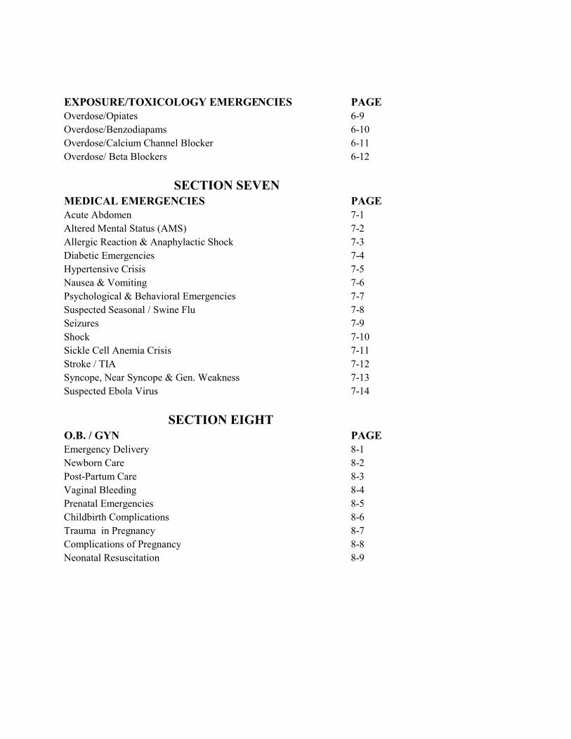
EXPOSURE/TOXICOLOGY EMERGENCIES PAGEOverdose/Opiates 6-9Overdose/Benzodiapams 6-10Overdose/Calcium Channel Blocker 6-11Overdose/ Beta Blockers 6-12
SECTION SEVENMEDICAL EMERGENCIES PAGEAcute Abdomen 7-1Altered Mental Status (AMS) 7-2Allergic Reaction & Anaphylactic Shock 7-3Diabetic Emergencies 7-4Hypertensive Crisis 7-5Nausea & Vomiting 7-6Psychological & Behavioral Emergencies 7-7Suspected Seasonal / Swine Flu 7-8Seizures 7-9Shock 7-10Sickle Cell Anemia Crisis 7-11Stroke / TIA 7-12Syncope, Near Syncope & Gen. Weakness 7-13Suspected Ebola Virus 7-14
SECTION EIGHT O.B. / GYN PAGEEmergency Delivery 8-1Newborn Care 8-2Post-Partum Care 8-3Vaginal Bleeding 8-4Prenatal Emergencies 8-5Childbirth Complications 8-6Trauma in Pregnancy 8-7Complications of Pregnancy 8-8Neonatal Resuscitation 8-9

SECTION NINEPEDIATRIC EMERGENCIES PAGEAsthma / Bronchiolitis 9-1Croup / Epiglottisis 9-2Pediatric Asystole 9-3Pediatric Bradycardia 9-4Pediatric Febrile Emergency 9-5Pediatric Pulseless Electrical Activity 9-6Pediatric Tachycardia 9-7Pediatric V Fib / Pulseless V tach 9-8Pediatric Altered Level of Consciousness 9-9Pediatric Seizure 9-10Pediatric Nausea & Vomiting 9-11Pediatric Fluid and Drug Administration 9-12
SECTION TEN TRAUMA EMERGENCIES PAGEBurns 10-1Chest Injuries 10-2Head Injuries 10-3Orthopedic Injuries 10-4Opthalmic Injuries 10-5Nosebleed (Epistaxis) 10-6Soft Tissue Injuires 10-7Major Traumatic Injuries 10-8Spinal Care 10-9-10,11Suspected Abuse &Neglect 10-11Suspected Sexual Assault 10-12 PHARMACOLOGY APPENDIX A A-1 thru A-47
BLS PROCEDURES APPENDIX B B-1 thru B-46
ALS PROCEDURES APPENDIX C C-1 thru C-47
TRAUMA ALERT PROTOCOL APPENDIX D D-1 thru D-4
CHARTS & SCORECARDS APPENDIX E E-1 thru E-4

MEDICATION CALCULATIONS APPENDIX F F-1 thru F-3
TRIAGE "START", "JumpSTART" APPENDIX G G-1 thru G-8
ABBREVIATIONS / SYMBOLS APPENDIX H H-1 thru H-5
TRANSPORT DESTINATIONS APPENDIX I I-1 thru I-4
NOTES APPENDIX N

GENERAL ADMINISTRATIVE PROTOCOLS
SECTION 1

HEMS Protocols
1-1 Return to: Revision: 40.6 Table of Contents
GENERAL POLICIES
All personnel shall conduct themselves in a professional manner at all times during which they are on duty, or in uniform.
All personnel shall provide care encompassing procedures and medication
administrations only up to their level of training, certification, Protocols, and pre-approved departmental authorization.
Hudson EMS is at the service of all the citizens and visitors to the City of Hudson and
will not deny care or medical service to any patient based on their race, creed, religion, sexual preference, ability to pay, location, or pre arrival care.
All patients and family members are to be treated with due respect.
All personnel are individually responsible for being up to date on all departmental policies, procedures, and protocols.
This department’s Medical Direction authorization exists only while functioning under the
auspices of this department.
Medical records, (Patient Care Reports), and associated documentation shall be accurate, complete, and timely.
Patient confidentiality is to be respected at all times.
All personnel shall conduct operations in a manner so as to minimize undue risk, harm, or
injury to themselves and their crews.
All personnel shall maintain departmental, State, and other regulatory credentials as required, and shall provide documentation of such as requested by the Medical Director or departmental director.
All medications, procedures, and ancillary medical equipment are to be specifically
approved by the Medical Director.
The Medical Director is the only physician who can provide authorization, (Medical Direction), to function in a medical capacity, or related support capacity, within this department.
The Medical Director may limit, suspend, withdraw, or revoke an individual’s Medical
Direction authorization at any time, at the sole discretion of the Medical Director.
Medical Direction and this department recognize a “Zero Tolerance” policy regarding illicit drug and alcohol usage amongst health care providers.

HEMS Protocols
Revision: 40.6 Return to: 1-2 Table of Contents
SCOPE OF PRACTICE
The Hudson Emergency Medical Service will, as a general principle, provide an Advanced Life Support (ALS) level of service and patient assessment to each patient encountered by our service. For the purposes of this policy, an ALS response presumes an ALS patient assessment by an EMT-P, (Paramedic), will occur. When HEMS resources preclude an ALS assessment, it may be undertaken through other means such as mutual aid. Non-ALS assessment may, on rare occasion, be appropriate. Examples include patients with minor concerns who otherwise meet the On Line Medical Control contact exemption policy, or those with significant concerns facing a significant delay in transport pending an ALS response. Non-ALS transport may be undertaken, when medically appropriate, following both an ALS assessment and On Line Medical Control authorization. It is acknowledged that on occasion deviation from this protocol may be required due to scene safety, scene management, or due to other extenuating circumstances. Such events shall be noted within the patient's PCR, or via the appropriate form. Sanctioned deviation is at the sole discretion of the Medical Director. Multiple simultaneous patient encounters, (e.g. LVI’s & MCI’s), are excluded from this policy. Either ALS or BLS assessment may be undertaken, as appropriate, per Incident Command – Medical Control guidance.
REACTION TIME It is the goal of this department to have the responding ambulance be en route to the scene of a call within 1 minute, (60 seconds), of the crew being toned/alerted. It is noted that this may not be met when verifying unfamiliar addresses, or mutual aid response addresses. This goal is in effect 24 hours a day. This goal does not apply to secondary responding units, (e.g. departmental officers, field supervisors, etc.). This policy specifically addresses the time to mobilize the responding crew. It does not pertain to the en route response time.
HEMS Protocols
1-3 Return to: Revision: 40.6 Table of Contents
COMMUNICATION
A member of the pre-hospital care team must contact Medical Control at the earliest time conducive to good patient care. This may be a brief early notification or “heads up”. It may mean that the hospital is contacted from the scene if assistance is needed in the patient's immediate care or permission is required for part of the patient care deemed necessary by the paramedic or EMT in charge. When possible, the member of the team most knowledgeable about the patient should be the one calling in the report. Reports should contain the key information in an organized manner, while striving to be concise. Additional information can be provided during one’s beside report at the receiving facility. If multiple victims are present on the scene a preliminary report will be given to Medical Control. This should be an overview of the scene, including the number of victims, seriousness of the injuries, estimated on-scene and transport times to the control hospital or possible other nearby facilities. Western Reserve Hospital and Children’s Hospital Medical Center of Akron will be used as On Line Medical Control for any controversial decisions that the crew may need assistance with on the scene. Western Reserve Hospital and Children’s Hospital Medical Center of Akron will be used as On Line Medical Control for patients requiring Non transport advisories and refusal. ON LINE MEDICAL CONTROL, (OLMC),: The medical facility that will function as the primary guidance of medical treatment not covered or as directed by this protocol. The caregiver is directed to contact OLMC unless a transport destination has been established. When the transport destination has been established, that facility will be contacted for On Line Medical Control. If you do not receive the orders you are requesting from that ER, document the denial on your PCR and continue transport. Do Not request orders from any other facility after receiving the denial. The Medical Director may provide orders in addition to, or in lieu of, On Line Medical Control.
HEMS Protocols
Revision: 40.6 Return to: 1-4 Table of Contents
RADIO/TELEPHONE REPORT FORMAT
FOR ALL TREATED AND/OR TRANSPORTED PATIENTS PROVIDE THE FOLLOWING INFORMATION IN A TIEMLY MANNER. 1. Squad and paramedic Identification with emergent or non emergent radio traffic. Pause, wait for response 2. Requesting a Code 1, refusal, patient info report. Pause, wait for response 3. State if physician's consult or request for orders is desired. 4. Patient age, sex, and (approximate weight, if pediatric pt). 5. Level of consciousness and orientation to Person, Place, Time, and Incident. 6. Chief Complaint: Mechanism of injury / history of present illness / pertinent scene information. Symptoms, degree of distress. Pertinent negative/denials. 7. Brief Medical History, Medications, Allergies, only if relevant to Chief Complaint and / or
treatments. 8. Clinical Findings:
Assessment findings.
EKG assessments.
Vital signs: Blood Pressure: auscultated or palpated.
Pulse: rate, regularity, quality.
Respirations: rate, depth, pattern.
Skin: color, temperature, moisture, turgor.
SpO2 numeric value, if indicated
SpCO numeric value, if indicated
Temperature: if indicated.
Other pertinent observations. 9. Treatment initiated and response to that treatment. 10. Estimated Time of Arrival. 11. Update patient status to receiving facility if patient deteriorates.

HEMS Protocols
1-5 Return to: Revision: 40.6 Table of Contents
PATIENT RESTRAINT PROTOCOL Policy: Identifying and Handling Uncooperative Patients Purpose: We will make every effort to create a comfortable and secure experience for all our patients. We use restraints as a last resort when the patient is a threat to themselves or our personnel and medical treatment is required. Procedure: These are protocols to follow when presented with the difficult situation that an uncooperative patient is in need of Pre-Hospital medical care. Restraints will only be used to the extent needed to secure a patient against harming himself or herself or a member of our department, and the method of restraint is approved by this department. Restraining a Combative Patient The combative patient may be one that demonstrates resistance to patient care attempts. The patient may be semi-conscious, or neurologically altered, and may be flailing arms and legs in an unintentional manner. There are no intentional or directed acts to do harm to self, bystander or department member. These patients may be restrained. The possibility of this type of patient having a serious medical condition that requires immediate care is highly probable; i.e. closed head injury, diabetic. Restraining this type of patient may be required to accomplish treatment goals. This will be accomplished by the use of soft restraints supplied by the department. Never restrain the patient face down, unless it is due to a medical condition. I.e. Protruding object from the posterior side of the body. Never sandwich a patient between 2 LSBs. Never restrain the patient by tying anything across the chest or abdomen that could effect respirations or injure the abdominal cavity. The initiation of the application of steel restraints is prohibited by non law enforcement personnel. If steel restraints are used by law enforcement on a patient for us to transport, it is requested that they accompany the restrained patient in the back of our squad. While restraining the patient, always leave access to upper extremities for vitals and IV access. Our goal is to provide Pre-Hospital care without allowing the patient to harm his/her self or any of our personnel.
Violent Patient The violent patient is typically conscious and illustrating intentional or aggressive behavior that is directed towards self, bystander or department members with the intent to do harm. Attempts to restrain these patients create a high risk of injury to the patient and department members. This type of patient is to be considered a threat to all members, therefore law enforcement will be requested and all members are to retreat to a safe distance until this patient has been secured. Law enforcement will be requested to accompany the patient to the hospital with the means to protect the officer and our members. It is preferable to have the officer accompany the patient and crew within the EMS vehicle, rather than follow in their law enforcement vehicle. In the event a patient becomes a threat to our members after they have been loaded into the unit, the members are to stop the vehicle out of the direct flow of traffic and exit the vehicle. Remove the portable radios and keys when leaving the vehicle. Our personnel should not get into any physical confrontation with a patient. If physical force is necessary, it will be used only to the extent to provide an escape for our members. RETREAT – Law Enforcement will be the aggressor.
HEMS Protocols
Revision: 40.6 Return to: 1-6 Table of Contents
PATIENT RESTRAINT PROTOCOL (continued) Law Enforcement Officer Accompanying If a law enforcement officer requests to accompany a patient on transport to the hospital they will be permitted to do so. This would typically be for an incarcerated, combative, or agitated psychiatric patient, but is not limited to these categories alone. The patient’s EMS report shall reflect that a law enforcement officer accompanied the patient and crew on transport. Incarcerated Patient Transport Request A law enforcement officer may request that EMS transport an incarcerated patient to a hospital for medical evaluation. EMS shall comply with the officer’s request providing the destination facility is one recognized as a receiving facility by the EMS service. The officer may, but is not required, to accompany EMS on the transport. It may, at times, be reasonable for an officer to follow EMS to the hospital in their police vehicle. The patient’s EMS report shall reflect that a law enforcement officer requested that the patient be transported to the hospital for evaluation. Although Medical Control may deem that the patient does not require EMS transport to seek ED care, (i.e. meets NTA criteria), these patients shall be transported by EMS as requested by the law enforcement officer.
Incompetent Patients We will presume an adult patient to be incompetent to refuse care or transportation when: He/she is disoriented, has an altered level of consciousness and/or appears impaired. He/she is unable to demonstrate his/her understanding of his/her medical condition.
Be sure to evaluate the patient adequately to determine medical condition, mental status and decision-making capacity. The hostile, angry, unwilling patient with decision-making capacity may refuse treatment. Interfacility Transfer of the Combative Patient The patient will have 4-point soft restraints applied prior to transfer, and if chemically sedated, document the medication, route and time last administered.
HEMS Protocols
1-7 Return to: Revision: 40.6 Table of Contents
PATIENT RESTRAINT PROTOCOL (continued)
Protocols for Physical Restraining Patients Whenever possible, attempt to reason with patients and gain consent for medical procedures and transportation. Diplomacy and tact can go a long way in avoiding situations requiring patient restraints and leave our patients with a positive feeling toward our department. If physical restraint is the only alternative, it should be planned and coordinated with 5 members, each being given a "limb" assignment and restrain on cue to gain rapid control of the patient. If the use of physical restraint offers a significant risk of injury to our personnel, the idea should be abandoned or additional personnel resources must be called. Physical restraint should be accomplished using accepted restraints such as soft restraints. Straps should not cause circulatory impairment of the extremities and should only be used to the extent necessary to gain control of the patient while minimizing the potential for injuries. The patient should be immobilized in the supine position on a backboard to ensure access to his/her airway and access to address other problems. In the event the patient’s airway becomes a problem due to a fluid obstruction, the entire backboard may be turned on its side to facilitate drainage without releasing the restraints. Consider the use of goggles, gloves, etc., to protect yourself from airborne saliva, emesis and blood. Explain your actions to bystanders and family members. Document the events and other information that led to the physical restraint of the patient, and the methods used. AVOID:
Causing unnecessary pain. The use of unreasonable force. Leaving a restrained patient unattended. Removing any restraints before arrival at the hospital.

HEMS Protocols
Revision: 40.6 Return to: 1-8 Table of Contents
MEDICAL CONTROL CONTACT EXEMPTION
The vast majority of patient’s require communications with On Line Medical Control. This includes, but is not limited to, all patients who are transported, all patients who receive medications, and all patients with significant medical or trauma presentations. On-line communications is required for all patients who demonstrate an altered level of consciousness, or whom appear to be intoxicated. On-line communications may be established, if desired, even in those cases which are exempt from doing so.
CONTACT EXEMPTION It is not necessary to establish contact with On Line Medical Control, (OLMC) in the
following circumstances.
1. Exemption is granted for on-station, asymptomatic, blood pressure checks meeting the following criteria:
Asymptomatic, (No headache, chest pain, shortness of breath, etc.) BP: Systolic < 200, Diastolic < 100 Non-ill in appearance
Patient’s name and B/P are to be recorded in the station Log Book. 2. Exemption is granted for minor, first aid type calls, such as giving a band aid to a patient
who does not require other care. 3. Exemption is granted for calls where there is no injury claimed by the supposed victim,
and none is noted by EMS personnel. An example would be the victim of an MVA where EMS was activated by a third party, and the person involved denies any injury, and there is no injury apparent to EMS personnel. Physical exam not performed.
An ePCR is to be completed. Include the patient’s name if it is provided. 4. Exemption is granted for calls where there is no injury claimed by the supposed victim, a
mechanism of injury did exist, but there was no apparent injury on EMS evaluation. Note that a medical record is required, which documents that an examination was
performed, and that it was negative for apparent injury. Physical exam performed.
A full ePCR is required. 5. Exemption is granted for personnel receiving routine “Rest and Rehabilitation” services in
whom there is no indication for medical intervention. For example, firefighters rotating through “Rehab” for rest and oral hydration are exempt. However, while an ePCR is not required, rehab documentation must be completed. Those requiring parenteral hydration, oxygen, aerosol treatment, etc., are not exempt.
6. Exemption is granted for minor injuries where private transport for medical evaluation is
available, and acceptable to the patient and/or family. An example would be a ‘stoved’ finger or a broken toe, without deformity, in which distal
functions are intact both before and after splinting, if immobilization is indicated.
An example would include a minor laceration with intact distal functions and no active bleeding.
A full ePCR is required.
HEMS Protocols
1-9 Return to: Revision: 40.6 Table of Contents
MEDICAL CONTROL CONTACT EXEMPTION (continued)
7. Exemption is granted for insect stings and bites which demonstrate only a local reaction,
and meet ALL of the following conditions: a) The patient has no past history of any significant reaction to insect stings or bites. b) More than one hour has elapsed since the sting or bite occurred. c) The patient denies any respiratory distress, throat swelling, or diffuse itching. d) Physical exam reveals an absence of:
Respiratory distress Wheezing Pharyngeal edema or stridor Generalized (diffuse) urticaria (hives) Hypotension
A full ePCR is required.
8. Exemption is granted for calls for unskilled nursing assistance, (e.g. lifting assistance). An example would be a call to assist in moving an elderly individual who does NOT
have any acute problem. This would include helping an individual back into bed who has fallen, if a full examination reveals no evidence of trauma, and the history reliably excludes an acute condition as having precipitated the fall. If the patient desires transport, appears ill, demonstrates evidence of trauma, or the cause for the fall is uncertain, then transport is indicated. An ePCR is required for all such patients, transported or not.

HEMS Protocols
Revision: 40.6 Return to: 1-10 Table of Contents
NON-TRANSPORT ADVISORY (Code I)
Hudson EMS is at the service of all the citizens and visitors to the City of Hudson and will not deny care or medical service to any patient based on their race, creed, religion, sexual preference, ability to pay, location, or pre arrival care.
Any and all individuals that are involved as patients or potential patients should receive proper evaluation and treatment. The majority of patients warrant transportation to an appropriate medical facility. Non transported patients fall under two (2) categories: Code 1,s and refusals. Code 1 patients undergo an appropriate evaluation and are deemed by EMS and Medical Control to not require EMS transport. Refusal patients are patients that refuse evaluation, treatment, and/or transport. Pre-hospital personnel should utilize the appropriate refusal of care protocol in situations in which a patient refuses evaluation, treatment, and/or transportation. NON – TRANSPORT ADVISORY (Code 1) – REQUIRES APPROVAL OF ON LINE MEDICAL CONTROL.
This category covers all minor illness and injury circumstances and the patient is in no danger of developing significant signs and symptoms. This advisory will be explained to the patient in detail that this is in no way a statement that they do not need medical attention, in fact medical treatment may have already been provided. It is a statement that they do not need to be transported by EMS to obtain additional attention. If the patient has no other way of getting the medical attention needed, then you will offer transport to an approved destination facility.
NON – TRANSPORT ADVISORY (Code 1) – DOES NOT REQUIRES APPROVAL OF ON LINE MEDICAL CONTROL if it falls within the MEDICAL CONTROL CONTACT EXEMPTION protocol. NON – TRANSPORT ADVISORY (Code 1) – For minors: Ohio Statute defines a minor child as anyone under the age of 18 that has not been emancipated. An injured or ill minor should be transported to a hospital unless the minor’s parent or guardian is on scene to assume care of the patient and sign the ePCR. If the minor is uninjured and without a chief complaint, a parent or guardian not on scene may consent by phone to the paramedic for a non-transport advisory (Code 1). If the parent or guardian of the patient is not available then adult can sign for the minor as long as s/he is a responsible adult which may be a family friend, neighbor, school bus driver, teacher, school official, police officer, social worker, or other person at the discretion of the paramedic. Detailed circumstances of the non-transport advisory (Code 1) will be documented on an ePCR.

HEMS Protocols
1-11 Return to: Revision: 40.6 Table of Contents
REFUSAL OF CARE Refusal (AMA) – REQUIRES APPROVAL OF ON LINE MEDICAL CONTROL This refusal is obtained as a result of a patient’s refusing to accept an evaluation,
treatment and/or transport against medical advice, AMA. Two sets of vitals will be documented on the PCR. This will be used to document complete refusals of care as well as any specific treatment and/or transport to any specific destination.
A. Patients ABLE to Refuse Care:
1. A person can refuse medical care if they meet both of the following requirements: a. Competent - defined by the ability to understand the nature and consequences of their actions by refusing an evaluation, medical care and/or transportation. b. Adult - eighteen (18) years of age or older and/or an emancipated minor.
2. A legal representative for the patient, (parent, guardian, or individual with Durable Power of Attorney for Health Care).
B. Patients NOT ABLE to Refuse Care:
1. A person may be considered incompetent to refuse medical care and/or transportation if the severity of their medical condition prevents them from making an informed,
rational decision regarding their medical care. Therefore, they may not refuse medical care and/or transportation based on the following presentations:
a. Altered level of consciousness, (e.g. head injury or impaired by alcohol and/or drugs).
b. Suicide, (attempt or verbal threat). c. Severely altered vital signs. d. Mental retardation and/or deficiency. e. Under eighteen, (18), years of age, (except those outlined in above section).
C. Implied Consent:
1. If a person is determined to be incompetent, they may be treated and transported under an "implied consent," (what the reasonable individual would consent to under the same circumstances).
2. If the patient is transported and/or treated on the basis of implied consent, members
should use reasonable measures to ensure safe transport to the closest appropriate facility.
Refusal of Care by a Minor
An injured or ill minor should be transported to a hospital unless the minor’s parent or guardian is on scene to assume care of the patient and sign the ePCR. If the minor is uninjured and without a chief complaint, a parent or guardian not on scene may consent by phone to the paramedic for a non-transport advisory (Code 1). If the parent or guardian of the patient is not available then an adult can sign for the minor as long as s/he is a responsible adult which may be a family friend, neighbor teacher, school official, police officer, social worker, or other person at the discretion of the paramedic. Detailed circumstances of the non-transport advisory (Code 1) will be documented on an ePCR.

HEMS Protocols
Revision: 40.6 Return to: 1-12 Table of Contents
REFUSAL OF CARE
(continued) Emancipation: (a) Married minor (b) Minor that is a member of the Armed Forces (c) Minor with a child can refuse on behalf of her child, however, a minor mother should be able to demonstrate the capacity of a "mature minor" before treatment is provided without parental consent to the minor mother. (d) Minors living financially independently of parents and self – supporting. Emancipation does not result merely from a minor child giving birth and becoming a parent. Emancipation becomes a matter of law when a minor leaves home permanently, whether to
join the armed forces, marry, or to secures his/her own living quarters, and becomes completely self-supporting, parents paying none of his/her bills. Once emancipation is established, the parent is no longer liable for the child's debts, including those for "necessities" such as medical treatment.
Married Minors: Any minor who is married, even if divorced or widowed, may give consent.
Unwed Pregnant Minor or Minor Mother, Consent to Medical Care: 1. An unwed pregnant minor may consent to care relating to her pregnancy. 2. An unwed mature minor mother may consent to care for her child. Emergency Medical Care to Minors Without Parental Consent: The circumstances that should be present in order for such an emergency include the patient being incapacitated to the point of being unable to give an informed choice, the circumstances are life-threatening or serious enough that immediate treatment is required, and it would be impossible or imprudent to try to obtain consent from someone regarding the patient. In these cases, consent of the parent is presumed, since otherwise the minor would suffer avoidable injury. This applies only when parental consent cannot be obtained for the following reasons: 1. The minor's condition causes him/her to be unable to reveal the identity of parents/guardian and that information is also unknown to anyone who is with the minor. 2. The parents/guardian cannot be located. Notification must be made as soon as possible after emergency medical care is administered. The Patient Care Report/ Refusal Form must indicate the reason consent was not obtained. Other Persons Who May Consent for a Minor: Any of the following persons, in order of priority listed, may consent / refuse medical care for a minor: 1. A person who possesses a power of attorney to provide medical consent for the child. 2. Stepparent. 3. Grandparent. 4. Adult brother or sister. 5. Adult aunt or uncle.

HEMS Protocols
1-13 Return to: Revision: 40.6 Table of Contents
REFUSAL OF CARE (continued) The electronic Patient Care Report shall reflect that a reasonable attempt was made to contact the patient’s parent or guardian. .
1. All refusals will be obtained following a consult with OLMC. 2. If the patient or responsible party will not sign the release, then document this on the ePCR. If available, witness signatures should be obtained. 3. Minor patients will be left in the care of adult family, friends, or responsible parties.
4. Carefully document the assessment and vital signs.
5. Document issues and circumstances leading to refusal.
HEMS Protocols
Revision: 40.6 Return to: 1-14 Table of Contents
LEVEL OF CARE REASSIGNMENT For single patient encounters the overall patient care is the responsibility of the highest level of care provider on the scene. If, following an appropriate assessment by the highest level of care provider on the scene it is determined that appropriate care can be provided by a "lower level" of care provider, care may be transferred to the lower level of care provider on scene if the following criteria are met: On Line Medical Control authorization is not required for application of this Protocol.
Patient's condition may be appropriately cared for by the lower level of care provider. No higher level provider skills or procedures have been attempted or performed. No medications restricted to the higher level of care provider have been administered. Both providers are comfortable with the lower level of care provider assuming patient
care. The lower level of care provider is willing to assume patient care. There is no foreseeable likelihood of the patient requiring the care of the higher level of
care provider. Appropriate care and services are not being omitted through this process, (e.g.
Appropriate analgesics are being withheld in order to have the lower level of care provider assume the care of the patient).
The higher level of care provider is required to co-sign the patient's medical record, (PCR).
This Protocol applies, for example, to a two person crew consisting of an EMT and a Paramedic where only EMT patient care is required and it is desired for the EMT to care for the patient during transport while the Paramedic drives the squad. This Protocol could also apply in the scenario where the higher level of care provider elects to remain in service, within their jurisdiction, and not accompany the patient during their transport to the Emergency Department, (assuming all other conditions of this Protocol are met). This Protocol is not in effect when a higher level of care provider remains with the patient during transport, and is simply supervising the care provided by a lower level of care provider. It is noted that the higher level of care provider retains joint responsibility for the care rendered to the patient. Care may be resumed by the higher level of care provider at any time. This Protocol is not in effect during multiple patient encounters where standard EMS triage processes may determine the assignment of resources, including personnel, and care provided. Numerous patient presentations warrant ongoing care by the highest level of care provider on the scene, even if specific assessments, skills, and/or medications limited to the higher level of care provider have not yet been required. In these cases transfer of care to a lower level of care provider is unauthorized. (Examples include, but are not limited to: Cardiac type chest pain, shortness of breath, syncope, unresponsiveness, major trauma, anaphylaxis, shock, etc.)

HEMS Protocols
1-15 Return to: Revision: 40.6 Table of Contents
DO NOT RESUSCITATE COMFORT CARE
This Protocol is divided into separate sections that cover the different situations of death in the field that the paramedic will be presented with. All patients found in cardiac arrest will receive cardiopulmonary resuscitation unless an exception is met as outlined in the following sections:
I. Advanced Directives / Do Not Resuscitate Order (DNRO). II. Determination of Death. III. Discontinuance of CPR.
DO NOT RESUSCITATE - COMFORT CARE
The State of Ohio Do Not Resuscitate Comfort Care Program will be followed. 1. Immediately determine the patient’s DNR status. It is expected that the crew make a reasonable effort
to confirm that DNR papers presented apply to the patient cared for.
DNR Comfort Care (DNR-CC) – Shall be honored immediately upon confirmation of the patient’s wishes. DNR Comfort Care - Arrest (DNR-CCA) – Shall receive care following pre-hospital Protocols until cardiac and/or respiratory arrest is present. CPR and its components shall then be withheld. Living Will and other forms of DNR – Shall be honored immediately upon confirmation of the patient’s wishes and confirmation with Medical Control. 2. Confirm patient’s DNR status by one of the following:
DNR Comfort Care order form. DNR Comfort Care wallet card. DNR Comfort Care bracelet or necklace.
1. All home care Do Not Resuscitate (DNR) orders must be dated and signed by the patient and at least two witnesses.
A. Home care DNRs shall not expire unless the document specifies a time for expiration. If the patient lacks capacity to make informed health care decisions on the date the DNR would expire, then the DNR shall continue in effect until the patient regains the capacity to make informed health care decisions for himself.
2. DNRs set forth in long-term care facility medical records shall be signed by the attending physician and dated.
A. DNRs set forth in long-term care facility medical records shall not expire unless the document specifies a time for expiration. If the patient lacks capacity to make informed health care decisions on the date the DNR would expire, then the DNR shall continue in effect until the patient regains the capacity to make informed health care decisions for himself.
HEMS Protocols
Revision: 40.6 Return to: 1-16 Table of Contents
DO NOT RESUSCITATE COMFORT CARE (continued) 3. In the event a DNR is presented to a Paramedic, communication with OLMC, EMS Medical Director, family physician or physician on the scene shall be established.
A. A DNR may be honored in accordance with the provisions of this protocol where it is determined that the patient is in a terminal condition and the patient is no longer capable of making informed decisions. B. A DNR may not be honored where the patient is pregnant, where withholding CPR would terminate the pregnancy, and where it is probable that the fetus will develop to the point of live birth if treatment is provided. C. If the Paramedic believes a DNR is valid, there is no need to commence CPR while waiting for physician orders. If the Paramedic has any doubt regarding the DNR validity, they may commence CPR pending physician’s guidance. The physician’s guidance shall be documented in the PCR.
4. In the case of any doubt or reservation as to the validity or authenticity of any DNR, and absent authorization by a OLMC, EMS Medical Director, family physician or physician on the scene to withhold CPR, the Paramedic shall provide CPR to the patient and shall document the reasons for not complying with the DNR. 5. In the event resuscitation is initiated on a patient and then a valid DNR insubsequently identified, resuscitation will be terminated. Documentation shall be made on the PCR indicating the events that happened set forth in chronological order, including the authentication of the DNR order to stop CPR in the field. In the event a DNR is identified after a patient has been intubated, the tube shall not be removed in the pre-hospital setting. If the initial resuscitation has restored cardiac rhythm, the patient should be transported to the nearest appropriate medical facility with no further procedures or pharmacological measures undertaken, except by authorization from the OLMC, Medical Director, or attending physician. Communication with a physician should be established. 6. A DNR signed by both parents of a minor child or by the spouse of a patient in a terminal condition who is no longer able to make informed decisions, and signed by two witnesses, may be honored. 7. A copy of all DNR paperwork should be attached to the medical record. This paperwork should be attached whether or not the DNR was exercised. NOTE: A patient always retains the right to revoke their DNR and request resuscitation, even if a valid DNR exists.
HEMS Protocols
1-17 Return to: Revision: 40.6 Table of Contents
DO NOT RESUSCITATE COMFORT CARE - (continued)
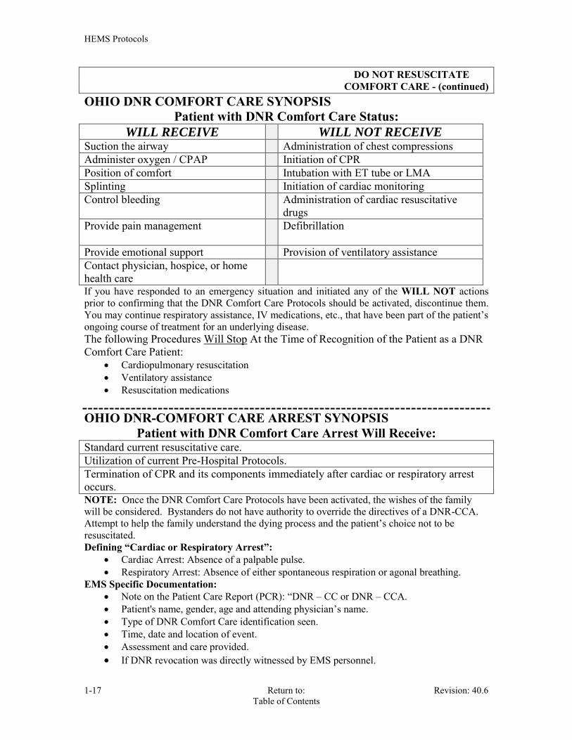
OHIO DNR COMFORT CARE SYNOPSIS Patient with DNR Comfort Care Status:
WILL RECEIVE WILL NOT RECEIVE Suction the airway Administration of chest compressions Administer oxygen / CPAP Initiation of CPR Position of comfort Intubation with ET tube or LMA Splinting Initiation of cardiac monitoring Control bleeding Administration of cardiac resuscitative
drugs Provide pain management
Defibrillation
Provide emotional support Provision of ventilatory assistance Contact physician, hospice, or home health care
If you have responded to an emergency situation and initiated any of the WILL NOT actions prior to confirming that the DNR Comfort Care Protocols should be activated, discontinue them. You may continue respiratory assistance, IV medications, etc., that have been part of the patient’s ongoing course of treatment for an underlying disease. The following Procedures Will Stop At the Time of Recognition of the Patient as a DNR Comfort Care Patient:
Cardiopulmonary resuscitation Ventilatory assistance Resuscitation medications
OHIO DNR-COMFORT CARE ARREST SYNOPSIS Patient with DNR Comfort Care Arrest Will Receive:
Standard current resuscitative care. Utilization of current Pre-Hospital Protocols. Termination of CPR and its components immediately after cardiac or respiratory arrest occurs. NOTE: Once the DNR Comfort Care Protocols have been activated, the wishes of the family will be considered. Bystanders do not have authority to override the directives of a DNR-CCA. Attempt to help the family understand the dying process and the patient’s choice not to be resuscitated. Defining “Cardiac or Respiratory Arrest”:
Cardiac Arrest: Absence of a palpable pulse. Respiratory Arrest: Absence of either spontaneous respiration or agonal breathing.
EMS Specific Documentation: Note on the Patient Care Report (PCR): “DNR – CC or DNR – CCA. Patient's name, gender, age and attending physician’s name. Type of DNR Comfort Care identification seen. Time, date and location of event. Assessment and care provided. If DNR revocation was directly witnessed by EMS personnel.
HEMS Protocols
Revision: 40.6 Return to: 1-18 Table of Contents
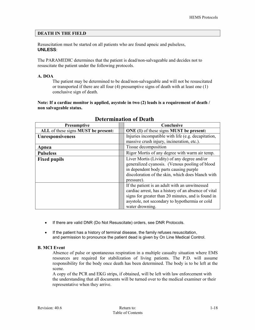
DEATH IN THE FIELD
Resuscitation must be started on all patients who are found apneic and pulseless, UNLESS: The PARAMEDIC determines that the patient is dead/non-salvageable and decides not to resuscitate the patient under the following protocols. A. DOA The patient may be determined to be dead/non-salvageable and will not be resuscitated or transported if there are all four (4) presumptive signs of death with at least one (1) conclusive sign of death.
Note: If a cardiac monitor is applied, asystole in two (2) leads is a requirement of death / non salvageable status. Determination of Death
Presumptive Conclusive ALL of these signs MUST be present: ONE (1) of these signs MUST be present:
Unresponsiveness Injuries incompatible with life (e.g. decapitation, massive crush injury, incineration, etc.).
Apnea Tissue decomposition Pulseless Rigor Mortis of any degree with warm air temp. Fixed pupils Liver Mortis (Lividity) of any degree and/or
generalized cyanosis. (Venous pooling of blood in dependent body parts causing purple discoloration of the skin, which does blanch with pressure).
If the patient is an adult with an unwitnessed cardiac arrest, has a history of an absence of vital signs for greater than 20 minutes, and is found in asystole, not secondary to hypothermia or cold water drowning.
If there are valid DNR (Do Not Resuscitate) orders, see DNR Protocols.
If the patient has a history of terminal disease, the family refuses resuscitation, and permission to pronounce the patient dead is given by On Line Medical Control. B. MCI Event Absence of pulse or spontaneous respiration in a multiple casualty situation where EMS
resources are required for stabilization of living patients. The P.D. will assume responsibility for the body once death has been determined. The body is to be left at the scene.
A copy of the PCR and EKG strips, if obtained, will be left with law enforcement with the understanding that all documents will be turned over to the medical examiner or their representative when they arrive.

HEMS Protocols
1-19 Return to: Revision: 40.6 Table of Contents
DEATH IN THE FIELD (continued)
C. Trauma Death A trauma victim who does not meet the "Determination of Death" criteria listed above
may be determined to be dead/non-salvageable based on the following criteria: 1. Blunt Trauma Arrest
2. Arrest from Primary Brain Injury
NOTE: Patients with suspected hypothermia, barbiturate overdose, or electrocution requires full ALS resuscitation unless there are injuries incompatible with life or tissue decomposition. Consideration should be given for the possibility of organ harvest, however this should not be the sole reason for resuscitation.
CAUTION: IF ANY DOUBT EXISTS THAT THE VICTIM IS DEAD AT THE TIME OF ARRIVAL OF EMS, RESUSCITATIVE MEASURES SHOULD BE INSTITUTED IMMEDIATELY. WHENEVER RESUSCITATIVE MEASURES ARE INSTITUTED, THEY MUST BE CONTINUED UNTIL ARRIVAL AT A HOSPITAL OR UNTIL A PHYSICIAN HAS PRONOUNCED THE VICTIM DEAD OR A VALID DNR IS PRODUCED.

HEMS Protocols
Revision: 40.6 Return to: 1-20 Table of Contents
TERMINATION OF RESUSCITATIVE EFFORTS Once resuscitative efforts have been initiated they must be continued until a physician, in person, or via OLMC, terminates the resuscitation. Resuscitation may be discontinued in the pre-hospital setting when the patient is nonresuscitable after an adequate trial of ACLS. When all of the following circumstances exist, resuscitative efforts may be discontinued prior to hospital arrival: A. This patient is an adult patient who experienced an unwitnessed cardiac arrest as a result of anything other than: drowning, hypothermia, acute airway obstruction, overdose, electrocution, lightning strike, or trauma. B. There has been early, successful endotracheal intubation and medication administration. C. ACLS in accordance with HEMS protocols has been carried out for over 20 minutes. D. There has NOT been any restoration of spontaneous circulation with a spontaneous palpable pulse for at least one, five-minute period at any time during the resuscitation. E. The patient does NOT have spontaneous respiration, eye opening, motor response, or other continued neurologic activity at the time stopping the resuscitation is contemplated. F. The cardiac rhythm is not persistent or recurrent ventricular fibrillation or ventricular tachycardia. If persistent or recurrent ventricular fibrillation or ventricular tachycardia is present, then resuscitative efforts should be continued. When the above conditions have been met, the paramedic may contact OLMC and request permission to terminate all resuscitation efforts. The lead paramedic and the OLMC must both be in agreement concerning termination of resuscitative efforts. Exception: When there is a delay in presenting a DNR to EMS personnel, resuscitation must be started. However, once the DNR is presented to EMS personnel, the crew can terminate resuscitation efforts, without first contacting OLMC.
HEMS Protocols
1-21 Return to: Revision: 40.6 Table of Contents
REMOTE DEATH PRONOUNCEMENT Under Ohio administrative code rule 4731-14-01, only a licensed physician can pronounce a person dead. A physician does not have to personally examine the body of the deceased “if a competent observer has recited the facts of the deceased’s present medical condition to the physician and the physician is satisfied that death has occurred.” Competent observers are individuals who by virtue of their training and licensure are able to determine vital signs or the absence of vital signs and assist the physician in making the determination of death. Competent observers are not permitted to make a pronouncement of death. Therefore the following protocol will be followed upon the determination that death has occurred and resuscitative efforts are not applicable. Requesting pronouncement of death
Western Reserve Hospital will be contacted and a full description of the scene, ALS patient assessment and the results will be relayed to the OLMC.
A directive will be requested from the physician to withhold resuscitative efforts and for the pronouncement of death.
Document the name of the physician acting as OLMC along with the time and method of communication on your PCR.
Contact the office of the Medical Examiner, (Summit County Coroner’s Office), at 330-643-2101 with the location of the deceased, circumstances surrounding the death, the deceased’s SSN, DOB, next of kin and their contact number, and the physician that pronounced.
The M.E. will advise if they are going to take jurisdiction of the decease or if they will release the body.
If the deceased is to be transported to the M.E.’s office, contact a private ambulance service to do the body removal. Once pick up arrangements has been made, turn the deceased and the PCR over to the P.D.
If the decease is released by the M.E.’s office, assist the family with funeral arrangements and turn the body over to the P.D.
P.D. will remain on scene until the body has been picked up. Complete the patient care report and provide the agency you are surrendering the body
to with a copy including EKG strips, if applicable. Provide support to the family of the deceased. Always handle a deceased body with kindness and respect.
Note: The Medical Examiner will not pronounce a patient deceased. This must be done by the protocol above and before the notification of their office. Hospice Patients – pronouncement of death A hospice patient will most likely have a DNR at their bedside. This is not a requirement of hospice but is a normal procedure. The hospice patient has already been cleared by a physician and the physician has agreed to sign the death certificate. Deceased hospice patient procedure:
Notify the hospice agency of the deceased. Give 30 minutes for hospice to call back. If they do not call back, contact OLMC. Obtain the name of the person you spoke to as well as an estimated time of their arrival. Confirm the physician has agreed to sign the death certificate (D.C.) and obtain the name
of the physician. Document the name of the physician agreeing to sign the D.C. on your PCR. Contact the Medical Examiner’s office at 330-643-2101 with the location of the deceased,
that the patient is hospice, and the physician that will be signing the D.C. Turn over a copy of your PCR and EKG strips, if applicable, to the same agency you turn
the decease over to.

HEMS Protocols
Revision: 40.6 Return to: 1-22 Table of Contents
CHILDREN WITH SPECIAL HEALTH CARE NEEDS (CSHCN)
EMS and Children with Special Health Care Needs:
The medically fragile child is one who depends on some form of technological assistance. This
can be anything from a nasal cannula to a child who requires total ventilatory support. Caring for a medically fragile child requires a full TEAM = Trust Every Available Member. Do not be concerned about removing the family from the crisis situation but inform them about what you are doing and include them in your plan of care. In most cases, the parents and/or home care providers can be of great assistance to the EMS providers. It is vitally important that their knowledge and experience is utilized when treating the child. Most importantly, they can console, comfort and calm their child. A. Treat the ABC’s first. Treat the child, not the equipment. If the emergency is due to an
equipment malfunction, manage the child appropriately using your own equipment. B. Children formerly cared for in hospitals or chronic care facilities are often cared for in
homes by parents or other caretakers. These children may have self limiting or chronic diseases. There are a multitude of underlying medical conditions that may categorize children as having special needs. Many are often unstable and may frequently involve the EMS system for evaluation, stabilization, and transport. Special needs children include technology-assisted children such as those with tracheostomy tubes with or without assisted ventilation, children with gastrostomy tubes, and children with indwelling central lines.
C. CSHCN may have many allergies. Children with spina bifida are often allergic to latex. Before treating a patient, ask the caregivers if the child is allergic to latex or has any other allergies.
D. Listen carefully to the caregiver’s guidance regarding their child’s treatment. E. Children with chronic illnesses often have different physical development from well
children. Therefore, their baseline vital signs may differ from normal standards. The size and developmental level may be different from age-based norms and length based tapes used to calculate drug dosages. Ask the caregiver if the child normally has abnormal vital signs, (i.e. a fast heart rate or a low pulse oximeter reading).
F. Some CSHCN may have sensory deficits, (i.e. they may be hearing impaired or blind),
yet may have age-appropriate cognitive abilities. Follow the caregiver’s lead in talking to and comforting a child during treatment and transport. Do not assume that a CSHCN is developmentally delayed.
HEMS Protocols
1-23 Return to: Revision: 40.6 Table of Contents
CHILDREN WITH SPECIAL HEALTH CARE NEEDS
(CSHCN) (continued)
G. When moving a special needs child, a slow careful transfer with two or more people is preferable. Do not try to straighten or unnecessarily manipulate contracted extremities as it may cause injury or pain to the child.
H. Caregivers of CSHCN often carry “go bags” or diaper bags that contain supplies to use with the child’s medical technologies and additional equipment such as extra tracheostomy tubes, adapters for feeding tubes, suction catheters, etc. Before leaving the scene, ask the caregivers if they have a “go bag” and carry it with you.
I. Caregivers may also carry a brief medical information form or card. The child may be
enrolled in a medical alert program whereby emergency personnel can get quick access to the child’s medical history. Ask the caregivers if they have an emergency information form or some other form of medical information for their child.
J. Caregivers of CSHCN often prefer that their child be transported to the hospital where the child is regularly followed or the “home” hospital. When making the decision as to where to transport a CSHCN, take into account: the child’s condition, capabilities of the local hospital, caregiver’s request, and the choice of approved destination facilities.

HEMS Protocols
Revision: 40.6 Return to: 1-24 Table of Contents
HEAVY PATIENTS As patients, these individuals are frequently classified as high risk because of the increased medical complications associated with their excess weight. Within EMS they present additional challenges involving movement and transportation. These individuals have the right to expect prompt and expert emergency medical care. The following protocol facilitates appropriate care while minimizing the risk of injury to EMS personnel. A. In managing a patient with weight over 300 lbs., at no time should the patient be
moved without sufficient manpower. At the scene, EMS personnel may be supplemented by police or other safety personnel as needed. If sufficient manpower is not available, mutual aid will be required.
B. It may be necessary to remove doors, walls or windows. The situation is no
different than extrication from a vehicle, although property damage may be higher. At all times the patient's life must be the first priority.
C. The patient is to be placed on at least 2, (double), backboards or other adequate
transfer device for support. D. The patient is to be loaded on a cot that is in the down position, and the cot is to
be kept in the down position at all times. E. It is NECESSARY TO NOTIFY THE HOSPITAL WELL IN ADVANCE of
arrival so that preparations can be completed in a timely fashion to assist in unloading the patient.
F. If individuals in the community are known to fall within this special category it is
appropriate to inform them in advance of the type of assistance they can expect from the EMS system, and help them make plans well in advance to assist you. When calling for the squad, if they identify themselves and their special needs, it will promote the timeliness of our efforts.

HEMS Protocols
1-25 Return to: Revision: 40.6 Table of Contents
PHYSICIAN AT THE SCENE Good Samaritan Physician: This is a physician with no previous relationship to the patient, who is not the patient's private physician, but is offering assistance in caring for the patient. The following criteria must be met for this physician to assume any responsibility for the care of the patient:
1. Ideally, if no further assistance is needed, the offer should be respectfully declined.
2. OLMC may be contacted for guidance.
3. The physician must have proof they are a physician. They should be able to show you their medical license. Notation of physician name, address and license number must be documented on the PCR.
4. The physician must be willing to assume responsibility for the patient until relieved
by another physician, usually at the emergency department.
5. The physician must not require the EMT to perform any procedures or institute any treatment that would vary from Protocols and/or procedure.
If the physician is not willing or able to comply with all the above requirements, his assistance must be courteously declined. Physician in his/her office, or Urgent Care Center:
EMS should perform its duties as usual under the auspices of OLMC or by Protocols.
The physician may elect to treat the patient in their office.
The EMS personnel should not provide any treatment under the physician's direction that varies from protocols. If requested to do so by the physician or his/her staff, the EMS personnel should decline until contact is made with OLMC.
Once the patient has been transferred into the squad, the patient's care comes under
Hudson EMS Protocols and OLMC.
ON SCENE EMT INTERVENER On an EMS run where an unknown EMT from outside the responding EMS agency wishes to assist / intervene in the care of patients, the following steps should be initiated:
Ideally, if no further assistance is needed, the offer should be declined.
If the intervener's assistance is needed or may contribute to the care of the patient an attempt should be made to obtain both proper identification and a valid EMT card. Notation of intervener’s name, address and certification numbers must be documented on the run report. Whenever possible, only “Non-critical” actions should be delegated to an unknown EMT.

HEMS Protocols
Revision: 40.6 Return to: 1-26 Table of Contents
ADVANCED CARE MEDICATIONS EMS may, at times, be called upon to transport patients whose care has been initiated by a physician prior to EMS assuming care of the patient. The transport of a patient from a physician’s office, urgent care center, and nursing home are examples of this. If the patient is on an I.V. infusion, receiving fluids, medications, TPN, or blood products not otherwise specifically sited within this protocols, the patient may be continued on it provided that the crew is paramedic staffed, and has received specific directions for the infusion by an on scene physician, or by OLMC. The physician / medical control is to be specifically informed that the given medication is not a “Standard EMS Medication”, and that it may be either discontinued for the transport, or continued if the paramedic is provided with the following information. Such instructions are to include what adjustments are to be made to the administration should the patient experience:
Hypertension Hypotension Symptomatic bradycardia Malignant tachycardia, (e.g. HR > 150, VT, etc.) Anaphylactic symptoms, possibly from the medication, (e.g. Stop the medication) Other specific instructions as provided.
The purpose of this Protocol is to facilitate the continuation of specific, physician ordered treatment, indicated to enhance patient care, particularly where its discontinuation may worsen the patient’s condition. Typical examples of this scenario include, but are not limited to:
A pediatric patient on an antibiotic, such as Rocephin. A cardiac patient on a nitroglycerine drip. A diabetic patient on a D5 or D10 type IV fluid.
NON-HOSPITAL TRANSFERS Non-hospital location to a Non-hospital location
HOME TO HOSPICE HOSPICE TO HOME
On occasion, one may be called upon to transport a patient from a non-hospital location to another non-hospital facility such as a Hospice Center, or from Hospice to home, or to a doctor’s office. The provider(s) will follow the written or pre-existing orders of the patient’s physician or physician approved hospice Center orders for the transport. At times, a Hospice nurse may arrive or already be at the scene. He/she should be able to help review orders and/or care directives such as DNR or “Support Care” orders to enable transport in accordance with the wishes of the patient and his/her family. A Hospice patient by definition is DNR. Medical Control does not need to be contacted unless the DNR is revoked. However, if the provider(s) feels the need to contact Medical Control for advice or direction, the provider(s) will clearly advise Medical Control of the patient’s terminal condition and DNR status. These patients require a history, physical exam, vital signs, and a complete medical record, as do all patients.

HEMS Protocols
1-27 Return to: Revision: 40.6 Table of Contents
MEDICAL PATIENT TRANSPORT DESTINATION
STABLE PATIENT: 1. Stable patients will be transported to the pre-approved destination facility of their choice. 2. If the patient / family request transport to a non-approved facility they may be turned over to a private ambulance service. This constitutes a refusal of care and a refusal form must be completed. Hudson EMS will facilitate contacting a private ambulance service and will remain on the scene until the arrival of the private service. A copy of the PCR will be turned over with the patient to the transporting agency. UNSTABLE PATIENT:
The definition of an unstable patient is one who presents with any of the following: SIGNIFICANT DISCOMFORT OF SUSPECTED CARDIAC ORIGIN, SEVERE DYSPNEA, ALTERED MENTAL STATUS, OR HYPOTENSION WITH SIGNS OF DECREASED TISSUE PERFUSION. 1. All patients whose condition meets the definition of UNSTABLE will be transported by HEMS to the closest appropriate approved facility. 2. If several approved destination hospitals are within the same approximate distance from the
scene, permit the patient and/or patient’s family to select the destination facility.
3. If an unstable patient refuses transport to the closest hospital, explain the danger involved in
their decision, up to and including a possible result of death. If the patient still refuses to comply with the direction of the paramedic and/or OLMC, document it on a PCR and have the patient sign a refusal AMA.
DESTINATION FACILITIES: See Appendix I

HEMS Protocols
Revision: 40.6 Return to: 1-28 Table of Contents
EMERGENCY TRANSPORT 1. Emergent transport may be indicated to optimize patient care. Each case will be unique and
compelling reasons MUST be documented. 2. If the situation warrants, DO NOT delay at the scene. EXAMPLES OF EMERGENCY TRANSPORT SITUATIONS INCLUDE BUT ARE NOT LIMITED TO: Inability to establish or maintain a patent airway or effective ventilations Complicated obstetrical presentation Acute ST elevation MI, (STEMI) Acute CVA Respiratory arrest Cardiac Arrest, if patient may significantly benefit from rapid transport Shock Massive internal / external hemorrhage Trauma Alert Criteria The use of emergency transport must be weighed against the potential injury to both the patient and EMS personnel. The few minutes that this type of transport may gain must be significantly more beneficial than the added stress and potential for injury to the patient, our personnel, and the public. The decision to transport in an emergent mode is to be made by the crew chief for the benefit to the patient. At no time will the squad be placed in the emergent mode for the benefit of the crew.

HEMS Protocols
1-29 Return to: Revision: 40.6 Table of Contents
FIREFIGHTER REHABILITATION PROTOCOL
Purpose: The purpose of this protocol is to ensure that the physical and mental condition of our fire department personnel operating at the scene of an emergency incident or training exercise does not deteriorate to the point that it adversely impacts the safety of the firefighter, or others on the scene. Definitions: Establishment of REHAB: Rehab should be established as soon as possible for any incident which will require an extended commitment of resources. Staffing of REHAB: At least one BLS crew should be committed to designate and manage the Rehab area. This crew will ensure that firefighters have access to beverages, and that personnel get a sufficient break. This crew will assist in establishing an appropriate environment to benefit the rehab process. Location of REHAB: Environment: Weather is a major consideration and the site should provide relief from extreme weather conditions. Space must be available for firefighters to sit, remove equipment, etc. The site should be far enough from the incident to isolate firefighters from hazards yet near enough for easy access to the incident. Noise and exhaust from fire apparatus is also to be considered. Resources: Fluids: Water, activity beverage, oral electrolyte solutions, ice. Rest: All members shall be sent to rehabilitation following the use of two 30-minute SCBA cylinders or one 45- to 60-minute SCBA cylinder. Shorter times might be considered during extreme weather conditions. Rest and cooling/warming rehab intervals will not be less than 10 to 20 minutes. REHAB Assessment: The REHAB EMS crew will be responsible for screening members entering REHAB and documenting their vitals and the beginning REHAB time. The crew will assess the firefighter’s B/P, Pulse, SpCO and SpO2 - Pulse will be WNL (60 – 100) prior to release back to duty. A HR that is abnormally fast, slow or irregular should be turned over to the EMS crew for evaluation. Check Temperature: Elevated temperature, noted by touch or measured, should alert the rehab crew to the possibility of heat-related illness. A member whose blood pressure is greater than 160 systolic and/or 100 diastolic should not be released from rehabilitation. Respiratory rate will be WNL (12 – 20) prior to being released back to duty. Record evaluation and treatment times, all findings and time returned to duty on the Rehab report. No Firefighter will return to duty without being released by the rehab crew. NFPA 1584. EMS Assessment: Any firefighter that after rehab assessment and a reasonable amount of time does not rebound to a healthy state will be turned over to EMS for care.

HEMS Protocols
Revision: 40.6 Return to: 1-30 Table of Contents
TRANSPORTATION OF SERVICE ANIMALS EMS may encounter patients who are assisted by service animals, including guide dogs for the visually impaired, and other types of service animals assisting persons with disabilities. Because of conflict between the nature of the services we provide and the federally mandated Americans with Disabilities Act (ADA) and its potential penalties and liability, decisions may be required as to whether or not a patient and a service animal should be separated in any particular situation involving patients with such animals. This protocol establishes a bias in such situations in favor of ADA requirements for allowing service animals to accompany disabled patients. The overwhelming factor which must govern the paramedic’s determination on transporting a service animal is the City’s requirement to comply with the ADA, weighed against any objectively real risk to the patient or crew. Due to ADA regulations, only clear and compelling concerns for the patient or crew member’s safety should be utilized in denying transport of any service animal (within the squad). Absent of documentation of clear and compelling circumstances supported by objective factual criteria, the service animal and patient should remain together. Members should be guided by this protocol in determining whether service animals should be transported with the individual in the squad or whether alternate methods of transporting the service animal should be utilized. Criteria: Any call involving a patient with a service animal.
Procedure All Patients with Service Animals:
Service animals, for example, guide dogs utilized by visually impaired persons or other animals assisting persons with disabilities, shall be permitted to accompany the patient in the squad unless the presence of the service animal is anticipated (based on clear and perceptible factors) to disrupt emergency or urgent patient care. If there is a clear and perceptible basis for the crew members to believe that the safety of the crew, the patient or others would be compromised by the presence of the service animal then alternate transportation must be arranged.
Members should assess the level of care required to provide competent medical attention to the patient and assess the service animal for transport continuity.
When all facts pertaining to the matter clearly establish that the presence of a given
service animal in the squad will interfere with patient care or jeopardize the safety of the crew or the patient, arrangements should be made for simultaneous transport of the service animal to the hospital. In such cases, unless emergency conditions dictate otherwise, absolutely every effort must be made to reunite the patient with the service animal at the time of the patient’s arrival at the hospital or other destination.

HEMS Protocols
1-31 Return to: Revision: 40.6 Table of Contents
TRANSPORTATION OF SERVICE ANIMALS
(continued)
Acceptable alternative methods of transporting a service animal to the hospital include, but are not necessarily limited to, a family member, friends or neighbors of the patient, Animal Control Officers, or a Law Enforcement Officer. Attempt to obtain and document the consent of the patient for transport of the service animal by such person. If you are unable to accomplish transportation have additional manpower respond to transport the service animal.
Personnel should document on the patient care report instances where the patient utilizes a service animal, and should document on the patient care report whether or not the service animal was transported with the patient. If the service animal is not transported
with the patient, you must document the specific clear factual circumstances under which the decision was made and the clear and compelling reasons why such decision was required under the circumstances, and you must further document the means of service animal transportation, including when the service animal was reunited with his owner, if known.

UNIVERSAL PATIENT CARE
MEDICAL/TRAUMA
ASSESSMENT & TREATMENT
PROTOCOL
SECTION 2
HEMS Protocols
2-1 Return to: Revision: 40.6 Table of Contents
INTRODUCTION to UNIVERSAL PATIENT CARE ASSESSMENT AND MANAGEMENT PROTOCOL
These Protocols of an Adult Initial Assessment and a Pediatric Initial Assessment are designed to guide the EMT, AEMT and Paramedic in his or her initial approach to assessment and management of adult and pediatric patients. The Pediatric Initial Assessment Protocol should be used for infant and pediatric patients. The care is specified as EMT, AEMT and Paramedic; AEMT and Paramedic: and Paramedic. Adult: An individual who is 8 years of age or older, or greater than 35kg. (Medical) An individual who is 16 years of age or older. (Trauma)
Pediatric: An individual who is less than 8 years of age, or weighs less than 35 kg. (Medical) An individual with the anatomical characteristics of a person less than sixteen (16)
years or younger. (Trauma)
Adult Initial Assessment & Management should be used on all adult patients. During this assessment, if the EMT, AEMT or Paramedic determines that there is a need for airway management, Airway Management Protocol will be used. Other Protocols are frequently referred to by this Protocol which may or may not override them in recommending more specific therapy. Medical Initial Assessment & Management presents the basic components of preparation for transport of medical patients. Due to the significant differences in priorities and packaging in the pre-hospital care of trauma and hypovolemia cases, a separate Trauma Initial Assessment and Management Protocol exists. After following the Adult Initial Assessment & Management Protocol , the Medical Supportive Care Protocol or Trauma Supportive Care Protocol may be the only Protocol used in medical emergency situations where a specific diagnostic impression and choice of additional Protocol (s) cannot be made. Judgment must be used in determining whether patients require ALS or BLS level care. UNIVERSAL MEDICAL PATIENT CARE ASSESSMENT & MANAGEMENT PROTOCOL EMT, AEMT and Paramedic Scene Size-up
A. Review of Dispatch Information. B. Body Substance Isolation / Universal Precautions. C. Assessment of Scene Safety. D. Determine Mechanism of Injury/Nature of illness E. Determine Number and Location of Patients. F. Determine Need for Additional Resources.
Initial Assessment A. General Impression: The overall impression of the patient's condition including
severity of distress. B. Determine Responsiveness / Level of Consciousness (LOC)*
A- Alert V- Verbal P- Painful U- Unresponsive

HEMS Protocols
Revision: 40.6 Return to: 2-2 Table of Contents
UNIVERSAL PATIENT CARE MEDICAL ASSESSMENT & MANAGEMENT PROTOCOL (continued)
C. ABC's 1. Cardiac arrest: Place AED / cardiac monitor’s combo/ defib pads on patient. 2. Airway: Establish and maintain airway. Utilize cervical spine precautions when indicated. 3. Breathing: Provide or assist ventilations as indicated. 4. Circulation: Check pulse and control hemorrhage as indicated.
D. ORIENTATION
Alert & Oriented X 4. (A & O X 4) 4/4: Person, Place, Time and Event
E. FOCUSED AND DETAILED HISTORY AND PHYSICAL EXAM 1. History of present illness or injury. 2. Past medical history, drugs, allergies. 3. Systematic head-to-toe assessment. 4. Vital signs every 5 minutes (unstable) to 15 minutes (stable) patient, barring higher prioritizes.
PERFORM INITIAL MEDICAL CARE, LEVEL BASED, WITH THERAPIES: A. Loosen tight clothing and reassure patient. B. Place patient in position of comfort if not contraindicated. C. Paramedic should decide within three (3) minutes after patient contact whether the
patient requires ALS intervention. All patients receiving oxygen therapy should receive the following care:
1. Administer the appropriate dosage and route of supplemental O2 as necessary to alleviate the patient's chief complaint, keep O2 sats > 94% and keep the patient's skin condition pink, warm and dry. All ALS patients will have pulse oximetry monitored and documented. 2. If indicated by ineffective breathing pattern or respiratory distress, provide advanced airway interventions and suction as necessary. 3. Capnography will be connected to the patient if the C/C is moderate to severe respiratory distress (sidestream) or a rescue airway (ETT, Combi tube, EOA, LMA)(mainstream) has been established. This patient will receive continuous monitoring of their ETCO2 until they are turned over to the ER staff. 4. Capnography will be used if the patient has been intubated. This patient will receive continuous monitoring of their ETCO2 until the patient is turned over to the ED staff. 5. Establish IV of Normal Saline with macro-drip tubing at KVO or IV Saline Lock as indicated by patient condition. Micro-drip tubing or IV infusion pumps will be used for pediatric patients. Attempt IV X 2 unless situation demands further repeated attempts. Failure to obtain IV access does not preclude the intervention of other definitive therapy.

HEMS Protocols
2-3 Return to: Revision: 40.6 Table of Contents
UNIVERSAL PATIENT CARE MEDICAL ASSESSMENT & MANAGEMENT PROTOCOL (continued)
6. Once a medication route has been established, administer medication as indicated per appropriate protocols, (e.g., IV/IO).
7. Use cardiac monitor to observe cardiac rhythm as indicated per protocols. Confirm assessment in another lead if necessary to correctly identify rhythm. Record strips to document rhythm changes and to document intervention necessities and outcome. 8. Assess blood glucose with glucometer as indicated per protocols. 9. Assess COHb with the RAD 57 as indicated per protocols.
10. Assess vital signs and patient condition every 5 minutes (unstable) to 15 minutes (stable),
barring higher prioritizes, and before and after medication administration. UNIVERSAL PATIENT CARE TRAUMA ASSESSMENT & MANAGEMENT PROTOCOL EMT, AEMT and Paramedic Scene Size-up
A. Review of Dispatch Information. B. Body Substance Isolation (As Appropriate) C. Assessment of Scene Safety. D. Determine Mechanism of Injury/Nature of illness E. Determine Number and Location of Patients. F. Determine Need for Additional Resources.
Initial Assessment:
A. General Impression: The overall impression of the patient's condition including severity of distress. B. Determine Responsiveness / Level of Consciousness (LOC)
A- Alert V- Verbal P- Painful U- Unresponsive
C. ABCDE 1. AIRWAY / C-SPINE: Manual C-spine immobilization is indicated until the C-spine has been cleared per protocol. Position airway and suction as needed. If intubation is required, utilize in-line technique. Refer to airway management protocol.
2. BREATHING: Check for inadequate breath sounds, JVD, tracheal deviation, and the use of accessory muscles of respiration. Assess for tension pneumothorax and treat as per PLEURAL DECOMPRESSION protocol, if indicated.
a. Administer the appropriate dosage and route of supplemental O2 as necessary to alleviate the patient's chief complaint, keep O2 sats > 94% and keep the patient's skin condition pink, warm and dry. All patients receiving oxygen therapy will have pulse oximetry monitored and documented.

HEMS Protocols
Revision: 40.6 Return to: 2-4 Table of Contents
UNIVERSAL PATIENT CARE TRAUMA ASSESSMENT & MANAGEMENT PROTOCOL (continued)
b. If indicated by ineffective breathing pattern or respiratory distress, assist ventilations, apply level specific airway intervention, and suction as necessary.
c. Post-intubation, confirm proper tube placement using the following methods: i. Auscultating the lungs bilaterally, then the gastric area.
ii. Confirmation by the EDD and noting color change on the end-tidal CO2 detector, (if capnography is not available).
iii. Capnography will be connected to the cardiac monitor and will remain connected to the ET tube/King Airway until the patient is turned over to the ER staff.
3. CIRCULATION: Evaluate peripheral pulses for presence, quality and equality. CPR as indicated. Treat severe external hemorrhage with direct pressure.
a. Apply QuickClot or TQ, if indicated. b. ADEQUATE PERFUSION: Establish IV large bore @ KVO (If assessment indicates possible deterioration refer to SHOCK Protocol), enroute unless delayed by extrication. c. INADEQUATE PERFUSION: Refer to SHOCK Protocol and initiate IV, enroute, unless delayed by extrication.
4. DISABILITY: Assess distal CSM for impairment
C- Circulation S- Sensory M- Motor
5. EXPOSE: Remove clothing as applicable to assess for hidden injuries while protecting the patient’s modesty.
a. Orientation : Alert & Oriented X 4 (A & O X 4) 4/4- Person, Place, Time and Event
FOCUSED AND DETAILED HISTORY AND PHYSICAL EXAM 1. History of present illness or injury. 2. Past medical history, drugs, allergies. 3. Systematic head-to-toe assessment. 4. Vital signs every 5 minutes, (unstable), to 15 minutes, (stable), patient, barring higher prioritizes.

HEMS Protocols
2-5 Return to: Revision: 40.6 Table of Contents
UNIVERSAL PATIENT CARE TRAUMA ASSESSMENT & MANAGEMENT PROTOCOL (continued) TRAUMA- FOCUSED HISTORY & PHYSICAL EXAM FOCUSED HISTORY, PHYSICAL EXAM AND INTERVENTIONS A. NEURO ASSESSMENT Check for Level of Consciousness, (LOC), central nervous system, (CNS), motor and sensory function and symmetry of each. B. HEAD Inspect and palpate the head and face, and check for symmetry. Note any drainage from the ears or nose. C. EYES Re-inspect pupils for size, shape, equality, and reactivity. Note occular motions or deviations. Note any trauma to eyes, lids, or orbits. D. NECK Total spinal immobilization as indicated. Note presence of carotid pulses, point tenderness, JVD, subcutaneous emphysema, or tracheal deviations prior to applying c-collar. E. CHEST Inspect, palpate, and auscultate for signs of injury. F. ABDOMEN Inspect and palpate for signs of injury. If evisceration, cover with sterile moist saline dressings. G. SOFT-TISSUE / MUSCULOSKELETAL / BACK Inspect and palpate for signs of injury. Assess vascular, motor, and sensory function distal to injuries, before and after immobilization. Immobilize limbs and / or joints as indicated. H. REASSESS Reassess and record vital signs every 5 minutes, (unstable), to 15 minutes ,(stable), barring higher prioritizes.

HEMS Protocols
Revision: 40.6 Return to: 2-6 Table of Contents
UNIVERSAL PATIENT CARE MEDICAL PROTOCOL EMT, AEMT and Paramedic 1. Initial Assessment & Management protocol. 2. Airway Management protocol. 3. If pulseless, refer to Cardiac Arrest protocol. 4. Establish hospital contact for notification of incoming patient and OLMC for additional orders. AEMT and Paramedic 5. Monitor EKG as appropriate. 6. Establish IV as indicated. 7. When unable to establish an IV in a patient that requires life saving fluids/medication, an intraosseous (IO) line may be established. Notes:
a. Authorized IV routes includes all peripheral venous sites including external jugular, (EJ).
b. For hypotension, administer a fluid challenge, adult 500 ml of NS.
Pediatric fluid challenge is 20 ml / kg N.S. Repeat fluid challenge until desired clinical effects are achieved. Assess for pulmonary congestion before and after each infusion.
c. Saline lock may be used in lieu of an IV, when appropriate.
d. 12 lead EKG will be recorded on all patient experiencing active chest pain, syncope and moderate to severe respiratory complaints, excluding patients with classic asthma presentations.

HEMS Protocols
2-7 Return to: Revision: 40.6 Table of Contents
UNIVERSAL PATIENT CARE TRAUMA PROTOCOL EMT, AEMT and Paramedic 1. Initial Assessment & Management Protocol.
2. Airway Management Protocol, (manually stabilize c-spine as indicated). 3. Correct any open wound/sucking chest wound, (3 sided occlusive dressing). 4. Control hemorrhage. 5. Conduct focused history and a trauma physical exam. 6. If mechanism or signs/symptoms of injury suggests injury to the head, neck, thorax, or pelvis, immobilize patient per cervical immobilization procedure. AEMT and Paramedic 7. Monitor EKG as indicated.
8. Establish IV of Normal Saline with appropriate infusion set.
Moderate to severe trauma IV/N.S. on a macro drip and titrate to effect. Use large bore catheters.
Intravenous access attempts shall not delay transport.
All trauma patients warrant at least one (1) large bore I.V. of N.S..
Every effort will be made to establish an I.V. line using a catheter no smaller than 18 gauge.
Second IV line if indicated. 9. Intraosseous Infusions will be used if fluid replacement is essential and alternative IV access is unsuccessful. Paramedic 10. Immediately stabilize any massive flail segment and treat any tension pneumothorax, (chest decompression).

HEMS Protocols
Revision: 40.6 Return to: 2-8 Table of Contents
ALS CARDIO-RESPIRATORY MONITORING Paramedic Cardio-respiratory monitoring is indicated for the majority of patient presentations, including, but not limited to:
Unconsciousness Syncope, Near syncope Chest pain Respiratory distress Altered mental status Significant trauma Abnormal vital signs Anaphylaxis Environmental emergencies Dizziness, Lightheadedness
Cardio-respiratory monitoring is indicated for all patients receiving pre-hospital medications, including supplemental oxygen. For many patients an EKG rhythm strip and pulse oximetry are sufficient. For those patients experiencing chest pain, CHF, or pulmonary edema a 12 lead EKG shall be obtained. Geriatric and adult diabetic patients experiencing fatigue and nausea warrant a 12 lead EKG to check for silent ischemia. It may be obtained in other patients at the crew’s discretion. A 12 lead EKG, when obtained for patients experiencing chest pain, is ideally obtained prior to treatment with nitrates. Pre and post treatment EKGs may be significantly different, and this is pertinent to the patient’s definitive care. All patients with syncope, near-syncope, or unconsciousness, are to be placed on a cardiac monitor immediately upon EMS personnel arrival. Arrhythmias are a known cause for such events, and are often transient and episodic. Pulse oximetry monitoring should be continuous monitoring in those patients with significant cardio-respiratory symptoms. Non-continuous monitoring may be used in less ill/injured patients. If no reading can be obtained, the chart should be marked: “Unable” or “Poor Signal”. A poor signal is often due to poor peripheral circulation. The patient should be assessed closely for their hemodynamic status, (shock), and treated as indicated. Cold extremities may also result in poor peripheral perfusion resulting in a poor signal, and can be treated by warming the patient. All patients needing to be monitored throughout the transport should be monitored until they are transferred to the hospital bed. Monitoring shall not be discontinued within the squad, prior to taking the patient into the ED. All patients with a measured carboxyhemoglobin level, (COHb), require its documentation. All patients are to have a minimum of two sets of vital signs, including BP, HR, RR. Exception:
Death in the field Medical Control contact exemptions In station asymptomatic B/P checks
HEMS Protocols
2-9 Return to: Revision: 40.6 Table of Contents
AIRWAY MANAGEMENT PROTOCOL EMT, AEMT and Paramedic Administer the appropriate dosage and route of supplemental O2 as necessary to alleviate the patient's chief complaint, keep O2 sats > 94% and keep the patient's skin condition pink, warm and dry. All ALS patients will have pulse oximetry monitored and documented. 1. Initial Assessment Protocol. 2. If spontaneous breathing is present without compromise:
A. Monitor breathing during transport. B. Administer oxygen via nasal cannula, (2-6 L/min), as indicated.
3. If spontaneous breathing is present with compromise:
A. Maintain airway, (e.g. modified jaw thrust). B. Administer oxygen via non-rebreather mask, (10-15 L/min). C. If unconscious, insert a rescue airway, (oropharyngeal, nasopharyngeal, King LT-D).
D. Assist ventilations with BVM as indicated when ventilations are less than 12 min, or otherwise insufficient or impaired.
E. Suction as indicated.
All patients receiving the King airway will receive constant EtCO2 monitoring. Be sure to assess for an obstructed airway as the cause for the patient’s apnea. Paramedic
F. If patient accepts oropharyngeal airway, secure airway with endotracheal intubation, (confirm ETT placement). If unsuccessful after two attempts – insert a rescue airway.
EMT, AEMT and Paramedic
4. If spontaneous breathing is absent or markedly compromised, (APNEIC):
A. Maintain airway, (e.g. modified jaw thrust). B. Assist ventilations with BVM.
C. A two person BVM technique, further assisted with cricoid pressure, may improve the ventilation provided.
D. If unconscious, insert a rescue airway. E. Suction as indicated.
Paramedic F. If patient accepts oropharyngeal airway, secure airway with endotracheal intubation, (confirm ETT placement). If unsuccessful after two attempts – insert a rescue airway.
All patients receiving the King airway or ETT will receive constant EtCO2 monitoring. Be sure to assess for an obstructed airway as the cause for the patient’s apnea.

HEMS Protocols
Revision: 40.6 Return to: 2-10 Table of Contents
AEROSOLIZED BRONCHODILATOR THERAPY PROTOCOL
AEMT and Paramedic This protocol will be utilized any time a patient requires treatment for respiratory distress secondary to a constriction of the bronchioles and presents with wheezing or constriction to the point of breath sounds being absent during auscultation of the lung fields.
I. Administer a Beta-2 bronchodilator, either Xopenex OR Albuterol/Atrovent, by Aerosol nebulizer. If wheezing or poor air exchange persists, this is to be repeated twice, at ~ 15 minutes intervals, prior to obtaining further OLMC orders.
Xopenex is to be used if the patient is already on Xopenex, or if the patient has an elevated heart rate, (HR > 100). The patient’s heart rate may be elevated from their respiratory insufficiency, or as a side effect of medications they may have already
self–administered. Xopenex is contra-indicated in patient’s under age 6 unless otherwise ordered by OLMC. Xopenex: Adults and Peds ≥ 12 years old: Xopenex 1.25 mg in 3 ml via nebulizer, q 15 minutes PRN, total of 3 prior to OLMC contact. Peds < 12 years old: Xopenex 0.63 mg in 3 ml via nebulizer, q 15 minutes PRN, total of 3 prior to OLMC contact. Peds < 6 years old: Xopenex is contra-indicated in patient’s under age 6 unless otherwise ordered by OLMC.
Atrovent is to be administered immediately following the completed delivery of the first Xopenex dose.
Albuterol/Atrovent: Albuterol 2.5 mg via nebulizer, q 15 minutes PRN, total of 3 prior to OLMC contact. This dosage may be given to patients of any age.
The first administration of Albuterol is to be delivered with Atrovent. All other administrations of Albuterol are delivered without adding additional Atrovent.
II. Atrovent: Atrovent may be mixed in with the first Albuterol treatment, giving both simultaneously; or it may be given immediately following completion of the first Xopenex treatment.
Patient > 2 years old: Atrovent 0.5 mg in 2.5 ml via nebulizer, once. Given its long duration of action it is given once in the pre-hospital setting, unless ordered otherwise by OLMC.
HEMS Protocols
2-11 Return to: Revision: 40.6 Table of Contents
CAPNOGRAPHY MAIN STREAM AND SIDESTREAM EMT, AEMT and Paramedic
Capnography is defined as the noninvasive, continuous measurement of exhaled carbon dioxide (EtCO2) concentration over time. Digital display provides EtCO2 value provides a distinct waveform (tracing) for each respiratory cycle. Non-Intubated Applications: Sidestream capnography is measure through a nasal cannula and is used to measure the exhaled CO2 of a breathing patient. This will be applied to all patients that are in experiencing any of the following medical emergencies: Bronchospasms (Asthma, COPD, Anaphylaxis), Hypoventilation (Drugs, Stroke, CHF, Post-Ictal), Shock and circulatory compromise, Hyperventilation syndrome, any time CPAP is applied or pain or sedation management is implemented. Intubated applications: Mainstream capnography is measured through a T sensor connected to the ET tube or King airway. EtCO2 Values Normal 35 – 45 mmHg Hypoventilation >45 mmHg Hyperventilation < 35 mmHg

HEMS Protocols
Revision: 40.6 Return to: 2-12 Table of Contents
CONTINUOUS POSITIVE AIRWAY PRESSURE PROTOCOL EMT, AEMT and Paramedic
CPAP is defined as the application of positive end expiratory pressure by facemask for relief of hypoxemia, which doesn’t respond to conventional therapy. In order for CPAP to be used the patient must be breathing.
CPAP may be utilized in the treatment of patients with severe respiratory distress caused by CHF or COPD. In addition to improving oxygenation, and decreasing the work of breathing, CPAP significantly improves the administration and effectiveness of nebulizer aerosol administered medications. In determining which patients demonstrate severe respiratory distress warranting CPAP administration one should consider: patient appearance, respiratory rate, air exchange, retractions, pulse oximetry data, heart rate and blood pressure, and mental status, in addition to confirming the absence of any contraindications. CPAP pressure is limited to 7.5 cm H2O in treating patients whose primary cause for their respiratory insufficiency is COPD or pneumonia, or 10 cm H2O for Pulmonary Edema. CPAP is contraindicated in patients who are apneic, or otherwise demonstrating insufficient spontaneous respirations. Such patients require ventilation with a BVM, and application of the Advanced Airway Procedures Protocol. Patients with COPD who receive CPAP therapy have a higher risk of developing a pneumothorax than do patients with CHF or Pulmonary Edema. Be alert to this possible complication of therapy. Protocol: 1. Ensure that the patient is on continuous cardiac monitoring and pulse oximetry. 2. Explain the procedure to the patient. 3. Ensure adequate oxygen supply and assemble CPAP mask, circuit, and EtCO2 device. 4. Turn CPAP adjustment knob clockwise to start airflow at 2.5 cm to 5cm H20. 5. Place the mask over the mouth and nose. 6. Secure the mask with straps. 7. Adjust the CPAP pressure to 10 cm H2O for Pulmonary Edema, or 7.5 cm H2O for all other purposes 8. Check for air leaks and adjust mask if needed. 9. Continue to coach patient to keep mask in place, however if the patient is experiencing increasing anxiety, see sedation protocol. 10. Reassess patient’s vital signs and response to CPAP every 5 minutes. 11. If the patient’s status improves continue CPAP until the patient is transferred to the care of the receiving hospital. 12. If patient’s status deteriorates discontinue CPAP and implement the advanced airway protocol. 13. Notify destination hospital that CPAP is being used. ** Consider sedation protocol

HEMS Protocols
2-13 Return to: Revision: 40.6 Table of Contents
STERIOD ADMINISTRATION PROTOCOL
Paramedic Solumedrol can be administered for multiple purposes, including the treatment of anaphylaxis, COPD, and asthma. It is to be administered as follows: Intravenously: If the patient has peripheral IV access, Solumedrol is to be administer in the pre-hospital setting. Do not initiate IO access solely for the purpose of solumedrol administration. ADULT:
Solumedrol 125 mg I.V., over 2 minutes. PEDIATRIC:
Solumedrol 1 mg / kg, rounded up to the nearest 10 mgs, I.V., over 2 minutes. Maximum of 125 mg. For this medication, patients who weigh over 60 kg, (132 lbs), are to be given the adult dosage.
Intramuscular: If a peripheral IV access is not present, patients with severe respiratory distress are to be treated with IM Solumedrol. Those patients with less severe symptoms may await steroid administration within the ED setting. ADULT:
Solumedrol 40 mg IM, (Additional dose likely to be given within the ED). PEDIATRIC:
Solumedrol 1 mg / kg, rounded up to the nearest 10 mgs, IM. Maximum IM dose of 40 mg.
HEMS Protocols
Revision: 40.6 Return to: 2-14 Table of Contents
EPINEPHRINE SQ PROTOCOL Epinephrine administration used in the treatment of a systemic anaphylactic reaction. EMT, AEMT and Paramedic Indication: Systemic anaphylactic reaction, manifested by any of the following:
Wheezing SOB Stridor Diffuse urticaria, (widespread hives) Hypotension Oropharyngeal edema, (airway swelling)
Contra-indication: Local allergic reaction, e.g. pain and swelling only at the site of an insect sting, without any of the above symptoms. EMT Adult dosing: EMT- Epi pen auto-injector 1:1,000. May repeat once in 5 to 10 minutes, if needed. Pediatric dosing: EMT-Epi pen Jr. auto-injector 1:2,000. May repeat once in 5 to 10 minutes if needed. AEMT & Paramedic Adult dosing: Advanced EMT/Paramedic - Epinephrine 1:1,000, 0.3 – 0.5 ml SQ, repeated every 5 – 10 minutes, if needed. Maximum of 3 doses prior to contacting OLMC. Epi pen auto-injector 1:1,000 repeated every 5 – 10 minutes, Maximum of 3 doses prior to contacting OLMC. Pediatric dosing: Advanced EMT & Paramedic -Epinephrine 1:1,000, 0.01 ml/kg SQ, repeated every 5 – 10 minutes, if needed. Maximum of 0.5 ml per dose. Maximum of 3 doses prior to contacting OLMC. Epi pen Jr. auto-injector 1:2,000 repeated every 5 – 10 minutes, Maximum of 3 doses prior to contacting OLMC. Paramedic If the patient is over 50 years of age, or has a known history of coronary artery disease, (angina, history of MI, stent, etc.), then the preferred method of epinephrine administration is via an I.V. drip infusion. If the patient is in critical condition, or an I.V. cannot be rapidly obtained, administer epinephrine SQ as above, otherwise see Epinephrine Infusion Protocol. Notes:
In the treatment of anaphylaxis epinephrine is given as a subcutaneous injection, or as an I.V. drip infusion.
When given SQ, give the entire dose at one site, using normal injection sites. Do not inject a partial dose at the site of a sting. Never inject epinephrine into a finger, toe, ear, or nose.
Do not confuse epinephrine 1:1,000, used for the treatment of anaphylaxis, with epinephrine 1:10,000, used in the treatment of cardiac arrest.
Cardio-respiratory monitory protocol applies. Auto injectors are primarily for initial dosing in time critical situations.

HEMS Protocols
2-15 Return to: Revision: 40.6 Table of Contents
EPINEPHRINE INFUSION
PROTOCOL Epinephrine I.V. drip infusion administration is used in the treatment of systemic anaphylactic reactions. Epinephrine I.V. drip infusion may be used in the treatment of non-traumatic shock by OLMC order only. Paramedic
If the patient is over 50 years of age, or has a known history of coronary artery disease, (angina, history of MI, stent, etc.), then the preferred method of epinephrine administration is via an i.v. drip infusion.
If the patient is in critical condition, or an I.V. cannot be rapidly obtained, administer epinephrine SQ as per Epinephrine Protocol, regardless of their age or past medical history.
Indication: (Three of these are required)
Systemic anaphylactic reaction, manifested by any of the following: SOB, Wheezing and/or Stridor Diffuse urticaria, (widespread hives) Hypotension Oropharyngeal edema, (airway swelling)
Age over 50, or a known history of coronary artery disease. Rapid peripheral vascular access is obtainable.
Contra-indication: Local allergic reaction, e.g., pain and swelling only at the site of an insect sting, without any of the above symptoms. Dosing: Epinephrine I.V. drip infusion:
Initiate infusion at 1 ug/min, IV./IO, (1 microgram / minute). Titrate up by 1 ug/min, every five minutes, to a maximum of 4 ug/min, to obtain the
desired clinical response. Titrate down or off if the patient demonstrates significant side effects from the infusion:
Significant tachycardia V. Tach / V. Fib Anginal type chest pain, or equivalent Significant hypertension: SBP> 180 mmHg, DBP> 100 mmHg)
Note: Epinephrine drips are not to be run “wide open”. Drip Preparation:
Mix 1 mg epinephrine in 500 ml NS. The concentration is 2 ug/ml. Initiate the drip at 1ug/min., which is 0.5 ml/min. With a 60 drops/ml micro drip chamber, this is 30 drops/min. This is 1 drop every other
second. To increase this from 1 to 2 ug/min, double the drip rate, to 1 drop every second. The maximum rate is 4 ug/min, or 2 drops every second.

HEMS Protocols
Revision: 40.6 Return to: 2-16 Table of Contents
EPINEPHRINE INFUSION PROTOCOL (continued) Infusion Setup: It is preferable to administer epinephrine infusions using an I.V. infusion pump. If a pump is not readily available, then they are to be administered using a 60 ml / gtt infusion setup. Documentation:
The EMS report shall indicate the presence of a systemic allergic reaction, and the patient’s age or prior cardiac history.
The I.V. bag must be clearly labeled as an epinephrine drip. Notes:
Administration of epinephrine by drip allows for careful titration of the amount delivered, to maximize the beneficial effects, and minimize the deleterious side effects. It may also be easily turned off, immediately ceasing any further drug administration. This is not possible with subcutaneous administration.
Cardio-respiratory monitory protocol applies.
Any patient demonstrating increased ventricular ectopy, bursts of V. tachycardia, or
V. fibrillation is to have the drip immediately stopped. It may be re-instituted at a lower dosage rate when the ectopy, etc., has resolved. Arrhythmias are treated as per standard ACLS protocols. Sinus tachycardia is not an indication for cessation of the drip.
.
HEMS Protocols
2-17 Return to: Revision: 40.6 Table of Contents
TYLENOL PROTOCOL The use of the Tylenol Jr. Meltaways contains 160mg of acetaminophen and is for the treatment of fevers above 101oF . The administration of this medication is at the discretion of the AEMTntermediate or paramedic per the applicable protocol. The amount of the medication necessary for this treatment will vary from patient to patient. It is of utmost importance to administer the correct dosage. Paramedic Tylenol, (acetaminophen): For patients with fever above 101oF that have had no fever reducing medications, motrin, Tylenol, etc. in the last four hours. Dosage
1a. Dose is age / weight based up to 12yr / 96lbs., (see chart below). 2a. Patient’s 12 years of age and older will receive four tylenol chewable Meltaways PO. 2a. Dissolve in mouth or Chew thoroughly before swallowing. 3a. May administer a small drink of water after swallowing medication.
Note: DO NOT administer to Children under 8 years of age without approval of OLMC. TABLE 1
AGE GROUP 8-10 yrs
10-12 yrs
12 yrs &-
older
WEIGHT (if possible use weight to dose; otherwise use
age)
48-70 lbs
70-96 lbs
96 lbs and over
JR Tylenol Meltaways
Acetaminophen 160 mg
2 tablets
3 tablets
4 tablets

HEMS Protocols
Revision: 40.6 Return to: 2-18 Table of Contents
SEDATION PROTOCOL The use of a medication to sedate a patient is authorized and at the discretion of the paramedic. The amount of the medication necessary to sedate a patient will vary from patient to patient. It is of utmost importance to monitor the patient’s vital signs, cardiac status and oxygen saturation while the patient is sedated. AEMT and Paramedic Valium (Diazepam): Adult Dosage
1a. Initial dose is 5mg IV over 1 minute. (Alternative route: IO/deep IM), 2a. After two-minute period to allow the medication to become effective, additional doses as above may be administered.
3a. Maximum allowable dosage is 10 mg.
Pediatric Dosage
1p. Initial dose is 0.2 mg/kg IV over 1 minute. (Alternative route: IO/deep IM/PR) 2p. After two-minute period to allow the medication to become effective, additional doses as above may be administered.
3p. Maximum allowable dosage is 10 mg.
Versed (Midazolam): Adult Dosage
1a. Initial dose is 4mg IV over 2 minutes. (Alternative route: IO/IM/IN)
2a. After a two-minute period to allow the medication to become effective, additional doses as above may be administered and titrated to effect.
3a. Maximum allowable dosage is 10 mg. 4a. Lower doses may be used in elderly patients.
Pediatric Dosage
1p. Initial dose is 0.1 mg/kg IV over 2 minutes. (Alternative route: IO/IM/IN/PR)
2p. After two-minute period to allow the medication to become effective, additional doses as above may be administered and titrated to effect.
3p. Maximum allowable dosage is 10 mg.
NOTE: Controlled substance tracking applies.
HEMS Protocols
2-19 Return to: Revision: 40.6 Table of Contents
PAIN MANAGEMENT PROTOCOL Toradol (Ketoralac tromethamine) EMT, AEMT and Paramedic
A. Assess ABC, LOC, reassurance, POC. B. Consider oxygen. C. Pad and splint injuries. D. Ensure patient is conscious and can validate drug allergies or reactions. E. Use Baker-Wong or 1 thru 10 scale to determine severity, document pain at intervention
and periodically. AEMT and Paramedic Toradol It belongs to the group of medicines called nonsteroidal anti-inflammatory drugs (NSAIDs). Ketorolac is not a narcotic and is not habit-forming. It will not cause physical or mental dependence, as narcotics can. Indications:
A. Kidney stones, Renal Colic. B. Migraine headache with history of migraines C. Musculoskeletal back pain D. Orthopedic Strains & Sprains E. Moderate Pain not requiring surgical intervention.
Contraindications:
A. Allergic to ketorolac, aspirin, or other NSAIDs. B. Severe renal disease or kidney transplant C. Bleeding or blood clotting disorder D. Closed head injury or bleeding in brain E. Patient is anticipated to need surgery F. Surgical candidate with open fracture or fracture deformities G. If patient is breast-feeding a baby
Dosing:
A. One dose only B. Adult only
Adult:
A. 30mg IV/IO over 15 seconds B. 60mg IM large muscle C. 15 mg IV slowly over 15 seconds in pts over 65 years old patients and those
weighing less than 50 kgs (110 lbs) Pediatric: Do Not Administer Toradol to anyone under the age of 16.

HEMS Protocols
Revision: 40.6 Return to: 2-20 Table of Contents
PAIN MANAGEMENT PROTOCOL Fentanyl EMT, AEMT and Paramedic
A. Assess ABC, LOC, reassurance, POC. B. Consider oxygen. C. Pad and splint injuries. D. Ensure patient is conscious and can validate drug allergies or reactions. E. Use Baker-Wong or 1 thru 10 scale to determine severity, document pain at intervention
and periodically. AEMT and Paramedic Fentanyl For pain control can be administered without a physician on-line order in the following conditions. Fentanyl is to be administered IV/IO/IM/IN. It should be given in small, incremental doses, to titrate for beneficial effect while monitoring for side effects, (e.g. over sedation, hypotension). Indications:
A. General moderate to severe pain. B. Burns C. Orthopedic Trauma D. Chest and abdominal pain and injuries.
Contraindications: A. Known hypersensitivity or allergy to fentanyl. B. Respiratory rate less than 14 or a significant respiratory depression. Spo2 less than 90%. D. The presence or suspicion of intoxication by alcohol. E. The presence or suspicion of a significant head injury. G. Chief complaint of headache. Dosing:
A. Adult – 50mcg – 100mcg, IV, IO, IM, IN. May repeat in 20 minutes or 1 mcg/kg, IV, IO, IM, IN May repeat in 20 minutes. Incremental, partial dosing is appropriate. Maximum of 200mcg w/o OLMC orders.
B. Pediatric – 1mcg/kg, IV, IO, IM, IN. May repeat in 20 minute Incremental, partial dosing is appropriate. Maximum of 100mcg w/o OLMC orders.
If narcotic analgesics are being administered, antiemetics can be administered prophylactically. See Nausea and vomiting protocols. NOTE: A. Ensure the readiness of resuscitative measures. Anticipate respiratory support, reversal
agent/antidote (Narcan), and antiemetic therapy (Zofran). Zofran is by Physician’s Order through OLMC in pediatric patients <8 y/o.
B. Incremental dosing is particularly important for pediatric and geriatric patients. C. Controlled substance tracking applies.
CONTACT OLMC IF ADDITIONAL DOSAGES ARE NEEDED

CARDIAC ARREST
SECTION 3

HEMS Protocols
3-1 Return to: Revision: 40.6 Table of Contents
CARDIAC ARREST – Initial Approach This Protocol is to be used in the initial management of all cardiac arrest patients. First Responders and EMTs shall provide BLS resuscitative care until the arrival of an ALS unit. Paramedics shall consult this Protocol prior to advancing to other more specific cardiac arrest Protocols. EMT, AEMT and Paramedic 1. Medical Supportive Care Protocol. 2. Assess responsiveness. 3. Airway: check for breathing 4. Circulation: Assess pulse for no longer than 10 seconds. a. If pulse present, perform rescue breathing, (1 breath every 5 seconds.) b. If no pulse, proceed to next line. 5 Give 30 compressions
6. Open the airway and give 2 breaths
7. Resume compressions
INITIAL APPROACH
CPR Protocols
Adult
< 12 y/o
Child
1y/o – 12 y/o
Infant
1 Rescuer
Under 1 year
Infant
2 Rescuer
Under 1 year
Rate At least 100 / minute
At least 100/minute
At least 100 / min.
At least 100 / min.
Ratio
30 to 2
30 to 2 30 to 2 15 to 2
Compression Depth At least 2” Center of chest
At least 1/3 the depth of the chest, approx. 2 inches.
At least 1/3 of the depth of the chest, approx.. 1 1/2” inches.
At least 1/3 of the depth of
the chest, approx.. 1 1/2”
inches EMT / AEMT Paramedic
2 minutes of CPR Apply the AED electrodes
Power on the AED
Auto Analyze (“Clear!”)
Shock, (“Clear!”)
Perform CPR for 2 minute
Assess pulse, if no pulse
Auto Analyze (“Clear!”)
Shock, (Clear!”)
Continue
2 minutes of CPR Apply the cardiac monitor Determine Rhythm Refer to appropriate cardiac arrest
Protocol.
HEMS Protocols
Revision: 40.6 Return to: 3-2 Table of Contents
ASYSTOLE This Protocol is to be used for patients in Asystolic Cardiac Arrest. As with PEA, the only hope for resuscitation of a person in asystole is to identify and treat a reversible cause. EMT, AEMT and Paramedic Cardiac Arrest Initial Approach Protocol 1. Medical Supportive Care Protocol. A. Hypoxia / acidosis. B. Drug overdose,Exposure / Toxicology Protocol. C. Hypothermia, Exposure / Toxicology Protocol. D. If diabetic, check glucose, treat hypoglycemia per protocol. E. Look for valid State Of Ohio DNR. F. If no DNR, initiate CPR. OXYGEN via BVM. AEMT and Paramedic 2. Establish IV / IO infusing Normal Saline. Paramedic Confirm asystole in two (2) leads. 3. Airway Management Protocol. 4. Epinephrine 1:10,000, 1 mg, IV/IO, then 1 mg every 3-5 minutes.(a) (b) (c) (d) 5. If prolonged resuscitation, (over 20 minutes), or metabolic acidosis is suspected, consider 1 mEq/kg of Sodium Bicarbonate, additional Bicarbonate as situation indicates at 0.5 mEq/kg. Notes: (a) If Transcutaneous Pacing (TCP) is considered, Do Immediately. Set rate at 80 bpm and milliamps at 200. Reattempt every 10 minutes. (b) Each medication bolus will be followed by a 10ml N.S. flush. (c) If the patient is combative post resuscitation, see the sedation protocol. (d) VASOPRESSIN: 40 units IV may replace either the first or second dose of Epi. Allow 10 minutes between Vasopressin and the next Epi.

HEMS Protocols
3-3 Return to: Revision: 40.6 Table of Contents
VENTRICULAR FIBRILLATION OR PULSELESS WIDE COMPLEX TACHYCARDIA EMT, AEMT and Paramedic Cardiac Arrest Initial Approach Protocol 1. Medical Supportive Care Protocols. AEMT and Paramedic 2. Establish IV / IO infusing Normal Saline. Paramedic 3. Defibrillate @ 200 joules (b or c)(e)(f)
4. Airway Management Protocol
5. Epinephrine 1:10,000, 1mg/10ml IV/IO. Administer every 3 -5 minutes. If converted, monitor
patient for ventricular irritability, (R on T, runs of VT), proceed to (a)(b or c)(d)(e)
6. Defibrillate @ 300 joules (b or c)(e)(f)
7. Epinephrine 1:10,000, 1mg/10ml IV/IO. Administer every 3 -5 minutes. If
converted, monitor patient. For ventricular irritability, (R on T, runs of VT), proceed to(a)(b
or,c)(d)(e).
8. Defibrillate @ 360 joules (b or c) (e)(f)
9. A) Amiodarone 300mg IV/IO over 2 – 3 minutes. (b)(d) OR B) Lidocaine 1.5 mg/kg IV/IO. If converted, monitor patient for ventricular irritability, (R on T, runs of VT), proceed to (c)(d)
10. Defibrillate @ 360 joules (b or c)(e)(f)
11. A) After 10 minutes, administer Amiodarone 150mg IV/IO over 2 – 3 minutes if the
patient’s condition is unchanged,. (b)(d)
OR
B) Lidocaine 1.5 mg/kg IV/IO. If converted, monitor patient for ventricular irritability,
(R on T, runs of VT), proceed to (c)(d)
12. Defibrillate @ 360 joules (b or c) (e)(f)
13. Consider Sodium Bicarbonate @ 1 mEq/kg, if prolonged resuscitation, (over 20 minutes), or
acidosis is suspected. (d)
Notes: (a) VASOPRESSIN: 40 units IV may replace either the first or second Epi. After the vasopressin has been given, the next Epi does not need to be given for 10 min. (b) If converted to a pulse producing rhythm, administer 150mg of Amiodorane IV/IO. (c) If converted to a pulse-producing rhythm, administer Lidocaine 50mg IV/IO q 20 minutes. (If patient is over 70 years of age, has liver disease, or a history of CHF, boluses and maximum dosage are to be cut in half). (d) Each medication bolus will be followed by a 10ml N.S. flush. (e) If combative post resuscitation, see the sedation protocol. (f) Check for pulse and rhythm change after 5 cycles of CPR, (about 2 minutes).
HEMS Protocols
Revision: 40.6 Return to: 3-4 Table of Contents
PULSELESS ELECTRICAL ACTIVITY
This Protocol is to be used for patients who present with Pulseless Electrical Activity (PEA). PEA is often associated with specific clinical states that can be reversed when identified early and treated appropriately. The most frequent causes of PEA are:
Hypovolemia Tablets (drug overdose, accidents) Hypoxia Tamponade, Cardiac Hydrogen Ion – acidosis Tension Pneumothorax Hyper-Hypokalemia Thrombosis, Coronary (ACS) Hypothermia Thrombosis, Pulmonary (embolism)
EMT, AEMT and Paramedic Cardiac Arrest Initial Approach Protocol 1. Medical Supportive Care Protocol.
2. Search for treatable cause. AEMT and Paramedic 3. Establish IV / IO infusing Normal Saline. Paramedic 4. Airway Management Protocol 5. If volume challenge indicated: Administer 500ml for adults, and 20ml/kg in peds. Titrate fluid to desired effects. Fluid bolus may be repeated PRN. 6. Epinephrine 1:10,000, 1mg IV/IO,then 1 mg every 3-5 minutes. (a)(b) 7. If prolonged resuscitation, (over 20 minutes), or metabolic acidosis is suspected, consider 1mEq/kg of Sodium Bicarbonate IV/IO. Note: (a) VASOPRESSIN: 40 units IV/IO may replace either the first or second dose of Epi. Allow 10 minutes between Vasopressin and the next Epi. (b) Each medication bolus will be followed by a 10ml N.S. flush.
CARDIAC EMERGENCIES
Section 4
HEMS Protocols
4-1 Return to: Revision 40.6 Table of Contents
ACUTE MYOCARDIAL INFARCTION This Protocol is used for the patient that is experiencing chest pain or discomfort consistent with a cardiac etiology. Other signs and/or symptoms that may or may not be present include: dyspnea, diaphoresis, nausea/vomiting, weakness/fatigue, etc. If these additional signs and symptoms are present in the absence of chest pain or discomfort, AMI may still be present. This Protocol should be followed when an AMI is suspected. EMT, AEMT and Paramedic
1. Medical Supportive Care Protocol.
2. Baby ASA 162 mg chewable, (81mg X 2), if no recent or current bleeding problem and not allergic to ASA. 3. May assist patients in taking their own prescribed nitroglycerin. (a) AEMT and Paramedic 4. Establish IV infusing Normal Saline. 5. Nitrolingual tab 0.4 mg, (If systolic B/P is greater than 100 mm Hg). May be repeated twice in 5 minutes intervals, if systolic B/P is greater than 100 mmHg. See nitro administration notes below, a,b,c,d).. 6. Pain Management Protocol. Paramedic 7 . Record a 12 Lead EKG.
8. If patient has nausea or vomiting, follow the nausea and vomiting protocol. (IF) History and Exam consistent with an AMI AND Cardiac monitor’s internal interpretation verifies acute ST segment elevation MI. STEMI 9 . Brilinta 180 mg PO (2x 90mg tablets), with a small sip of water. Note: DO NOT allow the patient to exert him/herself - e.g. walking, standing unassisted to transfer to the stretcher, etc. Nitro Administration notes:
(a) Withhold NTG if patient has taken VIAGRA or other similar acting medications within the preceding 36 hours.
(b) May consider establishing an IV prior to NTG administration if B/P is borderline hypotensive. If the patient becomes hypotensive after NTG administration, treat aggressively with an IV fluid bolus.
(c) Withhold Nitro if one suspects an acute Posterior (right-sided), (STEMI). (d) Obtain an IV prior to Nitro administration in the setting of an acute Inferior MI, (Inferior
STEMI).

HEMS Protocols
Revision 40.6 Return to: 4-5 Table of Contents
ACUTE PULMONARY EDEMA,
HYPOTENSION and CARDIOGENIC SHOCK This Protocol is to be used for patients with CHF and/or hemodynamic instability from acute myocardial infarction. The combination of hypotension (B/P < 100 mm Hg) and pulmonary edema constitutes clinical cardiogenic shock. EMT, AEMT and Paramedic 1. Medical Supportive Care Protocol. AEMT and Paramedic Acute Pulmonary Edema: 2. Establish IV / IO infusing Normal Saline. 3. Nitrolingual tablet 0.4 mg, (If systolic B/P is greater than 100 mm Hg). May be repeated, twice, q 5 minutes, if systolic B/P is greater than 100 mm Hg. (a)(b) 4. Apply Continuous Positive Airway Pressure (CPAP) protocol, (Do not withhold if unable to establish IV).
5. Consider Sedation protocol 6. If wheezing is present, see Aerosolized Bronchodilator Therapy protocol. 7. Pain management protocol.
Paramedic 8. Lasix, (Minimum 40 mg -- Max of 100 mg), slow IV/IO (A) Administer patients daily dose of Lasix, in mg, IV/IO. (B) If patient is not currently on Lasix: 40 mg IV/IO. 9. Obtain a 12 lead EKG if possible. See AMI protocol if indicated.
Hypotension: Paramedic 10. Dopamine drip: (400 mg/250ml/D5W or premix bag), start 10mcg/kg/min and titrate to achieve a blood pressure of 100 systolic in adults and 80-90 in peds. Note:
(a)Withhold NTG if patient has taken Viagra or other similar acting medications within the preceding 36 hours.
(b) May consider establishing an IV prior to NTG administration if B/P is borderline hypotensive. If the patient becomes hypotensive after NTG administration, treat aggressively with IV fluid bolus.

HEMS Protocols
4-3 Return to: Revision 40.6 Table of Contents
WIDE COMPLEX TACHYCARDIA This Protocol shall be used in the management of patients in Ventricular Tachycardia. Patients diagnosed as “unstable” will immediately be treated with urgent, electrical therapy. EMT, AEMT and Paramedic 1. Medical Supportive Care Protocol. AEMT and Paramedic 2. Establish IV / IO infusing Normal Saline. Paramedic Stable Patient: Monomorphic VTach : 3. (A) Amiodarone, 150mg IV/IO over 2 – 3 minutes. Repeat once in 10 minutes if not converted. OR (B) Lidocaine, 1.5 mg/kg,IV/IO. Repeat every 5 minutes at half dose, (0.75mg/kg), to a maximum total dose of 3mg/kg.(a) Unstable Patient: Monomorphic VTach: 2. Synchronize Cardiovert at 100, 200, 300 and 360 joules.(b)(c)
If IV established prior to patient becoming UNSTABLE, see conscious sedation Protocol.
Stable Patient: Polymorphic VTach, (suspected Torsade de Pointe). 3. Magnesium Sulfate 2g in 10cc in NS over 1-2 minutes. Unstable Patient: Polymorphic VTach –significant chest pain, shortness of breath, decreased level of consciousness, relative hypotension, or pulmonary edema. Defibrillate as per Defib protocol.(b) (c) EMT, AEMT and Paramedic 1. Medical Supportive Care Protocol. AEMT and Paramedic 2. Establish IV / IO infusing Normal Saline. Paramedic 3. Defibrillate @ 200 joules.
If IV established prior to patient becoming UNSTABLE, see sedation Protocol. 4. Follow the Ventricular Fibrillation Protocol.
Notes: Decrease LIDOCAINE administration by half in patients who manifest tachycardia with hypotension, altered mental status, widening of the QRS complex, and patients over age 70 or for those with known hepatic disease.
(a) If Lidocaine converts rhythm, administer Lidocaine 50mg IV/IO q 20 minutes. (b) If there is any doubt whether monomorphic or polymorphic VT , do not delay shock
delivery for detailed rhythm analysis—provide high-energy unsynchronized shocks (i.e.V Fib or Pulseless Wide complex Tachycardia Protocol) defibrillation doses).
(c) Consider sedation protocol.

HEMS Protocols
Revision 40.6 Return to: 4-5 Table of Contents
SYMPTOMATIC ATRIAL FIBRILLATION & FLUTTER
Stable Patient EMT, AEMT and Paramedic 1. Medical Supportive Care Protocol. AEMT and Paramedic 2. Establish IV / IO infusing Normal Saline. Paramedic
Do not cardiovert the patient’s rhythm if it has been present for over 48 hours unless the status of the patient dictates doing so. Symptomatic A.Fib/Flutter with rapid ventricular response, (RVR), with HR>120:
3. Cardizem: 0.25 mg/kg, IV over 2 minutes, typical dosage is 20 mg
4. Initiate a cardizem drip at 10 mg/hr, (Range: 5 to 15mg/hr). Prepare by putting 25 mg of cardizem in 500 ml 0.9% Saline and infuse with a 10 drop/ml set @ 33 gtt/min. Infuse by IV infusion pump, if available. 5. If tachycardia precise after 15 minutes:
Rebolus Cardizem 0.35 mg/kg , IV over 2 minutes, typical dosage is 25mg.
Increase Cardizem drip to 15mg per hour,( 50 drop per minute).
Unstable Patient: Significant Chest pain, or shortness of breath: decreased level of consciousness, hypotension, or pulmonary edema then: EMT, AEMT and Paramedic 1. Medical Supportive Care Protocol AEMT and Paramedic 2. Establish IV / IO infusing Normal Saline.
Paramedic 3. Have patient attempt Valsalva maneuver if patient is < 40 y/o.
Consider Sedation Protocol. 4. SYNCHRONIZED CARDIOVERSION @ 100, 200, 300, 360 Joules.

HEMS Protocols
4-5 Return to: Revision 40.6 Table of Contents
PREMATURE VENTRICULAR CONTRACTION, (PVCS), HR > 60 This Protocol is to be used in the management of patients who have premature ventricular contractions and a heart rate greater than 60. EMT, AEMT and Paramedic 1. Medical Supportive Care Protocol – High flow O2 via non rebreather mask.
AEMT and Paramedic 2. Establish IV / IO infusing Normal Saline. Paramedic 3. Treat only if patient is SYMPTOMATIC from: - Multi-focal PVCs with hemodynamic compromise, or - Unifocal / Multifocal couplets with hemodynamic compromise, or - Short runs of Ventricular Tachycardia, or - R on T phenomenon
Do not treat unifocal PVCs that are not coupling or R on T.
4. Lidocaine: 1.5 mg/kg IV (a) 5. If PVCs are not eradicated in 5 to 10 minutes then rebolus with
Lidocaine 0.5 to 0.75 mg/kg IV. (a) 6. If unsuccessful, repeat Lidocaine @ 0.5mg/kg q 5 minutes up to a maximum dose of 3mg/kg (a).
Notes: (a) If Lidocaine abolishes the PVCs, continue to administer a Lidocaine bolus of 50mg IV every 20 minutes.
Treat possible Lidocaine induced seizures with: Stopping lidocaine administration Seizure protocol

HEMS Protocols
Revision 40.6 Return to: 4-5 Table of Contents
SYMPTOMATIC BRADYCARDIA & A.V. BLOCKS
This Protocol is to be used in the management of symptomatic bradycardia. Symptoms include: hypotension, (systolic BP <90 mmHg), ventricular escape beats, altered mental status, chest pain, dyspnea, or ischemia/infarction on 12 lead EKG. EMT, AEMT and Paramedic 1. Medical Supportive Care Protocol.
AEMT and Paramedic
2. Establish IV / IO infusing Normal Saline. Paramedic STABLE: 3. For 2nd Degree, Type II or 3rd Degree Heart Block, apply Transcutaneous pacing, (TCP) on stand-by mode. UNSTABLE: 2. ATROPINE SULFATE 0.5 mg IV every 3 - 5 minutes ,(maximum total dose of 3 mg), or until the rate stabilizes. If atropine is ineffective, begin TCP. For 2nd degree AV block type II Atropine may be administered while preparing for pacing. 3. If patient is experiencing chest pain in the presents of bradycardia with hypotension, perform 12 lead EKG. Consider pacing and IV fluids prior to the use of Atropine.
a) Consider pacing before maximum dose of Atropine. 4. Initiate TCP. Apply sedation Protocols prior to or soon after initiating TCP. Set the rate for 60 BPM and start at 30 milli-amp. Increase every 10 seconds by 10 milli amps increments until electrical capture. Continually assess for pulses during pacing. 5. If hypotension with signs of decreased tissue perfusion exists, administer DOPAMINE 10 – 20 mcg / kg / min. Titrated to systolic BP > 100 mm Hg. DO NOT GIVE LIDOCAINE TO BRADYCARDIC RHYTHMS.
Note: Type II Blocks: Mobitz I, (Wenkebach), Progressive prolongation of PR interval followed by
dropped beats. Mobitz II, Intermittently non conductive P waves

HEMS Protocols
4-7 Return to: Revision 40.6 Table of Contents
SUPRAVENTRICULAR TACHYCARDIA, (SVT) HR > 160 This Protocol is to be used in the management of patients with supra-ventricular tachycardias. EMT, AEMT and Paramedic 1. Medical Supportive Care Protocol. AEMT and Paramedic 2. Establish IV / IO infusing Normal Saline. Paramedic Be sure to obtain a rhythm strip prior to treatment, during adenosine treatment, and post conversion. Obtain an initial EKG before treatment. 3. If stable, and age <40 consider vagal stimulation.
If the patient is on Methylxanthines, Persantine, or any product containing Dipyradamole, use caution when giving Adenocard. Large bore antecubital IVs are preferred to allow for rapid medication administration. Adenosine is contraindicated in patients with known Wolff Parkinson White disorder.
4. Administer ADENOSINE 6 mg RAPID IV, followed by a 10ml N.S. fluid bolus. 5. If NO response in 2 minutes, ADENOSINE 12 mg RAPID IV, followed by a 10ml N.S. fluid
bolus. 6. If REFRACTORY, ADENOSINE 12 mg RAPID IV, followed by a 10ml N.S. fluid bolus.
If Adenocard is contraindicated, or does not work: 7. Cardizem 0.25mg/kg over 2 minutes. a. Typically 20 mg
8. Second dose of 0.35 mg/kg over 2 minutes can be given after 15 minutes. a. Typically 25 mg.
If the patient is unstable 3. If IV established prior to patient becoming UNSTABLE, may administer ADENOSINE 6 mg RAPID IVP.
If unrelieved, and situation and time permits, consider sedation Protocol. 4. SYNCHRONIZED CARDIOVERSION @ 100, 200, 300, 360 Joules.

RESPIRATORY EMERGENCIES
Section 5
HEMS Protocols
5-1 Return to: Revision: 40.6 Table of Contents
CHOKING Causes of choking can be upper airway obstruction due to the tongue, foreign bodies, swelling of the upper airway due to edema and trauma to the airway, as well as an obstruction of the esophagus. Differentiation of the cause of upper airway obstruction is essential to determining the proper treatment. Airway Obstruction: EMT, AEMT and Paramedic
1. Medical Supportive Care Protocol.
2. If air exchange is inadequate and there is a reasonable suspicion of foreign body airway obstruction, (FBAO), apply abdominal thrusts.
Paramedic
3. If unable to relieve FBAO, attempt visualization with a laryngoscope and extract foreign body with Magill forceps.
4. If obstruction is due to trauma and/or edema, or if uncontrollable bleeding into the airway
causes life-threatening ventilatory impairment, perform endotracheal intubation.
5. If unable to intubate, patient is unconscious and patient cannot be adequately ventilated by other means, perform Quick - Trach.
Note:
If air exchange is adequate with a partial airway obstruction, do not interfere. Encourage patient to cough up obstruction. Continue to monitor for adequacy of air exchange. If air exchange becomes inadequate continue with Protocol.
Esophageal Obstruction: EMT, AEMT and Paramedic
1. Medical Supportive Care Protocol. AEMT & Paramedic
2. Glucagon I mg IV/IM.

HEMS Protocols
Revision: 40.6 Return to: 5-2 Table of Contents
ASTHMA / BRONCHIOLITIS EMT, AEMT and Paramedic 1. Medical Supportive Care Protocol. USE HUMIDFIED OXYGEN. 2. Obtain history of patient's current respiratory medications and time of last dosage.
3. Assist patient in taking his/her inhaler.
AEMT & Paramedic 4. Establish IV / IO infusing Normal Saline. 5. Apply Aerosolized Bronchodilator Therapy Protocol. 6. If the patient is decompensating, apply the Epinephrine SQ administration protocol. Paramedic 8. Apply Steriod Administration Protocol. 7. If the patient is decompensating, apply the Epinephrine administration and infusion protocol. DO NOT DELAY TRANSPORT WAITING FOR RESPONSE OF TREATMENT
If respiratory failure is imminent, or patient exhibits with an altered mental status, ventilate with BVM. Consider intubation, see the Airway Management protocol.

HEMS Protocols
5-3 Return to: Revision: 40.6 Table of Contents
CHRONIC OBSTRUCTIVE PULMONARY DISEASE EMT, AEMT and Paramedic 1. Medical Supportive Care Protocol. 2. Assist patient in taking his/her inhaler. 3. Patients with COPD who are on supplemental oxygen should receive HUMIDFIED OXYGEN. 4. Apply Continuous Positive Airway Pressure (CPAP) Protocol, if indicated. AEMT & Paramedic STABLE 5. Establish IV / IO infusing Normal Saline. 6. Obtain history of patient's current respiratory medications and time of last dosage. 7. Apply Aerosolized Bronchodilator Therapy Protocol. UNSTABLE Signs and Symptoms of significant hypoxia are present EMT, AEMT and Paramedic 5. Assist ventilation’s with BVM 100% OXYGEN. 6. Apply Aerosolized Bronchodilator Therapy Protocol. AEMT and Paramedic 7. Establish IV / IO infusing Normal Saline. 8. If the patient is decompensating, see epinephrine SQ administration protocol. Paramedic 9. Apply Steriod Administration Protocol. 10. Consider Airway Management Protocol.

HEMS Protocols
Revision: 40.6 Return to: 5-4 Table of Contents
HYPERVENTILATION SYNDROME EMT, AEMT and Paramedic 1. Medical Supportive Care Protocol. 2. Be calm and reassuring. Carefully explain to the patient why this is happening. 3. Successful treatment requires that the patient gain control of his / her own breathing.
This may require firm instruction to the patient to slow his / her breathing down.
4. Administer O2 at 6 LPM via N.C. 5. Monitor pulse oximetry. AEMT and Paramedic 6. Consider Sedation Protocol. Note: Look for other causes for the patient’s shortness of breath, (e.g. spontaneous pneumothorax, DKA, ASA overdose, pulmonary embolism, etc.).

EXPOSURES & TOXICOLOGY
EMERGENCIES
SECTION 6

HEMS Protocols
6-1 Return to: Revision: 40.6 Table of Contents
CARBON MONOXIDE INHALATION
DESCRIPTION: Colorless, odorless, tasteless, non-irritating gas. Converts hemoglobin into carboxyhemoglobin, a non-oxygen carrying compound causing chemical asphyxiation. Pulse oximetry will usually indicate a falsely elevated value. SIGNIFICANT EXPOSURE:
Non – smoker with CO > 5 and symptomatic. Smoker with CO > 10 and symptomatic.
POSSIBLE SIGNS AND SYMPTOMS: May be as vague as “flu-like” symptoms. Consider the possibility of environmental exposure. A. Headache B. Dizziness C. Weakness D. Cutaneous flush, (only 2-3% of cases). E. Ringing in the ears F. Altered Mental Status G. Dyspnea H. Nausea, vomiting I. Seizures J. Arrhythmias- ST depression, T-wave abnormalities, A-fib and / or PVC’s K. Cerebral edema L. Coma / death EMT, AEMT and Paramedic HazMat concerns apply 1. Remove patient from source to a safe environment. 2. Medical / Trauma Supportive Care Protocol. 3. Place a tight fitting non-rebreather mask with 10-15 lpm of oxygen. 4. Record Carboxyhemoglobin, COHb reading using the RAD 57. 5. Keep patient as calm as possible to minimize oxygen needs.
Be aware, pulse oximetry may indicate a falsely elevated value. AEMT and Paramedic 6. Establish IV with 0.9% N.S. IV fluids should be TKO unless warranted by hypotension as Significant CO poisoning may causes cerebral edema. Paramedic Only 7. Treat dysrhythmia per specific Protocol(s). 8. Airway Management Protocol. Notes:
Transport to a site with a Hyperbaric Chamber, if possible. Consider co-existant cyanide exposure.
HEMS Protocols
Revision 40.6 Return to: 6-2 Table of Contents
COLD EXPOSURES EMT, AEMT and Paramedic 1. All patients should have temperature assessed. 2. Trauma/Medical Supportive Care Protocol. FROSTBITE: 3. Remove wet garments and move patient to a warm environment ASAP. 4. Elevate affected part and cover with loosely applied, dry, sterile dressings. AEMT and Paramedic 5. Establish IV / IO infusing Normal Saline. Use warm fluids. 6. Consider Pain Management Protocol. MILD / MODERATE SYSTEMIC HYPOTHERMIA: Body temperature of 90-95 F: Patient may be conscious or have an altered mental status, with shivering. EMT, AEMT and Paramedic 3. Place patient in horizontal position avoiding rough movement and excess activity. 4. Completely dry patient and cover patient with insulated blanket. 5. Heated humidified OXYGEN @ 100% via NRB mask. AEMT and Paramedic 6. Establish IV / IO infusing Normal Saline. Use warm fluids. SEVERE SYSTEMIC HYPOTHERMIA: Body temperature of 90 F or less: Patient may appear uncoordinated with poor muscle control or stiffness, simulating rigor mortis. There will be NO shivering. Altered mental status: confused, withdrawn, disoriented, or comatose. Paramedic
DO NOT INTUBATE UNLESS PATIENT IS APNEIC. Used heated humidified oxygen and IV fluids, if available. Administer no more than 3 defibrillations, if indicated, until re-warmed.

HEMS Protocols
6-3 Return to: Revision: 40.6 Table of Contents
CYANIDE POISONING (Smoke Inhalation) DESCRIPTION: Hydrogen cyanide is produced by the incomplete combustion of both natural fibers, (such as wool and silk), and synthetic polymers, (such as polyurethane, polyacrylonitrile, nylon and melamine), widely used in building materials and home furnishings. • Synthetic polymers are increasingly used in very large quantities in residential and commercial buildings as well as vehicles such as cars and airplanes. Many contain nitrogen or halogen, resulting in the release of hydrogen cyanide and inorganic acids in fire smoke. In an enclosed-space fire, toxic levels of cyanide are known to contribute to death. • Studies have shown that cyanide may play a significant role in causing death in some smoke inhalation victims. EMT, AEMT and Paramedic 1. Trauma/Medical Supportive Care Protocol. 2. High Flow Oxygen via NRB mask. AEMT and Paramedic 3. Establish IV / IO infusing Normal Saline. Paramedic 4. Intubate if needed For known or suspected cyanide poisoning: 5. HYDROXOCOBALAMIN: 5 gm dose IV/IO over 15 min. Using a Cyanokit**,
The kit contains two 2.5 g vials of hydroxocobalamin as a lyophilized powder. Each vial should be reconstituted with 100 mL of normal saline. Dosing for adults is 5 g administered as an IV/IO infusion over 15 minutes.
Pediatric patients should receive 70 mg/kg as an initial dose.
Note: Hydroxocobalamin, the active ingredient in Cyanokit, works by binding directly to the cyanide ions, creating cyanocobalamin, a form of vitamin B12, which is excreted in the urine.

HEMS Protocols
Revision 40.6 Return to: 6-4 Table of Contents
NEAR DROWNING EMT, AEMT and Paramedic 1. Trauma Supportive Care Protocol. 2. Determine pertinent history, (duration of submersion, depth, water temperature, possible seizure,
drug and/or alcohol use). AEMT and Paramedic
3. Establish IV / IO infusing Normal Saline. 4. Treat dysrhythmia per specific Protocol(s). 5. Maintain body temperature, dry and warm patient. 6. All near drowning patients should be transported to the hospital, regardless of how well they
may seem to have recovered. Delayed death or complications due to pulmonary edema or aspiration pneumonia are not uncommon.
Note: Consider the possibility of a C spine injury, based upon the mechanism of injury.

HEMS Protocols
6-5 Return to: Revision: 40.6 Table of Contents
HAZARDOUS MATERIALS A TOXIC EXPOSURE SHALL BE DEFINED AS: The intentional or unintentional ingestion, inhalation, injection, dermal or ocular application of a medication, chemical, radioactive, or biologically active substance, resulting in immediate or potentially delayed effects. EMT, AEMT and Paramedic 1. PROTECT YOURSELF, OTHER EMERGENCY PERSONNEL AND CIVILIANS! Do not attempt to treat or remove the patient from exposure without appropriate personal protective equipment. 2. Request response of Fire Department / Haz-Mat Team.
a. Maintain safe zone and assess scene from appropriate distance.
b. Request information from Haz-Mat Incident Commander as to safety of assessing victims. 3. After obtaining clearance from Haz-Mat Team and / or after the patient is decontaminated: • While wearing heavy rubber gloves or two pair of regular exam gloves remove ALL patient’s clothing and place all materials, including the gloves, in a red biohazard bag. Turn bag over to Haz-mat team. • Irrigate patient with water, unless otherwise contraindicated.
c. Advise On Line Medical Control of situation as soon as possible to facilitate early hospital preparations.
4. Trauma/ Medical Supportive Care Protocol.

HEMS Protocols
Revision 40.6 Return to: 6-6 Table of Contents
NERVE AGENT EXPOSURE WMD
AEMT and Paramedic DUODOTE AutoInjectors Purpose: These are antidotes to be used in instances of exposure to a nerve or organophosphate agent. Contents: A combination of 2 mg Atropine & 600 mg 2-PAM, Pralidoxime Chloride per injection
NOTE: These injectors are not to be used as a prophylactic modality. There is to be no self – administration of the antidote.
II: Auto Injector Use:
(a) Atropine / Pralidoxime, (2 – PAM CL), may be administered by qualified emergency personnel and designated emergency responders who have training in onsite recognition and treatment of nerve and/or organophosphate agent intoxication in the event of a chemical release. This is specific to the disaster setting.
(b) Medical treatment is directed to relieving repiratory distress and treating seizures.
(c) Apply appropriate Medical Protocol(s). III: Indications for use of the Duo-Dote Auto Injectors :
(a) It is a concern that the use of auto – injectors could lead to administration of inappropriate and harmful doses during a non – chemical agent or minimal exposure situations. The auto – injectors are to be used only if the patient presents with: SLUDGEM and RESPIRATION and AGITATION
(b) The Atropine / Pralidoxime, (2- PAM CL), auto injectors should be used by qualified emergency medical personnel and designated emergency responders only after the following events have occurred:
1. The recognition of the existence of a potential chemical or organophosphate agent released in the area.
2. Some or all of the symptoms of the nerve agent poisoning cited below are present: SLUDGEM + RESPIRATORY DISTRESS and AGITATION
S – salivation, (excessive drooling). L – lacrimation, (tearing). U – urination. D – defecation / diarrhea. G – GI upset, (cramps). E – emesis, (vomiting). M – muscle, (twitching, spasm, “bag of worms” ). and RESPIRATORY DISTRESS – difficulty breathing / distress, (sob, wheezing). and AGITATION + CNS SIGNS – confusion, agitation, seizures, coma.
3. If symptoms resolve, then only monitoring is necessary.
HEMS Protocols
6-7 Return to: Revision: 40.6 Table of Contents
NERVE AGENT EXP. WMD (CONTINUED) The Duo-Dote Auto-injector is most effective if administered immediately after the poisoning, especially for severe exposures. If seizures persist, see seizure protocol. When the nerve agent has been ingested, exposure may continue for some time due to slow absorption from the lower bowel, and fatal relapses have been reported after initial improvement. Continued medical monitoring and transport is mandatory. If dermal exposure has occurred, decontamination is critical and should be done with standard decontamination procedures. Patient monitoring should be directed to the same signs and symptoms as with all nerve or oganophosphate exposures.
DuoDote autoinjector has a limit of 3 injections. This will administer a total 1,800 mg of 2-PAM.
Atropine: Should be administered in 2 mg increments IM until SLUDGEM ceases. There is no maximum dose for Atropine.
SEVERITY CHOLINERGIC AGENT
SIGNS & SYMPTOMS ADULT TREATMENT STANDING ORDERS
Mild Runny nose Cough Pupils may be pinpoint Eye pain Lacramation
Decontaminate Administer 100% oxygen Administer One DuoDote kit IM may repeat every 3-5 minutes until symptoms improve. Maximum of 3 doses. May administer Atropine 2 mg IM every 3-5 minutes until symptoms improve.
Moderate Runny nose Cough Sweating, twitching Nausea, abdominal cramping Weakness Localized sweating (seen with dermal exposure) Eye pain, trouble seeing Wheezing, shortness of breath
Decontaminate Administer 100% oxygen. Administer Two DuoDote kits IM May administer 1 additional DuoDote in 3-5 minutes. May administer Atropine 2 mg IM every 3-5 minutes until symptoms improve.
Severe All the above plus: Vomiting Diarrhea Drooling, copious respiratory secretions Significant weakness Seizures Decreased level of consciousness Apnea
Decontaminate Administer 100% oxygen Administer Three DuoDote kits IM May administer Atropine 2 mg IM every 3-5 minutes until symptoms improve. &
one of the following:
Diazepam 10 mg IM/IV OR, Midazolam 5-10 mg IM/IV

HEMS Protocols
Revision 40.6 Return to: 6-8 Table of Contents
HEAT RELATED ILLNESS EMT, AEMT and Paramedic 1. Medical Supportive Care Protocol.
2. Move the patient to a cool shaded environment. 3. Remove clothing as appropriate, and if necessary cool patient with wet towels. 4. DO NOT massage cramping areas. Heat Cramps AEMT and Paramedic 5. Establish IV / IO infusing Normal Saline. Heat Exhaustion EMT, AEMT and Paramedic Measure patient’s temperature, if possible. 6. Place in supine position with feet elevated. 7. Sponge with cool water or cover with wet sheet. AEMT and Paramedic 5. Establish IV / IO infusing Normal Saline. 6. If systolic BP < 100 mm Hg, incremental fluid boluses to titrate systolic BP > 100 mm Hg. Heat Stroke EMT, AEMT and Paramedic Measure patient’s temperature, if possible. 6. Semi-reclining position with head elevated 15 - 30 degrees if systolic BP > 100 mm Hg. Evaluate and take precautions for increased intracranial pressure. 7. Initiate rapid cooling:
Remove as much clothing as possible to facilitate cooling.
Sponge the patient with tepid water or cover with wet sheet. Blow ambient air across the patient to maximize evaporation and cooling.
AEMT and Paramedic 5. Establish IV / IO infusing Normal Saline.
7. If systolic BP < 100 mm Hg, incremental fluid boluses to titrate systolic BP > 100 mm Hg.
HEMS Protocols
6-9 Return to: Revision: 40.6 Table of Contents
OVERDOSE / OPIATES EMT, AEMT and Paramedic 1. Medical Supportive Care Protocol. 2. Determine what was taken, when, how much, and what has been done prior to your arrival.
Consult with the Receiving Emergency Department, OLMC, or poisoning control, to determine treatment strategy. Example of commonly prescribed opiates. Buprunex Hydrocodone Morphine Endocet Suboxone Lorcet Percocet Hydromorphone Methadone Opium Percodan Loratab Codeine Oxycotin Dilaudid Darvocet Demerol Vicodin Fentanyl Ultran Herion Vicoprofen Oxycodone Ultracet POISON CONTROL 1-800-222-1222
All OD patients require contact with OLMC. AEMT & Paramedic 3. Establish IV / IO infusing Normal Saline. Intranasal naloxone protocol for opiate overdoses: 4. For patients with an unprotected airway and inadequate respirations, slowly titrate Narcan in
0.4mg increments IV/IO up to 4 mg until the patient has a respiratory rate capable of maintaining an SpO2 sat > 94%. Alternatively:
Administer 2mg/2ml narcan (IN) Intranasal. 1ml in each nostril. Administer 2mg IM in each hip if unable to establish an IV/IO. Additional Narcan doses may be required.
Comment: Most "failures" of IN naloxone are due to being in a hurry to see the patient wake up. IN naloxone takes 3-5 minutes to begin working. The patient may demonstrates improved respiratory function with less sedation reversal and agitation than with IV Narcan. DO NOT ADMINISTER NARCAN TO ANY INTUBATED PATIENT.

HEMS Protocols
Revision 40.6 Return to: 6-10 Table of Contents
OVERDOSE / BENZODIAPAMS
EMT, AEMT and Paramedic 1. Medical Supportive Care Protocol. 2. Determine what was taken, when, how much, and what has been done prior to your arrival.
Consult with the Receiving Emergency Department, OLMC, or poisoning control, to determine treatment strategy.
Example of commonly prescribed Benzodiapams. Valium Ativan Versed
POISON CONTROL 1-800-222-1222 All OD patients require contact with OLMC. AEMT & Paramedic 3. Establish IV / IO infusing Normal Saline. Paramedic Romazacon 5. For patients suspected of overdosing on Benzodiazepines that present with an unprotected airway and inadequate respirations, administer 0.2mg IV over 30 seconds. Then administer 0.3 to 0.5mg q 30 seconds up to a total dose of 3.0 mg.
DO NOT administer Romazacon to patients that are known to be dependent on a benzodiazepine or are known to have a history of a seizure disorder.
6. See the Airway Management Protocol if patient has an unprotected, unsecured airway, and fails to demonstrate prompt improvement with narcan.
HEMS Protocols
6-11 Return to: Revision: 40.6 Table of Contents
CALCIUM CHANNEL BLOCKER OVERDOSE Calcium Channel Blocker OD EMT, AEMT and Paramedic 1. Medical Supportive Care Protocol.
2. Determine what was taken, when, how much, and what has been done prior to your arrival.
Example of commonly prescribed calcium channel blockers. Amlodipine (Norvasc) Bepridil (Vascor) Diltiazem (Cardizem, Dilacor) Felodipine (Plendil) Isradipine (DynaCirc) Nicardipine (Cardene) Nifedipine (Adalat, Procardia) Nimodipine (Nimotop) Verapamil (Calan, Isoptin, Verelan)
3. Airway Management Protocol AEMT & Paramedic 4. Establish IV / IO infusing Normal Saline. 5. 1gram Calcium Chloride 10% slow IVP.
6. Fluid bolus as needed. Pediatric: Fluid bolus 20 mg/kg (0.2 ml/kg) slow IV/IO push, repeat for documented conditions. Calcium Chloride 60 mg / kg IV/IO over 10 minutes If no response Dopamine 2 – 20 mcg / kg / min IV / IO

HEMS Protocols
Revision 40.6 Return to: 6-12 Table of Contents
BETA BLOCKER OVERDOSE EMT, AEMT and Paramedic 1. Medical Supportive Care Protocol.
2. Determine what was taken, when, how much, and what has been done prior to your arrival. 3. Determine what was taken, when, how much, and what has been done prior to your arrival.
Example of commonly prescribed Beta blockers. Acebutolol(Sectral), Atenolol(Apo-atenolol) Betaxolol(Kerlone) Bisoprolol (Zebta), Carteolol(Cartrol) Esmolol (Brevibloc) Labetalol (Normodyne) Metoprolol (Toprol) Nadolol (Corgard), Sotalol (Betapace) Oxprenolol (Trasicor) Penbutolol (Levatol) Pindolol (Novo-pindol) Propranolol (Inderal) Timolol (Apo-timol)
4. Airway Management Protocol.
AEMT & Paramedic
5. Fluid bolus as needed for hypotension. 6. Glucagon 0.5 to 1.0 unit IM or IVP. Pediatric: Fluid bolus as needed 20ml/kg for hypotension Glucagon 0.5 mg IV/IO If no response Dopamine 2 – 20 mcg / kg / min IV / IO All OD patients require contact with OLMC.
Consult with the Receiving Emergency Department, OLMC, or poisoning control, to determine treatment strategy.

MEDICAL EMERGENCIES
Section 7
HEMS Protocols
7-1 Return to: Revision: 40.6 Table of Contents
ACUTE ABDOMEN EMT, AEMT and Paramedic 1. Medical Supportive Care Protocol. AEMT and Paramedic 2. Establish IV(s) / IO with large bore catheter infusing Normal Saline. 3. If hypotension / shock is present, refer to SHOCK Protocol. 4. Administer nothing by mouth. 5. Assess distal / femoral pulses. Paramedic See Nausea and Vomiting Protocol
HEMS Protocols
Revision: 40.6 Return to: 7-2 Table of Contents
ALTERED MENTAL STATUS
EMT, AEMT and Paramedic 1. Medical Supportive Care Protocol. 2. Assess blood glucose level.
3. Assess CO level with RAD 57.
AEMT and Paramedic 5. Establish IV / IO infusing Normal Saline. 6. If blood sugar < 80, Refer to Diabetic Emergencies Protocol. 7. If substance abuse suspected, refer to Overdose/Opiates Protocol. 8. Administer NALOXONE 2mg IV/IO/IM in 0.4mg to 0.8mg increments. (a)(b) Administer 2mg/2ml narcan (IN) Intranasal. 1ml in each nostril. In 5 minute if no response: 9. Repeat NALOXONE as above. Intranasal naloxone protocol for opiate overdoses: For patients with an unprotected airway and inadequate respirations, slowly titrate Narcan in 0.4mg increments up to 4 mg until the patient has a respiratory rate capable of maintaining an SpO2 sat > 94%.
Administer 2mg/2ml narcan (IN) Intranasal. 1ml in each nostril. Administer 2mg IM in each hip if unable to establish an IV/IO.
Comment: Most "failures" of IN naloxone are due to being in a hurry to see the patient wake up. IN naloxone takes 3-5 minutes to begin working. The patient may demonstrates improved respiratory function with less sedation reversal and agitation than with IV Narcan. Note: (a) Consider NALOXONE 4mg IM if vascular access is not available., (Given as two deep IM injections of 2mg each). (b) Pediatric administration of NALOXONE is 0.1mg/kg, IV/IO/IM/IN. (c) Consider the possibility of trauma, toxicologic, and Haz Mat causes for patients Altered Mental Status.
HEMS Protocols
7-3 Return to: Revision: 40.6 Table of Contents
ALLERGIC REACTIONS & ANAPHYLACTIC SHOCK Treatment of Systemic Allergic / Anaphylactic Reaction: Presents with any or all of the following:
SOB, Wheezing, and/or Stridor Diffuse urticaria, (widespread hives) Hypotension Oropharyngeal edema, (airway swelling) Diffuse pruritis, (widespread itching)
EMT, AEMT and Paramedic 1. Medical Supportive Care Protocol.(a)
Remove the patient from the source, if applicable. Trendelenberg position, if applicable. Remove the stinger(s), if applicable. Ice pack to sting site(s), if applicable. Ice pack to sting site(s), if applicable.
Administer Epinephrine as per Epinephrine SQ Protocols
AEMT and Paramedic 2. Administer Epinephrine as per Epinephrine SQ Protocols. This takes priority over all
other pharmacologic interventions.
3. Administer Benadryl, (Diphenhydramine): a. Adult Dosage:
25 – 50 mg IV/IO/IM 50 mg maximum without OLMC order.
b. Pediatric Dosage: 1 – 2 mg/kg IV/IO/IM 50 mg maximum without OLMC order.
4. Administer Aerosolized Bronchodilator per Protocol.
Paramedic 2. Administer Epinephrine as per Epinephrine SQ / Infusion Protocols. This takes priority
over all other pharmacologic interventions.
5. Administer steroids per protocol.
(a) Consider a, (Non-Transport Advisory), if all three apply: Local Reaction: Presents as redness and/or swelling at the site of an insect sting or bite. There may be pruritis, (itching), at the site.
If the patient has a Local Reaction, with no symptoms of a systemic reaction. At least 20 minutes have elapsed from the time of the sting/bite. The patient has no history of a prior, significant, reaction.
Otherwise transport or Refusal of Care Protocol is indicated. OLMC may suggest ice, Benadryl, and/or tetanus status review. Be sure to advise the patient / family that they may call EMS back if the patient
should develop any new symptoms. Notes: If intubation becomes necessary, select a smaller ET Tube than one would otherwise use, to allow for airway swelling which has taken place.

HEMS Protocols
Revision: 40.6 Return to: 7-4 Table of Contents
DIABETIC EMERGENCIES EMT, AEMT and Paramedic 1. Medical Supportive Care Protocol. 2. Perform glucose test with finger stick. If glucose is 80 mg/dL or below and patient displays
signs and symptoms of insulin shock: Administer Oral-Glucose or sugar-based food or drink if patient is symptomatic,
including neuro-related deficits. Administer only if patient has an intact gag reflex and can protect their airway.
AEMT and Paramedic
Establish IV / IO infusing Normal Saline. HYPOGLYCEMIA 3. Administer DEXTROSE 50% 25 gm IV/IO for adults (a)(b)
If IV cannot be established, administer GLUCAGON 1 mg IM. If LIMITED or NO response after initial DEXTROSE 50%:
Repeat glucose check. If glucose level remains below 80mg/dl:
(a) Repeat DEXTROSE 50% 25 gm IVP for adults. (a)(b) For pediatric administration: If blood sugar is below 80 mg / dl:
Age newborn to 1 y/o: D10 2 mg/kg or D12.5 2 mg/kg, IV,IO, (see appendix I Glucose Table)
Age: 1 y/o to 8 y/o: D25 2 mg/kg, IV,IO. Pediatric administration of GLUCAGON 0.5 mg IM, if IV/IO cannot be established.
Make every attempt to transport the patient to an E.R. after the patient has received IV or IM
medications. Contact with OLMC is required.
(a) To avoid infiltration and resultant tissue necrosis, Dextrose 50% must be given slow IV with intermittent check of IV line to confirm IV patency.
(b) Patients that refuse transport after an ALS procedure has been performed or the chief
complaint is ALS in nature, will be documented as a Refusal of Transport AMA. HYPERGLYCEMIA / KETOACIDOSIS (DKA): BLOOD SUGAR LEVEL >400 WITH SIGNS AND SYMPTOMS of dehydration: 1. Administer a fluid bolus of 500 ml of Normal Saline if not contraindicated.
Continue IV infusion KVO.

HEMS Protocols
7-5 Return to: Revision: 40.6 Table of Contents
HYPERTENSIVE CRISIS PHARMACEUTICAL TREATMENT FOR HYPERTENSION CAUSED BY A HYPERTENSIVE CRISIS REQUIRES AN ORDER FROM OLMC. TREATABLE HYPERTENSION IS DEFINED AS A SYSTOLIC B/P > 220 mm/Hg OR DIASTOLIC > 120 mm/Hg, WITH ASSOCIATED SYMPTOMS. Symptoms of Hypertensive Crisis Include: Headache, N & V, AMS, weakness, focal weakness, seizure, chest pain, SOB, change in vision/speech. CONSIDER MEDICAL ETIOLOGY OF HYPERTENSIVE CRISIS AND REFER TO APPROPRIATE PROTOCOL: A. PULMONARY EDEMA. B. ACUTE CORONARY SYNDROME. C. CEREBROVASCULAR ACCIDENT, (Do Not treat Hypertension in the setting of an Acute CVA). EMT, AEMT and Paramedic 1. Medical Supportive Care Protocol. AEMT and Paramedic 2. Establish IV / IO infusing Normal Saline. OLMC prior to pharmaceutical treatment to lower B/P. 3. Administer Nitroglycerin 0.4 mg SL, (Do not withhold if unable to establish IV), repeat every 5 minutes X 3to achieve a systolic BP < 180 mm Hg or a diastolic BP < 110 mm Hg. (a) 4. Monitor and document BP frequently, target every 5 minutes. Paramedic 5. If the patient has severe nausea & vomiting , see Nausea and Vomiting protocol. 6. Transport in high fowlers position. Note: (a) Withhold NTG if patient has taken VIAGRA or other similar acting medications
within 36 hours.

HEMS Protocols
Revision: 40.6 Return to: 7-6 Table of Contents
NAUSEA AND VOMITING Patients complaining of severe uncomplicated nausea and vomiting. Aminetics can also be administered prophylactically to patient receiving narcotic analgesics. EMT, AEMT, Paramedics
1. Medical Supportive Care Protocol.
2. Place patient in position of comfort.
AEMT and Paramedic 3. If the patient is complaining of abdominal pain, refer to Acute Abdomen Protocol. 4. Manage ABC’s as necessary.
5. Monitor Vital Signs including pulse oximetry. 6. Establish IV access and infuse NS TKO.
7. Consider the causes of the nausea and vomiting and begin other treatments as
necessary.
Paramedic 8. ZOFRAN 4mg slow IV over 1 minute or IM, or ODT.
Pediatric: REQUIRES ON LINE MEDICAL CONTROL if less than 8 years of age.
ZOFRAN: Age < 8 y/o: 0.1mg/kg IV/IO/IM, Maximum dose 4mg via OLMC

HEMS Protocols
7-7 Return to: Revision: 40.6 Table of Contents
PSYCHOLOGICAL & BEHAVIORAL EMERGENCIES CONSIDER MEDICAL ETIOLOGY OF PSYCHOLOGICAL / BEHAVIORAL DISORDERS EMT, AEMT and Paramedic 1 Establish SCENE AND PERSONAL SAFETY - Call law enforcement personnel to scene. Above all, DO NOTHING TO JEOPARDIZE YOUR OWN SAFETY. 2. Determine and document if patient is a threat to self or others or if patient is unable to care or provide for self. Protect patient from harm to self or harming others. 3. Attempt to calm the patient. Physically restrain as necessary if indicated per the patient restraint protocol. Document use, type, time applied, and reasons for application. Document distal circulation, sensory, and motor assessments frequently and upon arrival at destination hospital. 4. Medical Supportive Care Protocol. 5. If applicable, consult law enforcement for assistance with securing the unwilling patient for transport. AEMT and Paramedic 6. Establish IV / IO infusing Normal Saline. Paramedic 7. If patient becomes severely agitated, to the point of interfering with patient care and / or becomes a physical danger to the crew’s well being:
o Attempt to determine patient’s allergies.
a. Administer the following three, (3), medications in 3 separate injections: o Haldol 5mg, IM (a) o Versed 2mg, IM o Benadryl 25mg IM
b. If no response in 15 minutes, then:
c. Haldol 5 mg IM, (second dose), (a)
Pediatrics: By OLMC order only. Note: (a) Haldol may cause dystonic reactions. Treat these with Benadryl, 25 mg IV or 50 mg IM.

HEMS Protocols
Revision: 40.6 Return to: 7-8 Table of Contents
SUSPECTED SEASONAL / SWINE FLU DESCRIPTION: Patients with complaints consistent with the Flu: Fever, headache, cough, fatigue, nausea, vomiting or diarrhea. This protocol does not apply to patients suspected of having pneumonia, or other febrile illnesses. EMT, AEMT and Paramedic Your initial interview of the patient should be from at least 6.5 feet away to determine if PPE precautions are necessary. Since the donning of N-95 respirators and eye protecting will take place in most cases after your initial assessment and interview of your patient. 1. Medical Supportive Care Protocol. a. Place a surgical mask on all patients with suspected symptoms.
b. Use a non-re-breather mask when oxygen is required. c. Obtain patient’s temperature.
AEMT and Paramedic 2. Check Glucose, SpO2, SpCO, (RAD 57), and treat as indicated. 3. Establish IV / IO infusing Normal Saline. 4. If symptomatic wheezing, apply the Epinephrine SQ Protocol.
Droplet producing procedures should be avoided whenever possible. If a bag-valve-mask, suctioning or intubation is required; N-95 respirators and eye
protection will be used. Paramedic 5. Document EKG assessment. 6. Apply the Tylenol Protocol, (for fever reduction).
For patients with increased fever above 101oF that have had no fever reducing medications, motrin, Tylenol, etc. in the last four hours.
If symptomatic wheezing, patients should be treated with bronchodialators. In these
patient aerosolized medications are to be avoided if at all possible to minimize the risk of spreading the infection.
7. If symptomatic wheezing exists, apply the Epinephrine SQ Protocol. 8. In patient over the age of 50, administer Epinephrine 1:1,000, 0.25ml SQ instead of initiating an Epinephrine drip. 9. Apply Nausea protocol as needed. Recommended PPE:
Gloves Goggles/face shields N95 respirators
PPE should be donned and doffed so as to prevent cross contamination, including eye and gown protection when splash or airborne contamination is possible.

HEMS Protocols
7-9 Return to: Revision: 40.6 Table of Contents
SEIZURES This Protocol should be used when the patient has a witnessed grand mal seizure,(generalized tonic-clonic seizure), or repeating episodes of seizure without regaining consciousness between the seizures. EMT, AEMT and Paramedic 1. Medical Supportive Care Protocol. 2. Do not try to restrain patient, move dangerous objects away from patient. 3. If unknown etiology, or mechanism for spinal cord injury is present, immobilize as appropriate. AEMT and Paramedic 4. Establish IV / IO infusing Normal Saline. 5. Vomiting / aspiration precautions. Place nothing in mouth during seizure. 6. If blood sugar < 80 or > 400, refer to Diabetic Emergencies Protocol. 7. (a) Valium (DIAZEPAM): 0.2mg/kg, IV/IO/deep IM/PR. Maximum 5 mg per dose. May Repeat in 5 minutes, twice, prior to OLMC, if indicated.
OR
7 (b) VERSED, (Midazolam): 2 – 5mg IV/IO/ IM/IN. Maximum 5mg per dose. May repeat in 5 minutes, twice, prior to OLMC, if indicated. 8. Observe patient's sensorium and airway during postictal period. CONSIDER ETIOLOGY and refer to AMS Protocol. Be sure to check Glucose, SpO2, SpCO (RAD 57), and rhythm. Note: If the second dose of intranasal Versed is administered use the other nostril.
HEMS Protocols
Revision: 40.6 Return to: 7-10 Table of Contents
SHOCK EMT, AEMT and Paramedic 1. Trauma/Medical Supportive Care Protocol. 2. Control external hemorrhage, if applicable, (direct pressure/elevation/ice pack/pressure point). 3. Place in trendelenburg according to signs/symptoms, and respiratory status. 4. Apply PASG/MAST, as needed, for pelvic stabilization, or treatment of a suspected AAA.
AEMT and Paramedic 5. Establish 1-2 large bore IV/IO lines. Rapid infusion of fluids in 500ml increments, re- evaluating after each bolus. Titrate systolic B/P > 100 mm Hg. (Contact OLMC prior to infusing more than 3 liters of IV fluids). (a)(b) Pediatric: Infuse IV fluids in 20ml / kg boluses, OR refer to the length based tape. Paramedic 6. In non-trauma mediated shock, if no response or condition worsens, start a DOPAMINE DRIP at 5 mcg / kg / min. Increase the drip in 5 mcg / kg / min increments q 5 min. Titrate to systolic B/P > 100 mm Hg. (Maximum of 20 mcg / kg / min). Discontinue if VTach/ VFib develops.
(a) Withhold fluid boluses in the present of Pulmonary Edema. (b) Treat hypotension caused by Pulmonary Edema with Dopamine, not fluid boluses.
Note:
Consider heart rate etiology and treat per appropriate Protocol.

HEMS Protocols
7-11 Return to: Revision: 40.6 Table of Contents
SICKLE CELL ANEMIA CRISIS
POSSIBLE SIGNS AND SYMPTOMS: A. Severe dyspnea. B. Severe pain secondary to hypoxia or vaso-occlusive disease process. C. Pain may include any large muscle mass, or chest pain. EMT, AEMT and Paramedic 1. Medical Supportive Care Protocol. 2. Keep patient as quiet as possible to minimize oxygen needs. AEMT and Paramedic 3. Establish IV / IO infusing Normal Saline. 4. Administer a fluid bolus of 500ml, Pediatric 20ml/kg, continue IV fluids as appropriate.
5. Refer to pain management Protocol.

HEMS Protocols
Revision: 40.6 Return to: 7-12 Table of Contents
STROKE / T.I.A. This Protocol is used for those patients exhibiting signs consistent with acute Stroke / CVA / "Brain Attack", (altered mental status, slurred speech, loss of function of any body part, hemiplegia, loss of vision, weakness of facial muscles, loss of sensation, drooling, etc.). Other causes should be ruled out,(hypoglycemia, drug overdose, hypoxia, etc.).
Stroke is a medical emergency and if symptoms have been present for less than 2 hours requires rapid assessment and transport. DO NOT DELAY. EMT, AEMT and Paramedic 1. Medical Supportive Care Protocol. 2. Place patient supine. 3. Conduct focused history and medical physical exam. 4. Complete Stroke Alert Checklist. AEMT and Paramedic 5. Establish IV / IO infusing Normal Saline. Paramedic 6. EKG / 12 Lead 7. If nausea and/or vomiting, refer to Nausea & Vomiting Protocol. 8. Transport with head of the stretcher elevated. Note: DO NOT treat hypertension in the setting of an acute Stroke.

HEMS Protocols
7-13 Return to: Revision: 40.6 Table of Contents
SYNCOPE, NEAR SYNCOPE
& GENERAL WEAKNESS EMT, AEMT and Paramedic 1. Trauma/Medical Supportive Care Protocol.
Spinal precautions as indicated. AEMT and Paramedic All patients with syncope or near syncope are to be immediately placed on a cardiac monitor for their initial rhythm assessment. 2. Check Glucose, SpO2, SpCO (RAD 57), cardiac rhythm, and treat as indicated.
3. Establish IV / IO infusing Normal Saline. Paramedic 4. Document EKG assessment for all syncope related patients.
Look for the causes for syncope, near syncope and General Weakness. These episodes may be indicative of a severe problem. Correct underlying abnormalities
and refer to the appropriate protocol.
HEMS Protocols
Revision: 40.6 Return to: 7-14 Table of Contents
SUSPECTED EBOLA VIRUS Application: This protocol is applicable for both high risk and known Ebola patients. Description: Ebola is a viral infectious disease. Ebola is often fatal, having a mortality rate of 50 – 90+ %. Similar to many viral infections, such as the common cold, or influenza, the initial symptoms often include fever, headache, muscle aches, and fatigue. Abdominal pain, nausea, vomiting, and diarrhea then occur. These are typically followed by respiratory symptoms. As the disease progresses significant bleeding and septic shock appear. There is no specific antibiotic or treatment for Ebola, the treatment is symptomatic, (hydration, electrolyte replenishment, blood pressure support, respiratory support). Limited experimental drug therapies also exist. A contagious individual can spread the infection to others through contact with their blood and body fluids, including saliva, sweat, urine, vomit, stool, etc. A contagious individual’s cough or sneeze will contain saliva and should therefore also be considered infectious. A contaminated needle stick injury, a mucous membrane exposure, (including one’s eyes), and exposure through non-intact skin, (e.g. an abrasion), are high risk exposures, but even contact with one’s own intact skin is to be avoided. One can also be exposed to live virus while cleaning, (decontaminating), the squad in which a contagious person was transported. Patients are considered to be contagious when they are experiencing symptoms. This phase of the illness may begin anywhere from approximately day 2 to day 21 post infection. Asymptomatic patients are not considered infectious. Patient Screening for determining High Risk Ebola status: Patients are to be considered high risk Ebola patients, (this protocol applies to their care), if they are positive for any of the screening questions AND have clinical symptoms consistent with Ebola. Screening Questions: 1) Within the past 21 days have you been in West Africa? If so, were you in Guinea, Sierra Leone, Liberia, or Cote d’Ivoire? 2) Have you, personally, had contact with any patients known or suspected of having Ebola? (Close proximity to an asymptomatic individual does not qualify as a "yes".) 3) Are you presently under any form of quarantine, isolation, or health monitoring by the Health Department or any other agency? Screening Symptoms: Is the patient experiencing fever, headache, muscle aches, vomiting, diarrhea, abdominal pain, or unexplained bleeding? (Fever can be subjective, or objective, (>= 100.4'F, 38.0'C)). If questions exist regarding the high risk / known Ebola status of the patient, EMS may seek further guidance from On Line Medical Control to assist in making this determination.
Initial Approach to the Patient: If one has reason to suspect the patient is a high risk or known Ebola patient from dispatch or other Information then appropriate PPE should be donned prior to approaching or interacting with the patient. Otherwise, based upon the situation, one should maintain at least a 6.5 foot distance from the patient while interviewing the patient and those nearby for the preliminary history and patient screening for identification of high risk or known Ebola patients. This may be performed by a single healthcare provider, with the other healthcare provider remaining at a further distance from the patient. If the patient is deemed to fall into the high risk or known Ebola patient category, the personnel should vacate the immediate area and don patient appropriate PPE prior to proceeding with further patient interaction. If the screening is negative, one may proceed with their usual patient care.

HEMS Protocols
7-15 Return to: Revision: 40.6 Table of Contents
(Continued) SUSPECTED EBOLA VIRUS Personal Protective Equipment, (PPE): Appropriate PPE for personnel caring for high risk or know Ebola patients, and for decontaminating and cleaning the squad, includes:
Full coverage of the care provider’s skin, including their head, face, and neck. A gown or suit which is impervious to blood and body fluids. Two pairs of gloves, worn simultaneously. If the patient presents with extensive blood and body fluid exposure risks, a second,
outer, gown or suit is appropriate. If the PPE utilizes a gown instead of a full suit, then overlapping leg and shoe coverage is required, as is head gear to protect the full head, face, and neck regions from exposure.
Dual eye coverage, with both goggles and a face shield is appropriate. Latitude exists in this regard based upon the specific PPE utilized.
A face mask is required, (an N95 rating is desirable). This should be double covered with a face shield or secondary mask.
A full hood with an intrinsic face shield negates the requirement for dual eye, mouth, and nose coverage, although they may still be utilized.
Donning and doffing of PPE is to be supervised by another individual who is trained in the process, and who is able to stop and redirect the procedure as necessary. The specifics for the donning and doffing of PPE in a safe manner is covered separately and is outside the scope of this protocol. Doffing one’s PPE is considered to be a high risk procedure, and is to be undertaken with extreme care, in a slow and purposeful manner, with attention to detail, and while under the supervision of a second individual. Removed PPE is to be, at a minimum, double red bagged. Patient Care: Patients being managed under the Ebola protocol are a carefully selected subset of one’s normal patients. This protocol is to be used in conjunction with existing infectious disease and HAZMAT protocols, policies, and practices. When addressed within this protocol, the following treatment authorizations supersede other protocols and general treatment practices. In the earlier phases of infection these patients may not appear to be overly ill, (such as a patient with a “common cold”), while in the later stages the patients may present in fulminant shock. The overlying principle is to provide appropriate care and transportation to these individuals while minimizing the risk of infection to the healthcare providers. The majority of care is to be deferred to the hospital setting. Patients being managed under the Ebola protocol will be treated under a modified BLS scope of care. This is outlined as follows, and applied as indicated:
All patients will have either a barrier type mask or an oxygen mask applied to the patient.
The use of nasal cannulas, CPAP, intubation, aerosolized medications, (e.g. albuterol via nebulizer), suctioning, and other airway adjuncts, including oral and naso-pharyngeal airways, are contra-indicated.
Vascular access is contra-indicated except as outlined below.
Cardiac monitoring, pulse oximetry, and EtCO2 monitoring are contra-indicated.
HEMS Protocols
Revision: 40.6 Return to: 7-16 Table of Contents
SUSPECTED EBOLA VIRUS (Continued)
As the EMS provider is to be wearing PPE, which includes protection of the head and face, auscultation of the patient’s breath sounds and blood pressure are contra-indicated. Vital signs are limited to the patient’s heart rate, (obtained via pulse), respiratory rate, and temperature, (if available).
It is acceptable, when the patient’s condition so permits, for the patient to ambulate to the vehicle.
Markedly ill patients, or patients experiencing significant diarrhea may be placed within an "isolation suit", (a.k.a. body bag), to assist in blood and body fluid containment. Obviously the patient's head and upper chest need to remain exposed.
Contact the destination facility or On Line Medical Control early on in the call to allow them ample time to prepare for the patient's arrival. Ideally this call should be made by cell phone, not radio, to facilitate increased communication’s security. The treatment of known acute reversible disease processes: EMS may encounter a high risk or known Ebola patient who is experiencing a known, acute, reversible disease process. Examples include a patient with diabetes who is experiencing a hypoglycemic episode, or a patient with COPD/Asthma who is experiencing bronchospasm. In such cases one may initiate limited emergent interventions using extreme caution. Vascular access can be obtained, if one is able to do so without difficulty, for the administration of D50, assuming the patient is not a candidate for oral treatment. Glucagon may be utilized if indicated. Bronchospasm may be treated with Epinephrine 1:1000, SQ/IM, as per the respiratory distress / anaphylaxis protocols. These patients are still not candidates for receiving nebulized medications, CPAP, airway adjuncts, etc. If treatment is initiated under this section of the Protocol it should be undertaken while on scene, or in a stationary vehicle, and not while in a moving vehicle. Use extreme caution to prevent a contaminated needle stick exposure. Resuscitation Status: As a general rule, CPR and ACLS are indicated for patients presenting with an acute, reversible, cause for their cardiac arrest. Examples include a subject who experienced an accidental electric shock, or a sudden cardiac arrhythmia. Patients being treated under the Ebola protocol, when in later stages of the disease, may present in septic and hypovolemic shock. If one of these patients experiences cardiac arrest this is the end result of a fatal disease process which is neither readily treatable nor rapidly reversible. As such, these patients are not candidates for CPR or ACLS in the pre-hospital setting. Such treatments present a significantly increased exposure risk to the health care provider, while having a negligible chance for success in this patient population. If a patient being cared for under the Ebola protocol is found to be in cardiac arrest, or sustains a cardiac arrest while under EMS care, resuscitative efforts will not be undertaken. The patient will be considered deceased and the appropriate associated protocols followed. (Scene pronouncement or On Line Medical Control pronouncement, coroner’s office notification by either EMS or law enforcement, etc.) For the purposes of this protocol, cardiac arrest shall be determined by the absence of a palpable pulse and the absence of spontaneous respirations. Cardiac monitoring, pulse oximetry, end tidal CO2 monitoring, and / or other adjuncts are contra-indicated.
HEMS Protocols
7-17 Return to: Revision: 40.6 Table of Contents
(Continued) SUSPECTED EBOLA VIRUS Destination Facility: These patients will be transported to a hospital based ED, not to a free standing ED. If a designated “Ebola Hospital” exists within the region, then that is the destination of choice. This may require transport to a hospital to which one does not normally transport patients. Personnel should be familiar with the routes to such facilities. The benefit to the patient and to the regional health care system to such “extended” transports outweighs the inconvenience and longer out of service times associated with such transports. A designated facility could include a temporary health care facility, (e.g. a school, hotel, D-Mat hospital, etc.) Transport Vehicle Preparation: High risk / known Eboli patients who appear generally healthy, and are not experiencing vomiting and diarrhea, may be transported in a squad without encapsulation of the patient care compartment with a plastic liner. For patients who are experiencing significant diarrhea, or emesis, or who are profoundly ill, it would be beneficial to encapsulate the patient care compartment with a plastic liner, or alternatively place the patient within an “isolation suit”. In either case, it is desirable to isolate the driver's cab compartment from the patient care compartment. Additionally, the patient care compartment should have its heating and air conditioning off. This is to help minimize air born spread of particulate matter. Patient compartment encapsulation and post call decontamination guidelines are provided separately. Transport Mode: These patients are to be transported in a non-emergent, (no lights and siren), mode. Crew duties: The crew is generally expected to be comprised of three people. Two people will provide patient care and load / unload the cot from the vehicle, the third individual is the vehicle’s driver. The driver is not to provide patient care, load/unload the cot, enter the back of the vehicle, enter the patient’s dwelling, or otherwise assist in patient care. This is to limit the number of individuals exposed to the patient, and to prevent possible contamination of the vehicle’s cab. When possible, care is to be provided by a single care provider, thereby limiting the number of care providers exposed to the patient. Additional care providers may be required for moving the patient and loading/unloading the cot. All care providers are to be in appropriate PPE. Dedicated Transport Team / Vehicle: If a dedicated “Ebola” transport team and / or vehicle are available for the care and transport of high risk or known Ebola patients they may be activated and care of the patient turned over to them. In this case at least one member of the initial crew is to await the arrival of the dedicated transport team and provide them with a patient report and formal transfer of care. If the initially responding crew member is in appropriate PPE, they may provide care as outlined elsewhere within this protocol while awaiting the arrival of the specialty team. Otherwise, they are to maintain a safe distance from the patient and are not to proceed with further patient assessment and care. If a non-dedicated squad is to be used for the transport it may be stripped of non-essential equipment and supplies prior to the transport. This is to minimize the contamination risk to non-essential equipment and supplies, and to facilitate the subsequent decontamination and cleaning process.

HEMS Protocols
Revision: 40.6 Return to: 7-18 Table of Contents
SUSPECTED EBOLA VIRUS (Continued)) Dispatch / EMD: The indications and appropriate application of screening questions by dispatch are outside of the scope of this protocol. Vehicle Decontamination: Appropriate vehicle decontamination is a crucial part of caring for a high risk or known Ebola patient. The cot, exposed surfaces, etc., may harbor live virus post transport. Vehicle decontamination is outlined separately and is outside the scope of this protocol. Required Documentation: It is understood that neither electronic charting nor paper charting will be undertaken by the primary care providers while in the presence of the patient, and that the patient’s medical record will be completed after the fact, or via communications to another healthcare provider who is in the warm zone. The patient care report / medical record is to specifically state that the patient was being cared for under the Suspected Ebola protocol. Known or Suspected Exposure to know or suspected positive Blood or Body Fluids: Any health care provider who sustains a known blood or body fluid exposure from a high risk / known Ebola patient should immediately cease patient care, and wash a skin exposure with soap and water, or perform eye irrigation with water or normal saline, etc. The incident should be reported to one's supervisor in a timely manner. Future Revisions: This Ebola protocol supersedes all prior versions of this protocol. At the time of this protocol’s release there are no confirmed cases of Ebola within Ohio. This protocol is subject to rapid revisions based upon disease prevalence, new information available from the CDC, State, and local health departments, and other dynamic factors. Watch for and become immediately familiar with new protocol revisions as they are released.
OB / GYN
Section 8

HEMS Protocols
8-1 Return to: Revision: 40.6 Table of Contents
EMERGENCY DELIVERY EMT, AEMT and Paramedic 1. Medical Supportive Care Protocol. PHASE I: LABOR 1. Obtain history and determine if there is adequate time to transport. 2. Inspect for bulging perineum, crowning or whether patient is involuntarily pushing with contractions. If contractions are two minutes apart or less, or any of the above are present, prepare for delivery. DO NOT ATTEMPT TO RESTRAIN OR DELAY DELIVERY UNLESS a PROLAPSED CORD is present. 3. If mother is hyperventilating, encourage eye contact and coach her to take slow, deep breaths.
If mother becomes lightheaded at any time, apply OXYGEN @ 100% via NRB mask or assist with BVM.
If patient becomes hypotensive, position her in the left lateral recumbent position, (left side down).
4. Position mother supine on flat surface if possible. If time permits, put on full blood and body secretion barriers. 5. Open OB pack. Place drapes over mother's abdomen and beneath perineum. Prepare bulb syringe, cord clamps and chux to receive infant. PHASE II: DELIVERY 1. Allow head to deliver passively. Control rate of delivery by placing palm of one hand gently over occiput. Protect perineum with pressure from other hand. 2. Gently suction infant's nose and mouth as soon as the head delivers if time allows. 3. Once head is delivered, allow it to passively turn to one side to allow shoulders to deliver. 4. Feel around the infant's neck for the umbilical cord, (nuchal cord). If present, attempt to gently lift it over the baby's head. If unsuccessful, double clamp and cut the cord between the clamps. 5. To facilitate delivery of the upper shoulder, gently guide the head downward. Support and lift the head and neck slightly to deliver the lower shoulder. 6. The rest of the infant should deliver quickly with one contraction. Firmly grasp the infant as it emerges. Baby will be wet and slippery. 7. Keep newborn level with perineum until the cord stops pulsating and is double clamped and cut. Clamp the cord at appropriately 6 and 8 inches from the baby’s abdomen and cut between the clamps. Proceed to NEWBORN and POST-PARTUM CARE PRACTICE Protocol.
If able, mother may hold infant on chest / abdomen during transport. AEMT and Paramedic IV – Normal Saline 8. If mother becomes hypotensive or lightheaded at any time, treat with fluid boluses in increments of 500ml, to titrate systolic BP > 100 mm Hg.
HEMS Protocols
Revision: 40.6 Return to: 8-2 Table of Contents
NEWBORN CARE EMT, AEMT and Paramedic 1. Deliver infant according to EMERGENCY DELIVERY PROTOCOL, Noting the TIME of delivery.
• Record APGAR score at 1 and 5 minutes. 2. Continue to suction mouth FIRST, then nose of infant with bulb syringe. MECONIUM- STAINED INFANTS SHOULD NOT BE STIMULATED TO BREATHE UNTIL SUCTIONING HAS OCCURRED: • Suction upon delivery of the head. 3. Continually assess respiratory rate. If Respiratory Status Adequate / Pt. Crying
If Respiratory Status Shallow, Slow or Absence:
Place infant on side in trendelenburg position
Assist ventilations with 100% OXYGEN @ 100% via BVM.
Assess color and provide OXYGEN @ 100% via NRB mask if central cyanosis present.
If respirations improve, provide OXYGEN @ 100% via NRB mask.
If respirations improve, administer “blow by” OXYGEN.
4. Assess heart rate: • If the heart rate is < 100 beats per minute, positive-pressure ventilations with 100% oxygen should be initiated immediately. • If the heart rate is < 60 beats per minute or 60 to 80 beats per minute and not increasing rapidly despite effective ventilation with 100% oxygen, CHEST COMPRESSIONS
SHOULD BE INITIATED.
APGAR SCALE
SIGN 0 1 2 Appearance Pale / Blue Pink Body, Blue
Extremeties Pink body, Pink
Extremeties Pulse Absent Less than 100 100 or greater
Grimace No Response Grimace Cough, sneeze Activity Limp Some flexion Action
Respiratory Efforts Absence Slow, Irregular Strong Cry The APGAR score equals the sum of the points for each category, (0-2), giving a score of 0 – 10. Note: Meconium is thick green secretions compromising the airway.
HEMS Protocols
8-3 Return to: Revision: 40.6 Table of Contents
POST-PARTUM CARE EMT, AEMT and Paramedic 1. Medical Supportive Care Protocol. 2. Placenta should deliver in 20 - 30 minutes. If delivered, collect in plastic bag. Do NOT pull on cord to facilitate delivery of the placenta. 3. If perineum torn and bleeding apply direct pressure with sanitary pads and have patient bring
her legs together. TRANSPORT AS SOON AS POSSIBLE, EVEN IF PLACENTA HAS NOT DELIVERED. AEMT and Paramedic 4. Establish IV / IO infusing Normal Saline. 5. If significant blood loss or signs of hypoperfusion:
• Fluid boluses in increments of 500ml, to titrate systolic BP > 100 mm Hg. • Gently massage abdomen over uterus, until uterus is firm.
6. If altered sensorium, or signs of hypoperfusion despite above, refer to SHOCK Protocol.

HEMS Protocols
Revision: 40.6 Return to: 8-4 Table of Contents
VAGINAL BLEEDING EMT, AEMT and Paramedic 1. Medical Supportive Care Protocol. 2. Determine date of LMP, note passage of clots or tissue and save for analysis. DO NOT perform vaginal examination. 3. Unknown pregnancy status and / or LMP, and / or known pregnancy in first six Months
If hypotensive, refer to SHOCK PROTOCOL.
If in active labor, refer to EMERGENCY DELIVERY PROTOCOL. AEMT and Paramedic Third trimester bleeding: 4. Establish IV / IO infusing Normal Saline. • If the patient is hypotensive, place patient in left lateral recumbent position.
HEMS Protocols
8-5 Return to: Revision: 40.6 Table of Contents
PRENATAL EMERGENCIES EMT, AEMT and Paramedic 1. Medical Supportive Care Protocol. PREGNANCY INDUCED HYPERTENSION AEMT and Paramedic 2. Establish IV / IO infusing Normal Saline. 3. GENTLE HANDLING. Minimal CNS stimulation. 4. Position patient on left side or raise right side of backboard approximately 30 degrees. Paramedic 5. If seizure occurs, refer to ECLAMPSIA Protocol. 6. Treat hypertension per HYPERTENSIVE CRISIS Protocol by OLMC order only. PRE – ECLAMPSIA AEMT and Paramedic 2. Establish IV / IO infusing Normal Saline. 3. Closely observe patient for possibility of seizure activity, (CNS, Disturbances, and Auras). Paramedic 4. MAGNESIUM SULFATE 4 Grams in 250 ml of D5W infused over 30 minutes. ECLAMPSIA AEMT and Paramedic 2. Establish IV / IO infusing Normal Saline. 3. If the patient is actively seizing, see seizure protocol. Paramedic 4. Administer MAGNESIUM SULFATE 4 Grams in 250 ml of D5W infused over 30 minutes. Interfacility Transports: If Magnesium Sulfate drip has already been established, continue the infusion per transferring facility order. See Advanced Care Medications Protocol.

HEMS Protocols
Revision: 40.6 Return to: 8-6 Table of Contents
CHILDBIRTH COMPLICATIONS EMT, AEMT and Paramedic 1. Medical Supportive Care Protocol.
Contact OLMC as soon as any complication is discovered TRANSPORT AS SOON AS POSSIBLE – DO NOT DELAY ON SCENE BREECH BIRTH: 2. NEVER ATTEMPT TO PULL THE INFANT FROM THE VAGINA. 3. As soon as the legs are delivered, support the baby's body wrapped in a towel. 4. After the shoulders are delivered, if the neonate is face down, gently elevate the legs and trunk to facilitate delivery of the head. 5. Head should deliver in 30 seconds. If NOT, reach two gloved fingers into vagina to locate baby's mouth. Push vaginal wall away from baby's mouth to form an airway. 6. Apply gentle pressure to the fundus. If head does NOT deliver in 2 minutes, keep your fingers in place to maintain the airway. PROLAPSED CORD: 2. Elevate mother's hips. 3. Place sterile gloved hand into vagina between pubic bone and presenting part with cord between two fingers to monitor cord pulsations and exert counter-pressure on presenting part. 4. Cover exposed cord with moist dressing and keep warm.

HEMS Protocols
8-7 Return to: Revision: 40.6 Table of Contents
TRAUMA IN PREGNANCY EMT, AEMT and Paramedic 1. Medical Supportive Care Protocol. 2. Check for uterine contractions, vaginal bleeding, and / or leaking amniotic fluid. Assess for fetal movements. 3. Raise right side of backboard approximately 30 degrees, unless increased patient distress / pain. AEMT & Paramedic 4. Establish IV / IO infusing Normal Saline. 5. If hypotension / shock present, refer to SHOCK Protocol.

HEMS Protocols
Revision: 40.6 Return to: 8-8 Table of Contents
COMPLICATIONS OF PREGNANCY Placenta Previa, (Bleeding without pain).: Placenta is formed in an abnormal location, usually the lower uterine wall, and will not allow for a normal carrying of the fetus. Bleeding bright red with no uterine tenderness. Abruptio Placenta, (Bleeding with Pain): Placenta separates from the uterine wall. Usually seen in the third trimester. Abdominal pain may be referred to the shoulder. This is a true OB emergency. May result in abortion of the fetus. Bleeding may be dark and uterus tender.
Contact OLMC as soon as any complication is discovered
EMT, AEMT and Paramedic 1. Medical Supportive Care Protocol. 2. Reassure the patient. 3. Place a bulky dressing over the vagina. AEMT and Paramedic 4. Establish IV / IO infusing Normal Saline.
I - 2 Large bore IV, as indicated. 5. Place patient in trendelenburg according to signs/symptoms.
6. If patient has signs/symptoms of hypovolemia, immediate transport indicated in left lateral
recumbent position.
HEMS Protocols
8-9 Return to: Revision: 40.6 Table of Contents
NEONATAL RESUSCITATION EMT, AEMT and Paramedic 1. Dry the Newborn and provide warmth. 2. Position, clear airway, as necessary.
A. Place blanket or towels under shoulders to maintain proper head position.
B. Suction mouth then nose with bulb syringe.
3. If fluid contains meconium and infant has absent or decreased respirations, decreased muscle tone, or heart rate < 100 bpm, perform direct laryngoscopy after birth for suctioning of residual meconium from the hypopharynx and trachea.
MECHANICAL SUCTION SHOULD BE SET NO HIGHER THAN 100mmHg.
4. Dry, stimulate, reposition. 5. Give O2, as necessary. 6. Evaluate respirations, heart rate, and color.
A. If breathing is normal and heart rate > 100 provide supportive care
B. If Apneic or heart rate < 100 ventilatory support. 7. Provide bag-valve-mask ventilations.
A. If heart rate > 100 provide ongoing care
B. If heart rate < 60 apply CPR
8. Administer chest compressions.
A. Maintain a 3:1 ratio of compressions to ventilations, (approximately 120 events per minute).
B. If heart rate remains < 60 continue CPR AEMT and Paramedic 9. Administer Epinephrine, 1:10,000, 0.01 mg/kg IV/IO, (Refer to length based measuring
tape). Repeat every 3 to 5 minutes as indicated. An EKG monitor will be placed prior to the administration of Epinephrine. 10. If vascular access is needed for the newborn utilize an IO. A. Meconium suctioning, if indicated.

PEDIATRIC EMERGENCIES
SECTION 9
HEMS Protocols
Return to: Revision: 40.6 Table of Contents
9-1
ASTHMA / BRONCHIOLITIS EMT, AEMT and Paramedic 1. Medical Supportive Care Protocol. 2. Assist patient in taking his/her own inhaler.
2a. Apply capnography AGE < 8 years -- USE HUMIDFIED OXYGEN. 3. Obtain history of patient's current respiratory medications and time of last dosage. AEMT and Paramedic 4. Establish IV / IO infusing Normal Saline. 5. See Aerosolized Bronchodilator Therapy Protocol. If the patient is intolerant of the nebulizer administration device, use a “blow-by” technique. Paramedic 6. See Steroid Administration Protocol. 7. If the patient is decompensating, administer per EPINEPHRINE Protocols. DO NOT DELAY TRANSPORT WAITING FOR RESPONSE OF TREATMENT
HEMS Protocols
Revision: 40.6 Return to: 9-2 Table of contents
CROUP / EPIGLOTTITIS Pediatric Infectious Upper Airway Obstruction When severe, patient will be stridorous and in respiratory distress. Remember to consider foreign body aspiration in your differential diagnosis. Watch for drooling, (common in epiglottitis), and listen for a barking cough, (common in croup). EMT, AEMT and Paramedic 1. Medical Supportive Care Protocol. 2. Administer humidified OXYGEN via Pediatric mask or "blow-by" technique. Significant
attempts should be made to keep child CALM at all times; including allowing parents close contact with patient during transport.
2a. Apply Capnography Do not attempt aggressive airway intervention unless airway becomes compromised.
3. Obtain history and assess respiratory status.
Get the patient into the bathroom and fill the room full of steam if ALS transport is more than 10 minutes away, or take the patient outside into the fresh air.
Do not insert any object in the breathing patient’s oropharynx.
AEMT and Paramedic 4. If wheezing present, administer Aerosolized Bronchodilator Therapy Protocol. (May use
blow-by technique to reduce anxiety). 5. If patient is in significant respiratory distress, and has audible stridor AT REST (i.e. when not crying), administer one dose only of RACEMIC EPINEPHRINE 0.5ml of a 2.25% solution in 3.0 ml Normal Saline.
May repeat dose once in 20 – 30 minutes, if indicated.
Initiate IV access ONLY after airway control is established and if patient's condition warrants this intervention.
6. If breathing becomes labored, assist ventilations with BVM. 7. If respiratory status is compromised, see Airway Management Protocol.
HEMS Protocols
Return to: Revision: 40.6 Table of Contents
9-3
PEDIATRIC ASYSTOLE
This Protocol is to be used for patients in Asystolic Cardiac Arrest. As with PEA, the only hope for resuscitation of a person in asystole is to identify and treat a reversible cause:
A. Hypoxia B. Chest Injuries C. Suffocation caused by a foreign body, FBAO D. Smoke inhalation, Burns E. Infection F. Hypothermia
EMT, AEMT and Paramedic 1. Medical Supportive Care Protocol. AEMT and Paramedic 2. Establish IV / IO infusing Normal Saline.
Paramedic 3. Confirm asystole in two (2) leads. 4. Intubate and monitor capnography. 5. Administer EPINEPHRINE 1:10,000 0.01 mg / kg IVP or IO.(a)(b)(c) Notes: (a) Each medication bolus will be followed by a 5ml N.S. flush. IV/IO. (b) Refer to Length based Tape for medication administration based on weight / length. (c) OLMC ONLY: If prolonged resuscitation, or metabolic acidosis is suspected, consider 1 mEq/kg of Sodium Bicarbonate, additional Bicarbonate as situation indicates at 0.5 mEq/kg. INTUBATION, VENTILATION, AND OXYGENATION SHOULD ALWAYS PRECEDE DRUG THERAPY.
HEMS Protocols
Revision: 40.6 Return to: 9-4 Table of contents
PEDIATRIC BRADYCARDIA
Bradycardia in infants and children should be aggressively treated with BLS measures. Pediatric Bradycardia should be viewed as a sign of an underlying problem, usually respiratory compromise. EMT, AEMT and Paramedic 1. Medical Supportive Care Protocol. 2. Monitor patient for deterioration 3. Treat underlying causes per protocol and Length Based Tape. AEMT and Paramedic 4. Establish IV / IO infusing Normal Saline. Paramedic 5. If respiratory compromise INTUBATE and VENTILATE with a BVM. 6. If symptomatic and in need of further medications. 7. If symptomatic and the heart rate < 60/minutes, administer Epinephrine, 1:10,000, 0.01 mg/kg IVP/IO, can repeat once. If this does not increase the HEART RATE proceed with atropine. (a)(b) 8. If symptomatic and the heart rate < 60 beats / minute, administer ATROPINE SULFATE 0.02 mg / kg, (minimum dosage is 0.1 mg), every 5 minutes until HR > 60 bpm or a maximum dose of 1 mg has been administered. (a)(b) Notes: (a) Each medication bolus will be followed by a 5ml N.S. flush. IV/IO. (b) Refer to Length based Tape for medication administration based on weight / length. INTUBATION, VENTILATION, AND OXYGENATION SHOULD ALWAYS PRECEDE DRUG THERAPY.

HEMS Protocols
Return to: Revision: 40.6 Table of Contents
9-5
PEDIATRIC FEBRILE EMERGENCY
ALL PATIENTS SHOULD HAVE THEIR TEMPERATURE ASSESSED. EMT, AEMT and Paramedic 1. Measure temperature. 2. Medical Supportive Care Protocol.
3. Move patient to cool environment. (Cool environment defined as cooler than surrounding air or body temperature) - DO NOT CHILL PATIENT. 4. Remove clothing or blankets, if appropriate.
5. Transport all infants <12 weeks of age with a reported temperature of > 100.4F or (38C) as they require further evaluation for possible sepsis.
AEMT and Paramedic 6. Consider establishing IV infusing Normal Saline.
Paramedic 7. Refer to Seizure Protocol, if indicated. Do not give Aspirin to pediatric patients as it is may be associated with Reye’s syndrome.

HEMS Protocols
Revision: 40.6 Return to: 9-6 Table of contents
PEDIATRIC PULSELESS ELECTRICAL ACTIVITY
This Protocol is to be used for patients who present with Pulseless Electrical Activity (PEA). PEA is often associated with specific clinical states that can be reversed when identified early and treated appropriately. The most frequent causes of PEA are:
Hypovolemia Tablets, (drug overdose, accidents). Hypoxia Tamponade, Cardiac. Hydrogen Ion – acidosis Tension Pneumothorax. Hyper-Hypokalemia Thrombosis, Coronary, (ACS). Hypothermia Thrombosis, Pulmonary, (embolism).
EMT, AEMT and Paramedic 1. Medical Supportive Care Protocol. 2. Search for treatable cause. AEMT and Paramedic 3. Establish IV / IO infusing Normal Saline. Paramedic 4.Intubate, monitor EtCO2 with capnography 5. If hypovolemia suspected, fluid bolus 20 ml / kg. 6. Administer EPINEPHRINE, 1:10,000, 0.01 mg / kg IV/ IO, (0.1ml/kg). (b)(c) Notes: (a) OLMC ORDER: If prolonged resuscitation, or metabolic acidosis is suspected, consider 1 mEq/kg of Sodium Bicarbonate, additional Bicarbonate as situation indicates at 0.5 mEq/kg.(c) (b) Each medication bolus will be followed by a 5ml N.S. flush. IV/IO. (c) Refer to Length based Tape for medication administration based on weight / length. INTUBATION, VENTILATION, AND OXYGENATION SHOULD ALWAYS PRECEDE DRUG THERAPY. Refer to Length Based Tape for medication administration based on weight / length.

HEMS Protocols
Return to: Revision: 40.6 Table of Contents
9-7
PEDIATRIC TACHYCARDIA STABLE EMT, AEMT and Paramedic 1. Medical Supportive Care Protocol. 2. Monitor patient for deterioration. 3. Treat underlying causes per protocol and Length Based Tape. AEMT and Paramedic 4. Establish IV / IO infusing Normal Saline. 5. Give a 20ml / kg fluid bolus if hypovolemia suspected. Paramedic 6. If respiratory compromise INTUBATE and VENTILATE with a BVM. 7. If PSVT suspected, consider vagal maneuver.
Crushed ice in a plastic bag/glove applied to the face without obstructing ventilation. Have child blow through a straw.
8. If patient is symptomatic, (poor perfusion, shock, hypotension, respiratory difficulty, SOB, signs of CHF, altered LOC), and rate is greater than 240 bpm:
Administer Adenosine, 0.1mg/kg, (maximum 6mg), RAPID IV bolus with a 5cc bolus of Normal Saline.
If no conversion, repeat Adenosine in 1-2 minutes, 0.2mg/kg, (maximum 12mg), RAPID IV bolus followed IMMEDIATELY with a 5-10cc bolus of saline. bolus with a 5cc bolus of saline flush.
UNSTABLE EMT, AEMT and Paramedic 1. Medical Supportive Care Protocol. AEMT and Paramedic 2. Consider Sedation Protocol. Paramedic 3. Synchronous cardioversion at: (a) 0.5 joules/kg (a) (b) 1.0 joules/kg (a) (c) 2.0 joules/kg Refer to Length Based Tape for medication administration and electrical therapy based on weight / length.

HEMS Protocols
Revision: 40.6 Return to: 9-8 Table of contents
PEDIATRIC V-FIB & PULSELESS V-TACH
EMT, AEMT and Paramedic 1. Medical Supportive Care Protocol. 2. Cardiac Arrest. Initial Approach Protocol. Refer to Length Based Tape, if needed. 3. CPR X 2 min. Apply cardiac monitor. AEMT and Paramedic 4. DEFIBRILLATION @ 2 J / kg. 5. CPR X 2 minutes. Establish IV/IO, Establish airway.(Intubation preferred) and apply capnography. EPINEPHRINE 1:10,000 0.1 mg/kg IVP/IO 6. DEFIBRILLATION @ 4J kg. 7. CPR X 2 minutes. 8a. AMIODARONE 5 mg/kg IVP/IO, may repeat 2 times for refractory VF/pulseless VT. Or 8b. LIDOCAINE 1 mg/kg IV/IO. May repeat up to 3mg/kg. 9. DEFIBRILLATION @ 4 J/kg. 10. CPR X 2 minutes. 11. EPINEPHRINE 1:10,000 0.01 mg / kg IV/ IO, (0.1ml/kg). Repeat EPI every 3 – 5 minutes of continued arrest. 12. If hypovolemia suspected, fluid bolus 20 ml/kg. 13. DEFIBRILLATION @ 4 j/kg. 14. CPR X 2 minutes. 15a.AMIODARONE 5 mg/kg IVP/IO may repeat 1 more time. Or 15b.LIDOCAINE 1 mg/kg IV/IO. DO NOT exceed 3mg/kg. 16.DEFIBRILLATION @ 4 J / kg. (a) 17. CPR X 2 minutes. OLMC for 1 mq/kg of SODIUM BICARB

HEMS Protocols
Return to: Revision: 40.6 Table of Contents
9-9
PEDIATRIC ALTERED MENTAL STATUS EMT, AEMT and Paramedic 1. Medical Supportive Care Protocol. AEMT and Paramedic 2. EKG rhythm strip 3. Establish IV / IO infusing Normal Saline.
4. Check Blood Sugar a. If blood sugar is below 80mg dl. Age: newborn to 1 y/o: D10 2mg/kg or D12.5 2 mg/kg (see appendix I glucose table) Age: >1 y/o to 8 y/o: D25 2mg/kg
May be repeated in 10 minutes if blood sugar remains below 80mg dl. b. If blood sugar is above 400mg /dl:
Administer IV fluid bolus 20ml/kg of Normal Saline. May repeated fluid bolus if indicated, based on vitals and perfusion.
5. Assess SpO2 and SpCO (RAD 57) levels. 6. Assess airway adequacy and assist ventilation with bag-valve-mask while administering 100% oxygen. See Airway Management Protocol, if indicated. For Altered Mental Status unknown etiology: (Eg, excluding known trauma, known post-ictal state). 7. Narcan administration
Administer Narcan 0.1 mg/kg IV, IO, IN up to a maximum of 2mg per dose. Additional Narcan doses may be required.
Note: Refer to length based tape, if needed.
HEMS Protocols
Revision: 40.6 Return to: 9-10 Table of contents
PEDIATRIC SEIZURE
EMT, AEMT and Paramedic 1. Medical Supportive Care Protocol. 2. If suspected febrile seizure, aggressively cool the patient, (remove pt’s clothing). AEMT and Paramedic 3. EKG 4. Establish IV / IO infusing Normal Saline.
5. a. If blood sugar is below 80 mg / dl:
Age newborn to 1 y/o: D10 2 mg/kg or D12.5 2 mg/kg, IV,IO. Age: 1 y/o to 8 y/o: D25 2 mg/kg, IV,IO.
6. (a) Valium (DIAZEPAM): 0.3mg/kg – 0.5 mg/kg, IV/IO/deep IM/PR. Maximum 5 mg per dose.
If seizure persist repeat dose of Valium
(b) Repeat Valium 0.25mg/kg IV/IO/deep IM/PR
May repeat in 5 minutes, twice, prior to OLMC, if indicated. Maximum dose 20mg.
OR
7. (a) Versed 0.1mg/kg to 0.2mg/kg IV/IO/IM/IN
If seizure persist for 5 minutes Repeat dose 0.1mg/kg to 0.2mg/kg IV/ IO/ IM/IN
Maximum 5mg
May repeat in 5 minutes, twice, prior to OLMC, if indicated. Note:
Be sure to check Glucose, SpO2, SpCO (RAD 57) and EKG rhythm . Refer to length Based Tape, if needed.

HEMS Protocols
Return to: Revision: 40.6 Table of Contents
9-11
PEDIATRIC NAUSEA AND VOMITING Inclusion criteria: Patients that are complaining of severe uncomplicated nausea and vomiting. Aminetics can also be administered prophylactically to patient receiving narcotic analgesics.
EMT, AEMT, Paramedics
1. Medical Supportive Care Protocol.
2. Place patient in position of comfort.
AEMT and Paramedic 3. If the patient is complaining of abdominal pain, refer to Acute Abdomen Protocol. 4. Manage ABC’s as necessary.
5. Monitor Vital Signs including pulse oximetry 6. Establish IV / IO infusing Normal Saline, if indicated.
7. Consider the causes of the nausea and vomiting and begin other treatments as
necessary.
Paramedic
REQUIRES ON LINE MEDICAL CONTROL if less than 8 years of age. ZOFRAN: Age<8 y/o: 0.1mg/kg IV/IO/IM, ODT, Maximum dose 4 mg.
HEMS Protocols
Revision: 40.6 Return to: 9-12 Table of contents
PEDIATRIC FLUID AND DRUG ADMINISTRATION
EMT, AEMT, Paramedics 1. Medical Supportive Care Protocol.
AEMT and Paramedic 2. Peripheral venous access lines will be the first route for fluid and drug administration for any life or limb threatening emergency situation. If venous access is delayed and/or unsuccessful and fluids or medications are necessary, secure Introsseous Infusion.
Note: Unless there are compelling factors, no more than two attempts at peripheral access should be made in the pediatric patient.
3. In a life threatening situation, where venous access appears futile, immediately establish Intraosseous access.
4. Establish IV / IO infusing Normal Saline, administer increments of 20ml/kg to maintain acceptable vital signs.
5. Intramuscular, (IM), route may be used for Fentanyl, if indicated.
6. Rectal route, (PR), may be used for Valium (Diazepam), if indicated.

TRAUMA EMERGENCIES Section 10

HEMS Protocols
Return to: Revision: 40.6 Table of Contents
10-1
BURNS EMT, AEMT and Paramedic 1. Trauma Supportive Care Protocol. 2. Secure source and remove victim. 3. Remove all clothing and jewelry from the area. 4. Complete burn scale. (a)(b) 5. Measure SpCO with RAD 57. Treat as per carbon monoxide protocol, if indicated. Thermal Burns
Cover significant burns with sterile burn dressings. (b)(c) Small burn may be covered with saline moistened dressings.
Radiation Burns Treat as thermal burns unless it is contaminated with a radioactive source. Wear appropriate protective clothing Contact HazMat Team for assistance in contaminated cases.
Chemical Burns If dry powder, brush away as much as possible and then flood area with copious amount
of irrigation saline or water for at least 20 minutes. If liquid, flood the area for a minimum of 20 minutes.
Ultraviolet Burns Bandage both eyes with sterile eye pads, if tolerated by the patient.
Minor Burn injury: 1st degree 2nd degree < 15% BSA in adults 2nd degree < 5% BSA in children 3rd degree < 2% BSA
Moderate Burn injury: 2nd degree 16 – 25% BSA adults 2nd degree 5 – 20% BSA in children 3rd degree 2 – 10% BSA
Major Burn injury: 2nd degree >25% BSA in adults 2nd degree >10% BSA in children 3rd degree >10% BSA Burns involving the hands, face, eyes, ears, feet and perineum Most patients with inhalation injury, electrical injury, concomitant major trauma, or significant pre-existing diseases.
AEMT and Paramedic 6. Large Bore IV(s) : Normal Saline. 7. Refer to Pain Management Protocol. 8. Refer to Airway Management Protocol. Note: (a) Evaluate depth of burn and estimate extent using Rule-of-Nines.
(b) Do NOT break blisters. (c) Open clean sterile sheet, (burn sheet), on stretcher before placing patient for transport. (d) Cover patient with dry, sterile sheet and blanket to maintain body temperature. Presence of wheezing, hoarseness, stridor, carbonaceous sputum/cough, singed nasal hair, eye brows/lashes or any other respiratory compromise consider early intubation.
HEMS Protocols
Revision: 40.6 Return to: 10-2 Table of Contents
CHEST INJURIES EMT, AEMT and Paramedic 1. Trauma Supportive Care Protocol. SUCKING CHEST WOUND / OPEN PNEUMOTHORAX: 2. Apply occlusive dressing taped on three sides to create a flutter valve or a Asherman Chest Seal, if available. 3. If patient deteriorates, remove dressing temporarily to allow air to escape. FLAIL SEGMENT: 2. Restrict flail segment movement to involved area of chest with soft splinting material, assess for ventilatory compromise. 3. If patient deteriorates, assess for possible pneumothorax. TENSION PNEUMOTHORAX: Suspect when patient presents with severe respiratory distress, hypotension, distended neck veins, absent breath sounds on the involved side, and / or tracheal deviation. Also suspect when marked resistance is met while assisting ventilation. 3. Pleural Decompression on affected side. PERICARDIAL TAMPONADE: Suspect when patient presents with hypotension, distended neck veins, muffled heart tones and good breath sounds bilaterally, or penetrating chest injuries between the right midclavicular line and the left anterior axillary line. AEMT and Paramedic 3. Administer fluid boluses of N.S. in 500ml increments while enroute. TREAT DURING TRANSPORT. DO NOT DELAY TRANSPORT PERFORMING PROCEDURES.

HEMS Protocols
Return to: Revision: 40.6 Table of Contents
10-3
HEAD INJURIES EMT, AEMT and Paramedic 1. Trauma Supportive Care Protocol. 2. C – spine assessment. 3. If patient’s systolic BP >100 mmHg, elevate head of backboard 30 degrees, (12-18 inches). 4. If patient is without signs / symptoms of shock, restrict IV fluids to a minimum, KVO.
5. If ventilatory support is indicated, then:
Adult ventilations: 20/min Pediatric ventilations: 30/min Infant ventilations: 35/min

HEMS Protocols
Revision: 40.6 Return to: 10-4 Table of Contents
ORTHOPEDIC INJURIES EMT, AEMT and Paramedic 1. Trauma Supportive Care Protocol. 2. Check for motor movement, sensation, and circulation. 3. Apply steady traction if necessary. (a) 4. Apply a dressing between the injured site and an ice pack. 5. Apply a splint to the affected site, splinting as found, covering open fracture sites with a
sterile dressing, splinting the joint above/below the fracture site. 6. Reassess and document motor movement, circulation and sensation. AEMT and Paramedic 7. Initiate Large Bore IV N.S. on known or possible long bone fractures, dislocations, open
fractures, or pelvic fractures. 8. Refer to Pain Management Protocol.
(a) Do not reduce a compound fracture if it will take a contaminated bone fragment and move it back under the skin. Angulated fractures with absent distal circulation shall be realigned, which may restore the pulse.

HEMS Protocols
Return to: Revision: 40.6 Table of Contents
10-5
OPTHALMIC INJURIES VISUAL ACUITY SHOULD BE ASSESSED AND DOCUMENTED. THIS ASSESSMENT INVOLVES READING ½ INCH LETTERS, (NAME TAG OR OTHER APPROPRIATE MATERIAL), AT A DISTANCE OF 18 INCHES. EMT, AEMT and Paramedic 1. Trauma Supportive Care Protocol. CHEMICAL: 2. Quickly assess gross visual acuity, ie: “How many fingers am I holding up”? 3. Thoroughly irrigate affected eye(s) using continuous NORMAL SALINE irrigation. Start irrigation as soon as possible and continue while enroute to hospital. Paramedic 4. Irrigate eye, see Morgan Eye Lens procedure. BLUNT TRAUMA: 2. If blood noted in anterior chamber (hyphema), transport with head elevated at least 60 degrees. 3. Dim lights to patient comfort. 4. Apply eye shield if needed. PENETRATING: 2. DO NOT remove an impaled object. 3. Protect injury by applying eye shield and bandage over eye(s) avoiding any pressure onto the eye itself. 4. Patch other eye, if tolerated by the patient. 5. Transport supine, unless otherwise contraindicated. AEMT and Paramedic 6. Establish IV / IO infusing Normal Saline. 7. Refer to Pain Management Protocol.

HEMS Protocols
Revision: 40.6 Return to: 10-6 Table of Contents
NOSEBLEED (Epistaxis)
The primary concern is maintenance of an adequate airway and ventilation. Swallowed blood frequently causes nausea, so anticipate vomiting and the potential for aspiration. EMT, AEMT and Paramedic 1. Trauma / Medical Supportive Care Protocol. 2. Establish an airway, maintain as indicated. 3. Suction as needed. 4. Apply Ice pack(s) to back of neck. 5. If the nose is still bleeding, have the patient blow his nose to remove clots and then pinch the nostrils together. 6. Compress nostrils with the nose clip. Once applied: Transport: Do Not remove the clip during transport. Non Transport Advisory: Instruct patient to leave nose clip on for at least 1 hour. 7. Tilt head forward to decrease risk of aspiration. 8. Consider checking orthostatic blood pressure. 9. Transport in position of comfort (often sitting up, leaning forward). AEMT and Paramedic 10. Consider establishing an IV infusing Normal Saline. 11. Refer to Pain Management Protocol, if necessary. Note: Anticoagulants include aspirin, Coumadin, (warfarin), NSAIDS and several OTC headache and pain relief preparations.
HEMS Protocols
Return to: Revision: 40.6 Table of Contents
10-7
SOFT TISSUE INJURIES
EMT, AEMT and Paramedic 1. Trauma Supportive Care Protocol. 2. Place in Trendelenburg according to signs and symptoms and treat per Shock Protocol. CLOSED WOUNDS:
Apply a dressing between the injured site and ice pack, splint if necessary. AEMT and Paramedic
Consider establishing an IV infusing Normal Saline.
Refer to Pain Management Protocol, if necessary. OPEN WOUNDS: EMT, AEMT and Paramedic
Expose the wound and remove foreign matter from surface.
Control bleeding by direct pressure, ice pack, elevation, and pressure point.
Quick Clot
The Tourniquet may be used when Life threatening extremity bleeding exist and cannot be other wised controlled.
AEMT and Paramedic
Consider establishing an IV infusing Normal Saline.
Refer to Pain Management Protocol, if necessary.
HEMS Protocols
Revision: 40.6 Return to: 10-8 Table of Contents
MAJOR TRAUMATIC INJURIES EMT, AEMT and Paramedic 1. Trauma / Medical Supportive Care Protocol. AMPUTATIONS/AVULSED PARTS:
Place tissue on a N.S. moistened dressing and place in a plastic bag. Then place this bag in a larger plastic bag containing cold water, on ice.
PUNCTURE WOUNDS/ IMPALED OBJECTS:
Do not remove object. Cover area with a moist sterile dressing and then cover this with a dry dressing. Treat per Shock Protocol.
EVISCERATION:
Do not replace organs. Cover area with a moist sterile dressing and then cover this with a dry dressing. If possible have patient flex their legs.
AEMT and Paramedic 3. Establish IV / IO infusing Normal Saline. 4. Refer to Pain Management Protocol, if necessary.

HEMS Protocols
Return to: Revision: 40.6 Table of Contents
10-9
SPINAL CARE Indication: This Protocol addresses the assessment and treatment for trauma patients with potential cervical, thoracic, or lower spinal injuries. When indicated, Spinal Motion Restriction, (SMR) is performed by the application of a rigid cervical collar. 1) SMR is to be applied to trauma patients meeting any of the following criteria:
Patient complains of neck or upper back pain. Patient has or had motor weakness, numbness / tingling, or loss of feeling to any
extremity. Patient has a MOI consistent with a possible spinal injury and:
o Has an altered mental status, (i.e. is not A&O x4 or GCS is not 15). o Has a communications barrier preventing a complete and meaningful assessment,
(e.g. Language barrier, young pediatric patients, patients with a CVA or dementia preventing or limiting assessment, patients with significant MR / developmental delay, etc.).
o Exam suggests that the patient may be impaired based upon alcohol intoxication, drugs, or other medications.
o Has a distracting injury, (i.e. other significantly painful injury which could mask symptoms from a spinal injury).
Has pain or tenderness on palpation of the neck or upper thoracic spine. Has pain or tenderness on cervical range of motion assessment.
2) If the patient's MOI is such that a cervical spinal injury could exist, and they have not had SMR applied based upon the above criteria, then perform the cervical range of motion (CRM) assessment. Stop the assessment, have the patient return to the neutral position, and apply SMR if the patient experiences pain, discomfort, numbness or tingling to an extremity, or other such symptoms. 3) Otherwise, SMR is not indicated. Cervical Range of Motion Assessment: CRM assessment is not to be performed if the patient meets any of the other SMR criteria above. CRM testing is to be performed by the patient themselves, **EMS personnel are not to move the patient's head.
Have the patient gently flex their cervical spine by bringing their chin down to their chest, and then extend their cervical spine by tilting backwards to look upwards.
From the neutral position then have the patient rotate their head to the left and right, by bringing their chin over to towards their shoulders.

HEMS Protocols
Revision: 40.6 Return to: 10-10 Table of Contents
(Continued) SPINAL CARE Mechanism of Injury, (MOI): Trauma patients experiencing the following MOI's require a SMR assessment.
Fall from standing position with the possibility of having hit their head Fall from any height Vehicle crash, (MVC, ATV, motorcycle, bicycle, skimobile, skateboard, etc.) Pedestrian struck by a vehicle Swimming, diving, or near drowning incident High voltage or lightning injury Altercation with potential for spinal injury Other event consistent with a possible spinal injury
Patient's Medical Record: If a spinal care assessment was performed then clearly document on the patient's medical record why SMR was indicated, or that it was not indicated. Backboard, (Long Spine Board), Utilization: The backboard is to be considered an extrication device, not a treatment modality. Patients warranting SMR may be transported on the EMS cot with a rigid cervical collar in place and without a backboard, or CID, (head blocks, etc.). If a backboard, Reeve's stretcher, scoop stretcher, or similar device is utilized for extrication most patients should be removed from them as soon as possible, i.e. prior to transport. Backboards may be utilized for extrication and / or transport of major trauma patients, patients who are semiconscious / unconscious, for those who are otherwise difficult to move, or in whom possible pelvic or hip injuries preclude patient movement without the backboard. The backboard may be padded. Application of the backboard to a standing patient is contraindicated. Full body vacuum mattresses, (with or without a backboard for additional support), may be used for both extrication and transport as needed. Self Extrication /Ambulation: Excluding major trauma / rapid roll out patients, patients involved in a MVC should be assessed for SMR prior to their removal from the vehicle. If indicated, a rigid cervical collar should be applied while the patient is still within the vehicle. If the patient is able to do so, they may be assisted in exiting the vehicle without the use of either a short or long spine board, (backboard). Manual stabilization of the patient's cervical spine by EMS personnel during extrication is an alternative to early cervical collar application.
The patient's motor and sensory exam of the extremities is to be assessed, (and subsequently documented), both prior to and following extrication.
A patient who is otherwise able to do so may walk several steps, with or without a rigid cervical collar in place, as indicated, to either a stair chair or cot. For example: A patient involved in a MVC who is experiencing neck pain could be placed in a rigid cervical collar while in the vehicle, and then be assisted in exiting the vehicle and walking several steps to a cot.
If spinal care assessment is deferred prior to extrication the reason for doing so is to be documented in the patient's medical record, (e.g. patient in extremis, rapid rollout, vehicle was on fire, vehicle was under water, etc.).

HEMS Protocols
Return to: Revision: 40.6 Table of Contents
10-11
(Continued) SPINAL CARE Low Back Pain: A patient with low back pain, without major trauma or pelvic injuries, and without indications as above for SMR, may be transported on the EMS cot without the use of a backboard.
A full body vacuum mattress may be used if deemed appropriate to do so. Cot Positioning: A patient with or without a cervical collar in place may be transported in their position of comfort, (supine, partially reclined, or upright), barring other indications for specific positioning, (e.g. Supine for a patient in shock). Exclusion Criteria: Penetrating injuries, (GSW, knife, etc.), to the head, neck, and torso do not require SMR unless the patient is awake and complaining of a new neurological, (motor or sensory), deficit, and immobilization can be performed without otherwise compromising the patient's airway management. Patients experiencing an exacerbation of chronic back pain, without having experienced a new traumatic event, do not require SMR. Alternative Immobilization options: In patients for whom SMR is indicated, but from whom an appropriately fitting rigid cervical collar is not available, alternative methods of restricting the spinal motion may be employed. These include, but are not limited to, using a towel roll or a full body vacuum mattress. High Risk individuals: Keep in mind that geriatric patients, patients with prior spinal surgery, dialysis patients, and those with known metastatic cancer are at a higher risk of sustaining spinal injuries. SUSPECTED ABUSE / NEGLECT EMT, AEMT and Paramedic 1. Trauma / Medical Supportive Care Protocol. 2. Note environment, patient’s interaction with caregivers, discrepancies in the history obtained from patient and caregivers, and any signs of obvious injury. 3. If parents / guardians refuse to let you transport the patient, leave scene, remain in a safe location, and enlist aid from law enforcement. 4. A transport, Non Transport Advisory, or a Refusal, requires OLMC contact. 5. It is mandatory to report your suspicions to the ER physician upon arrival. 6. Carefully document history and physical exam findings as well as environmental / circumstantial data on the run report.
HEMS Protocols
Revision: 40.6 Return to: 10-12 Table of Contents
SUSPECTED SEXUAL ASSAULT
EMT, AEMT and Paramedic 1. Trauma Supportive Care Protocol. 2. Calm patient and protect privacy of patient and crime scene. 3. Do not remove clothing unless medically necessary for treatment.
Advise Law Enforcement if you suspect your patient is a victim of sexual assault.
4. At conclusion of the transport, leave the stretcher sheets in the patient’s room. 5. Transport with a female crewmember present, if appropriate.

PHARMACOLOGY REFERENCE
APPENDIX A
Revision 40.6
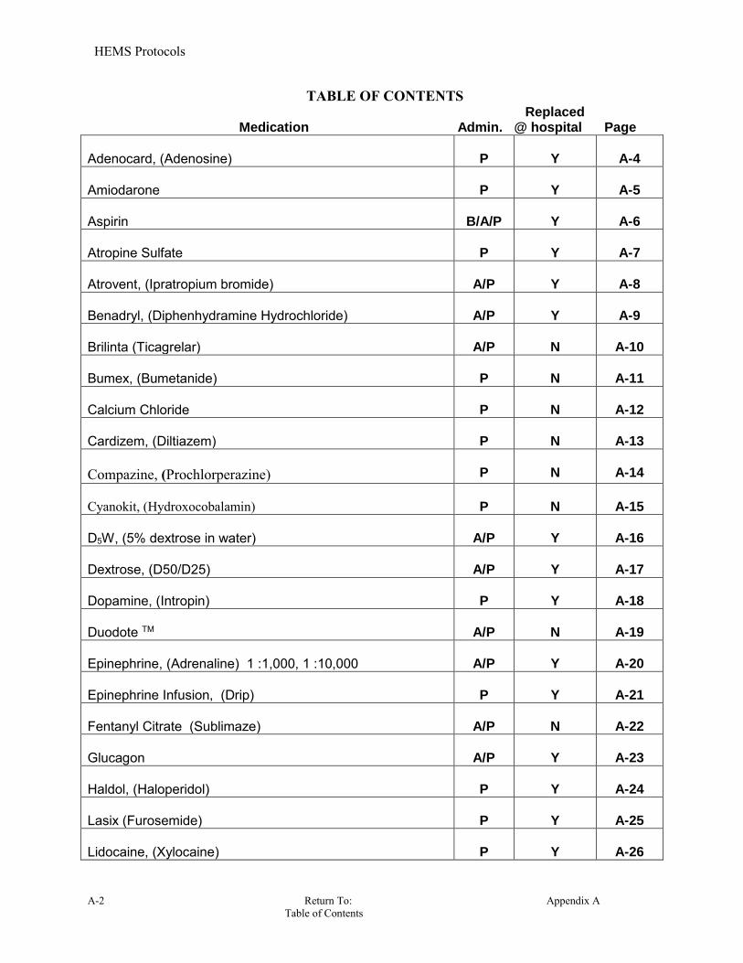
HEMS Protocols
A-2 Return To: Appendix A Table of Contents
TABLE OF CONTENTS Replaced Medication Admin. @ hospital Page
Adenocard, (Adenosine)
P
Y
A-4
Amiodarone
P
Y
A-5
Aspirin
B/A/P
Y
A-6
Atropine Sulfate
P
Y
A-7
Atrovent, (Ipratropium bromide)
A/P
Y
A-8
Benadryl, (Diphenhydramine Hydrochloride)
A/P
Y
A-9
Brilinta (Ticagrelar)
A/P
N
A-10
Bumex, (Bumetanide)
P
N
A-11
Calcium Chloride
P
N
A-12
Cardizem, (Diltiazem)
P
N
A-13
Compazine, (Prochlorperazine)
P
N
A-14
Cyanokit, (Hydroxocobalamin)
P
N
A-15
D5W, (5% dextrose in water)
A/P
Y
A-16
Dextrose, (D50/D25)
A/P
Y
A-17
Dopamine, (Intropin)
P
Y
A-18
Duodote TM
A/P
N
A-19
Epinephrine, (Adrenaline) 1 :1,000, 1 :10,000
A/P
Y
A-20
Epinephrine Infusion, (Drip)
P
Y
A-21
Fentanyl Citrate (Sublimaze)
A/P
N
A-22
Glucagon
A/P
Y
A-23
Haldol, (Haloperidol)
P
Y
A-24
Lasix (Furosemide)
P
Y
A-25
Lidocaine, (Xylocaine)
P
Y
A-26

HEMS Protocols
Appendix A Return To: A-3 Table of Contents
TABLE OF CONTENTS
Admin.
Replaced @ hospital
Page
Magnesium Sulfate
P
N
A-27
Narcan, (Naloxone)
A/P
Y
A-28
Nitroglycerin tablet
A/P
Y
A-29
Normal Saline, (0.9% Sodium Chloride)
A/P
Y
A-30
Oral Glucose
B/A/P
N
A-31
Oxygen (O2)
B/A/P
N
A-32
Phenergan, (promethazine)
P
Y
A-33
Proventil, (Albuterol Sulfate)
A/P
Y
A-34
Racemic Epinephrine
P
N
A-35
Romazacon, (Flumazenil)
P
N
A-36
Sodium Bicarbonate
P
Y
A-37
Solumedrol, (methylprednisolone)
P
Y
A-38
Sterile Water
B/A/P
N
A-39
Tetracaine, (Pontocaine)
P
Y
A-40
Toradol (Ketorolac Tromethamine)
A/P
Y
A-41
Tylenol (Acetamenaphen)
P
N
A-42
Valium, (Diazepam)
A/P
N
A-43
Vasopressin
P
Y
A-44
Versed, (Midazolam)
A/P
Y
A-45
Xopenex, (Levalbuterol HCL)
A/P
N
A-46
Zofran (Ondansetron)
P
N
A-47
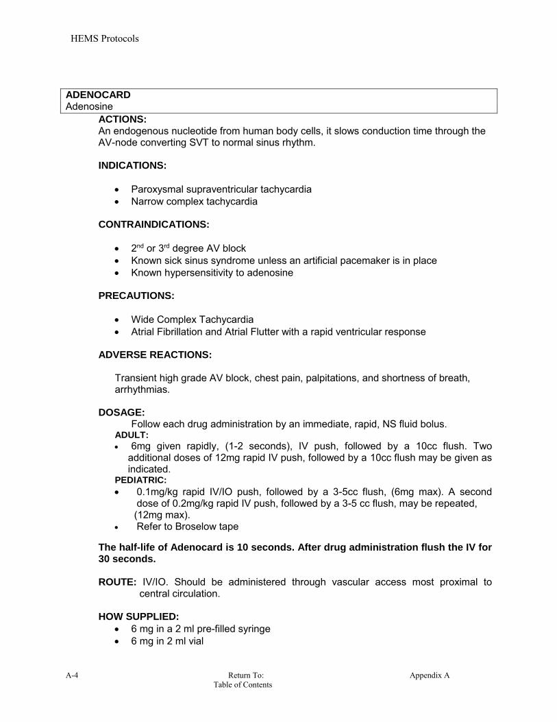
HEMS Protocols
A-4 Return To: Appendix A Table of Contents
ADENOCARD Adenosine
ACTIONS: An endogenous nucleotide from human body cells, it slows conduction time through the AV-node converting SVT to normal sinus rhythm. INDICATIONS:
Paroxysmal supraventricular tachycardia Narrow complex tachycardia
CONTRAINDICATIONS:
2nd or 3rd degree AV block
Known sick sinus syndrome unless an artificial pacemaker is in place
Known hypersensitivity to adenosine PRECAUTIONS:
Wide Complex Tachycardia Atrial Fibrillation and Atrial Flutter with a rapid ventricular response
ADVERSE REACTIONS:
Transient high grade AV block, chest pain, palpitations, and shortness of breath, arrhythmias.
DOSAGE: Follow each drug administration by an immediate, rapid, NS fluid bolus.
ADULT:
6mg given rapidly, (1-2 seconds), IV push, followed by a 10cc flush. Two additional doses of 12mg rapid IV push, followed by a 10cc flush may be given as indicated.
PEDIATRIC:
0.1mg/kg rapid IV/IO push, followed by a 3-5cc flush, (6mg max). A second dose of 0.2mg/kg rapid IV push, followed by a 3-5 cc flush, may be repeated,
(12mg max). Refer to Broselow tape
The half-life of Adenocard is 10 seconds. After drug administration flush the IV for 30 seconds. ROUTE: IV/IO. Should be administered through vascular access most proximal to
central circulation. HOW SUPPLIED:
6 mg in a 2 ml pre-filled syringe 6 mg in 2 ml vial
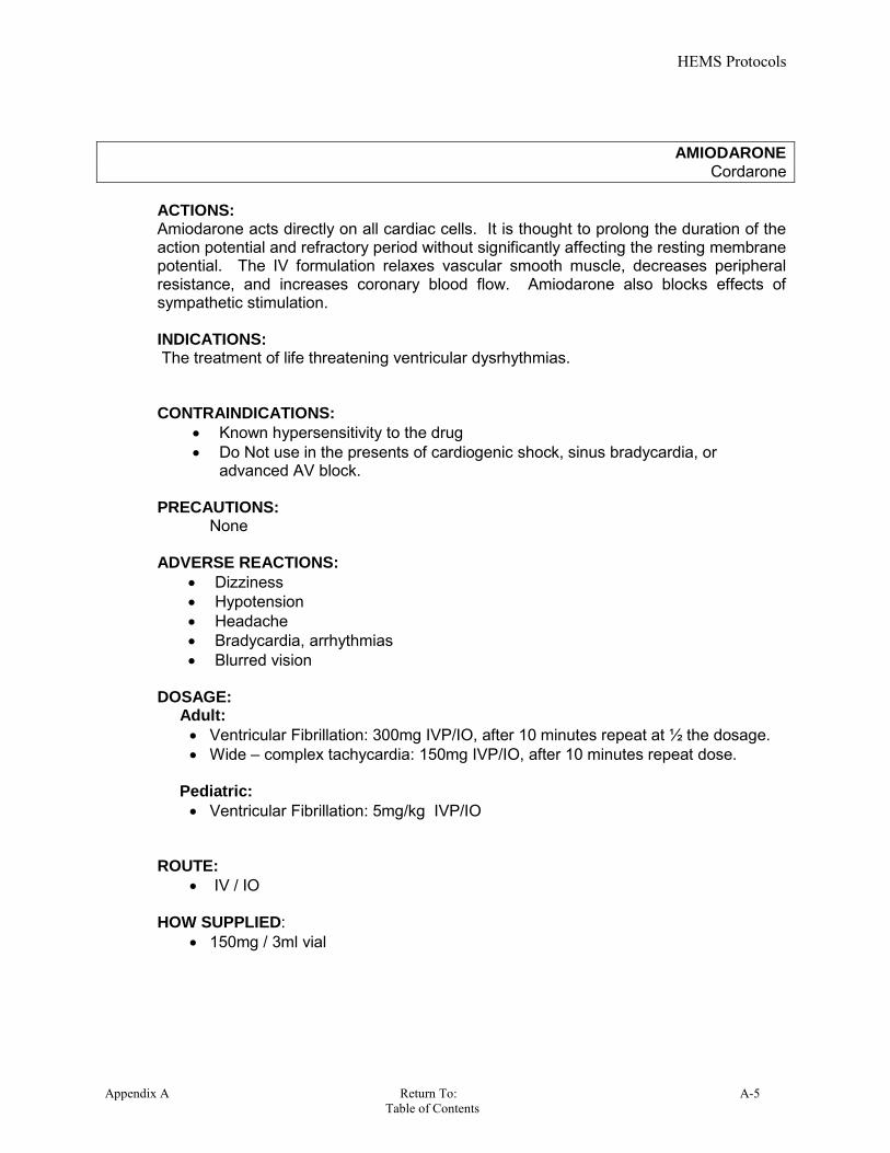
HEMS Protocols
Appendix A Return To: A-5 Table of Contents
AMIODARONE Cordarone
ACTIONS:
Amiodarone acts directly on all cardiac cells. It is thought to prolong the duration of the action potential and refractory period without significantly affecting the resting membrane potential. The IV formulation relaxes vascular smooth muscle, decreases peripheral resistance, and increases coronary blood flow. Amiodarone also blocks effects of sympathetic stimulation.
INDICATIONS:
The treatment of life threatening ventricular dysrhythmias.
CONTRAINDICATIONS:
Known hypersensitivity to the drug Do Not use in the presents of cardiogenic shock, sinus bradycardia, or
advanced AV block.
PRECAUTIONS: None
ADVERSE REACTIONS:
Dizziness Hypotension Headache Bradycardia, arrhythmias Blurred vision
DOSAGE: Adult:
Ventricular Fibrillation: 300mg IVP/IO, after 10 minutes repeat at ½ the dosage. Wide – complex tachycardia: 150mg IVP/IO, after 10 minutes repeat dose.
Pediatric:
Ventricular Fibrillation: 5mg/kg IVP/IO ROUTE:
IV / IO
HOW SUPPLIED: 150mg / 3ml vial
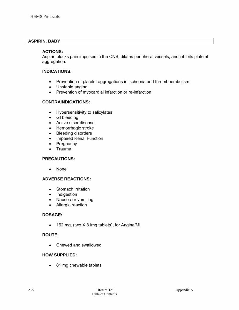
HEMS Protocols
A-6 Return To: Appendix A Table of Contents
ASPIRIN, BABY
ACTIONS: Aspirin blocks pain impulses in the CNS, dilates peripheral vessels, and inhibits platelet aggregation.
INDICATIONS:
Prevention of platelet aggregations in ischemia and thromboembolism Unstable angina Prevention of myocardial infarction or re-infarction
CONTRAINDICATIONS:
Hypersensitivity to salicylates GI bleeding Active ulcer disease Hemorrhagic stroke Bleeding disorders Impaired Renal Function Pregnancy Trauma
PRECAUTIONS:
None
ADVERSE REACTIONS:
Stomach irritation Indigestion Nausea or vomiting Allergic reaction
DOSAGE:
162 mg, (two X 81mg tablets), for Angina/MI
ROUTE:
Chewed and swallowed
HOW SUPPLIED:
81 mg chewable tablets
HEMS Protocols
Appendix A Return To: A-7 Table of Contents
ATROPINE Atropine Sulfate
ACTIONS: Parasympathetic blocker, reducing vagal tone. Atropine is a Parasympatholytic, (Anticholinergic), that acts to block acetylcholine receptors, thus inhibiting parasympathetic stimulation. INDICATIONS:
Hemodynamically symptomatic bradycardias Heart blocks Organanophosphate poisoning WMD
CONTRAINDICATIONS:
Tachycardias
Atrial fibrillation/flutter with a rapid ventricular response, (RVR). Glaucoma
PRECAUTIONS:
If given in too small a dose, or if given too slowly, bradycardia may worsen. ADVERSE REACTIONS:
Dryness of the mouth and nose, blurred vision, dilated pupils, tachycardia, headache and restlessness
DOSAGE: No maximum dose on organophosphate poisoning treatment
ADULT:
Brady-Dysrhythmias: 0.5mg every 5 minutes to maximum dosage of 3 mg Organophosphate Poisoning: 2mg slow IVP or IM every 5 – 10 minutes until sign / symptoms are resolve.
PEDIATRIC:
Bradycardia: 0.02 mg / kg, (0.1mg minimum dose). May be repeated q 5 minutes to a max total dose of 1 mg in a child and 2 mg in an adolescent.
Refer to length based tape for resuscitation dose ROUTE:
IV, IM, IO
HOW SUPPLIED: 1 mg in 10 ml pre-filled syringe 8 mg in 20 ml multi-dose vial

HEMS Protocols
A-8 Return To: Appendix A Table of Contents
ATROVENT Ipratropium bromide
ACTIONS: It is a parasympatholytic that causes bronchodilation and dries respiratory tract secretions. It blocks acetylcholine receptors, thus inhibiting parasympathetic stimulation. INDICATIONS: Bronchospasms associated with:
COPD Asthma
CONTRAINDICATIONS:
History of sensitivity to atrovent or atropine
PRECAUTIONS:
Avoid getting this medication in your eyes. If this happens, rinse your eyes with water or NS.
ADVERSE REACTIONS:
Headache or dizziness Dry mouth, cough or hoarseness Nausea or vomiting Blurred vision
DOSAGE: Adults & Pediatrics 2 years of age and older:
Atrovent 0.5 mg mixed with 2.5 mg proventil, (or given alone).
Under the age of 2 years old requires OLMC order. As its duration of action is 3 to 4 hours it is typically only given as a single dose in the pre-hospital setting. ROUTE:
Inhalation by nebulizer
HOW SUPPLIED: 0.5 mg / 2.5ml plastic vial
HEMS Protocols
Appendix A Return To: A-9 Table of Contents
BENADRYL Diphenhydramine Hydrochloride ACTION:: Benadryl, (diphenhydramine), is an anti-histamine. It is the second line treatment for anaphylactic / allergic reactions. Benadryl can induce, or worsen, hypotension. Therefore significant hypotension should be treated, (fluid bolus, epi drip, etc.), prior to benadryl administration. Due to its duration of action a repeat dose is not indicated in the pre-hospital setting. INDICATIONS:
Anaphylaxis, (Administered after epinephrine) Allergic Reactions Dystonic and extrapyramidal reactions
PRECAUTIONS:
Use with caution in patients with a history of asthma, cardiovascular disease, and hypertension.
Sedative effects are more pronounced when patient has ingested alcohol or other CNS depressants, (barbiturates, phenothiazines, antidepressants, or narcotics).
ADVERSE REACTION:
Tachycardia Hypotension Central Nervous System depression Nausea and vomiting
DOSAGE:
Adult: Benadryl 50 mg, IV or IM If an i.v. is in place it is reasonable to give Benadryl 25 mg i.v. AND 25 mg I.M. Pediatric: 2 – 6 years of age: 6.25 mg IV /IO/ IM 6 – 12 years of age: 12.5 to 25 mg IV/IO/IM OR weight based: 1 – 2 mg / Kg,IV / IO / IM, to a Max of 50 mg.
ROUTE:
IV, IM, IO
HOW SUPPLIED:
50 mg in a 1ml vial 50 mg in a 1 ml pre-filled syringe

HEMS Protocols
A-10 Return To: Appendix A Table of Contents
BRILINTA ticagrilor
ACTION: Platelet inhibitor
Brilinta (ticagrelor) keeps the platelets in your blood from coagulating (clotting) to prevent unwanted blood clots that can occur with certain heart or blood vessel conditions.
Brilinta is used to lower your risk of having a stroke or serious heart problems after you have had a heart attack or severe chest pain (angina).
INDICATIONS:
STEMI Acute Coronary Syndrones
PRECAUTIONS:
Do not take BRILINTA if you are bleeding now, especially from your stomach or intestine (ulcer).
History of bleeding in the brain. Have severe liver problems.
ADVERSE REACTION:
General risk of bleeding (may be fatal) Dyspnea Hyper- or hypotension Chest pain
DOSAGE: ADULT: 180mg (90mg x 2) with a small sip of water Pediatric: Not established ROUTE: P.O. can be taken with or without food. How Supplied:
90mg is supplied as a round, biconvex, yellow, film coated tablet marked with a “90” above “T” on one side.

HEMS Protocols
Appendix A Return To: A-11 Table of Contents
BUMEX Bumetanide
ACTION: A potent diurectic with a rapid rate of onset and a short duration of action. Like furosemide, bumetanide is a loop diurectic that inhibits the reabsorption of sodium chloride in the kidneys and thus causes a net diuresis.
Bumex 1mg IV = 40 mg IV Lasix Bumex 1.5mg IV = 60 mg IV Lasix Bumex 2mg IV = 80 mg IV Lasix Bumex 2.5mg IV = 100 mg IV Lasix INDICATIONS:
Congestive Heart Failure, (CHF)) Pulmonary Edema, (PE)
PRECAUTIONS:
Should be limited to life-threatening situation in pregnant patients.
ADVERSE REACTION:
Muscle cramps Dizziness Nausea Hypotention Headache
DOSAGE:
ADULT:
1mg over a period of 1 – 2 minutes
PEDIATRIC:
PER OLMC ROUTE:
IV, IO
HOW SUPPLIED:
0.5 mg in 2 ml of solvent ampule, (0.25 mg / ml) 2,4, and 10 ml vials

HEMS Protocols
A-12 Return To: Appendix A Table of Contents
CALCIUM CHLORIDE (CaCl2)
ACTIONS: Couples electrical and mechanical events of the myocardium. Increases myocardial contractility. Increases ventricular irritability.
INDICATIONS:
Hyperkalemia Overdose of calcium channel blockers Antidote for Magnesium Sulfate toxicity
PRECAUTIONS:
Patients taking digitalis based medications IV line should be flushed between calcium chloride and Sodium Bicarb. Extravasation may cause tissue necrosis.
SIDE EFFECTS:
Bradycardia Hypotension Syncope
DOSAGE:
Adult:
10 mg/kg slow IV push of a 10% solution; may be repeated at 10-minute intervals.
Pediatric:
20mg/kg IV /IO 0.2 ml/kg slow IV push of a 10% solution, (1gm / 10ml prefilled)
ROUTE: SLOW IVP / IO HOW SUPPLIED:
1gm / 10 ml prefilled syringe
HEMS Protocols
Appendix A Return To: A-13 Table of Contents
CARDIZEM Diltiazem
ACTION: Cardizem is a calcium channel blocker. Cardizem inhibits the influx of extra cellular calcium across both the myocardial and vascular smooth muscle cell membranes. This decreases the contractility of the myocardial smooth muscle, and causes dilation of the coronary and systemic arteries. It decreases AV nodal conduction. INDICATIONS:
Symptomatic Atrial Fibrillation Symptomatic Atrial Flutter Paroxysmal supraventricular tachycardia, (PSVT), refractory to adenosine
Document pre and post treatment rhythm strips. CONTRAINDICATIONS:
Patients with cardiogenic shock, or patients with a systolic B/P <90mmHg. Patients with known accessory pathway conditions, (e.g. WPW)
PRECAUTIONS: Use with caution in patients with ventricular dysfunction, severe bradycardia or with previous conduction abnormalities.
It may be used in pregnant patients by OLMC order only. ADVERSE REACTION:
Hypotension
Bradycardia
Heart blocks
Asystole DOSAGE: ADULT:
0.25 mg/kg, IV over 2 minutes. May Repeat after 15 minutes: 25 mg or 0.35 mg/kg, IV over 2 minutes
Infusion
IV Drip @ 10 mg / hr. Prepare by putting 25 mg of cardizem in 500 ml 0.9% Saline and infuse with a 10 drop/ml set @ 33 gtt/min. Infuse by IV infusion pump, if available.
PEDIATRIC: OLMC ONLY
ROUTE:
IV, IO HOW SUPPLIED:
25 mg in a 5 ml pre-filled syringe. 5 ml & 10 ml vials of 5 mg / ml

HEMS Protocols
A-14 Return To: Appendix A Table of Contents
COMPAZINE Prochlorperazine
ACTION:
Antiemetic.
INDICATIONS:
For control of severe nausea and vomiting
CONTRAINDICATIONS: Comatose states Patients with a demished gag reflex. Patients with a history of hypersensitivity to the drug.
PRECAUTIONS:
Extrapyramidal and dystonic reactions may occur. Treat these with iv benadryl.
ADVERSE REACTIONS: May impair mental and physical ability Drowsiness
DOSAGE : ADULT :
5-10 mg slow IV, (over at least 2 minutes), or IM
PEDIATRIC:
0.15 mg/kg IM, (iv by OLMC order only). Under 4 y/o: OLMC only
ROUTE:
IVP, (Slow over 2 minutes) IM, (Deep, not deltoid)
HOW SUPPLIED:
10mg / 2ml Vial
HEMS Protocols
Appendix A Return To: A-15 Table of Contents
CYANOKIT Hydroxocobalamin
(A precursor of vitamin B12)
ACTION: Hydroxocobalamin is a precursor to Vitamin B12. It is used in the treatment of known or suspected cyanide poisoning based upon its ability to bind cyanide ions, forming cyanocobalamin, which is then excreted in the urine. INDICATIONS:
For known or suspected cyanide poisoning Use in conjunction with the RAD – 57, (If elevated HbCO, consider cyanide, too).
CONTRAINDICATIONS:
None PRECAUTIONS:
Allergic reactions have been observed. May interfere with some clinical lab test. Patients should avoid direct sunlight for 24 hours after receiving medication.
ADVERSE REACTIONS:
Chromaturia, (red colored urine) Erythema, (skin redness) Rash Increased blood pressure Nausea Headache
DOSAGE: Single dose therapy ADULT:
5 g / 200 ml N.S.administered as an IV infusion over 15 minutes. Pediatric:
70mg/kg in 200 ml N.S. administered as an IV infusion over 15 minutes.
ROUTE: IV infusion
HOW SUPPLIED: The kit contains two, 2.5 g vials of hydroxocobalamin as a lyophilized powder. Each vial should be reconstituted with 100 mL of normal saline. An adult will require two vials. Note: Diazepam, dobutamine, dopamine, fentanyl, nitroglycerin, pentobarbital, propofol, and thiopental are physically incompatible, (particle formation), with hydroxocobalamin and should not be administered simultaneously through the same IV line as hydroxocobalamin.

HEMS Protocols
A-16 Return To: Appendix A Table of Contents
D5W 5% Dextrose in Water
ACTION: D5W is a hypotonic glucose solution, used to keep a vein open and to supply calories necessary for cell metabolism. It contains 5 g of dextrose per 100 ml. INDICATIONS:
Infusion of Magnesium Sulfate
CONTRAINDICATIONS:
None
PRECAUTIONS:
N/A ADVERSE REACTIONS:
N/A
DOSAGE:
250 ml 100 ml
ROUTE:
IV, IO infusion HOW SUPPLIED:
250 ml bags 100 ml bags

HEMS Protocols
Appendix A Return To: A-17 Table of Contents
DEXTROSE D25/D50
ACTION: Dextrose in water provides supplemental glucose to treat hypoglycemia. D50 is a hypertonic solution primarily used to elevate the blood sugar. It may also be used to decrease intracranial pressure. INDICATIONS:
Hypoglycemia Coma of unknown origin Cardiac arrest – PEA, (known diabetic)
CONTRAINDICATIONS:
Patients with intracranial hemorrhage or known CVA with a normal blood glucose level.
PRECAUTIONS:
Perform a glucometer check prior to administration, when possible. Localized venous irritation and tissue necrosis may result from infiltrated IV line.
ADVERSE REACTIONS:
Hyperglycemia Thrombophlebitis D50 may precipitate severe neurological symptoms , (Wernicke's
encephalopathy) ,in thiamine-deficient patients.
DOSAGE:
ADULT:
25 grams of D50, (may repeat as clinically indicated) PEDIATRIC:
0.5 gm / kg of D25 Refer to length based tape
ROUTE:
IV, IO HOW SUPPLIED:
D50: 25 grams of glucose in a 50 ml D25: 12.5 grams of glucose in a 10 ml

HEMS Protocols
A-18 Return To: Appendix A Table of Contents
DOPAMINE Intropin
ACTION: Vasopressor – dose dependent alpha, beta, and dopaminergic agonist.
Low dose, (0.5-2 mcg/kg/min.): Causes vasodilatation in renal, mesenteric, cerebral, and coronary arteries, via activation of the dopamine receptor sites.
Intermediate doses, (2-10 mcg/kg/min.): Produce a step-wise increase in
contractility, automaticity, and conductivity via beta-receptor effects. High doses, (10-20 mcg/kg/min.): The alpha receptor effects predominate
producing peripheral vasoconstriction. Extremely high dose, (>20 mcg/kg): Renal and mesenteric vessels constrict
resulting in decreased blood flow. NOT used in the Pre-Hospital setting. INDICATIONS:
SHOCK CONTRAINDICATIONS:
Tachydysrhythmias VT/VF
PRECAUTIONS:
Correct any volume deficit before instituting dopamine therapy, unless otherwise directed.
Do not mix with Sodium Bicarbonate.
ADVERSE REACTIONS:
Ectopy, local tissue narcosis if line infiltrates, tachycardia, palpitations, dysrhythmia, hypertension, nausea and vomiting.
DOSAGE: ADULT & PEDIATRIC:
400 mg/250 cc premixed bag yields 1600 mcg/ ml Initiate therapy at rate of 5 mcg/kg/min and titrate to a blood pressure of 100
systolic in adults and 80-90 in pediatric patients. ROUTE:
IV, IO infusion HOW SUPPLIED:
400 mg / 250 ml premixed

HEMS Protocols
Appendix A Return To: A-19 Table of Contents
DUODOTE
ACTION: Atropine competively blocks the effects of aceycholine, including excess acetylcholine due to organophosphate poisoning, at muscarinic cholinergic receptors on smooth muscle, and secretory gland cells and in peripheral autonomic ganglia and the central nervous system. Pralidoxine reactivates acetylcholinesterase which was inactivated by phosphorylation due to an organophosphate nerve agent or insecticide. However, pralidoxine does not reactivate acetylcholinesterase inactivated by all organophosphorous nerve agents, (e.g. soman). Reactivated acetylcholinesterase hydrolyzes excess acetylcholine resulting from organophosphorous poisoning to help restore impaired cholinergic neural function. Reactivation is clinically important because only a small proportion of active acetylcholinesterase is needed to maintain vital functions. Pralidoxine cannot reactivate phosphorylated acetylcholinsterases that have undergone a further chemical reaction known as “aging”. INDICATIONS:
Chemical Nerve Agent Insecticide, (Organophosphate), Poisoning
CONTRAINDICATIONS:
In the event of a chemical nerve agent attack, there are no contraindications. PRECAUTIONS:
Duodote should only be administered to patients experiencing symptoms of orgonophosphate poisoning in a situation where exposure is known or suspected. Duodote should be administered as soon as symptoms of orgonophosphate poisoning appear.
DO NOT confused with Duoneb ADVERSE REACTIONS:
When Atropine and Pralidoxine are used together, the signs of atropinization may occur earlier than might be expected when atropine is used alone.
DOSAGE: ADULT: Mild symptoms - Administer One DuoDote autoinjector, may repeat every 10-15 minutes until symptoms improve. Maximum of 3 doses. Moderate symptoms - Administer Two(2) DuoDote autoinjectors in rapid succession. May administer 1 additional DuoDote in 10-15 minutes. Maximum of 3 doses. Severe symptoms – Administer Three DuoDote autoinjectors in rapid succession. ROUTE: Autoinjector deep IM into the mid-lateral thigh :. The autoinjector can inject through clothing. Firmly press the green tip straight down , (900 angle), against the mid-outer thigh. Continue to firmly push until you feel the injector trigger. Hold down for 10 seconds to deliver medication. Remove from thigh and check to insure the needle is visible. Treat as any other SHARP. HOW SUPPLIED:
Autoinjector: 2mg Atropine & 600 mg pralidoxine, (2-PAM).

HEMS Protocols
A-20 Return To: Appendix A Table of Contents
EPINEPHRINE Adrenaline
ACTION: Alpha and beta adrenergic agonist that stimulates the sympathetic nervous system. Major sympathetic effects include: positive chronotropic effect, (Increase heart rate), positive inotropic effect, (Increased force of contraction), increased systemic vascular resistance, bronchodilation, assist in the conversion of ventricular fibrillation, and increased cerebral blood flow in cardiac arrest. INDICATIONS:
Anaphylaxis Asthma, bronchospasm Asystole Pulseless electrical activity Heart blocks, hemodynamically unstable bradycardias Ventricular fibrillation Pulseless ventricular tachycardia
CONTRAINDICATIONS:
None in a life threatening situation PRECAUTIONS:
Give cautiously in patients with a history of angina, hypertension, tachycardia, coronary artery disease or who are pregnant.
Do not mix with Sodium Bicarbonate. ADVERSE REACTIONS:
Ectopic beats, tachycardia, palpitations, anxiety and headache, VF/VT Increased myocardial oxygen demand
DOSAGE: ADULT:
Ventricular Fibrillation, Pulseless Ventricular Tachycardia, Pulseless Electrical Activity and Asystole: 1 mg of 1:10,000 repeated every three to five minutes.
Asthma and Anaphylactic reactions: 0.3 – 0.5 ml of 1:1,000 is given SQ. EMT Basic Anaphylactic reactions > 30kg. EpiPen auto-injector 0.3mg (0.3ml
1:1,1,000) EMT-I & Paramedic--Anaphylactic reactions: > 30kg. EpiPen auto-injector 0.3mg
0.3ml 1:1,1,000) PEDIATRICS:
Resuscitation dose: 0.01mg/kg IV/IO. Refer to Broselow tape. Asthma and Anaphylactic reactions: 0.3 ml of 1:1,000 is given sq. EMT Basic -- Anaphylactic reactions: 15kg – 30kg. EpiPen Jr auto-injector
0.15mg (0.3ml, 1:2,000). AEMT & Paramedic -- Anaphylactic reactions: 15kg – 30kg. EpiPen Jr auto-
injector 0.15mg (0.3ml, 1:2,000). ROUTE:
IV, IM, IO, SQ, auto injector HOW SUPPLIED:
EpiPen 0.3mg (0.3ml, 1:1,000) in auto-injector. Warning: Hold the epi pen in your fist. DO NOT hold your thumb over the end of the auto injector.
EpiPen Jr 0.15mg(0.3ml, 1:2,000) in auto-injector, 1:1,000: 1 mg in 1 ml ampule 1:10,000: 1 mg in a 10 ml pre-filled syringe.
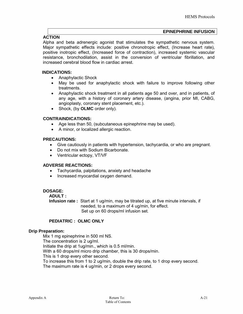
HEMS Protocols
Appendix A Return To: A-21 Table of Contents
EPINEPHRINE INFUSION
ACTION Alpha and beta adrenergic agonist that stimulates the sympathetic nervous system. Major sympathetic effects include: positive chronotropic effect, (Increase heart rate), positive inotropic effect, (Increased force of contraction), increased systemic vascular resistance, bronchodilation, assist in the conversion of ventricular fibrillation, and increased cerebral blood flow in cardiac arrest.
INDICATIONS: Anaphylactic Shock May be used for anaphylactic shock with failure to improve following other
treatments. Anaphylactic shock treatment in all patients age 50 and over, and in patients, of
any age, with a history of coronary artery disease, (angina, prior MI, CABG, angioplasty, coronary stent placement, etc.).
Shock, (by OLMC order only).
CONTRAINDICATIONS:
Age less than 50, (subcutaneous epinephrine may be used). A minor, or localized allergic reaction.
PRECAUTIONS:
Give cautiously in patients with hypertension, tachycardia, or who are pregnant. Do not mix with Sodium Bicarbonate. Ventricular ectopy, VT/VF
ADVERSE REACTIONS:
Tachycardia, palpitations, anxiety and headache Increased myocardial oxygen demand.
DOSAGE: ADULT : Infusion rate : Start at 1 ug/min, may be titrated up, at five minute intervals, if needed, to a maximum of 4 ug/min, for effect. Set up on 60 drops/ml infusion set. PEDIATRIC : OLMC ONLY
Drip Preparation: Mix 1 mg epinephrine in 500 ml NS. The concentration is 2 ug/ml. Initiate the drip at 1ug/min., which is 0.5 ml/min. With a 60 drops/ml micro drip chamber, this is 30 drops/min. This is 1 drop every other second. To increase this from 1 to 2 ug/min, double the drip rate, to 1 drop every second. The maximum rate is 4 ug/min, or 2 drops every second.
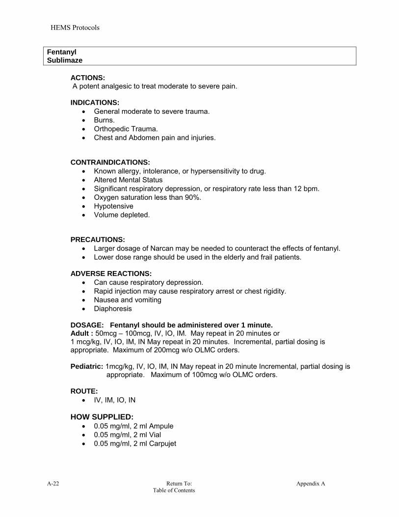
HEMS Protocols
A-22 Return To: Appendix A Table of Contents
Fentanyl Sublimaze
ACTIONS: A potent analgesic to treat moderate to severe pain. INDICATIONS:
General moderate to severe trauma. Burns. Orthopedic Trauma. Chest and Abdomen pain and injuries.
CONTRAINDICATIONS:
Known allergy, intolerance, or hypersensitivity to drug. Altered Mental Status Significant respiratory depression, or respiratory rate less than 12 bpm. Oxygen saturation less than 90%. Hypotensive
Volume depleted. PRECAUTIONS:
Larger dosage of Narcan may be needed to counteract the effects of fentanyl. Lower dose range should be used in the elderly and frail patients.
ADVERSE REACTIONS:
Can cause respiratory depression. Rapid injection may cause respiratory arrest or chest rigidity. Nausea and vomiting Diaphoresis
DOSAGE: Fentanyl should be administered over 1 minute. Adult : 50mcg – 100mcg, IV, IO, IM. May repeat in 20 minutes or 1 mcg/kg, IV, IO, IM, IN May repeat in 20 minutes. Incremental, partial dosing is appropriate. Maximum of 200mcg w/o OLMC orders. Pediatric: 1mcg/kg, IV, IO, IM, IN May repeat in 20 minute Incremental, partial dosing is appropriate. Maximum of 100mcg w/o OLMC orders. ROUTE:
IV, IM, IO, IN
HOW SUPPLIED: 0.05 mg/ml, 2 ml Ampule 0.05 mg/ml, 2 ml Vial 0.05 mg/ml, 2 ml Carpujet

HEMS Protocols
Appendix A Return To: A-23 Table of Contents
GLUCAGON
ACTION: Pancreatic hormone, insulin antagonist. Increases the breakdown of glycogen to glucose and stimulates glucose synthesis, resulting in blood glucose elevation. This is only effective if liver glycogen is available. May be ineffective in chronic states of hypoglycemia, starvation, and adrenal insufficiency. Glucagon also acts as a smooth muscle relaxer, and improves cardiac muscle function. INDICATIONS:
Symptomatic hypoglycemia treatment when unable to obtain IV access. Beta Blocker Overdose Esophageal foreign body Severe anaphylactic shock Cardiogenic shock
CONTRAINDICATIONS:
Hypersensitivity PRECAUTIONS:
None ADVERSE REACTIONS:
Tachycardia Hypotension Nausea and vomiting Urticaria
DOSAGE: Note : 1 international unit = 1 mg
ADULT:
Hypoglycemia : 1 mg IM, (single dose only) Esophageal foreign body: 1 mg IVP, (single dose only)
PEDIATRIC:
Hypoglycemia: < 20kg 0.5 mg IM (single dose only), (Otherwise administer adult dose).
Esophageal foreign body: <20kg, 0.5mg IVP, (single dose only). Otherwise administer adult dosage.
ROUTE:
SQ, IM, IV HOW SUPPLIED:
I mg / vial, with 1 ml diluent / vial. Dilute 1 unit, (1 mg), white powder with 1 ml of diluting solution, (1 mg / ml). Glucagon must be reconstituted, (with provided diluent), before
administration. Do not mix white powder with saline.
HEMS Protocols
A-24 Return To: Appendix A Table of Contents
HALDOL Haloperidol
ACTION: Blocks the dopamine receptors in the brain that are responsible for mood behavior. Has antiemetic properties. INDICATIONS:
Acute psychotic episodes Emergency sedation of severely agitated or delirious patients
CONTRAINDICATIONS:
None PRECAUTIONS:
None
ADVERSE REACTIONS: Arrhythmias including torsade de pointes Dystonic reactions, (which are treated with Bendryl). Neuroleptic malignant syndrome, (high fever, altered mental status, DEATH)
DOSAGE:
ADULT:
2mg – 5mg
Pediatric:
OLMC only ROUTE:
IM only
HOW SUPPLIED:
5mg / 1ml
HEMS Protocols
Appendix A Return To: A-25 Table of Contents
LASIX Furosemide
ACTION: Potent vasodilator and diuretic. Blocks the re-absorption of sodium in the Loop of Henle as well as the distal and proximal tubules within the kidney. INDICATIONS:
Congestive heart failure Pulmonary Edema
CONTRAINDICATIONS:
Dehydrated patients PRECAUTIONS:
Patients with hypersensitivity to sulfa may experience reactions to Furosemide May need a higher dosage in patients with renal failure Systolic Blood Pressure < 90 mmHg
ADVERSE REACTIONS: Hypotension Hypokalemia, electrolyte abnormalities, muscle cramps, weakness, thirst, light
headiness, dizziness, nausea and vomiting DOSAGE:
ADULT:
Up to double the prescribe daily dose If not taking daily, administer a maximum of 40 mg.
PEDIATRIC:
OLMC ONLY
ROUTE:
IM, IV, IO
HOW SUPPLIED:
100 mg in 10 ml pre-filled syringe 100 mg in 10 ml vial 40 mg in 4 ml vial
HEMS Protocols
A-26 Return To: Appendix A Table of Contents
LIDOCAINE Xylocaine
ACTIONS: Suppresses ventricular ectopic activity, increases ventricular fibrillation threshold, reduces velocity of electrical impulse through the conductive system. INDICATIONS:
Ventricular Fibrillation Pulseless Ventricular Tachycardia Significant ventricular ectopy, (PVCs), in the setting of myocardial
ischemia/infarction Stable Ventricular Tachycardia Wide Complex Tachycardia of undetermined etiology
CONTRAINDICATIONS:
High-degree heart blocks PVCs in conjunction with Bradycardia, (escape beats)
PRECAUTIONS:
Prophylactic use in MI’s is not indicated Reduce dose, (1/2 normal dose), if known liver impairment or LV dysfunction
ADVERSE REACTIONS:
Lidocaine Toxicity, (Light headiness, dizziness, blurred vision, nausea, vomiting, seizures, hypotension, bradycardia and central nervous system depression).
DOSAGE:
ADULT: Cardiac arrest from VF/VT, 1.5 mg/kg. Refractory VF, give an additional 0.5 mg/kg dose q 5 minutes to a maximum
dose of 3 mg/kg. Stable VT, Wide Complex Tachycardia, significant ventricular ectopy, 1.5 mg/kg
IV push. Maintenance infusion alternative: Rebolus 0.5mg / kg q 15 – 20 minutes max.
dose 3 mg/kg.
PEDIATRIC (up to 38kgs): Loading dose: 1 mg/kg, (Refer to Broselow tape.) ROUTE: IVP, IO
HOW SUPPLIED:
100 mg in a 5 ml
HEMS Protocols
Appendix A Return To: A-27 Table of Contents
MAGNESIUM SULFATE
ACTIONS: CNS depressant, anticonvulsant, antiarrhyhmic. INDICATIONS:
Refractory Ventricular Fibrillation / Ventricular Tach Torsade de Pointes Eclampsia
CONTRAINDICATIONS:
Heart Block
PRECAUTIONS: Magnesium should be administered slowly to minimize side effects. Maintain continuous cardiac monitoring. Use with caution in patient’s with renal failure.
ADVERSE REACTIONS:
Flushing of the skin, sweating Central Nervous System depression Respiratory depression Hypotension Bradycardias and cardiac arrhythmias
DOSAGE:
ADULT:
Pulseless VF/VT or torsade de pointes: 2 grams IVP over 2 minutes Pulse producing VT, torsade de pointes: 2 gram in 250 ml D5W over 10-20 min. Eclampsia: 4 grams in 250cc D5W over 30 minutes
PEDIATRIC:
50 mg/kg, (Refer to length based tape), over 10-20 minutes: Maximum 2 grams. ROUTE: IV, IO, IV infusion
HOW SUPPLIED:
1 grams in 2 ml vial 5 grams in 10 ml pre-filled syringe

HEMS Protocols
A-28 Return To: Appendix A Table of Contents
NARCAN Naloxone
ACTION: Narcotic antagonist reverses the central nervous system and respiratory depression effects of narcotics; reverses the cardiovascular effects to a lesser extent. Naloxone competes for narcotic receptor sites in the brain, and displaces narcotic molecules from the opiate receptors. INDICATIONS:
Known or suspected narcotic overdoses, such as: Morphine Demerol Heroin Darvon
Percocet Dilaudid Codeine Percodan Fentanyl Methadone Nubian Talwin Stadol
Unwitnessed cardiac arrests of suspected overdose etiology. CONTRAINDICATIONS:
Known allergy to Narcan PRECAUTIONS:
Narcan should be administered cautiously to patients who are known or suspected to be physically dependent on narcotics. Abrupt and complete reversal of narcotic effects by Naloxone can cause withdrawal type effects, agitation, and violence.
ADVERSE REACTIONS:
Hypotension/hypertension Nausea/Vomiting Acute narcotic withdrawal syndrome, (nausea, vomiting, sweating, tachycardia,
hypertension, tremor, agitation, diarrhea, abdominal cramps, seizures, and cardiac arrest).
DOSAGE: ADULT:
0.4 mg increments up to a maximum of 4 mg PEDIATRIC:
0.1 mg/kg with a maximum single dose of 2 mg. (Refer to Broselow tape)
ROUTE: IV, IM, IO, SQ, IN
HOW SUPPLIED:
2 mg / 2 ml vial 2mg 2ml pre-filled syringe 1 mg / 2 ml pre-filled syringe 0.4mg / 1 ml vial
HEMS Protocols
Appendix A Return To: A-29 Table of Contents
NITROGLYCERIN TABLET
ACTION:
Anti-anginal medication: Nitroglycerin, NTG, improves coronary circulation, decreased pre-load and afterload, and decreased cardiac workload. This improves coronary circulation, deceasing ischemia. INDICATIONS:
Angina pectoris Myocardial infarction Congestive heart failure / Pulmonary Edema Hypertension
CONTRAINDICATIONS:
Hypotension, (systolic <100 mmHg) VIAGRA and other similar acting medications within the preceding 36 hours
PRECAUTIONS: Volume-depleted patients may experience exaggerated hypotensive response Postural hypotension
ADVERSE REACTIONS:
Hypotension Headache Nausea/Vomiting Tachycardia Dizziness Palpitations
DOSAGE: ADULT:
0.4 mg SL q 5 minutes , total of (3) three. Hold for systolic B/P < 100 mmHg. PEDIATRIC: N/A ROUTE:
Sublingual HOW SUPPLIED:
0.4 mg / tablet, 200/bottle

HEMS Protocols
A-30 Return To: Appendix A Table of Contents
NORMAL SALINE 0.9% Sodium Chloride
ACTION: Normal saline, (0.9% N.S.), is an isotonic crystalloid solution used for fluid and electrolyte replacement. INDICATIONS:
Diabetic ketoacidosis Dehydration Shock Heat related emergencies Keep vein open
CONTRAINDICATIONS:
Congestive Heart Failure Pulmonary Edema
PRECAUTIONS: Circulatory overload Renal disease Large volume infusion may dilute other electrolytes
ADVERSE REACTIONS: N/A
DOSAGE:
Titrate to effect; large volumes may be required in patients suffering DKA
ROUTE: IV, IO
HOW SUPPLIED: 500 ml bag 1,000 ml bag
10 ml pre-filled syringe for saline lock
3.0 ml plastic vial for neubilizer
HEMS Protocols
Appendix A Return To: A-31 Table of Contents
ORAL GLUCOSE
ACTION: This medication is used to raise the patient's serum glucose. INDICATIONS:
Patients with altered mental status or other associated signs/symptoms of diabetes or hypoglycemia.
Symptomatic patients with a known history of diabetes or hypoglycemia. Patient is able to swallow without gagging or aspirating the medication.
CONTRAINDICATIONS:
Unresponsive or unconscious patient. Known diabetic that has not taken insulin for days, (ie.. suspected hyperglycemia). Patient is unable to swallow without gagging or aspirating the medication.
PRECAUTIONS:
Do not administer a large portion at one time. ADVERSE REACTIONS:
Aspiration if unable to swallow.
DOSAGE: ADULT:
15 grams oral PEDIATRIC:
7.5 grams oral ROUTE:
Oral: Administer in small increments between the cheek and the gum. It may take up to 20 minutes to see noticeable improvement. HOW SUPPLIED:
15 grams pliable tube.

HEMS Protocols
A-32 Return To: Appendix A Table of Contents
OXYGEN
ACTION: Oxygen supports life. Increased concentrations of oxygen increase the saturation level in the hemoglobin in the red blood cell. This results in an increased oxygenation level at the tissue. Oxygen is required for the efficient breakdown of glucose into a usable energy form. INDICATIONS:
Hypoxia Respiratory distress Chest pain Shock Trauma Oxygen should be used In all patients where an increased oxygen level will
decrease tissue hypoxia. CONTRAINDICATIONS:
None
PRECAUTIONS: Possible suppression of respiratory drive in COPD patients. Never permit smoking in an oxygen enriched environment. Never deprive the hypoxic patient of oxygen for fear of respiratory depression.
ADVERSE REACTIONS:
None DOSAGE: ADULT & PEDIATRIC:
Patients in mild distress should receive 4 to 6 liters via a nasal cannula. Patients in moderate to severe distress from should receive 100% oxygen via a
100% non-rebreather mask. Severe multiple trauma patients, cardiac arrest patients, severe pulmonary
edema, etc., may warrant advanced airway management. ROUTE:
NRB, NC, Ambu bag, ETT, King airway, Blow by, CPAP, etc. HOW SUPPLIED:
D, H, and M tanks.

HEMS Protocols
Appendix A Return To: A-33 Table of Contents
PHENERGAN Promethazine
ACTIONS:
Antiemetic, anticholinergic, sedative
INDICATIONS:
Severe nausea and vomiting
CONTRAINDICATIONS:
Phenergan should not be given if the patient is hypotensive. Unresponsive or sedated patients, (morphine may potentiate the sedative side
effects of Phenergan). Patient with allergies to Phenothiazines, (found in some tranquilizers and anti-
histamines). Pediatrics patients less than 2 years of age is contraindicated in the pre-hospital
setting.
PRECAUTIONS:
Watch for dystonic reactions. Treat these with iv benadryl.
ADVERSE REACTIONS:
Sedation, dizziness, dysrhythmia, hyperexcitability, hallucinations, seizures, dystonic reactions.
DOSAGE:
ADULT : 12.5 mg to 25 mg IV / IO or deep IM
PEDIATRIC:
Age 4 y/o and older: 0.5 mg / kg Maximum 25 mg. < Age 4 y/o by OLMC only < Age 2 y/o contraindicated.
ROUTE:
IV, IO or IM
HOW SUPPLIED:
25 mg / ml
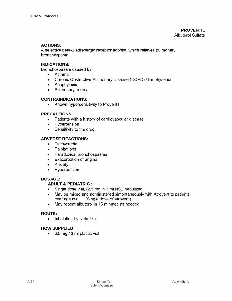
HEMS Protocols
A-34 Return To: Appendix A Table of Contents
PROVENTIL Albuterol Sulfate
ACTIONS: A selective beta-2 adrenergic receptor agonist, which relieves pulmonary bronchospasm. INDICATIONS:
Bronchospasam caused by: Asthma Chronic Obstructive Pulmonary Disease (COPD) / Emphysema Anaphylaxis Pulmonary edema
CONTRAINDICATIONS:
Known hypersensitivity to Proventil
PRECAUTIONS: Patients with a history of cardiovascular disease Hypertension Sensitivity to the drug
ADVERSE REACTIONS:
Tachycardia Palpitations Paradoxical bronchospasms Exacerbation of angina Anxiety Hypertension
DOSAGE:
ADULT & PEDIATRIC :
Single dose vial, (2.5 mg in 3 ml NS), nebulized. May be mixed and administered simontaneously with Atrovent to patients
over age two. (Single dose of atrovent) May repeat albuterol in 15 minutes as needed.
ROUTE:
Inhalation by Nebulizer HOW SUPPLIED:
2.5 mg / 3 ml plastic vial

HEMS Protocols
Appendix A Return To: A-35 Table of Contents
RACEMIC EPINEPHRINE
VapoNefrin
ACTION: Effective in reversing upper airway edema when administered with a nebulizer. Proposed mechanism of action is alpha-adrenergic receptor-mediated vasoconstriction of edematous tissues. Racemic epinephrine also causes bronchodilation, increases one’s heart rate, and increases one’s cardiac contractile force.
INDICATIONS:
Croup, (laryngotracheobronchitis)
CONTRAINDICATIONS: Hypersensitivity to the drug
PRECAUTIONS:
Vital signs should be constantly monitored.
ADVERSE REACTIONS: Palpitations Anxiety Headache
DOSAGE:
ADULT & PEDIATRIC:
0.5ml of a 2.25% solution in 3.0 ml Normal Saline. May repeat dose once in 20 – 30 minutes, if indicated.
ROUTE:
Inhalation by nebulizer
HOW SUPPLIED: 2.25% solution in 0.5ml unit dose, to be mixed in 3.0 ml N.S. nebulizer.

HEMS Protocols
A-36 Return To: Appendix A Table of Contents
ROMAZACON Flumazenil
ACTIONS: Benzodiazepine antagonist. Reverses effects of benzodiazepines. Reversal is typically evident within two minutes of administration, peaking at 6 to 10 minutes. INDICATIONS:
To reverse CNS and respiratory depression caused by Benzodiazepine overdose, or mixed alcohol and benzodiazepine overdose.
CONTRAINDICATIONS:
Romazacon should not be used as a diagnostic agent. Known or suspected post-ectal state. Known or suspected tricyclic overdose. Known hypersensitivity to the drug.
PRECAUTIONS:
Administer with caution, if at all, to patients dependent upon Benzodiazepines as it may induce seizures.
ADVERSE REACTIONS: Seizures Arrhythmias
DOSAGE:
ADULT :
0.2mg IV over 30 seconds. Then administer 0.3 to 0.5mg q 30 seconds up to a total dose of 3.0 mg. PEDIATRIC:
0.01 mg / kg IV, (Maximum of 0.2 mg / dose), over 1 minute, repeated q 2 minutes if needed, to a maximum dose of 1 mg.
ROUTE: IV/IO
HOW SUPPLIED:
0.5mg / 5ml Vial

HEMS Protocols
Appendix A Return To: A-37 Table of Contents
SODIUM BICARBONATE
ACTION: Alkalinizing agent used in the treatment of metabolic acidosis, and in the treatment of symptomatic tricyclic antidepressant overdose. INDICATIONS:
Any life – threatening acidosis Cardiac Arrest, (after defibrillation, airway management, and other
pharmaceutical interventions). Rarely administered in the first 20 minutes of resuscitation.
Tricyclic antidepressant overdose Known hyperkalemia, used in conjunction with other medications. Phenobarbital overdose
CONTRAINDICATIONS:
Respiratory acidosis PRECAUTIONS:
Inactivates catecholamines ADVERSE REACTIONS:
Metabolic alkalosis Tissue necrosis if the IV infiltrates.
DOSAGE:
ADULT
1meq/kg of 8.4% repeated in 10 minutes if necessary at 0.5 meq/kg
PEDIATRIC 1meq/kg of 4.2% repeated in 10 minutes if necessary at 0.5 meq/kg
(ie.. Must dilute as 50% NS 50% Sodium Bicarb for administration to pediatric patients).
ROUTE: IV, IO
HOW SUPPLIED:
8.4% solution: 50mEq in 50 ml, pre-filled syringe.
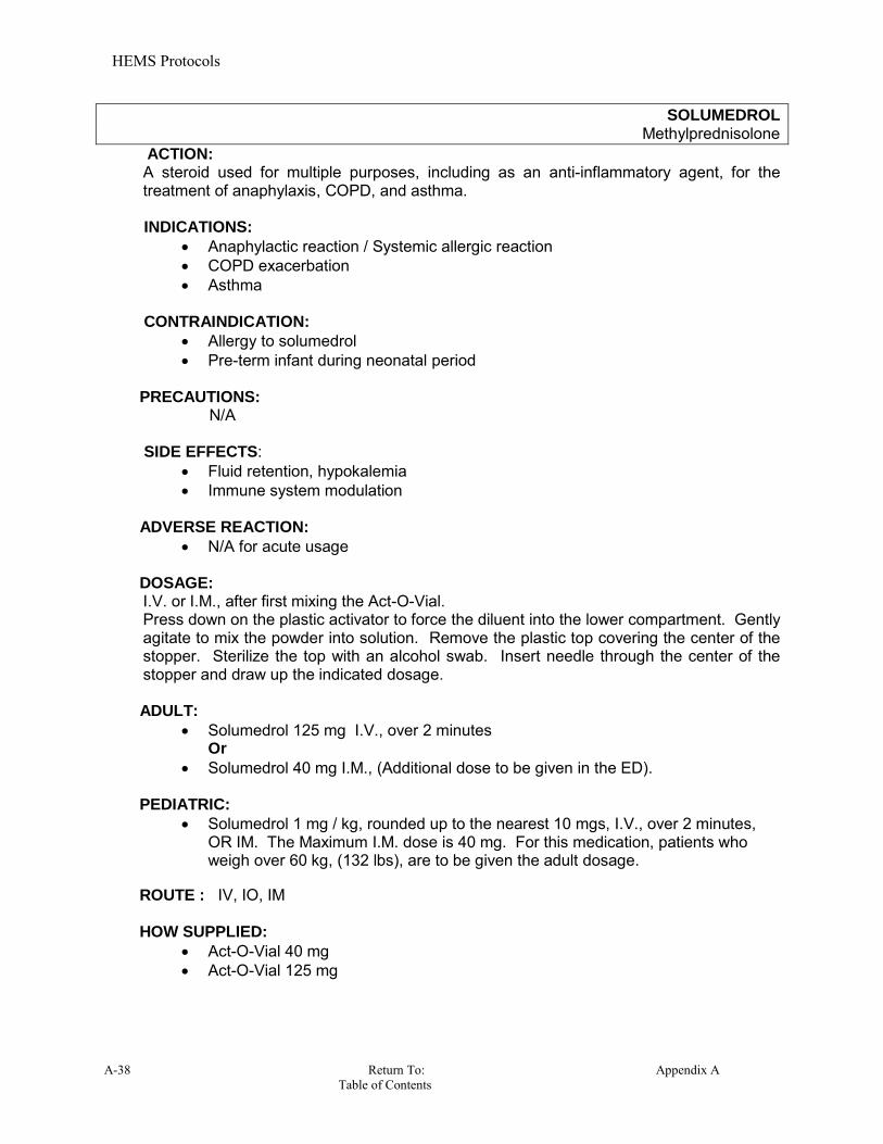
HEMS Protocols
A-38 Return To: Appendix A Table of Contents
SOLUMEDROL Methylprednisolone
ACTION: A steroid used for multiple purposes, including as an anti-inflammatory agent, for the treatment of anaphylaxis, COPD, and asthma.
INDICATIONS:
Anaphylactic reaction / Systemic allergic reaction COPD exacerbation Asthma
CONTRAINDICATION:
Allergy to solumedrol Pre-term infant during neonatal period
PRECAUTIONS:
N/A SIDE EFFECTS:
Fluid retention, hypokalemia Immune system modulation
ADVERSE REACTION:
N/A for acute usage DOSAGE:
I.V. or I.M., after first mixing the Act-O-Vial. Press down on the plastic activator to force the diluent into the lower compartment. Gently agitate to mix the powder into solution. Remove the plastic top covering the center of the stopper. Sterilize the top with an alcohol swab. Insert needle through the center of the stopper and draw up the indicated dosage.
ADULT:
Solumedrol 125 mg I.V., over 2 minutes Or
Solumedrol 40 mg I.M., (Additional dose to be given in the ED). PEDIATRIC:
Solumedrol 1 mg / kg, rounded up to the nearest 10 mgs, I.V., over 2 minutes, OR IM. The Maximum I.M. dose is 40 mg. For this medication, patients who weigh over 60 kg, (132 lbs), are to be given the adult dosage.
ROUTE : IV, IO, IM HOW SUPPLIED:
Act-O-Vial 40 mg Act-O-Vial 125 mg

HEMS Protocols
Appendix A Return To: A-39 Table of Contents
STERILE WATER H2O
ACTION: Used as a diluent for various medications, as a wound cleaner, and to humidify supplemental oxygen. INDICATIONS: CONTRAINDICATIONS: PRECAUTIONS: SIDE EFFECTS: ADVERSE REACTION: DOSAGE: Based upon the medication being dissolved / diluted into the water. ROUTE: Based upon the medication being dissolved / diluted into the water. HOW SUPPLIED:
3 ml vial 500 ml bottle Humified O2

HEMS Protocols
A-40 Return To: Appendix A Table of Contents
TETRACAINE OPTHALMIC Pontocaine
ACTION: Is used for rapid, brief, ocular anesthesia. The agent inhibits conduction of nerve impulses from sensory nerves. Tetracaine causes a brief burning or stinging when initially instilled into the eye.
INDICATION:
Short-term relief from eye pain or irritation. Insertion of the Morgan eye lense for subsequent irragation.
CONTRAINDICATION:
Patients with known hypersensitivity to tetracaine Open eye injuries
ADVERSE REACTIONS:
Do not allow the patient to rub their eyes once they are anesthetized.
DOSAGE: ADULT & PEDIATRIC:
2 drops to the effective eye, may be repeated in 10 minutes, if needed. Gently pull down the lower eye lid and instill 2 drops into the lower eye sac.
ROUTE:
Topical opthalmic
HOW SUPPLIED:
0.5% in 2 ml plastic bottle

HEMS Protocols
Appendix A Return To: A-41 Table of Contents
Toradol Ketorolac Tromethamine
ACTIONS: Non-steroidal Anti-inflammatory, (NSAID). Ketorolac (Toradol) is an injectable non-steroidal anti-inflammatory drug (NSAID) that demonstarates analgesic, anti-inflammatory and antipyretic activity. Usually effective in 20 – 30 minutes. INDICATIONS: Suspected kidney stones, any moderate pain that is not associated with fractures, Head injuries or any surgical candidates.
CONTRAINDICATIONS:
Allergic to ketorolac, aspirin, or other NSAIDs. severe renal disease or kidney transplant a bleeding or blood clotting disorder a closed head injury or bleeding in brain patients needing surgery a surgical candidate with open fracture or fracture deformities if breast-feeding a baby
PRECAUTIONS:
Any liver disease Kidney disease Blood disorder Asthma Nursing females
ADVERSE REACTIONS:
Nausea and vomiting Sweating Blurred vision
DOSAGE: 1 dose only
60 mg IM 30 mg IV slowly over 15 seconds. 15 mg IV slowly over 15 seconds in pts over 65 years old patients and those
weighing less than 50 kgs (110 lbs) ROUTE:
IV, IM, IO
HOW SUPPLIED:
30mg, 1ml Carpujet 30mg, 1ml Vial 60mg, 2ml Vial 60mg, 2ml Carpujet

HEMS Protocols
A-42 Return To: Appendix A Table of Contents
Tylenol Acetaminophen
Action: (Fever-Reducer) It reduces fever through its action on the heat-regulating center of the brain. Specifically, it tells the center to lower the body's temperature when the temperature is elevated.
INDICATIONS: Body temperature in excess of 101o F CONTRAINDICATIONS: History of allergy to Tylenol. PRECAUTIONS: Liver disease Alcohol dependency Pregnancy Actively nursing mother ADVERSE REACTIONS: Over dosing with this medication can cause severe liver damage. DOSAGE: 12 years old or older and >96 lbs. : four, 160mg Meltaway tablets = 640mg of Tylenol. Under 12 or under 96 lbs see Table 1 for administration. Under 8 years old requires OLMC orders. ROUTE: Oral HOW SUPPLIED: Tylenol Meltaways contain 160mg of acetaminophen each. Administer by placing the tablets in the patient’s mouth and letting it dissolve or chewing thoroughly. A small drink of water may be given to the patient after the medication has been ingested. TABLE 1
AGE GROUP 8-10 yrs
10-12 yrs
12 yrs &-
older
WEIGHT (if possible use weight to dose; otherwise use
age)
48-70 lbs
70-96 lbs
96 lbs and over
JR Tylenol Meltaways
Acetaminophen 160 mg
2 tablets
3 tablets
4 tablets
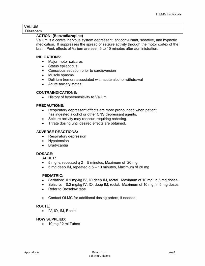
HEMS Protocols
Appendix A Return To: A-43 Table of Contents
VALIUM Diazepam
ACTION: (Benzodiazapine) Valium is a central nervous system depressant, anticonvulsant, sedative, and hypnotic medication. It suppresses the spread of seizure activity through the motor cortex of the brain. Peek effects of Valium are seen 5 to 10 minutes after administration. INDICATIONS:
Major motor seizures Status epilepticus Conscious sedation prior to cardioversion Muscle spasms Delirium tremors associated with acute alcohol withdrawal Acute anxiety states
CONTRAINDICATIONS:
History of hypersensitivity to Valium
PRECAUTIONS:
Respiratory depressant effects are more pronounced when patient has ingested alcohol or other CNS depressant agents. Seizure activity may reoccur, requiring redosing. Titrate dosing until desired effects are obtained.
ADVERSE REACTIONS:
Respiratory depression Hypotension Bradycardia
DOSAGE:
ADULT:
5 mg iv, repeated q 2 – 5 minutes, Maximum of 20 mg 5 mg deep IM, repeated q 5 – 10 minutes, Maximum of 20 mg
PEDIATRIC:
Sedation: 0.1 mg/kg IV, IO,deep IM, rectal. Maximum of 10 mg, in 5 mg doses. Seizure: 0.2 mg/kg IV, IO, deep IM, rectal. Maximum of 10 mg, in 5 mg doses. Refer to Broselow tape Contact OLMC for additional dosing orders, if needed.
ROUTE: IV, IO, IM, Rectal
HOW SUPPLIED:
10 mg / 2 ml Tubex
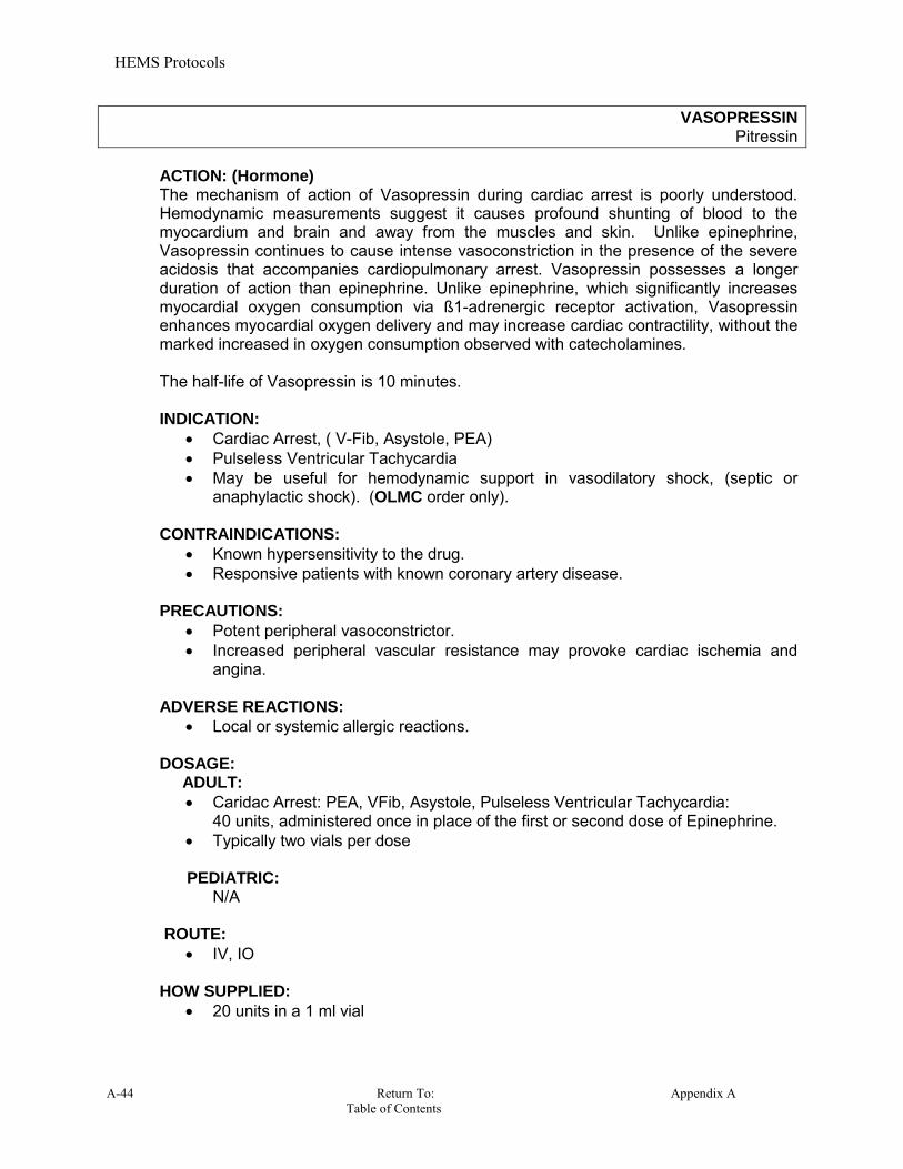
HEMS Protocols
A-44 Return To: Appendix A Table of Contents
VASOPRESSIN Pitressin
ACTION: (Hormone)
The mechanism of action of Vasopressin during cardiac arrest is poorly understood. Hemodynamic measurements suggest it causes profound shunting of blood to the myocardium and brain and away from the muscles and skin. Unlike epinephrine, Vasopressin continues to cause intense vasoconstriction in the presence of the severe acidosis that accompanies cardiopulmonary arrest. Vasopressin possesses a longer duration of action than epinephrine. Unlike epinephrine, which significantly increases myocardial oxygen consumption via ß1-adrenergic receptor activation, Vasopressin enhances myocardial oxygen delivery and may increase cardiac contractility, without the marked increased in oxygen consumption observed with catecholamines. The half-life of Vasopressin is 10 minutes. INDICATION:
Cardiac Arrest, ( V-Fib, Asystole, PEA) Pulseless Ventricular Tachycardia May be useful for hemodynamic support in vasodilatory shock, (septic or
anaphylactic shock). (OLMC order only). CONTRAINDICATIONS:
Known hypersensitivity to the drug. Responsive patients with known coronary artery disease.
PRECAUTIONS:
Potent peripheral vasoconstrictor. Increased peripheral vascular resistance may provoke cardiac ischemia and
angina.
ADVERSE REACTIONS:
Local or systemic allergic reactions. DOSAGE: ADULT:
Caridac Arrest: PEA, VFib, Asystole, Pulseless Ventricular Tachycardia: 40 units, administered once in place of the first or second dose of Epinephrine. Typically two vials per dose
PEDIATRIC:
N/A
ROUTE:
IV, IO
HOW SUPPLIED: 20 units in a 1 ml vial

HEMS Protocols
Appendix A Return To: A-45 Table of Contents
VERSED Midazolam
ACTION: Versed is a potent, short-acting Benzodiazapine with strong hypnotic and amnesic properties. It is widely used as a sedative prior to cardioversion. Versed is 3-4 times more potent than Valium, with a 1.5 minute onset of action when administered intravenously and 15 minutes when administered intramuscularly. Versed has impressive amnesic properties making it the drug of choice for conscious sedation. Like all Benzodiazapine class drugs, Versed is a central nervous system depressant. INDICATIONS:
Conscious sedation of patients prior to procedures, (cardioversion, etc.). Treatment of seizures
CONTRAINDICATIONS:
Hypersensitivity to the drug Narrow-angle glaucoma Not to be administered to patients with CNS depression, patients
taking narcotics, or patients taking Lithium Carbonate.
PRECAUTIONS:
A slight to moderate decrease in mean arterial pressure, cardiac output, systemic vascular resistance and heart rate may be seen.
Lower dosages should be considered in elderly patients, and in patients that are debilitated or chronically ill.
ADVERSE REACTIONS:
Respiratory depression Laryngospasm Bronchospasm Respiratory depressant effects are more pronounced when patient has ingested
alcohol or other CNS depressant agents. Hypotension, (treated with a fluid bolus).
DOSAGE:
ADULT:
2 - 5mg IM / IV / IO / IN over 2 minutes, repeat in 5 minutes if needed, to a Maximum of 10 mg.
PEDIATRIC: 0.1mg/kg to 0.2mg/kg IM / IV / IO / IN over 2 minutes, repeat in 5 minutes if
needed, to a Maximum of 5 mg. ROUTE: IV, IO, IM, IN
HOW SUPPLIED: 2 mg / 2 ml vial. 5mg / 1 ml vial
5mg / 1 ml carpujet 10mg / 2ml vial
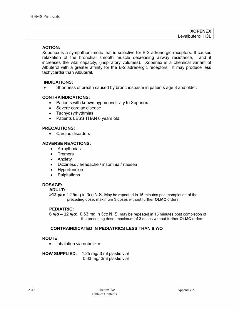
HEMS Protocols
A-46 Return To: Appendix A Table of Contents
XOPENEX Levalbuterol HCL
ACTION: Xopenex is a sympathomimetic that is selective for B-2 adrenergic receptors. It causes relaxation of the bronchial smooth muscle decreasing airway resistance, and it increases the vital capacity, (inspiratory volumes). Xopenex is a chemical variant of Albuterol with a greater affinity for the B-2 adrenergic receptors. It may produce less tachycardia than Albuteral.
INDICATIONS:
Shortness of breath caused by bronchospasm in patients age 8 and older.
CONTRAINDICATIONS:
Patients with known hypersensitivity to Xopenex. Severe cardiac disease Tachydsyrhythmias Patients LESS THAN 6 years old.
PRECAUTIONS:
Cardiac disorders ADVERSE REACTIONS:
Arrhythmias Tremors Anxiety Dizziness / headache / insomnia / nausea Hypertension Palpitations
DOSAGE:
ADULT: >12 y/o: 1.25mg in 3cc N.S. May be repeated in 15 minutes post completion of the
preceding dose, maximum 3 doses without further OLMC orders. PEDIATRIC: 6 y/o – 12 y/o: 0.63 mg in 3cc N. S. may be repeated in 15 minutes post completion of the preceding dose, maximum of 3 doses without further OLMC orders. CONTRAINDICATED IN PEDIATRICS LESS THAN 6 Y/O
ROUTE:
Inhalation via nebulizer
HOW SUPPLIED: 1.25 mg/ 3 ml plastic vial 0.63 mg/ 3ml plastic vial
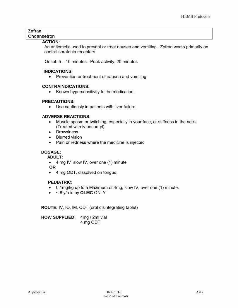
HEMS Protocols
Appendix A Return To: A-47 Table of Contents
Zofran
Ondansetron ACTION: An antiemetic used to prevent or treat nausea and vomiting. Zofran works primarily on central seratonin receptors.
Onset: 5 – 10 minutes. Peak activity: 20 minutes INDICATIONS:
Prevention or treatment of nausea and vomiting. CONTRAINDICATIONS:
Known hypersensitivity to the medication.
PRECAUTIONS:
Use cautiously in patients with liver failure. ADVERSE REACTIONS:
Muscle spasm or twitching, especially in your face; or stiffness in the neck. (Treated with iv benadryl). Drowsiness Blurred vision Pain or redness where the medicine is injected
DOSAGE: ADULT:
4 mg IV slow IV, over one (1) minute OR
4 mg ODT, dissolved on tongue. PEDIATRIC:
0.1mg/kg up to a Maximum of 4mg, slow IV, over one (1) minute. < 8 y/o is by OLMC ONLY
ROUTE: IV, IO, IM, ODT (oral disintegrating tablet) HOW SUPPLIED: 4mg / 2ml vial 4 mg ODT

Return to: Appendix B Table of Contents
BASIC LIFE
SUPPORT PROCEDURES
Paramedics Advanced EMTs Emergency Medical Technicians APPENDIX B
Revision: 40.6
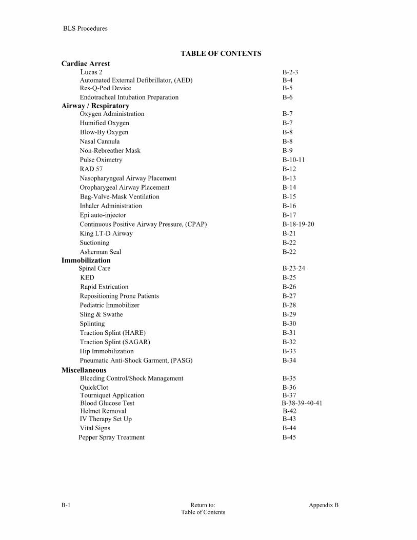
BLS Procedures
B-1 Return to: Appendix B Table of Contents
TABLE OF CONTENTS Cardiac Arrest Lucas 2 B-2-3
Automated External Defibrillator, (AED) B-4 Res-Q-Pod Device B-5 Endotracheal Intubation Preparation B-6
Airway / Respiratory Oxygen Administration B-7 Humified Oxygen B-7 Blow-By Oxygen B-8 Nasal Cannula B-8 Non-Rebreather Mask B-9 Pulse Oximetry B-10-11 RAD 57 B-12 Nasopharyngeal Airway Placement B-13 Oropharygeal Airway Placement B-14 Bag-Valve-Mask Ventilation B-15 Inhaler Administration B-16 Epi auto-injector B-17 Continuous Positive Airway Pressure, (CPAP) B-18-19-20 King LT-D Airway B-21 Suctioning B-22 Asherman Seal B-22
Immobilization Spinal Care B-23-24 KED B-25 Rapid Extrication B-26
Repositioning Prone Patients B-27 Pediatric Immobilizer B-28 Sling & Swathe B-29 Splinting B-30 Traction Splint (HARE) B-31 Traction Splint (SAGAR) B-32 Hip Immobilization B-33 Pneumatic Anti-Shock Garment, (PASG) B-34
Miscellaneous Bleeding Control/Shock Management B-35 QuickClot B-36
Tourniquet Application B-37 Blood Glucose Test B-38-39-40-41 Helmet Removal B-42
IV Therapy Set Up B-43 Vital Signs B-44
Pepper Spray Treatment B-45

BLS Procedures
Appendix B Return to: B-2 Table of Contents
LUCAS 2 Indications: The Lucas 2 device is use only when the patient is pulseless and apneic. Contraindications / Precautions: Do NOT use the LUCAS Chest Compression System in these cases: If it is not possible to position LUCAS safely or correctly on the patient's chest. a) Too small patient: If you cannot enter the PAUSE mode or ACTIVE mode when the pressure pad touches the patient's chest and LUCAS alarms with 3 fast signals. b) Too large patient: If you cannot lock the Upper Part of LUCAS to the Back Plate without compressing the patient's chest. Equipment Needed: Lucas 2 Device, Reeves or LBB. Push ON/OFF on the User Control Panel for 1 second to power up LUCAS in the bag and start the self-test. The green LED adjacent to the ADJUST key illuminates when LUCAS is ready for use. Place the LUCAS Back Plate under the patient, immediately below the arm pits.
Attach the support legs to the Back Plate, so that the two support legs lock against the Back Plate. Listen for click. (Pull up once to make sure that the parts are correctly attached). Adjust the height of the Suction Cup to set the Start Position. a) Make sure that LUCAS is in the ADJUST mode. b) Push the Suction Cup down with two fingers until the pressure pad makes firm contact with the sternum without compressing the chest. Push PAUSE to lock the Start Position - then remove your fingers from the Suction Cup. a) Check for proper position. If not, push ADJUST, pull up the Suction Cup to readjust the central and/or height position for a new Start Position. Then Push PAUSE. b) Push ACTIVE (continuous) OR ACTIVE (30:2) to start the compressions. Secure the patient's arms: When you move the patient, secure the patient's arms with the Patient Straps on the LUCAS. This makes it easier to move the patient. Attaching Stabilization strap. Carefully lift the patient's head and attach stabilization strap behind the patient’s neck. Position the cushion as near the patient's shoulders as possible. Connect the buckles on the support leg straps with the buckles on the cushion strap. Make sure the straps are not twisted. Change the Battery 1. Push PAUSE to temporarily stop the compressions. 2. Pull the Battery out and then upwards to remove it. 3. Install a fully-charged LUCAS Battery by putting it in from above. 4. Wait until the green PAUSE mode LED illuminates.

BLS Procedures
B-3 Return to: Appendix B Table of Contents
LUCAS 2 5. Push ACTIVE (continuous) or ACTIVE (30:2) to start the chest compressions again. The LUCAS Smart Restart feature remembers the settings and Start Position for 60 seconds. Connect to the external Power To use the Power Supply cable: a) Connect the Power Supply cable to LUCAS. b) Connect the main cable to the wall mains outlet (100-240V, 50/60Hz) Caution - The Battery must always be installed for LUCAS to be able to operate, even when powered by the external Power Supply. Remove LUCAS™ from the patient: a) Push ON/OFF for 1 second to power off the device. b) If a LUCAS Stabilization Strap is attached to LUCAS, remove the cushion strap, which is part of the Stabilization Strap, from the support leg straps. c) Pull the release rings to remove the Upper Part from the Back Plate. d) If the patient's condition allows it, remove the Back Plate.
Note: LUCAS powers down automatically after 5 minutes if you let it stay in the ADJUST mode. Note: If the Battery change takes more than 60 seconds, LUCAS does a self-test and you must adjust the Start Position again.
Remove and install a new Suction Cup after each patient usage. a) Pull the Suction Cup off the black mounting tube. b) Discard the Suction Cup as contaminated medical waste. c) Bend a new Suction Cup onto the black mounting tube. d) Make sure the Suction Cup is safely attached on the mounting tube.
Procedure Satisfactory Unsatisfactory 1
Push ON/OFF on the User Control Panel for 1 second to power up LUCAS in the bag and start the self-test. The green LED adjacent to the ADJUST key illuminates when LUCAS is ready for use.
2
Place the LUCAS Back Plate under the patient, immediately below the arm pits.
3
Attach the support legs to the Back Plate, so that the two support legs lock against the Back Plate. Listen for click.
4
With the Lucas is in the ADJUST position. Use to two fingers to push the Suction Cup down until it is place in the center of the Sternum with the bottom of the suction Cup just above the bottom of the sternum then press Pause to lock the Start Position.
5 Press ACTIVE to start compression. Intubated or King select 100/min. Any other airway select 30/2.
6 Secure the hands to the patient straps.
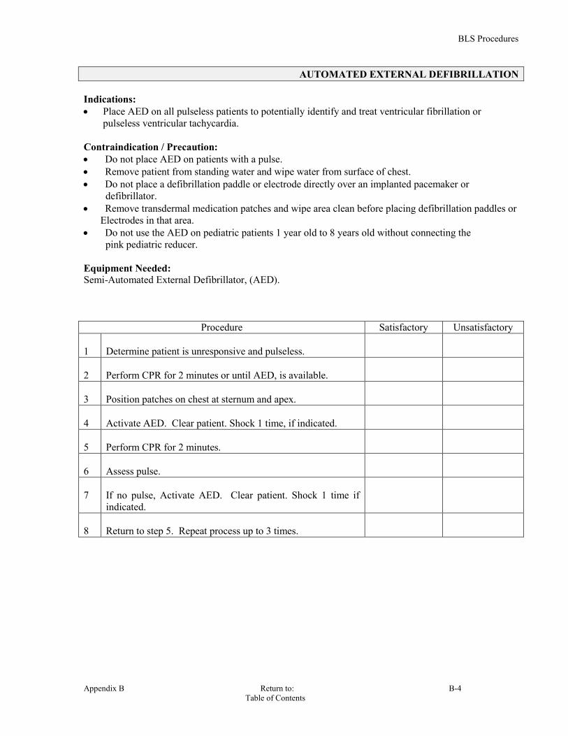
BLS Procedures
Appendix B Return to: B-4 Table of Contents
AUTOMATED EXTERNAL DEFIBRILLATION Indications: Place AED on all pulseless patients to potentially identify and treat ventricular fibrillation or
pulseless ventricular tachycardia. Contraindication / Precaution: Do not place AED on patients with a pulse. Remove patient from standing water and wipe water from surface of chest. Do not place a defibrillation paddle or electrode directly over an implanted pacemaker or
defibrillator. Remove transdermal medication patches and wipe area clean before placing defibrillation paddles or
Electrodes in that area. Do not use the AED on pediatric patients 1 year old to 8 years old without connecting the
pink pediatric reducer. Equipment Needed: Semi-Automated External Defibrillator, (AED).
Procedure Satisfactory Unsatisfactory 1
Determine patient is unresponsive and pulseless.
2
Perform CPR for 2 minutes or until AED, is available.
3
Position patches on chest at sternum and apex.
4
Activate AED. Clear patient. Shock 1 time, if indicated.
5
Perform CPR for 2 minutes.
6
Assess pulse.
7
If no pulse, Activate AED. Clear patient. Shock 1 time if indicated.
8
Return to step 5. Repeat process up to 3 times.
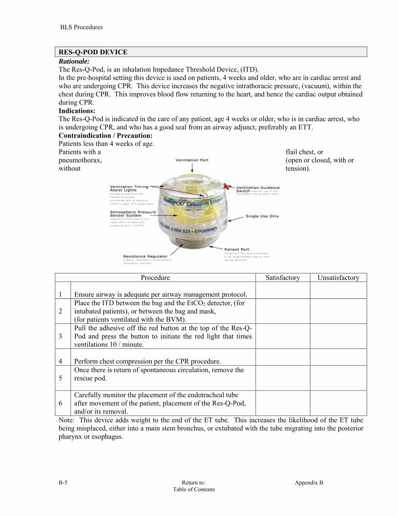
BLS Procedures
B-5 Return to: Appendix B Table of Contents
RES-Q-POD DEVICE Rationale:
The Res-Q-Pod, is an inhalation Impedance Threshold Device, (ITD). In the pre-hospital setting this device is used on patients, 4 weeks and older, who are in cardiac arrest and who are undergoing CPR. This device increases the negative intrathoracic pressure, (vacuum), within the chest during CPR. This improves blood flow returning to the heart, and hence the cardiac output obtained during CPR. Indications: The Res-Q-Pod is indicated in the care of any patient, age 4 weeks or older, who is in cardiac arrest, who is undergoing CPR, and who has a good seal from an airway adjunct, preferably an ETT. Contraindication / Precaution: Patients less than 4 weeks of age. Patients with a flail chest, or pneumothorax, (open or closed, with or without tension).
Procedure Satisfactory Unsatisfactory 1
Ensure airway is adequate per airway management protocol.
2
Place the ITD between the bag and the EtCO2 detector, (for intubated patients), or between the bag and mask, (for patients ventilated with the BVM).
3
Pull the adhesive off the red button at the top of the Res-Q-Pod and press the button to initiate the red light that times ventilations 10 / minute.
4
Perform chest compression per the CPR procedure.
5
Once there is return of spontaneous circulation, remove the rescue pod.
6
Carefully monitor the placement of the endotracheal tube after movement of the patient, placement of the Res-Q-Pod, and/or its removal.
Note: This device adds weight to the end of the ET tube. This increases the likelihood of the ET tube being misplaced, either into a main stem bronchus, or extubated with the tube migrating into the posterior pharynx or esophagus.

BLS Procedures
Appendix B Return to: B-6 Table of Contents
ENDOTRACHEAL INTUBATION PREPARATION
Indications: When Endotracheal Intubation is required. Contraindication / Precaution: None Equipment Needed: Proper size ET tube. Prepare suction equipment. Laryngoscope and proper blade for patient age. Proper size stylette. 10cc syringe. BVM Oral tracheal or nasal pharyngeal airway. Endotracheal stabilizing device.
Procedure Satisfactory Unsatisfactory 1
Open airway with jaw thrust or head tilt chin lift.
2
Ventilate patient using BVM with high flow oxygen.
3
Pre-oxygenate patient until ready to begin intubation attempts.
4 Prepare suction equipment.
5
Prepare and connect EtCO2 monitoring equipment.
6
Make sure all equipment is ready and operational, (cuff check, blade light, etc).

BLS Procedures
B-7 Return to: Appendix B Table of Contents
OXYGEN ADMINISTRATION
Indications: Nasal Cannula: For the spontaneously adequately breathing patient with no significant
compromise or potential compromise in condition. Choice is determined by severity of condition, practice parameters and patient tolerance.
Non-Rebreather Mask: For any patient whose condition or complaint suggests that severe hypoxia or ischemia may be a problem. Use on all multi-trauma patients and all patients who present with sign and symptoms of shock.
Bag Valve Mask, (BVM): Assist ventilations in the conscious or unconscious hypoxemic patient who is not moving air adequately. Ventilate the apneic patient, or those with insufficient ventilations.
Equipment: Nasal Cannula: 2 - 6 liters/minute delivers 25 - 40 % of oxygen.
Non-Rebreather Mask (NRBM): 8 - 15 liters/minute delivers > 90 % of oxygen.
Bag Valve Mask, (BVM): with supplemental oxygen at 15 liters/minute and reservoir attached delivers nearly 100% oxygen.
HUMIDIFIED OXYGEN Indications: May be used with:
COPD / Emphysema Asthma Croup
Heated Humidified Oxygen:
Hypothermia
Contraindication / Precaution:
CHF / Pulmonary Edema
Equipment Needed: Oxygen Regulator H2O Reservoir

BLS Procedures
Appendix B Return to: B-8 Table of Contents
BLOW-BY OXYGEN Indications:
Infant / child that will not tolerate a mask or nasal cannula. Patient requiring supplemental low concentration oxygen.
Contraindication / Precaution:
Do not blow oxygen directly in the eye’s of a newborn. Equipment Needed:
Oxygen tubing Oxygen bottle and regulator with flow meter.
Procedure Satisfactory Unsatisfactory 1
Explain procedure to patient, if possible.
2
Check Tank Pressure.
3
Attach oxygen tubing to oxygen regulator.
4
Adjust liter flow to 4-6 LPM.
5
Place tubing approximately 1-2 inches from patient’s. nose/mouth
6
Monitor patient as appropriate.
NASAL CANNULA Indications:
Spontaneous breathing patient without respiratory compromise. Patient unable to tolerate a mask.
Contraindication / Precaution: Epistaxis
Equipment Needed: Nasal Cannula Oxygen source with regulator and flow meter.
Procedure Satisfactory Unsatisfactory 1
Explain procedure to patient.
2
Check Tank Pressure.
3
Attach nasal cannula to oxygen regulator.
4
Adjust liter flow to 0.5 - 6 liters/minute.
5
Apply nasal cannula to patient.
BLS Procedures
B-9 Return to: Appendix B Table of Contents
NON-REBREATHER MASK NRBM Indications: Patient requiring high concentrations of oxygen. Respiratory distress Cardiac related symptoms Shock / Trauma
Contraindication / Precaution: None for short term use.
Equipment Needed: Non-rebreather mask. Oxygen source and regulator with flow meter.
Procedure Satisfactory Unsatisfactory 1
Explain procedure to patient.
2
Check tank pressure.
3
Attach NRBM to oxygen regulator.
4
Pre - fill reservoir bag.
5
Adjust liter from 10 – 15 LPM flow to ensure reservoir bag stays inflated.
6
Apply and adjust mask to the patient.
7
Monitor reservoir bag for constant inflation.
BLS Procedures
Appendix B Return to: B-10 Table of Contents
PULSE OXIMETERY Pulse oximetry is used in conjunction with other assessment processes to determine the actual available oxygen in the blood for use by body tissue. Pulse oximetry measures the oxygen saturation of the red blood cells, (%SpO
2).
Use of pulse oximetry in conjunction with other assessment processes may sometimes identify those patients in mild to moderate hypoxia, and with proper intervention profound hypoxia can be prevented. Pulse oximetry should be used on all patients as part of obtaining their vital signs. Pulse oximetry should be continuously monitored on all patients with shock, altered LOC, or cardio/respiratory symptoms.
Indications:
To determine effective oxygenation. Initial vital signs on a ll patients.
Contraindication / Precaution:
Hypothermia may cause false readings. Hypotension causing poor peripheral perfusion. Nail polish Jaundice Vasoconstrictive drugs. An SpO2 reading does not replace clinical assessment of the patient’s respiratory status.
Equipment Needed:
Pulse oximeter with proper probe Procedure Satisfactory Unsatisfactory NORMAL VALUES 94% -- 100% 1
Turn on device.
2
Place probe on proper body part.
3
Read and document SpO2.
REMEMBER, INITIATE NORMAL AIRWAY AND OXYGENATION SUPPORT REGARDLESS OF THE AVAILABILITY OF PULSE OXIMETRY.
NEVER BASE ANY TREATMENT OR OXYGEN THERAPY SOLELY ON THE READING FROM THE PULSE OXIMETER.
BLS Procedures
B-11 Return to: Appendix B Table of Contents
PULSE OXIMETERY (continued)
Interpretation of Reading: 95% to 100% Ideal Range - Maintain oxygen and airway support methods being used. 90% to 94% Mild to Moderate Hypoxemia - Check airway and increase oxygen support until ideal range is achieved. 85% to 89% Severe Hypoxemia - Aggressive airway and oxygen support is essential. Look for and treat cause: i.e. COPD, CHF, etc. Below 85% BE PREPARED TO INTUBATE AND/OR ASSIST VENTILATION. CONSIDERATIONS
Hypovolemic, hypothermic, and peripheral vascular disease patients may not be suitable candidates for pulse oximetry due to poor peripheral circulation.
Be aware that there may be a 30 to 60 second delay between oxygenation and %SpO
2
readings.
A pulse must be detected by the oximeter to determine the %SpO2 .
Pulse oximetry monitoring is not indicated in carbon monoxide poisoning, use the RAD 57.
COPD patients MAY have a low %SpO
2 as a baseline. They should be treated with O2
based upon their clinical status.

BLS Procedures
Appendix B Return to: B-12 Table of Contents
RAD 57 The RAD-57 monitor is designed to non-invasively measure Carboxyhemoglobin, (Carbon Monoxide Poisoning). This device measures the level of carbon monoxide in the blood, allowing early detection and treatment of potentially life-threatening conditions. Indications:
Headache, dizziness, syncope, weakness. Altered LOC. Firefighter rehabilitation. Extended time on or near fire-ground. Occupants of Buildings on fire or smoke filled. Multi-patient presentation. Nausea, abdominal complaints. Any ill or injured patient with vague complaints found in the interior of a building or in a car. Any scenario where both the patient(s), and their pet(s) are ill.
Contraindication / Precaution:
Be sure to shield the ambient light from the sensor. Equipment Needed:
RAD 57 monitor Finger sensor Acetone wipe
Procedure Satisfactory Unsatisfactory 1 Press on / off button.
2 Place probe on the patient’s finger.
3 Wire from probe should be on top of finger.
4 Position Markers on side of probe over middle of fingernail.
5 Use Ring Finger on Non Dominate hand whenever possible. Finger should be warm and clean.
6 Device will take 6-10 seconds to calibrate, (you will see zeros).
7 Device will display pulse rate and SpO2. Press SpCO button to display Carboxyhemoglobin %.
8 Display will return to pulse rate and SpO2 in 10 seconds.
9
Document Readings.

BLS Procedures
B-13 Return to: Appendix B Table of Contents
NASOPHARYNGEAL AIRWAY PLACEMENT Indications: Patient not fully responsive. Patient with a gag reflex. Assistance needed in maintaining an open airway.
Contraindication / Precaution: Improper sized airway. Fractured facial bones. Basilar skull fractures.
Equipment Needed: Assorted sizes of nasopharyngeal airways. Water soluble lubricant.
Procedure Satisfactory Unsatisfactory 1
Explain procedure to patient, if necessary.
2
Select appropriate airway by measuring from the tip of the nose to the ear lobe.
3
Lubricate airway with a water soluble lubricant.
4
Insert the airway into the larger or more open nostril with the bevel facing towards the septum.
5
If you meet resistance, gently rotate from side to side as you insert. If resistance continues remove and try the other nostril.
6
Airway should rest against the flare of the nostril. May withdraw slightly if gag flex is stimulated.

BLS Procedures
Appendix B Return to: B-14 Table of Contents
OROPHARYNGEAL AIRWAY PLACEMENT Indications: Unconscious patient. No gag reflex.
Contraindication / Precaution: Responsive patient. Gag reflex.
Equipment Needed: Assorted sizes of oropharyngeal airways. Suction.
Procedure Satisfactory Unsatisfactory 1
Select appropriate size airway by measuring from the center of the mouth to the angle of the jaw or corner of the mouth to the ear lobe.
2
Insert airway upside down using the cross finger technique with the tip pointing to the roof of the mouth.
3
When airway comes in contact with the soft palate at the back of the roof of the mouth, gently rotate 180 degrees while continuing to advance the airway until the flat flange at the top of the airway rests on the patient’s front teeth.
4
In pediatrics place directly in following the natural curvature of the airway.
5
If patient gags during insertion remove the airway.
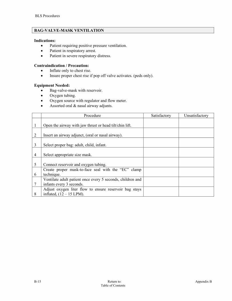
BLS Procedures
B-15 Return to: Appendix B Table of Contents
BAG-VALVE-MASK VENTILATION Indications:
Patient requiring positive pressure ventilation. Patient in respiratory arrest. Patient in severe respiratory distress.
Contraindication / Precaution:
Inflate only to chest rise. Insure proper chest rise if pop off valve activates. (peds only).
Equipment Needed:
Bag-valve-mask with reservoir. Oxygen tubing. Oxygen source with regulator and flow meter. Assorted oral & nasal airway adjunts.
Procedure Satisfactory Unsatisfactory 1
Open the airway with jaw thrust or head tilt/chin lift.
2
Insert an airway adjunct, (oral or nasal airway).
3
Select proper bag: adult, child, infant.
4
Select appropriate size mask.
5
Connect reservoir and oxygen tubing.
6
Create proper mask-to-face seal with the “EC” clamp technique.
7
Ventilate adult patient once every 5 seconds, children and infants every 3 seconds.
8
Adjust oxygen liter flow to ensure reservoir bag stays inflated, (12 – 15 LPM).
BLS Procedures
Appendix B Return to: B-16 Table of Contents
INHALER ADMINISTRATION Indications:
Prescribed to patients with pulmonary disease. Signs and symptoms of respiratory difficulty.
Contraindication / Precaution:
Altered mental status. Inhaler is not prescribed to the patient. Patient has already reach the maximum dose. Inhaler expired.
Equipment Needed: Patient’s prescribed metered dose inhaler. By OLMC order Procedure Satisfactory Unsatisfactory 1
Shake canister and mouthpiece well.
2
Invert the device and hold it close to the patient’s mouth.
3
Advise patient to exhale, pushing as much air from lungs as possible.
4
Place mouthpiece in patient’s mouth and instruct patient to close his/her lips loosely around the mouthpiece with tongue underneath.
5
Advise patient to inhale deeply, press down on canister quickly then release it, (over 5 sec).
6
Instruct patient to hold his/her breath for 5 to 10 seconds before exhaling.
7
Monitor patient for desired effects.

BLS Procedures
B-17 Return to: Appendix B Table of Contents
EPINEPHRINE INJECTION VIA EPI PEN Epinephrine is administered to effect reversal of the shock and bronchial constriction which occurs with anaphylaxis. Indications: Allergic reaction with respiratory distress and hypotension. Contraindication / Precaution: None Equipment Needed: EPI-Pen & Alcohol swab Procedure Satisfactory Unsatisfactory 1
Locate the injection site. Mid femur lateral aspect
2
Cleanse the injection site with an alcohol swab.
3
Inspect EPI-PEN,(color & date; if brownish, outdated or contains any precipitate discard).
4
Pull off gray safety cap.
5
Place black tip on thigh at right angle to leg.
6
Press hard into thigh until Auto-Injector functions and hold in place for ten seconds.
7
Remove and discard EPI Pen in a “sharp safe” container.
8
Massage injection area for 10 seconds.
9
Repeat vital signs and patient assessment every 5 minutes until stable.
10
If no improvement in 5 minutes, repeat injection.

BLS Procedures
Appendix B Return to: B-18 Table of Contents
CONTINUOUS POSITIVE AIRWAY PRESSURE (CPAP)
Continuous Positive Airway Pressure, (CPAP), is defined as the application of positive end expiratory pressure by facemask for relief of hypoxemia, which doesn’t respond to conventional therapy. In order for CPAP to be used, the patient must be breathing. CPAP works by “splinting” the lungs with a constant pressure of air, which reduces the work of breathing. In CHF it forces the excess fluid out of the alveoli and interstitial space back into the vasculature as well as decreases venous return to the heart thereby lessening its workload. CPAP can also be a palliative intervention for patients with DNR orders due to the non invasion nature of pressure support versus ventilatory support. Indications:
Congestive Heart Failure/Pulmonary Edema. Pneumonia Pulmonary Edema secondary to near drowning. COPD
Contraindication / Precaution:
Penetrating chest trauma. Pneumothorax Severe facial injury. Severe hypotension. Persistent nausea and / or vomiting. Respiratory / Cardiac arrest. Patient must be conscious and have the ability to maintain their own airway. Questionable ability to protect airway, (e.g. stroke, obtundation etc.).
Equipment Needed:
CPAP Flow generator. Oxygen tank. Quick connect pigtail. Cardiac Monitor. SpO2 Monitor.
Pre-CPAP Patient Assessment: 1. Assess patient, record vitals including baseline SpO2. 2. Gather appropriate equipment. 3. Administer O2 by non-rebreather mask while preparing equipment.

BLS Procedures
B-19 Return to: Appendix B Table of Contents
CONTINUOUS POSITIVE AIRWAY PRESSURE (CPAP) (continued)
PORT O2 VENT CPAP EQUIPMENT

BLS Procedures
Appendix B Return to: B-20 Table of Contents
CONTINUOUS POSITIVE AIRWAY PRESSURE (CPAP)
(continued) Procedure Satisfactory Unsatisfactory 1
Place patient in a seated position with legs dependent, (if possible).
2
Monitor EKG and vital signs: B/P, heart rate, respiratory rate, SpO2.
3
Takes infection control precautions/BSI.
4
Identifies acute respiratory distress of CHF origin.
5
Verbalizes inclusion criteria: (At least 2 of the following). 1) RR>25. 2) Retractions, accessory muscle use or fatigue. 3) SAO2< 94% at any time.
6
Assembles required equipment: 1) Oxygen cylinder with quick connect adapter 2) CPAP device 3) Breathing circuit 4) Proper size mask with head harness
7
Explains the procedure to the patient.
8
Turns CPAP adjustment knob clockwise to start airflow at the same time placing mask over patient’s mouth and nose.
9 Secures the mask with head harness.
10
Sets CPAP pressure to 10 cm H2O for CHF, Pul. Edema, Sets CPAP pressure to 7.5 cm H2O for pneumonia, COPD
11
Checks for air leaks and adjusts mask if needed.
12
Reassess the patient every 5 minutes during the use of CPAP.
13
Verbalizes the importance of notifying the receiving hospital that CPAP is in use.

BLS Procedures
B-21 Return to: Appendix B Table of Contents
KING LT-D AIRWAY Indications:
Apneic patient when endotracheal intubation is not possible or not available. Unresponsive patients without an intact gag reflex.
Contraindication / Precaution:
Intact gag reflex. Known esophageal disease such as cancer. Caustic ingestion patient.
Procedure: Choose correct size: Size Height (ft) Cuff Volume (ml) Connector Color
2 12kg - 25 kg (35 - 45in.) 25-35 (Green) 2.5 25kg - 35kg (41 - 51in) 30-40 (Orange) 3 4' - 5' 50 (Yellow) 4 5' – 6' 70 (Red) 5 6' + 80 (Purple)
Equipment Needed:
King Airway Adjunct 80cc Syringe Lubricant Thomas Tube Holder EtCO /Capnography Suction equipment
Procedure Satisfactory Unsatisfactory 1 Secure the device. 2 Test cuffs for leaks. 3
Lubricate device with water soluble lubricant.
4
Pre-oxygenate and hyperventilate the patient, if time permits.
5
Grasp the patient’s tongue and jaw with your gloved hand and pull forward.
6
With the King LT-D rotated laterally at 45-90 degrees such that the blue orientation line is touching the corner of the mouth, introduce tip into mouth and advance behind base of tongue.
7
As tube tip passes under tongue, rotate tube back to midline, (blue orientation line faces chin).
8 Advance tube until base of connector is aligned with teeth or gums.
9 Inflate cuffs to appropriate volume as listed above. 10
Connect the King LT-D to a bag-valve device and ventilate the patient.
11
Assess for adequate placement by auscultation, (equal breath sounds over the chest and lack of sounds over the epigastrium with bagging), while gently bagging the patient to assess ventilations, simultaneously withdraw the airway until ventilations are easy and free flowing.

BLS Procedures
Appendix B Return to: B-22 Table of Contents
SUCTIONING Indications:
Removal of blood, emesis, food, debris, or secretions from the airway. Contraindication / Precaution:
Ensure pre and post oxygenation, if indicated. Equipment Needed:
Appropriate suction device Proper suction catheter
Procedure Satisfactory Unsatisfactory 1. Turn on and prepare suction device.
2. Assure presence of suction vacuum.
3. Select proper suction catheter.
4. Insert proper suction tip without applying suction.
5. Apply suction to the oropharynx / nasopharynx during removal of the catheter.
6. Limit suction times: adult 15 seconds, infants and children 5 seconds, per pass.
7. Stop suctioning immediately if heart rate drops in infants and children.
ASHERMAN SEAL The ACS™ (Asherman Chest Seal) is a sterile occlusive dressing for treating open pneumothorax and preventing tension pneumothorax in chest injuries from gunshots, stab wounds, or other penetrating chest trauma. The unique one-way valve is designed to let air and blood escape while preventing re-entry of either.
Procedure Satisfactory Unsatisfactory 1.
Use the 4 X 4 to clean and dry the area around the chest wound.
2.
Peel off the protective paper liner, exposing the adhesive.
3.
Place the chest seal over the wound.
4.
With each breath, more air will be forced out through the flutter valve, which also keeps outside air from returning to the pleural space.

BLS Procedures
B-23 Return to: Appendix B Table of Contents
SPINAL CARE CERVICAL RANGE OF MOTION (CRM)
ASSESSMENT
Indication: Per Spinal Care protocol. Contraindication: Per spinal care protocol indications exist for spinal motion restriction.
CERVICAL RANGE OF MOTION (CRM) CRM ASSESSMENT Assessed Not Assessed
1.
Stop the assessment, have the patient return to the neutral position, and apply SMR if the patient experiences pain, discomfort, numbness or tingling to an extremity, or other such symptoms.
2. CRM testing is to be performed by the patient doing gentle range of motion themselves, EMS personnel are not to move the patient’s head.
3.
Have the patient gently flex their cervical spine by bringing their chin down to their chest, and then extend their cervical spine by tilting backwards to look upwards.
4. From the neutral position then have the patient rotate their head to the left and right, by bringing their chin over to towards their shoulders.
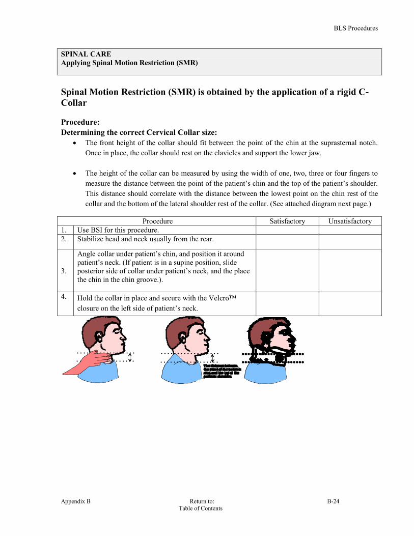
BLS Procedures
Appendix B Return to: B-24 Table of Contents
SPINAL CARE Applying Spinal Motion Restriction (SMR)
Spinal Motion Restriction (SMR) is obtained by the application of a rigid C-Collar Procedure: Determining the correct Cervical Collar size:
The front height of the collar should fit between the point of the chin at the suprasternal notch. Once in place, the collar should rest on the clavicles and support the lower jaw.
The height of the collar can be measured by using the width of one, two, three or four fingers to
measure the distance between the point of the patient’s chin and the top of the patient’s shoulder. This distance should correlate with the distance between the lowest point on the chin rest of the collar and the bottom of the lateral shoulder rest of the collar. (See attached diagram next page.)
Procedure Satisfactory Unsatisfactory
1. Use BSI for this procedure. 2. Stabilize head and neck usually from the rear.
3.
Angle collar under patient’s chin, and position it around patient’s neck. (If patient is in a supine position, slide posterior side of collar under patient’s neck, and the place the chin in the chin groove.).
4. Hold the collar in place and secure with the Velcro™ closure on the left side of patient’s neck.
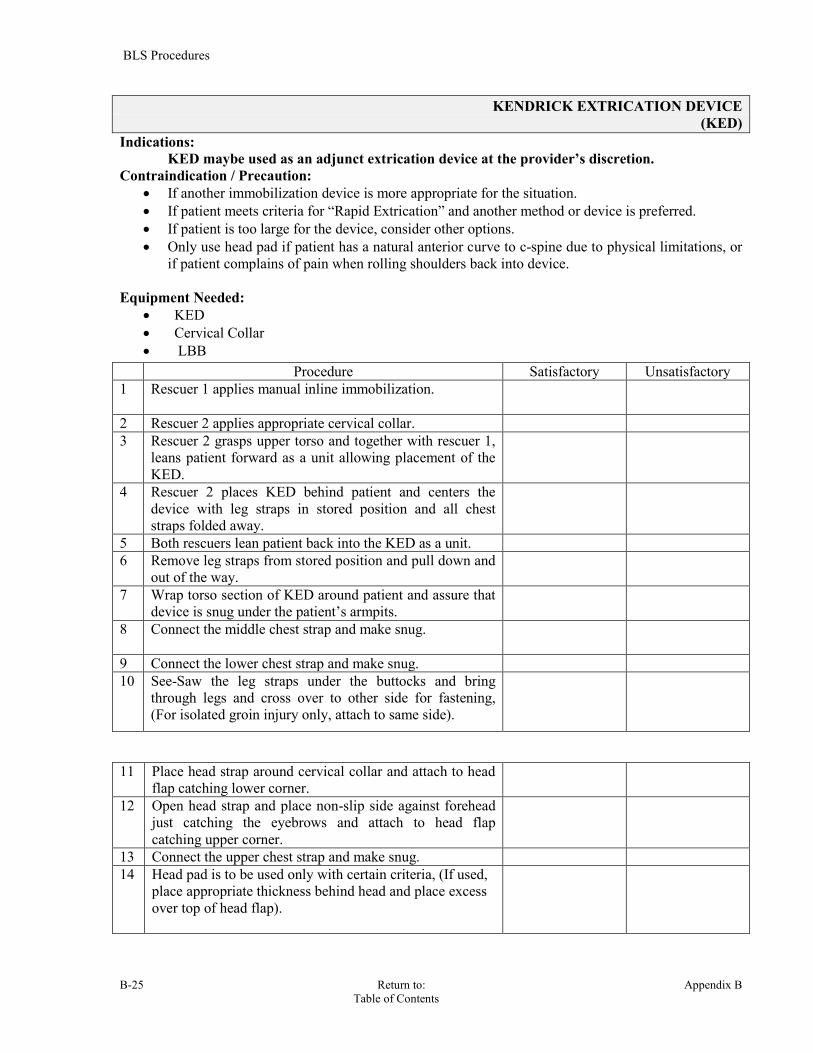
BLS Procedures
B-25 Return to: Appendix B Table of Contents
KENDRICK EXTRICATION DEVICE (KED)
Indications: KED maybe used as an adjunct extrication device at the provider’s discretion. Contraindication / Precaution:
If another immobilization device is more appropriate for the situation. If patient meets criteria for “Rapid Extrication” and another method or device is preferred. If patient is too large for the device, consider other options. Only use head pad if patient has a natural anterior curve to c-spine due to physical limitations, or
if patient complains of pain when rolling shoulders back into device. Equipment Needed:
KED Cervical Collar LBB
Procedure Satisfactory Unsatisfactory 1 Rescuer 1 applies manual inline immobilization.
2 Rescuer 2 applies appropriate cervical collar. 3 Rescuer 2 grasps upper torso and together with rescuer 1,
leans patient forward as a unit allowing placement of the KED.
4 Rescuer 2 places KED behind patient and centers the device with leg straps in stored position and all chest straps folded away.
5 Both rescuers lean patient back into the KED as a unit. 6 Remove leg straps from stored position and pull down and
out of the way.
7 Wrap torso section of KED around patient and assure that device is snug under the patient’s armpits.
8 Connect the middle chest strap and make snug.
9 Connect the lower chest strap and make snug. 10 See-Saw the leg straps under the buttocks and bring
through legs and cross over to other side for fastening, (For isolated groin injury only, attach to same side).
11 Place head strap around cervical collar and attach to head flap catching lower corner.
12 Open head strap and place non-slip side against forehead just catching the eyebrows and attach to head flap catching upper corner.
13 Connect the upper chest strap and make snug. 14 Head pad is to be used only with certain criteria, (If used,
place appropriate thickness behind head and place excess over top of head flap).

BLS Procedures
Appendix B Return to: B-26 Table of Contents
RAPID EXTRICATION Indications:
If the patient’s life or the life of the rescuer is in immediate danger. If the patient’s condition requires immediate life saving intervention that cannot be done
in the vehicle. If a stable patient needs to be removed to gain access to a patient that requires immediate life saving intervention that cannot be done in the vehicle.
Contraindication / Precaution: Stable patients. Not to be implemented out of convenience. Any patient that does not meet any of the above three criteria.
Equipment Needed: Cervical collar Procedure Satisfactory Unsatisfactory 1
Manually immobilize patient’s head, situation permitted.
2
Consider Spinal Care protocol, (C-Collar), if practical.
3
Extricate patient as able.

BLS Procedures
B-27 Return to: Appendix B Table of Contents
REPOSITIONING PRONE PATIENTS Indications:
Prone patients requiring repositioning. Equipment Needed: Extrication device (e.g. Vacuum Mattress, Reeve’s stretcher, backboard, etc.) (Logrolling Technique, minimum of 3, preferably 4 rescuers): Procedure Satisfactory Unsatisfactory 1 Address spinal care. 2 RESCUER applies a cervical collar, if indicated per step 1 then
maintain in-line stabilization.
3 RESCUER kneels alongside patient. and grasps patient’s shoulder and hip.
4 RESCUER kneels alongside patient. and grasps patient’s back and behind the knee.
5 RESCUER (if available), kneels alongside patient. and grasps patient’s upper leg and lower leg.
6 Together as a unit and under the command of the rescuer at the head, roll patient onto side toward rescuers, leaning against rescuers upper legs.
7 RESCUER 4 or another person stabilize the extrication device behind the patient.
8 Together as a unit and under the command of the rescuer at the head, roll patient onto the extrication device.
9 If needed to center patient then have rescuers straddle patient. With one rescuer at head, one grasping under the armpits, one grasping the hips, and one grasping the legs.
10 On order of the rescuer at the head, slide patient as a unit to proper position onto the extrication device.

BLS Procedures
Appendix B Return to: B-28 Table of Contents
PEDIATRIC IMMOBILIZER Indications: The pediatric immobilizer is an optional adjunct to pediatric care. Contraindication / Precaution:
Weight over 65lbs.
Equipment Needed: Pediatric Immobilizer Cervical collar, if indicated per Spinal Care protocol.
Procedure Satisfactory Unsatisfactory 1 Begin with manual immobilization of the head in a neutral, in-
line position, unless contraindicated. Manual immobilization should be provided without interruption until complete patient immobilization is accomplished.
2
Size and apply the appropriate cervical collar, (per spinal care protocol).
3 While maintaining manual stabilization with a cervical collar in place:
3a) Position the Pediatric immobilizer next to the patient so that the head of the immobilizer is approximately 6-12 inches above the patient's head.
3b) Log roll the patient onto the immobilizer in a supine position.
3c)
Reposition patient, in order to center on immobilizer, by sliding patient in an upward motion, (axially), on the immobilizer. Do not slide patient in a direct lateral position, as this may manipulate the spine.
4
Pad the space, as needed, between the back of the head and the immobilizer to prevent hyperextension of the cervical vertebrae.
5 Secure the patient's body to the immobilizer with the attached straps.
5a)
Immobilize the upper torso to prevent upward sliding of patient's body during movement and transportation. This is accomplished by bringing straps over the shoulders and across the chest to make an X. The cross straps velcro into the strap that crosses the abdomen.
5b)
Apply the attached straps across the chest, abdomen and legs. Take care not to leave any space between the straps and the sides of the patient. If the patient is so small that there is a space left between straps and sides of patient, fill the space with pads, (eg. blanket, towel, etc.).
5c)
Arms should be placed at the patient's side to prevent movement of the shoulder girdle.
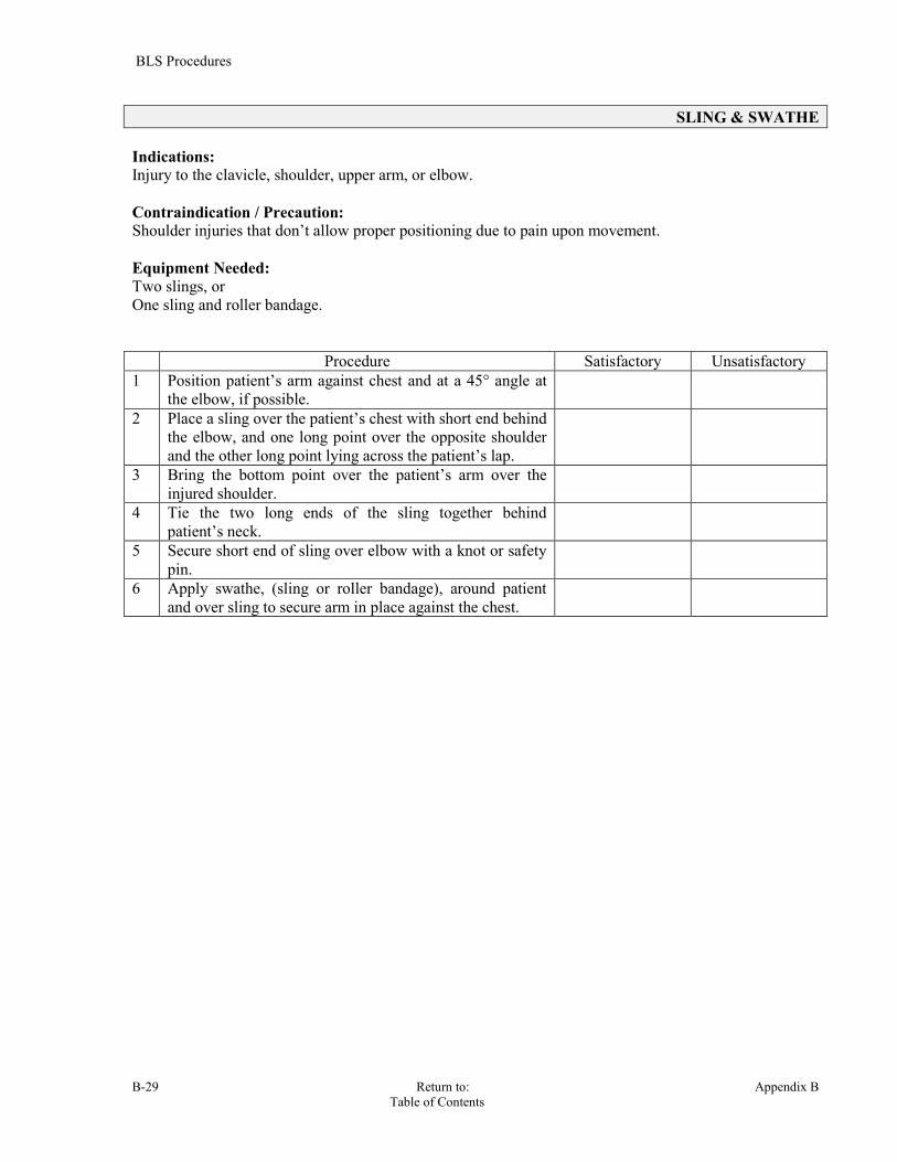
BLS Procedures
B-29 Return to: Appendix B Table of Contents
SLING & SWATHE Indications: Injury to the clavicle, shoulder, upper arm, or elbow.
Contraindication / Precaution: Shoulder injuries that don’t allow proper positioning due to pain upon movement.
Equipment Needed: Two slings, or One sling and roller bandage. Procedure Satisfactory Unsatisfactory 1 Position patient’s arm against chest and at a 45° angle at
the elbow, if possible.
2 Place a sling over the patient’s chest with short end behind the elbow, and one long point over the opposite shoulder and the other long point lying across the patient’s lap.
3 Bring the bottom point over the patient’s arm over the injured shoulder.
4 Tie the two long ends of the sling together behind patient’s neck.
5 Secure short end of sling over elbow with a knot or safety pin.
6 Apply swathe, (sling or roller bandage), around patient and over sling to secure arm in place against the chest.
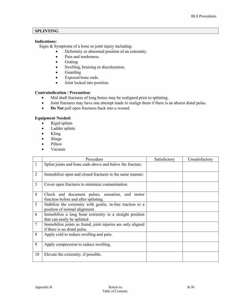
BLS Procedures
Appendix B Return to: B-30 Table of Contents
SPLINTING Indications: Signs & Symptoms of a bone or joint injury including:
Deformity or abnormal position of an extremity. Pain and tenderness. Grating Swelling, bruising or discoloration. Guarding Exposed bone ends. Joint locked into position.
Contraindication / Precaution:
Mid shaft fractures of long bones may be realigned prior to splinting. Joint fractures may have one attempt made to realign them if there is an absent distal pulse. Do Not pull open fractures back into a wound.
Equipment Needed:
Rigid splints Ladder splints Kling Slings Pillow Vacuum
Procedure Satisfactory Unsatisfactory 1 Splint joints and bone ends above and below the fracture.
2 Immobilize open and closed fractures in the same manner.
3 Cover open fractures to minimize contamination.
4 Check and document pulses, sensation, and motor function before and after splinting.
5 Stabilize the extremity with gentle, in-line traction to a position of normal alignment.
6 Immobilize a long bone extremity in a straight position that can easily be splinted.
7 Immobilize joints as found, joint injuries are only aligned if there is no distal pulse.
8 Apply cold to reduce swelling and pain.
9 Apply compression to reduce swelling.
10 Elevate the extremity, if possible.
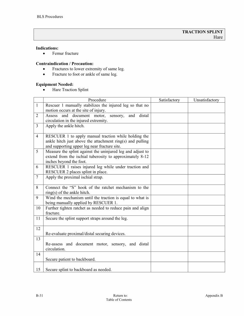
BLS Procedures
B-31 Return to: Appendix B Table of Contents
TRACTION SPLINT Hare
Indications:
Femur fracture
Contraindication / Precaution: Fractures to lower extremity of same leg. Fracture to foot or ankle of same leg.
Equipment Needed: Hare Traction Splint
Procedure Satisfactory Unsatisfactory 1 Rescuer 1 manually stabilizes the injured leg so that no
motion occurs at the site of injury.
2 Assess and document motor, sensory, and distal circulation in the injured extremity.
3 Apply the ankle hitch.
4 RESCUER 1 to apply manual traction while holding the ankle hitch just above the attachment ring(s) and pulling and supporting upper leg near fracture site.
5 Measure the splint against the uninjured leg and adjust to extend from the ischial tuberosity to approximately 8-12 inches beyond the foot.
6 RESCUER 1 raises injured leg while under traction and RESCUER 2 places splint in place.
7 Apply the proximal ischial strap.
8 Connect the “S” hook of the ratchet mechanism to the ring(s) of the ankle hitch.
9 Wind the mechanism until the traction is equal to what is being manually applied by RESCUER 1.
10 Further tighten ratchet as needed to reduce pain and align fracture.
11 Secure the splint support straps around the leg.
12 Re-evaluate proximal/distal securing devices.
13 Re-assess and document motor, sensory, and distal circulation.
14 Secure patient to backboard.
15
Secure splint to backboard as needed.

BLS Procedures
Appendix B Return to: B-32 Table of Contents
TRACTION SPLINT SAGAR This splint is different in several ways from the Hare Traction splint. It works by providing counter-traction against the pubic ramus and the ischial tuberosity medial to the shaft of the femur, and thus does not go under the leg. The hip does not have to be slightly flexed as with the Hare splint. It can also splint both legs with one splint, if needed. Indications:
Fractures of the shaft of the femur. Contraindications:
Fractures involving or distal to the knee.
Fractured pelvis.
Hip injury with gross displacement.
Equipment:
Sager splint.
Ankle hitch with D-ring.
Procedure Satisfactory Unsatisfactory 1 Expose injury site. 2 Assess and record pulse, sensation, and motor function
distal to injury site.
3 Position patient on a long back board. 4 Rescuer 1 supports the leg and maintains gentle traction. 5 Rescuer 2 positions the splint against the inside of injured
leg with the padded bar fitted snugly against pelvis in the groin. The splint can be used on the outside of leg, using strap to maintain traction against the pubis. BE CAREFUL NOT TO COMPRESS THE GENITALS UNDER THE BAR OR STRAP.
6 While maintaining gentle manual traction, attach the padded ankle hitch to the foot and ankle.
7 Extend splint until the correct tension, (10% of the patient’s body weight in lbs), on the pulley wheel is obtained. Maximum of 15 lbs per pulley wheel.
8 Release manual traction and recheck pulse, sensation, and motor function distal to injury site.
9 Apply elastic straps above and below knee. Strap ankles and feet together. Secure patient to the long back board.

BLS Procedures
B-33 Return to: Appendix B Table of Contents
HIP IMMOBILIZATION Indications:
Hip fracture Shortened and externally rotated.
Hip dislocation Shortened and internally rotated.
Contraindication / Precaution:
Assessment of neurovascular status.
Equipment Needed: Pillow Kling or cravats Scoop stretcher PASG Vacuum mattress
Procedure Satisfactory Unsatisfactory 1 Slide cravats or similar lengths of Kling under knee area
of both legs and position one high under the upper leg, one under the lower half of the upper leg and one just below the knees.
2 Spread legs open by moving the unaffected leg.
3 Place a pillow or blanket lengthways between legs and move unaffected leg back in position.
4 Tie cravats or Kling around both legs.
5 Disassemble scoop stretcher.
6 Slide half of scoop stretcher under affected side while lifting up side of patient only enough to get stretcher in place.
7 Slide other half of scoop stretcher under the unaffected side lifting up on patient only enough to attach both ends of scoop together.
8 Pad as necessary for patient comfort.
9 Secure patient to scoop with four straps.
Consider: The Vacuum mattress can also be utilized for the purpose of immobilizing a hip fracture. This would be more comfortable and the chances of aggravating the patient’s injury will be minimized.

BLS Procedures
Appendix B Return to: B-34 Table of Contents
PNEUMATIC ANTI-SHOCK GARMENT, (PASG/MAST)
Indications: Shock believed to be from a ruptured AAA. Stabilization of a pelvic fracture.
These conditions require that BOTH the legs and abdominal section be inflated. Contraindication / Precaution:
Acute Pulmonary Edema. Pregnancy - Do not inflate abdominal section. Cardiogenic Shock. Open wounds of the chest.
Equipment Needed:
Pneumatic Anti-Shock Garment. Foot Pump
Procedure Satisfactory Unsatisfactory 1 Unfold PASG completely and lay on a stretcher or
backboard.
2 Put patient on the PASG face up, (supine), so that the top of garment will be just below the lowest rib.
3 Wrap the left leg first, then the right leg, then the abdomen. Each should be snug and all Velcro should be fastened. Following this sequence will facilitate quicker application of the garment.
4 Check the victim's vital signs and breath sounds. If symptoms of shock are present, inflate PASG.
5 Attach foot pump to PASG at the valves and inflate each section, starting with the leg sections, until: patient’s systolic BP reaches 100, velcro fasteners crackle, air escapes the safety valves and/or bleeding stops.
6 IF USING AS A SPLINT, INFLATE ONLY UNTIL GARMENT IS FIRM.
7 Close valves, leave hoses and pump attached for transport.
Removal: PASGs should not be deflated until:
A physician is present and has taken charge of the patient. Fluids are available for infusion.

BLS Procedures
B-35 Return to: Appendix B Table of Contents
BLEEDING CONTROL / SHOCK MANAGEMENT
Procedure Satisfactory Unsatisfactory
1 Uses body substance isolation precautions.
2
Applies direct pressure to the wound.
3
Elevates the extremity.
4
NOTE: Wound continues to bleed.
5
Applies an additional dressing to the wound.
6
NOTE: Wound continues to bleed. Second dressing does not control bleeding.
7
Locates and applies pressure to the appropriate arterial pressure point.
8
NOTE: Bleeding is controlled.
9
Bandages the wound.
10
NOTE: Patient is exhibiting signs and symptoms of hypoperfusion.
11
Properly positions the patient, (Trendelenburg).
12
Administers high concentration oxygen.
13
Initiates steps to prevent heat loss from the patient.
14
Indicates the need for immediate transportation.

BLS Procedures
Appendix B Return to: B-36 Table of Contents
QUICKCLOT
A sterile, traumatic wound treatment that rapidly arrests high-volume blood loss in large wounds, Creates coagulation in moderate-to-severe wounds, including high-volume venous and arterial bleeding. Indications:
Hemorrhage – High volume blood loss. Procedure Satisfactory Unsatisfactory 1
Tear open package and remove the mesh bag. DO NOT discard package.
2
Remove excess pooled blood from wound, while preserving any clots that have already formed.
3
Pack QuickClot tightly and directly onto the bleeding source. (More than one may be required).
4
Quickly apply pressure until bleeding stops. Suggested time 3 -5 minutes of continuous pressure.
5
Leave QuickClot in place and wrap to secure the product in the wound.
6
Do Not remove bandage or QuickClot. Elevate and evaluate as needed.
7
Turn QuickClot outer package over to the ER staff.
8
Removal instructions to the ER are on the back of the package.
Directions for Emergency Room removal 1
Gently remove mesh bag(s) from wound.
2
If any beads are present in the wound they may be removed by normal irrigation and/or suction until all loose beads are removed.
3 Device contains a blue X-ray detectable element.

BLS Procedures
B-37 Return to: Appendix B Table of Contents
COMBAT APPLICATION TOURNIQUET (C-A-T) APPLICATION The C-A-T Tourniquet is a compact, lightweight tourniquet that can be self-applied using just one hand. It's windlass system provides true circumferential pressure to an extremity to occlude blood flow in the event of a traumatic wound with a significant hemorrhage. Indications:
Life-threatening hemorrhage from an extremity that cannot be controlled by direct pressure, pressure points and elevation.
Contraindication / Precaution:
None when faced with uncontrolled life threatening hemorrhaging. Equipment needed:
C-A-T tourniquet
Procedure Satisfactory Unsatisfactory 1 Place the CAT around the injured limb at least 2 to 3 inches
above the wound site to allow for possible vessel retraction.
2
Pass the free-running end through the inside slit of the buckle and pass band through the outside slit of the buckle.
3
Pull the band tight and securely fasten the band back on itself.
4
Twist the windlass rod until arterial bleeding has stopped.
5
Lock the rod in the U shaped clip.
6
Secure the rod with the strap over the U shaped clip.
7
Note the time the tourniquet was applied in your PCR.
8
Notify the receiving hospital that a tourniquet is in place.
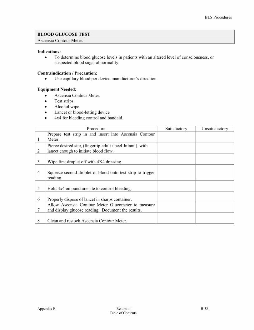
BLS Procedures
Appendix B Return to: B-38 Table of Contents
BLOOD GLUCOSE TEST Ascensia Contour Meter. Indications:
To determine blood glucose levels in patients with an altered level of consciousness, or suspected blood sugar abnormality.
Contraindication / Precaution:
Use capillary blood per device manufacturer’s direction. Equipment Needed:
Ascensia Contour Meter. Test strips Alcohol wipe Lancet or blood-letting device 4x4 for bleeding control and bandaid.
Procedure Satisfactory Unsatisfactory
1
Prepare test strip in and insert into Ascensia Contour Meter.
2
Pierce desired site, (fingertip-adult / heel-Infant ), with lancet enough to initiate blood flow.
3
Wipe first droplet off with 4X4 dressing.
4
Squeeze second droplet of blood onto test strip to trigger reading.
5
Hold 4x4 on puncture site to control bleeding.
6
Properly dispose of lancet in sharps container.
7
Allow Ascensia Contour Meter Glucometer to measure and display glucose reading. Document the results.
8
Clean and restock Ascensia Contour Meter.
BLS Procedures
B-39 Return to: Appendix B Table of Contents
BLOOD GLUCOSE TEST (continued)
CONTROL TEST
The Control Test confirms that the system is functioning properly and must be done at the beginning of each week, (1st shift, each Monday), and anytime blood glucose test results are in question. 1. Equipment:
Ascensia Contour Meter. Ascensia Contour or Microfill blood glucose test Strip. Ascensia Contour or Ascensia Microfill Control Solution
2. Control Test:
Remove the test strip from the plastic bottle and tightly close the bottle immediately after removing the test strips.
Hold the round end of the test strip with the gray electrode side up. Insert the test strip into the meter until it stops. The meter will run a quick self-test and then prompt you to apply blood by showing you a picture
of a drop of blood being drawn into the test strip. This is your signal to apply the control solution. Prepare the control solution by gently rocking the control solution bottle before you open it to
ensure it is mixed well. Squeeze a small drop of control solution on a clean nonabsorbent surface (such as a clean piece
of wax paper). Do not apply control solution to the test trip directly from the bottle. Touch the tip of the test strip to the drop of control solution. Hold it until a beep is heard indicating the test strip has an adequate amount of solution to run the
test. The meter will begin counting down from 5 seconds. Compare your normal control test result with the range printed on the test strip bottle label. Record and date the control test results.
BLS Procedures
Appendix B Return to: B-40 Table of Contents
BLOOD GLUCOSE TEST (continued)
Meter and Test Strip Problems
PROBLEM CAUSE SOLUTION
No display appears when Test Strip is inserted into the Meter.
1. Batteries are not installed correctly.
2. Three minutes have passed since
inserting and unit has turned off. 3. Strip not fully inserted. 4. Battery voltage is low.
1. Install batteries correctly. 2. Do not insert Test Strip until you
are ready to test. 3. Insert until tab is inside Meter. 4. Replace with new batteries.
Check Strip test result out of range.
1. Check Strip, (meter end), dirty or damaged.
2. Meter electronics failure.
1. Wipe with clean dry tissue and visually examine and retest.
2. Contact HEMS Ops Mngr.
Meter fails to start after blood or control solution is drawn into the Test Strip.
1. Not enough blood or control solution is drawn into the Test Strip.
2. Test Strip is defective or has
deteriorated. 3. Three minutes have passed since
inserting and unit has turned off.
1. Form a larger drop of blood or control solution and rerun test using a new Test Strip.
2. Make sure strips used are within
expiration date printed on foil and carton. Rerun test using a new Test Strip.
3. Repeat test with a new Test
Strip. Do not insert Test Strip until you are ready to test.
Meter will not count down. 1. Test Strip inserted incorrectly. 1. Insert Test Strip correctly, (meter end first).
"BATT" display. 1. Battery voltage low. 1. Replace batteries.

BLS Procedures
B-41 Return to: Appendix B Table of Contents
BLOOD GLUCOSE TEST (continued)
Test Result Problems
PROBLEM CAUSE SOLUTION "Lo" display. 1. The blood glucose value may be
below 10 mg/dl, 2. Test Strip not filled completely. 3. Test Strip is defective or has
deteriorated.
1. Possible hypoglycemic condition, (low blood sugar).
2. Rerun test with new strip and
apply sufficient sample to fill Test Strip. Hold TEST END to sample until after the beep.
3. Rerun test using a new strip from
unexpired pack. "Hi" display. The blood glucose value may be
above 600 mg/dl,
Possible hyperglycemic condition, (high blood sugar).
Blood glucose or control test results are inconsistent.
1. Not enough blood or control solution is drawn into the Test Strip.
2. Test Strips have passed their
expiration date. 3. Deteriorated Test Strip caused by
heat or humidity gives low and/or high readings.
4. Meter and/or Test Strip are not at
room temperature when used.
1. Rerun test with new strip and apply sufficient sample to fill Test Strip. Hold TEST END to sample until after the beep.
2. Obtain new Test Strips that are
within their expiration date. 3. Run a Control Test using a new
strip. If results are still out of range, replace carton of Test Strips.
4. Allow time for Meter and/or Test
Strips to come to room temperature before use.
IMPORTANT: There can be as much as a 7% difference in blood glucose readings between capillary, (finger stick), and venous IV blood.

BLS Procedures
Appendix B Return to: B-42 Table of Contents
HELMET REMOVAL
BLS Procedures
B-43 Return to: Appendix B Table of Contents
IV THERAPY SET UP Indications:
For fluid replacement and / or medication administration. Contraindication / Precaution: Selecting proper fluid and administration set as directed.
Mini drip set, (60 drops / ml), for medication administration or fluid restriction. Macro drip set, (10/15 drops / ml), for fluid infusion. 0.9% NaCl, (Normal Saline), for fluid replacement on medical patients.
Equipment Needed:
IV fluid Administration set Alcohol wipes Occlusive dressing Gauze
Procedure Satisfactory Unsatisfactory
1
Obtain and set up alcohol wipes, constricting band, and gauze.
2
Examine IV solution for proper type, clarity and expiration date.
3
Review administration set for proper type, and remove from container.
4
With flow valve shut off, attach IV tubing to IV solution.
5
Squeeze drip chamber until half full.
6
Open flow valve and allow solution to run through entire tubing, expelling all air.
7
Do not contaminate either the connection at the IV bag, or the connection at the IV site.
BLS Procedures
Appendix B Return to: B-44 Table of Contents
VITAL SIGNS Indications:
Frequent assessment on all patients. Every 5 minutes on unstable patients, whenever possible.
Every 15 minutes on stable patients.
At least 2 sets taken on all BLS patients. At least 3 sets taken on all ALS patients.
Contraindication / Precaution:
None Equipment Needed:
Watch BP Cuff Stethoscope Penlight
Procedure Satisfactory Unsatisfactory
1 Assess Respirations, (observe rate for 1 minute, quality, depth, pattern, effort, and breath sounds).
2 Assess Pulse, if regular count for 15 seconds X 4 = rate, feeling for quality and regularity. Assess Pulse for 1 minute if irregular. Sites are: radials, carotid, brachial, femoral, posterior tibial and dorsalis pedis.
3 Assess Skin, (observe for color, temperature, moisture, capillary refill < 2 seconds).
4 Assess Pupillary Reaction, (observe size and reactivity to light ).
5 Blood Pressure, (Taken by palpation and/or auscultation). In all cases, the first B/P is to be taken manually.

Return to: Appendix B Table of Contents
PEPPER SPRAY TREATMENT Action: Sudecon Decontamination Wipes neutralizes pepper spray and tear gas even when running water is not available. It is like an antidote to pepper spray, minimizing the effects. Sudecon works fast and consistently to clean up individuals exposed to OC, CS and CN (pepper spray or tear gas). It strips the chemical agents from the skin and takes away the burn allowing one to open their eyes in 7 to 15 minutes. This requires NO soap or water, contain all natural ingredients and NO alcohol. Indications: Exposure to Pepper spray or Teat Gas: OC (Oleoresin Capsicum) , CS ( Orthochlorobenzalmalonitrile), and CN (alphachloroacetaphenone) Contraindication / Precaution: • Non-toxic, non-flammable, non-irritating. Sudecon® is a topical solution that is used on closed eyes. Equipment Needed:
Procedure Satisfactory Unsatisfactory 1 Tear off the top portions of a pouch and remove and
unfold the damp cloth.
2 Immediately place the towelette over the patient’s face and thoroughly wipe the OC and CS off their face, eyes, nose and exposed areas.
3 With eyes closed, using a second fresh towelette, squeeze towelette over the eyes allowing membranes to absorb the solution. (Squeeze towelette to extract solution.)
4 It is of the utmost importance to squeeze the towelette over the eyes while allowing the liquid to flow around the eyes.
5 After doing so, lay towelette over face to soothe burning sensation.
Then continue to wipe the OC and CS off their face, concentrating on their eyes, nose and mouth. Do this until they can voluntarily open their eyes. Do not use water with the towelettes; it will only dilute the decontamination formula.
ADVANCED LIFE
SUPPORT PROCEDURES
PARAMEDICS & ADVANCED EMTS
APPENDIX C
Revision: 40.6

HEMS ALS Procedures
C-2 Return to: Appendix C Table of Contents
TABLE OF CONTENTS Defib / Monitor
Cardiac Monitoring C-3 –C-4 EKG, 12-Lead C-5 –C-7 Biphasic Defib/Cardioversion Guidelines C-8 Synchronized Cardioversion C-9 Transcutaneous Pacing C-10 Defibrillation, Manual, (Adult) C-11 Defibrillation, Manual, (Pediatric) C-12 Vagal Nerve Stimulation C-13
Airway / Respiratory End –Tidal CO2 Detectors C-14 Capnography C-15 Intubation, Endotracheal C-16-C-17 Intubation, In-Line C-18 ETT position confirmation Devices, (EDD) C-19 Glidescope Intubations C-20 Gum Bougie C-21–C-22 Needle Chest Decompression, (Anterior Approach) C-23–C-24 Cricothyrotomy, (Quick - Trach) C-25 -C-27 Meconium Aspirator C-28 S.A.L.T. C-29
IV/IO Injections / Infusions IV Access – Peripheral C-30 Infusion Pressure Bag C-31 Infusion Pump C-31 Intraosseous Infusion EZ-IO C-32–C-34 Intraosseous Infusion FAST 1 C-35–C-36 Medication Administration, (IVP) C-37 Medication Administration, (Nebulizer) C-38 Medication Administration, (Intramuscular Injection) C-39 Medication Administration, (Rectal) C-40 Medication Administration, (SQ, Subcutaneous administration) C-41 Medication Administration, ( auto-injector) C-42 Medication Administration, (Nasal Spray-Atomizer) C-43 Medications, Reconstitution of C-44
Misc DuoDote C-45-C-46 Morgan Therapeutic Lenses C-47-C-48 Taser Injuries C-49

HEMS ALS Procedures
Appendix C Return to: C-3 Table of Contents
CARDIAC MONITORING Indications:
All ALS patients shall have their EKG rhythm monitored. Contraindication / Precaution:
Do not delay transport of trauma patients to attach the EKG monitor. Equipment Needed:
Monitor/defibrillator. Electrodes. Razor.
Procedure Satisfactory Unsatisfactory
1
Treats patient per appropriate protocol.
2
Shave excessive hair on chest to maximize electrode adhesion.
3
Place electrodes on limbs or on thorax adjacent limbs. Leads will be marked accordingly. (LA – left arm, RA – right arm, LL – left leg, RL – right leg).
4
Adjust gain to the proper level.
5
Obtain baseline EKG tracing.
6 Analyze EKG for: -- Rate -- Rhythm -- Ectopy -- AV block -- Pacing
HEMS ALS Procedures
C-4 Return to: Appendix C Table of Contents
CARDIAC MONITORING (continued)
MULTIFUNCTION PAD METHOD 1. Indications: Determination and monitoring of cardiac rhythms with anticipation of defibrillation. 2. Procedure: Remove clothing from patient's chest. Apply Defib/Pacing Pad: Apply PAD and STERNUM wire to upper sternum slightly toward right shoulder.
Apply PAD and APEX wire to the anterior, (mid-axillary), line below the nipple. Ensure the lead selection is in the PADS mode.
HEMS ALS Procedures
Appendix C Return to: C-5 Table of Contents
EKG, 12 LEAD
Indications:
Complaints of chest pain or discomfort. Drug overdoses. Epigastric pain Unexplained diaphoresis. Dyspnea Unexplained syncope. CHF/Pulmonary Edema. Thoracic back pain in the absence of trauma. Dysrhythmia
Contraindication / Precaution:
Do not perform 12 Lead EKG until life-threatening conditions are managed. Equipment Needed:
Monitor incorporating 12 Lead EKG. Electrodes. Associated monitoring cables. Razor.
Procedure Satisfactory Unsatisfactory 1
Treat patient per AMI protocol, if indicated.
2 Shave excessive hair on chest to maximize electrode adhesion.
3
Place electrodes on limbs, (L arm, R arm, L leg, R leg).
4 Place electrodes on chest: - V1: 4th interspace right parasternal. - V2: 4th interspace left parasternal. - V3: Diagonally between V2 and V4. - V4: 5th interspace at mid-clavicular line. - V5: Anterior-axillary line in line with V4. - V6: Mid-axillary line in line with V4 and V5.
5
Perform 12 Lead EKG.
6 Interpret EKG: - Rhythmn, rate & blocks. - ST-segment elevation. - Ischemic T-wave inversion. - Nondiagnostic or normal EKG.
NOTE: PERCARDITIS CAN MIMMICK ISCHEMIA OF THE MYOCARDIUM. A LBB CAN MASK AN ACUTE MI.

HEMS ALS Procedures
C-6 Return to: Appendix C Table of Contents
1 2 3
4 5 6
EKG, 12 LEAD (continued) The six precordial, (chest), leads are placed on specific locations on the chest. Proper placement is important for accurate diagnosis and should be identified as shown below:
Locating the V1 position, (fourth intercostal space), is critically important because it is the reference point for locating the placement of the remaining V leads. To locate the V1 position: 1. Place your finger at the notch in the top of the sternum. 2. Move your finger slowly downward about 1.5 inches until you feel a slight horizontal ridge
or elevation. This is the “angle of Louis” where the manubrium joins the body of the sternum. 3. Locate the second intercostal space on the right side, lateral to and just below the angle of
Louis. 4. Move your finger down two more intercostal spaces to the fourth intercostal space which is
the V1 position. Other important considerations:
When placing electrodes on female patients, always place leads V3 - V6 under the breast rather than on the breast.
Never use the nipples as reference points for locating the electrodes for men or women patients because nipple locations may vary widely.
Lead Location V1 Fourth intercostal space to the right of the sternum V2 Fourth intercostal space to the left of the sternum. V3 Directly between leads V2 and V4. V4 Fifth intercostal space at midclavicular line. V5 Level with V4 at left anterior axillary line. V6 Level with V5 at left midaxillary line, (Midpoint of
armpit).

HEMS ALS Procedures
Appendix C Return to: C-7 Table of Contents
EKG, 12 LEAD
(continued)
PROCEDURE FOR EVALUATING RIGHT SIDED ISCHEMIA / AMI Indication:
Per protocol or OLMC request Suspicion of possible right sided AMI
To rule out a RIGHT sided MI, move lead V4 to Fifth intercostal space at midclavicular line on the right side, (V4R). Repeat the 12 lead. On the 12 lead print out in the V4 space write in V4R to note the right sided look. Look for an ST elevation, if present, this will assist in verifying the presence of a RIGHT sided MI. PLEASE SEE BELOW FOR LEAD PLACEMENT.
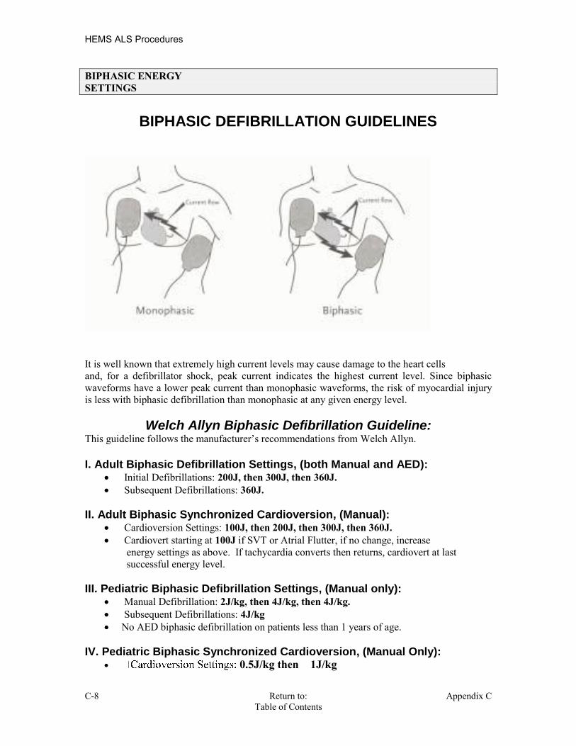
HEMS ALS Procedures
C-8 Return to: Appendix C Table of Contents
BIPHASIC ENERGY SETTINGS
BIPHASIC DEFIBRILLATION GUIDELINES
It is well known that extremely high current levels may cause damage to the heart cells and, for a defibrillator shock, peak current indicates the highest current level. Since biphasic waveforms have a lower peak current than monophasic waveforms, the risk of myocardial injury is less with biphasic defibrillation than monophasic at any given energy level.
Welch Allyn Biphasic Defibrillation Guideline: This guideline follows the manufacturer’s recommendations from Welch Allyn. I. Adult Biphasic Defibrillation Settings, (both Manual and AED):
Initial Defibrillations: 200J, then 300J, then 360J. Subsequent Defibrillations: 360J.
II. Adult Biphasic Synchronized Cardioversion, (Manual): Cardioversion Settings: 100J, then 200J, then 300J, then 360J. Cardiovert starting at 100J if SVT or Atrial Flutter, if no change, increase
energy settings as above. If tachycardia converts then returns, cardiovert at last successful energy level.
III. Pediatric Biphasic Defibrillation Settings, (Manual only):
Manual Defibrillation: 2J/kg, then 4J/kg, then 4J/kg. Subsequent Defibrillations: 4J/kg No AED biphasic defibrillation on patients less than 1 years of age.
IV. Pediatric Biphasic Synchronized Cardioversion, (Manual Only):
0.5J/kg then 1J/kg
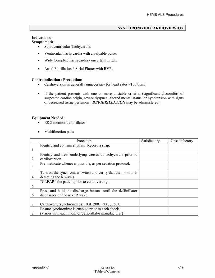
HEMS ALS Procedures
Appendix C Return to: C-9 Table of Contents
SYNCHRONIZED CARDIOVERSION Indications: Symptomatic
Supraventricular Tachycardia.
Ventricular Tachycardia with a palpable pulse.
Wide Complex Tachycardia - uncertain Origin.
Atrial Fibrillation / Atrial Flutter with RVR.
Contraindication / Precaution: Cardioversion is generally unnecessary for heart rates <150 bpm.
If the patient presents with one or more unstable criteria, (significant discomfort of
suspected cardiac origin, severe dyspnea, altered mental status, or hypotension with signs of decreased tissue perfusion), DEFIBRILLATION may be administered.
Equipment Needed:
EKG monitor/defibrillator
Multifunction pads
Procedure Satisfactory Unsatisfactory 1
Identify and confirm rhythm. Record a strip.
2
Identify and treat underlying causes of tachycardia prior to cardioversion.
3
Pre-medicate whenever possible, as per sedation protocol.
4
Turn on the synchronizer switch and verify that the monitor is detecting the R waves.
5
“CLEAR” the patient prior to cardioverting.
6
Press and hold the discharge buttons until the defibrillator discharges on the next R wave.
7
Cardiovert, (synchronized): 100J, 200J, 300J, 360J.
8
Ensure synchronizer is enabled prior to each shock. (Varies with each monitor/defibrillator manufacturer)

HEMS ALS Procedures
C-10 Return to: Appendix C Table of Contents
TRANSCUTANEOUS PACING Indications:
May be used for all symptomatic bradycardias. Asystolic cardiac arrest.
Contraindication / Precaution:
Do not pace patients with severe hypothermia. Asystolic cardiac arrest for greater than 20 minutes.
Equipment Needed:
EKG monitor/defibrillator/pacer. Peripheral IV supplies.
Procedure Satisfactory Unsatisfactory
1
Treat patient per Bradycardia Protocol.
2
Identify rhythm on the cardiac monitor.
3
Obtain vascular access as soon as possible.
4
If patient is conscious and aware of situation during pacing refer to Conscious Sedation Protocol.
5
Apply pacing electrodes.
6
Set the pacemaker to 60 beats per minute.
7
Set the output setting to 0.
8
Turn on the pacer.
9
Slowly increase the current, (mA), until ventricular capture is detected.
10
Reassess the vital signs. Adjust the rate and current as necessary to maintain capture and perfusion.
Procedure Satisfactory Unsatisfactory
1
Treat patient per Asystole Protocol.
2
Apply pacing electrodes.
3
Set the pacemaker to 80 beats per minute.
4
Set the output setting to 200, (mAmp).
5
Turn on the pacer.
6
Check for mechanical capture, (pulse producing), If unsuccessful.
7
Reattempt at 10 minute intervals.

HEMS ALS Procedures
Appendix C Return to: C-11 Table of Contents
DEFIBRILLATION, ADULT Indications: Ventricular Fibrillation and Pulseless Ventricular Tachycardia. First Defibrillation: 200 -300 Joules Second Defibrillation 300 Joules Third Defibrillation 360 Joules
Procedure Satisfactory Unsatisfactory 1
Check the equipment: Turn on the monitor / defibrillator.
2
Apply monitor per cardiac monitoring procedure.
3
Confirm the rhythm.
4
Defibrillation:
4a Confirm the patient is unresponsive and pulseless.
4b Ensure synchronizer is turned off.
4c Check pads position and conduction.
4d Select appropriate energy level per protocol.
4e
Stop CPR and call ALL CLEAR. Check to ensure all people including you are clear of the patient.
4f
Discharge energy by pressing discharge button.
4g
Perform CPR for two minutes, then observe for rhythm change and check patient for a pulse.
4h
Repeat per Protocol.
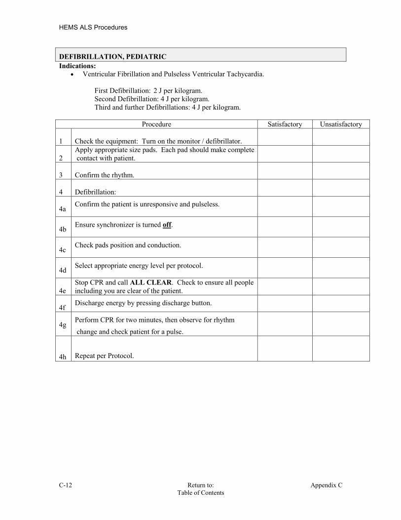
HEMS ALS Procedures
C-12 Return to: Appendix C Table of Contents
DEFIBRILLATION, PEDIATRIC Indications: Ventricular Fibrillation and Pulseless Ventricular Tachycardia. First Defibrillation: 2 J per kilogram. Second Defibrillation: 4 J per kilogram. Third and further Defibrillations: 4 J per kilogram.
Procedure Satisfactory Unsatisfactory 1
Check the equipment: Turn on the monitor / defibrillator.
2
Apply appropriate size pads. Each pad should make complete contact with patient.
3
Confirm the rhythm.
4
Defibrillation:
4a
Confirm the patient is unresponsive and pulseless.
4b Ensure synchronizer is turned off.
4c
Check pads position and conduction.
4d Select appropriate energy level per protocol.
4e
Stop CPR and call ALL CLEAR. Check to ensure all people including you are clear of the patient.
4f
Discharge energy by pressing discharge button.
4g
Perform CPR for two minutes, then observe for rhythm change and check patient for a pulse.
4h
Repeat per Protocol.
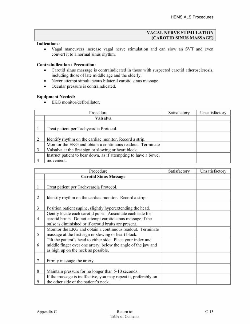
HEMS ALS Procedures
Appendix C Return to: C-13 Table of Contents
VAGAL NERVE STIMULATION (CAROTID SINUS MASSAGE)
Indications: Vagal maneuvers increase vagal nerve stimulation and can slow an SVT and even
convert it to a normal sinus rhythm. Contraindication / Precaution:
Carotid sinus massage is contraindicated in those with suspected carotid atherosclerosis, including those of late middle age and the elderly.
Never attempt simultaneous bilateral carotid sinus massage. Occular pressure is contraindicated.
Equipment Needed:
EKG monitor/defibrillator.
Procedure Satisfactory Unsatisfactory Valsalva 1
Treat patient per Tachycardia Protocol.
2
Identify rhythm on the cardiac monitor. Record a strip.
3
Monitor the EKG and obtain a continuous readout. Terminate Valsalva at the first sign or slowing or heart block.
4
Instruct patient to bear down, as if attempting to have a bowel movement.
Procedure Satisfactory Unsatisfactory
Carotid Sinus Massage 1
Treat patient per Tachycardia Protocol.
2
Identify rhythm on the cardiac monitor. Record a strip.
3
Position patient supine, slightly hyperextending the head.
4
Gently locate each carotid pulse. Auscultate each side for carotid bruits. Do not attempt carotid sinus massage if the pulse is diminished or if carotid bruits are present.
5
Monitor the EKG and obtain a continuous readout. Terminate massage at the first sign or slowing or heart block.
6
Tilt the patient’s head to either side. Place your index and middle finger over one artery, below the angle of the jaw and as high up on the neck as possible.
7
Firmly massage the artery.
8
Maintain pressure for no longer than 5-10 seconds.
9
If the massage is ineffective, you may repeat it, preferably on the other side of the patient’s neck.

HEMS ALS Procedures
C-14 Return to: Appendix C Table of Contents
END –TIDAL CO2 DETECTORS Indications:
EtCO2 adjuncts are used to help verify endotracheal tube placement, by displaying either colorimetric or numeric indicators.
Contraindication / Precaution:
Results may be affected by recent consumption of carbonated beverages, (transient finding), or in cases of low cardiac output.
Always use this device in concert with frequent reassessment of lung sounds, pulse oximetry tracking, patient color and overall condition.
Equipment Needed:
EtCO2 detector, ( BVM with built in colorimetric device ) EtCO2 detector, (color metric device placed between ET tube and BVM ) Electronic EtCO2 monitor, ( Placed between ET tube and BVM )
Procedure Satisfactory Unsatisfactory
1
Complete intubation procedure and initial assessment of tube placement.
2
Attach EtCO2 monitor between ET tube and BVM
3
Ventilate with at least 6 cleansing breaths.
4
Observe for color change, (purple -.03% to yellow – 5% ).
5
If electronic, monitor numerical values. The goal being 35-45 mmHg.
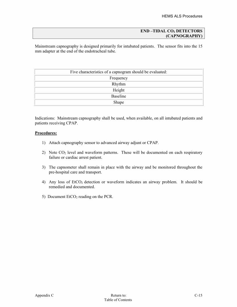
HEMS ALS Procedures
Appendix C Return to: C-15 Table of Contents
END –TIDAL CO2 DETECTORS (CAPNOGRAPHY)
Mainstream capnography is designed primarily for intubated patients. The sensor fits into the 15 mm adapter at the end of the endotracheal tube.
Five characteristics of a capnogram should be evaluated: Frequency
Rhythm Height
Baseline Shape
Indications: Mainstream capnography shall be used, when available, on all intubated patients and patients receiving CPAP. Procedures:
1) Attach capnography sensor to advanced airway adjunt or CPAP. 2) Note CO2 level and waveform patterns. These will be documented on each respiratory
failure or cardiac arrest patient.
3) The capnometer shall remain in place with the airway and be monitored throughout the pre-hospital care and transport.
4) Any loss of EtCO2 detection or waveform indicates an airway problem. It should be
remedied and documented.
5) Document EtCO2 reading on the PCR.

HEMS ALS Procedures
C-16 Return to: Appendix C Table of Contents
INTUBATION, ENDOTRACHEAL Indications: Respiratory or cardiac arrest. Relative Indications: Glasgow Coma Scale of 8 or less. Inadequate ventilations. Possible airway obstruction.
When a patient cannot maintain his/her own airway.
Contraindication / Precaution: Severe oral trauma Patient needs to be well oxygenated prior to intubation attempts. In-line stabilization should be performed for suspected cervical injury patients. All EMS intubations are to be performed with a stylet, (or Gum Bougie). Avoid injuries to tongue and teeth.
Equipment Needed: Suction
Laryngoscope handle with appropriate size blade or Glidescope. Proper size endotracheal tube. Water soluble lubrication gel, (lubricate distal end of tube at cuff). 10 cc syringe, (check cuff for patency). Stylet, (Insert into ET tube). Tape or endotracheal securing device. Proper size oral or nasal pharyngeal airway. BVM C-Collar if indicated. Stethoscope
Gum Bougie Res-Q-Pod EDD device EtCO2 device
HEMS ALS Procedures
Appendix C Return to: C-17 Table of Contents
INTUBATION, ENDOTRACHEAL (continued)
Procedure: Satisfactory Unsatisfactory 1
Prepare suction equipment and place stylet in ET tube.
2
Position the head properly and remove oral / nasal airway. Maintain cervical alignment if indicated.
3
Pre-oxygenate the patient, if indicated.
4
Remove dentures or broken teeth, if indicated.
5
With the left hand, insert the laryngoscope blade while displacing the tongue to the left.
6
Direct the blade downward until in proper position with direct visualization of the glottic opening.
7 Introduce the ET tube through the right corner of the mouth and advance the cuff through the glottic opening to approximately ½ - 1 inch past the vocal cords.
8
Suction and remove debris as indicated.
9
Remove stylette.
10
Inflate the cuff with 10–12cc of air and disconnect the syringe.
11
Ventilate the patient with appropriate device.
12
Attach Capnography per protocol.
13
Confirm proper placement by: auscultation of lungs bilaterally and over epigastrium, chest rise/fall, appropriate numeric value / wave form on EtC02 device, note the ET insertion depth, and EDD.
14
If abdominal sounds are heard, deflate the endotracheal cuff and remove the endotracheal tube immediately. Ventilate the patient and attempt intubation again.
15
If lung sounds are unequal, deflate the endotracheal cuff and reposition the endotracheal tube. Inflate endotracheal cuff and reassess lung sounds. If lung sounds are still unequal, assess the patient for Pneumothorax, (simple or tension).
16
Secure the ET tube with appropriate device, and reassess ET tube position.
17
Document confirmation of ET tube position, (confirmation through auscultation, visualization and mechanical method, (EDD, colormetric or mechanical EtCO2 detector).

HEMS ALS Procedures
C-18 Return to: Appendix C Table of Contents
INTUBATION, IN-LINE Indications:
Patients with possible spinal injuries.
Contraindication / Precaution: Requires two rescuers to properly perform.
Equipment Needed: Intubation equipment. Second rescuer.
Follow intubation procedures, with the following additions: Procedure Satisfactory Unsatisfactory 1
Rescuer 1 to apply manual in-line stabilization from the patient’s side.
2
Proper position will be placing hands over patient’s ears with little fingers under the occipital skull and the thumbs over the maxillary sinuses.
3
Stabilization should be maintained in a neutral position throughout the intubation procedure.
4
Re-secure C–collar / CID.

HEMS ALS Procedures
Appendix C Return to: C-19 Table of Contents
ETT POSITION CONFIRMATION DEVICE (EDD)
Indications:
Aid in determination of correct ET tube placement. Contraindication / Precaution:
None Equipment Needed:
Bulb Type device Syringe Type device
Procedure Satisfactory Unsatisfactory Bulb Type Device: 1
Compress the bulb and place the device on the end of the ET tube.
2
If the device easily refills, the tube is in the trachea.
3
If the device is difficult or fails to refill, the tube is in the esophagus.
Procedure Satisfactory Unsatisfactory Syringe Type Device: 1
Place syringe on the end of the ET tube.
2
Create negative pressure on the syringe.
3
If syringe is easily aspirated, the tube is in the trachea.
4
If the syringe is difficult or fails to aspirate, the tube is in the esophagus.
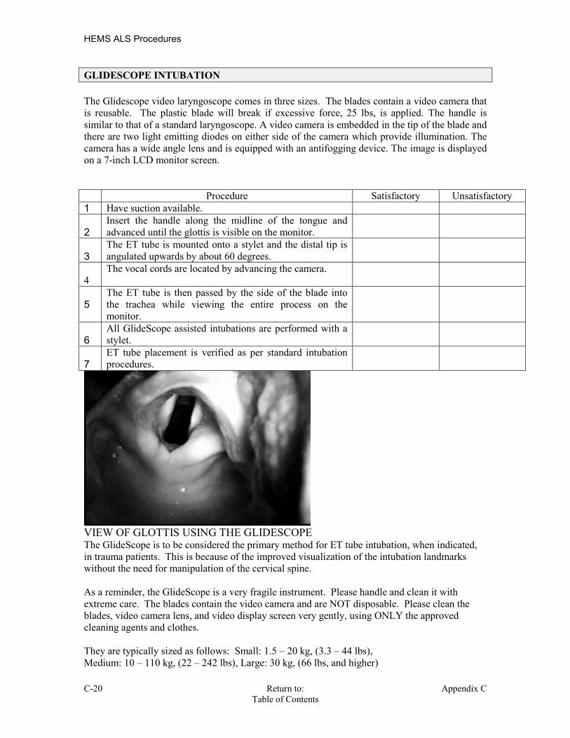
HEMS ALS Procedures
C-20 Return to: Appendix C Table of Contents
GLIDESCOPE INTUBATION The Glidescope video laryngoscope comes in three sizes. The blades contain a video camera that is reusable. The plastic blade will break if excessive force, 25 lbs, is applied. The handle is similar to that of a standard laryngoscope. A video camera is embedded in the tip of the blade and there are two light emitting diodes on either side of the camera which provide illumination. The camera has a wide angle lens and is equipped with an antifogging device. The image is displayed on a 7-inch LCD monitor screen. Procedure Satisfactory Unsatisfactory 1 Have suction available. 2
Insert the handle along the midline of the tongue and advanced until the glottis is visible on the monitor.
3
The ET tube is mounted onto a stylet and the distal tip is angulated upwards by about 60 degrees.
4
The vocal cords are located by advancing the camera.
5
The ET tube is then passed by the side of the blade into the trachea while viewing the entire process on the monitor.
6
All GlideScope assisted intubations are performed with a stylet.
7
ET tube placement is verified as per standard intubation procedures.
VIEW OF GLOTTIS USING THE GLIDESCOPE The GlideScope is to be considered the primary method for ET tube intubation, when indicated, in trauma patients. This is because of the improved visualization of the intubation landmarks without the need for manipulation of the cervical spine. As a reminder, the GlideScope is a very fragile instrument. Please handle and clean it with extreme care. The blades contain the video camera and are NOT disposable. Please clean the blades, video camera lens, and video display screen very gently, using ONLY the approved cleaning agents and clothes. They are typically sized as follows: Small: 1.5 – 20 kg, (3.3 – 44 lbs), Medium: 10 – 110 kg, (22 – 242 lbs), Large: 30 kg, (66 lbs, and higher)

HEMS ALS Procedures
Appendix C Return to: C-21 Table of Contents
GUM BOUGIE
The Gum Bougie, also known as an ET tube exchanger, is a long, flexible, soft tipped catheter. At times one may only be able to visualize a small portion of the tracheal opening and the vocal cords, inferior to the epiglottis, during oral intubation. With only minimal visualization of the important landmarks used for ET tube placement, they may become completely obscured as the tube is advanced into the region. The Gum Bougie may be significantly easier to advance into the trachea. Its small size facilitates improved visualization of the anatomy and landmarks, and its flexibility facilitates manipulation to the desired location. Following placement, one then threads the tube over the bougie. The Gum bougie is then removed, and the ET tube position is confirmed per protocol. Indications 1. Difficult endotracheal intubation, with limited visualization of the vocal cords. 2. Anticipated ET tube of size 6.0 mm or larger. 3. The epiglottis must be visible. For pre-hospital use this is considered a disposable device.

HEMS ALS Procedures
C-22 Return to: Appendix C Table of Contents
GUM BOUGIE, (continued)
Procedure Satisfactory Unsatisfactory 1
Prepare, position and oxygenate the patient with 100% O2.
2
Select proper ET tube without stylet, test cuff and prepare suction.
3
Lubricate the distal end and cuff of the endotracheal tube, (ETT), and the distal ½ of the Endotracheal Tube Introducer, (Bougie), (Note: Failure to lubricate the Bougie and the ETT may result in being unable to pass the ETT).
4
Using laryngoscopic techniques, visualize the vocal cords if possible using Sellick’s as needed.
5
Introduce the Bougie with curved tip anterior and visualize the tip passing the vocal cords or above the arytenoids if the cords cannot be visualized.
6
Once inserted, gently advance the Bougie until you meet resistance or “hold-up”, (if you do not meet resistance you have a probable esophageal placement and insertion should be re-attempted or alternative methods implemented).
7
While maintaining a firm grasp on the proximal Bougie, introduce the ET tube over the Bougie passing the tube to its appropriate depth.
8
If you are unable to advance the ETT into the trachea and the Bougie and ETT are adequately lubricated, withdraw the ETT slightly and rotate the ETT 90 degrees COUNTER clockwise to turn the bevel of the ETT posteriorly. If this technique fails to facilitate passing of the ETT you may attempt direct laryngoscopy while advancing the ETT, (This will require an assistant to maintain the position of the Bougie and, if so desired, advance the ETT).
9
Once the ETT is correctly placed, hold the ET tube securely and remove the Bougie.
10 Confirm tracheal placement according to the intubation protocol. Secure the tube.

HEMS ALS Procedures
Appendix C Return to: C-23 Table of Contents
NEEDLE CHEST DECOMPRESSION (Anterior Approach)
Indications: Tension Pneumothorax associated with closed chest trauma and any of the following
signs and symptoms: Respiratory distress, anxiety or restlessness. JVD, (if not hypovolemic). Decreasing LOC. Initially tachycardic, but later will be bradycardic. Hypotension. Tracheal deviation, (very late sign). Absent breath sounds.
Contraindication / Precaution: Not all signs and symptoms listed above will be present. Must enter skin above the ribs to avoid neurovascular bundle located just underneath
each rib. Creation of a pneumothorax may occur if one is not already present. Laceration of the lung is possible if poor technique is used. Be sure to decompress the side with the pneumothorax.
Equipment Needed: 10cc syringe 14 ga IV catheter/minimum 3 ½ ” length. 3-way stopcock.
Procedure Satisfactory Unsatisfactory 1
Attach the needle to syringe and prep skin.
2
Insert needle/syringe straight into the second intercostal space in the midclavicular line, just above the top of the rib, perpendicular to the chest wall.
3
Advance the catheter forward while applying negative pressure to the plunger until enter is confirmed, or the hub of the needle is in contact with the patient’s skin.
4
Confirmation of tension pneumothorax will be evident by the plunger of the syringe being pushed out, or ease of pulling back on plunger.
5
If confirmed, remove needle and syringe, leaving the catheter in place.
6
Attach 3-way stopcock and close until relief of pressure is needed again, (optional).
7
If negative pressure is attained when pulling back on plunger, remove entire catheter.
8
If blood is present when pulling back on plunger, remove entire catheter.
Note: One may utilize a finger of a glove to create a flutter valve, if desired. Insert the needle through the glove finger, from the inside, before inserting it into the patient’s chest.

HEMS ALS Procedures
C-24 Return to: Appendix C Table of Contents
NEEDLE CHEST DECOMPRESSION (Anterior Approach)(continued)

HEMS ALS Procedures
Appendix C Return to: C-25 Table of Contents
CRICOTHYROTOMY (QUICK-TRACH)
Indications 1. Acute upper airway obstruction, which cannot be relieved using basic airway maneuvers: finger sweep, or endotracheal visualization or Magill forceps removal. 2. Respiratory arrest with facial or neck injury, or abnormal anatomy, which makes endotracheal intubation impossible. 3. Inability to ventilate patient with a bag valve mask. SIZING: Adult, (4.0 mm) Quick Trach: Any patient greater than 100 pounds, (45kg). Pediatric, (2.0mm) Quick Trach: Any patient less than 100 pounds, (45 kg). Causes of Upper Airway Obstruction: 1. Massive facial trauma. 2. Foreign body aspiration. 3. Laryngoedema 4. Laryngospasms 5. Airway/facial burns. 6. Epiglotitis 7. Fractured larynx. Complications: 1. Bleeding 2. Vocal cord injury. 3. Failure to place the catheter in the trachea.
HEMS ALS Procedures
C-26 Return to: Appendix C Table of Contents
CRICOTHYROTOMY (QUICK-TRACH)(continued)
Procedure Satisfactory Unsatisfactory 1
Expose the neck.
2
Identify the cricoid membrane located between the cricoid cartilage and the thyroid cartilage.
3
Prep the skin as appropriate.
4
Puncture the cricothyroid membrane at a 90-degree angle with the catheter/syringe assembly.
5
Aspirate for air upon introducing the catheter.
6
Upon aspiration of air, redirect the catheter in a 60-degree angle, (toward feet), and advance until the stopper meets the skin.
7
Remove the stop guide.
8
Advance the catheter, (not the needle), until the flange rests on the skin.
9
Remove the needle-syringe assembly.
10
Attach the connecting tube to the 15mm adaptor.
11
Attach a bag valve mask, (BVM), to the other end of the connecting tube.
12
Carefully secure the Quick-Trach.
13
Ventilate the patient using the BVM.
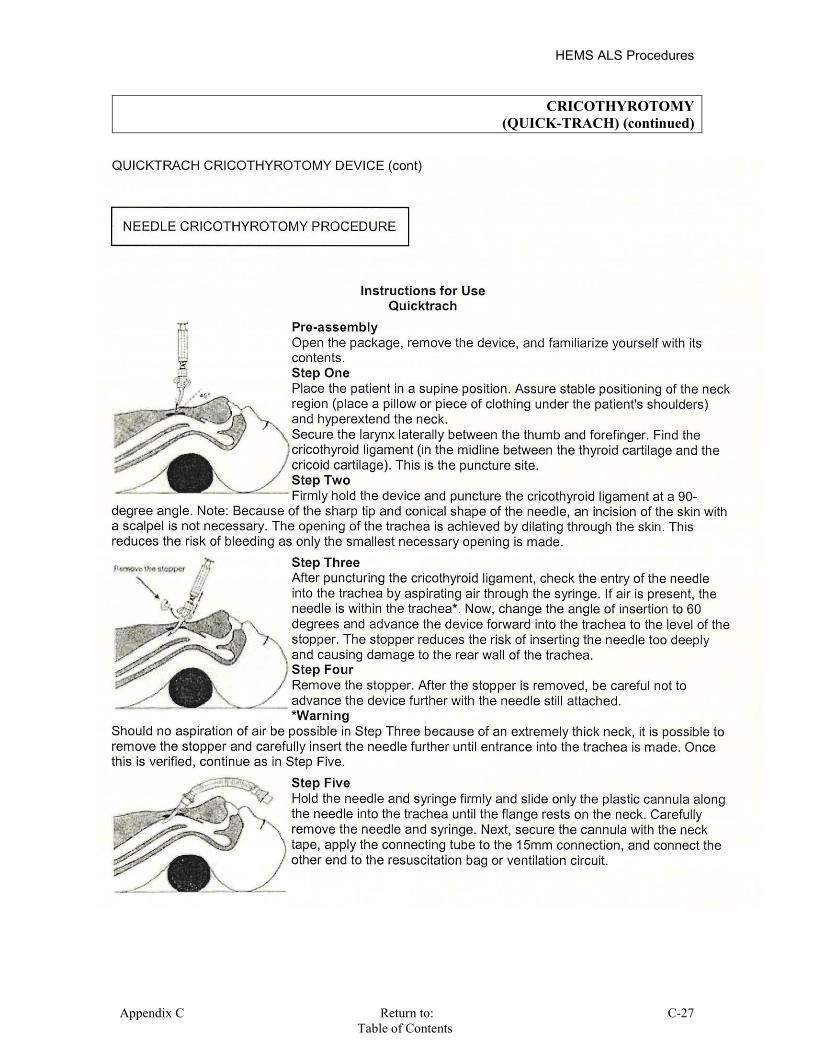
HEMS ALS Procedures
Appendix C Return to: C-27 Table of Contents
CRICOTHYROTOMY (QUICK-TRACH) (continued)
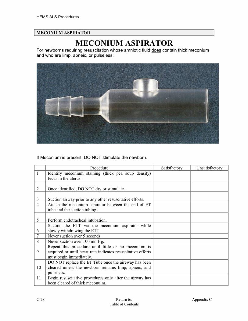
HEMS ALS Procedures
C-28 Return to: Appendix C Table of Contents
MECONIUM ASPIRATOR
MECONIUM ASPIRATOR For newborns requiring resuscitation whose amniotic fluid does contain thick meconium and who are limp, apneic, or pulseless:
If Meconium is present, DO NOT stimulate the newborn. Procedure Satisfactory Unsatisfactory 1 Identify meconium staining (thick pea soup density)
fecus in the uterus.
2
Once identified, DO NOT dry or stimulate.
3
Suction airway prior to any other resuscitative efforts.
4 Attach the meconium aspirator between the end of ET tube and the suction tubing.
5
Perform endotracheal intubation.
6
Suction the ETT via the meconium aspirator while slowly withdrawing the ETT.
7 Never suction over 5 seconds. 8 Never suction over 100 mmHg. 9
Repeat this procedure until little or no meconium is acquired or until heart rate indicates resuscitative efforts must begin immediately.
10
DO NOT replace the ET Tube once the aireway has been cleared unless the newborn remains limp, apneic, and pulseless.
11 Begin resuscitative procedures only after the airway has been cleared of thick meconuim.
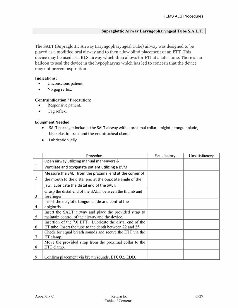
HEMS ALS Procedures
Appendix C Return to: C-29 Table of Contents
Supraglottic Airway Laryngopharyngeal Tube S.A.L.T. The SALT (Supraglottic Airway Laryngopharyngeal Tube) airway was designed to be
placed as a modified oral airway and to then allow blind placement of an ETT. This
device may be used as a BLS airway which then allows for ETI at a later time. There is no
balloon to seal the device in the hypopharynx which has led to concern that the device
may not prevent aspiration.
Indications: Unconscious patient. No gag reflex.
Contraindication / Precaution: Responsive patient. Gag reflex.
Equipment Needed:
SALT package: Includes the SALT airway with a proximal collar, epiglotic tongue blade,
blue elastic strap, and the endotracheal clamp.
Lubrication jelly
Procedure Satisfactory Unsatisfactory 1
Open airway utilizing manual maneuvers &
Ventilate and oxygenate patient utilizing a BVM.
2
Measure the SALT from the proximal end at the corner of
the mouth to the distal end at the opposite angle of the
jaw. Lubricate the distal end of the SALT.
3
Grasp the distal end of the SALT between the thumb and forefinger.
4
Insert the epiglotic tongue blade and control the epiglottis.
5
Insert the SALT airway and place the provided strap to maintain control of the airway and the device.
6
Insertion of the 7.0 ETT. Lubricate the distal end of the ET tube. Insert the tube to the depth between 22 and 25.
7
Check for equal breath sounds and secure the ETT via the ET clamp.
8
Move the provided strap from the proximal collar to the ETT clamp.
9
Confirm placement via breath sounds, ETCO2, EDD.

HEMS ALS Procedures
C-30 Return to: Appendix C Table of Contents
IV ACCESS - PERIPHERAL Indications:
Peripheral vascular access including external jugular, scalp and foot veins, for the administration of medications or fluids.
Contraindication / Precaution:
Do not cannulate at or distal to fracture sites. Use caution when initiating IV access into feet or ankles. DO NOT reinsert needle into a catheter once withdrawn.
Equipment Needed:
IV fluid Administration set Alcohol wipes Transparent dressing Gauze IV Catheter
Procedure Satisfactory Unsatisfactory 1 Explain procedure to patient. 2 Set up IV bag and tubing per protocol. 3 Apply constricting band or Blood Pressure cuff as a TQ. 4 Locate vein of choice. 5 Clean site with alcohol swab using aseptic technique. 6 Stabilize vein by holding pressure distal to point of
insertion.
7 Enter vein with bevel up. 8 Observe flash, advance catheter over the needle. 9 Remove constricting band. 10
Remove needle, placing in sharps container. Apply pressure to proximal end of IV catheter to prevent blood return, while holding catheter hub.
11
Attach IV tubing to catheter, releasing pressure over IV catheter.
12
Infuse IV fluid, and observe for tissue swelling or resistance to infusion.
13
Secure site with transparent dressing. Secure tubing and consider arm board.
14 Set fluid rate as directed. 15 Document date, time, site, catheter size, fluid used, rate
of infusion and person performing the procedure.
16 Document all successful and unsuccessful IV attempts. Troubleshooting the IV, (if the IV is not working well): Make sure the tourniquet is off. Check the IV insertion site for infiltration. Check the IV tubing clamp to make sure it is open. Check the drip chamber to make sure it is half full. Lower the IV bag below IV site and watch for blood to return into the tubing.

HEMS ALS Procedures
Appendix C Return to: C-31 Table of Contents
INFUSION PRESSURE BAGS Indication: Infusion pressure bags may be used to facilitate the rapid infusion of I.V. fluids when rapid infusion is indicated. Their usage is discretionary. Document its usage. Be sure to monitor both the infusion site and the remaining fluid quantity closely when I.V. pressure infusion bags are utilized.
INFUSION PUMPS Indications:
Regulated intravenous infusions of medications and fluids. Large amount of fluid to be infused. Transfer of a patient on an established pump infusion.
Contraindication / Precaution:
Major trauma & cardiac arrest patients. Unfamiliar with the setup or operations of the specific pump.
Equipment Needed:
Specific type of infusion pump /syringe. Specific type of tubing.
Patients who are already on an infusion pump may be continued on that pump during EMS care and transport, provided the crew is familiar with the pump being used.

HEMS ALS Procedures
C-32 Return to: Appendix C Table of Contents
INTRAOSSEOUS, ADULT INFUSION EZ-IO
Indications: The EZ-IO is indicated whenever traditional vascular access techniques are not possible or require too much time to achieve a successful insertion. This may include:
Hemodynamic instability. Respiratory compromise. Patients requiring EMERGENCY medication or volume replacement. Altered mentation: GCS < 8.
May be use immediately in the following patients:
Patients in Cardiac Arrest. Patients who are Hypovolemic / Hypotensive with profoundly altered mental status.
Viable patients receiving IO lines warrant transport.
Size Specific: EZ-IO AD (Adult, Blue) : For patients weighing 40 kg and greater. EZ-IO PD (Pedi, Pink): For patients weighing between 3 and 39 kg. Contraindications:
1. Any patient that may receive thrombolytic therapy; specific to Acute MI and/or Stroke. 2. Suspected fracture of the associated tibia or femur. 3. Previous orthopedic procedures: i.e. knee replacement. 4. Extremity that is compromised by a pre-existing medical condition, i.e. tumor or PVD. 5. Overlying skin infection/trauma at placement site. 6. Inability to locate the 3 anatomical landmarks for insertion, which are the patella, tibial.
tuberosity, and 1 finger width medical to the tibial tuberosity. 7. Excessive tissue over the insertion site.
Equipment:
EZ-IO Driver EZ-IO Needle Set Provo-iodine, Betadine or Iodine swabs or prep pads. IV setup and interconnecting tubing. 10cc or 20cc Syringe. Roller gauze
Procedure: 1. Use universal precautions.
2. Determine if EZ-IO is indicated and no contraindications are present. 3. Locate proper site for EZ-IO insertion.
a. Feel the front of the leg and locate the patella. b. Locate the tibial tuberosity inferior to the patella. c. Place 1 finger medial to the tibial tuberosity. Insertion location is 1 finger width
medial of the tibial tuberosity. d. On younger patients the tibial tuberosity can be difficult if not impossible to
palpate. If it cannot be located the insertion site is two finger widths below the patella and then medial along the flat aspect of the tibia.

HEMS ALS Procedures
Appendix C Return to: C-33 Table of Contents
INTRAOSSEOUS, ADULT INFUSION EZ-IO (continued)
4. Cleanse the insertion with betadine/iodine or similar prep-pads using accepted technique. Remember to work from the inside to the outside in concentric circles.
5. If patient is conscious, inform patient of the EMERGENT need to perform procedure and that they might feel some discomfort until Lidocaine is administered.
6. Prepare the EZ-IO Driver and Needle Set. a. Open the case and remove the driver and needle set cartridge.
b. Open the cartridge and attach the needle set to the driver. c. Remove needle set from the cartridge. d. Remove the cap from the needle set.
7. Begin insertion of the EZ-IO a. Hold the EZ-IO Driver in one hand and stabilize the leg near the insertion site
with the opposite hand. b. Position the driver at the insertion site at a 90-degree angle to the bone surface. c. Power the driver through the skin at the insertion site until it makes contact with
bone. Let the driver do the work! STOP WHEN YOU FEEL THE POP! d. Evaluate the EZ-IO needle for the 5mm mark.
8. Power the EZ-IO Driver and continue insertion until the flange, (base) of the EZ-IO needle set touches the skin OR a sudden lack of resistance is felt, indicating entry into the marrow cavity.
9. Remove the driver from the needle set. 10. Remove the stylet from the catheter. DO NOT REPLACE or ATTEMPT to recap the needle set.
11. Confirm proper EZ-IO Catheter tip position by checking for at least one of the following: a. IO catheter standing at 90 degrees and firmly seated in tibia.
b. Blood at tip of the stylet. c. A free-flow of fluid through the needle with no evidence of
extravasation. d. Aspirate a small amount of blood or marrow into the tubing. 12. Connect the included mini extension set and begin infusion. 13. Rapid Bolus site with 10 ml of NS to flush out any bone plugs, (5ml flush in pediatric pts). 14. If site does not flow, consider pressure infusion and/or rotate needle 180 degrees.
15. Dress site with roller gauze to prevent accidental dislodgement. 16. Place the yellow wrist EZ-IO band on the patient’s arm.
17. Avoid rocking the needle during usage.
IF PATIENT IS CONSCIOUS FOR PROCEDURE
1. If patient is experiencing pain from the infusion, administer up to 50 mg, (2.5 ml) of Lidocaine 2% SLOW push for local analgesia. This should provide pain relief for up to 1 hour.
Warning: DO NOT hold your hand directly behind the insertion site when inserting an IO needle of any type.

HEMS ALS Procedures
C-34 Return to: Appendix C Table of Contents
INTRAOSSEOUS, ADULT INFUSION EZ-IO (continued)
Anterior , (Front) Anterior , (Front) Medial , (middle) Tuberosities 2 Fingers on Tibial Anterio Anterior , (

HEMS ALS Procedures
Appendix C Return to: C-35 Table of Contents
INTRAOSSEOUS, ADULT INFUSION FAST 1 Indications:
Patients requiring EMERGENCY medicinal therapy or volume replacement. Peripheral IV access has been attempted a minimum of TWO times, without success. Patient is unconscious and unresponsive to painful stimuli. Adult or child weighing more than 100 lbs, (45kg).
Contraindication / Precaution: The patient is NOT unconscious and unresponsive. The patient is postictal and is awakening. The patient has an estimated weight of less than 100 lbs., (45kg). Vascular access is prophylactic and is not emergently required. Patient has an infection or injury overlying the upper sternum, (manubrium). Inability to accurately determine the landmarks require for placement.
Equipment Needed:
HEMS ALS Procedures
C-36 Return to: Appendix C Table of Contents
INTRAOSSEOUS, ADULT INFUSION FAST 1 (continued)
Identify the insertion site. The FAST 1 Intraosseous Infusion System site is the manubrium on the midline and 1.5 cm below, (inferior), to the suprasternal notch. Proper placement of the patch helps assure insertion at this site. This is referred to as the “Target Zone”.
Procedure Satisfactory Unsatisfactory 1
Use universal precautions.
2
Prep site with sterile iodine solution such as Betadine, and clean with alcohol as a sterile procedure.
3
Locate the sternal notch with index finger and apply patch and verify that target zone is midline over the manubrium.
4
Remove sharp protector and position introducer in the target zone to the skin.
5 Push introducer with gradually increasing force until the introducer release is heard and felt.
6
Remove the introducer and replace the sharp protector.
7
Connect the infusion tubing to the male connector on the patch.
8
Attach the female connector and begin running fluids.
9
Check for infiltration.
10
Apply protector dome to site.
11
Ensure the removal kit accompanies the patient to the hospital and is given to ED personnel with removal instructions.
12
Any medications or fluids that would be given via a normal peripheral IV can be given via the FAST 1 System.
ONCE TRIGGERED THE DEVICE MAY NOT BE REUSED / REATTEMPTED.
HEMS ALS Procedures
Appendix C Return to: C-37 Table of Contents
MEDICATION ADMINISTRATION, INTRAVENOUS (IV) Indications:
For the administration of all IV medications. Contraindication / Precaution:
Allergic Reactions. Untoward Reactions, (hypotension, etc. as related to each specific drug’s effects). Whenever possible, have another paramedic verify medication, dosage, and route.
Equipment Needed:
Alcohol wipes Syringes / Needles / Medication. IV Fluid
Procedure Satisfactory Unsatisfactory
1
Confirms: Right patient / Right drug / Right dose Right route / Right time. Determines allergies.
2
Verifies medication is not expired.
3
Verifies fluid is not cloudy, discolored, and box has not been tampered with.
4
Properly prepares medication.
5
Expels air from syringe.
6
Clean off injections site with alcohol wipe.
7
Insert needle, or blunt tip cannula into injection site.
8
Clamp off proximal IV tubing for the duration of the injection.
9
Administer desired dose.
10
Remove syringe and dispose of it properly.
11
Flush medication with 10ml of fluid, (IV or bolus).
12
Monitor patient for positive or adverse effects.
13
Document name of drug given, time given, route, dose, name of person administering drug and effects of administration.
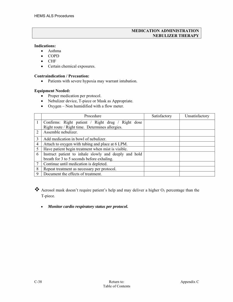
HEMS ALS Procedures
C-38 Return to: Appendix C Table of Contents
MEDICATION ADMINISTRATION NEBULIZER THERAPY
Indications:
Asthma COPD CHF Certain chemical exposures.
Contraindication / Precaution:
Patients with severe hypoxia may warrant intubation. Equipment Needed:
Proper medication per protocol. Nebulizer device, T-piece or Mask as Appropriate. Oxygen – Non humidified with a flow meter.
Procedure Satisfactory Unsatisfactory 1 Confirms: Right patient / Right drug / Right dose
Right route / Right time. Determines allergies.
2 Assemble nebulizer. 3 Add medication in bowl of nebulizer. 4 Attach to oxygen with tubing and place at 6 LPM. 5 Have patient begin treatment when mist is visible. 6 Instruct patient to inhale slowly and deeply and hold
breath for 3 to 5 seconds before exhaling.
7 Continue until medication is depleted. 8 Repeat treatment as necessary per protocol. 9 Document the effects of treatment.
Aerosol mask doesn’t require patient’s help and may deliver a higher O2 percentage than the
T-piece. Monitor cardio respiratory status per protocol.
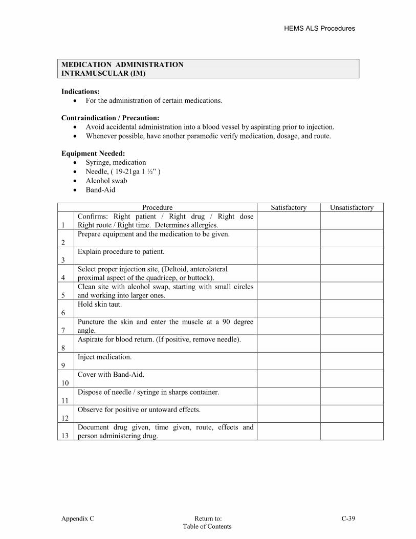
HEMS ALS Procedures
Appendix C Return to: C-39 Table of Contents
MEDICATION ADMINISTRATION INTRAMUSCULAR (IM)
Indications:
For the administration of certain medications. Contraindication / Precaution:
Avoid accidental administration into a blood vessel by aspirating prior to injection. Whenever possible, have another paramedic verify medication, dosage, and route.
Equipment Needed:
Syringe, medication Needle, ( 19-21ga 1 ½” ) Alcohol swab Band-Aid
Procedure Satisfactory Unsatisfactory
1
Confirms: Right patient / Right drug / Right dose Right route / Right time. Determines allergies.
2
Prepare equipment and the medication to be given.
3
Explain procedure to patient.
4
Select proper injection site, (Deltoid, anterolateral proximal aspect of the quadricep, or buttock).
5
Clean site with alcohol swap, starting with small circles and working into larger ones.
6
Hold skin taut.
7
Puncture the skin and enter the muscle at a 90 degree angle.
8
Aspirate for blood return. (If positive, remove needle).
9
Inject medication.
10
Cover with Band-Aid.
11
Dispose of needle / syringe in sharps container.
12
Observe for positive or untoward effects.
13
Document drug given, time given, route, effects and person administering drug.

HEMS ALS Procedures
C-40 Return to: Appendix C Table of Contents
MEDICATION ADMINISTRATION, RECTAL, PR
INDICATION
Administration of medications in pediatric seizure patients when IV or IO access is not possible or would delay the administration of medication in the case of status epilepticus seizure.
Other medications per protocol. CONTRAINDICATION/PRECAUTIONS
Hypersensitivity to medication to be administered. EQUIPMENT NEEDED
Lubricant Tuberculin or 3-5cc syringe without needle. OR Existing Carpujet with 14ga IV catheter without needle. OR Feeding Tube 3cc syringe with saline.
Procedure Satisfactory Unsatisfactory
1
Confirms: Right patient / Right drug / Right dose Right route / Right time. Determines allergies.
2 Prepare all necessary equipment.
3 Apply universal precautions.
4
Position the patient in lateral decubitus or supine position.
5
Insert lubricated feeding tube, syringe, or Carpujet with catheter about 5cm into rectum.
6 Inject medication.
7
Remove syringe or Carpujet and attach saline syringe.
8 Flush catheter with 1cc saline.
9
Remove syringe and catheter holding buttocks together.
10 Tape buttocks closed as needed.
11 Observe for positive or untoward effects. 12
Document drug given, time given, route, effects and person administering drug.
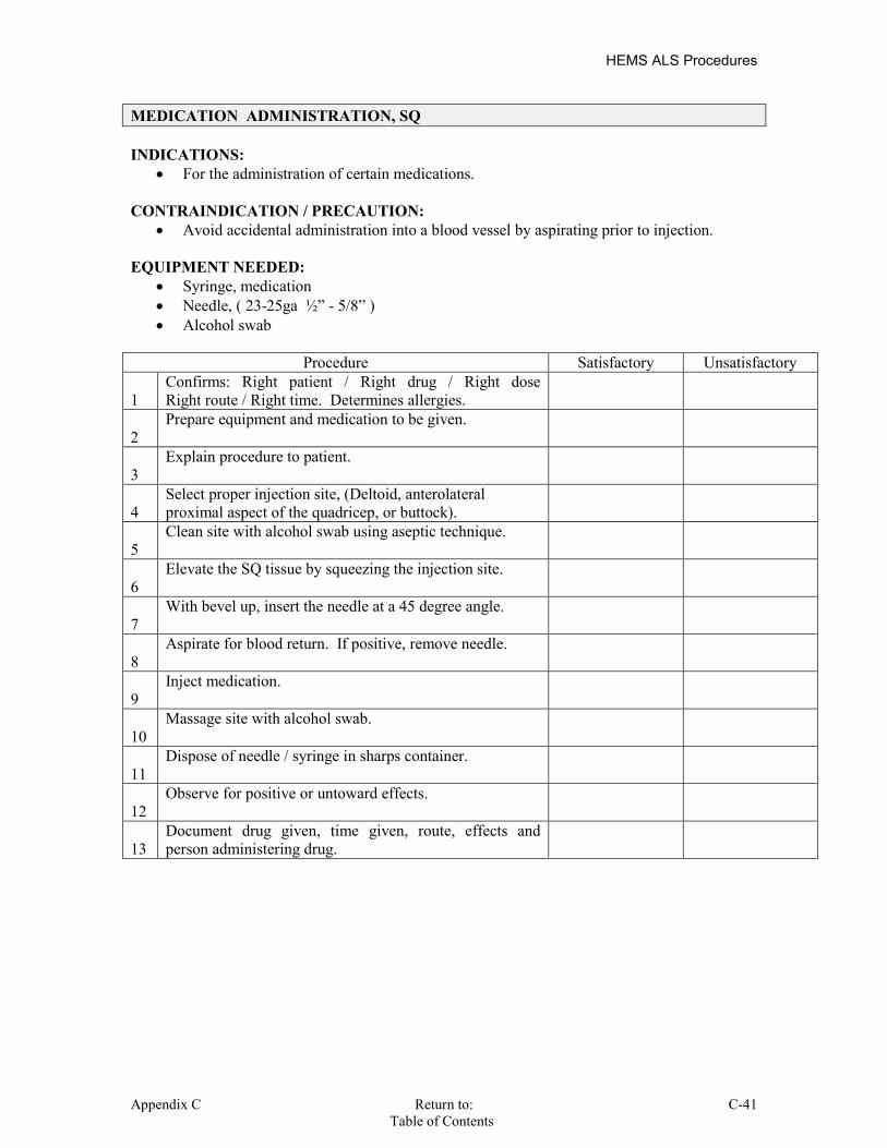
HEMS ALS Procedures
Appendix C Return to: C-41 Table of Contents
MEDICATION ADMINISTRATION, SQ
INDICATIONS: For the administration of certain medications.
CONTRAINDICATION / PRECAUTION:
Avoid accidental administration into a blood vessel by aspirating prior to injection. EQUIPMENT NEEDED:
Syringe, medication Needle, ( 23-25ga ½” - 5/8” ) Alcohol swab
Procedure Satisfactory Unsatisfactory
1
Confirms: Right patient / Right drug / Right dose Right route / Right time. Determines allergies.
2
Prepare equipment and medication to be given.
3
Explain procedure to patient.
4
Select proper injection site, (Deltoid, anterolateral proximal aspect of the quadricep, or buttock).
5
Clean site with alcohol swab using aseptic technique.
6
Elevate the SQ tissue by squeezing the injection site.
7
With bevel up, insert the needle at a 45 degree angle.
8
Aspirate for blood return. If positive, remove needle.
9
Inject medication.
10
Massage site with alcohol swab.
11
Dispose of needle / syringe in sharps container.
12
Observe for positive or untoward effects.
13
Document drug given, time given, route, effects and person administering drug.

HEMS ALS Procedures
C-42 Return to: Appendix C Table of Contents
EPINEPHRINE INJECTION VIA EPI PEN Epinephrine is administered to effect reversal of the shock and bronchial constriction which occurs with anaphylaxis. Indications: Allergic reaction with respiratory distress and hypotension. Contraindication / Precaution: None Equipment Needed: EPI-Pen & Alcohol swab Procedure Satisfactory Unsatisfactory 1
Locate the injection site. Mid femur lateral aspect
2
Cleanse the injection site with an alcohol swab.
3
Inspect EPI-PEN,(color & date; if brownish, outdated or contains any precipitate.
4
Pull off gray safety cap.
5
Place black tip on thigh at right angle to leg.
6
Press hard into thigh until Auto-Injector functions and hold in place for ten seconds.
7
Remove and discard EPI Pen in a “sharp safe” container.
8
Massage injection area for 10 seconds.
9
Repeat vital signs and patient assessment every 5 minutes until stable.
10
If no improvement in 5 minutes, repeat injection.

HEMS ALS Procedures
Appendix C Return to: C-43 Table of Contents
MEDICATIONS, ATOMIZER Intranasal (IN)
Indications: No IV access with the following symptoms: Active Grand Mal seizure Suspected narcotic overdose Pain Management Equipment: Muscosal Atomizer Device (MAD) adapter Syringe Tranfer needle Alcohol prep pad Contraindication: Epistaxis Complete mucosal blockage of both nostrils Any recognizable nasal septal abnormalities Retropharyngeal lacerations/dissections Procedure Satisfactory Unsatisfactory
1
The patient can have the medication delivered from any position (sitting, lying down, prone, on side)
2
Aspirate the proper volume of highly concentrated medication required to treat the patient (an extra 0.1 ml of medication should be drawn up to account for the dead space within the atomizer at the end of the procedure.)
3
Remove air from syringe
4
Attach the atomizer tip via Luer lock mechanism – it twists into place. Slip Luer is also effective as long as the tip is firmly seated on the syringe tip
5
Using your free hand to hold the crown of the head stable, place the tip of the atomizer snugly against the nostril aiming slightly up and inward (towards the top of the opposite ear).
Timing the respirations, depress the plunger rapidly when patient fully exhales and before inhalation
6
Briskly compress the syringe plunger to deliver half of the medication into the nostril.
7
Do not exceed 0.5 – 1.0 ml per nostril
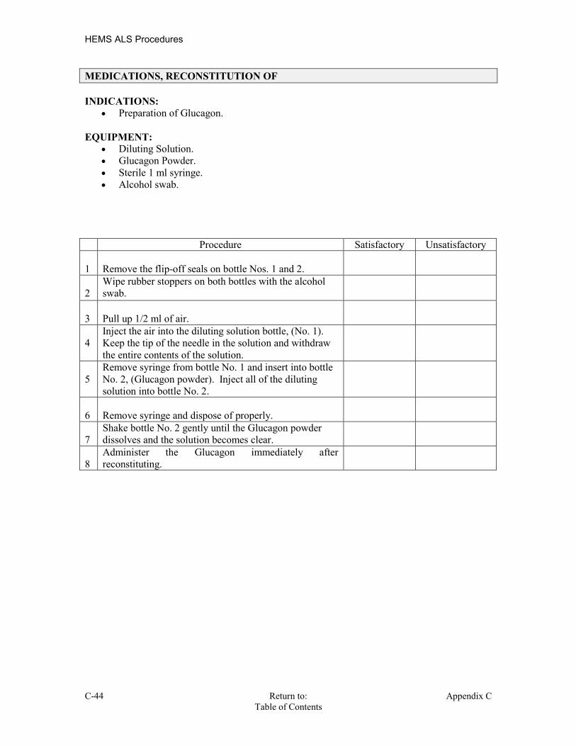
HEMS ALS Procedures
C-44 Return to: Appendix C Table of Contents
MEDICATIONS, RECONSTITUTION OF
INDICATIONS: Preparation of Glucagon. EQUIPMENT: Diluting Solution. Glucagon Powder. Sterile 1 ml syringe. Alcohol swab. Procedure Satisfactory Unsatisfactory
1
Remove the flip-off seals on bottle Nos. 1 and 2.
2
Wipe rubber stoppers on both bottles with the alcohol swab.
3
Pull up 1/2 ml of air.
4
Inject the air into the diluting solution bottle, (No. 1). Keep the tip of the needle in the solution and withdraw the entire contents of the solution.
5
Remove syringe from bottle No. 1 and insert into bottle No. 2, (Glucagon powder). Inject all of the diluting solution into bottle No. 2.
6
Remove syringe and dispose of properly.
7
Shake bottle No. 2 gently until the Glucagon powder dissolves and the solution becomes clear.
8
Administer the Glucagon immediately after reconstituting.
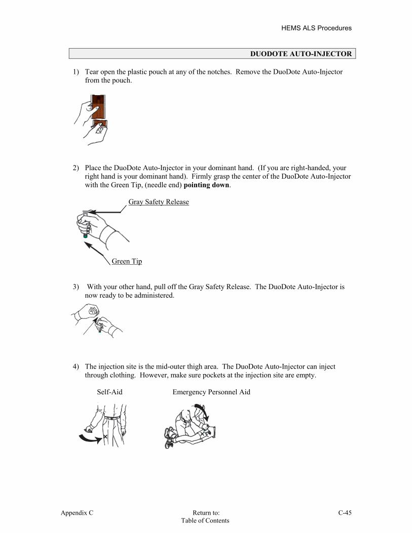
HEMS ALS Procedures
Appendix C Return to: C-45 Table of Contents
DUODOTE AUTO-INJECTOR
1) Tear open the plastic pouch at any of the notches. Remove the DuoDote Auto-Injector from the pouch.
2) Place the DuoDote Auto-Injector in your dominant hand. (If you are right-handed, your
right hand is your dominant hand). Firmly grasp the center of the DuoDote Auto-Injector with the Green Tip, (needle end) pointing down.
Gray Safety Release
Green Tip 3) With your other hand, pull off the Gray Safety Release. The DuoDote Auto-Injector is
now ready to be administered.
4) The injection site is the mid-outer thigh area. The DuoDote Auto-Injector can inject
through clothing. However, make sure pockets at the injection site are empty. Self-Aid Emergency Personnel Aid

HEMS ALS Procedures
C-46 Return to: Appendix C Table of Contents
DUODOTE AUTO-INJECTOR (continued) 5) Swing and firmly push the Green Tip straight down, (a 90o angle), against the mid-outer
thigh. Continue to push until you feel the DuoDote Auto-Injector trigger. Self Aid Emergency Personnel Aid
6) Hold injector at site for 10 seconds to allow medication to be administered.

HEMS ALS Procedures
Appendix C Return to: C-47 Table of Contents
MORGAN THERAPEUTIC LENS The Morgan Therapeutic Lens is indicated for flushing of the eye to remove contaminates, (eg. chemicals). It should not be used for patients with penetrating eye trauma or in cases where foriegn material may be imbedded in the eye, (eg. Broken Glass, sand, etc.). Procedure Satisfactory Unsatisfactory 1
Remove contact lenses, if applicable.
2
Instill topical local anesthetic, (Tetracaine HCl 0.5% Eye Drops), to the affected eye (s). Lavage with Sodium Chloride and set for high continuous flow.
3
Have the patient look down, insert edge of the lens under the upper lid. Have the patient look up, retract the lower lid.
4
Release lower lid over the lens and continue flow. Tape tube and adaptor to patient's forehead to prevent accidental lens removal. Absorb outflow with towels.
Precaution Have the normal saline solution running as one inserts the
lens.
Removal Have patient look up. Retract lower lid behind lower border of
the lens. Hold position. Have patient look down, retract upper lid and slide lens out.

HEMS ALS Procedures
C-48 Return to: Appendix C Table of Contents
MORGAN THERAPEUTIC LENS

HEMS ALS Procedures
Appendix C Return to: C-49 Table of Contents
TASER INJURIES A TASER, is a handheld device which shoots two metal electrodes, (probes), several feet, which then puncture the victim’s skin. The electrode tips incorporate a “fish hook” design and are attached to the TASER unit by two thin wires. An electric shock may then be delivered to the victim, via the wires and electrodes, while maintaining a safe distance from the victim. TASERs put out a high voltage, high frequency, electric shock which is designed to incapacitate the victim, while minimizing the risk of inducing an arrhythmia, apnea, syncope, seizure, or other undesired biophysiologic effects. Trauma, such as head injury or cervical spine injury, can occur as the result of the victim involuntarily falling to the ground when shocked. Iatrogenic injury can also occur from efforts to restrain the victim once they are temporarily incapacitated by the TASER. The electrodes, wires, and the victim pose a risk to shocking rescuers if one comes in contact with them while the TASER is energized. EMS should insure that the device is on SAFETY if they are evaluating an individual who is still connected to the electrodes. Once the electrodes have been removed from the victim, the victim poses no further threat of shock to the rescuers.
DO NOT REMOVE: Face Head Neck, (above the collar bons) Genital region Wrists and Hands Ankles and feet
REMOVE: Chest Back Extremeties,(proximal to wrist and
ankles) Abdomen
Procedure Satisfactory Unsatisfactory
1 Assure Scene and personal safety, Universal precautions. 2 If already removed, administer wound care per protocol. 3 To remove, firmly grasps the electrode and quickly tugging it
out, perpendicular to the entry site.
4 Inspect the electrode to ensure the barb is intact. marking the skin puncture site with a pen if the barb is missing.
5 Mark the entry site of the electrode by drawing a circle around the puncture wound.
6 Treat as any other biohazard. Utilize your SHARPs container unless law enforcement request the electrode be turned over to them.
7 Apply wound care as per protocol. 8 NO incision is to made at the electrode entry site. 9 Perform a neurological and cervical spine exam to assess for
possible head and neck injuries.
10 History, physical exam and vitals are required. 11 EKG rhythm strip is required. 12 TASER related events require contact with OLMC . 13 These patients do not qualify for Medical Control contact
exemption, even if only minor puncture wounds or no injuries at all, are noted.

HEMS ALS Procedures
C-50 Return to: Appendix C Table of Contents
INTENTIONALLY
LEFT
BLANK

CITY OF HUDSON
EMERGENCY MEDICAL SERVICE
TRAUMA ALERT PROTOCOL
APPENDIX D
Ohio Administrative Code sections
3727.10 and 3727.101 [3727.10.1] Revised 4.5
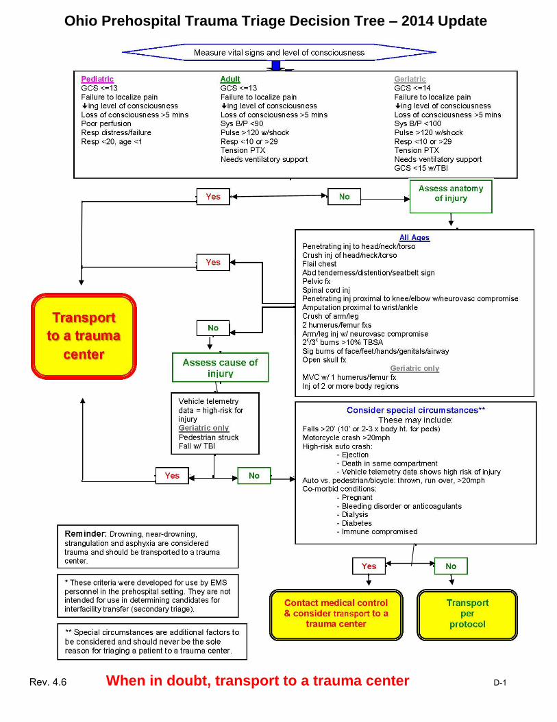
Ohio Prehospital Trauma Triage Decision Tree – 2014 Update
Rev. 4.6 When in doubt, transport to a trauma center D-1

TRAUMA ALERT PROTOCOL
(continued)
TRAUMA CENTERS
INITIAL RECEIVING FACILITIES
Akron City Hospital (TL1) 525 East Market Street
Akron, Ohio 44309-2090 (330) 375- 3361
Akron General Medical Center (TL1)
400 Wabash Avenue Akron, OH 44307 (330) 344-6000
Akron Children’s Hospital (TL II) One Perkins Square
Akron, OH 44308-1062 (330) 543-1000
Toll-Free in Ohio: (800) 262-0333
Bedford Medical Center
44 Blaine Avenue
Bedford, Ohio 44146
(440) 735-3900
Western Reserve Hospital
1900 23rd Street
Cuyahoga Falls, Oh.
44223-1404
(330) 971-7436
Ahuja Medical Center
3999 Richmond Road
Beachwood, oh, 44122
(216)593-5500
UH Twinsburg Health Center
8819 Commons Blvd.
Twinsburg, Oh. 44087
(330) 405-1500
Summa Akron City Hospital (TL1) 525 East Market Street
Akron, Ohio 44309-2090 (330) 375- 3361
D-2
University Hospital Portage Medical Center (UHPMC)
6847 N. Chestnut Street
Ravenna, Ohio
(330) 297-0811
Akron General Medical Center – North 4300 Allen Road Stow, Oh. 44224 (330) 945-3111
Cleveland Clinic Twinsburg Family Health & Surgery
Center
2365 Edison Blvd.
Twinsburg, Oh. 44087
(330) 888-4000
Akron General Medical Center (TL1) 400 Wabash Avenue
Akron, OH 44307 (330) 344-6000
Akron Children’s Hospital (TL II) One Perkins Square
Akron, OH 44308-1062 (330) 543-1000
Toll-Free in Ohio: (800) 262-0333
Return to:
Table of Contents Rev. 4.6

TRAUMA ALERT PROTOCOL
AIR MEDICAL TRANSPORT
(Metro Life Flight)
Air medical transport may be indicated for the care and transport of severely injured trauma patients with compromised
and uncontrollable deficiencies in the management of their ABC’s.
The decision to utilize air medical services is optional, and is at the discretion of the senior medical care provider.
Logistical issues, (timing, LZ location, weather, etc.), may preclude its utilization, even when it is felt to be medically
indicated.
Air medical transport requires activation of additional resources to set up a safe landing zone. This must be initiated as
soon as the decision to utilize air medical services is made.
Air medical transport is contra-indicated in the following:
Patient is in cardiac arrest. Patient has a medical concern, and is not major trauma patient. The time for patient arrival at the hospital would be longer via air medical services than via ground
transport. The weather precludes safe aircraft operations, (e.g. icing conditions, low ceiling (cloud layer), low
visibility). There is no adjacent or reasonably close landing zone. Air medical services response is not immediately available.
1. Determine potential need for air medical transport of the patient.
2. Criteria that suggest the need for air medical transport may include, but are not limited to:
Severely injured trauma patients with compromised and uncontrollable deficiencies in the management of their ABC’s.
Major trauma with prolonged extrication.
Traffic conditions or geographic terrain which prohibit adequate ground access to the victim.
Situations in which the time differential between air and ground transport, or care provided, may substantially
impact the outcome of the patient.
Mass casualty incidents.
3. The paramedic in charge of the patient is responsible for determining if air medical transport is warranted. The
paramedic in charge should initiate air medical transport as soon as possible in order to minimize response and
transport times.
Rev. 4.6 Return to: D-3
Table of Contents
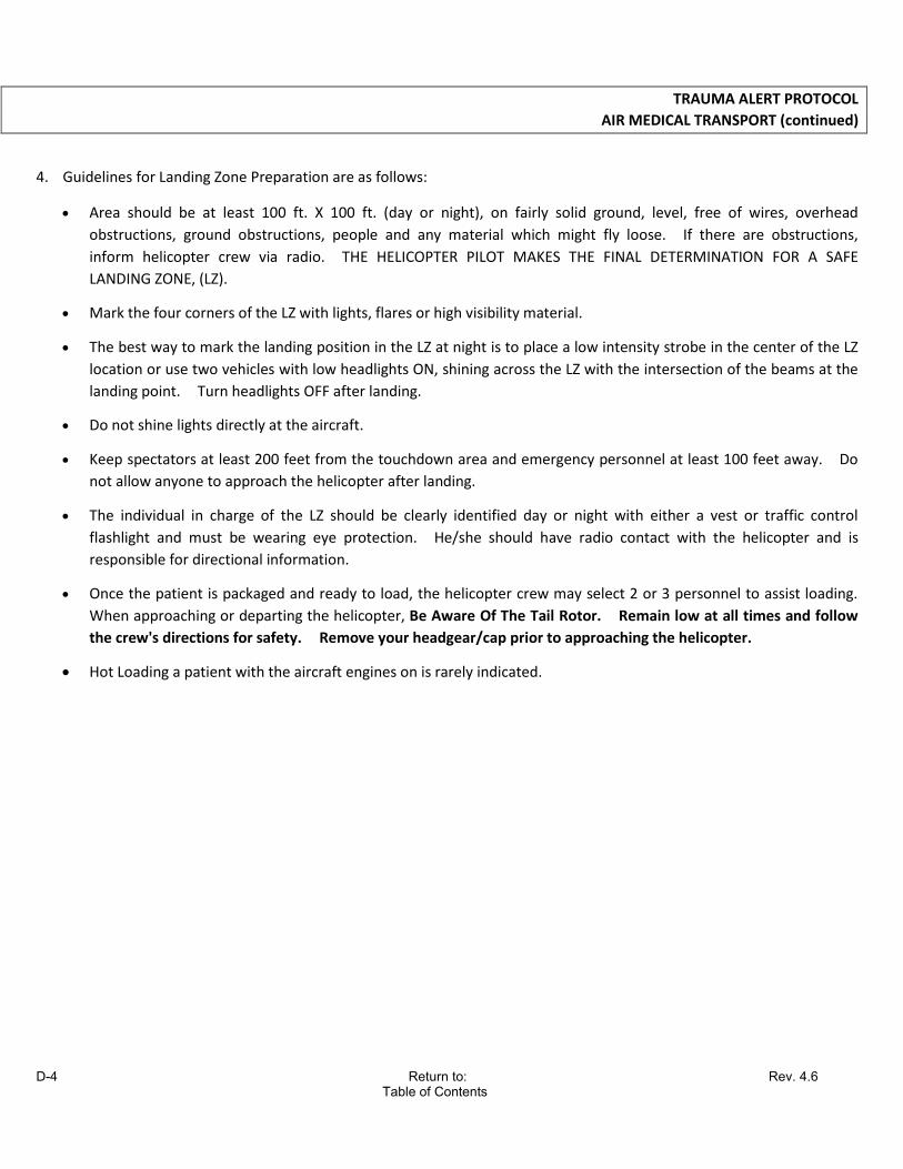
TRAUMA ALERT PROTOCOL
AIR MEDICAL TRANSPORT (continued)
4. Guidelines for Landing Zone Preparation are as follows:
Area should be at least 100 ft. X 100 ft. (day or night), on fairly solid ground, level, free of wires, overhead
obstructions, ground obstructions, people and any material which might fly loose. If there are obstructions,
inform helicopter crew via radio. THE HELICOPTER PILOT MAKES THE FINAL DETERMINATION FOR A SAFE
LANDING ZONE, (LZ).
Mark the four corners of the LZ with lights, flares or high visibility material.
The best way to mark the landing position in the LZ at night is to place a low intensity strobe in the center of the LZ
location or use two vehicles with low headlights ON, shining across the LZ with the intersection of the beams at the
landing point. Turn headlights OFF after landing.
Do not shine lights directly at the aircraft.
Keep spectators at least 200 feet from the touchdown area and emergency personnel at least 100 feet away. Do
not allow anyone to approach the helicopter after landing.
The individual in charge of the LZ should be clearly identified day or night with either a vest or traffic control
flashlight and must be wearing eye protection. He/she should have radio contact with the helicopter and is
responsible for directional information.
Once the patient is packaged and ready to load, the helicopter crew may select 2 or 3 personnel to assist loading.
When approaching or departing the helicopter, Be Aware Of The Tail Rotor. Remain low at all times and follow
the crew's directions for safety. Remove your headgear/cap prior to approaching the helicopter.
Hot Loading a patient with the aircraft engines on is rarely indicated.
D-4 Return to: Rev. 4.6 Table of Contents

HUDSON EMERGENCY MEDICAL SERVICE
CHARTS & SCORECARDS
APPENDIX E
Revision 40.6

Appendix E
BURN ASSESSMENT RULE OF 9’S
NOTE: Generally Patient’s Palm = 1% of Body Surface Area
First-degree burns are red and very sensitive to touch, and the skin will appear blanched when light pressure is applied. First-degree burns involve minimal tissue damage and they involve the epidermis, (skin surface). These burns affect the outer-layer of skin causing pain, redness and swelling. Sunburn is a good example of a first-degree burn Second-degree burns affect both the outer-layer, (epidermis), and the under lying layer of skin, (dermis), causing redness, pain, swelling and blisters. These burns often affect sweat glands, and hair follicles.
Third-degree burns affect the epidermis, dermis and hypodermis, causing charring of skin or a translucent white color, with coagulated vessels visible just below the skin surface. These burn areas may be numb, but the person may complain of pain. This pain is usually because of second-degree burns. Healing from third-degree burns is very slow due the skin tissue and structures being destroyed. Third-degree burns usually result in extensive scarring.

HEMS Protocols
Appendix E
The Glasgow Coma Scale For Adults: EYE OPENING 4 Spontaneous - At this point, with no further stimulation, patient has eyes open. 3 To Voice - If patient's eyes are unopened, a request to "open your eyes" should be spoken, and if necessary, should be shouted. 2 To Pain - If verbal stimulation is unsuccessful in eliciting eye opening, the standard painful stimulus is applied. NOTE: Document if eyes are closed due to swelling or facial injuries, etc. 1 None - No eye opening. BEST VERBAL RESPONSE 5 Oriented - After the patient is aroused, he is asked who he is, where he is, and what the year and month are. If accurate answers are obtained , this is recorded as oriented. 4 Confused - Although the patient is unable to give correct answers to previous questions, he is capable of producing complete phrases, sentences, and even conversational exchange. 3 Inappropriate Words - The patient speaks or exclaims only a word or two. Such a response is usually obtained only by physical stimulation rather
than a verbal stimulus, although occasionally a patient will shout obscenities or call relatives names for no apparent reasons.
2 Incomprehensible Words - The patient's response consists of groans, moans, or indistinct mumbling and does not contain any intelligible words. 1 No Verbal Response - Prolonged and, if necessary, repeated stimulation does not produce any phonation. BEST MOTOR RESPONSE 6 Obeys command - This requires an ability to comprehend instructions, usually given in some form of verbal commands but sometimes by gestures and writing. The patient is required to perform the specific movements requested. The command is given to hold up two fingers, (if physically feasible); the patient should respond appropriately. 5 Localizes Pain - If the patient does not obey commands, a painful stimulus may be applied as firm pressure to the sternum or nail bed for 5 seconds.
reaches to and/or tries to remove source of pain
4 Withdrawals - After painful stimulus: elbow flexes rapid movement no muscle stiffness arm is drawn away from the torso

Appendix E
The Glasgow Coma Scale For Adults
3 Flexion Response - After painful stimulation: slow movement accompanied by stiffness forearm and head held against the body limbs assume hemiplegic position
2 Extension Response - After painful stimulation: legs and arms extend accompanied by stiffness internal rotation of shoulder and forearm
1 None - No motor response Note The Glasgow Coma Scale measures cognitive ability. Therefore, if injury, (chronic or acute), has caused paraplegia or quadraplegia, alternate methods of assessing motor response must be used, (eg. ability to blink eyes = obeys commands). The Glasgow Coma Scale for Children < 5 Years Old
EYES OPENING Score Birth to (1) One year (1) One year and older 4 Spontaneously Spontaneously 3 To shout To verbal command 2 To pain To pain 1 No response No response
BEST MOTOR RESPONSE Score Birth to (1) One year (1) One year and older 6 Obeys 5 Localizes pain Localizes pain 4 Flexion withdrawal Flexion withdrawal
3 Flexion abnormal (decorticate) Flexion abnormal (decorticate) 2 Extension (decerebrate) Extension (decerebrate) 1 No response No response

HEMS Protocols
Appendix E
BEST VERBAL RESPONSE 5 Cried appropriately, coos, smiles Appropriate words & phrases 4 Irritable cry Inappropriate words 3 Inappropriate crying or screaming Cries and/or screams 2 Grunts Grunts 1 No response No response For pediatric patient over the age of 5, use the adult GCS.
REVISED TRAUMA SCORE Glasgow Coma Scale
(GCS) Systolic Blood Pressure
(SBP) Respiratory Rate
(RR) Coded Value
13-15 >89 10-29 4
9-12 76-89 >29 3
6-8 50-75 6-9 2
4-5 1-49 1-5 1
3 0 0 0
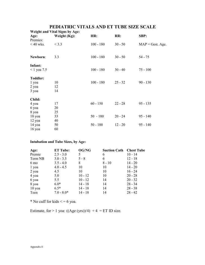
Appendix E
PEDIATRIC VITALS AND ET TUBE SIZE SCALE Weight and Vital Signs by Age: Age: Weight (Kg): HR: RR: SBP: Premies: < 40 wks. < 3.3 100 - 180 30 - 50 MAP = Gest. Age. Newborn: 3.3 100 - 180 30 - 50 54 - 75 Infant: < 1 yoa 7.5 100 - 180 30 - 40 75 - 100 Toddler: 1 yoa 10 100 - 180 25 - 32 90 - 130 2 yoa 12 3 yoa 14 Child: 4 yoa 17 60 - 150 22 - 28 95 - 135 6 yoa 20 8 yoa 25 10 yoa 33 50 - 100 20 - 24 95 - 140 12 yoa 40 14 yoa 50 50 - 100 12 - 20 95 - 140 16 yoa 60 Intubation and Tube Sizes, by Age: Age: ET Tube: OG/NG Suction Cath Chest Tube Premie 2.5 - 3.0 5 6 10 - 14 Term NB 3.0 - 3.5 5 - 8 6 12 - 18 6 mo 3.5 - 4.0 8 8 - 10 14 - 20 1 yoa 4.0 - 4.5 10 10 14 - 20 2 yoa 4.5 10 10 16 - 24 4 yoa 5.0 10 - 12 10 20 - 28 6 yoa 5.5 10 - 12 14 20 - 32 8 yoa 6.0* 14 - 18 14 28 - 34 10 yoa 6.5* 14 - 18 14 28 - 38 Teen 7.0 - 8.0* 14 - 18 14 28 - 42 * No cuff for kids < = 6 yoa. Estimate, for > 1 yoa: ((Age (yrs))/4) + 4 = ET ID size.
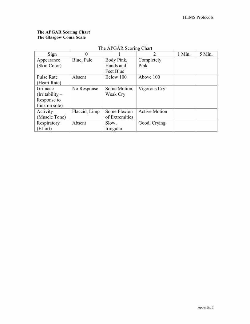
HEMS Protocols
Appendix E
The APGAR Scoring Chart The Glasgow Coma Scale
The APGAR Scoring Chart
Sign 0 1 2 1 Min. 5 Min. Appearance (Skin Color)
Blue, Pale Body Pink, Hands and Feet Blue
Completely Pink
Pulse Rate (Heart Rate)
Absent Below 100 Above 100
Grimace (Irritability – Response to flick on sole)
No Response Some Motion, Weak Cry
Vigorous Cry
Activity (Muscle Tone)
Flaccid, Limp Some Flexion of Extremities
Active Motion
Respiratory (Effort)
Absent Slow, Irregular
Good, Crying

Appendix E
Bites and Envenomations: History:
Type of bite / sting Description or bring
creature / photo with patient for identification
Time, location, size of bite / sting
Previous reaction to bite / sting
Domestic vs. Wild Tetanus and Rabies risk Immunocompromised
patient
Signs and Symptoms: Rash, skin break,
wound Pain, soft tissue
swelling, redness Blood oozing from
the bite wound Evidence of infection Shortness of breath,
wheezing Allergic reaction,
hives, itching Hypotension or
shock
Differential:
Animal bite
Human bite
Snake bite (poisonous)
Spider bite (poisonous)
Insect sting / bite (bee, wasp, ant, tick)
Infection risk
Rabies risk
Tetanus risk
Human bites are much worse than animal bites due to normal mouth bacteria. Carnivore bites are much more likely to become infected and all have risk of Rabies
exposure. Cat bites may progress to infection rapidly due to a specific bacteria, (Pasteurella
multocida). Black Widow spider bites tend to be minimally painful but over a few hours muscular pain
and severe abdominal pain may develop, (spider is black with red hourglass on belly). Brown Recluse spider bites are minimally painful to painless. Little reaction is noted
initially but tissue necrosis at the site of the bite develops over the next few days, (brown spider with fiddle shape on back).
Evidence of infection: swelling, redness, drainage, fever, red streaks proximal to wound. Immunocompromised patients are at an increased risk for infection: diabetes,
chemotherapy, transplant patients.
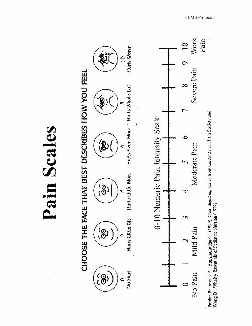
HEMS Protocols
Appendix E

Appendix E
HUDSON EMS PRE-HOSPITAL STROKE WORKSHEET
Patient Name:________________________________Time of Evaluation:__________ Onset Date and Time:____________________________ Every attempt must be made to determine the time the signs and symptoms first began.
Patients that awaken with symptoms, onset time is the last time a patient’s neurological status was known to be normal.
Facial Droop, (Have the patient smile and show their teeth). _____ Normal – Both sides move equally well. _____ Abnormal – One side does not move as well as the other. Arm Drift, (Have the patient close their eyes and hold both arms out palms up). _____ Normal – Both arms move the same or not at all. _____ Abnormal – One arm drifts down or palm turns down compared to the other. Speech, (Have the patient repeat “The sky is blue over Akron). _____ Normal – Patient states correctly without slurring. _____ Abnormal – Patient slurs their words, uses inappropriate words or is unable to speak. Blood Glucose level:____________ ALERT THE HOSPITAL TO A POSSBLE STROKE ASAP.
RAPID TRANSPORT
Assessment Perform by:______________________________ Date:______________
Revised: 6/08
HEMS Protocol
Appendix F Return To: Revision 40.5
Table of Contents P
age1
CITY OF HUDSON EMERGENCY MEDICAL SERVICE
MEDICATION CALCULATIONS
APPENDIX F
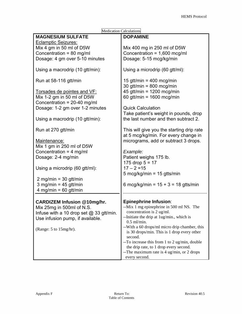
HEMS Protocol
Appendix F Return To: Revision 40.5
Table of Contents P
age2
Medication Calculations
MAGNESIUM SULFATE Eclamptic Seizures: Mix 4 gm in 50 ml of D5W Concentration = 80 mg/ml Dosage: 4 gm over 5-10 minutes Using a macrodrip (10 gtt/min): Run at 58-116 gtt/min Torsades de pointes and VF: Mix 1-2 gm in 50 ml of D5W Concentration = 20-40 mg/ml Dosage: 1-2 gm over 1-2 minutes Using a macrodrip (10 gtt/min): Run at 270 gtt/min Maintenance: Mix 1 gm in 250 ml of D5W Concentration = 4 mg/ml Dosage: 2-4 mg/min Using a microdrip (60 gtt/ml): 2 mg/min = 30 gtt/min 3 mg/min = 45 gtt/min 4 mg/min = 60 gtt/min CARDIZEM Infusion @10mg/hr. Mix 25mg in 500ml of N.S. Infuse with a 10 drop set @ 33 gtt/min. Use infusion pump, if available. (Range: 5 to 15mg/hr).
DOPAMINE Mix 400 mg in 250 ml of D5W Concentration = 1,600 mcg/ml Dosage: 5-15 mcg/kg/min Using a microdrip (60 gtt/ml): 15 gtt/min = 400 mcg/min 30 gtt/min = 800 mcg/min 45 gtt/min = 1200 mcg/min 60 gtt/min = 1600 mcg/min Quick Calculation Take patient’s weight in pounds, drop the last number and then subtract 2. This will give you the starting drip rate at 5 mcg/kg/min. For every change in micrograms, add or subtract 3 drops. Example: Patient weighs 175 lb. 175 drop 5 = 17 17 – 2 =15 5 mcg/kg/min = 15 gtts/min 6 mcg/kg/min = 15 + 3 = 18 gtts/min Epinephrine Infusion: --Mix 1 mg epinephrine in 500 ml NS. The
concentration is 2 ug/ml.
--Initiate the drip at 1ug/min., which is
0.5 ml/min.
--With a 60 drops/ml micro drip chamber, this
is 30 drops/min. This is 1 drop every other
second.
--To increase this from 1 to 2 ug/min, double
the drip rate, to 1 drop every second.
--The maximum rate is 4 ug/min, or 2 drops
every second.

HEMS Protocol
Appendix F Return To: Revision 40.5
Table of Contents
Pag
e3
PEDIATRIC DRUG LIST
Age 50% Weigh
Dose 0.1mg/k
g
Dose 0.1mg/kg
Dose 0.2 mg/kg
Dose 2.0ml/kg
Dose 0.5mg/k
g
Dose 0.03mg/
kg
Dose 0.05mg/
kg
Dose 0.03mg/
kg
Dose 0.1mg/k
g
0.1mg/kgDose
Dose 20ml/kg
Dose 0.5J/kg
Newborn 3kg 0.3 ml 0.3 ml 1.0 ml 6 ml 0.15 ml 0.10 ml 0.15 ml 0.10ml 0.5ml 0.10 ml 50 ml 5 Joules
1 month 4kg 0.4 ml 0.4 ml 1.0 ml 8 ml 0.20 ml 0.10 ml 0.20 ml 0.10ml 0.5ml 0.10 ml 80 ml 5 Joules
3 mo 6kg 0.6 ml 0.6 ml 1.0 ml 12 ml 0.30 ml 0.15 ml 0.30 ml 0.20ml 1.0ml 0.20 ml 120 ml 5 Joules
6 mo 8 kg 0.8 ml 0.8 ml 1.0 ml 16 ml 0.40 ml 0.20 ml 0.40 ml 0.25ml 1.0ml 0.24 ml 160 ml 5 Joules
1 yr 10kg 1.0 ml 1.0 ml 2.0 ml 20 ml 0.50 ml 0.25 ml 0.50 ml 0.5ml 1.0ml 0.30 ml 200 ml 5 Joules
2 yr 12kg 1.2 ml 1.2 ml 2.5 ml 24 ml 0.50 ml 0.30 ml 0.50 ml 0.5ml 1.0ml 0.36 ml 240 ml 5 Joules
3 yr 14kg 1.4 ml 1.4 ml 3.0 ml 28 ml 0.50 ml 0.35 ml 0.50 ml 0.5ml 1.5ml 0.42 ml 280 ml 10 Joules
4 yr 16kg 1.6 ml 1.6 ml 3.0 ml 32 ml 0.50 ml 0.40 ml 0.50 ml 0.5ml 1.5ml 0.48 ml 320 ml 10 Joules
5 yr 18kg 1.8 ml 1.8 ml 3.5 ml 36 ml 0.90 ml 0.45 ml 0.90 ml 0.5ml 2.0ml 0.54 ml 360 ml 10 Joules
6 yr 20kg 2.0 ml 2.0 ml 4.0 ml 40 ml 1.00 ml 0.50 ml 1.00 ml 0.5ml 2.0ml 0.60 ml 400 ml 10 Joules
7 yr 22kg 2.2 ml 2.2 ml 4.5 ml 44 ml 1.00 ml 0.55 ml 1.00 ml 0.5ml 2.0ml 0.66 ml 440 ml 10 Joules
8 yr 25kg 2.5 ml 2.5 ml 5.0 ml 50 ml 1.00 ml 0.60 ml 1.00 ml 0.5ml 2.0ml 0.75 ml 500 ml 10 Joules
9 yr 28kg 2.8 ml 2.8 ml 5.5 ml 56 ml 1.00 ml 0.70 ml 1.00 ml 0.5ml 2.0ml 0.84 ml 560 ml 15 Joules
10 yr 34kg 3.4 ml 3.4 ml 7.0 ml 68 ml 1.00 ml 0.85 ml 1.00 ml 0.5ml 2.0ml 1.00 ml 680 ml 15 Joules
12yr 40kg 4.0 ml 4.0 ml 8.0 ml 80 ml 1.00 ml 1.00 ml 1.00 ml 0.5ml 2.0ml 1.20 ml 1,000 ml 20 Joules
14 yr 50kg 5.0 ml 5.0 ml 10.0 ml 100 ml 1.00 ml 1.00 ml 1.00 ml 0.5ml 2.0ml 1.50 ml 1,000 ml 25 Joules
16 yr 60kg 6.0 ml 6.0 ml 10.0 ml 100 ml 1.00 ml 1.00 ml 1.00 ml 0.5ml 2.0ml 2.00 ml 1,000 ml 30 Joules
Maxi DOSE 10.0 ml 10.0 ml 10.0 ml 100 ml 1.00 ml 1.00 ml 1.00 ml 0.5ml 2.0ml 4 ml 1,000 ml 50 Joules
Note: Six (6) years of age and older may receive D25 or D50.
Epinephrine Epinephrine Atropine Dextrose D25 Versed Diazapam Diazapam Albuterol Naloxine Adenosine Fluid Bolus Synchronized
(Standard) (High Dose) (IV,IO) (IV, IO) (IV,IO,IM) (IV, IO) (Rectal) (Neb) (IV, IO) (IV, IO) (IV, IO) Cardioversion
(IV/IO) (IV/IO)
1:10,000 1:1,000
(0.1mg/ml) (1mg/ml) (0.1 mg/ml) (0.25g/ml) (1mg/ml) (10 mg/ml) (10mg/ml) (5 mg/ml) (1mg/ml) (3 mg/ml) (N.S.) (Joules)
0.01 mg/kg 0.1 mg/kg 0.02 mg/kg 0.3 g/kg 0.05mg/kg 0.3mg/kg 0.5mg/ml 0.15mg/kg 0.1mg/kg 0.1mg/kg

HEMS Protocol
Appendix F Return To: Revision 40.5
Table of Contents
Pag
e4
PEDIATRIC PREHOSPITAL MEDICATIONS Medication Dose Route Remarks
Acetaminophen (Tylenol) 10 mg/kg PO Useful for musculoskeletal pain and fever control Activated charcoal 1 gm/kg PO Do not give to child with altered level of consciousness
Adenosine 0.1 mg/kg IV, IO Indicated for SVT. First dose 6mg, second dose 12mg. third dose 12mg. Follow with a 20cc N.S. flush
Albuterol 2.5 mg Aerosol Indicated for wheezing as per protocol Atropine 0.02 mg/kg
Minimum dose 0.1 mg; max dose for child 0.5 mg; max
dose for adolescent 1.0 mg;
IV, IO May repeat x1; Also useful before intubating children, blocks bradycardia due to vagal nerve stimulation.
Dextrose 25% 2mL/kg IV, IO Try to obtain bedside glucose level before administering ----administer if blood glucose < 80; 25% or dilute 50% 1:1 with sterile water; consult On Line Medical Control if infant < 1 month as solution may need to be further diluted.
Diazepam (Valium 0.2-0.3 mg/kg IV Indicated for uncontrolled seizure mg/kg activity; anticipate respiratory depression. Max. dose 10 mg.
Diazepam (Valium) 0.5 mg/kg Rectal Indicated for uncontrolled seizure activity; anticipate respiratory depression. Max. dose 10 mg.
Diphenhydramine (Benadryl)
1 mg/kg IV Useful in allergic reactions and anaphylaxis. Max dose 50 mg
Epinephrine (1:10,000)
(0.01 mg/kg)
IV, IO Commonly used in cardiac arrest rhythms as first dose. Increase second dose 10 X (may use 1:1,000 solution).
Epinephrine (1:1,000)
0.1 mL/kg IV, IO Commonly used in cardiac arrest rhythms. Use for all ET doses, and second and subsequent IV/IO doses. *The ET route has limited absorption, use IV/IO route whenever possible: Used for anaphylaxis. Max dose is 0.3ml
Lidocaine
1 mg/kg IV, IO If successful, rebolus 0.5mg/kg q 20 minutes.
Morphine 0.1 mg/kg IV/IM Useful for moderate pain, may cause respiratory depression. Hypotension and reflex bradycardia may develop from histamine Release.
Midazolam (Versed) 0.1 mg/kg IV/IO/IM Indicated for uncontrolled seizure activity; anticipate respiratory depression Useful to facilitate advanced airway management in combative patients
Naloxone (Narcan)
0.1 mg/kg IV, IO Useful for unknown unconscious, known narcotic overdoses
Xopenex
(>12 y/o 1.25 mg/ 3cc NS) (6 –12 y/o 0.63mg/3cc N.S.) (<6 contraindicated )
Aerosol Indicated for wheezing as per protocol

HUDSON VOLUNTEER EMERGENCY MEDICAL SERVICE
TRIAGE SYSTEM
“To do the best for the most
with the least.”
START & JumpSTART PROGRAM (Adult) (Pediatric)
APPENDIX G
Revision 40.6

G-2 Return To: Appendix G Table of Contents
TRIAGE SYSTEM “START” USING A TRIAGE SYSTEM FOCUSES YOUR ACTIVITIES DURING A CHAOTIC AND CONFUSING ENVIRONMENT. THE GOAL IS TO RAPIDLY IDENTIFY PATIENT’S INJURIES AND SORT THEM ACCORDING TO THEIR SEVERITY AND NEED FOR TREATMENT. THE METHOD LISTED BELOW IS BASED ON THE S.T.A.R.T. METHOD OF SIMPLE TRIAGE AND RAPID TREATMENT. A. ENROUTE - (Pre-Arrival Planning) While you are responding to the scene, you should prepare yourself mentally for what you may find. Perhaps you’ve been to the same location before. Where might additional resources come from? How long might they take to arrive? B. INITIAL ASSESSMENT - STAY CALM - The first priority is to stay calm and get an overview of the scene. This initial “size-up” will give you an initial impression of the situation, including the potential number of patients, and possibly the mechanism and severity of their injury. This “size-up” will guide you as to additional resources that may be needed. The initial scene size up should include a scene safety survey for possible hazards: wires, fires, weapons, hostiles, animals, traffic and/or HazMat. C. INITIAL REPORT - As you prepare to give the first initial report, use clear and concise information. The key points to communicate are:
1. Location of the incident 2. Type of incident 3. Any hazards 4. Approximate number of victims 5. Type of additional resources needed
D. SORTING THE PATIENTS - It is important not to become involved with the treatment of the first or second patient you initially encounter on scene. Your job is to get to each and every patient as quickly as possible. Each patient should receive a rapid assessment and be assigned to broad categories based on their need for treatment. You must not stop during this survey, except to correct airway and severe bleeding problems quickly. Other rescuers will provide further treatment. E. WALKING WOUNDED - Upon arrival at the scene, patients presenting ambulatory will be directed to a specific area by the triage officer. The triage officer will assign a member of EMS to monitor their movement and arrival to the area. F. THE S.T.A.R.T. SYSTEM- This system is based upon 3 observations: R.P.M. defined as RESPIRATIONS, PERFUSION and MENTAL STATUS.
1. RESPIRATIONS - If the patient is breathing, you need to determine the respiratory rate. Patients
with respiratory rates greater than 30 per minute are triaged as “IMMEDIATE, (RED)”. These patients are showing one of the signs of shock and need immediate care. If the patient is breathing less than 30 times per minute, move on to step # 2. If the patient is not breathing, quickly clear the mouth of foreign matter. Use a head tilt or jaw-thrust, (as applicable), to open the airway. During an MCI the initial triage assessment is the only time in emergency care when there may not be time to properly stabilize every injured patient’s spine. If the patient begins to breathe, they are categorized as IMMEDIATE, (RED). If they don’t spontaneously begin to breathe, the patient should be categorized as “DEAD, (BLACK)”.
Appendix G Return to: G-3 Table of Contents
TRIAGE SYSTEM “START” (continued)
2. PERFUSION - The best field method for checking circulation and distal perfusion is to check the patient’s radial pulse. If the radial pulse is absent, weak or irregular, the patient is categorized as “IMMEDIATE, (RED)”.
3. MENTAL STATUS- The last part of the initial triage is to assess the patient’s mental status.
This assessment is done on patients who have adequate breathing and circulation. Test the patient’s mental status by having the patient follow a simple command such as “open your eyes”, “close your eyes”, or “squeeze my hand”. Patients who can follow these simple commands are tagged as “DELAYED, (YELLOW)”. A patient who is unresponsive or cannot follow these simple commands is categorized as “IMMEDIATE, (RED)”.
A. The START system is designed to assist rescuers in identifying the most seriously injured patients. The initial triage should take 30 seconds or less. As more rescuers arrive on scene, the remaining patients will be re-triaged for further evaluation, treatment, stabilization, and transportation. A patient may be re-triaged multiple times during the course of the incident.
B. When resources are limited or the number of injured patients is greater than the number of rescue personnel, (patient to paramedic ratio), the paramedic must focus treatment and stabilization efforts on the “IMMEDIATE, (RED)” patients and assign supportive personnel to care for the “DELAYED, (YELLOW),” patients. In the event of more than one “IMMEDIATE, (RED),” patient, the initial rescuer must choose the most viable of the “IMMEDIATE, (RED),” patients upon which to focus their care efforts. Other patients can be cared for by assisting personnel until additional resources arrive. If circumstances permit, the other patients can be moved to a central location by which the paramedic can monitor additional patients, so long as it doesn’t interfere with care of the primary “IMMEDIATE, (RED),” patient selected. Initial units on scene: Stabilize as per categories.
Each patient is to have visibly affixed to them a triage tag with at least the following: time triaged, initial findings, (general), initial vitals.
The triage officer is to tear off one-half of the bottom part of the tag of the appropriate triage category to retain as a receipt of that patient. At the end of the initial triage component on an incident, those category receipts are to be turned over to medical command for the purpose of accounting for patients on the scene.
Responding personnel will utilize the START tags whenever there are six or more patients involved in one incident. However, the START tags can be utilized whenever the crew chief feels it will benefit the movement of patients and scene control, most often from a multiple vehicle collision. Use of the START triage system can improve initial scene organization, and enhance its use during mass casualty incidents.
Once patients are in the designated triage area, continue treatment and assessment as per categories. Document any changes in condition by tearing off additional tabs. Documentation of treatment is to be placed on their triage tag. (i.e., extent of injuries, vitals, fluid or oxygen therapy, etc.)

G-4 Return To: Appendix G Table of Contents
TRIAGE SYSTEM “START” (continued) The Personal Property Receipt at the top of the Triage Tag is to address the issue of collecting and returning the victim’s personal property back to them with some degree of security and accountability. Place all the patient’s belongings in a personal belongings bag and remove the Personal Property Receipt and place it in the bag.
The Personal Property Receipt and mode at the top of the tag will be removed as the patient is loaded into the transport unit and retained by the transportation officer. If known, place the patient’s name on the tag.
Recall that the personal effects such as wallets, watches, jewelry, etc,, may help in identifying individuals. “CONTAMINATED” strip affixed to the tag is designed for three (3) functions:
A. It is designed to prompt the first arriving EMS crew to consider contaminants. If no contamination hazard exists, simply remove and discard the contamination strip and use the tag as a simple triage tag. If a contaminated hazard does exist, it will prompt responders to take appropriate safety precautions and actions before making contact with the victims.
B. It is designed to identify contaminated victims prior to decontamination. The contaminated strip is to be removed from the tag when the victim’s clothes have been removed. Both the contaminated clothing and the contaminated strip are placed inside a clear personal belongings bag.
C. It is designed to identify possible contaminated evidence for law enforcement. DETAILED FLOWCHART

Appendix G Return to: G-5 Table of Contents
TRIAGE SYSTEM “JumpSTART”
The START triage system is modified for the pediatric population. The Jump START triage system was designed to address the needs of the pediatric, (age 1y/o to 8y/o), population during crisis management. In a crisis situation, it will be difficult, if not impossible, to accurately determine a patient’s age. If the patient looks like an adult, use the START triage criteria. If the patient looks like a child, use the Jump START triage guidelines. Some pediatric patients who are minimally injured are developmentally unable to walk. The adult START triage system does not have provisions for these patients tagged green despite minimal or no injury. JumpSTART's objectives are: 1) To optimize the primary triage of injured children in the MCI setting. 2) To enhance the effectiveness of resource allocation for all MCI victims. 3) To reduce the emotional burden on triage personnel who may have to make rapid life-or-death decisions about injured children in chaotic circumstances. Ambulatory:
Pediatric patients who are ambulatory are tagged green, but must undergo a secondary triage to assess for further injuries as well as potential for rapid deterioration. If the child is unable to ambulate, the respiratory status should be assessed immediately. Non- ambulatory children with a normal respiratory rate and a palpable pulse are then assessed for responsiveness.
Respirations: (Normal 15 – 45)
A child with a normal respiratory rate and no palpable pulse is tagged red and transported immediately. If the child is not breathing, reposition the airway. If respirations resume with repositioning, the child is tagged red and should be transported immediately. If the patient remains apneic with repositioning of the airway, assess the pulse. If the apneic child has a pulse, five rescue breaths should be given. If respirations resume, the child is tagged red and transported immediately. If apnea persists, the child is unsalvageable and tagged black. A respiratory rate greater than 30 is normal in a young child and it would be inappropriate to triage these patients with red tags. The majority of cardiac arrests in the pediatric population are due to a respiratory arrest. Therefore, a slow respiratory rate is an ominous symptom in a child and should be addressed immediately,(i.e. red tag). If the child is not ambulatory but has respirations, the respiratory rate should be measured and classified as normal, (15-45 breaths per minute), or abnormal, (less than 15 or greater than 45 breaths per minute). Non-ambulatory children with an abnormal respiratory rate are tagged red and require immediate transport. If the respiratory rate is normal, check for a pulse.
Pulse /Capillary refill: If the pulse is absent, the child is deceased and tagged black. In a crisis situation, the lighting and cool temperatures may make capillary refill difficult to assess. Mental Status: (AVPU)
Children who are unresponsive, respond inappropriately to pain, or are posturing are tagged red and transported immediately. The non-ambulatory child who is alert or responds appropriately to verbal or painful stimuli is tagged yellow and can be transported to a health care facility in a delayed fashion.
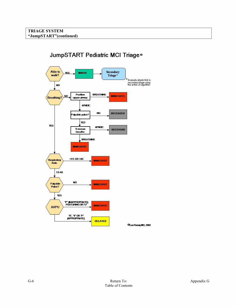
G-6 Return To: Appendix G Table of Contents
TRIAGE SYSTEM “JumpSTART”(continued)

Appendix G Return to: G-7 Table of Contents
TRIAGE SYSTEM “ START & JumpSTART”(continued)
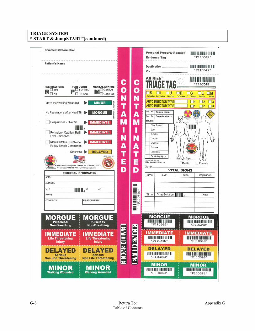
G-8 Return To: Appendix G Table of Contents
TRIAGE SYSTEM “ START & JumpSTART”(continued)

CITY OF HUDSON EMERGENCY MEDICAL SERVICE
APPROVED
ABBREVIATIONS & SYMBOLS
APPENDIX H
Revision 40.4

Abbreviation Definition AA&OX4 alert and oriented to person, place, time, and event AAA abdomenal aortic aneurysmABC airway, breathing and circulationAbd abdomen or abdominalACLS advanced cardiac life supportA-Fib atrial FibrillationAKA above the knee amputationALOC altered level of consciousnessALS advanced life supportAMA against medical adviceAMI acute myocardial infarctionAMS altered mental status AMT amountAOS arrive on sceneApprox approximately ASA aspirinATF arrived to find BBBB bundle branch blockBGL blood glucose levelBGT blood glucose testBilat bilateral or bilaterally BLS basic life supportBM bowel movement BP blood pressure BS breath soundsBVM bag valve mask CC/C chief complaint C/O complaint of, complains of, or complaining of CA cancerCABG cornary artery bypass graftCAD cornary artery diseaseCATH catheterCHF congested heart failureCID cervical immobilization device CNS central nervous systemCO2 carbon dioxide COHb carboxyhemoglobinCOPD chronic obstruction pulmonary diseaseCP chest painCPR cardiopulmonary resuscitation CSF cerebrospinal fluidC-Spine cervical spine
2Return to:
Table of Contents Appendix H

CT CAT scanCVA cerebral vascular accident DD5W 5% dextrose in water DKA diabetic ketoacidosis DNR do not resuscitate DOA dead on arrival DT delirum tremorsDx Diagnosis EECG electrocardiogramET endotrachealETA estimated time of arrival ETOH ethanol alcohol ETT endotracheal tube FF femaleFB foreign bodyFHR fetal heart rate Fx fracture Gg grams GI gatrointestinal GSW gun shot woundgtts drops per minutesgtts/min. drops per minutesGYN gynnecology, gynecological HH/A headache HEENT head, eyes, ears, nose and throatHR heart rate per minute HTN hypertensionHx history IICP intercranial pressure ICU intensive care unitIDDM insulin dependent diabetic mellitus IM intramuscular IO intraosseousIV intravenous JJVD jugular vein distension
Appendix HReturn to:
Table of Contents 3

Kkg kilogramKVO keep vein open L(L) leftL&D labor and deliveryL/S spine lumbarsacral spineLat laterallb poundLBB long backboardLBBB left bundle branch blockLLL left lower lobeLLQ left lower quadrant LMP last menstrual period LOC level of consciousnessLR lactated ringersL-spine lumbar spineLUL left upper lobeLUQ left upper quadrant MM maleMAEW moves all extremeties wellMAST military anti-shock trousersmcg microgramsMED medicine mg milligramMI myocardial infarctionmin minute or minimumMOI mechanism of injuryMS mental status or muscular sclerosis MS mental status changeMS04 morphine sulfate MSPs motor, sensory, pulsesMVC motor vehicle collision NN/A not applicable N/V nausea and vomiting N/V/D nausea, vomiting and diarrheaNAD no apparent distressNAT no apparent trauma NC nasal cannulaNEB nebulizerNIDDM non insulin dependent diabetes mellitusNKDA no known drug allergiesNRB non rebreather NRM normal range of motion
4Return to:
Table of Contents Appendix H

NS normal salineNSR normal sinus rhythm OO2 oxygenOB/GYN obstretrics and gynecology PPAC premature atrial contractionPALP palpitationPE physical examination or pulmonary embolusPEA pulseless electrical activity PERRL pupils equal round reactive to light PMHx past medical historyPO by mouthPOC position of comfortPOP pain on palpationPR rectally PRN as neededPT patient PTA prior to arrivalPVC premature ventricular contraction R® rightRBBB right bundle branch blockRLQ right lower quadrant ROM range of motionRR respiratory rate per minuteRUQ right upper quadrant Rx medicine SS/S signs and symptoms SIDS sudden infant death syndromeSNT soft and non tenderSOB shortness of breathSpCO Saturation of peripheral carbon monoxide SpO2 Saturation of peripheral oxygenSQ subcutaneousST sinus tachycardia SVT supraventricular tachycardiaSZ seizure TT temperature TB tuberculosis TCA tricyclic antidepressant TIA transient ischemic attackTKO to keep open
Appendix HReturn to:
Table of Contents 5

T-spine thoracic spineTx treatment UUOA upon our arrivalURI aupper respiratory infectionUTI urinary tract infection VVF ventricular fibrillationVS vital signs VT ventricular tachycardia WWNL within normal limits YYO years old
SYMBOLS? questions positive negative approximately< less than> greater than= equal↑ higher or increased ↓ lower or decreased 1o primary2o secondaryD change ♀ female ♂ male ĉ with (Flat bar not carrot)
ŝ without (Flat bar not carrot)
ā beforep after @ at
6Return to:
Table of Contents Appendix H

I-1 Return To: Appendix I Table of Contents
HUDSON EMERGENCY MEDICAL SERVICE
TRANSPORT
DESTINATIONS
APPENDIX I
Revision 40.6
I-2 Return To: Appendix I Table of Contents
TRANSPORT DESTINATIONS
Hudson EMS has no preferred destination facilities. There is a wide range of "Approved" destination facilities to which our patient's may be transported. "Approved", in this case, is based upon both the services able to be provided to the patient at that facility and upon geography. Hudson EMS does not transport to facilities in downtown Cleveland, Canton, Youngstown, or similar sites. The destination facility should be the patient's choice when such choice is "appropriate". A patient presenting with a STEMI or acute CVA, for example, may appropriately select any Hudson EMS approved destination facility with the capability to do an emergent heart catheterization, or provide acute stroke intervention. It would not be appropriate to transport such a patient to a facility without those capabilities. The freestanding emergency departments on the approved destination facilities list do not have such capabilities. Patients in serious condition, who are being transported "Code Red", should generally be transported to the "nearest" appropriate facility. This is a general truism in EMS, and is generally NOT a concern for Hudson EMS, as the difference in transport time between our various receiving facilities is rather minimal. The crew's expectation as to whether or not the patient is likely to be treated and released from the ED, or admitted to the hospital is also a crucial factor in determining the destination facility. As a general rule, patients whom the crew suspects will require admission should be transported to a hospital, not to a freestanding emergency department. In spite of the patient's and crew's decision on the intended destination facility, it is acknowledged that squads may be diverted to another facility by on-line medical control. On occasion a patient may adamantly request to be transported to a destination facility to which Hudson EMS does not transport, (e.g. University Hospitals of Cleveland, in Cleveland; or Cleveland Clinic Foundation, in Cleveland). On such occasions additional judgment and negotiations with the patient may need to take place. Options might include:
Transport to the nearest (Trauma) facility, with the potential for transfer once the patient has
been stabilized.
Transport to UH Ahuja, for UH patients.
Transport to CCF, Twinsburg, for CCF patients, WITH the understanding that they will require a
secondary transport to a hospital.
Transfer of care to, and transport by, a private ambulance service.
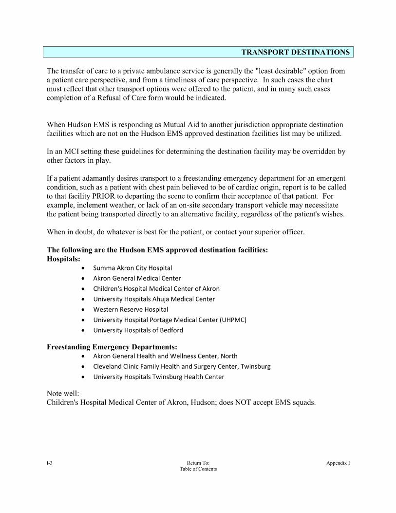
I-3 Return To: Appendix I Table of Contents
TRANSPORT DESTINATIONS The transfer of care to a private ambulance service is generally the "least desirable" option from a patient care perspective, and from a timeliness of care perspective. In such cases the chart must reflect that other transport options were offered to the patient, and in many such cases completion of a Refusal of Care form would be indicated. When Hudson EMS is responding as Mutual Aid to another jurisdiction appropriate destination facilities which are not on the Hudson EMS approved destination facilities list may be utilized. In an MCI setting these guidelines for determining the destination facility may be overridden by other factors in play. If a patient adamantly desires transport to a freestanding emergency department for an emergent condition, such as a patient with chest pain believed to be of cardiac origin, report is to be called to that facility PRIOR to departing the scene to confirm their acceptance of that patient. For example, inclement weather, or lack of an on-site secondary transport vehicle may necessitate the patient being transported directly to an alternative facility, regardless of the patient's wishes. When in doubt, do whatever is best for the patient, or contact your superior officer. The following are the Hudson EMS approved destination facilities: Hospitals:
Summa Akron City Hospital
Akron General Medical Center
Children's Hospital Medical Center of Akron
University Hospitals Ahuja Medical Center
Western Reserve Hospital
University Hospital Portage Medical Center (UHPMC)
University Hospitals of Bedford
Freestanding Emergency Departments: Akron General Health and Wellness Center, North
Cleveland Clinic Family Health and Surgery Center, Twinsburg
University Hospitals Twinsburg Health Center
Note well: Children's Hospital Medical Center of Akron, Hudson; does NOT accept EMS squads.
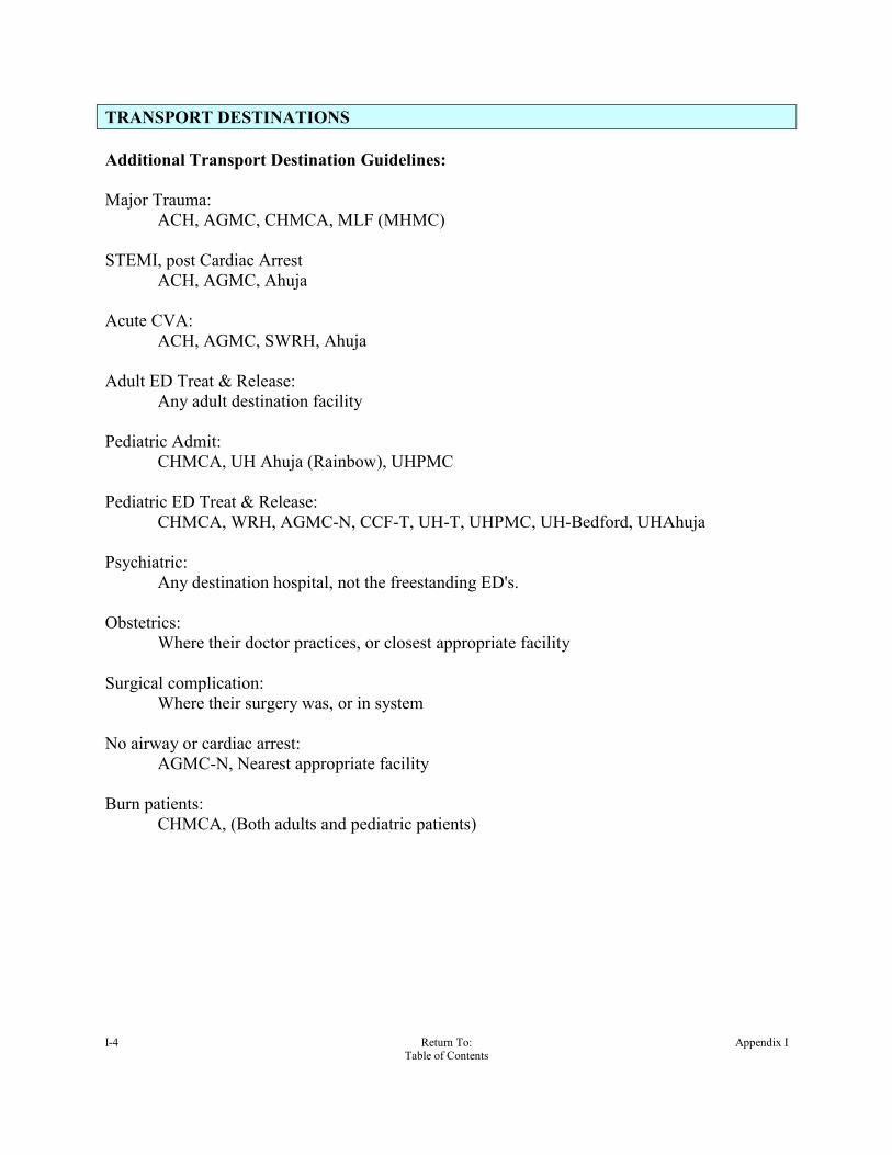
I-4 Return To: Appendix I Table of Contents
TRANSPORT DESTINATIONS Additional Transport Destination Guidelines: Major Trauma: ACH, AGMC, CHMCA, MLF (MHMC) STEMI, post Cardiac Arrest ACH, AGMC, Ahuja Acute CVA: ACH, AGMC, SWRH, Ahuja Adult ED Treat & Release: Any adult destination facility Pediatric Admit: CHMCA, UH Ahuja (Rainbow), UHPMC Pediatric ED Treat & Release: CHMCA, WRH, AGMC-N, CCF-T, UH-T, UHPMC, UH-Bedford, UHAhuja Psychiatric: Any destination hospital, not the freestanding ED's. Obstetrics: Where their doctor practices, or closest appropriate facility Surgical complication: Where their surgery was, or in system No airway or cardiac arrest: AGMC-N, Nearest appropriate facility Burn patients: CHMCA, (Both adults and pediatric patients)

CITY OF HUDSON EMERGENCY MEDICAL SERVICE
PROTOCOL
NOTES & ERRATA:
APPENDIX N Revision 40.6

HEMS Protocols
N-1 Return to: Appendix N Table of Contents
.
Related Documents