Adequacy of EMS systems of care protocols for adults with OHCA, STEMI & Stroke in Oregon: a structured review Paul S. Rostykus, MD, MPH Disclosure: The author is the medical director and supervising physician for the ambulance agencies in Jackson County. This project is supported by the Health Resources and Services Administration (HRSA) of the U.S. Department of Health and Human Services (HHS) under grant number H54RH00049, Rural Hospital Flexibility Program. This information or content and conclusions are those of the author and should not be construed as the official position or policy of, nor should any endorsements be inferred by HRSA, HHS or the U.S. Government.

Welcome message from author
This document is posted to help you gain knowledge. Please leave a comment to let me know what you think about it! Share it to your friends and learn new things together.
Transcript

Adequacy of EMS systems of care protocols for adults with OHCA, STEMI & Stroke in Oregon:
a structured review
Paul S. Rostykus, MD, MPH
Disclosure:
The author is the medical director and supervising physician for the ambulance agencies in Jackson County. This project is supported by the Health Resources and Services Administration (HRSA) of the U.S. Department of Health and Human Services (HHS) under grant number H54RH00049, Rural Hospital Flexibility Program. This information or content and conclusions are those of the author and should not be construed as the official position or policy of, nor should any endorsements be inferred by HRSA, HHS or the U.S. Government.

Adequacy of EMS Systems of Care protocols in Oregon 10/17/2016
2"
Title
Adequacy of EMS Systems of Care protocols for OHCA, STEMI & Stroke in Oregon: a structured review
Author
Paul Rostykus, MD, MPH1,2 1 Oregon Health & Science University, Portland, OR USA 2Jackson County EMS, Oregon, USA
Corresponding Author
Paul Rostykus, MD, MPH
436 Grandview Drive, Ashland, OR 97520
541-601-9709
Running Title
Adequacy of EMS Systems of Care protocols in Oregon
Key words
STEMI, Stroke, OHCA, Systems of Care, Emergency Medical Services (EMS), Variability, Clinical Practice Variations
Adequacy of EMS Systems of Care protocols in Oregon 10/17/2016
3"
Abstract Background
Out of hospital Cardiac Arrest (OHCA), ST-segment Elevation Myocardial Infarction (STEMI) and stroke
are 3 commonly encountered prehospital conditions for which systems of care have been developed to
improve adult patient outcomes. Several published evidence-based guidelines suggest best practices for
EMS treatment protocols for these conditions.
Objective
To examine the adequacy in Oregon ambulance protocols for OHCA, STEMI & stroke systems of care in
Oregon.
Methods
Oregon ambulance treatment protocols for OHCA, STEMI and stroke received were reviewed in a
structured fashion. Treatment protocols elements were abstracted and analyzed. Descriptive statistics and
chi-square were used to summarize the findings.
Results
Protocols were received from 95 Oregon ambulance agencies from 34 of Oregon’s 36 counties. There
were 31 different protocols used in the 60 Rural agencies and 9 different protocols used in the 35 Non-rural
agencies. At least 75% of the protocols were dated within the prior 4 years; more so in protocols from Non-
rural agencies compared to Rural agencies. Evidence-based guideline elements were mentioned or followed
variably ranging from 0-100% of the time. In general, elements from Non-rural agencies were mentioned or
specified more often than those from the Rural agencies. The most common elements missing from the
protocols were specific event times and tidal volumes for ventilations.
Conclusions
In Oregon, EMS protocols for OHCA, STEMI or stroke systems of care were quite varied. Of the
evidence-based guideline elements, two were found in all protocols for OHCA, four in all protocols for STEMI,
and one in all protocols for stroke. All the guideline recommendations for stroke were included in protocols
from 3 agencies; no protocols for OHCA or STEMI included all the recommended elements.

Adequacy of EMS Systems of Care protocols in Oregon 10/17/2016
4"
Introduction
The purpose of this study was to examine the adequacy of EMS protocols for adult patients with Out of
Hospital Cardiac Arrest (OHCA), STEMI (ST Elevation Myocardial Infarction) and stroke for ambulance
agencies throughout the State of Oregon. The study was to determine the number of EMS protocols from
rural ambulance agencies, which met all the evidence-based guidelines and which met selected evidence-
guided or best practices in each of these 3 systems of care. A tool kit of model protocols using evidence-
based best practices was developed for these three systems of care that might be beneficial across the state
(Appendix A).
Adequacy of EMS Systems of Care protocols in Oregon 10/17/2016
5"
Methods
Study Design
Cross-sectional review of current ambulance EMS protocols for adults for OHCA, STEMI and stroke
systems of care in the State of Oregon.
Study Population and Setting
EMS protocols were collected from Oregon ambulance agencies, which provided service in one or more
Ambulance Service Areas (ASAs). Copies of the EMS protocols were received from the EMS agency
or downloaded from the EMS agency website.
Variables and Data Collection
Protocols were reviewed by the author in a structured format using a standardized data collection tool for
abstracted data elements (Table 1, 2, 3), which had been piloted and refined after the first 10 protocols.
Evidence-guided elements or best practices (marked with an *) were defined as those listed in the
appropriate NASEMSO Model EMS Clinical Guidelines1 or American Heart Association (AHA)
Guidelines2,3,4,5,6,7,8. A few additional elements were added by the author based on his EMS medical
direction education, experience and clinical judgment.
Statistical Analysis
Descriptive statistics, chi-square and z-test were used to characterize findings.
Research ethics review
This study was reviewed and determined by the Oregon Health & Science University to be"research"not"
involving"human"subjects
Adequacy of EMS Systems of Care protocols in Oregon 10/17/2016
6"
Results
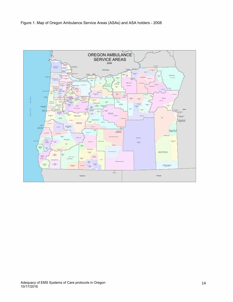
Ambulance service in Oregon is provided by a licensed ambulance provider in each county-managed
Ambulance Service Area (ASA) 9 (Fig. 1). Air ambulance service is not governed by this ASA process.
According to the State of Oregon EMS & Trauma Office there are 140 licensed ambulance agencies in
Oregon, of which 9 provide only air ambulance service and another 10 have a licensed ambulance, but are
not an ambulance provider in an ASA. EMS protocols for adults with OHCA, STEMI and stroke were received
from 95 of the 121 Oregon ground ambulance agencies, which provide service in one or more Oregon ASAs.
Of the 95 EMS agencies from which protocols were received, 60 (63%) were located in counties considered
to be Rural or Frontier by the Oregon Office of Rural Health and designated Rural for the purposes of this
study; the other 35 (37%) counties were designated Non-rural. There were 38 distinct sets of protocols
received, as several regions have developed protocols covering multiple agencies (range 1-10) in a region, a
county, part of a county, or group of counties. The 60 Rural counties had 31 separate protocol sets and the
35 Non-rural counties had 9 separate protocol sets.
Protocols were obtained from EMS agencies in 34 of the 36 Oregon counties (Fig. 2). The number of
protocols obtained per county ranged from 1-10. At least 75% of all the protocols were dated within the prior
4 years (Table 4). At least 97% of the protocols from the Non-rural agencies had an effective date within the
prior 4 years. A smaller percentage (77-84%) from Rural agencies had an effective date within the prior 4
years. Almost one tenth of the protocols received from the Rural EMS agencies did not list a date, compared
with none of those from Non-rural agencies.
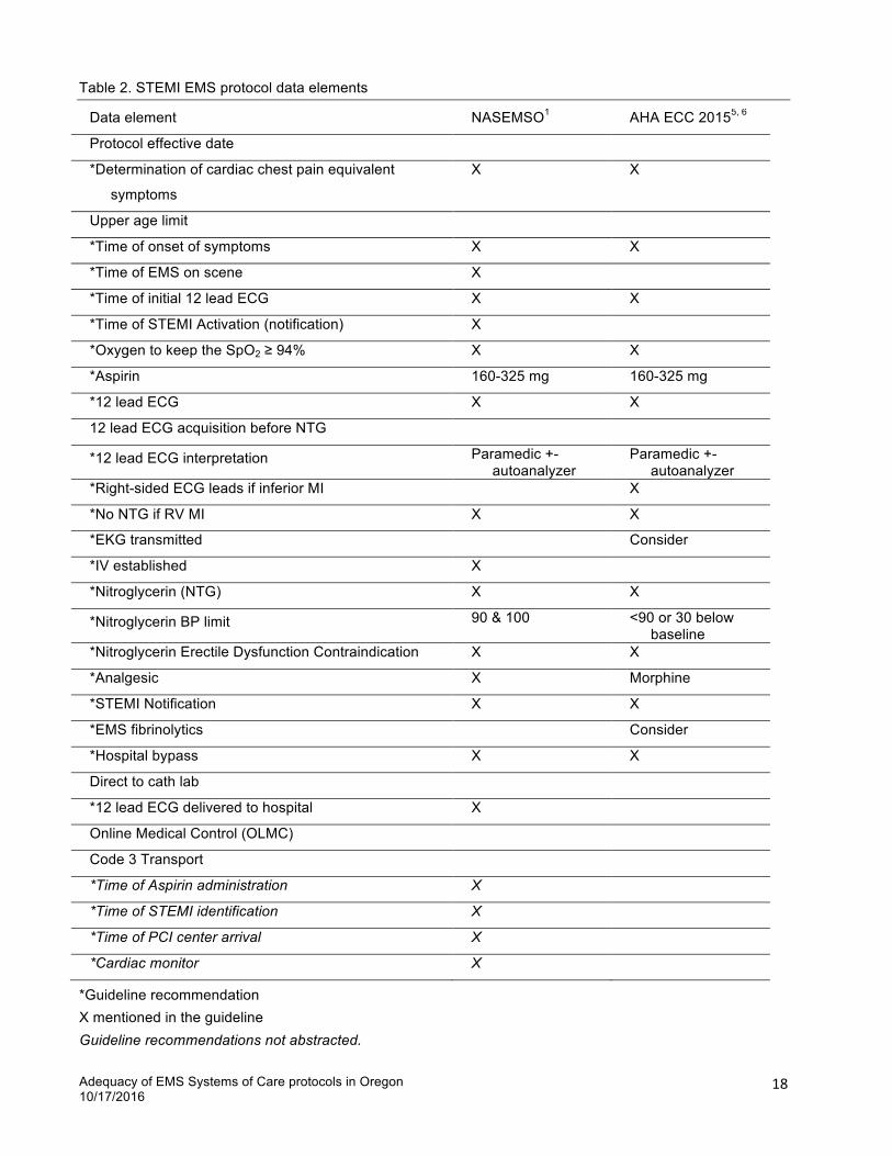
Data element presence ranged from 0% to 100% in protocols for OHCA (Tables 5 & 6), STEMI (Table 7)
and stroke (Table 8). All OHCA protocols mentioned initial vasopressor and initial advanced airway. All
STEMI protocols required a 12 lead ECG, noted the establishment of intravenous access, and mentioned the
administration of nitroglycerin (NTG) and an analgesic. All stroke protocols specified time of onset of
symptoms. None of the OHCA protocols required Code 3 (lights & siren) transport. None of the STEMI or
stroke protocols noted EMS fibrinolytic administration. None of the stroke protocols required the EMS provider
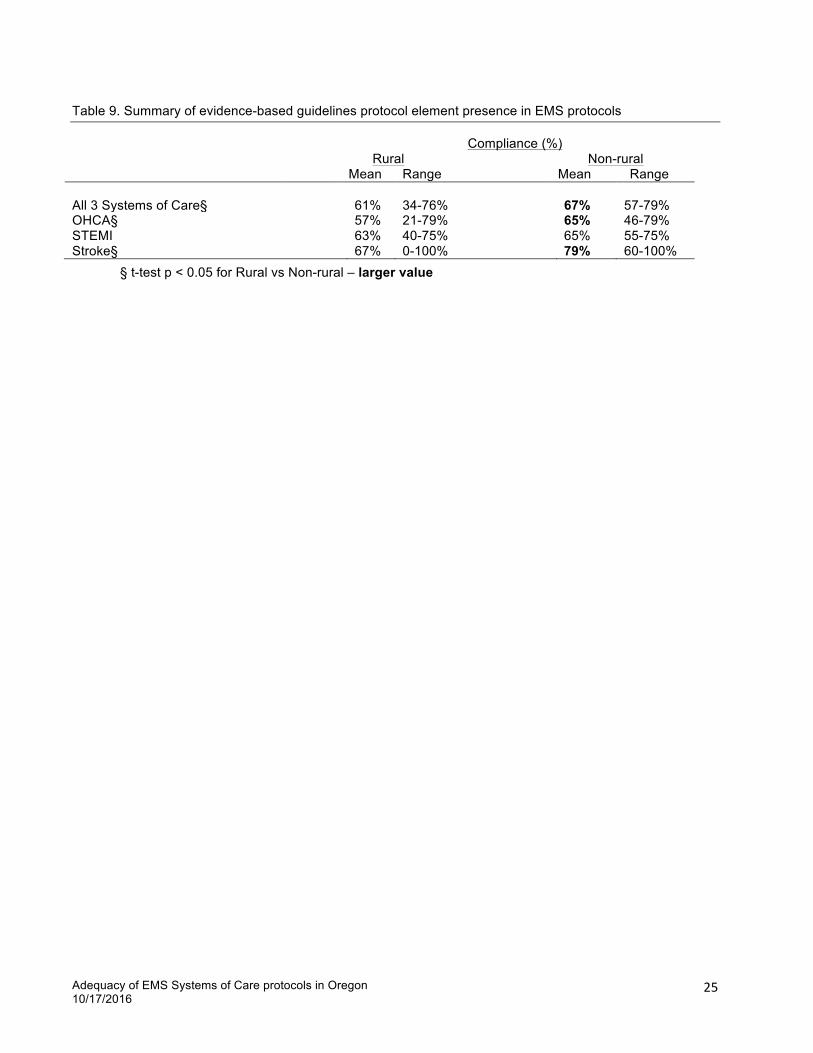
to obtain Online Medical Control (OLMC). Protocols from 3 agencies contained all the evidence-based
guideline elements for stroke (Table 9). Two of these were from Rural agencies using the same protocols
and one was a Non-rural agency. None of the protocols for OHCA or STEMI contained all the evidence-based

Adequacy of EMS Systems of Care protocols in Oregon 10/17/2016
7"
guideline elements. In general, the protocols from the Non-rural agencies were more likely to mention a data
element than were those from Rural agencies. Three Rural agencies supplied protocols for OHCA and
STEMI, but not for stroke. Variation was noted in individual data elements for protocols for all 3 systems of
care.
OHCA evidence-based guideline elements were noted quite variably ranging from initial vasopressor and
initial advanced airway in all protocols to Time CPR Started in less than 10%. Epinephrine was listed as the
initial vasopressor in 60% of the protocols and either epinephrine or vasopressin in 40%. A variety of initial
advanced airways were listed including endotracheal tube, supraglottic airway, King airway, LMA, pharyngeal
esophageal airway device, CombiTube, dual lumen airway device, blind insertion device and cuffed
pharyngeal airway device. The initial anti-arrhythmic listed was usually amiodarone (in 82% of the protocols)
and less likely lidocaine (in 12%) or either (in 4%). Early AED/defibrillator use was noted, often with the
caveat of after 1-2 minutes of CPR if the downtime was more than 5 minutes or if the arrest was unwitnessed
(Table 6). Defibrillation energy was variably noted as specific to one device or per manufacturer’s
recommendation. Monophasic defibrillation was mentioned infrequently. Most protocols listed escalating
defibrillation energies. Some protocols noted double sequential defibrillation for ventricular fibrillation
unresponsive to ongoing CPR, standard defibrillation, vasopressor and antiarrhythmic administration. Most of
the protocols specified a chest compression rate of 100 per minute or more as recommended in the
guidelines. Chest compression depth was usually listed as at least 2 inches or 2-2.4 inches, although one
protocol listed a depth of 1.5-2 inches. Ventilation rates were noted in more than half the protocols and split
between a specific rate and a ratio with chest compressions. Interrupted chest compressions were specified
slightly more often than continuous chest compressions. Defibrillator download for post-event review of
resuscitation was specified in only 4 protocols. Rapid determination of cardiac arrest was specified in less
than one third of the EMS protocols, although this is really a 911 dispatch process. Only about one in ten
protocols mentioned mechanical CPR.
If ROSC was obtained following OHCA, 12 lead ECG analysis was specified in about half the protocols.
Cooling by EMS with ice packs or iced saline IV was noted in about half the protocols, more often in Non-rural
protocols than in Rural protocols. Less than half the protocols noted hospital bypass for patients with ROSC.
A few protocols called for specific hospital ROSC notification with the term “Cooling Activation”.

Adequacy of EMS Systems of Care protocols in Oregon 10/17/2016
8"
For STEMI protocols, IV established was noted in all, including details such as saline lock unless IV fluid or
medication was needed, 2 or even 3 IVs if a STEMI was present, and to avoid the right wrist if possible.
Hospital bypass and notification were required in 77% or more, using terms including “STEMI Activation”,
“STEMI Alert”, “Cath Alert”, “STEMI patient”, “Code Heart”, Heart One” or “AMI”. Paramedic reading or auto-
analysis for 12 lead ECG interpretation was included in three-quarters of the protocols and paramedic reading
only in about 10%. All the protocols detailed nitroglycerin administration, with a minimum systolic blood
pressure limit of 100 mm Hg twice as often as 90 mm Hg. Likewise, analgesic administration was detailed in
all, usually with morphine, slightly more often than with fentanyl, and infrequently with nalbuphine, meperidine
or hydromorphone. About half the protocols noted 12 lead ECG transmission, often with the caveat of “if
available” and sometimes for “non-paramedic”. Less than half the protocols had an upper age limit for STEMI
activation, which included 85, 86 and 90 years. Specific event times were mentioned in less than half the
protocols. Code 3 transport for STEMI patients was required infrequently.
All of the EMS protocols for stroke specified a time of onset (hours since last seen normal) which ranged
from 2 to 6 hours, most commonly 3, 4.5 or 6 hours. Almost all (97% or more) specified hypoglycemia
treatment, establishment of an IV, use of a stroke evaluation method and blood glucose determination.
Elevation of the head of the stroke patient’s bed (ranging from 0-30°) during transport was noted in more than
half the protocols. About half the protocols noted hospital bypass, obtaining a 12 lead ECG and hospital
notification using the terms “Stroke Alert”, Stroke One”, “Stroke Activation” or ”Stroke Team Alert”. Code 3
transport for stroke patients was required in less than one third of the protocols.
During the review comments were noted in some protocols, which were not analyzed further (Table 10).
Adequacy of EMS Systems of Care protocols in Oregon 10/17/2016
9"
Discussion
Most of the Oregon EMS protocols received and reviewed noted many of the evidence-based guidelines or
best practices for OHCA, STEMI or stroke. The most common elements lacking from the protocols were
specific event times and tidal volumes for ventilations. Some of the guidelines are fairly explicit such as a
CPR chest compression rate of 100-120 per minute or a 12 lead ECG for all patients with a possible STEMI.
Other guidelines are less specific, allowing for variation depending on the local situation, such as interrupted
or continuous CPR chest compressions, the use of mechanical CPR, the aspirin dose for STEMI, or the time
threshold of 3, 4.5, or 6 hours for acute stroke intervention. Hospital bypass specifications were not usually
detailed in the protocols and may be very dependent on local and time-dependent conditions such as
geography, ambulance agency resources, weather, or available aircraft.
Oregon model guidelines were developed for each system of care: OHCA - High Performance CPR &
VF/VT, STEMI and stroke (Appendix A) based primarily on published guidelines and also on the author’s best
professional judgment. As noted above, the guidelines sometimes noted options and these items are shown
in italics in the model protocols. Some protocol items may be very system-specific and were not further
delineated, such as the number of IV sites for patients with STEMI or stroke or the specific destination
hospital for patients with STEMI or stroke. None of the protocols examined specified fibrinolytics for patients
with STEMI or stroke so this modality was omitted from the model protocols.
Although not a purpose of this project, during the protocol abstraction process a number of observations
were made about the protocols themselves. Many of the protocols appeared very similar to others,
suggesting that protocols may be shared amongst agencies, some of which are quite geographically distant
from each other. Protocols appeared to be organized in outline form, as flow charts, or, infrequently, as
narratives. Most of the protocols obtained were searchable: for .pdf files using Acrobat Reader (Adobe
Acrobat Reader DC, Version 2015.010.20059) or for .doc or .rtf files using WORD (Microsoft® Word for Mac
2011, Version 14.5.9), suggesting that they were generated directly from computer software. A few of the
protocols were not searchable with the software or had misaligned text or image artifacts suggesting hole
punches, indicating that these were paper versions which had subsequently been scanned before being
emailed to the investigator.
Adequacy of EMS Systems of Care protocols in Oregon 10/17/2016
10"
EMS protocols in the United States have been described as individual (as is the case in Oregon), regional,
model statewide or mandatory statewide10. Where generally agreed upon EMS guidelines exist, such as to a
large degree for OHCA, STEMI and stroke, an argument can be made that EMS protocols should be
established statewide or at least regionally, based upon the institution(s) likely to receive patients transported
by EMS. The capabilities of the receiving institutions may vary significantly. Especially in the more rural parts
of Oregon, options for long-distance air ambulance transport may be limited by weather conditions and the
available aircraft. The Oregon trauma system11 with its State Trauma Advisory Board (STAB) and Area
Trauma Advisory Boards (ATABs) could provide a model for the further development of these other time
sensitive systems of care.
EMS agency medical direction in Oregon is provided by physicians on a part-time basis in addition to their
other responsibilities, who provide EMS medical direction ranging from about half-time to a few hours per
month or even less. The larger number of older or undated protocols from Rural agencies suggests that
these EMS medical directors may have less time and energy to spend on EMS protocols. The creation and
review of EMS protocols may be an easier task in many ways when a number of medical directors work
together in a geographic region, as opposed to the very part-time medical director who works in a distant part
of the state and has limited contact with nearby peers.
The development of regional EMS protocols may enhance safety by reducing variability, which has been
associated with fewer errors12. Error reduction in medicine13 and in EMS14 in particular have been national
topics of concern for a number of years. Variability has been noted in other studies that have looked at
statewide EMS protocols9, as well as EMS protocols for naloxone administration15, pelvic binding16, blood
glucose measurement in seizure patients which was associated with delayed seizure treatment17, and
hypoglycemia treatment18. EMS providers may work in a number of different agencies or move to a different
area and encounter protocol differences that may not always be recognized in a timely fashion in the dynamic
prehospital work environment. If there is no physiologic or scientific basis for these differences, protocol
standardization may result in fewer errors and enhance patient safety. The frequently heard adage “If you’ve
seen one EMS system, then you’ve seen one EMS system” applies to EMS protocols as well. As the body of
prehospital and EMS knowledge and research expands, the spread of evidence-based guidelines in the

Adequacy of EMS Systems of Care protocols in Oregon 10/17/2016
11"
development of EMS protocols to improve patient care, as has been promulgated19, is worthy of further
consideration in Oregon.

Adequacy of EMS Systems of Care protocols in Oregon 10/17/2016
12"
Limitations
Limitations to this survey include missing protocols, the denominator chosen, the point-in-time survey, and
challenges of data abstraction.
EMS protocols were requested by the Oregon Health Authority’s EMS & Trauma Office from Oregon
ambulance agencies providing emergency patient transportation in one of the ASAs. Most were sent by the
EMS agency and some were found posted on the agency’s website. Protocols from some agencies were not
received despite multiple requests. The State of Oregon was not able to provide a list of ASAs and the
assigned ambulance agencies so it is possible that some ambulance agencies providing patient transport in
an ASA were missed or that a few non-ASA ambulance agencies were included, particularly if the ASA
assignee subcontracts with a different ambulance agency or if there is more than one ambulance provider in
an ASA. More than one protocol was received from some agencies, sometimes with different effective dates
suggesting the possibility of different protocol details. Some of the protocols were in the process of being
updated and may have changed between the time of review and the time this report was completed.
The denominator used in this study was ambulance agencies, not separate protocols. Non-rural agencies
had an average of 0.26 protocols/agency compared to 0.52 protocols/agency for Rural agencies, which is
significantly different (z test p = 0.0134). Use of agencies as the denominator emphasizes the effect of the
relatively large proportion of common protocols in Non-rural agencies compared to Rural agencies.
Protocols reviewed noted a variety of effective dates and some with no date at all. Some of the protocols
were received with a note that the protocols were in the process of being revised. Updated evidence-based
guidelines are published every few years as new science is learned and put into practice. Research and
updating of EMS protocols is a time-consuming process that EMS medical directors do periodically, every
year to few years, so that the findings of this study would likely be different from a similar review if done at a
future date.
Data elements were abstracted from EMS protocols to determine positive (Yes) answers or negative (Not
listed) answers. Negative answers could mean either that the element was not present or could not be
located. Some EMS agencies sent their entire set of protocols, others sent only a limited set. Most of the
data elements for STEMI were abstracted from cardiac chest pain or STEMI protocols, for stroke from stroke
Adequacy of EMS Systems of Care protocols in Oregon 10/17/2016
13"
or CVA protocols, and for OHCA from cardiac arrest or ventricular fibrillation protocols. Some elements were
not listed in any of these protocols and other protocols, if available, were searched for details of CPR,
termination of resuscitation (TOR), destination, morphine, nitroglycerin (NTG), oxygen, aspirin, magnesium,
airway management or universal patient care. Specific event time and ventilation tidal volume were the least
frequently note data elements which may have been listed in different places in the protocols or “assumed” to
be basic EMS provider care and not mentioned at all. Some of the guideline elements allowed for options or
were not always clearly specified so the author used his experience and professional judgment to interpret
some of the guideline elements or best practices. Other experienced reviewers might make slightly different
choices for some of these elements.
Finally, this study reviewed written EMS protocols, which may differ from what actually occurs during
prehospital care.
Conclusion
In Oregon, EMS adult protocols for OHCA, STEMI or stroke systems of care were quite varied. Two of
the evidence-based guideline elements were noted in all OHCA protocols, four in all STEMI protocols and one
in all stroke protocols. Three agencies, using 2 different sets of protocols, listed all the stroke evidence-based
guideline elements. Some elements were mentioned frequently and still others rather infrequently. Specific
event times and tidal volumes were detailed least often. Further study may help determine the minimum set
of evidence-based guideline elements, which should be included in each of the protocols for OHCA, STEMI
and stroke. Processes can then be developed to insure that all EMS protocols contain the essential elements
for each system of care.
Acknowledgements
Ken Kempner, PhD and Meredith Guardino for editorial review.
Adequacy of EMS Systems of Care protocols in Oregon 10/17/2016
14"
Figure 1. Map of Oregon Ambulance Service Areas (ASAs) and ASA holders - 2008
Idaho
Washington
California Nevada
HARNEY CO.ASA
WALLOWA ASA
VALEASA
BEND ASA
BAKER ASA
MERCY FLIGHTS
MADRASASA
ASA-4
LAKEVIEW DISASTER UNIT
TRI-CITYASA
BENTON ASA
HEPPNER ASA
DRAIN ASA
MITCHELLAMBULANCE
LA PINE ASA
SILVER LAKE EMS
CLACKAMAS ASA
NORTH LAKE COUNTY EMSROSEBURGASA
CONDONASA
SWEET HOME ASA
RAGER RESCUE
ASA-3
BLY ASA
MORO ASA
CHEMULT ASA
COASTAL ASA
SISTERSASA
PENDLETON ASA
CROOK COUNTYFIRE & RESCUE
SENECA ASA
DIAMOND LAKE
PAISLEY DISASTER UNIT
KENO FIRE
WASHINGTON CO.ASA
LA GRANDE ASA
NORTHJOSEPHINE CO.
ASA
CHILOQUINASA
MEDIC 400
MULTNOMAH ASA
Crescent Fire
ASHLAND FIRE &RESCUE
SUNRIVERASA
CENTRAL CURRY CO.
ASA
TRIBAL EMS
RICHLAND ASA
LEBANONASA
ASA 6
OAKRIDGE FIRE &AMBULANCE DEPT.
SOUTHJOSEPHINE CO.
ASA
ASA-2
MCMINNVILLEASA
ASA-5
SPRINGFIELD DEPT OFFIRE & LIFE SAFETY
FIREDISTRICT
#1
GLENDALE ASA
WARM SPRINGSASA
ARLINGTONASA
CALAPOOYA ASA
ASA 7
FOSSILASA
HARRMANFIRE
SOUTHERNCURRY CO.
ASA
WARM SPRINGSASALYONS ASA
HUNTINGTONASA
SPRY AMB
MOLALLAASA
EUGENEFIRE & EMS
WINSTONASA
PARKDALEEMS
REDMONDASA
UNION ASA
LONG CREEKASA
CAMPSHERMAN
ASA
ALBANYASA
MILTON-FREEWATERASA
SADDLEMOUNTAIN
ASA 4
NO COVERAGE
WEST VALLEYASA
BURNSASA
IDANHA/DETROIT
ASA 3
LYONSR.F.P.D.
NOCOVERAGE
RUFUS ASA
BASIN ASA
JORDAN VALLEYASA
ROME AROCK AMBULANCE/JORDAN VALLEY AMBULANCE
WESTERN LANEASA
PACWEST ASA
PRAIRIE CITYASA
MCDERMITT AMBULANCE(SERVICED BY HUMBOLDT
COUNTY, NV)
DAYVILLEASA
COTTAGE GROVEFIRE & ABMULANCE
DEPT.
NORTHERNCURRY CO.
ASA
TILLAMOOK ASA
BONANZAASA
MONUMENTASA
ELGINASA
CRATERLAKE NP
ROGUERIVERFIRE
BOARDMANASA
HERMISTONASA
MT VERNONASA
BAY CITIES ASA
JOHN DAYASA
NAVYASA
MYRTLEPOINT ASA
IDANHA/DETROITR.F.P.D.
PACIFIC CITY ASA
LA PINEFIRE
NEWBERGASA
ASA-6
ADRIAN ASA (SERVICEDBY TREASURE VALLEYPARAMEDIC - NYSSA)
ASA 5
LANE RURALFIRE & RESCUE
ASA
WOODBURNASA
SISTERSASA
SHERIDANASA
MANZANITA ASA
ASA 1PACWEST
ASA
COQUILLE ASA
POWERSASA
BAKERASA
ASA-1
HOODRIVER EMS
COLUMBIA RIVERFIRE & RESCUE ASA
HERMISTONASA
SANTIAMASA
SPRINGFIELDASA
CORVALLISASA
MIST-BIRKENFELDRFPD ASA
CANBYASA
CLATSKANIERFPD ASA
METROWEST ASA
BAY CITIES ASA
ASA 2
CENTRAL OREGONCOAST ASA
TREASURE VALLEYPARAMEDIC
SCAPPOOSERFPD ASA
NOCOVERAGE
SANTIAMMEMORIAL
HOSPITAL ASA
REDMONDASA
IRRIGON ASA
MARIONCO. FIRE
DISTRICT # 1
WEISER ANNEXASA
JEFFERSONASA
ST PAULRFPD
ASA-7
SALEMASA
HUNTINGTONASA
SOUTHLINCOLN
ASA
TURNERRFPD
MARIONCOUNTY IGA
GARIBALDI ASA
CASCADELOCKS EMS
EUGENE
NOCOVERAGE
SPRINGFIELDASA
LOWER UMPQUAHOSPITAL - EMS
UMATILLA ASA
NYSSA ASA (SERVICEDBY TREASURE VALLEYPARAMEDIC - NYSSA)
LOWER BRIDGE ASA
WALLA WALLAASA
KEIZERFD
PACWESTASA
NOCOVERAGE
NORTH LAKECOUNTY EMS -
HARNEY MUTUAL AID
CROOKED RIVERRANCH ASA
NORTHALBANY ASA
LINCOLN CITY ASA
NOCOVERAGE
BLACK BUTTERANCH ASA
WASHINGTONCO. IGA
MEDIX ASA
CORVALLIS FIRE &AMBULANCE DEPT.
POLK CO.FIRE DISTRICT #1
FIREDISTRICT
#4
AMR DISTRICT # 1
LakeLake
HarneyHarney
MalheurMalheur
LaneLane
GrantGrant
KlamathKlamath
DouglasDouglas
BakerBaker
LinnLinn
CrookCrook
UmatillaUmatil laWallowaWallowa
WascoWasco
UnionUnion
JacksonJackson
CoosCoos
CurryCurry
DeschutesDeschutes
MorrowMorrow
WheelerWheeler
JeffersonJefferson
MarionMarion
ClackamasClackamasGill iamGilliam
PolkPolk
JosephineJosephine
LincolnLincoln
ClatsopClatsop
TillamookTillamook
YamhillYamhill
ShermanSherman
WashingtonWashington
Hood RiverHood River
BentonBenton
ColumbiaColumbia
MultnomahMultnomah
OREGON AMBULANCESERVICE AREAS
2008
Pa
cif
ic
O
ce
an

Adequacy of EMS Systems of Care protocols in Oregon 10/17/2016
15"
Figure 2. Map of Oregon counties showing the number of EMS agencies from which protocols were received
No protocols received 1 EMS agency 2-5 EMS agencies 6-10 EMS agencies

Adequacy of EMS Systems of Care protocols in Oregon 10/17/2016
16"
Table 1. Out of Hospital Cardiac Arrest (OHCA) EMS protocol data elements
Data element NASEMSO1 AHA ECC 2015 BLS3
AHA ECC 2015 ALS4
Protocol effective date
*Rapid determination of cardiac arrest (Location,
Unconscious, Abnormal breathing, Initiate
CPR)
X
*Initial cardiac rhythm X
Time of cardiac arrest
*Time CPR started X
Time of ROSC
*Rate 100-120 100-120
*Depth ≥ 2 in 2-2.4 in
*Continuous vs interrupted 30:2 30:2
*Rotate compressors Every 1-2 minutes
Every 2 minutes
*Early AED/defibrillator use Early AED or 30
sec - 2 min CPR if unwitnessed
CPR with early AED use
*Defibrillation energy
Manufacturer’s recommendation or Maximum
Biphasic > monophasic
Manufacturer’s recommendation or Maximum
*IV or IO X Either
*Oxygen Highest flow possible
High flow
*Initial Vasopressor Epinephrine or Vasopressin
Epinephrine
*Subsequent Vasopressor Epinephrine Epinephrine
*Initial Antiarrythmic Amiodarone Amiodarone
*Subsequent Antiarrythmic Lidocaine Lidocaine
*Magnesium if Torsades Yes
*Tidal volume
Limited Chest rise – 500-600 ml = 6-7 ml/kg – avoid excessive
*Ventilation rate BVM 30:2 30:2
*Initial advanced airway Supraglottic No preference
No interruption *Back-up advanced airway Endotracheal

Adequacy of EMS Systems of Care protocols in Oregon 10/17/2016
17"
Data element NASEMSO1 AHA ECC 2015 BLS3
AHA ECC 2015 ALS4
*Ventilation rate airway Every 6-8 seconds
Every 6 seconds
*ETCO2 monitoring Yes X
*12 lead ECG if ROSC X X
*EMS Cooling Consider Not recommended
*Mechanical CPR Consider for transport
OHCA notification
*Hospital bypass Consider Consider
Online Medical Control (OLMC)
Code 3 Transport
*Termination of Resuscitation (TOR) protocol X X
Defibrillator download
Review of CPR Quality X
*Time of EMS on scene X
*Time of EMS to patient X
*Time of 1st shock X
*Bystander CPR performed? X
*Guideline recommendation X mentioned in the guideline Guideline recommendation not abstracted.
Adequacy of EMS Systems of Care protocols in Oregon 10/17/2016
18"
Table 2. STEMI EMS protocol data elements
Data element NASEMSO1 AHA ECC 20155, 6
Protocol effective date
*Determination of cardiac chest pain equivalent
symptoms
X X
Upper age limit
*Time of onset of symptoms X X
*Time of EMS on scene X
*Time of initial 12 lead ECG X X
*Time of STEMI Activation (notification) X
*Oxygen to keep the SpO2 ≥ 94% X X
*Aspirin 160-325 mg 160-325 mg
*12 lead ECG X X
12 lead ECG acquisition before NTG
*12 lead ECG interpretation Paramedic +- autoanalyzer
Paramedic +- autoanalyzer
*Right-sided ECG leads if inferior MI X
*No NTG if RV MI X X
*EKG transmitted Consider
*IV established X
*Nitroglycerin (NTG) X X
*Nitroglycerin BP limit 90 & 100 <90 or 30 below baseline
*Nitroglycerin Erectile Dysfunction Contraindication X X
*Analgesic X Morphine
*STEMI Notification X X
*EMS fibrinolytics Consider
*Hospital bypass X X
Direct to cath lab
*12 lead ECG delivered to hospital X
Online Medical Control (OLMC)
Code 3 Transport
*Time of Aspirin administration X
*Time of STEMI identification X
*Time of PCI center arrival X
*Cardiac monitor X
*Guideline recommendation X mentioned in the guideline Guideline recommendations not abstracted.

Adequacy of EMS Systems of Care protocols in Oregon 10/17/2016
19"
Table 3. Stroke EMS protocol data elements
Data element NASEMSO1 20107 20138
Protocol effective date
*Time of onset of symptoms (last seen normal) X 3-4.5
med/6 for cath
X 3-4.5 med/6 for cath
*Stroke evaluation method Specific
validated stroke score
X Prehospital stroke assessment tool
*Blood glucose determination (CBG) X X X
*Hypoglycemia treatment CBG < 60 CBG < 60
*Oxygen to keep the SpO2 ≥ 94% X X
*IV established Avoid multiple
X
*12 lead ECG If possible
EMS Fibrinolytics
*Stroke Notification X X
*Hospital bypass
Nearest stroke center or stroke capable facility
X X
Online Medical Control (OLMC)
*Head of bed elevated 15-30 if BP > 100
Code 3 Transport
*Stroke checklist X
*Do not treat hypertension X
*Cardiac monitor X X
*Guideline recommendation X mentioned in the guideline Guideline recommendations not abstracted.

Adequacy of EMS Systems of Care protocols in Oregon 10/17/2016
20"
Table 4. Protocol Effective Date
Rural Non-rural N (%) N (%) OHCA Protocol Effective Date § Total = 60 Total = 35 Within 1 year 30 (50%) 28 (80%) Within 2 years 39 (65%) 35 (100%) Within 4 years 46 (77%) 35 (100%) 5 or more years 6 (10%) 0 (0%) Undated 8 (13%) 0 (0%) STEMI Protocol Effective Date Total = 60 Total = 35 Within 1 year 34 (57%) 28 (80%) Within 2 years 44 (73%) 34 (97%) Within 4 years 49 (82%) 34 (97%) 5 or more years 5 (8%) 1 (3%) Undated 6 (10%) 0 (0%) Stroke Protocol Effective Date Total = 57 Total = 35 Within 1 year 32 (56%) 28 (80%) Within 2 years 40 (70%) 34 (97%) Within 4 years 48 (84%) 34 (97%) 5 or more years 4 (7%) 1 (3%) Undated 5 (9%) 0 (0%) § Chi-square p-value < 0.05 for Rural vs Non-rural

Adequacy of EMS Systems of Care protocols in Oregon 10/17/2016
21"
Table 5. OHCA data elements present in protocols from EMS agencies Rural Non-rural N (%) N (%)
(listed by decreasing sum of N Rural + N Non-rural)
*Initial vasopressor 60 (100%) 35 (100%) *Initial vasopressor 60 (100%) 35 (100%) *Initial advanced airway 60 (100%) 35 (100%) *Initial anti-arrhythmic 58 (97%) 35 (100%) *Early AED/defibrillator use (detailed in Table 3) 56 (93%) 35 (100%) *Defibrillation energy 56 (93%) 35 (100%) *Magnesium if Torsades § 52 (87%) 35 (100%) *Chest compression rate § (detailed in Table 3) 51 (85%) 34 (97%) *IV or IO 55 (92%) 28 (80%) *Chest compression type (detailed in Table 3) 49 (82%) 28 (80%) *High flow oxygen § 51 (85%) 22 (63%) *Termination of Resuscitation (TOR) protocol § 39 (65%) 32 (91%) *ETCO2 monitoring § 37 (62%) 34 (97%) *Ventilation rate - airway 40 (67%) 28 (80%) *Chest compression depth § 34 (57%) 28 (80%) *Ventilation rate - BVM (detailed in Table 3) § 33 (55%) 28 (80%) *Subsequent anti-arrhythmic 37 (62%) 18 (51%) *12 lead ECG if ROSC § 25 (42%) 24 (69%) *EMS Cooling 26 (43%) 20 (57%) *Back-up advanced airway 27 (45%) 10 (29%) *Rotate compressors 20 (33%) 16 (46%) *Initial cardiac rhythm 18 (30%) 17 (49%) Time of cardiac arrest 20 (33%) 14 (40%) *Subsequent vasopressor 19 (32%) 15 (43%) *Hospital bypass specified 19 (32%) 15 (43%) *Rapid determination of cardiac arrest 10 (17%) 10 (29%) *Tidal volume 7 (12%) 5 (14%) *Mechanical CPR 6 (10%) 5 (14%) Specific OHCA notification 4 (7%) 3 (9%) *Time CPR started 3 (5%) 4 (11%) Time of ROSC 3 (5%) 3 (9%) Defibrillator download 4 (7%) 0 (0%) Online Medical Control (OLMC) required 1 (2%) 0 (0%) Code 3 Transport required 0 (0%) 0 (0%)
*Guideline recommendation1,2,3,4 § Chi-square p-value < 0.05 for Rural vs Non-rural – larger value
Adequacy of EMS Systems of Care protocols in Oregon 10/17/2016
22"
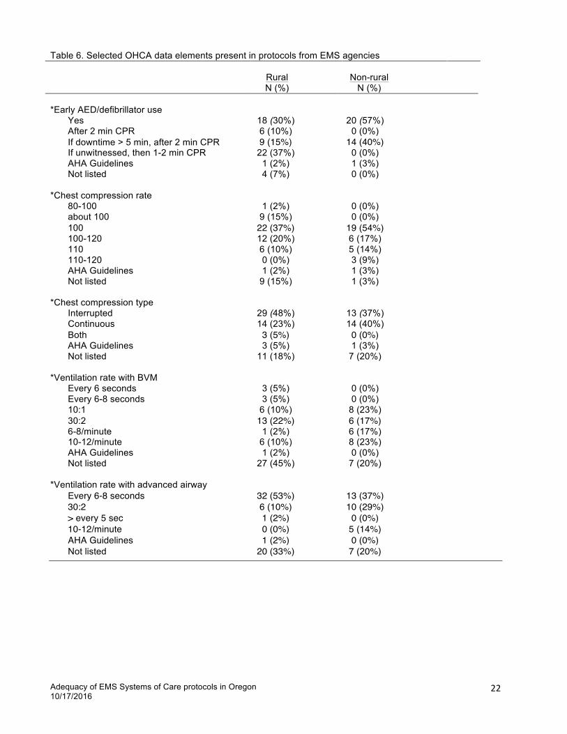
Table 6. Selected OHCA data elements present in protocols from EMS agencies Rural Non-rural N (%) N (%)
*Early AED/defibrillator use
Yes 18 (30%) 20 (57%) After 2 min CPR 6 (10%) 0 (0%) If downtime > 5 min, after 2 min CPR 9 (15%) 14 (40%) If unwitnessed, then 1-2 min CPR 22 (37%) 0 (0%) AHA Guidelines 1 (2%) 1 (3%) Not listed 4 (7%) 0 (0%)
*Chest compression rate
80-100 1 (2%) 0 (0%) about 100 9 (15%) 0 (0%) 100 22 (37%) 19 (54%) 100-120 12 (20%) 6 (17%) 110 6 (10%) 5 (14%) 110-120 0 (0%) 3 (9%) AHA Guidelines 1 (2%) 1 (3%) Not listed 9 (15%) 1 (3%)
*Chest compression type
Interrupted 29 (48%) 13 (37%) Continuous 14 (23%) 14 (40%) Both 3 (5%) 0 (0%) AHA Guidelines 3 (5%) 1 (3%) Not listed 11 (18%) 7 (20%)
*Ventilation rate with BVM
Every 6 seconds 3 (5%) 0 (0%) Every 6-8 seconds 3 (5%) 0 (0%) 10:1 6 (10%) 8 (23%) 30:2 13 (22%) 6 (17%) 6-8/minute 1 (2%) 6 (17%) 10-12/minute 6 (10%) 8 (23%) AHA Guidelines 1 (2%) 0 (0%) Not listed 27 (45%) 7 (20%)
*Ventilation rate with advanced airway
Every 6-8 seconds 32 (53%) 13 (37%) 30:2 6 (10%) 10 (29%) > every 5 sec 1 (2%) 0 (0%) 10-12/minute 0 (0%) 5 (14%) AHA Guidelines 1 (2%) 0 (0%) Not listed 20 (33%) 7 (20%)

Adequacy of EMS Systems of Care protocols in Oregon 10/17/2016
23"
Table 7. STEMI data elements present in protocols from EMS agencies Rural Non-rural N (%) N (%)
(listed by decreasing sum of N Rural + N Non-rural)
*12 lead ECG required 60 (100%) 35 (100%) *IV established 60 (100%) 35 (100%) *Nitroglycerin (NTG) administration 60 (100%) 35 (100%) *Analgesic administration 60 (100%) 35 (100%) *Aspirin administration (detailed below) 59 (98%) 35 (100%) *Nitroglycerin BP limit 56 (93%) 35 (100%) *12 lead ECG interpretation 54 (90%) 35 (100%) *Nitroglycerin Erectile Dysfunction contraindication 54 (90%) 35 (100%) *Hospital bypass specified § 46 (77%) 34 (97%) *STEMI notification required 47 (78%) 31 (89%) *Determination of cardiac chest pain equivalent § 51 (85%) 23 (66%) *EKG transmitted 39 (65%) 18 (51%) *Oxygen to keep the SpO2 ≥ 94% 32 (53%) 20 (57%) *12 lead ECG delivered to hospital 28 (47%) 23 (66%) Upper age limit 25 (42%) 8 (23%) *Time of onset of symptoms 25 (42%) 8 (23%) *No NTG if RV MI 16 (27%) 8 (23%) 12 lead ECG acquisition before NTG § 19 (32%) 4 (11%) *Right-sided ECG leads if inferior MI § 8 (13%) 0 (0%) Code 3 Transport required § 1 (2%) 6 (17%) Online Medical Control (OLMC) required 4 (7%) 0 (0%) *Time of initial 12 lead 1 (2%) 3 (9%) *Time of EMS on scene § 0 (0%) 3 (9%) Direct transfer to cath lab § 0 (0%) 3 (9%) *Time of STEMI notification 0 (0%) 1 (3%) *EMS fibrinolytics administration 0 (0%) 0 (0%)
§ Chi-square p-value < 0.05 for Rural vs Non-rural – larger value
*Aspirin administration 162 mg 11 (18%) 3 (9%) 162-324 mg 10 (17%) 4 (11%) 243-324 mg 0 (0%) 1 (3%) 324 mg 35 (58%) 27 (77%) Amount not specified 3 (5%) 0 (0%) Not listed 1 (2%) 0 (0%)
*Guideline recommendation1,5,6

Adequacy of EMS Systems of Care protocols in Oregon 10/17/2016
24"
Table 8. Stroke data elements present in protocols from EMS agencies Rural Non-rural N (%) N (%)
(listed by decreasing sum of N Rural + N Non-rural)
*Time of onset of symptoms (detailed below) 57 (100%) 35 (100%) *Hypoglycemia treatment 57 (100%) 34 (97%) *IV established 55 (97%) 35 (100%) *Stroke evaluation method (detailed below) § 52 (91%) 35 (100%) *Blood glucose determination (CBG) § 55 (97%) 27 (77%) *Head of bed elevated § 26 (46%) 32 (91%) *Stroke notification required 28 (49%) 20 (57%) *12 lead ECG 24 (42%) 19 (54%) *Hospital bypass specified 24 (42%) 18 (51%) *Oxygen to keep the SpO2 ≥ 94% 21 (37%) 20 (57%) Code 3 Transport 6 (11%) 11 (31%) *EMS Fibrinolytics 0 (0%) 0 (0%) Online Medical Control (OLMC) required 0 (0%) 0 (0%)
§ Chi-square p-value < 0.05 for Rural vs Non-rural – larger value *Time of onset of symptoms (hours since last seen normal)
2 4 (7%) 0 (0%) 2-3 1 (2%) 0 (0%) 3 17 (30%) 6 (17%) 3.5 2 (4%) 1 (3%) 3.5/6 6 (11%) 4 (11%) 3 if thrombolytic /6 if intravascular intervention 1 (2%) 0 (0%) 4 1 (2%) 6 (17%) 4.5 9 (16%) 9 (26%) 5 0 (0%) 5 (14%) 6 12 (21%) 3 (9%) Yes 4 (7%) 1 (3%) Not listed 3 (5%) 0 (0%)
*Stroke evaluation method §
Cincinnati Prehospital Stroke Scale 16 (28%) 21 (60%) Cincinnati Prehospital Stroke Scale &/or Los
Angeles Prehospital Stroke Screen 1 (2%) 0 (0%) F.A.S.T. and Cincinnati Prehospital Stroke Scale 1 (2%) 0 (0%) FAST 15 (26%) 5 (14%) Facial droop. Arm drift. Abnormal speech with “You
can’t teach an old dog new tricks”. 3 (5%) 0 (0%) H&P 8 (14%) 0 (0%) Modified Los Angeles Prehospital Stroke Screen 6 (11%) 9 (26%) Not listed 8 (14%) 0 (0%)
*Guideline recommendation1,7,8
Adequacy of EMS Systems of Care protocols in Oregon 10/17/2016
25"
Table 9. Summary of evidence-based guidelines protocol element presence in EMS protocols
Compliance (%) Rural Non-rural Mean Range Mean Range All 3 Systems of Care§ 61% 34-76% 67% 57-79% OHCA§ 57% 21-79% 65% 46-79% STEMI 63% 40-75% 65% 55-75% Stroke§ 67% 0-100% 79% 60-100%
§ t-test p < 0.05 for Rural vs Non-rural – larger value

Adequacy of EMS Systems of Care protocols in Oregon 10/17/2016
26"
Table 10. Summary of evidence-based guidelines protocol element presence in EMS protocols
General comments
Some protocols did not contain a physician (EMS medical director) signature.
A few “protocols” were exceptions to a multi-agency protocol.
OHCA comments
Metronome or CPR feedback device whenever possible.
Passive oxygenation via nasal cannula until 3rd EMS provider arrives.
Central Line access is preferred over IO placement in Cardiac Arrest.
Interrupt compressions only per AED/Airway procedures.
Do not transport until ROSC.
STEMI comments
Administration of clopidogrel, heparin or metoprolol and blood draw for labs
Labetalol for hypertension
Lidocaine for PVC if ischemia
Do not delay administration of aspirin to obtain 12 lead.
ECG before ASA, NTG, morphine.
Long distance to cath lab or other hospitals.
Stroke comments
Patient is not a candidate for stroke therapy if they have a valid POLST with DNR or Comfort
Measures Only
POLST "Comfort Measures Only" do not get Stroke Activation.
Must be less than 80 years old.
Thiamine if alcoholism.
Labetolol if BP > 190/110.
NTG if DBP > 135.

Adequacy of EMS Systems of Care protocols in Oregon 10/17/2016
27"
References
"""""""""""""""""""""""""""""""""""""""""""""""""""""""""""""1http://www.nasemso.org/Projects/ModelEMSClinicalGuidelines/documents/National-Model-EMS-Clinical-
Guidelines-23Oct2014.pdf. Accessed March 21, 2015. 2 Neumar RW, Shuster M, Callaway CW, Gent LM, Atkins DL, Bhanji F, Brooks SC, de Caen AR, Donnino
MW, Ferrer JM, Kleinman ME, Kronick SL, Lavonas EJ, Link MS, Mancini ME, Morrison LJ, O'Connor RE, Samson RA, Schexnayder SM, Singletary EM, Sinz EH, Travers AH, Wyckoff MH, Hazinski MF. Part 1: Executive Summary: 2015 American Heart Association Guidelines Update for Cardiopulmonary Resuscitation and Emergency Cardiovascular Care. Circulation. 2015 Nov 3;132(18 Suppl 2):S315-67.
3 Kleinman ME, Brennan EE, Goldberger ZD, Swor RA, Terry M, Bobrow BJ, Gazmuri RJ, Travers AH, Rea T. Part 5: Adult Basic Life Support and Cardiopulmonary Resuscitation Quality: 2015 American Heart Association Guidelines Update for Cardiopulmonary Resuscitation and Emergency Cardiovascular Care. Circulation. 2015 Nov 3;132(18 Suppl 2):S414-35.
4 Link MS, Berkow LC, Kudenchuk PJ, Halperin HR, Hess EP, Moitra VK, Neumar RW, O'Neil BJ, Paxton JH, Silvers SM, White RD, Yannopoulos D, Donnino MW. Part 7: Adult Advanced Cardiovascular Life Support: 2015 American Heart Association Guidelines Update for Cardiopulmonary Resuscitation and Emergency Cardiovascular Care. Circulation. 2015 Nov 3;132(18 Suppl 2):S444-64.
5 O’Gara PT, et al. 2013 ACCF/AHA Guideline for the management of ST-elevation myocardial infarction - A report of the American College of Cardiology Foundation/American Heart Association Task Force on practice guidelines. J Amer Coll Cardiol 2013;61:e78-e140.
6 Part 9: Acute Coronary Syndromes: 2015 American Heart Association O'Connor RE, Al Ali AS, Brady WJ, Ghaemmaghami CA, Menon V, Welsford M, Shuster M. Guidelines Update for Cardiopulmonary Resuscitation and Emergency Cardiovascular Care. Circulation. 2015 Nov 3;132(18 Suppl 2):S483-500.
7 Field JM, Hazinski MF, Sayre MR, Chameides L, Schexnayder SM, Hemphill R, Samson RA, Kattwinkel J, Berg RA, Bhanji F, Cave DM, Jauch EC, Kudenchuk PJ, Neumar RW, Peberdy MA, Perlman JM, Sinz E, Travers AH, Berg MD, Billi JE, Eigel B, Hickey RW, Kleinman ME, Link MS, Morrison LJ, O’Connor RE, Shuster M, Callaway CW, Cucchiara B, Ferguson JD, Rea TD, Vanden Hoek TL. Part 1: executive summary: 2010 American Heart Association Guidelines for Cardiopulmonary Resuscitation and Emergency Cardiovascular Care. Circulation. 2010;122(suppl 3):S640–S656.
8 Jauch EC, Saver JL, Adams HP Jr, Bruno A, Connors JJ, Demaerschalk BM, Khatri P, McMullan PW Jr, Qureshi AI, Rosenfield K, Scott PA, Summers DR, Wang DZ, Wintermark M, Yonas H; American Heart Association Stroke Council; Council on Cardiovascular Nursing; Council on Peripheral Vascular Disease; Council on Clinical Cardiology. Guidelines for the early management of patients with acute ischemic stroke: a guideline for healthcare professionals from the American Heart Association/American Stroke Association. Stroke. 2013;44:870–947.
9 OAR 333-260-0010 (3) 10 Kupas DF, Schenk E, Sholl JM, Kamin R. Characteristics of statewide protocols for emergency medical
services in the United States. Prehospital Emergency Care 2015;19(2):292-301. 11"OAR 333-200"12"Hinckley CM. Error management: Make no mistake—errors can be controlled. Qual Saf Health Care
2003;12:359-365."13 Kohn LT, Corrigan JM, Donaldson MS. To Err Is Human: Building a Safer Health System. Washington,
DC: Committee on Quality of Health Care in America, Institute of Medicine, 1999.

Adequacy of EMS Systems of Care protocols in Oregon 10/17/2016
28"
""""""""""""""""""""""""""""""""""""""""""""""""""""""""""""""""""""""""""""""""""""""""""""""""""""""""""""""""""""""""""""""""""""""""""""""""""""""""""""""""""""""""""""""""""""""""""""""""""""""""""""14 Bigham BL, Buick JE, Brooks SC, Morrison M, Shojania KG, Morrison LJ. Patient safety in emergency
medical services: a systematic review of the literature. Prehospital Emergency Care 2012;16:1–20. 15 Corey SD, Southwell, JK, Niehaus, VR, Walley, AY, Dailey, MW. Emergency Medical Services Naloxone
Access: A National Systematic Legal Review. Academic Emergency Medicine, 2014;21:1173–1177. 16 Yamaguchi Y, Zhu Z, Hodroj M, Daya M. Prehospital Pelvic Binding for Suspected Pelvic Injury: A
Nationwide Survey [abstract]. Prehospital Emergency Care 2015;19(1):146. 17 Beskind DL, Rhodes SM, Stolz U, Birrer B, Mayfield TR, Bourn S, Denninghoff K. When Should You Test
for and Treat Hypoglycemia in Prehospital Seizure Patients? Prehospital Emergency Care 2014;18(3) 433-441
18 Rostykus P, Kennel J, Adair K, Fillinger M, Palmberg R, Quinn A, Ripley J, Daya M. Variability in the Treatment of Prehospital Hypoglycemia: A Structured Review of EMS Protocols in the United States. Prehosp Emerg Care. Advance online publication. doi:10.3109/10903127.2015.1128031.
19 Martin-Gill C, Gaither JB, Bigham BL, Myers JB, Kupas DF, Spaite DW. National Prehospital Evidence-Based Guidelines Strategy: A Summary for EMS Stakeholders. Prehosp Emerg Care 2016;20(2):175-83. doi: 10.3109/10903127.2015.1102995. Epub 2016 Jan 25.
!
May 28, 2016 Model Protocols !
Appendix A – Model Protocols
High Performance CPR - Adult
Ventricular Fibrillation/Pulseless Ventricular Tachycardia (VF/VT)
Cardiac Chest Pain & STEMI
Stroke!
!!! !
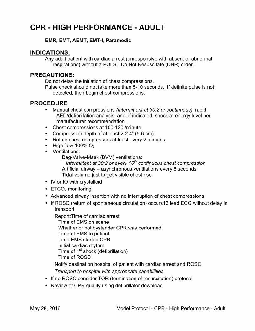
May 28, 2016 Model Protocol - CPR - High Performance - Adult
CPR - HIGH PERFORMANCE - ADULT EMR, EMT, AEMT, EMT-I, Paramedic
INDICATIONS: Any adult patient with cardiac arrest (unresponsive with absent or abnormal
respirations) without a POLST Do Not Resuscitate (DNR) order.
PRECAUTIONS: Do not delay the initiation of chest compressions. Pulse check should not take more than 5-10 seconds. If definite pulse is not
detected, then begin chest compressions.
PROCEDURE • Manual chest compressions (intermittent at 30:2 or continuous), rapid
AED/defibrillation analysis, and, if indicated, shock at energy level per manufacturer recommendation
• Chest compressions at 100-120 /minute • Compression depth of at least 2-2.4” (5-6 cm) • Rotate chest compressors at least every 2 minutes • High flow 100% O2 • Ventilations:
Bag-Valve-Mask (BVM) ventilations: Intermittent at 30:2 or every 10th continuous chest compression
Artificial airway – asynchronous ventilations every 6 seconds Tidal volume just to get visible chest rise
• IV or IO with crystalloid • ETCO2 monitoring • Advanced airway insertion with no interruption of chest compressions • If ROSC (return of spontaneous circulation) occurs12 lead ECG without delay in
transport Report:Time of cardiac arrest
Time of EMS on scene Whether or not bystander CPR was performed Time of EMS to patient Time EMS started CPR Initial cardiac rhythm Time of 1st shock (defibrillation) Time of ROSC
Notify destination hospital of patient with cardiac arrest and ROSC Transport to hospital with appropriate capabilities
• If no ROSC consider TOR (termination of resuscitation) protocol • Review of CPR quality using defibrillator download
May 28, 2016 Model Protocol - Ventricular Fibrillation/Pulseless Tachycardia
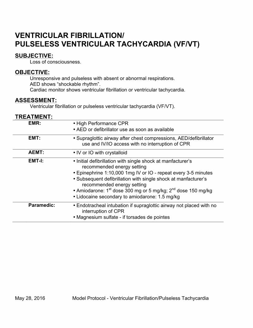
VENTRICULAR FIBRILLATION/ PULSELESS VENTRICULAR TACHYCARDIA (VF/VT) SUBJECTIVE:
Loss of consciousness.
OBJECTIVE: Unresponsive and pulseless with absent or abnormal respirations. AED shows “shockable rhythm”. Cardiac monitor shows ventricular fibrillation or ventricular tachycardia.
ASSESSMENT: Ventricular fibrillation or pulseless ventricular tachycardia (VF/VT).
TREATMENT: EMR: • High Performance CPR
• AED or defibrillator use as soon as available
EMT: • Supraglottic airway after chest compressions, AED/defibrillator use and IV/IO access with no interruption of CPR
AEMT: • IV or IO with crystalloid EMT-I: • Initial defibrillation with single shock at manfacturer’s
recommended energy setting • Epinephrine 1:10,000 1mg IV or IO - repeat every 3-5 minutes • Subsequent defibrillation with single shock at manfacturer’s
recommended energy setting • Amiodarone: 1st dose 300 mg or 5 mg/kg; 2nd dose 150 mg/kg • Lidocaine secondary to amiodarone: 1.5 mg/kg
Paramedic: • Endotracheal intubation if supraglottic airway not placed with no interruption of CPR
• Magnesium sulfate - if torsades de pointes

May 28, 2016 Model Protocol - Cardiac Chest Pain & STEMI
CARDIAC CHEST PAIN
SUBJECTIVE: Chest or epigastric discomfort lasting minutes to hours – not usually seconds or days
Discomfort may originate, be limited to, or may radiate to neck, jaw, shoulder, inner arm or elbow
May be associated with diaphoresis, nausea, vomiting, SOB , weakness, lightheadedness or palpitations.
May be brought on by exertion, stress or occur spontaneously. Relieved by rest or nitroglycerine. May have PMH of bypass surgery, angioplasty, angina, heart attack or myocardial
infarction. Atypical presentations are common and may include no discomfort.
OBJECTIVE: Examination may be normal. Patient may appear ashen or sweaty. Patient may be
hypotensive, bradycardic or have evidence of pulmonary edema (rales). Cardiac rhythm is monitored to detect the occurrence of ventricular or atrial dysrhythmias.
ASSESSMENT: Diagnosis of cardiac chest pain or heart equivalent discomfort is made on the basis of the
patient’s history. Other causes of chest discomfort include chest wall trauma, esophageal reflux, gastritis, peptic ulcer disease, pneumonia, pericarditis, pleurisy, pancreatitis, costochondritis, gall bladder disease, aortic dissection, aortic aneurysm, pulmonary embolism and anxiety.
TREATMENT:
EMR: • 12 lead ECG • Aspirin - 160-325 mg chewed – give even if taking other
anticoagulant or “blood thinner” medications • Oxygen only to maintain SpO2 = 94% or above
EMT:
• May assist with self-administration of patient’s own nitroglycerin
AEMT: • Nitroglycerin - if systolic BP > (90-100) mm Hg and no recent erectile dysfunction medication
• IV (20 or 18 gauge preferred) with saline lock unless crystalloid or medications indicated
EMT-I: • Cardiac monitor • Morphine or Fentanyl
Paramedic: • STEMI protocol - next page
May 28, 2016 Model Protocol - Cardiac Chest Pain & STEMI
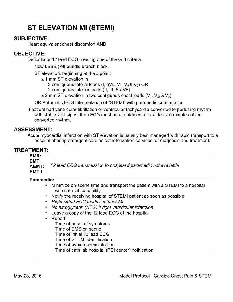
ST ELEVATION MI (STEMI) SUBJECTIVE:
Heart equivalent chest discomfort AND
OBJECTIVE: Defibrillator 12 lead ECG meeting one of these 3 criteria:
New LBBB (left bundle branch block,
ST elevation, beginning at the J point: ≥ 1 mm ST elevation in
2 contiguous lateral leads (I, aVL, V4, V5 & V6) OR 2 contiguous inferior leads (II, III, & aVF)
≥ 2 mm ST elevation in two contiguous chest leads (V1, V2, & V3)
OR Automatic ECG interpretation of “STEMI” with paramedic confirmation
If patient had ventricular fibrillation or ventricular tachycardia converted to perfusing rhythm with stable vital signs, then ECG must be at obtained after at least 5 minutes of the converted rhythm.
ASSESSMENT: Acute myocardial infarction with ST elevation is usually best managed with rapid transport to a
hospital offering emergent cardiac catheterization services for diagnosis and treatment.
TREATMENT:
EMR: EMT: AEMT: EMT-I
12 lead ECG transmission to hospital if paramedic not available
Paramedic: • Minimize on-scene time and transport the patient with a STEMI to a hospital
with cath lab capability. • Notify the receiving hospital of STEMI patient as soon as possible • Right-sided ECG leads if inferior MI • No nitroglycerin (NTG) if right ventricular infarction • Leave a copy of the 12 lead ECG at the hospital • Report:
Time of onset of symptoms Time of EMS on scene Time of initial 12 lead ECG Time of STEMI identification Time of aspirin administration Time of cath lab hospital (PCI center) notification
May 28, 2016 Model Protocol - Stroke - CVA
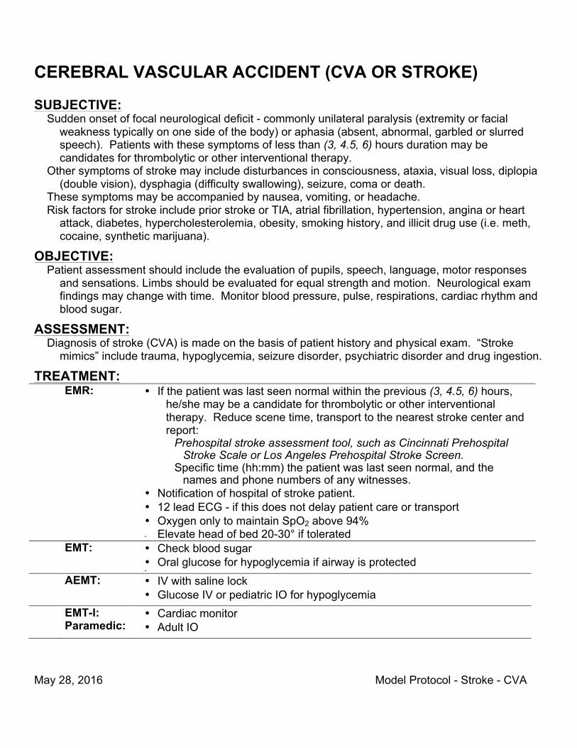
CEREBRAL VASCULAR ACCIDENT (CVA OR STROKE)
SUBJECTIVE: Sudden onset of focal neurological deficit - commonly unilateral paralysis (extremity or facial
weakness typically on one side of the body) or aphasia (absent, abnormal, garbled or slurred speech). Patients with these symptoms of less than (3, 4.5, 6) hours duration may be candidates for thrombolytic or other interventional therapy.
Other symptoms of stroke may include disturbances in consciousness, ataxia, visual loss, diplopia (double vision), dysphagia (difficulty swallowing), seizure, coma or death.
These symptoms may be accompanied by nausea, vomiting, or headache. Risk factors for stroke include prior stroke or TIA, atrial fibrillation, hypertension, angina or heart
attack, diabetes, hypercholesterolemia, obesity, smoking history, and illicit drug use (i.e. meth, cocaine, synthetic marijuana).
OBJECTIVE: Patient assessment should include the evaluation of pupils, speech, language, motor responses
and sensations. Limbs should be evaluated for equal strength and motion. Neurological exam findings may change with time. Monitor blood pressure, pulse, respirations, cardiac rhythm and blood sugar.
ASSESSMENT: Diagnosis of stroke (CVA) is made on the basis of patient history and physical exam. “Stroke
mimics” include trauma, hypoglycemia, seizure disorder, psychiatric disorder and drug ingestion.
TREATMENT: EMR: • If the patient was last seen normal within the previous (3, 4.5, 6) hours,
he/she may be a candidate for thrombolytic or other interventional therapy. Reduce scene time, transport to the nearest stroke center and report:
Prehospital stroke assessment tool, such as Cincinnati Prehospital Stroke Scale or Los Angeles Prehospital Stroke Screen.
Specific time (hh:mm) the patient was last seen normal, and the names and phone numbers of any witnesses.
• Notification of hospital of stroke patient. • 12 lead ECG - if this does not delay patient care or transport • Oxygen only to maintain SpO2 above 94% • Elevate head of bed 20-30° if tolerated
EMT: • Check blood sugar • Oral glucose for hypoglycemia if airway is protected •
AEMT: • IV with saline lock • Glucose IV or pediatric IO for hypoglycemia
EMT-I: Paramedic:
• Cardiac monitor • Adult IO
Related Documents