Chapter 10 *Corresponding Author: Urs Schmidt‑Ott—University of Chicago, Department of Organismal Biology and Anatomy, 920 E. 58th Street, Chicago, IL 60637. Email: [email protected] Hox Genes: Studies om the 20th to the 21st Century, edited by Jean S. Deutsch. ©2010 Landes Bioscience and Springer Science+Business Media. Hox3/zen and the Evolution of Extraembryonic Epithelia in Insects Urs Schmidt‑Ott,* Ab. Matteen Rafiqi and Steffen Lemke Abstract I nsects have undergone dramatic evolutionary changes in extraembryonic development, which correlate with changes in the expression of the class‑3 Hox gene zen. Here, we review the evo‑ lution of this gene in insects and point out how changes in zen expression may have affected extraembryonic development at the morphological and the genetic level. Introduction During the early radiation of insects, Hox3 abandoned its ancestral role in specifying segmental identity along the anteroposterior axis of the embryo and acquired a new role in extraembryonic tissue. 1 is evolutionary transition may have occurred in the stem lineage of modern Pterygota (winged insects), as indicated by expression data and protein comparisons. Canonical Hox proteins share a hexapeptide or YPWM motif, 1 which enables them to interact with the Hox‑cofactor Exd/ Pbx. 2,3 In the context of overlapping Hox gene expression patterns, e.g., during axis‑specification, this interaction is important because it contributes indirectly to the DNA‑binding specificity of individual Hox proteins. 4 e Hox3 gene of the apterygotan firebrat ermobia is expressed in a nested arrangement with other Hox genes in the prospective gnathocephalon and in the growth zone and encodes a protein with the YPWM motif. 5,6 In contrast, Hox3 genes of Pterygota (named zen aſter their Drosophila prototype) 7 are expressed in extraembryonic tissue, lack expression in the germband and encode proteins without YPWM motif. 6 e present chapter is devoted to the evolutionary history of extraembryonic zen expression in insects. We cover data from five orders including the silverfish ermobia (ysanura), the grasshopper Schistocerca (Orthoptera), the bug Oncopeltus (Hemiptera), the beetle Tribolium (Coleoptera) and various fly species (Diptera). Yet, the focus of this review is on Diptera as this is the only insect order for which data on the expression and function of zen are available from multiple distantly related species. Even in this order, some relevant traits have been poorly sampled. Despite the ‘patchiness’ of the data matrix, we hope that as current working hypotheses our phylogentic inferences will help to advance the field. To set the stage, we briefly review morphological aspects of extraembryonic development. In the following sections, we focus on variants of zen expression and their evolutionary significance. We propose that changes in zen expression underlie major reorganizations in extraembryonic development of pterygote insects. . n o i t u b i r t s i D r o f t o N . e c n e i c s o i B s e d n a L t h g i r y p o C 10 0 2 ©

Welcome message from author
This document is posted to help you gain knowledge. Please leave a comment to let me know what you think about it! Share it to your friends and learn new things together.
Transcript

Chapter 10
*Corresponding Author: Urs Schmidt‑Ott—University of Chicago, Department of Organismal Biology and Anatomy, 920 E. 58th Street, Chicago, IL 60637. Email: [email protected]
Hox Genes: Studies from the 20th to the 21st Century, edited by Jean S. Deutsch. ©2010 Landes Bioscience and Springer Science+Business Media.
Hox3/zen and the Evolution of Extraembryonic Epithelia in InsectsUrs Schmidt‑Ott,* Ab. Matteen Rafiqi and Steffen Lemke
Abstract
Insects have undergone dramatic evolutionary changes in extraembryonic development, which correlate with changes in the expression of the class‑3 Hox gene zen. Here, we review the evo‑lution of this gene in insects and point out how changes in zen expression may have affected
extraembryonic development at the morphological and the genetic level.
IntroductionDuring the early radiation of insects, Hox3 abandoned its ancestral role in specifying segmental
identity along the anteroposterior axis of the embryo and acquired a new role in extraembryonic tissue.1 This evolutionary transition may have occurred in the stem lineage of modern Pterygota (winged insects), as indicated by expression data and protein comparisons. Canonical Hox proteins share a hexapeptide or YPWM motif,1 which enables them to interact with the Hox‑cofactor Exd/Pbx.2,3 In the context of overlapping Hox gene expression patterns, e.g., during axis‑specification, this interaction is important because it contributes indirectly to the DNA‑binding specificity of individual Hox proteins.4 The Hox3 gene of the apterygotan firebrat Thermobia is expressed in a nested arrangement with other Hox genes in the prospective gnathocephalon and in the growth zone and encodes a protein with the YPWM motif.5,6 In contrast, Hox3 genes of Pterygota (named zen after their Drosophila prototype)7 are expressed in extraembryonic tissue, lack expression in the germband and encode proteins without YPWM motif.6 The present chapter is devoted to the evolutionary history of extraembryonic zen expression in insects. We cover data from five orders including the silverfish Thermobia (Thysanura), the grasshopper Schistocerca (Orthoptera), the bug Oncopeltus (Hemiptera), the beetle Tribolium (Coleoptera) and various fly species (Diptera). Yet, the focus of this review is on Diptera as this is the only insect order for which data on the expression and function of zen are available from multiple distantly related species. Even in this order, some relevant traits have been poorly sampled. Despite the ‘patchiness’ of the data matrix, we hope that as current working hypotheses our phylogentic inferences will help to advance the field. To set the stage, we briefly review morphological aspects of extraembryonic development. In the following sections, we focus on variants of zen expression and their evolutionary significance. We propose that changes in zen expression underlie major reorganizations in extraembryonic development of pterygote insects.
.noitubirtsiD rof to
N .ecneicsoiB sednaL thgirypoC
1002©
134 Hox Genes: Studies from the 20th to the 21st Century
Setting the Stage: Morphological Evolution of Extraembryonic Development
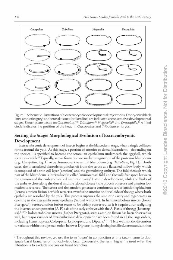
Extraembryonic development of insects begins at the blastoderm stage, when a single cell layer forms around the yolk. At this stage, a portion of anterior or dorsal blastoderm—depending on the species—is specified to become the serosa, an epithelium underneath the eggshell, which secretes a cuticle.8 Typically, serosa formation occurs by invagination of the posterior blastoderm (e.g., Oncopeltus, Fig. 1), or by closure over the ventral blastoderm (e.g., Tribolium, Fig. 1). In both cases, the internalized blastoderm pinches off from the serosa as a flattened hollow body, which is composed of a thin cell layer (amnion) and the gastrulating embryo. The fold through which part of the blastoderm is internalized is called ‘amnioserosal fold’ and the yolk‑free space between the amnion and the embryo is called ‘amniotic cavity’. Later in development, while the flanks of the embryo close along the dorsal midline (dorsal closure), the process of serosa and amnion for‑mation is reversed. The serosa and the amnion generate a continuous serosa‑amnion epithelium (‘serosa‑amnion fusion’), which retracts towards the anterior or dorsal side of the egg where both epithelia are resorbed by the yolk. This process ruptures the amniotic cavity and regenerates an opening in the extraembryonic epithelia (‘serosal window’). In hemimetabolous insects (lower Pterygotaa), serosa‑amnion fusion seems to be widely conserved, as it is required for realigning the inverted anteroposterior (A‑P) axis of the early embryo with the A‑P axis of the egg (katatrep‑sis).9,10 In holometabolous insects (higher Pterygota), serosa‑amnion fusion has been observed as well, but major variants of extraembryonic development have been found in all the large orders, including Hymenoptera, Coleoptera, Lepidoptera and Diptera.8,11‑13 Here we limit the discussion to variants within the dipteran order. In lower Diptera (noncyclorrhaphan flies), serosa and amnion
a Throughout this review, we use the term ‘lower’ in conjunction with a taxon name to des‑ignate basal branches of monophyletic taxa. Conversely, the term ‘higher’ is used when the intention is to exclude species on basal branches.
Figure 1. Schematic illustrations of extraembryonic developmental trajectories. Embryonic (black line), amniotic (grey) and serosal tissues (broken line) are indicated at consecutive developmental stages. Sketches are based on Oncopeltus,9,10 Tribolium,23 Megaselia18 and Drosophila.19 A filled circle indicates the position of the head in Oncopeltus and Tribolium embryos.
.noitubirtsiD rof to
N .ecneicsoiB sednaL thgirypoC
1002©

135Hox3/zen and the Evolution of Extraembryonic Epithelia in Insects
are generated essentially in the same way as in the beetle Tribolium (Fig. 1).14 However, it seems that in many lower dipterans the two mature epithelia fail to fuse again and persist or degenerate independently, while dorsal closure proceeds.15‑17 More radical departures from the ancestral tra‑jectory have been reported for higher dipterans (Cyclorrhapha). Lower cyclorrhaphan flies such as the phorid fly Megaselia abdita or the syrphid fly Episyrphus balteatus initiate the formation of an amnioserosal fold, but while their serosa expands ventrally, their amnion disjoins from the leading edge of the serosa and grows over the dorsal side of the yolk sac (e.g., Megaselia, Fig. 1).18 In these species, serosa‑amnion fusion does not occur and only the amnion is resorbed by the yolk. Finally, in higher cyclorrhaphan flies, while a rudimentary amnioserosal fold is transiently visible at the morphological level, the extraembryonic tissue does not disjoin at the edge of this fold and does not expand over the germband (e.g., Drosophila, Fig. 1). Instead, the extraembryonic anlage gives rise to a single epithelium, called amnioserosa, which is later resorbed by the yolk,19,20 like the dorsal amnion in lower Cyclorrhapha. Taken together, the data suggest that extraembryonic devel‑opment in the dipteran lineage evolved in three distinct steps. First, mature serosal and amniotic epithelia failed to fuse and retract as a continuous serosa‑amnion epithelium during the process of dorsal closure. Second, ventral closure of the amnion was suppressed and substituted by closure of the amnion over the dorsal yolk sac. Third, serosa and dorsal amnion were transformed into an amnioserosa. All these evolutionary transitions seem to correlate with changes in the activity pattern of zen, which will be reviewed in the next section.
Variants of zen Expression and Function in Insects and Possible Morphological Correlates
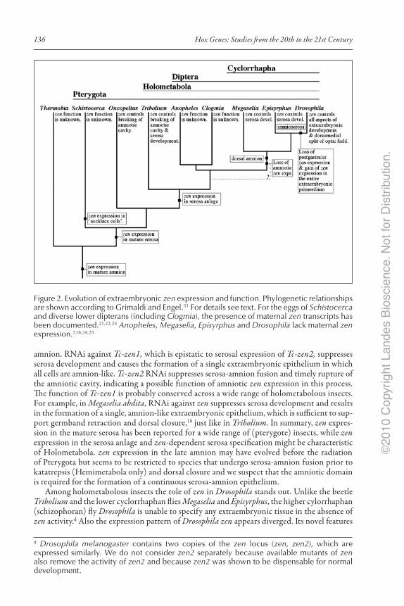
Variants of zen expression and function in insects (Fig. 2) suggest that this gene played an im‑portant role in the evolution of extraembryonic tissue. The apterygotan insect Thermobia expresses Hox3 not only in the embryo (see Introduction) but also in the mature (completed) amnion.5,b Hemimetabolous Pterygota (e.g., Schistocerca, Oncopeltus) exhibit zen expression transiently during blastoderm formation and subsequently in the serosa and parts of the amnion.9,21,22 In the grasshopper Schistocerca gregaria, extraembryonic zen expression begins in the leading edge of the developing serosa (‘necklace cells’) but in other species comparable zen expression has not been observed. Hence, zen expression in necklace cells might have evolved in the Schistocerca lineage. During later stages, Schistocerca expresses zen throughout the completed serosa and in adjacent parts of the completed amnion. In the bug Oncopeltus fasciatus, zen expression has been observed throughout the completed serosa and in a rim of amniotic cells, which connect with the contracting serosa. RNA interference (RNAi) against Oncopeltus zen (Of‑zen) prevents rupture of the amniotic cavity and formation of a continuous serosa‑amnion epithelium, as well as katatrepsis and dorsal closure.9,10,c To explain this phenotype, zen activity in the serosa has been invoked as a regulator of global contractions,10 but an essential complementary role of amniotic zen expression in this process has not been ruled out.
In holometabolous insects, serosal tissue expresses zen at all stages.18,21,23‑25 Amniotic zen ex‑pression has been reported for a beetle (Tribolium) but only at the site where the serosal window opens up prior to dorsal closure. In species that lack this process (e.g., the lower cyclorrhaphan flies Megaselia and Episyrphus), amniotic zen expression has not been observed. In Tribolium castaneum, zen is critical for both serosa specification and serosa‑amnion fusion. The distinction of these functions is possible because of a recent duplication of the zen locus in the Tribolium lineage26 and subfunctionalization of the resulting Tribolium paralogs, Tc‑zen1 and Tc‑zen2.23 Both genes are expressed throughout the serosal tissue, while only Tc‑zen2 is expressed in the
b Amniotic expression was observed at the torpedo stage, when extraembryonic epithelia have already formed.c In Of‑zen RNAi embryos, the serosa and the amnion form a ‘serosal window pane’ that fails to rupture, keeping the amniotic cavity closed and detracting the flanks of the embryo, which under these conditions close ventrally.
.noitubirtsiD rof to
N .ecneicsoiB sednaL thgirypoC
1002©
136 Hox Genes: Studies from the 20th to the 21st Century
amnion. RNAi against Tc‑zen1, which is epistatic to serosal expression of Tc‑zen2, suppresses serosa development and causes the formation of a single extraembryonic epithelium in which all cells are amnion‑like. Tc‑zen2 RNAi suppresses serosa‑amnion fusion and timely rupture of the amniotic cavity, indicating a possible function of amniotic zen expression in this process. The function of Tc‑zen1 is probably conserved across a wide range of holometabolous insects. For example, in Megaselia abdita, RNAi against zen suppresses serosa development and results in the formation of a single, amnion‑like extraembryonic epithelium, which is sufficient to sup‑port germband retraction and dorsal closure,18 just like in Tribolium. In summary, zen expres‑sion in the mature serosa has been reported for a wide range of (pterygote) insects, while zen expression in the serosa anlage and zen‑dependent serosa specification might be characteristic of Holometabola. zen expression in the late amnion may have evolved before the radiation of Pterygota but seems to be restricted to species that undergo serosa‑amnion fusion prior to katatrepsis (Hemimetabola only) and dorsal closure and we suspect that the amniotic domain is required for the formation of a continuous serosa‑amnion epithelium.
Among holometabolous insects the role of zen in Drosophila stands out. Unlike the beetle Tribolium and the lower cyclorrhaphan flies Megaselia and Episyrphus, the higher cylorrhaphan (schizophoran) fly Drosophila is unable to specify any extraembryonic tissue in the absence of zen activity.d Also the expression pattern of Drosophila zen appears diverged. Its novel features
Figure 2. Evolution of extraembryonic zen expression and function. Phylogenetic relationships are shown according to Grimaldi and Engel.51 For details see text. For the eggs of Schistocerca and diverse lower dipterans (including Clogmia), the presence of maternal zen transcripts has been documented.21,22,25 Anopheles, Megaselia, Episyrphus and Drosophila lack maternal zen expression.7,18,24,25
d Drosophila melanogaster contains two copies of the zen locus (zen, zen2), which are expressed similarly. We do not consider zen2 separately because available mutants of zen also remove the activity of zen2 and because zen2 was shown to be dispensable for normal development.
.noitubirtsiD rof to
N .ecneicsoiB sednaL thgirypoC
1002©

137Hox3/zen and the Evolution of Extraembryonic Epithelia in Insects
include a broad dorsal expression domain at early blastoderm stages, a posteriorly expanded and laterally compressed expression domain of zen at late blastoderm stages and absence of zen expression after gastrulation (Fig. 3). Below, we discuss the evolutionary significance of these Drosophila‑specific features.
Suppression of Postgastrular zen Expression May Have Triggered the Origin of the Amnioserosa
In holometabolous insects, serosal zen expression is maintained from the time of serosa specification until after the completion of the serosa epithelium.18,23 However, in Drosophila, zen expression (transcript and protein) fades during stage 8, shortly after a pseudo‑amnioserosal fold has formed. We found in Megaselia that during early gastrulation zen expression is still critical for the formation of distinct serosal and amniotic epithelia.28 Suppression of zen expression at this stage alters the developmental trajectory of the extraembryonic epithelia: the serosa fails to expand over the germband and does not disjoin from the edge of the amnion but rather becomes part
Figure 3. Expression of Mab‑zen/zen transcripts in Megaselia abdita and Drosophila mela‑nogaster. Matching consecutive stages of Megaselia (left) and Drosophila (right) are shown before cellularization (A,B), during cellularization (C,D), at the beginning of gastrulation (E,F), during gastrulation (G,H) and after gastrulation (I,J). Horizontal bars indicate the posi‑tion of the amnioserosa anlage. Posterior shortening of the Mab‑zen domain is indicated by arrowheads and the zen‑positive invaginated portion of the amnioserosa is indicated by an arrow. Panels C, I and J from reference 18. Anterior is left and dorsal up.
.noitubirtsiD rof to
N .ecneicsoiB sednaL thgirypoC
1002©
138 Hox Genes: Studies from the 20th to the 21st Century
of it or disintegrates. Hence, the amnioserosa could have originated by the loss of postgastrular zen expression, which suppressed further serosa development while allowing the formation of a zen‑negative dorsal amnion. This hypothesis implies that late developmental stages of the ances‑tral amnioserosa (after stage 8) were equivalent to a dorsal amnion, whereas early developmental stages of the ancestral amnioserosa (until stage 8) were composed of zen‑expressing serosal and zen‑negative amniotic tissue. However, in Drosophila, all cells of the early amnioserosa express zen (including cells that invaginate with the proctodeum and which formerly gave rise to the amnion) and hence, might best be compared with early serosa cells of lower cyclorrhaphan flies. This novel feature of extraembryonic development may have evolved after the origin of the amnioserosa because at this point in time the distinction of serosal and amniotic primordia had become obsolete. The genetic mechanisms by which zen gained control over all amnion‑competent cells of the Drosophila blastoderm were probably different along the anteroposterior (A‑P) and the dorsoventral (D‑V) axis and will be discussed separately.
Reduction of the Amniotic Anlage along the A‑P AxisIn Megaselia, the expression domain of zen in the cellular blastoderm is shortened at the pos‑
terior end. The cells, in which zen has been actively repressed, invaginate with the proctodeum and become part of the amnion (cf. Figs. 1 and 3). The repressor has not been identified but could be dependent on Megaselia caudal (Mab‑cad), which is specifically expressed in amniotic blastoderm.29 Drosophila lacks expression of caudal in this domain of the blastoderm and does not down‑regulate zen there. As a result, zen is also expressed in cells that eventually invaginate with the proctodeum (cf. Figs. 1 and 3). These cells become part of the amnioserosa. Thus, the posterior expansion of zen expression in the cellular blastoderm of Drosophila may have caused a reduction of the amnion anlage along the A‑P axis. Consistent with this hypothesis, we recently found that the injection of capped Mab‑zen mRNA into syncytial Megaselia embryos represses genes with amniotic expression and causes defects in germband retraction and dorsal closure, but does not interfere with the formation of the serosa (unpublished data).
Reduction of the Amniotic Anlage along the D‑V AxisWhile derepression of posterior zen activity in the Drosophila lineage may account for the
reduction of the amniotic anlage along the A‑P axis, a different mechanism must account for the reduction of the amniotic anlage along the D‑V axis, because in the cellular blastoderm of Drosophila, the expression domain of zen is narrower than in other dipterans.24 How was this transi‑tion achieved? Below, we argue that the gain of early broad zen expression (spanning about 40% of the D‑V perimeter) in the Drosophila lineage was critical for the evolution of an all‑zen‑dependent extraembryonic anlage. To make the argument, it is necessary to introduce another Drosophila gene, decapentaplegic (dpp).30 This gene encodes a ligand of the bone morphogenetic protein (BMP) family. It is secreted into the space between the eggshell and the embryo and promotes pattern formation in the dorsal ectoderm in a concentration‑dependent manner (reviewed in ref. 31). The dorsal blastoderm of dpp‑deficient Drosophila embryos acquires a more ventral neurogenetic fate; moderate levels of dpp activity are sufficient to specify dorsal embryonic ectoderm (but insufficient for specifying the amnioserosa); and high‑level overexpression of dpp converts all dorsal cells to an amnioserosa fate.32
The activity of dpp can be visualized with an antibody against the phosphorylated form of the intracellular protein Mad, which transmits the Dpp‑dependent signal from the cytoplasm to the regulatory DNA sequences of target genes.33‑35 As secreted Dpp undergoes extracellular transport towards the dorsal midline, its activity range in the dorsal ectoderm is confined to a narrower portion than the distribution of dpp transcript (ca. 40% of the D‑V perimeter) would suggest (reviewed in ref. 31). Long‑range, extracellular transport of Dpp accounts for the shallow pMad gradient that is observed in the early Drosophila blastoderm and which spans about 20% of the D‑V perimeter with peak levels at the dorsal midline. In older embryos (undergoing blastoderm cellularization), pMAD levels experience an additional and much more dramatic lateral contrac‑tion (accompanied by dorsal increase) in response to a positive feedback loop that is at least in part
.noitubirtsiD rof to
N .ecneicsoiB sednaL thgirypoC
1002©

139Hox3/zen and the Evolution of Extraembryonic Epithelia in Insects
dependent on zen.36 The result of the refinement is a high‑level pMad domain that now spans only about 10% of the D‑V perimeter, precisely the width of the amnioserosa anlage.e The important point is that throughout blastoderm development, dpp activity of the Drosophila embryo does not extend beyond the expression domain of zen. This is different in lower dipterans.
Compared to Drosophila, the spatial relationship of Dpp activity and zen expression in the mosquito Anopheles appears to be inversed (Fig. 4). In Anopheles, the pMad domain is much broader than in Drosophila and extends at all blastoderm stages beyond the boundaries of the zen domain.24 Provided that Anopheles dpp is essential for the specification of amnion‑competent blastoderm, as it probably isf, and considering that zen represses amnion development in amnion‑competent blastoderm,18,23 the gain of early broad zen expression in the Drosophila lineage after the origin of the amnioserosa might well have delayed amnion specification until the end of gastrulation by promoting serosa development at early stages.
Figure 4. Evolution of the relationship of zen and dpp activities in the blastoderm. Blastoderm embryos of Anopheles (left) and Drosophila (right) are depicted as transverse sections at two consecutive stages with dpp activity in dark grey and zen activity in black. Note that in Anopheles, dpp activity extends beyond the expression domain of zen, while in Drosophila, dpp activity does not extend beyond the activity range of zen. For details and references see text.
e The evolutionary trend towards bistable Dpp activity in the late blastoderm could have gradually increased the ratio of serosal to amnion‑competent blastoderm and might have prevented ventral amnion closure in the stem lineage of schizophoran flies.f In Tribolium, the effect of dpp RNAi on extraembryonic development has been assessed using early markers for the amnion (Tc‑pnr) and a dorsal sector of the serosa anlage (Tc‑doc). Tc‑dpp RNAi abolishes Tc‑pnr as well as Tc‑doc expression. However, only amnion development is sup‑pressed, while the serosa primordium is merely reduced. In Tribolium, the specification of the serosa strongly depends on anterior signaling‑input from a receptor tyrosine kinase (‘terminal system’). In Oncopeltus, dpp RNAi suppresses the invagination of the germ rudiment, which might depend on the specification of amniotic tissue, but does not interfere with serosa development.
.noitubirtsiD rof to
N .ecneicsoiB sednaL thgirypoC
1002©
140 Hox Genes: Studies from the 20th to the 21st Century
Expression of zen in the Optic FieldDrosophila zen has also an embryonic function. In wildtype embryos of Drosophila, regulatory
genes that establish the optic field, such as sine oculis (so) and eyes absent (eya), are repressed at the dorsal midline in response to zen, which prevents a cyclopic phenotype.40 However, in Megaselia and Episyrphus, zen RNAi does not affect the visual system. In these species, the specification of amnion‑competent blastoderm under the control of dpp might be sufficient for splitting the optic field into left and right parts. In Drosophila, genes of the optic field may have acquired Zen binding sites in their cis‑regulatory DNA. This model implies a repressor function of Zen. Alternatively, Drosophila genes of the optic field may have retained the ancestral, Dpp‑dependent regulatory mechanism. In this case, it should be possible to rescue the phenotype of zen‑mutant embryos in the optic field by increasing Dpp activity along the dorsal midline.
The Amnioserosa Gene‑Network in Evolutionary PerspectiveAbove, we argued that in the Drosophila lineage, the loss of postgastrular zen expression ab‑
rogated serosa development and resulted in the formation of a single extraembryonic epithelium, while boundary conflation of the amnion‑competent cellular blastoderm and the zen expression domain (serosa anlage) generated a uniform extraembryonic primordium with early serosal fea‑tures. This simple model provides an evolutionary framework for understanding gene functions in the amnioserosa. In closing this chapter, we discuss a few examples that may serve to illustrate this point.
After stage 8 (when zen is shut off ), the maintenance of the amnioserosa depends on genes of the u‑shaped group (ush‑group), such as u‑shaped (ush), dorsocross (doc), hindsight (hnt), tail‑up (tup) or serpent (srp).41‑43 These genes are unrelated but share similar germband‑retraction and dorsal‑closure phenotypes due to the precocious disintegration of the amnioserosa. All of them are co‑expressed with zen (see Berkeley Drosophila Genome Project at http://www.fruitfly.org/cgi‑bin/ex/insitu.pl). However, none of them is required for the specification of the amnioserosa. In Anopheles24 and Megaselia (our unpublished data), doc, tup and hnt are expressed in the amnion. Thus, it is possible that the maintenance functions of ush‑group genes in the amnioserosa relate to functions of their homologs in the amnion of less derived dipterans.
Two of these genes, doc and hnt, activate Krüppel (Kr) in the late amnioserosa.42,43 In Megaselia, the putative ortholog of Kr (Mab‑Kr) is expressed in the early serosa and later in the amnion. In the early serosa, Mab‑Kr transcripts appear with gastrulation and persist at least until the forma‑tion of the amnioserosal fold. In the amnion, expression starts with stomodeum formation (like in Drosophila), i.e., after the completion of the serosa and persists at least until germband retraction (Fig. 5). The temporal correlation suggests that Kr expression in the amnioserosa is homologous to the amniotic expression in Megaselia. Consistent with this hypothesis, Mab‑zen RNAi embryos lack the early (serosal),18 but not the late (amniotic) phase of extraembryonic Mab‑Kr expression.28 Conversely, serosal Kr expression in Megaselia (and Episyrphus, S.L. unpublished observation) has no equivalent in Drosophila and may have been lost in this lineage because postgastrular serosa development is suppressed.
Another example, pannier (pnr), encodes a GATA transcription factor, which is expressed in the early amnioserosa and in the dorsal epidermis.44‑46 However, in the amnioserosa, the activity of pnr appears to be blocked and both the pnr transcript and the pnr protein disappear during stage 9.47,48 In Megaselia and Tribolium, pnr is expressed in the early amnion and adjacent embryonic tissue.18,23 In Megaselia, amniotic pnr transcript (protein data are not available) lasts until the early expansion phase of the serosa, which is roughly comparable to the stage when pnr is down‑regulated in Drosophila. Thus, it is possible that the ancestral extraembryonic function of pnr was restricted to the early phase of amnion development. According to our model, such a function should have been lost with the conflation of the boundaries of amnion‑competent and serosal blastoderm. Hence, we propose that pnr expression expanded into the amnioserosa without consequences because another factor in this tissue repressed its activity.
.noitubirtsiD rof to
N .ecneicsoiB sednaL thgirypoC
1002©

141Hox3/zen and the Evolution of Extraembryonic Epithelia in Insects
As a final example of how our hypothesis for the origin of the amnioserosa can provide context for the function of genes in the amnioserosa, we turn to the homeobox gene C15, which is activated in the amnioserosa anlage and a narrow strip of adjacent dorsal ectoderm by direct cis‑regulatory input of dpp effectors and Zen.49 C15 protein persists throughout the lifetime of the amnioserosa,43 but C15 null mutations do not interfere with the formation of viable larvae.50 Thus, C15 might affect amnioserosa development in minor ways. In Megaselia, C15 is predominantly expressed in the early amnion and in the dorsal epidermis.18 Transcript expression in the amnion fades before the expansion of the serosa, which is consistent with a developmental role of this gene in the early amnion. As in the case of pnr, the early amniotic function may have been lost in the Drosophila lineage as the boundary between amnion‑competence and zen expression dissolved.
ConclusionChanges in the expression of zen accompanied major reorganizations in extraembryonic devel‑
opment both at the morphological and the regulatory level. At this point, however, the data matrix for any molecular feature of extraembryonic development in insects remains very patchy. With this limitation in mind our tentative conclusions from the above discussion are as follows: 1. The ancestral extraembryonic role of zen in pterygote insects was in the mature serosa and
in parts of the mature amnion, mediating katatrepsis or a related blastokinetic movement. Holometabola abandoned katatrepsis but retained the functions of zen in controlling
Figure 5. Expression of Mab‑Kr/Kr transcripts in Megaselia abdita and Drosophila melano‑gaster. Matching consecutive stages of Megaselia (left) and Drosophila (right) are shown during gastrulation (A,B), shortly before germband retraction (C,D) and during germband retraction (E,F). Early serosal (asterisk) and amniotic/amnioserosal Kr expression domains (arrows) are marked. Anterior is left and dorsal up.
.noitubirtsiD rof to
N .ecneicsoiB sednaL thgirypoC
1002©
142 Hox Genes: Studies from the 20th to the 21st Century
rupture of the amniotic cavity and in generating a continuous serosa‑amnion epithelium. The loss of amniotic zen expression may underlie the suppression of this process in some holometabolous lineages.
2. Zen‑dependent serosa specification evolved before or during the early radiation of Holometabola.
3. In the course of dipteran evolution, the ratio of extraembryonic to embryonic blastoderm was reduced and the ratio of zen‑positive to zen‑negative amnion‑competent blastoderm was increased. The underlying mechanisms involved posterior derepression of zen and enhanced transport of Dpp towards the dorsal midline.
4. In schizophoran flies, postgastrular zen expression was lost. This change in zen expression may have triggered the origin of the amnioserosa in the schizophoran lineage by suppressing postgastrular serosa development, while allowing the completion of a dorsal amnion.
5. After the origin of the amnioserosa, extraembryonic development became entirely depen‑dent on zen. This change in the function of zen may have evolved in concert with a novel broad dorsal expression domain of zen in the early blastoderm and bistable Dpp signaling activity in the cellular blastoderm.
6. The requirement for zen activity in the optic field evolved with the loss of zen‑independent specification of amnion‑competent blastoderm.
AcknowledgementsWe thank Chip Ferguson for discussions and Kristen A. Panfilio, an anonymous reviewer and
Kacy L. Gordon for useful comments on the manuscript.
References 1. Hughes CL, Kaufman TC. Hox genes and the evolution of the arthropod body plan. Evol Dev 2002;
4:459‑499. 2. Mann RS, Chan SK. Extra specificity from extradenticle: the partnership between HOX and PBX/
EXD homeodomain proteins. Trends Genet 1996; 12:258‑62. 3. Moens CB, Selleri L. Hox cofactors in vertebrate development. Dev Biol 2006; 291:193‑206. 4. Joshi R, Passner JM, Rohs R et al. Functional specificity of a Hox protein mediated by the recognition
of minor groove structure. Cell 2007; 131:530‑43. 5. Hughes CL, Liu PZ, Kaufman TC. Expression patterns of the rogue Hox genes Hox3/zen and fushi
tarazu. Evol Dev 2004; 6:393‑401. 6. Panfilio KA, Akam M. A comparison of Hox3 and Zen protein coding sequences in taxa that span the
Hox3/zen divergence. Dev Genes Evol 2007; 217:323‑9. 7. Rushlow C, Doyle H, Hoey T et al. Molecular characterization of the zerknüllt region of the Anten‑
napedia gene complex in Drosophila. Genes Dev 1987; 1:1268‑1279. 8. Panfilio KA. Extraembryonic development in insects and the acrobatics of blastokinesis. Dev Biol 2008;
313:471‑91. 9. Panfilio KA, Liu PZ, Akam M et al. Oncopeltus fasciatus zen is essential for serosal tissue function in
katatrepsis. Dev Biol 2006; 292:226‑43. 10. Panfilio KA. Late extraembryonic morphogenesis and its zen RNAi‑induced failure in the milkweed
bug Oncopeltus fasciatus. Dev Biol 2009; 333:297‑311. 11. Machida R, Ando H. Evolutionary changes in developmental potentials of the embryo proper and
embryonic membranes along with the derivative structures in Atelocerata, with special reference to hexapoda (arthropoda). Proc Arthropod Embryol Soc Jpn 1998; 33:1‑12.
12. Sander K. In Insect Development. Lawrence PA, ed. Oxford: Blackwell Scientific Publications, 1976;35‑52.
13. Anderson DT. In Developmental Systems: insects. Counce SJ, Waddington CH, eds. London: Academic, 1972;165‑242.
14. Davis CWC. A comparative study of larval embryogenesis in the mosquito Culex fatigans Wiedemann (Diptera: Culicidae) and the sheep‑fly Lucilia sericata Meigen (Diptera: Calliphoridae). Aust J Zool 1967; 15:547‑579.
15. Kahle W. Die Paedogenesis der Cecidomyiden. Zoologica, Stuttgart 1908; 21:1‑80 (Plates I‑VI). 16. Gambrell FL. The embryology of the blackfly Simulium pinctipes Hagen. Ann Entomol Soc Am 1933;
26:641‑671. 17. Butt FH. Embryology of Sciara. Ann Entomol Soc Am 1934; 27:565‑579.
.noitubirtsiD rof to
N .ecneicsoiB sednaL thgirypoC
1002©

143Hox3/zen and the Evolution of Extraembryonic Epithelia in Insects
18. Rafiqi AM, Lemke SJ, Ferguson S et al. Evolutionary origin of the amnioserosa in cyclorrhaphan flies correlates with spatial and temporal expression changes of zen. Proc Natl Acad Sci USA 2008; 105:234‑239.
19. Campos‑Ortega JA, Hartenstein V. The embryonic development of Drosophila melanogaster (Springer‑Verlag, Berlin, Heidelberg, New York, 1997).
20. Reed BH, Wilk R, Schöck F et al. Integrin‑dependent apposition of Drosophila extraembryonic mem‑branes promotes morphogenesis and prevents anoikis. Curr Biol 2004; 14:372‑380.
21. Falciani F, Hausdorf B, Schröder R et al. Class 3 Hox genes in insects and the origin of zen. Proc Natl Acad Sci USA 1996; 93:8479‑8484.
22. Dearden P, Grbic M, Falciani F et al. Maternal expression and early zygotic regulation of the Hox3/zen gene in the grasshopper Schistocerca gregaria. Evol Dev 2000; 2:261‑270.
23. van der Zee M, Berns N, Roth S. Distinct functions of the Tribolium zerknüllt genes in serosa specifica‑tion and dorsal closure. Curr Biol 2005; 15:624‑636.
24. Goltsev Y, Fuse N, Frasch M et al. Evolution of the dorsal‑ventral patterning network in the mosquito, Anopheles gambiae. Development 2007;2415‑2424.
25. Stauber M, Prell A, Schmidt‑Ott U. A single Hox3 gene with composite bicoid and zerknüllt expression characteristics in non‑Cyclorrhaphan flies. Proc Natl Acad Sci USA 2002; 99:274‑279.
26. Brown S, Fellers J, Shippy T et al. A strategy for mapping bicoid on the phylogenetic tree. Curr Biol 2001; 11:R43‑R44.
27. Pultz, MA Diederich RJ, Cribbs DL et al. The proboscipedia locus of the antennapedia complex: a molecular and genetic analysis. Genes Dev 1988; 2:901‑920.
28. Rafiqi AM. Morphological transitions and the genetic basis of the evolution of extraembryonic tissues in flies. Ph.D. Thesis, Wageningen University (NL) 2008.
29. Stauber M, Lemke S, Schmidt‑Ott U. Expression and regulation of caudal in the lower cyclorrhaphan fly Megaselia. Dev Genes Evol 2008; 218:81‑7.
30. Newfeld SJ, Wisotzkey RG, Kumar S. Molecular evolution of a developmental pathway: phylogenetic analyses of transforming growth factor‑beta family ligands, receptors and Smad signal transducers. Genetics 1999; 152:783‑95.
31. O’Connor MB, Umulis D, Othmer HG et al. Shaping BMP morphogen gradients in the Drosophila embryo and pupal wing. Development 2006; 133:183‑93.
32. Ferguson EL, Anderson KV. Decapentaplegic acts as a morphogen to organize dorsal‑ventral pattern in the Drosophila embryo. Cell 1992; 71:451‑61.
33. Dorfman R, Shilo BZ. Biphasic activation of the BMP pathway patterns the Drosophila embryonic dorsal region. Development 2001; 128:965‑972.
34. Tanimoto H, Itoh S, ten Dijke P et al. Hedgehog creates a gradient of DPP activity in Drosophila wing imaginal discs. Mol Cell 2000; 5:59‑71.
35. Persson U, Izumi H, Souchelnytskyi S et al. The L45 loop in type I receptors for TGF‑beta family members is a critical determinant in specifying Smad isoform activation. FEBS Lett 1998; 434:83‑7.
36. Wang Y‑C, Ferguson EL. Spatial bistability of Dpp‑receptor interactions during Drosophila dorsal‑ventral patterning. Nature 2005; 434:229‑234.
37. van der Zee M, Stockhammer O, von Levetzow C et al. Sog/Chordin is required for ventral‑to‑dorsal Dpp/BMP transport and head formation in a short germ insect. Proc Natl Acad Sci USA 2006; 103:16307‑16312.
38. Schoppmeier M, Schröder R. Maternal torso signaling controls body axis elongation in a short germ insect. Curr Biol 2005; 15:2131‑6.
39. Angelini DR, Kaufman TC. Functional analyses in the milkweed bug (Hemiptera) support a role for Wnt signaling in body segmentation but not appendage development. Dev Biol 2005; 283:409‑23.
40. Chang T, Mazotta J, Dumstrei K et al. Dpp and Hh signaling in the Drosophila embryonic eye field. Development 2001; 128:4691‑4704.
41. Frank LH, Rushlow C. A group of genes required for maintenance of the amnioserosa tissue in Droso‑phila. Development 1996; 122:1343‑1352.
42. Yip MLR, Lamka ML, Lipshitz HD. Control of germ‑band retraction in Drosophila by the zinc‑finger protein Hindsight. Development 1997; 124:2129‑2141.
43. Reim I, Lee H‑H, Frasch M. The T‑box‑encoding dorsocross genes function in amnioserosa develop‑ment and the patterning of the dorsolateral germ band downstream of Dpp. Development 2003; 130:3187‑3204.
44. Winick J, Abel T, Leonard MW et al. A GATA family transcription factor is expressed along the embryonic dorsoventral axis in Drosophila melanogaster. Development 1993; 119:1055‑1065.
45. Heitzler P, Haenlin M, Ramain P et al. A genetic analysis of pannier, a gene necessary for viability of dorsal tissues and bristle positioning in Drosophila. Genetics 1996; 143:1271‑1286.
.noitubirtsiD rof to
N .ecneicsoiB sednaL thgirypoC
1002©
144 Hox Genes: Studies from the 20th to the 21st Century
46. Ashe HL, Mannervik M, Levine M. Dpp signaling thresholds in the dorsal ectoderm of the Drosophila embryo. Development 2000; 127:3305‑12.
47. Haenlin M, Cubadda Y, Blondeau F et al. Transcriptional activity of pannier is regulated negatively by heterodimerization of the GATA DNA‑binding domain with a cofactor encoded by the u‑shaped gene of Drosophila. Genes Dev 1997; 11:3096‑108.
48. Herranz H, Morata G. The functions of pannier during Drosophila embryogenesis. Development 2001; 128:4837‑46.
49. Lin MC, Park J, Kirov N et al. Threshold response of C15 to the Dpp gradient in Drosophila is established by the cumulative effect of Smad and Zen activators and negative cues. Development 2006; 133:4805‑13.
50. Campbell G. Regulation of gene expression in the distal region of the Drosophila leg by the Hox11 homolog, C15. Dev Biol 2005; 278:607‑18.
51. Grimaldi D, Engel MS. Evolution of insects. Cambridge: Cambridge University Press, 2005.
.noitubirtsiD rof to
N .ecneicsoiB sednaL thgirypoC
1002©
Related Documents