MARINE ECOLOGY PROGRESS SERIES Mar Ecol Prog Ser Vol. 505: 253–266, 2014 doi: 10.3354/meps10794 Published May 28 INTRODUCTION Stressful conditions can act as evolutionary forces, leading to adaptive changes in populations (Sørensen et al. 2003). A variety of stressors (physical, chemical, biological) operate in aquatic ecosystems, and among them, temperature is one of the most important. All organisms have a thermal performance curve, mean- ing that when temperature reaches values close to the organism’s upper thermal limits, performance decreases (see Huey & Stevenson 1979). Moreover, several studies defined the concept of oxygen-limited thermal tolerance, implying that the oxygen supply to organs is optimal between limits (lower and upper pejus temperatures; Pörtner 2001, 2010). Further- more, organisms cannot survive (except for a limited © Inter-Research 2014 · www.int-res.com *Corresponding author: [email protected] Histopathological alterations, physiological limits, and molecular changes of juvenile Sparus aurata in response to thermal stress Diana Madeira 1, *, Catarina Vinagre 2 , Pedro M. Costa 3 , Mário S. Diniz 1 1 REQUIMTE, Departamento de Química, Centro de Química Fina e Biotecnologia, Faculdade de Ciências e Tecnologia, Universidade Nova de Lisboa, 2829-516 Caparica, Portugal 2 Centro de Oceanografia, Faculdade de Ciências, Universidade de Lisboa, Campo Grande, 1749-016 Lisboa, Portugal 3 IMAR-Instituto do Mar, Departamento de Ciências e Engenharia do Ambiente, Faculdade de Ciências e Tecnologia, Universidade Nova de Lisboa, 2829-516 Caparica, Portugal ABSTRACT: Current concerns about climate change have led to a great body of literature on ther- mal tolerance. However, it is still difficult to understand and relate biological changes at different organizational levels. This is especially important in commercial species. We aimed to test the ef- fects of temperature stress in juvenile gilthead seabream Sparus aurata in order to contribute to the understanding of the vulnerability of this species to temperature changes, heat waves, and poten- tially climate warming. Here we applied an integrative approach, considering indicators at the bio- chemical, cellular, and physiological levels. Firstly, the upper thermal limit was estimated via the critical thermal maximum (CTMax); secondly, levels of Hsp70 (reversible protein damage) and total ubiquitin (irreversible protein damage) were quantified in several tissues via ELISAs; and thirdly, histological analyses were performed to identify cellular structural damage due to temper- ature and how it correlates to biochemical alterations. Results showed that mean (± SD) CTMax was 35.5 ± 0.5°C. Absolute amounts of both Hsp70 and total ubiquitin varied significantly among organs, with gills having the highest amounts of both. Biomarker and histopathological results in- dicated that S. aurata might be particularly sensitive to water temperatures ≥28°C. At 30°C, S. au- rata juveniles showed severe signs of stress, with increased biomarker levels in almost every organ tested and significant cellular damage (atrophy, inflammation, micro-hemorrhage, hyperemia, hy- perplasia). Therefore, S. aurata may be vulnerable to heat wave events that currently make water temperature attain 28°C for several weeks (30°C by 2100 under a climate change scenario). Thus, this species might be vulnerable to a rise in sea temperature, and our research may be important from a management perspective, as S. aurata is a major commercial species. KEY WORDS: Hsp70 · Ubiquitin · Cellular alterations · CTMax · Sea bream · Temperature Resale or republication not permitted without written consent of the publisher This authors' personal copy may not be publicly or systematically copied or distributed, or posted on the Open Web, except with written permission of the copyright holder(s). It may be distributed to interested individuals on request.

Welcome message from author
This document is posted to help you gain knowledge. Please leave a comment to let me know what you think about it! Share it to your friends and learn new things together.
Transcript
MARINE ECOLOGY PROGRESS SERIESMar Ecol Prog Ser
Vol. 505: 253–266, 2014doi: 10.3354/meps10794
Published May 28
INTRODUCTION
Stressful conditions can act as evolutionary forces,leading to adaptive changes in populations (Sørensenet al. 2003). A variety of stressors (physical, chemical,biological) operate in aquatic ecosystems, and amongthem, temperature is one of the most important. Allorganisms have a thermal performance curve, mean-
ing that when temperature reaches values close tothe organism’s upper thermal limits, performancedecreases (see Huey & Stevenson 1979). Moreover,several studies defined the concept of oxygen-limitedthermal tolerance, implying that the oxygen supplyto organs is optimal between limits (lower and upperpejus temperatures; Pörtner 2001, 2010). Further-more, organisms cannot survive (except for a limited
© Inter-Research 2014 · www.int-res.com*Corresponding author: [email protected]
Histopathological alterations, physiological limits,and molecular changes of juvenile Sparus aurata in
response to thermal stress
Diana Madeira1,*, Catarina Vinagre2, Pedro M. Costa3, Mário S. Diniz1
1REQUIMTE, Departamento de Química, Centro de Química Fina e Biotecnologia, Faculdade de Ciências e Tecnologia, Universidade Nova de Lisboa, 2829-516 Caparica, Portugal
2Centro de Oceanografia, Faculdade de Ciências, Universidade de Lisboa, Campo Grande, 1749-016 Lisboa, Portugal3IMAR-Instituto do Mar, Departamento de Ciências e Engenharia do Ambiente, Faculdade de Ciências e Tecnologia,
Universidade Nova de Lisboa, 2829-516 Caparica, Portugal
ABSTRACT: Current concerns about climate change have led to a great body of literature on ther-mal tolerance. However, it is still difficult to understand and relate biological changes at differentorganizational levels. This is especially important in commercial species. We aimed to test the ef-fects of temperature stress in juvenile gilthead seabream Sparus aurata in order to contribute to theunderstanding of the vulnerability of this species to temperature changes, heat waves, and poten-tially climate warming. Here we applied an integrative approach, considering indicators at the bio-chemical, cellular, and physiological levels. Firstly, the upper thermal limit was estimated via thecritical thermal maximum (CTMax); secondly, levels of Hsp70 (reversible protein damage) andtotal ubiquitin (irreversible protein damage) were quantified in several tissues via ELISAs; andthirdly, histological analyses were performed to identify cellular structural damage due to temper-ature and how it correlates to biochemical alterations. Results showed that mean (±SD) CTMaxwas 35.5 ± 0.5°C. Absolute amounts of both Hsp70 and total ubiquitin varied significantly amongorgans, with gills having the highest amounts of both. Biomarker and histopathological results in-dicated that S. aurata might be particularly sensitive to water temperatures ≥28°C. At 30°C, S. au-rata juveniles showed severe signs of stress, with increased biomarker levels in almost every organtested and significant cellular damage (atrophy, inflammation, micro-hemorrhage, hyperemia, hy-perplasia). Therefore, S. aurata may be vulnerable to heat wave events that currently make watertemperature attain 28°C for several weeks (30°C by 2100 under a climate change scenario). Thus,this species might be vulnerable to a rise in sea temperature, and our research may be importantfrom a management perspective, as S. aurata is a major commercial species.
KEY WORDS: Hsp70 · Ubiquitin · Cellular alterations · CTMax · Sea bream · Temperature
Resale or republication not permitted without written consent of the publisher
This authors' personal copy may not be publicly or systematically copied or distributed, or posted on the Open Web, except with written permission of the copyright holder(s). It may be distributed to interested individuals on request.
Mar Ecol Prog Ser 505: 253–266, 2014
period of time) at temperatures above or below thepejus temperatures (see Pörtner & Peck 2010 for areview).
Metabolic pathways and biochemical reactions areaffected by temperature, determining an organism’sphysiological state and behavior. Therefore, activi-ties such as foraging, reproduction, and growth willbe altered and may change the fitness of the individ-uals (Mora & Ospina 2001, Hochachka & Somero2002, Pörtner & Peck 2010) and community and eco-system structure (e.g. Pörtner & Farrell 2008).
Within marine aquatic environments, estuaries andother confined coastal areas are amongst the mostvariable. These areas are economically important,providing food resources and serving as nurseryareas for juveniles of many species (Haedrich 1983,Vinagre et al. 2010). Nevertheless, as estuaries areshallow water bodies, anthropogenic impacts (e.g.pollution, overfishing) and environmental change aregreater than in deep sea or open-ocean sites. Addi-tionally, significant variations in abiotic variables thatfollow seasonal or even tidal rhythms may be perma-nent sources of stress to residing biota. Hence, juve-nile fish inhabiting these ecosystems may endurestressful conditions, including significant tempera-ture variations (see Madeira et al. 2012a). Consider-ing climate change scenarios for Portuguese waters(a 2°C increase in mean seawater temperature by2100; Miranda et al. 2002), juveniles may undergosevere thermal stress which may compromise theirgrowth (Vinagre et al. 2012a, 2013a,b). Madeira et al.(2012a) showed that sea bream juveniles inhabitingPortuguese estuaries may already be subjected tothermal stress under the present conditions. It istherefore important to study the molecular and bio-chemical mechanisms behind the thermal stress re -sponse in commercial species to determine whetherthey may be undergoing cellular and tissue damage.Biomarker quantification may be very useful toachieve this aim. A biomarker is defined as ‘anymeasurement reflecting an inter action between abiological system and a potential hazard, which maybe chemical, physical, or bio logical. The measuredresponse may be functional and physiological, bio-chemical at the cellular level, or a molecular inter -action’ (World Health Organi zation 1993). Knowingand measuring these bio markers may help us under-stand ecological patterns of response to increasingseawater temperature and, there fore, improve themanagement of commercial fish populations.
From this perspective, the model chosen for thisstudy was the gilthead seabream Sparus aurata,which is a very important commercial species, both
in fishing activities and aquaculture. It is a eury -haline, warm-temperate species that occurs in coastalseagrass beds, on sandy bottoms, and in the surfzone, as well as in estuaries and coastal lagoons(Altimiras et al. 1994). It usually inhabits shallowwaters, from 1 to 150 m deep (commonly 1 to 30 m).Juveniles tend to school in coastal lagoons and estu-arine brackish waters (Craig et al. 2008), which arewarmer than coastal waters. Its native distributionis subtropical and extends from the Mediterranean tothe east Atlantic coast (including Cape Verde and theCanaries to the English Channel).
Amongst stress biomarkers, heat shock proteins(Hsps) have been the most widely studied, espe-cially Hsp70 (Tomanek 2011). Temperature leadsto proteotoxic stress and induces a heat shock re -sponse in several species (e.g. Somero 1995, Ma -deira et al. 2012b). Hsps have chaperoning activityand can be seen as molecular protection mecha-nisms with a selective value, contributing to theorganism’s and species’ success across environ -mental gradients (Hofmann 2005). They confer stresstolerance (Li et al. 1995, Feder 1996, Basu et al.2002, Sorte & Hofmann 2005) and are associatedwith an increase in the ability to temporarily survivetemperature extremes (Feder & Hofmann 1999, Hof-mann et al. 2002, Sørensen et al. 2003). Hsps areconsidered an indirect measure of protein damageand biochemical indicators of reversible protein un -folding in the cell (Hofmann 2005, Tomanek 2010,2011). Another im portant biomarker, which comple-ments the information provided by Hsps, is ubiqui-tin. Ubiquitination is critical to cell cycle regulation,transcription, apop tosis, protein sorting, and proteinquality control, among other cell processes (Hannaet al. 2007). The ubiquitin metabolic pathway in -volves the selective tagging of proteins for protea -somal degradation (Hershko & Ciechanover 1992)and falls into 4 categories: (1) continuous turnoverto rapidly modulate protein levels; (2) elimination ofproteins in response to specific signals; (3) elimina-tion of defective proteins that arise through physicalor chemical factors and transcriptional/translationalerrors or gene mutations; and (4) the turnover ofexcess free forms of proteins (from multi subunitcomplexes; Finley 1991). Regarding these aspects,ubiquitin levels are considered a direct measure ofprotein loss (Ciechanover 1998), indicating irre-versible protein damage in the cells (Hofmann &Somero 1995). Moreover, a relationship may existbetween the heat shock and ubiquitin systems (Finley et al. 1984, Alberti et al. 2002). However, it isstill unclear which features of damaged proteins
254A
utho
r cop
y
Madeira et al.: Temperature effects on juvenile Sparus aurata
mediate proteasome activity, whether they are elim-inated through ubiquitination or recovered by Hsps(Finley 1991, Patterson & Höhfeld 2006). None the -less, substrate recognition and processing may re -quire cooperation between these 2 systems (Patter-son & Höhfeld 2006). During stress, accumu lationsof misfolded proteins (aggresomes) tend to form inthe cells (Johnston et al. 1998, Kopito 2000). Inter-estingly, components of the ubiquitin system andchaperone system are present and seem to be activelyrecruited to these aggresomes (García-Mata et al.1999, Wyttenbach et al. 2000). Moreover, increasedHsp70 can reduce aggresome formation by increas-ing protein degradation (Dul et al. 2001). Thus,there seems to be a protein triage system within thecells, in which Hsps seem to play an important part.However, little information is available on thesematters concerning marine species. All of thesehypotheses were tested in mammals or human celllines, so further studies are needed using aquaticorganisms as models.
Histopathological changes are also important indi-cators of health changes in species of aquatic andother ecosystems (e.g. Bignell et al. 2011, Costa et al.2013a,b). Even though several studies have investi-gated histopathological changes induced by temper-ature, they mainly focused on freshwater fish (e.g.Day & Butler 2005, Sollid & Nilsson 2006). Thesechanges included morphometric changes in theattributes of muscle tissue, changes in axon and fiberdiameter and thickness, changes in the respiratorysurface area, and changes in several tissues (gills,liver, kidney, and heart), such as hyperplasia, promi-nent vacuolization, increased melanin production,canaliculi formation, hypertrophy, and necrosis ofcells.
The aims of this study were to test the effects oftemperature stress in juvenile gilthead seabreamS. aurata in order to contribute to the understandingof the vulnerability of this species to temperaturechanges, heat waves, and potentially climate warm-ing. In detail, our aims were (1) to test the upper ther-mal limit, (2) to assess the biochemical thermal stressresponse, quantifying the levels of Hsp70 (reversibleprotein damage) and total ubiquitin (irreversible pro-tein damage) in several tissues of this species (gills,intestine, muscle, brain, hepa topancreas−liver, andinfiltrated pancreatic acini), (3) to identify any corre-lations between Hsp70 levels and total ubiquitin lev-els, and (4) to perform histological analysis of thesamped tissues in order to identify cellular structuraldamage due to temperature and how it correlates tobiochemical alterations.
MATERIALS AND METHODS
Thermal tolerance method
Juvenile Sparus aurata (mean ± SD total length of92.1 ± 8.1 mm and weight of 12.2 ± 2.9 g) were ob -tained from a fish farm (MARESA, Spain) and accli-mated in a re-circulating system with 150 l aquariacontaining clean, permanently aerated sea water, ata constant temperature of 18°C and salinity of 35‰(maintenance conditions in the fish farm). The dis-solved O2 level of the water varied between 95 and100%. The fish were acclimated for 1 wk prior tobioassays and were fed commercial pellets ad libitumtwice a day. They were starved for 24 h before theexperiments. The use of farmed organisms reducespotential confounding factors (i.e. thermal history).According to several authors, thermal history andparental effects are determinant and induce irre-versible changes in the thermal tolerance of the spe-cies (Cossins & Bowler 1987, Schaefer & Ryan 2006).
Thermal tolerance was determined using thedynamic method described by Mora & Ospina (2001).The parameter measured was the critical thermalmaximum (CTMax, given in °C), which is defined asthe ‘arithmetic mean of the collective thermal pointsat which the end-point is reached’ (Mora & Ospina2001, p. 766). This end-point is defined as the loss ofequilibrium. The CTMax method was chosen for thisstudy because it has been the most used to determineupper thermal limits (Lutterschmidt & Hutchison1997) and because dynamic trials are accurate pre-dictors of the responses of organisms to natural con-ditions (Bennett & Judd 1992, Bennett & Beitinger1997).
To determine the CTMax, the fish were subjectedto a thermostatized bath, using a heated/refrigeratorcirculator (MultiTemp III, Pharmacia Biotech). Theywere placed in 40.5 l white plastic tanks (n = 5, 30 ×30 × 45 cm) with lids to prevent evaporation(10−11 fish tank−1). Each tank was equipped with anaeration system. During the experiment, animalswere exposed to a constant rate of water temperatureincrease of 1°C h−1, and observed continuously, untilthey reached the end-point. Fish were collectedevery 2°C, killed by cervical decapitation with theaid of a scalpel, and organs were removed. Samples(brain, gills, intestine, hepatopancreas, muscle) from6 individuals were taken and immediately frozen inliquid nitrogen or processed fresh for histologicalanalyses (in this case, samples of 3 animals per sam-pling point, out of the total of 6). The total number ofindividuals was 54.
255A
utho
r cop
y
Mar Ecol Prog Ser 505: 253–266, 2014
The temperature at which each animal reached itsend-point was measured with a digital thermometerand registered, and then CTMax (and its standarddeviation) were calculated as:
(1)
Where Tend-point is the temperature at which theend-point (loss of equilibrium) was reached for indi-vidual 1, individual 2, individual n, divided by the nindividuals that were in the sample. The experimentswere carried out in shaded daylight (15 h light:9 hdark). Total length and weight were measured at theend of each trial using an ichthyometer and a scale,respectively.
Protein extraction
Samples (approximately 200−250 mg of gills, muscle,intestine, brain, and hepatopancreas) were placed in1.5 ml microtubes and homogenized in 1 ml of phos-phate-buffered saline (PBS) solution (0.14 M NaCl,0.003 M KCl, 0.01 M Na2HPO4, 0.002 M KH2PO4, pH7.4) to extract most soluble cytosolic proteins, using aglass and teflon Potter Elvehjem tissue grinder, inice-cold conditions. The crude homo genates werethen centri fuged for 15 min at 10 000 × g. Afterwards,the supernatant was collected, transferred to newmicrotubes (1.5 ml), and frozen immediately (−80°C)until further biochemical analyses.
Hsp70 quantification
Hsp70 was quantified using an indirect enzyme-linked immunosorbent assay (ELISA) (Njemini etal. 2005) with 96-well microplates (Greiner). EitherELISA or Western blot can be successfully employedin Hsp quantification (Brun et al. 2008). Sampleswere diluted to 1:100 in 0.05 M carbonate-bicarbonatebuffer (Sigma-Aldrich). This dilution was chosen togive an optimum signal and was achieved after pre-vious optimization.
Three replicates of 50 μl were taken from eachdiluted sample, transferred to the microplate wells,and incubated overnight at 4°C. The microplate waswashed 3 times in PBS 0.05% Tween-20 and thenblocked by adding 200 μl of 1% BSA (%w/v; Sigma-Aldrich) in PBS. The microplate was incubated at37°C for 90 min. After microplate washing, the pri-mary antibody (anti-HSP70/HSC70; Acris), diluted to0.5 μg ml−1 in 1% BSA in PBS, was added to the
microplate wells (50 μl each). The microplates werethen incubated for 90 min at 37°C. After anotherwashing stage, the secondary antibody (anti-mouseIgG, fab specific, alkaline phosphatase conjugate;Sigma-Aldrich) was diluted (1 μg ml−1 in 1% BSA inPBS) and added (50 μl) to each well followed by incu-bation at 37°C for 90 min. After the washing stage,100 μl of substrate (SIGMA FAST ™ p-nitrophenylphosphate tablets, Sigma-Aldrich) was added to eachwell and incubated for 30 min at room temperature.Stop solution (3N NaOH, 50 μl) was added to eachwell, and the absorbance was read in a 96-well micro -plate reader at 405 nm (Benchmark, BIO-RAD). Forquantification purposes, a calibration curve was con-structed using serial dilutions of purified Hsp70 activeprotein (Acris) to give a range from 0 to 2 μg ml−1.
Ubiquitin and ubiquitinated/poly-ubiquitinatedprotein quantification
Quantification was achieved through a directELISA with 96-well microplates (Greiner). After pre-vious dilution optimization, samples were diluted to1:100 in 0.05 M carbonate-bicarbonate buffer (Sigma-Aldrich) to give an optimum signal.
Three replicates of 50 μl were taken from eachdiluted sample, transferred to the microplate wells,and incubated overnight at 4°C. The microplate waswashed 3 times in PBS 0.05% Tween-20 and thenblocked by adding 200 μl of 1% BSA in PBS (Sigma-Aldrich). The microplate was incubated at 37°C for90 min. After microplate washing, the primary anti-body (Ub P4D1, sc-8017, HRP conjugate, Santa CruzBiotechnology), diluted to 0.5 μg ml−1 in 1% BSA inPBS, was added to the microplate wells (50 μl each).The microplates were then incubated for 90 min at37°C. After another washing stage, 100 μl of sub-strate (TMB/E, Merck Millipore) were added to eachwell and incubated for 30 min at room temperature.Stop solution (1 N HCl, 100 μl) was added to eachwell, and the absorbance was read in a 96-wellmicroplate reader at 415 nm (Benchmark, BIO-RAD).For quantification purposes, a calibration curve wasconstructed using serial dilutions of purified ubiquitin(UbpBio, E-1100) to give a range from 0 to 2 μg ml−1.
For normalization purposes, the Bradford Assaywas used to quantify the total amount of protein ineach sample (Bradford 1976). The analysis was car-ried out in 96-well microplates (Nunc) by adding200 μl of Bradford reagent in each well and 10 μl ofeach sample or standards. After 10 min of reaction,the absorbance was read at 595 nm in a microplate
CTMax( )1 end-point
n
Tn
n∑=
256A
utho
r cop
y
Madeira et al.: Temperature effects on juvenile Sparus aurata
reader (Benchmark, BIO-RAD). A calibration curvewas constructed using BSA standards (0−10 μg ml−1).The results for Hsp70 and total ubiquitin weredivided by the total amount of protein in order to givethe final data values.
Histological analysis
The procedure followed essentially Martoja & Mar-toja-Pierson (1967) for light microscopy. In brief,freshly-dissected samples from 3 individuals per tem -perature group (of gills, muscle, intestine, hepato -pancreas), were fixed in Bouin’s solution (10% v/vformalin and 7% v/v acetic acid with picric acidadded to saturation) for 48 h at room temperature.The samples were dehydrated in a progressive seriesof ethanol and embedded in liquid paraffin (Panreac)overnight at 60°C. Sectioning was done with a JungRM2035 model rotary microtome. Sections (5 μmthick) were stained with Harris’ alum hematoxylin(Riedel-de Haën) and counterstained with alcoholiceosin Y (Panreac) for structural analysis. The slideswere mounted with DPX resinous media (VWR Inter-national). Microscopy analysis was performed with aDMLB model optical microscope equipped with aDFC 480 digital camera (all from Leica Microsystems).
Statistical analysis
Failure to comply with the assumptions of para -metric ANOVA (normality of the data and homo -scedasticity) led to the application of non-parametricstatistics. Kruskal-Wallis ANOVA by ranks and mul-tiple comparisons tests were used to compare Hsp70and ubiquitin levels between different temperaturegroups and different organs. Correlation analyses(Spearman R, since the data did not follow bivariatenormality) were performed for Hsp70 and ubiquitinlevels in each tissue. A significance level of α = 0.05was set for all statistics. Statistics were carried outusing the software Statistica (Version 10, StatSoft).
RESULTS
CTMax and Hsp70
Following Eq. (1), Sparus aurata juveniles had amean ± SD CTMax of 35.5 ± 0.5°C.
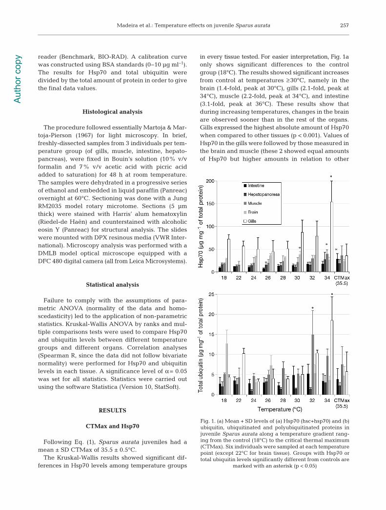
The Kruskal-Wallis results showed significant dif-ferences in Hsp70 levels among temperature groups
in every tissue tested. For easier interpretation, Fig. 1aonly shows significant differences to the controlgroup (18°C). The results showed significant in creasesfrom control at temperatures ≥30°C, namely in thebrain (1.4-fold, peak at 30°C), gills (2.1-fold, peak at34°C), muscle (2.2-fold, peak at 34°C), and intestine(3.1-fold, peak at 36°C). These results show that during increasing temperatures, changes in the brainare observed sooner than in the rest of the organs.Gills expressed the highest absolute amount of Hsp70when compared to other tissues (p < 0.001). Values ofHsp70 in the gills were followed by those measured inthe brain and muscle (these 2 showed equal amountsof Hsp70 but higher amounts in relation to other
257
Fig. 1. (a) Mean + SD levels of (a) Hsp70 (hsc+hsp70) and (b)ubiquitin, ubiquitinated and polyubiquitinated proteins injuvenile Sparus aurata along a temperature gradient rang-ing from the control (18°C) to the critical thermal maximum(CTMax). Six individuals were sampled at each temperaturepoint (except 22°C for brain tissue). Groups with Hsp70 ortotal ubiquitin levels significantly different from controls are
marked with an asterisk (p < 0.05)
Aut
hor c
opy
Mar Ecol Prog Ser 505: 253–266, 2014
tissues; p < 0.001). Hepatopancreas and intestineshowed equi valent amounts of Hsp70, but these lev-els were lower compared to other tissues (p < 0.001).Intraspecific variability in Hsp70 was elevated in all tissues.
Total ubiquitin
The Kruskal-Wallis results showed significant dif-ferences in total ubiquitin levels between tempera-ture groups in all tissues tested except intestine andbrain. For easier interpretation, Fig. 1b only showssignificant differences to the control group (18°C).The results showed significant in creases from con-trols in muscle (+2.9-fold, peak at 32°C) and gills(+4.1-fold, peak at 34°C), indicating that muscle re -sponds faster than gills. When comparing total ubiq-uitin amounts in different tissues, gills, brain, andmuscle had equivalent levels to each other, but hadsignificantly higher levels than hepatopancreas andintestine (p < 0.001). He pa topancreas and intestineshowed significant differences, with intestine havinghigher amounts of total ubiquitin (p < 0.01). Intraspe-cific variability in total ubiquitin was also elevated inall tissues.
Correlation between Hsp70 and total ubiquitin
Spearman R correlations showed that Hsp70and total ubiquitin were significantly correlated ingills (Spearman R = 0.43, p < 0.005), hepatopancreas(Spearman R = 0.44, p < 0.002), and muscle (Spear-man R = 0.67, p < 0.000). Brain (p > 0.61) and intes-tine (p > 0.18) did not show a significant correlationbetween these 2 biomarkers.
Histological observations
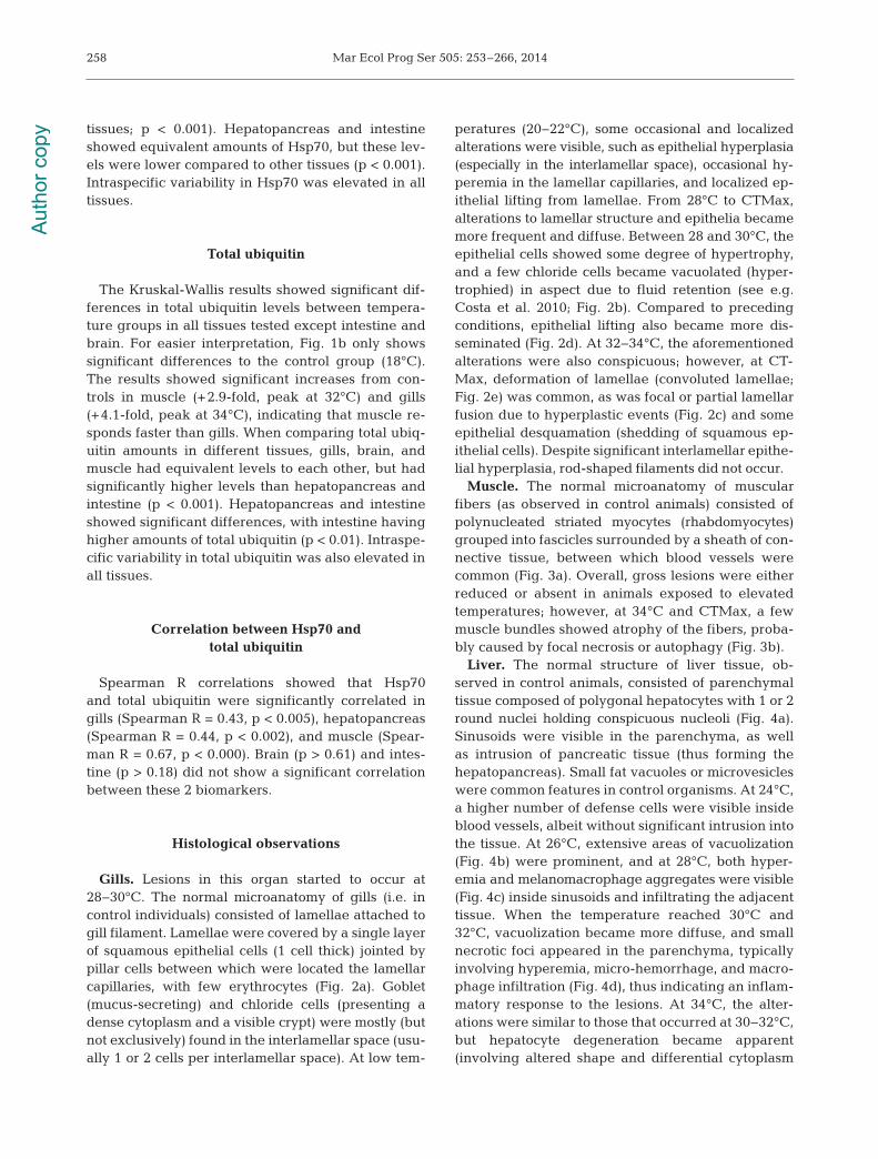
Gills. Lesions in this organ started to occur at28−30°C. The normal microanatomy of gills (i.e. incontrol individuals) consisted of lamellae attached togill filament. Lamellae were covered by a single layerof squamous epithelial cells (1 cell thick) jointed bypillar cells between which were located the lamellarcapillaries, with few erythrocytes (Fig. 2a). Goblet(mucus-secreting) and chloride cells (presenting adense cytoplasm and a visible crypt) were mostly (butnot exclusively) found in the interlamellar space (usu-ally 1 or 2 cells per interlamellar space). At low tem-
peratures (20−22°C), some occasional and localizedalterations were visible, such as epithelial hyperplasia(especially in the interlamellar space), occasional hy-peremia in the lamellar capillaries, and localized ep-ithelial lifting from lamellae. From 28°C to CTMax,alterations to lamellar structure and epi thelia becamemore frequent and diffuse. Be tween 28 and 30°C, theepithelial cells showed some degree of hypertrophy,and a few chloride cells became vacuolated (hyper-trophied) in aspect due to fluid retention (see e.g.Costa et al. 2010; Fig. 2b). Compared to precedingconditions, epithelial lifting also became more dis-seminated (Fig. 2d). At 32−34°C, the aforementionedalterations were also conspicuous; however, at CT-Max, deformation of lamellae (convoluted lamellae;Fig. 2e) was common, as was focal or partial lamellarfusion due to hyperplastic events (Fig. 2c) and someepithelial desquamation (shedding of squamous ep-ithelial cells). Despite significant interlamellar epithe-lial hyperplasia, rod-shaped filaments did not occur.
Muscle. The normal microanatomy of muscularfibers (as observed in control animals) consisted ofpolynucleated striated myocytes (rhabdomyocytes)grouped into fascicles surrounded by a sheath of con-nective tissue, between which blood vessels werecommon (Fig. 3a). Overall, gross lesions were eitherreduced or absent in animals exposed to elevatedtemperatures; however, at 34°C and CTMax, a fewmuscle bundles showed atrophy of the fibers, proba-bly caused by focal necrosis or autophagy (Fig. 3b).
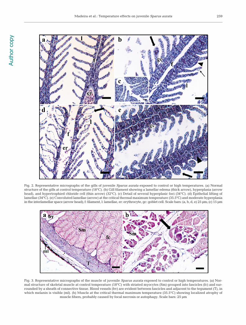
Liver. The normal structure of liver tissue, ob -served in control animals, consisted of parenchymaltissue composed of polygonal hepatocytes with 1 or 2round nuclei holding conspicuous nucleoli (Fig. 4a).Sinusoids were visible in the parenchyma, as wellas intrusion of pancreatic tissue (thus forming thehepatopancreas). Small fat vacuoles or microvesicleswere common features in control organisms. At 24°C,a higher number of defense cells were visible insideblood vessels, albeit without significant intrusion intothe tissue. At 26°C, extensive areas of vacuolization(Fig. 4b) were prominent, and at 28°C, both hyper-emia and me lanomacrophage aggregates were visible(Fig. 4c) inside sinusoids and infiltrating the adjacenttissue. When the temperature reached 30°C and32°C, vacuolization became more diffuse, and smallnecrotic foci appeared in the parenchyma, typicallyinvolving hyperemia, micro-hemorrhage, and macro-phage infiltration (Fig. 4d), thus indicating an inflam-matory response to the lesions. At 34°C, the alter-ations were similar to those that occurred at 30−32°C,but hepatocyte degeneration became apparent(involving altered shape and differential cytoplasm
258A
utho
r cop
y
Madeira et al.: Temperature effects on juvenile Sparus aurata 259
Fig. 3. Representative micrographs of the muscle of juvenile Sparus aurata exposed to control or high temperatures. (a) Nor-mal structure of skeletal muscle at control temperature (18°C) with striated myocytes (Sm) grouped into fascicles (fc) and sur-rounded by a sheath of connective tissue. Blood vessels (bv) are evident between fascicles and adjacent to the tegument (T), inwhich melanin is visible (ml). (b) Muscle at the critical thermal maximum temperature (35.5°C) showing localized atrophy of
muscle fibers, probably caused by focal necrosis or autophagy. Scale bars: 25 μm
Fig. 2. Representative micrographs of the gills of juvenile Sparus aurata exposed to control or high temperatures. (a) Normalstructure of the gills at control temperature (18°C). (b) Gill filament showing a lamellar edema (thick arrow), hyperplasia (arrowhead), and hypertrophied chloride cell (thin arrow) (32°C). (c) Detail of several hyperplasic foci (34°C). (d) Epithelial lifting oflamellae (34°C). (e) Convoluted lamellae (arrow) at the critical thermal maximum temperature (35.5°C) and moderate hyper plasiain the interlamellar space (arrow head); f: filament, l: lamellae, er: erythrocyte, gc: goblet cell. Scale bars: (a, b, d, e) 25 μm; (c) 15 μm
Aut
hor c
opy
Mar Ecol Prog Ser 505: 253–266, 2014
staining, probably indicative of a change in function;Fig. 4e). When organisms reached their CTMax, therewas a boost in hyperemia, and de generate he pa -tocytes were clearly more eosino philic, likely indica-ting an early stage of hepatocellular alteration (Fig. 4f).
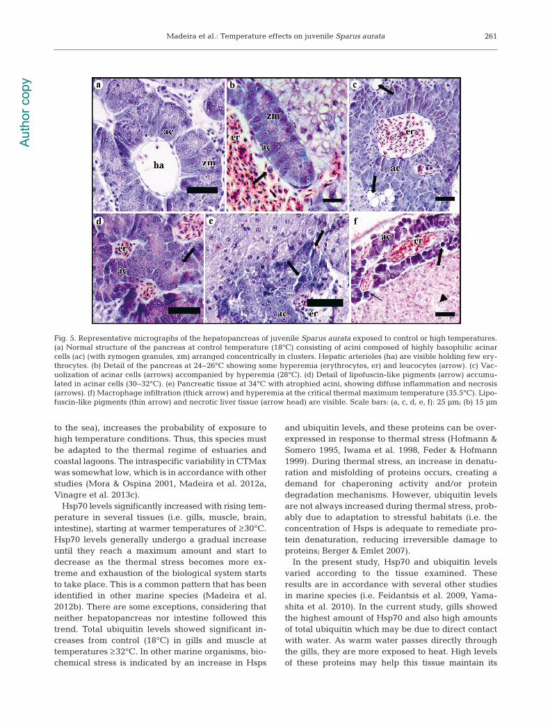
Pancreatic acini. Control animals (18°C) revealednormal micro anatomy of pancreatic acini, consistingof highly baso philic (with zymogen granules) cellsarranged concentrically in clusters (Fig. 5a). Pancre-atic tissue infiltrated the hepatic parenchyma, form-ing the hepato pancreas typical of this species andother teleosts. Overall, this tissue yielded lesions thatdeveloped in a gradual mode as temperature in -creased, similar to the liver. Occasional alterationsstarted to occur at 24°C, namely moderate hyperemiaand macrophage infiltration, which may indicate thebeginning of an inflammation process (Fig. 5b). At28°C, vacuolization of acinar cells be came evident,accompanied by hyperemia (Fig. 5c). At tempera-tures around 30−32°C, agglomerates of lipofuscin-like pigments were visible inside pancreatocytes andnearby melanomacrophages (Fig. 5d). At 34°C andCTMax, the acini were atrophied, showing diffuseinflammation and necrosis (Fig. 5e,f).
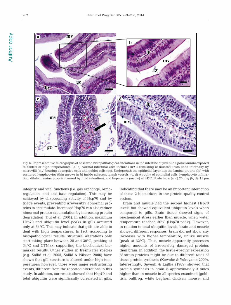
Intestine. Control animals (18°C) ex hibited the nor-mal intestinal architecture, consisting of muco salfolds lined internally by microvilli-bearing ab sorp -tive cells and goblet cells that secrete mucin (which,
in contact with water, forms mucus). Underneath theepithelial layer, a thin layer of connective tissue inthe middle (the lamina propria) was visible, withscattered lymphocytes in side adjacent lymph vessels(Fig. 6a,b). The muscularis of the mucosa was also vis-ible as well as the tunica submucosa, tunica muscu-laris externa, and tunica se rosa at the edge. At lowtemperatures (20 and 22°C), no alterations were visi-ble except for occasional epithelial vacuolization.Lesions started to become visible in fish subjected to28 and 30°C, namely with atrophy of epithelial cellsand defense cell infiltration within the lamina propria,accom panied by a slight hyperemia, which be camemore prominent at higher temperatures (Fig. 6c,d).At 34°C, hyperplasia of ab sorp tive cells seemed tooccur, as well as moderate focal necrosis in the mus-cle layer, which was more obvious in animals sub-jected to CTMax.
DISCUSSION
Sparus aurata juveniles had a CTMax value of 35.5± 0.5°C. As gilthead seabream is a warm temperatemarine species and juveniles inhabit warmer watersof estuaries and coastal lagoons, a relatively highCTMax value was expected. Living in these confinedhabitats, with a lower thermal inertia (in comparison
260
Fig. 4. Representative micrographs of the liver of juvenile Sparus aurata exposed to control or high temperatures. (a) Normalstructure of the liver at control temperature (18°C) consisting of parenchymal tissue composed of polygonal hepatocytes (hp)with round nuclei (1 or 2) and conspicuous nucleoli. Sinusoids with few erythrocytes (er) are visible in the parenchyma. (b) Fatvacuoles (arrow) and slight hyperemia in hepatic vessels (26°C). (c) Fat vacuoles (thick arrow), hyperemia, and melano -macrophage aggregates (thin arrows) (28°C). (d) Fat vacuoles (thick arrow), small necrotic foci in the parenchyma (arrowhead) involving hyperemia, micro-hemorrhage, and melanomacrophage infiltration (30°C). (e) Moderate hepatocellular alter-ation (arrow) and melanomacrophage infiltration (34°C). Note the alteration to hepatocyte size and shape. (f) Increased hyper-emia and hemorrhage, with hepatocytes clearly more eosinophilic (at the critical thermal maximum temperature of 35.5°C).
Scale bars: 25 μm
Aut
hor c
opy
Madeira et al.: Temperature effects on juvenile Sparus aurata
to the sea), increases the probability of exposure tohigh temperature conditions. Thus, this species mustbe adapted to the thermal regime of estuaries andcoastal lagoons. The intraspecific variability in CTMaxwas somewhat low, which is in accordance with otherstudies (Mora & Ospina 2001, Madeira et al. 2012a,Vinagre et al. 2013c).
Hsp70 levels significantly in creased with rising tem -perature in several tissues (i.e. gills, muscle, brain,intestine), starting at warmer tem peratures of ≥30°C.Hsp70 levels generally under go a gradual increaseuntil they reach a maximum amount and start todecrease as the thermal stress becomes more ex -treme and exhaustion of the bio logical system startsto take place. This is a common pattern that has beenidentified in other marine species (Madeira et al.2012b). There are some exceptions, considering thatneither hepatopancreas nor intestine followed thistrend. Total ubiquitin levels showed significant in -creases from control (18°C) in gills and muscle attemperatures ≥ 32°C. In other marine organisms, bio-chemical stress is indicated by an increase in Hsps
and ubiquitin levels, and these proteins can be over-expressed in response to thermal stress (Hofmann &Somero 1995, Iwama et al. 1998, Feder & Hofmann1999). During thermal stress, an increase in denatu-ration and misfolding of proteins occurs, creating ademand for chaperoning activity and/or proteindegradation mechanisms. However, ubiquitin levelsare not always increased during thermal stress, prob-ably due to adaptation to stressful habitats (i.e. theconcentration of Hsps is adequate to remediate pro-tein denaturation, reducing irreversible damage toproteins; Berger & Emlet 2007).
In the present study, Hsp70 and ubiquitin levelsvaried according to the tissue examined. Theseresults are in accordance with several other studiesin marine species (i.e. Feidantsis et al. 2009, Yama -shita et al. 2010). In the current study, gills showedthe highest amount of Hsp70 and also high amountsof total ubiquitin which may be due to direct contactwith water. As warm water passes directly throughthe gills, they are more exposed to heat. High levelsof these proteins may help this tissue maintain its
261
Fig. 5. Representative micrographs of the hepatopancreas of juvenile Sparus aurata exposed to control or high temperatures.(a) Normal structure of the pancreas at control temperature (18°C) consisting of acini composed of highly basophilic acinarcells (ac) (with zymogen granules, zm) arranged concentrically in clusters. Hepatic arterioles (ha) are visible holding few ery-throcytes. (b) Detail of the pancreas at 24−26°C showing some hyperemia (erythrocytes, er) and leucocytes (arrow). (c) Vac-uolization of acinar cells (arrows) accompanied by hyperemia (28°C). (d) Detail of lipofuscin-like pigments (arrow) accumu-lated in acinar cells (30−32°C). (e) Pancreatic tissue at 34°C with atrophied acini, showing diffuse inflammation and necrosis(arrows). (f) Macrophage infiltration (thick arrow) and hyperemia at the critical thermal maximum temperature (35.5°C). Lipo-fuscin-like pigments (thin arrow) and necrotic liver tissue (arrow head) are visible. Scale bars: (a, c, d, e, f): 25 μm; (b) 15 μm
Aut
hor c
opy
Mar Ecol Prog Ser 505: 253–266, 2014
integrity and vital functions (i.e. gas exchange, osmo -regulation, and acid-base regulation). This may beachieved by chaperoning activity of Hsp70 and bytriage events, preventing irreversibly abnormal pro-teins to accumulate. Increased Hsp70 can also reduceabnormal protein accumulation by increasing proteindegradation (Dul et al. 2001). In addition, maximumHsp70 and ubiquitin level peaks in gills occurredonly at 34°C. This may indicate that gills are able todeal with high temperatures. In fact, according tohistopathological results, structural alterations onlystart taking place between 28 and 30°C, peaking at34°C and CTMax, supporting the biochemical bio-marker results. Other studies in freshwater species(e.g. Sollid et al. 2005, Sollid & Nilsson 2006) haveshown that gill structure is altered under high tem-peratures; however, those were major restructuringevents, different from the reported alterations in thisstudy. In addition, our results showed that Hsp70 andtotal ubiquitin were significantly correlated in gills,
indicating that there may be an important interactionof these 2 biomarkers in the protein quality controlsystem.
Brain and muscle had the second highest Hsp70levels but showed equivalent ubiquitin levels whencompared to gills. Brain tissue showed signs of biochemical stress earlier than muscle, when water temperature reached 30°C (Hsp70 peak). However,in relation to total ubiquitin levels, brain and muscleshowed different responses: brain did not show anyincreases with higher temperature, unlike muscle(peak at 32°C). Thus, muscle apparently processeshigher amounts of irreversibly damaged proteinsthan brain. In addition, the tissue-specific expressionof stress proteins might be due to different rates oftissue protein synthesis (Kawabe & Yokoyama 2009).Interestingly, Sayegh & Lajtha (1989) showed thatprotein synthesis in brain is approximately 3 timeshigher than in muscle in all species examined (gold-fish, bullfrog, white Leghorn chicken, mouse, and
262
Fig. 6. Representative micrographs of observed histopathological alterations in the intestine of juvenile Sparus aurata exposedto control or high temperatures. (a, b) Normal intestinal architecture (18°C) consisting of mucosal folds lined internally by microvilli (mv) bearing absorptive cells and goblet cells (gc). Underneath the epithelial layer lies the lamina propria (lp) withscattered lymphocytes (thin arrows in b) inside adjacent lymph vessels. (c, d) Atrophy of epithelial cells, lymphocyte infiltra-tion, dilated lamina propria (caused by fluid retention), and hyperemia (arrow) at 34°C. Scale bars: (a, c) 25 μm; (b, d): 15 μm
Aut
hor c
opy
Madeira et al.: Temperature effects on juvenile Sparus aurata
Tokay lizard). That study and another study by Lajtha& Sershen (1975) also showed that protein metabolicrates increase with temperature in the brain of fishesand lizards. The increase in Hsp70 in the brain dur-ing thermal stress may be necessary due to the highamount of proteins there and their higher metabolicrate. In muscle, which is an organ of low protein turn-over rate (Loughna & Goldspink 1985), the cyto- protective efforts may be directed towards the main-tenance of existing proteins.
The fact that Hsp70 and ubiquitin were correlatedin muscle but not in brain tissue may support thishypothesis of tissue-specific protein expression andmetabolism. Histopathological results did not showany gross lesions in muscle, only atrophy of somemuscle bundles at higher temperatures (34°C andCTMax). In fact, some degree of atrophy of musclefibers at higher temperatures has also been shown inother organisms including mammals (Wang & Chen1999). In addition, Garcia de la serrana et al. (2012)found persistent effects of development temperatureon muscle growth patterns during early life stages ofS. aurata (20% more fibers of lower average dia -meter at low temperature, i.e. 17.5−18.5°C, than athigh temperature, i.e. 21−22°C; myogenesis and geneexpression patterns are not fixed during growth).Furthermore, the responses in brain and muscle maybe linked to the loss of equilibrium. This loss of right-ing reflex may indicate that nerve impulse transmis-sion is disturbed (Aslanidi et al. 2008). As describedabove, brain started to show signs of stress at 30°Cand muscle at 32−34°C. The combination of braindamage with an abnormal amount of aberrant pro-teins in the muscle plus fiber atrophy led to the lossof equilibrium at 35−36°C. This explains why theCTMax value was 35.5°C.
Hepatopancreas and intestine showed equivalentamounts of Hsp70 but showed different responsepatterns, as intestine had a significant increase atCTMax while hepatopancreas did not show anyincrease. In addition, hepatopancreas had lower totalubiquitin levels. These results somewhat match theresults of Feidantsis et al. (2013), who failed to findsignificant changes in Hsp70 in the liver of S. aurataexposed to seasonal temperature changes. However,these same authors reported changes in Hsp90, soperhaps other chaperones are more important inthe liver. Moreover, Hsp70 and Hsp90 expression istissue- and species-specific, which may explain thedifferent responses (see Deane & Woo 2003). Never-theless, liver and pancreatic acini started to showhistopathological lesions at 26−28°C, with more significant alterations (inflammation and necrosis) at
32−34°C. Additionally, intact hepatocytes showedsome degree of hypertrophy and eosinophilia, whichmay be regarded as a change in function, reflectingan alteration of regular metabolic pathways.
Hsps confer thermal tolerance (Moseley 1997). Still,this adaption is of short duration (hours to days) andundergoes a fast decline when Hsp contents start todecrease (Moseley 1997, Kregel 2002). Therefore,our results may indicate that S. aurata is particularlysensitive to prolonged exposures to elevated watertemperatures (i.e. ≥28−30°C). This result is supportedby other studies (e.g. Feidantsis et al. 2009), in whichS. aurata started to show signs of stress at tempera-tures above 20°C and were unable to acclimate totem peratures beyond 26°C. These results were basedon the analysis of several chaperones, kinases, de -hydro genases, and citrate synthase. The authors alsoconcluded that 30°C is the critical threshold for thisspecies (yielding a high increase in mortality, lactateaccumulation, high Hsp70 levels, and behavioralalterations). Even though the study of Feidantsis etal. (2009) was a 10 d experiment, their results some-what match our study. Nevertheless, Requena et al.(1997) stated that after a temperature rise from 20 to28°C (1°C d−1), the basal metabolism (oxygen con-sumption) of S. aurata was compensated within 2 wk.However, other studies (Madeira et al. 2012a, Vina-gre et al. 2012a,b,c, 2013a,b) concerning differentspecies (i.e. Dicentrarchus labrax, Solea senegalen-sis, and Diplodus sp.) also showed no acclimation totemperatures of 27−28°C, potentially rendering envi-ronmental heat waves detrimental to juvenile fish.Thus, there still might be some controversy about theability of warm-temperate fish to acclimate to hightemperatures. It is also reasonable to consider thatfish are very mobile organisms and thus may escapestressful conditions. Nonetheless, considering thatthe growth of juveniles occurs mainly in estuaries orlagoons, they might be more restrained to these shal-low warm habitats during this phase, limiting theirescape possibilities (as shown in other species ofcomparable size, life cycle, and ecological require-ments, see Vinagre et al. 2008, 2011). In the Tagus estuary (the largest in Western Europe), mean watertemperature during the winter is 12°C, increasing to24°C during the summer (Madeira et al. 2012a). Theseasonal variation in temperature is thus 12°C, andthe fish may be exposed to extreme temperaturesduring heat wave events. In a climate warming sce-nario, water temperature is expected to increase 2°Cby 2100. During current heat waves, water tem -perature attains 28°C in several locations (e.g. Por-tuguese waters: Vinagre et al. 2012a; Mediterranean:
263A
utho
r cop
y
Mar Ecol Prog Ser 505: 253–266, 2014
Damianidis & Chintiroglou 2000), which means thatby 2100, the seawater temperature may remain at30°C for several weeks. As mentioned above, at thistemperature, S. aurata juveniles show significantsigns of stress, with adverse consequences to theirhealth, growth, and therefore individual and popula-tional fitness. In addition, as S. aurata is a very impor-tant commercial species, fishing pressure may alsocontribute to the negative im pact upon S. aurata pop-ulations. Moreover, this species is one of the mostimportant in saline and hyper saline aquaculture inthe Mediterranean area (Feidantsis et al. 2009, 2013).It can be farmed in coastal ponds and lagoons, withextensive and semi-intensive methods, or in land-based installations and in sea cages, with intensivefarming systems. In many cases, the fish are kept inlow volumes of water and shallow depths, whichmakes the animals rather susceptible to warming.This potentially affects the health status of the fishand the quality of the end-product, with obviousimpacts on this economic activity.
It is also relevant to mention that thermal windowsmay differ between life stages (Pörtner & Farrell2008). According to these authors, juveniles have thegreatest aerobic thermal window. Thus, eggs, larvae,and adults may experience physiological and molec-ular changes earlier than juveniles. Moreover, Polo etal. (1991) showed that eggs of S. aurata did not hatchat high temperatures such as 30°C, and outsidethe range of 16−22°C, mortality and abnormalitiesincreased significantly. Georga ko poulou et al. (2010)also detected a significant effect of water tempera-ture on the development of several deformities in S.aurata, such as inside folded gill-cover, hemal lordo-sis, and malformations of the caudal and dorsal fin-supporting elements. Therefore, it is important toevaluate other life stages in relation to biochemicaland physiological alterations resulting from exposureto increased temperature.
CONCLUSIONS
The current work demonstrates that it is extremelyimportant to know the level of impact of rising seawater temperature, either due to natural envi -ronmental change or anthropogenic disturbances, onSparus aurata, since it has great commercial value.From this perspective, studies considering other lifestages of S. aurata are currently being performedby our team, and these may be crucial to understandthe population dynamics, recruitment success, sur-vival rates, fitness, and ultimately the vulnerability
and resilience of the species to climate and anthro-pogenic change. These results may be importantfrom an environmental management perspectiveconsidering a bottom-up approach. In detail, sub-cellular (e.g. proteins) and intermediate-level bio-markers (e.g. histopathological alterations) mayserve as early warning signals of stress and mayhelp to understand the health status of the individual.Responses occurring at higher levels of biologicalorganization (population, ecosystem) can ultimatelybe linked to biomarkers. Integrating biomarker in -formation for several life stages can thus be im -portant in predicting impacts at an ecological level.All together, the information can influence decisionmakers to improve marine resource sustainability byconstraining catch limits (in all or specific life stages)and applying financial penalties to those with un -sustainable practices, or giving incentives to thosewith sustainable ones (see Roberts & Brink 2010for more detailed information on marine resourcesmanagement).
Acknowledgements. This study had the support of the Por-tuguese Fundação para a Ciência e a Tecnologia (FCT)through grants SFRH/BPD/86566/2012 awarded to C.V.,SFRH/BPD/72564/2010 to P.M.C., and SFRH/BD/80613/2011 awarded to D.M.; through grant PTDC/MAR/ 119068/2010; and through the strategic project nos. Pest-C/EQB/LA0006/2011 granted to Requimte and Pest-OE/MAR/UI0199/2011 granted to the Centro de Oceanografia. Ourexperiments followed the Portuguese legislation for animalexperimentation. Moreover, 3 of the authors have a level Ccertification issued by the Federation of European Labora-tory Animal Science Associations.
LITERATURE CITED
Alberti S, Demand J, Esser C, Emmerich N, Schild H, Hoh -feld J (2002) Ubiquitylation of BAG-1 suggests a novelre gulatory mechanism during the sorting of chaperonesubstrates to the proteasome. J Biol Chem 277: 45920−45927
Altimiras J, Champion SR, Puigcerver M, Tort L (1994) Phys-iological responses of the gilthead sea bream (Sparusaurata) to hypoosmotic shock. Comp Biochem Physiol A108: 81−85
Aslanidi KB, Kharakoz DP, Chailakhyan LM (2008) Tem -perature shock and adaptation in fish. Dokl Biochem Biophys 422: 302−303
Basu N, Todgham AE, Ackerman PA, Bibeau MR, NakanoK, Schulte PM, Iwama GK (2002) Heat shock proteingenes and their functional significance in fish. Gene 295: 173−183
Bennett WA, Beitinger TL (1997) Temperature tolerance ofthe sheepshead minnow, Cyprinodon variegates. Copeia1997: 77−87
Bennett WA, Judd FW (1992) Comparison of methods fordetermining low temperature tolerance: experiments withpinfish, Lagodon rhomboides. Copeia 1992: 1059−1065
264A
utho
r cop
y
Madeira et al.: Temperature effects on juvenile Sparus aurata
Berger MS, Emlet RB (2007) Heat-shock response of theupper intertidal barnacle Balanus glandula: thermal stressand acclimation. Biol Bull (Woods Hole) 212: 232−241
Bignell JP, Stentiford GD, Taylor NGH, Lyons BP (2011)Histopathology of mussels (Mytilus sp.) from the Tamarestuary, UK. Mar Environ Res 72: 25−32
Bradford MM (1976) Rapid and sensitive method for thequantitation of microgram quantities of protein utilizingthe principle of protein-dye binding. Anal Biochem 72:248–254
Brun NT, Bricelj VM, MacRae TH, Ross NW (2008) Heatshock protein responses in thermally stressed bay scal-lops, Argopecten irradians, and sea scallops, Placopectenmagellanicus. J Exp Mar Biol Ecol 358: 151−162
Ciechanover A (1998) The ubiquitin-proteasome pathway: on protein death and cell life. EMBO J 17: 7151−7160
Cossins AR, Bowler K (1987) Temperature biology of ani-mals. Chapman & Hall, London
Costa PM, Caeiro S, Diniz MS, Lobo J and others (2010) Adescription of chloride cell and kidney tubule alterationsin the flatfish Solea senegalensis exposed to moderatelycontaminated sediments from the Sado estuary (Portu-gal). J Sea Res 64: 465−472
Costa PM, Caeiro S, Costa MH (2013a) Multi-organ histo-logical observations on juvenile Senegalese soles ex -posed to low concentrations of waterborne cadmium.Fish Physiol Biochem 39: 143−158
Costa PM, Carreira S, Costa MH, Caeiro S (2013b) Develop-ment of histopathological indices in a commercial marinebivalve (Ruditapes decussatus) to determine environ-mental quality. Aquat Toxicol 126: 442−454
Craig G, Paynter D, Coscia I, Mariani S (2008) Settlement ofgilthead sea bream Sparus aurata L. in a southern IrishSea coastal habitat. J Fish Biol 72: 287−291
Damianidis P, Chintiroglou CC (2000) Structure and functionsof polychaetofauna living in Mytilus galloprovincialisassemblages in Thermaikos Gulf (north Aegean Sea).Oceanol Acta 23: 323−337
Day N, Butler PJ (2005) The effects of acclimation toreversed seasonal temperatures on the swimming per-formance of adult brown trout Salmo trutta. J Exp Biol208: 2683−2692
Deane EE, Woo NYS (2003) Ontogeny of thyroid hormones,cortisol, hsp70 and hsp90 during silver sea bream larvaldevelopment. Life Sci 72: 805−818
Dul JL, Davis DP, Williamson EK, Stevens FJ, Argon Y (2001)Hsp70 and antifibrillogenic peptides promote degrada-tion and inhibit intracellular aggregation of amyloido-genic light chains. J Cell Biol 152: 705−716
Feder ME (1996) Ecological and evolutionary physiology ofstress proteins and the stress response: the Drosophilamelanogaster model. In: Johnston IA, Bennett AF (eds)Animals and temperature: phenotypic and evolutionaryadaptation. Cambridge University Press, Cambridge,p 79−102
Feder ME, Hofmann GE (1999) Heat-shock proteins, molec-ular chaperones, and the stress response: evolutionaryand ecological physiology. Annu Rev Physiol 61: 243−282
Feidantsis K, Pörtner HO, Lazou A, Kostoglou B, MichaelidisB (2009) Metabolic and molecular stress responses of thegilthead seabream Sparus aurata during long-term expo-sure to increasing temperatures. Mar Biol 156: 797−809
Feidantsis K, Antonopoulou E, Lazou A, Pörtner HO, Micha -elidis B (2013) Seasonal variations of cellular stressresponse of the gilthead sea bream (Sparus aurata).J Comp Physiol B Biochem Syst Environ Physiol 183: 625−639
Finley D (1991) Ubiquitination. Annu Rev Cell BioI 7: 25−69Finley D, Ciechanover A, Varshavsky A (1984) Thermolabil-
ity of ubiquitin-activating enzyme from the mammaliancell cycle mutant ts85. Cell 37: 43−55
Garcia de la serrana D, Vieira VLA, Andree KB, Darias M,Estévez A, Gisbert E, Johnston IA (2012) Developmenttemperature has persistent effects on muscle growthresponses in gilthead sea bream. PLoS ONE 7(12): e51884
García-Mata R, Bebok Z, Sorscher EJ, Sztul ES (1999) Characterization and dynamics of aggresome formationby a cytosolic GFP-chimera. J Cell Biol 146: 1239−1254
Georgakopoulou E, Katharios P, Divanach P, Koumoun -douros G (2010) Effect of temperature on the develop-ment of skeletal deformities in gilthead seabream (Sparusaurata Linnaeus, 1758). Aquaculture 308: 13−19
Haedrich RL (1983) Estuarine fishes. In: Ketchum B (ed) Ecosystems of the world, 26. Estuarine and enclosedseas. Elsevier, Amsterdam, p 183−207
Hanna J, Meides A, Zhang DP, Finley D (2007) A ubiquitinstress response induces altered proteasome composition.Cell 129: 747−759
Hershko A, Ciechanover A (1992) The ubiquitin system forprotein degradation. Annu Rev Biochem 61: 761−807
Hochachka PW, Somero GN (2002) Biochemical adaptation: mechanism and process in physiological evolution. OxfordUniversity Press, New York, NY
Hofmann GE (2005) Patterns of Hsp gene expression in ecto-thermic marine organisms on small to large biogeographicscales. Integr Comp Biol 45: 247−255
Hofmann GE, Somero GN (1995) Evidence for protein dam-age at environmental temperatures: seasonal changes inlevels of ubiquitin conjugates and hsp70 in the intertidalmussel Mytilus trossulus. J Exp Biol 198: 1509−1518
Hofmann GE, Buckley BA, Place SP, Zippay ML (2002)Molecular chaperones in ectothermic animals: biochem-ical function and gene expression. Integr Comp Biol 42: 808−814
Huey RB, Stevenson RD (1979) Integrating thermal physiol-ogy and ecology of ectotherms: a discussion of approaches.Am Zool 19: 357−366
Iwama GK, Thomas PT, Forsyth RB, Vijayan MM (1998)Heat shock protein expression in fish. Rev Fish Biol Fish8: 35−56
Johnston JA, Ward CL, Kopito RR (1998) Aggresomes: a cellular response to misfolded proteins. J Cell Biol 143: 1883−1898
Kawabe S, Yokoyama Y (2009) cDNA cloning and expres-sion of grp94 in the Pacific oyster Crassostrea gigas.Comp Biochem Physiol B Biochem Mol Biol 154: 290−297
Kopito RR (2000) Aggresomes, inclusion bodies and proteinaggregation. Trends Cell Biol 10: 524−530
Kregel KC (2002) Heat shock proteins: modifying factors inphysiological stress responses and acquired thermotoler-ance. J Appl Physiol 92: 2177−2186
Lajtha A, Sershen H (1975) Changes in the rates of proteinsynthesis in the brain of goldfish at various temperatures.Life Sci 17: 1861−1868
Li L, Shen G, Li GC (1995) Effects of expressing humanHsp70 and its deletion derivatives on heat killing and onRNA and protein synthesis. Exp Cell Res 217: 460−468
Loughna PT, Goldspink G (1985) Muscle protein synthesisrates during temperature acclimation in a eurythermal(Cyprimus carpio) and a stenothermal (Salmo gairdneri)species of teleost. J Exp Biol 118: 267−276
Lutterschmidt WI, Hutchison VH (1997) The critical thermalmaximum: history and critique. Can J Zool 75: 1561−1574
265
Aut
hor c
opy
Mar Ecol Prog Ser 505: 253–266, 2014
Madeira D, Narciso L, Cabral HN, Vinagre C (2012a) Ther-mal tolerance and potential impacts of climate change oncoastal and estuarine organisms. J Sea Res 70: 32−41
Madeira D, Narciso L, Cabral HN, Vinagre C, Diniz MS(2012b) HSP70 production patterns in coastal and estuar-ine organisms facing increasing temperatures. J Sea Res73: 137−147
Martoja R, Martoja-Pierson M (1967) Initiation aux tech-niques de l’histologie animale. Masson, Paris
Miranda PMA, Coelho FES, Tomé AR, Valente MA (2002)20th century Portuguese climate and climate scenarios.In: Santos FD, Forbes K, Moita R (eds) Climate change inPortugal: scenarios, impacts and adaptation measures(SIAM Project). Gradiva, Lisboa, p 23−83
Mora C, Ospina A (2001) Tolerance to high temperaturesand potential impact of sea warming on reef fishes ofGorgona Island (tropical eastern Pacific). Mar Biol 139: 765−769
Moseley PL (1997) Heat shock proteins and heat adaptationof the whole organism. J Appl Physiol 83: 1413−1417
Njemini R, Demanet C, Mets T (2005) Comparison oftwo ELISAs for the determination of Hsp70 in serum.J Immunol Methods 306: 176−182
Patterson C, Höhfeld J (2006) Molecular chaperones and theubiquitin−proteasome system. In: Mayer RJ, CiechanoverA, Rechsteiner M (eds) Protein degradation, Vol 2. Theubiquitin-proteasome system. Wiley-VCH, Weinheim,p 1–30
Polo A, Yúfera M, Pascual E (1991) Effects of temperatureon egg and larval development of Sparus aurata L.Aquaculture 92: 367−375
Pörtner HO (2001) Climate change and temperaturedependent biogeography: oxygen limitation of thermaltolerance in animals. Naturwissenschaften 88: 137−146
Pörtner HO (2010) Oxygen and capacity limitation of ther-mal tolerance: a matrix for integrating climate relatedstressors in marine ecosystems. J Exp Biol 213: 881−893
Pörtner HO, Farrell AP (2008) Physiology and climatechange. Science 322: 690−692
Pörtner HO, Peck MA (2010) Climate change effects onfishes and fisheries: towards a cause-and-effect under-standing. J Fish Biol 77: 1745−1779
Requena A, Fernádez-Borràs J, Planas J (1997) The effectsof a temperature rise on oxygen consumption and energybudget in gilthead sea bream. Aquacult Int 5: 415−426
Roberts SJ, Brink K (2010) Managing marine resources: sus-tainability. Environment 52: 44−52
Sayegh JF, Lajtha A (1989) In vivo rates of protein synthesisin brain, muscle, and liver of five vertebrate species.Neurochem Res 14: 1165−1168
Schaefer J, Ryan A (2006) Developmental plasticity in thethermal tolerance of zebrafish Danio rerio. J Fish Biol 69: 722−734
Sollid J, Nilsson GE (2006) Plasticity of respiratory struc-tures — adaptive remodeling of fish gills induced byambient oxygen and temperature. Respir Physiol Neuro-biol 154: 241−251
Sollid J, Weber RE, Nilsson GE (2005) Temperature altersthe respiratory surface area of crucian carp (Carassiuscarassius) and goldfish (Carassius auratus). J Exp Biol208: 1109−1116
Somero GN (1995) Proteins and temperature. Ann Rev Physiol 57: 43–68
Sørensen JG, Kristensen TN, Loeschcke V (2003) The evolutionary and ecological role of heat shock proteins.Ecol Lett 6: 1025−1037
Sorte CJB, Hofmann GE (2005) Thermotolerance and heat-shock protein expression in Northeastern Pacific Nucellaspecies with different biogeographical ranges. Mar Biol146: 985−993
Tomanek L (2010) Variation in the heat shock response andits implication for predicting the effect of global climatechange on species’ biogeographical distribution rangesand metabolic costs. J Exp Biol 213: 971−979
Tomanek L (2011) Environmental proteomics: changes inthe proteome of marine organisms in response to envi-ronmental stress, pollutants, infection, symbiosis, anddevelopment. Annu Rev Mar Sci 3: 373−399
Vinagre C, Salgado J, Costa MJ, Cabral HN (2008) Nurseryfidelity, primary sources of nutrition and food web inter-actions of the juveniles of Solea solea and S. senegalensisin the Tagus estuary (Portugal): a stable isotope approach.Estuar Coast Shelf Sci 76: 255−264
Vinagre C, Cabral HN, Costa MJ (2010) Relative importanceof estuarine nurseries for species of the genus Diplodus(Sparidae) along the Portuguese coast. Estuar CoastShelf Sci 86: 197−202
Vinagre C, Salgado J, Cabral HN, Costa MJ (2011) Foodweb structure and habitat connectivity in fish estuarinenurseries − impact of river flow. Estuaries Coasts 34: 663−674
Vinagre C, Madeira D, Narciso L, Cabral HN, Diniz MS(2012a) Impact of climate change on coastal versus estu-arine nursery areas: cellular and whole-animal indicatorsin juvenile seabass Dicentrarchus labrax. Mar Ecol ProgSer 464: 237−243
Vinagre C, Madeira D, Narciso L, Cabral HN, Diniz MS(2012b) Effect of temperature on oxidative stress in fish: lipid peroxidation and catalase activity in the muscle ofjuvenile seabass, Dicentrarchus labrax. Ecol Indic 23: 274−279
Vinagre C, Narciso L, Cabral HN, Costa MJ, Rosa R (2012c)Coastal versus estuarine nursery grounds: effect of differential temperature and heat waves on juvenile sea -bass, Dicentrarchus labrax. Estuar Coast Shelf Sci 109: 133e137
Vinagre C, Narciso L, Pimentel M, Cabral HN, Costa MJ,Rosa R (2013a) Contrasting impacts of climate changeacross seasons: effects on flatfish cohorts. Reg EnvironChange 13: 853−859
Vinagre C, Narciso L, Cabral HN, Costa MJ, Rosa R (2013b)Thermal sensitivity of native and invasive seabreams.Mar Ecol 2013: 1−6
Vinagre C, Dias M, Roma J, Silva A, Madeira D, Diniz MS(2013c) Critical thermal maxima of common rocky inter-tidal fish and shrimps—a preliminary assessment. J SeaRes 81: 10−12
Wang KT, Chen SC (1999) Morphological changes of muscleand capillary endothelial cells after aerobic exercisetraining under different temperature conditions. Kaohsi-ung J Med Sci 15: 326−336
World Health Organization (1993) International Programmeon Chemical Safety Biomarkers and Risk Assessment: concepts and principles. Available at www.inchem.org/documents/ehc/ehc/ehc155.htm (accessed February 2014)
Wyttenbach A, Carmichael J, Swartz J, Furlong RA, NarainY, Rankin J, Rubinsztein DC (2000) Effects of heat shock,heat shock protein 40 (HDJ-2), and proteasome inhibi-tion on protein aggregation in cellular models of Hunt-ington’s disease. Proc Natl Acad Sci USA 97: 2898−2903
Yamashita M, Yabu T, Ojima N (2010) Stress protein HSP70in fish. Aqua-BioScience Monogr 3: 111−141
266
Editorial responsibility: Inna Sokolova, Charlotte, North Carolina, USA
Submitted: December 4, 2013; Accepted: March 21, 2014Proofs received from author(s): May 18, 2014
Aut
hor c
opy
Related Documents