Histology • Instructors: Faris Mohammednoor Altaf MT. MS. Ph.D. ف ط ل د أ ي ع س ور ن مد ح م ارس د.فEmail: [email protected] Phone# 5720000 ext. 4179 Mohammad Afzal Khan M.B.B.S., M.Phil. Email: [email protected] Phone# 5270000 ext. 4149 • Text/Atlas: - Basic Histology Text and Atlas, Luiz Carlos Junqueira and Jose Carneiro, 11th Ed., McGraw-Hill Publishing, 2005. - Color Atlas of Histology (Paperback) by Leslie P Gartner, James L Hiatt 4 th Ed. (April 1, 2005)

Histology Instructors: Faris Mohammednoor Altaf MT. MS. Ph.D. د. فارس محمد نور سعيد ألطف Email: [email protected] Phone# 5720000 ext. 4179.
Dec 20, 2015
Welcome message from author
This document is posted to help you gain knowledge. Please leave a comment to let me know what you think about it! Share it to your friends and learn new things together.
Transcript
Histology• Instructors: Faris Mohammednoor Altaf MT. MS. Ph.D.ألطف. سعيد نور محمد فارس د Email: [email protected]
Phone# 5720000 ext. 4179
Mohammad Afzal Khan M.B.B.S., M.Phil. Email: [email protected]
Phone# 5270000 ext. 4149
• Text/Atlas: - Basic Histology Text and Atlas, Luiz Carlos Junqueira and Jose Carneiro, 11th
Ed., McGraw-Hill Publishing, 2005.- Color Atlas of Histology (Paperback)
by Leslie P Gartner, James L Hiatt 4th Ed. (April 1, 2005)
Introduction • The name "Histology" is derived from the Greek word
for a tissue "Histos", and "-logos" = the study of.• Four fundamental tissues are recognized: epithelial
tissue, connective tissue, muscular tissue, and nervous tissue.
• Tissues are made of cells and extracellular matrix.• Intense interaction between cells and matrix• cells and extracellular matrix form a continuum that
functions together and reacts to stimuli and inhibitors together
• The small size of cells and matrix components makes histology dependent on the use of microscopes

BASIC TECHNIQUES• Preparation of histological sections• 1. Fixation• 2. Embedding• 3. Microtomy • 4. Staining• 5. Permanent Mounting• Frozen sections • Total preparations • In some cases the tissue to be examined is a very thin membrane. • Cell Smears • blood or bone marrow, epithelial cells (e.g. from the oral cavity,
cervix uteri).
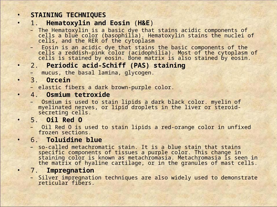
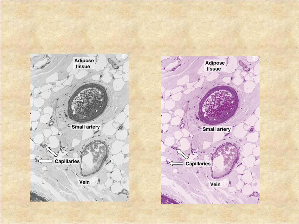
• STAINING TECHNIQUES • 1. Hematoxylin and Eosin (H&E)
– The Hematoxylin is a basic dye that stains acidic components of cells a blue color (basophilia). Hematoxylin stains the nuclei of cells, and the RER of the cytoplasm
– Eosin is an acidic dye that stains the basic components of the cells a reddish-pink color (acidophilia). Most of the cytoplasm of cells is stained by eosin. Bone matrix is also stained by eosin.
• 2. Periodic acid-Schiff (PAS) staining – mucus, the basal lamina, glycogen.
• 3. Orcein – elastic fibers a dark brown-purple color.
• 4. Osmium tetroxide – Osmium is used to stain lipids a dark black color. myelin of myelinated nerves, or lipid
droplets in the liver or steroid-secreting cells. • 5. Oil Red O
– Oil Red O is used to stain lipids a red-orange color in unfixed frozen sections. • 6. Toluidine blue
– so-called metachromatic stain. It is a blue stain that stains specific components of tissues a purple color. This change in staining color is known as metachromasia. Metachromasia is seen in the matrix of hyaline cartilage, or in the granules of mast cells.
• 7. Impregnation – Silver impregnation techniques are also widely used to demonstrate reticular fibers.
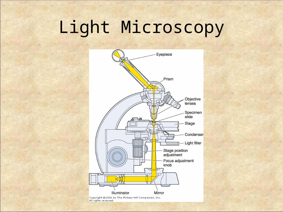
Light Microscopy
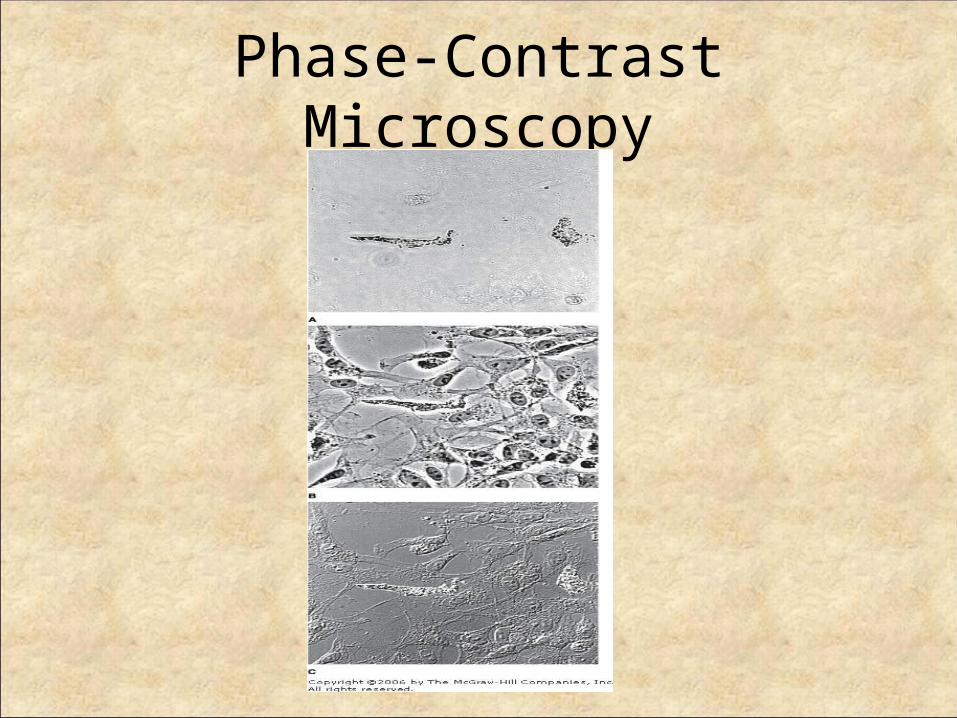
Phase-Contrast Microscopy

Polarizing Microscopy
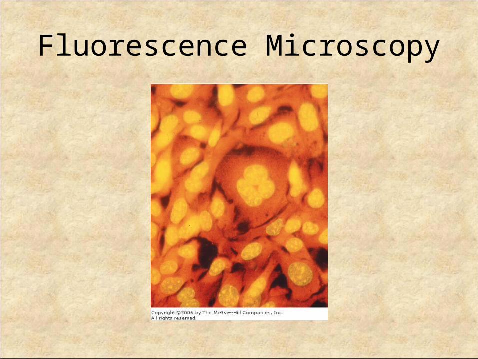
Fluorescence Microscopy
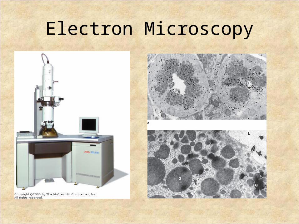
Electron Microscopy
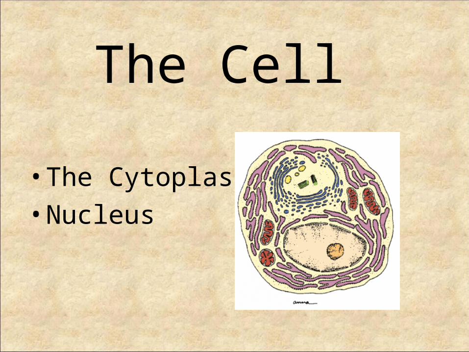
The Cell
• The Cytoplasm• Nucleus
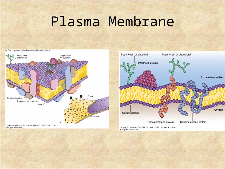
Plasma Membrane

Function of PM
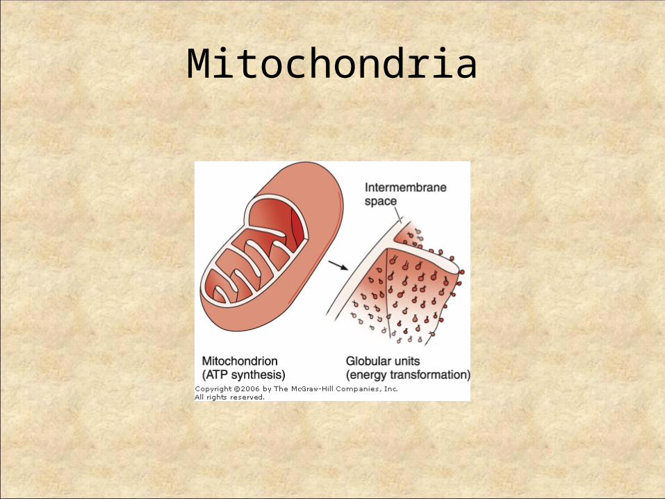
Mitochondria
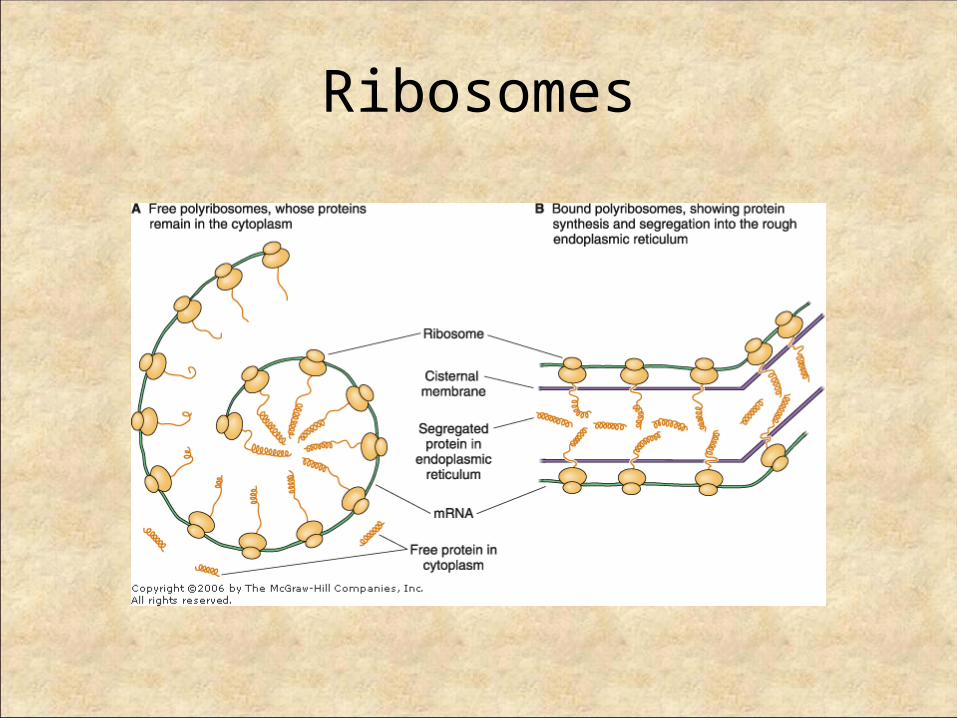
Ribosomes
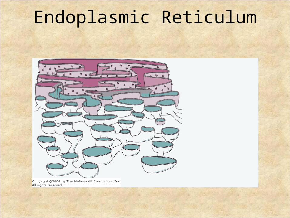
Endoplasmic Reticulum

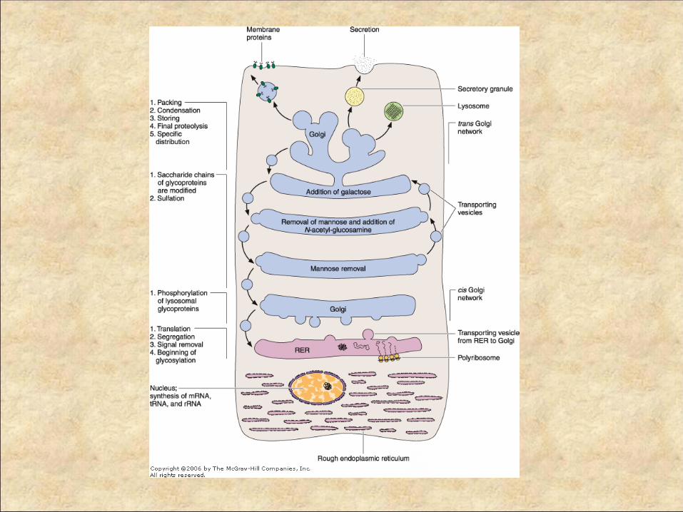
Golgi Complex
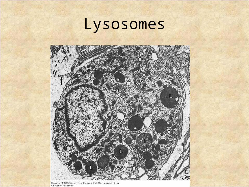
Lysosomes

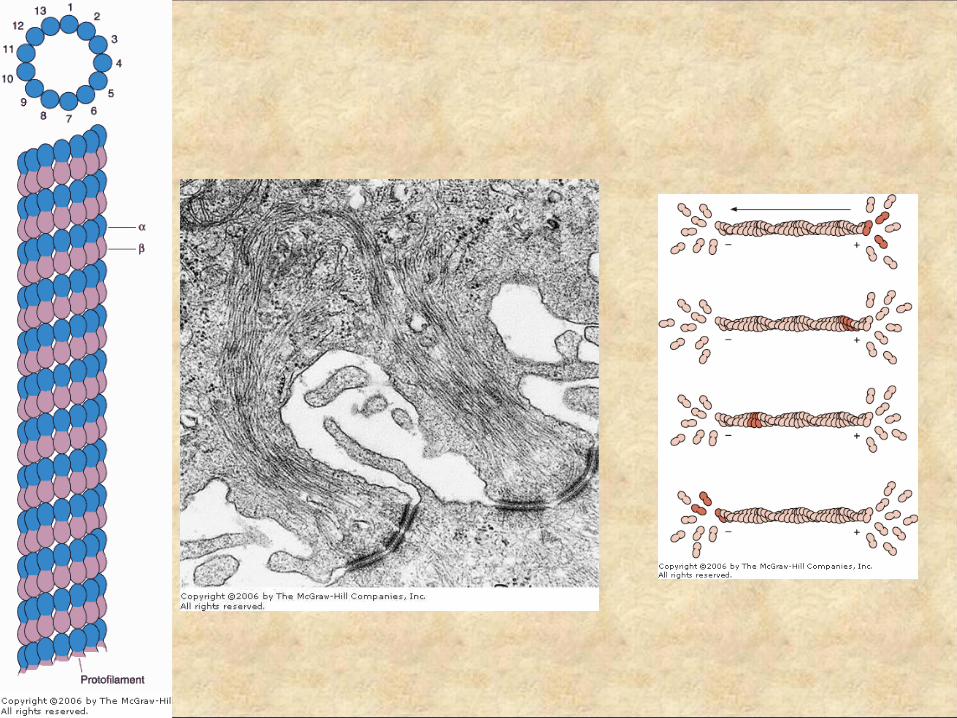
The Cytoskeleton
• Microtubules• Intermediate Filaments• Actin Filaments

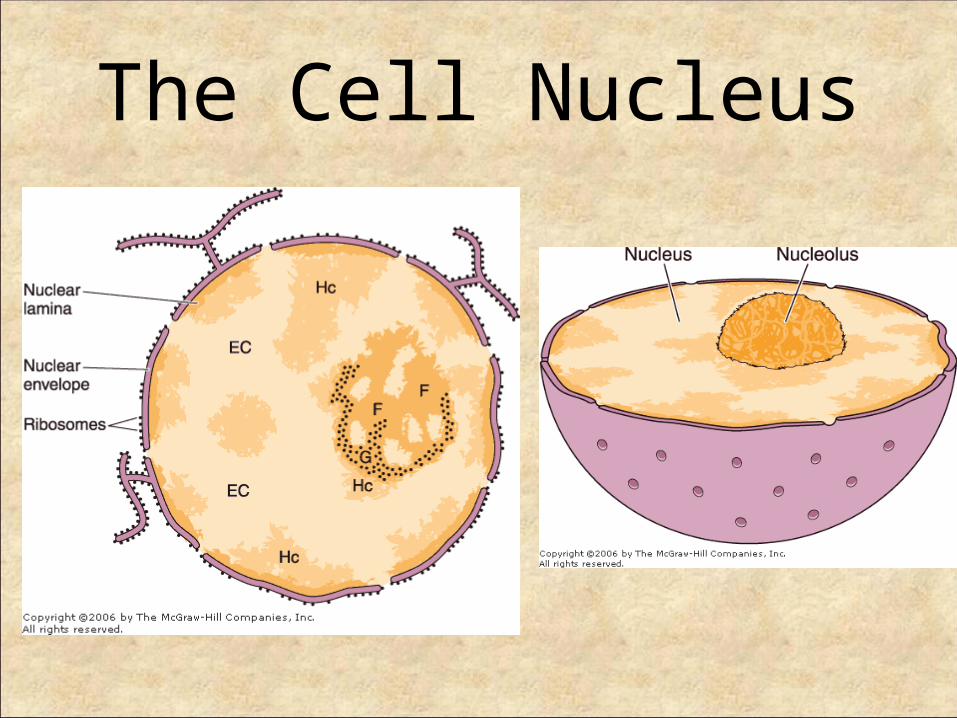
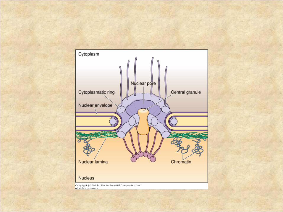
The Cell Nucleus
Related Documents