Citation: Cerva, C.; Salpini, R.; Alkhatib, M.; Malagnino, V.; Piermatteo, L.; Battisti, A.; Bertoli, A.; Gersch, J.; Holzmayer, V.; Kuhns, M.; et al. Highly Sensitive HBsAg, Anti-HBc and Anti HBsAg Titres in Early Diagnosis of HBV Reactivation in Anti-HBc-Positive Onco-Haematological Patients. Biomedicines 2022, 10, 443. https:// doi.org/10.3390/biomedicines10020443 Academic Editor: Giovanni Squadrito Received: 13 January 2022 Accepted: 10 February 2022 Published: 14 February 2022 Publisher’s Note: MDPI stays neutral with regard to jurisdictional claims in published maps and institutional affil- iations. Copyright: © 2022 by the authors. Licensee MDPI, Basel, Switzerland. This article is an open access article distributed under the terms and conditions of the Creative Commons Attribution (CC BY) license (https:// creativecommons.org/licenses/by/ 4.0/). biomedicines Article Highly Sensitive HBsAg, Anti-HBc and Anti HBsAg Titres in Early Diagnosis of HBV Reactivation in Anti-HBc-Positive Onco-Haematological Patients Carlotta Cerva 1,† , Romina Salpini 2,† , Mohammad Alkhatib 2 , Vincenzo Malagnino 3 , Lorenzo Piermatteo 2 , Arianna Battisti 2 , Ada Bertoli 2 , Jeff Gersch 4 , Vera Holzmayer 4 , Mary Kuhns 4 , Gavin Cloherty 4 , Ludovica Ferrari 3 , Campogiani Laura 3 , Elisabetta Teti 3 , Maria Cantonetti 5 , William Arcese 5 , Francesca Ceccherini-Silberstein 2 , Carlo-Federico Perno 6 , Massimo Andreoni 3 , Valentina Svicher 2,‡ and Loredana Sarmati 3, * ,‡ 1 I.N.M.I. “Lazzaro Spallanzani”, 00149 Rome, Italy; [email protected] 2 Department of Experimental Medicine, Tor Vergata University, 00133 Rome, Italy; [email protected] (R.S.); mohammad–[email protected] (M.A.); [email protected] (L.P.); [email protected] (A.B.); [email protected] (A.B.); [email protected] (F.C.-S.); [email protected] (V.S.) 3 Unit of Clinical Infectious Disease, Department of System Medicine, Tor Vergata University, 00133 Rome, Italy; [email protected] (V.M.); [email protected] (L.F.); [email protected] (C.L.); [email protected] (E.T.); [email protected] (M.A.) 4 Infectious Disease Research, Abbott Diagnostics, Abbott Park, Green Oaks, IL 60064, USA; [email protected] (J.G.); [email protected] (V.H.); [email protected] (M.K.); [email protected] (G.C.) 5 Stem Cell Transplant Unit, Department of Hematology, Tor Vergata University, 00133 Rome, Italy; [email protected] (M.C.); [email protected] (W.A.) 6 Children Hospital “Bambino Gesù”, 00165 Rome, Italy; [email protected] * Correspondence: [email protected] † These authors contributed equally to this work. ‡ These authors contributed equally to this work. Abstract: The role of novel HBV markers in predicting Hepatitis B virus reactivation (HBV-R) in HBsAg-negative/anti-HBc-positive oncohaematological patients was examined. One hundred and seven HBsAg-negative/anti-HBc-positive oncohaematological patients, receiving anti-HBV prophylaxis for >18 months, were included. At baseline, all patients had undetectable HBV DNA, and 67.3% were anti-HBs positive. HBV-R occurred in 17 (15.9%) patients: 6 during and 11 after the prophylaxis period. At HBV-R, the median (IQR) HBV-DNA was 44 (27–40509) IU/mL, and the alanine aminotransferase upper limit of normal (ULN) was 44% (median (IQR): 81 (49–541) U/L). An anti-HBc > 3 cut-off index (COI) plus anti-HBs persistently/declining to <50 mIU/mL was predictive for HBV-R (OR (95% CI): 9.1 (2.7–30.2); 63% of patients with vs. 15% without this combination experienced HBV-R (p < 0.001)). The detection of highly sensitive (HS) HBsAg and/or HBV-DNA confirmed at >2 time points, also predicts HBV-R (OR (95% CI): 13.8 (3.6–52.6); 50% of positive vs. 7% of negative patients to these markers experienced HBV-R (p = 0.001)). HS-HBs and anti-HBc titration proved to be useful early markers of HBV-R. The use of these markers demonstrated that HBV-R frequently occurs in oncohaematological patients with signs of resolved HBV infection, raising issues of proper HBV-R monitoring. Keywords: HBV reactivation; anti-HBc positivity; resolved HBV infection; oncohaematological patients; new HBV markers; antiviral prophylaxis; HSCT 1. Introduction HBsAg-negative/anti-HBc-positive patients with oncohaematological diseases are at high risk of hepatitis B virus reactivation (HBV-R). This is due to the intrahepatic Biomedicines 2022, 10, 443. https://doi.org/10.3390/biomedicines10020443 https://www.mdpi.com/journal/biomedicines

Welcome message from author
This document is posted to help you gain knowledge. Please leave a comment to let me know what you think about it! Share it to your friends and learn new things together.
Transcript

�����������������
Citation: Cerva, C.; Salpini, R.;
Alkhatib, M.; Malagnino, V.; Piermatteo,
L.; Battisti, A.; Bertoli, A.; Gersch, J.;
Holzmayer, V.; Kuhns, M.; et al. Highly
Sensitive HBsAg, Anti-HBc and Anti
HBsAg Titres in Early Diagnosis of
HBV Reactivation in Anti-HBc-Positive
Onco-Haematological Patients.
Biomedicines 2022, 10, 443. https://
doi.org/10.3390/biomedicines10020443
Academic Editor:
Giovanni Squadrito
Received: 13 January 2022
Accepted: 10 February 2022
Published: 14 February 2022
Publisher’s Note: MDPI stays neutral
with regard to jurisdictional claims in
published maps and institutional affil-
iations.
Copyright: © 2022 by the authors.
Licensee MDPI, Basel, Switzerland.
This article is an open access article
distributed under the terms and
conditions of the Creative Commons
Attribution (CC BY) license (https://
creativecommons.org/licenses/by/
4.0/).
biomedicines
Article
Highly Sensitive HBsAg, Anti-HBc and Anti HBsAg Titres inEarly Diagnosis of HBV Reactivation in Anti-HBc-PositiveOnco-Haematological PatientsCarlotta Cerva 1,† , Romina Salpini 2,† , Mohammad Alkhatib 2 , Vincenzo Malagnino 3 ,Lorenzo Piermatteo 2 , Arianna Battisti 2, Ada Bertoli 2, Jeff Gersch 4, Vera Holzmayer 4, Mary Kuhns 4,Gavin Cloherty 4, Ludovica Ferrari 3, Campogiani Laura 3, Elisabetta Teti 3, Maria Cantonetti 5, William Arcese 5 ,Francesca Ceccherini-Silberstein 2, Carlo-Federico Perno 6, Massimo Andreoni 3, Valentina Svicher 2,‡
and Loredana Sarmati 3,*,‡
1 I.N.M.I. “Lazzaro Spallanzani”, 00149 Rome, Italy; [email protected] Department of Experimental Medicine, Tor Vergata University, 00133 Rome, Italy; [email protected] (R.S.);
mohammad–[email protected] (M.A.); [email protected] (L.P.);[email protected] (A.B.); [email protected] (A.B.); [email protected] (F.C.-S.);[email protected] (V.S.)
3 Unit of Clinical Infectious Disease, Department of System Medicine, Tor Vergata University, 00133 Rome, Italy;[email protected] (V.M.); [email protected] (L.F.); [email protected] (C.L.);[email protected] (E.T.); [email protected] (M.A.)
4 Infectious Disease Research, Abbott Diagnostics, Abbott Park, Green Oaks, IL 60064, USA;[email protected] (J.G.); [email protected] (V.H.); [email protected] (M.K.);[email protected] (G.C.)
5 Stem Cell Transplant Unit, Department of Hematology, Tor Vergata University, 00133 Rome, Italy;[email protected] (M.C.); [email protected] (W.A.)
6 Children Hospital “Bambino Gesù”, 00165 Rome, Italy; [email protected]* Correspondence: [email protected]† These authors contributed equally to this work.‡ These authors contributed equally to this work.
Abstract: The role of novel HBV markers in predicting Hepatitis B virus reactivation (HBV-R)in HBsAg-negative/anti-HBc-positive oncohaematological patients was examined. One hundredand seven HBsAg-negative/anti-HBc-positive oncohaematological patients, receiving anti-HBVprophylaxis for >18 months, were included. At baseline, all patients had undetectable HBV DNA,and 67.3% were anti-HBs positive. HBV-R occurred in 17 (15.9%) patients: 6 during and 11 afterthe prophylaxis period. At HBV-R, the median (IQR) HBV-DNA was 44 (27–40509) IU/mL, and thealanine aminotransferase upper limit of normal (ULN) was 44% (median (IQR): 81 (49–541) U/L). Ananti-HBc > 3 cut-off index (COI) plus anti-HBs persistently/declining to <50 mIU/mL was predictivefor HBV-R (OR (95% CI): 9.1 (2.7–30.2); 63% of patients with vs. 15% without this combinationexperienced HBV-R (p < 0.001)). The detection of highly sensitive (HS) HBsAg and/or HBV-DNAconfirmed at >2 time points, also predicts HBV-R (OR (95% CI): 13.8 (3.6–52.6); 50% of positive vs. 7%of negative patients to these markers experienced HBV-R (p = 0.001)). HS-HBs and anti-HBc titrationproved to be useful early markers of HBV-R. The use of these markers demonstrated that HBV-Rfrequently occurs in oncohaematological patients with signs of resolved HBV infection, raising issuesof proper HBV-R monitoring.
Keywords: HBV reactivation; anti-HBc positivity; resolved HBV infection; oncohaematologicalpatients; new HBV markers; antiviral prophylaxis; HSCT
1. Introduction
HBsAg-negative/anti-HBc-positive patients with oncohaematological diseases areat high risk of hepatitis B virus reactivation (HBV-R). This is due to the intrahepatic
Biomedicines 2022, 10, 443. https://doi.org/10.3390/biomedicines10020443 https://www.mdpi.com/journal/biomedicines

Biomedicines 2022, 10, 443 2 of 11
persistence of HBV circular covalently closed DNA (cccDNA) coupled with profound, andoften durable, immunosuppression [1–4].
HBV-R has a negative impact on the clinical course of oncohaematological patients,since it is associated with liver injury and with delayed administration or premature ter-mination of immunosuppressive treatments, contributing to worsening prognosis [1]. Forthis reason, most international guidelines recommend starting antiviral prophylaxis inoncohaematological patients at high HBV-R risk. Nevertheless, defining the most appro-priate drug and duration for antiviral prophylaxis remains challenging, with importantdifferences in recommendations across international guidelines [2–5]. However, HBV-Rcases, occurring even several years after the last immunosuppressive therapy, have beenfrequently reported, particularly in patients undergoing haematological stem cell transplan-tation (HSCT), as a consequence of deep and long-lasting immunosuppression [6]. Thishighlights the need to better define proper antiviral prophylaxis in this special population.
Recently, novel biomarkers have been proposed to reflect the intrahepatic HBV reser-voir [7,8], providing information on the presence of active cccDNA. In this context, apromising biomarker is represented by anti-HBc titres. Notably, in immunocompetent anti-HBc-positive/HBsAg-negative individuals, anti-HBc titres with a >4 cut-off index (COI)has been associated with the presence of intrahepatic cccDNA [9], while in HIV-positivepatients, anti-HBc titres with a >15 COI is suggestive of cryptic HBV replication [10].
Moreover, serum HBV-RNA emerged as a novel marker reflecting cccDNA tran-scriptional activity [11]. This biomarker, measuring the number of virions containingpregenomic HBV-RNA (thus lacking the reverse transcriptase process) [12], is particularlyuseful in unravelling ongoing viral activity in patients on antiviral prophylaxis and/ortreatment.
The availability of these innovative markers also parallels the development of novelassays with increased sensitivity for detecting/quantifying HBV biomarkers (such asserum HBsAg and HBV-DNA) capable of identifying minimal HBV replicative activity,thus allowing an early HBV-R diagnosis and reducing the risk of developing severe clinicalmanifestations [13,14].
In this light, this study aims to (i) evaluate the rate of HBV-R in HBsAg-negative/anti-HBc-positive oncohaematological patients undergoing antiviral prophylaxis for >18 months;and (ii) define the role of highly sensitive detection of HBsAg (HS-HBs) and quantitativeanti-HBc titres in predicting HBV-R in the setting of haematological malignancies. In a sub-group of patients treated for HBV-R, the trend in the HBV-RNA values was also evaluated.
2. Materials and Methods2.1. Study Population
This longitudinal study included 107 adult patients followed for a haematologicalmalignancy at the University Hospital Tor Vergata, Rome, Italy, from January 2016 toDecember 2020. Patients were HBsAg negative/anti-HBc positive and HBV-DNA negativeat baseline screening. Patients received lamivudine prophylaxis for >18 months aftercompleting chemotherapy and/or HSCT according to EASL Guidelines [15]. Patients weremonitored every 3 months to assess HBV-R occurrence during or after completing HBVprophylaxis for a median (IQR) duration of 44 (31–56) months. All data were censoredin December 2020, with a target monitoring period of 2 years. HBV-R was defined asHBV-DNA reappearance (HBV-DNA ≥ 20 IU/mL), regardless of liver biochemistry [16].
All the patients’ personal information was treated in a confidential manner and allclinical data were collected anonymously to respect the patients’ privacy. Informed consentto study participation was obtained from all enrolled subjects. All methods were performedin accordance with the relevant guidelines and regulations. The study was approved bythe Tor Vergata University Ethical Committee (Code: RS.178.18).

Biomedicines 2022, 10, 443 3 of 11
2.2. HBV Laboratory Evaluation2.2.1. Classical HBV Markers
HBsAg was tested by the commercially available quantitative HBsAg detection sys-tem (Abbott Architect, lower limit of detection (LLoD): 50 mIU/mL (routinely used fordiagnostic purposes at University Hospital Tor Vergata) (Abbott Diagnostics, Abbott Park,IL, USA). Similarly, the qualitative detection of anti-HBc and anti-HBs titres was assessedby the Abbott Architect instrument (Abbott Diagnostics, Abbott Park, IL, USA). SerumHBV DNA was quantified by a real-time PCR system (COBAS Amplicor-COBAS TaqMan,Roche, Basel, Switzerland; lower limit of quantitation (LLoQ): 20 IU/m).
2.2.2. Innovative HBV MarkersHS-HBs
HS-HBs was assessed by an ARCHITECT HBsAg NEXT Qualitative assay (HBsAgNx;Abbott Diagnostics, Abbott Park, IL, USA, LLoD: 5 mIU/mL), while the highly sensitivequantification of HBsAg was by Lumipulse HBsAg-HQ (Fujirebio) (LLOQ: 5 mIU/mL),working on a LUMIPULSE®G system for the qualitative assay (Abbott).
Both assays are characterized by a 1 log IU/mL increase in sensitivity with respect toserological assays routinely used for HBsAg detection/quantification. HBsAg detection byboth assays showed a high concordance (98.1%).
Quantification of Anti-HBc Titer
Anti-HBc levels were quantified by using the Lumipulse®G HBcAb-N assay (Fujire-bio, Tokyo, Japan). The assay is based on a fully automated two-step sandwich CLEIAtechnology working on the LUMIPULSE®G system. Anti-HBc IgG levels are automaticallyreported as COI, calculated as a multiple of the cut-off value obtained from the calibrationdata (COI = S/C × 0.09). The lower limit of quantification reported by the manufacturerfor the assay was 1 COI.
Quantification of Serum HBV-RNA
The levels of serum pregenomic HBV-RNA were assessed by using the Abbott Real-Time HBV-RNA Research Use Only assay (Abbott Diagnostics, Abbott Park, IL, USA), asdescribed in [17]. Results are expressed as log10 U/mL. The lower limit of quantitationof the assay was 1.65 log10 U/mL. Serum HBV-RNA was quantified in 51 samples from6 HBV-reactivated patients at the diagnosis of HBV-R and during the following antiviraltherapy (median (IQR) time of treatment: 12 (11–24) months; median (IQR) number ofsamples collected for patient: 3 (2–6)).
2.3. Population-Based Sequencing of HBV Reverse Transcriptase
Population-based sequencing of HBV reverse transcriptase was performed on serumsamples at HBV-R [18].
In details, HBV-DNA was extracted using a commercially available kit (QIAmpDNAblood mini-kit, Qiagen Inc., Germantown, MD, USA), and amplified with Amplitaq-Gold-polymerase using the following primer pairs: 5′-GGTCACCATATTCTTGGGAAand 5′-GTGGGGGTTGCGTCAGCAAA. PCR conditions were: one cycle (93 ◦C 12 min),40 cycles (94 ◦C 50 s, 57 ◦C 50 s, 72 ◦C 1 min and 30 s). PCR-products were sequencedby using eight different sequence-specific primers, BigDye-terminator-v.3.1 sequencingkit (Applied-Biosystems Foster City, CA, USA ) and an automated sequencer (ABI-3130XL). The sequences were analysed by using SeqScape software. The quality endpoint wasensured by a double sequence coverage for each nucleotidic region.
2.4. Statistical Methods
The Mann–Whitney U-test for continuous variables and Fisher’s exact test for dis-crete variables were applied to define statistically significant differences. The cumulativeprobability of experiencing HBV-R was estimated by Kaplan–Meier analysis in the overall
Biomedicines 2022, 10, 443 4 of 11
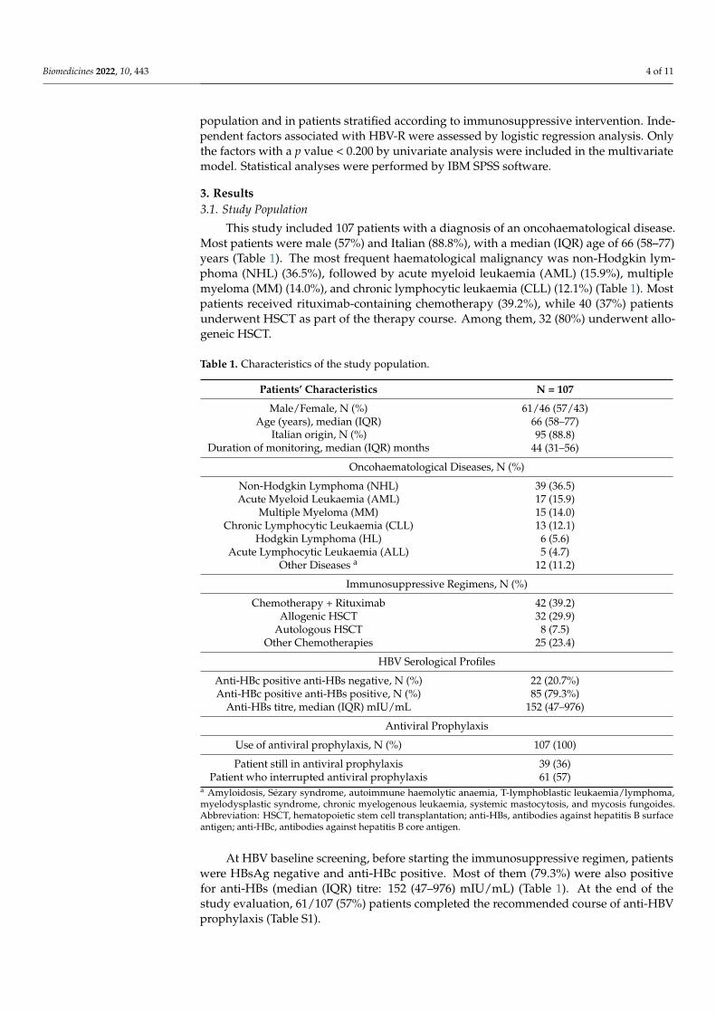
population and in patients stratified according to immunosuppressive intervention. Inde-pendent factors associated with HBV-R were assessed by logistic regression analysis. Onlythe factors with a p value < 0.200 by univariate analysis were included in the multivariatemodel. Statistical analyses were performed by IBM SPSS software.
3. Results3.1. Study Population
This study included 107 patients with a diagnosis of an oncohaematological disease.Most patients were male (57%) and Italian (88.8%), with a median (IQR) age of 66 (58–77)years (Table 1). The most frequent haematological malignancy was non-Hodgkin lym-phoma (NHL) (36.5%), followed by acute myeloid leukaemia (AML) (15.9%), multiplemyeloma (MM) (14.0%), and chronic lymphocytic leukaemia (CLL) (12.1%) (Table 1). Mostpatients received rituximab-containing chemotherapy (39.2%), while 40 (37%) patientsunderwent HSCT as part of the therapy course. Among them, 32 (80%) underwent allo-geneic HSCT.
Table 1. Characteristics of the study population.
Patients’ Characteristics N = 107
Male/Female, N (%) 61/46 (57/43)Age (years), median (IQR) 66 (58–77)
Italian origin, N (%)Duration of monitoring, median (IQR) months
95 (88.8)44 (31–56)
Oncohaematological Diseases, N (%)
Non-Hodgkin Lymphoma (NHL) 39 (36.5)Acute Myeloid Leukaemia (AML) 17 (15.9)
Multiple Myeloma (MM) 15 (14.0)Chronic Lymphocytic Leukaemia (CLL) 13 (12.1)
Hodgkin Lymphoma (HL) 6 (5.6)Acute Lymphocytic Leukaemia (ALL) 5 (4.7)
Other Diseases a 12 (11.2)
Immunosuppressive Regimens, N (%)
Chemotherapy + Rituximab 42 (39.2)Allogenic HSCT 32 (29.9)
Autologous HSCT 8 (7.5)Other Chemotherapies 25 (23.4)
HBV Serological Profiles
Anti-HBc positive anti-HBs negative, N (%) 22 (20.7%)Anti-HBc positive anti-HBs positive, N (%) 85 (79.3%)
Anti-HBs titre, median (IQR) mIU/mL 152 (47–976)
Antiviral Prophylaxis
Use of antiviral prophylaxis, N (%) 107 (100)
Patient still in antiviral prophylaxis 39 (36)Patient who interrupted antiviral prophylaxis 61 (57)
a Amyloidosis, Sézary syndrome, autoimmune haemolytic anaemia, T-lymphoblastic leukaemia/lymphoma,myelodysplastic syndrome, chronic myelogenous leukaemia, systemic mastocytosis, and mycosis fungoides.Abbreviation: HSCT, hematopoietic stem cell transplantation; anti-HBs, antibodies against hepatitis B surfaceantigen; anti-HBc, antibodies against hepatitis B core antigen.
At HBV baseline screening, before starting the immunosuppressive regimen, patientswere HBsAg negative and anti-HBc positive. Most of them (79.3%) were also positivefor anti-HBs (median (IQR) titre: 152 (47–976) mIU/mL) (Table 1). At the end of thestudy evaluation, 61/107 (57%) patients completed the recommended course of anti-HBVprophylaxis (Table S1).

Biomedicines 2022, 10, 443 5 of 11
3.2. Occurrence of HBV-R
During the entire patients’ monitoring (median (IQR duration) time: 44 [31–56]months), HBV-R was observed in 17/107 (15.8%) patients, with a 5-year cumulative riskof 27.4% (Table 2 and Figure 1A). The 5-year cumulative risk of HBV-R was significantlyhigher in patients undergoing HSCT (without statistically significant differences betweenautologous and allogenic HSCT: 54.3% vs. 51.1%) than in those receiving chemotherapieswith or without rituximab (Figure 1B).
Table 2. Characteristics of 17 patients at HBV-R.
Patients’ Characteristics N = 17
Serum HBV-DNA, median (IQR) IU/mL 44 (27–40,509)Serum ALT > UNL, N (%) 7 (44)
- Serum ALT, median (IQR) U/L 81 (49–541)
HBV Serological Profiles at HBV-R
HBsAg positive, N (%): 4 (23.5)HBsAg negative, N (%) 13 (76.5)
Anti-HBs positive, N (%): 8 (47)- Anti-HBs titre, range mIU/mL 13–505
Immunosuppressive Regimen, N (%)
Chemotherapy + Rituximab3 (17.6)
Allogeneic HSCT8 (47.2)
Autologous HSCT3 (17.6)
Other Chemotherapies3 (17.6)
HBV reactivation occurrence
- During prophylaxis, N (%) 6 (35.3)- After prophylaxis completion, N (%) 11 (64.7)
Months after prophylaxis completion, median(IQR) 4 (2–13)
Abbreviations: ALT, alanine aminotransferase; UNL; upper normal limit; HBsAg; hepatitis B surface antigen;anti-HBs, antibodies against hepatitis B surface antigen; HSCT, hematopoietic stem cell transplantation.
Figure 1. Survival analysis by competing risk estimates of the cumulative odds of HBV reactivation.The graph plots report the cumulative risk for the occurrence of HBV-reactivation in overall popula-tion (N = 107) (A) and in patients stratified according to HSCT (N = 40) versus RTX (N = 42) and/orother chemotherapies (N = 25) (B). The cumulative risk was calculated by Kaplan Meier. Statisticallysignificant difference was calculated by log-rank test.

Biomedicines 2022, 10, 443 6 of 11
Notably, most cases of HBV-R (11/17, 64.7%) occurred after completing anti-HBVprophylaxis (range: 1–27 months after prophylaxis completion). In the remaining 35.3%,HBV-R occurred while receiving antiviral prophylaxis (Table 2). No patients harboureddrug-resistant strains.
At HBV-R, the median (IQR) HBV-DNA was 44 (27–40,509) IU/mL, and ALT wasabove the normal level (>UNL) in 44% of patients (median (IQR) ALT: 81 (49–541) U/L),indicating a diagnosis of biochemical HBV-R in less than half of the patients (Table 2). Thesedata are indicative of an early HBV-R diagnosis as a result of strict patient monitoring (every3 months) followed in this study protocol. The median anti-HBc titre was 22 (3–46) COI.
The graph plots report the cumulative risk for the occurrence of HBV reactivation in theoverall population (N = 107) (A) and in patients stratified according to HSCT (N = 40) versuschemotherapies with (N = 42) or without rituximab (RTX) (N = 25) (B). The cumulativerisk was calculated by Kaplan–Meier analysis. Statistically significant differences werecalculated by the log-rank test.
3.3. Predictive Role of Serological HBV Markers in the Diagnosis of HBV-R
An anti-HBc > 3 COI combined with an anti-HBs persistent or declining to <50 mIU/mLduring patient monitoring was significantly correlated with a higher risk of developingHBV-R (OR (95% CI): 9.1 (2.7–30.2); p < 0.001). Indeed, 63% of patients with the combina-tion of an anti-HBc > 3 COI plus anti-HBs < 50 mIU/mL experienced HBV-R compared to15% of patients without this combination (p < 0.001) (Figure 2A). Notably, this result wasalso confirmed in the subset of patients experiencing HBV-R after completing anti-HBVprophylaxis (OR (95% CI): 8.8 (2.0–38); p = 0.005). In this setting, 55% of patients withthe combination of an anti-HBc > 3 COI plus anti-HBs < 50 mIU/mL experienced HBV-Rversus 14% without this combination (p = 0.005) (Figure 2B).
Figure 2. The combined usage of accurate HBV markers can predict the occurrence of HBV reacti-vation. The histogram reports the occurrence of HBV-reactivation in patients positive/negativeto anti-HBc > 3COI + anti-HBs < 50 mIU/mL (A,C) and in patients positive/negative HS-HBsAg

Biomedicines 2022, 10, 443 7 of 11
and/or HBV-DNA (B,D). Data in (A,B) are referred to overall population (N = 107), while in (C,D)are referred to the subset of patient completing the recommended course of antiviral prophylaxis(N = 61) during the entire follow-up. Statistically significant difference was calculated Chi-squiredtest for independence based on 2 × 2 contingency table.
A further step in this study was to evaluate the predictive role of virological markersin HBV-R diagnosis. The on-monitoring analysis revealed that the detection of HS_HBsand/or of serum HBV-DNA (target detected below LLOQ), confirmed at least at 2 timepoints, was a predictive factor for HBV-R (OR (95% CI): 13.8 (3.6–52.6); p < 0.001). Indeed,50% of patients positive to HS-HBsAg and/or to serum HBV-DNA versus 7% never positiveto these markers experienced HBV-R (p < 0.001) (Figure 2C). This result was also confirmedin the subset of patients experiencing HBV-R after completing anti-HBV prophylaxis(p < 0.001) (Figure 2D), suggesting that monitoring these biomarkers could be useful toidentify patients requiring prolonged or enhanced prophylaxis.
Multivariate analysis confirmed that either the combination of anti-HBc > 3 COI+ anti-HBs < 50 mIU/mL or positivity for HS-HBsAg and/or serum HBV-DNA (target detectedbelow LLOQ) were independent predictive factors for HBV-R (OR (95% CI): 7.2 (1.4–39.2),p = 0.020, and 5.3 (1.0–27.8), p = 0.049, respectively) (Table 3).
Table 3. Factors associated with HBV-R by multivariable logistic regression analysis.
Variables a Univariable Analysis Multivariable Analysis
Crude OR (95% CI) p-Value Adjusted OR (95% CI) p-Value
Gender (male vs. female) 2.0 (0.7–6.2) 0.224 - -Age (for 1 year increase) 1.0 (1.0–1.1) 0.101 1.1 (1.0–1.1) 0.435
HSCT vs. RTX and other chemotherapies 3.9 (1.3–11.5) 0.015 3.0 (0.5–17.2) 0.018Combination of anti-HBc > 3 COI +
anti-HBs < 50 mIU/mL 9.1 (2.7–30.2) <0.001 7.2 (1.4–39.2) 0.020
Detection to HS-HBs and/or to serumHBV-DNA < 20 IU/mL 13.8 (3.6–52.6) <0.001 5.3 (1.0–27.8) 0.049
a The logistic regression analysis was performed on 107 oncohaematological anti-HBc-positive/HBsAg-negativepatients. The following variables were considered: gender, age, HSCT vs. RXT and/or other chemotherapies,combined anti-HBc > 3 COI + anti-HBs < 50 mIU/mL, and positivity to HS-HBs or HBV-DNA detected <20 IU/mL.Only variables showing a p-value ≤ 0.200 in the univariable analysis were included in the multivariable analysis.Abbreviations: HSCT, hematopoietic stem cell transplantation; RTX, Rituximab; OR, odds ratio; CI, confidenceinterval; anti-HBs, antibodies against hepatitis B surface antigen; anti-HBc, antibodies against hepatitis B coreantigen; HS-HBs, high-sensitive hepatitis B surface antigen. The significant values appeared in bold.
The histogram reports the occurrence of HBV reactivation in patients with or withoutthe combination of anti-HBc > 3 COI + anti-HBs < 50 mIU/mL (A,B) and in patients posi-tive/negative for HS-HBsAg and/or HBV DNA (C,D). Data in (A,C) refer to the overallpopulation (N = 107), while those in (B,D) refer to the subset of patients completing therecommended course of antiviral prophylaxis (N = 61) during the entire follow-up. Statis-tically significant differences were calculated using the chi-square test for independencebased on a 2 × 2 contingency table.
3.4. Outcome of Patients Experiencing HBV-R
Among the 17 patients experiencing HBV-R, 16 received antiviral therapy with ente-cavir and/or tenofovir disoproxil fumarate/tenofovir alafenamide and were monitored fora median (IQR) time of 19 (15–34) months, while one patient was lost to follow-up.
By analysing the treatment outcome, ALT normalization was achieved by all patientsafter a median (IQR) time of 3 (3–9) months, and 81.3% (13/16) achieved virologicalsuppression within 6 months of antiviral therapy.
Overall, 25% of patients (4/16) died from the progression of the underlying onco-haematological disease, while no death due to hepatic failure occurred.
In a subset of 6 (35%) out of 17 patients, serum HBV RNA was also quantified at HBV-Rand during antiviral treatment (median (IQR) time of follow-up: 12 (11–24) months). At

Biomedicines 2022, 10, 443 8 of 11
HBV-R, serum HBV-RNA ranged from <LLOQ to 6.6 log IU/mL. In 5 out of 6 patients, bothserum HBV-DNA and HBV-RNA became undetectable within 6 months of treatment andremained undetectable in the subsequent follow-up, suggesting rapid and durable silencingof cccDNA transcriptional activity. A different scenario was observed in the remainingpatient, characterized at HBV-R by serum HBV-DNA and HBV-RNA of 6.0 log IU/mL and6.6 log IU/mL, respectively. Notably, after 9 months of treatment, a significant decay (up to2.8 log IU/mL) was observed exclusively for serum HBV-DNA, while serum HBV-RNAremained stable at approximately 6 log IU/mL, indicating elevated cccDNA transcriptionalactivity. To date, the patient is still in entecavir treatment, and the last (June 2021) HBV-DNAvalue was undetectable below 20 UI/mL. He is continuing regular follow up.
3.5. The Added Value of HS HBsAg Quantification: A Case Report
Here, a clinical case is reported, highlighting the importance of HS-HBsAg quantifica-tion in unravelling minimal HBV replicative activity.
The patient was a 58-year-old Italian male diagnosed with Hodgkin lymphoma. Thepatient was anti-HBc, isolated at baseline screening, and thus received proper antiviral pro-phylaxis before starting chemotherapy, containing adriamycin, bleomycin, vinblastyn, anddacabarzin. During 24 months of patient monitoring, serum HBV-DNA was persistentlyundetectable, and HBsAg was persistently negative by classical assays, while transam-inases fell within the normal values. Antiviral prophylaxis was suspended 18 monthsafter completing chemotherapy. Unexpectedly, 1 month after prophylactic suspension,the patient experienced HBV-R with HBV-DNA and HBsAg reappearance in serum andALT levels rising to 95 IU/mL (Figure S1). The highly sensitive quantification of HBsAgrevealed that HBsAg was already detected at each time point analysed during antiviralprophylaxis, supporting the intrahepatic activity of the HBV reservoir that can explainHBV-R occurrence immediately after the suspension of antiviral prophylaxis (Figure 3).
Figure 3. A clinical case shows the quantified persistence of high-sensitivity HBsAg.
The line chart reports the trend of serum HBV-DNA and ALT during patient moni-toring. At each time point, the highly sensitive detection of HBsAg was assessed by anARCHITECT HBsAg NEXT Qualitative assay (HBsAgNx; Abbott Diagnostics, AbbottPark, IL, USA, LLoD: 5 mIU/mL), while the highly sensitive quantification of HBsAgby Lumipulse HBsAg-HQ (Fujirebio) (LLOQ: 5 mIU/mL), working on a LUMIPULSE®Gsystem the qualitative assay (Abbott).

Biomedicines 2022, 10, 443 9 of 11
4. Discussion
This study shows the occurrence of HBV-R in almost 16% of oncohaematological anti-HBc-positive/HBsAg-negative patients. Notably, most of them (64.7%) experienced HBV-Rafter completing the recommended course of antiviral prophylaxis, supporting the need toreconsider its proper duration in a setting characterized by profound immunosuppression.These results may pave the way for designing further ad hoc studies aimed to define a newdesirable duration of antiviral prophylaxis.
At the same time, this study raises the issue to unravel the role of novel HBV biomark-ers in identifying patients at higher risk of HBV-R. In this direction, this study shows thatthe combination of an anti-HBc > 3 COI plus anti-HBs at or declining to <50 mIU/mLduring the follow-up is predictive for HBV-R. This datum, confirmed by multivariableanalysis, is also present in the subgroup of patients experiencing HBV-R after prophy-laxis interruption. Our finding is in keeping with a previous study on an Asian cohortof oncohaematological patients showing that the levels of anti-HBc > 6.41 IU/mL andanti-HBs < 56.48 mIU/mL may predict HBV-R in patients with lymphoma [19].
Overall, these findings support the concept that the progressive weakening of thehumoral response coupled with transcriptionally active cccDNA can play an importantrole in driving HBV-R. This corroborates the importance of integrating HBV biomarkers, re-flecting the activity of the intrahepatic HBV reservoir and the strength of anti-HBV immuneresponses, for the optimized management of severely immunosuppressed patients at risk ofHBV-R. In particular, the combination of an anti-HBc > 3 COI plus anti-HBs < 50 mIU/mLcould guide the identification of patients more prone to develop HBV-R who need closermonitoring, extended prophylaxis, or more potent anti-HBV drugs, such as tenofoviror entecavir.
Furthermore, the on-monitoring analysis of virological markers showed that thedetection of HS-HBs and/or a target detected serum HBV-DNA (below LLOQ) stronglyincreases HBV-R risk. This result, confirmed by multivariate analysis, is present in patientswho reactivated HBV both during prophylaxis and after its suspension, thus demonstratingthe ability of these biomarkers to identify patients with minimal viral replication. The roleof HS-HBs is supported by the paradigmatic clinical case showing persistent positivityfor this biomarker (not detected by the currently available assays) preceding HBV-R. Suchpositivity was also coupled with persistently high anti-HBc levels (ranging from 64 to130 COI), further reinforcing the presence of an already replicative active HBV reservoir,even several months before the clinical evidence of HBV-R diagnosis. This finding is alsoconsistent with a recent Japanese study showing that the detection of HS-HBs and a targetdetected serum HBV-DNA below the lower limit of quantification can help identify patientsmore prone to HBV-R [20].
In this context, highly sensitive assays for HBV-DNA quantification, such as thosebased on digital droplet PCR, capable of detecting even a few copies of HBV-DNA [14,21],could also represent a useful tool to demonstrate the presence of low-level HBV replica-tive activity.
Although based on a limited sample size, this study also supports the utility of serumHBV-RNA (although not yet available in clinical practice) in monitoring the therapeuticresponse of patients experiencing HBV-R. The loss of serum HBV-RNA, observed in mostpatients during treatment, may indicate exhausting or transcriptional silencing of cccDNA.As reported by previous studies, this highlights the potential role of serum HBV-RNA as abiomarker to guide the safe discontinuation of NUC therapy [22].
In the present study, almost half of patients with HBV-R underwent allogeneic HSCT,which represents a category of patients characterized by the highest grade of severe im-munosuppression. Little information is available on the history of immune recovery in ourstudy population, but it is known that it may take years post-HSCT. In this light, assessingthis issue could help to better define the correlation between the degree and quality ofimmunosuppression and HBV-R risk.

Biomedicines 2022, 10, 443 10 of 11
In conclusion, HBV-R frequently occurs in anti-HBc-positive/HBsAg-negative onco-haematological patients, particularly after completing the recommended course of antiviralprophylaxis, suggesting the need to reconsider the duration of prophylaxis in profoundimmunosuppression. In this setting, close monitoring based on the integrated use of novelHBV markers can help to detect minimal viral replication, thus guiding the identificationof patients at higher risk of developing HBV-R who would need to change the anti-HBVdrug towards more potent antivirals (tenofovir, entecavir) or to extend prophylaxis.
Supplementary Materials: The following supporting information can be downloaded at: https://www.mdpi.com/article/10.3390/biomedicines10020443/s1, Table S1: Characteristics of patientscompleting anti HBV prophylaxis.
Author Contributions: Conceptualization: L.S., V.S., R.S., C.C., V.M. and M.A. (Mohammad Alkhatib);data curation: M.A. (Mohammad Alkhatib), A.B. (Ada Bertoli), A.B. (Arianna Battisti), L.F., C.L.,E.T. and C.C; methodology: L.P., A.B. (Arianna Battisti), M.A. (Mohammad Alkhatib), V.H., J.G.,M.K. and G.C.; formal analysis: M.A. (Mohammad Alkhatib), L.P. and C.C.; investigation: R.S. andV.M.; writing: R.S., C.C., L.S. and V.S.; review and editing: F.C.-S., C.-F.P., M.A. (Massimo Andreoni),W.A., M.C., M.K. and G.C.; resources: J.G., M.K. and G.C. All authors have approved the submittedversion and agree to be personally accountable for the author’s own contributions and to ensurethat questions related to the accuracy or integrity of any part of the work, even those in which theauthor was not personally involved, are appropriately investigated, resolved, and documented in theliterature. All authors have read and agreed to the published version of the manuscript.
Funding: This research was, in part, financially supported by grant from the Gilead FellowshipProgram 2015 attributed to L.S.
Institutional Review Board Statement: The study was approved by the Tor Vergata UniversityEthical Committee (Code: RS.178.18).
Informed Consent Statement: Informed consent was obtained from all subjects involved in the study.
Data Availability Statement: Data is contained within the article and supplementary material.
Acknowledgments: We thank the AVI RALIA Foundation, VIRONET C Foundation, and the ItalianMinistry of University and Research (project protocol: PRIN 20179 JHAMZ) for the financial supportof the study. We are grateful to Corinna Orsini and Maria Assunta Melone (Fujirebio Italia) forproviding reagents and technical support. The Lumipulse® G HBcAb-N assay is currently notcommercially available in Europe and only in Japan. We also thank Massimiliano Bruni and AndreaBiddittu for data management.
Conflicts of Interest: Salpini R reports speaker fees and consultancy from Diasorin and FujiRebiooutside the submitted work. Sarmati L reports grants from Gilead Sciences and Merck Sharp &Dohme outside the submitted work. Andreoni M reports grants, consultancy and other from BristolMyers Squibbs, grants, consultancy and other from Gilead Sciences, grants, consultancy and otherfrom MSD, outside the submitted work. Ceccherini-Silberstein F reports grants, consultancy and otherfrom Bristol Myers Squibbs, grants, consultancy and other from Gilead Sciences, grants, consultancyand other from MSD, consultancy from Janssen-Cilag, grants and consultancy from Viiv Healthcare,consultancy from Roche Diagnostics, outside the submitted work. Perno CF reports grants andconsultancy from Bristol Myers Squibb, grants and consultancy from Gilead Sciences, consultancyfrom MSD, consultancy from Abbott Diagnostics, consultancy from Roche Diagnostics, outside thesubmitted work. Svicher V reports grants and consultancy from Roche, Gilead Sciences, grants, andDiasorin Diagnostics, Fujirebio, outside the submitted work. Other authors’ conflicts of interest: none.
References1. Sagnelli, C.; Pisaturo, M.; Calò, F.; Martini, S.; Sagnelli, E.; Coppola, N. Reactivation of hepatitis B virus infection in patients with
hemo-lymphoproliferative diseases, and its prevention. World J. Gastroenterol. 2019, 14, 3299–3312. [CrossRef] [PubMed]2. Myint, A.; Tong, M.J.; Beaven, S.W. Reactivation of Hepatitis B Virus: A Review of Clinical Guidelines. Clin. Liver Dis. 2020, 15,
162–167. [CrossRef]3. Terrault, N.A.; Lok, A.S.F.; McMahon, B.J.; Chang, K.-M.; Hwang, J.P.; Jonas, M.M.; Brown, R.S.B., Jr.; Bzowej, N.H.; Wong, J.B.
Update on prevention, diagnosis, and treatment of chronic hepatitis B: AASLD 2018 hepatitis B guidance. Hepatology 2018, 67,1560–1599. [CrossRef] [PubMed]

Biomedicines 2022, 10, 443 11 of 11
4. Sarin, S.K.; Kumar, M.P.; Lau, G.K.; Abbas, Z.; Chan, H.L.Y.; Chen, C.J.; Chen, D.-S.; Chen, H.L.; Chien, R.N.; Dokmeci, A.; et al.Asian-Pacific clinical practice guidelines on the management of hepatitis B: A 2015 update. Hepatol. Int. 2016, 10, 1–98. [CrossRef][PubMed]
5. Reddy, K.R.; Beavers, K.L.; Hammond, S.; Lim, J.K.; Falck-Ytter, Y.T. American Gastroenterological Association Institute Guidelineon the Prevention and Treatment of Hepatitis B Virus Reactivation During Immunosuppressive Drug Therapy. Gastroenterology2015, 148, 215–219. [CrossRef] [PubMed]
6. Cerva, C.; Colagrossi, L.; Maffongelli, G.; Salpini, R.; Di Carlo, D.; Malagnino, V.; Battisti, A.; Ricciardi, A.; Pollicita, M.; Bianchi,A.; et al. Persistent risk of HBV reactivation despite extensive lamivudine prophylaxis in haematopoietic stem cell transplantrecipients who are anti-HBc-positive or HBV-negative recipients with an anti-HBc-positive donor. Clin. Microbiol. Infect. 2016, 22,946.e1–946.e8. [CrossRef] [PubMed]
7. Inoue, T.; Tanaka, Y. Novel biomarkers for the management of chronic hepatitis B. Clin. Mol. Hepatol. 2020, 26, 261–279. [CrossRef]8. Charre, C.; Levrero, M.; Zoulim, F.; Scholtès, C. Non-invasive biomarkers for chronic hepatitis B virus infection management.
Antivir. Res. 2019, 169, 104553. [CrossRef]9. Caviglia, G.P.; Abate, M.L.; Tandoi, F.; Ciancio, A.; Amoroso, A.; Salizzoni, M.; Saracco, G.M.; Rizzetto, M.; Romagnoli, R.;
Smedile, A. Quantitation of HBV cccDNA in anti-HBc-positive liver donors by droplet digital PCR: A new tool to detect occultinfection. J. Hepatol. 2018, 69, 301–307. [CrossRef]
10. Salpini, R.; Malagnino, V.; Piermatteo, L.; Mulas, T.; Alkhatib, M.; Scutari, R.; Teti, E.; Cerva, C.; La Rosa, K.Y.; Brugneti,M.; et al. Cryptic HBV replicative activity is frequently revealed in anti-HBC-positive/HBsag-negative patients with HIVinfection by highly sensitive molecular assays, and can be predicted by integrating classical and novel serological HBV markers.Microorganisms 2020, 8, 1819. [CrossRef]
11. Anderson, M.; Gersch, J.; Luk, K.-C.; Dawson, G.; Carey, I.; Agarwal, K.; Shah, P.; Dusheiko, G.; Lau, D.; Cloherty, G. CirculatingPregenomic Hepatitis B Virus RNA Is Primarily Full-length in Chronic Hepatitis B Patients Undergoing Nucleos(t)ide AnalogueTherapy. Clin. Infect. Dis. 2021, 72, 2029–2031. [CrossRef] [PubMed]
12. Wang, J.; Shen, T.; Huang, X.; Kumar, G.R.; Chen, X.; Zeng, Z.; Zhang, R.; Chen, R.; Li, T.; Zhang, T.; et al. Serum hepatitis B virusRNA is encapsidated pregenome RNA that may be associated with persistence of viral infection and rebound. J. Hepatol. 2016, 65,700–710. [CrossRef] [PubMed]
13. Shinkai, N.; Kusumoto, S.; Murakami, S.; Ogawa, S.; Ri, M.; Matsui, T.; Tamori, A.; Toyoda, H.; Ishida, T.; Iida, S.; et al. Novelmonitoring of hepatitis B reactivation based on ultra-high sensitive hepatitis B surface antigen assay. Liver Int. 2017, 37, 1138–1147.[CrossRef] [PubMed]
14. Piermatteo, L.; Scutari, R.; Chirichiello, R.; Alkhatib, M.; Malagnino, V.; Bertoli, A.; Iapadre, N.; Ciotti, M.; Sarmati, L.; Andreoni,M.; et al. Droplet digital PCR assay as an innovative and promising highly sensitive assay to unveil residual and cryptic HBVreplication in peripheral compartment. Methods, 2021; in press. [CrossRef] [PubMed]
15. Lampertico, P.; Agarwal, K.; Berg, T.; Buti, M.; Janssen, H.L.A.; Papatheodoridis, G.V.; Zoulim, F.; Tacke, F. EASL 2017 ClinicalPractice Guidelines on the management of hepatitis B virus infection. J. Hepatol. 2017, 67, 370–398. [CrossRef] [PubMed]
16. Seto, W.-K.; Chan, T.S.-Y.; Hwang, Y.-Y.; Wong, D.K.-H.; Fung, J.; Liu, K.S.-H.; Gill, H.; Lam, Y.-F.; Lau, E.; Cheung, K.-S.;et al. Hepatitis B reactivation in occult viral carriers undergoing hematopoietic stem cell transplantation: A prospective study.Hepatology 2017, 65, 1451–1456. [CrossRef] [PubMed]
17. Butler, E.K.; Gersch, J.; McNamara, A.; Luk, K.-C.; Holzmayer, V.; De Medina, M.; Schiff, E.; Kuhns, M.; Cloherty, G.A. Hepatitis BVirus Serum DNA andRNA Levels in Nucleos(t)ide Analog-Treated or Untreated Patients During Chronic and Acute Infection.Hepatology 2018, 68, 2106–2117. [CrossRef]
18. Salpini, R.; Alteri, C.; Cento, V.; Pollicita, M.; Micheli, V.; Gubertini, G.; De Sanctis, G.; Visca, M.; Romano, S.; Sarrecchia, C.; et al.Snapshot on drug-resistance rate and profiles in patients with chronic hepatitis B receiving nucleos(t)ide analogues in clinicalpractice. J. Med. Virol. 2013, 85, 996–1004. [CrossRef]
19. Yang, H.-C.; Tsou, H.-H.; Pei, S.-N.; Chang, C.-S.; Chen, J.-H.; Yao, M.; Lin, S.-J.; Lin, J.; Yuan, Q.; Xia, N.; et al. Quantification ofHBV core antibodies may help predict HBV reactivation in patients with lymphoma and resolved HBV infection. J. Hepatol. 2018,69, 286–292. [CrossRef]
20. Kusumoto, S.; Tanaka, Y.; Suzuki, R.; Watanabe, T.; Nakata, M.; Sakai, R.; Fukushima, N.; Fukushima, T.; Moriuchi, Y.; Itoh, K.;et al. Ultra-high sensitivity HBsAg assay can diagnose HBV reactivation following rituximab-based therapy in patients withlymphoma. J. Hepatol. 2020, 73, 285–293. [CrossRef]
21. Yang, D.; Hu, T.; Wu, X.; Li, K.; Zhong, Q.; Liu, W. Droplet-digital polymerase chain reaction for detection of clinical hepatitis Bvirus DNA samples. J. Med. Virol. 2018, 90, 1868–1874. [CrossRef] [PubMed]
22. Wang, J.; Yu, Y.; Li, G.; Shen, C.; Meng, Z.; Zheng, J.; Jia, Y.; Chen, S.; Zhang, X.; Zhu, M.; et al. Relationship between serumHBV-RNA levels and intrahepatic viral as well as histologic activity markers in entecavir-treated patients. J. Hepatol. 2018, 68,16–24. [CrossRef] [PubMed]
Related Documents