ARTICLE Received 15 Oct 2012 | Accepted 12 Apr 2013 | Published 14 May 2013 High doses of CpG oligodeoxynucleotides stimulate a tolerogenic TLR9–TRIF pathway Claudia Volpi 1 , Francesca Fallarino 1 , Maria T. Pallotta 1 , Roberta Bianchi 1 , Carmine Vacca 1 , Maria L. Belladonna 1 , Ciriana Orabona 1 , Antonella De Luca 1 , Louis Boon 2 , Luigina Romani 1 , Ursula Grohmann 1, * & Paolo Puccetti 1, * CpG-rich oligodeoxynucleotides activate the immune system, leading to innate and acquired immune responses. The immune-stimulatory effects of CpG-rich oligodeoxynucleotides are being exploited as a therapeutic approach. Here we show that at high doses, CpG-rich oligodeoxynucleotides promote an opposite, tolerogenic response in mouse plasmacytoid dendritic cells in vivo and in a human in vitro model. Unveiling a previously undescribed role for TRIF and TRAF6 proteins in Toll-like receptor 9 (TLR9) signalling, we demonstrate that physical association of TLR9, TRIF and TRAF6 leads to activation of noncanonical NF-kB signalling and the induction of IRF3- and TGF-b-dependent immune-suppressive tryptophan catabolism. In vivo, the TLR9–TRIF circuit—but not MyD88 signalling—was required for CpG protection against allergic inflammation. Our findings may be relevant to an increased understanding of the complexity of Toll-like receptor signalling and optimal exploitation of CpG-rich oligodeoxynucleotides as immune modulators. DOI: 10.1038/ncomms2874 1 Department of Experimental Medicine, University of Perugia, Perugia 06126, Italy. 2 Bioceros BV, Utrecht 3584 CM, The Netherlands. *These authors contributed equally to this work. Correspondence and requests for materials should be addressed to P.P. (email: [email protected]). NATURE COMMUNICATIONS | 4:1852 | DOI: 10.1038/ncomms2874 | www.nature.com/naturecommunications 1 & 2013 Macmillan Publishers Limited. All rights reserved.

Welcome message from author
This document is posted to help you gain knowledge. Please leave a comment to let me know what you think about it! Share it to your friends and learn new things together.
Transcript

ARTICLE
Received 15 Oct 2012 | Accepted 12 Apr 2013 | Published 14 May 2013
High doses of CpG oligodeoxynucleotides stimulatea tolerogenic TLR9–TRIF pathwayClaudia Volpi1, Francesca Fallarino1, Maria T. Pallotta1, Roberta Bianchi1, Carmine Vacca1, Maria L. Belladonna1,
Ciriana Orabona1, Antonella De Luca1, Louis Boon2, Luigina Romani1, Ursula Grohmann1,* & Paolo Puccetti1,*
CpG-rich oligodeoxynucleotides activate the immune system, leading to innate and acquired
immune responses. The immune-stimulatory effects of CpG-rich oligodeoxynucleotides are
being exploited as a therapeutic approach. Here we show that at high doses, CpG-rich
oligodeoxynucleotides promote an opposite, tolerogenic response in mouse plasmacytoid
dendritic cells in vivo and in a human in vitro model. Unveiling a previously undescribed role
for TRIF and TRAF6 proteins in Toll-like receptor 9 (TLR9) signalling, we demonstrate that
physical association of TLR9, TRIF and TRAF6 leads to activation of noncanonical NF-kB
signalling and the induction of IRF3- and TGF-b-dependent immune-suppressive tryptophan
catabolism. In vivo, the TLR9–TRIF circuit—but not MyD88 signalling—was required for CpG
protection against allergic inflammation. Our findings may be relevant to an increased
understanding of the complexity of Toll-like receptor signalling and optimal exploitation of
CpG-rich oligodeoxynucleotides as immune modulators.
DOI: 10.1038/ncomms2874
1 Department of Experimental Medicine, University of Perugia, Perugia 06126, Italy. 2 Bioceros BV, Utrecht 3584 CM, The Netherlands. * These authorscontributed equally to this work. Correspondence and requests for materials should be addressed to P.P. (email: [email protected]).
NATURE COMMUNICATIONS | 4:1852 | DOI: 10.1038/ncomms2874 | www.nature.com/naturecommunications 1
& 2013 Macmillan Publishers Limited. All rights reserved.

Toll-like receptors (TLRs) are part of the innate immunesystem, and they belong to the pattern recognition receptorfamily. This family recognizes and binds conserved
pathogen-associated molecular patterns, which are not generatedby the host, and are restricted and essential to microorganisms.TLR9, which recognizes unmethylated cytosine–phosphate–guanosine (CpG) motifs, is a very promising target fortherapeutic activation. Stimulation of TLR9 activates humanplasmacytoid dendritic cells (pDCs) and B cells, and results inpotent Th1-type immune reactivity and antitumor responses inmouse tumour models and in patients1. While CpG-richoligodeoxynucleotides (CpG-ODNs) are potent immuneactivators in mice—accelerating and boosting antigen-specificimmune responses by 5–500-fold2—their stimulatory effects areoften less dramatic in humans, and this disparity between rodentsand mammals has been attributed to differences in TLR9expression in the different species3.
Immunotherapeutic applications of CpG-ODN TLR9 agonistsinclude not only approaches to enhance immune responses butalso immunosuppressive strategies for treating allergy4 andautoimmunity5. In both mice and humans, the immuno-suppressive activity of TLR9 agonists has been attributed to theregulatory function of indoleamine 2,3-dioxygenase 1 (IDO1)6,7,which is reportedly activated by CpG mainly as a function of itsroute of administration, with systemic application stronglysuppressing—rather than enhancing—T-cell immunity7,8.Human pDCs stimulated with TLR7 or TLR9 ligands induceCD8þ LAG-3þ Foxp3þ CTLA-4þ regulatory (Treg) cells thatsuppress alloreactive memory T cells via an IDO1-dependentmechanism9. Unlike TLR9, TLR7 is targeted by synthetic GpC-ODNs, yet its univocally tolerogenic signalling exploits IDO1 as adownstream effector mechanism10.
In the present study, by using purified mouse pDCs treatedwith CpG in vitro, we demonstrate a clear-cut dichotomy of pDC-based immunotherapy in vivo—immunostimulation as opposedto IDO1-dependent immunosuppression—depending onODN dosages. This could be traced to the respective dominanceof interleukin (IL)-23 versus transforming growth factor(TGF)-b production. Differential cytokine production was, inturn, underlain by disparate modalities of MyD88- versusTRIF-dependent signal transduction, resulting in mutuallyantagonistic pathways of canonical and noncanonical NF-kBactivation in the pDCs.
ResultsDifferent doses of CpG promote opposite effects. At least threestructurally distinct classes of CpG-ODNs have been described inprimates. ‘B’ type ODNs encode multiple CpG motifs on aphosphorothioate backbone, which replaces and stabilizes thephosphodiester backbone present in naturally occurring A-typeCpG-ODNs. B-type CpG-ODNs activate pDCs and trigger B cellsto proliferate and secrete cytokines2. Preliminary evidenceindicated that splenic pDCs (CD11cþ mPDCA-1þ 120G8þ )are made immunogenic by overnight exposure to 1mg ml� 1—butnot 10 mg ml� 1—of B-type CpG-ODN 1826 in a model system ofskin test reactivity to the tumour and self P815AB nona-peptide11,12, such that peptide-pulsed pDCs would effectivelysensitize recipient hosts to intrafootpad challenge with thepeptide in saline (Supplementary Fig. S1). As high-dosage CpG-ODN 1826 manifests pDC-dependent therapeutic activity inexperimental autoimmunity13, we examined whether the higherconcentration, 10 mg ml� 1, of CpG-ODN would, in fact, confertolerogenic activity on the pDCs. Cells were treated with 1 or10 mg ml� 1 CpG-ODN, and admixed with spontaneouslytolerogenic CD8þ DCs (Fig. 1a) or immunogenic CD8� DCs
(Fig. 1b) in a skin test assay. The immunoadjuvant effectselectively conferred on pDCs by 1 mg ml� 1 of CpG-ODN wasstrong enough to make cells surmount suppression by the CD8þ
DC fraction. In contrast, the higher CpG dosage induced suchstrong suppressive effects on the pDCs that immunogenic peptidepresentation by coadministered CD8� DCs could no longer beobserved. The suppressive potential conferred by CpG-ODN onpDCs was contingent on functional IDO1, as the effect wasablated by treating the CpG-conditioned pDCs with Ido1-specificsmall-interfering (si)RNA or by the IDO1 inhibitor 1-methyl-tryptophan (Fig. 1b; Supplementary Fig. S2). Both the lower-doseimmunostimulant activity (Supplementary Fig. S3a) and thehigher–dose–suppressive effect (Supplementary Fig. S3b) of CpG-ODN 1826 were shared by the corresponding ODN on a phos-phodiester backbone (CpG-A). Two non-CpG-ODNs, namely anegative control ODN (nc-ODN; a scrambled sequence fromODN 1826) and a GpC-ODN (in which the CpG motif had beenreplaced by GpC), both on a phosphorothioate backbone, lackedstimulatory activity (Supplementary Fig. S4). As expected10, theGpC-ODN manifested IDO1-dependent tolerogenic activity in adose-independent fashion (that is, at both 1 and 10 mg ml� 1)(Fig. 1c). Importantly, the nc-ODN lacked suppressive activityand, overall, showed no activity at all in our model system(Fig. 1c). The dual activity of CpG-ODN in our setting could bemimicked by lipopolysaccharide-free, bacterial DNA, which likelyrepresents the physiological TLR9 ligand (SupplementaryFig. S5). This confirmed the duality of function in splenic pDCstreated with CpG-ODN and the IDO1 dependency of thetolerogenic effect.
IL-23 and TGF-b dependency of opposing activities of CpG.Flexibility in DC presentation programs mostly reflects autocrineeffects by a set of cytokines, which either reinforce or subvert adefault presentation profile14. Typically, the IL-12–IL-23 pair15
and IL-616 induce immunogenic presentation, whereas TGF-b17
and type I or type II interferons (IFNs)18 promote tolerance, viatranscriptional19 or posttranslational20 regulation of IDO1. AfterpDC exposure to CpG-ODN, GpC-ODN or nc-ODN, at 1 or10 mg ml� 1, transcripts specific for IL-6, IL-10, IL-23 p19, IFN-a,IFN-b, IFN-g and TGF-b were measured by real-time PCR(Fig. 2a). Cytokine content in 24-h culture supernatants wasmeasured by enzyme-linked immunosorbent assay (ELISA)(Fig. 2b). At the transcriptional level, low-dose CpG was foundto promote Il23a induction, whereas high-dose CpG inducedTgfb1. However, while secreted IL-23 could be found in the earlyculture supernatants, TGF-b would apparently occur in a largelatent complex or as a biologically active cell surface molecule21,as demonstrated by concomitant staining for the intracellularcytokine (Fig. 2c and Supplementary Fig. S6). However, in 72-hculture supernatants, we could document CpG-induced TGF-bactivity in a specific in vitro assay (Fig. 2d). On the other hand,pDCs stimulation by GpC resulted in measurable TGF-b inculture supernatants at as early as 24 h, further underlining thequalitatively different mechanism of action of CpG and GpC inTLR signalling10.
The roles of IFN-a, IL-23 and TGF-b in the respective adjuvantand suppressive activities conferred on pDCs by CpG wereexamined in a skin test assay. In mice lacking a functional type IIFN receptor, the Ifnar� /� pDCs were not amenable to changesin their antigen presentation profile by either lower or higherCpG dosage, nor was GpC capable of any effect in the absence oftype I IFN receptor (Fig. 3a,b). In contrast, anti-p19 antibody—but not an anti-TGF-b reagent—added to the pDC culturesduring CpG exposure blocked pDC ability to overcome sup-pression by tolerogenic CD8þ DCs (Fig. 3c). The symmetric
ARTICLE NATURE COMMUNICATIONS | DOI: 10.1038/ncomms2874
2 NATURE COMMUNICATIONS | 4:1852 | DOI: 10.1038/ncomms2874 | www.nature.com/naturecommunications
& 2013 Macmillan Publishers Limited. All rights reserved.
experiment—using anti-TGF-b on combining pDCs and CD8�
DCs—demonstrated that the induction of suppressive activity byhigher-dose CpG was dependent on autocrine TGF-b butindependent of p19 (Fig. 3d). (As expected10, anti-TGF-b alsoablated the tolerogenic effect of either GpC dosage in this setting;Supplementary Fig. S7.) Immunoblot analysis of IDO1 expressionproved that adding anti-TGF-b to high-dosage CpG or GpC (andlow-dosage GpC as well) would ablate IDO1 protein expression(Fig. 3e). Neutralization of TGF-b did also negate IDO1functional activity, as measured by tryptophan conversion tokynurenine, regardless of the nature of the tolerogenic ODN
(Fig. 3f). Not only was the tolerogenic effect of higher-dosageCpG detectable as an induction of functional IDO1, but theconditioned pDCs were also able to bias naive T-celldifferentiation towards a Treg phenotype, a likely consequenceof the combined effects of tryptophan starvation and tryptophancatabolites22. Fluorescence-activated cell sorting analysis of Treg-associated Foxp3 expression in permeabilized CD4þ cells aftercoculture with CpG- or GpC-conditioned pDCs revealed a higherpercentage of Foxp3-expressing CD4þ cells (Supplementary Fig.S8). The specificity of the roles of autocrine IL-23 and TGF-b inthe setting of CpG-dependent modulation of pDC function was
pDC+ CD8+ DCCD8– DC CD8– DC
CD8– DC + pDCCD8– DC + pDC
CD8– DC + pDC/L-CpGCD8– DC + pDC/L-nc-ODNCD8– DC + pDC/H-nc-ODN
CD8– DC + pDC/H-CpGCD8– DC + pDC/L-GpCCD8– DC + pDC/L-GpCCD8– DC + pDC/L-GpCCD8– DC + pDC/H-GpCCD8– DC + pDC/H-GpCCD8– DC + pDC/H-GpC
CD8– DC + pDC/H-CpG
CD8– DC + pDC/H-CpG
CD8– DC + pDC/H-CpG
pDC/L-CpG + CD8+ DC
pDC/H-CpG + CD8+ DC
0 2 4 6 0
Footpad weight increase (mg)
**
**
**
**
**
*
*
***
**
nc siRNAIdo1 siRNA1-MT
2 4 6 0 2 4 6
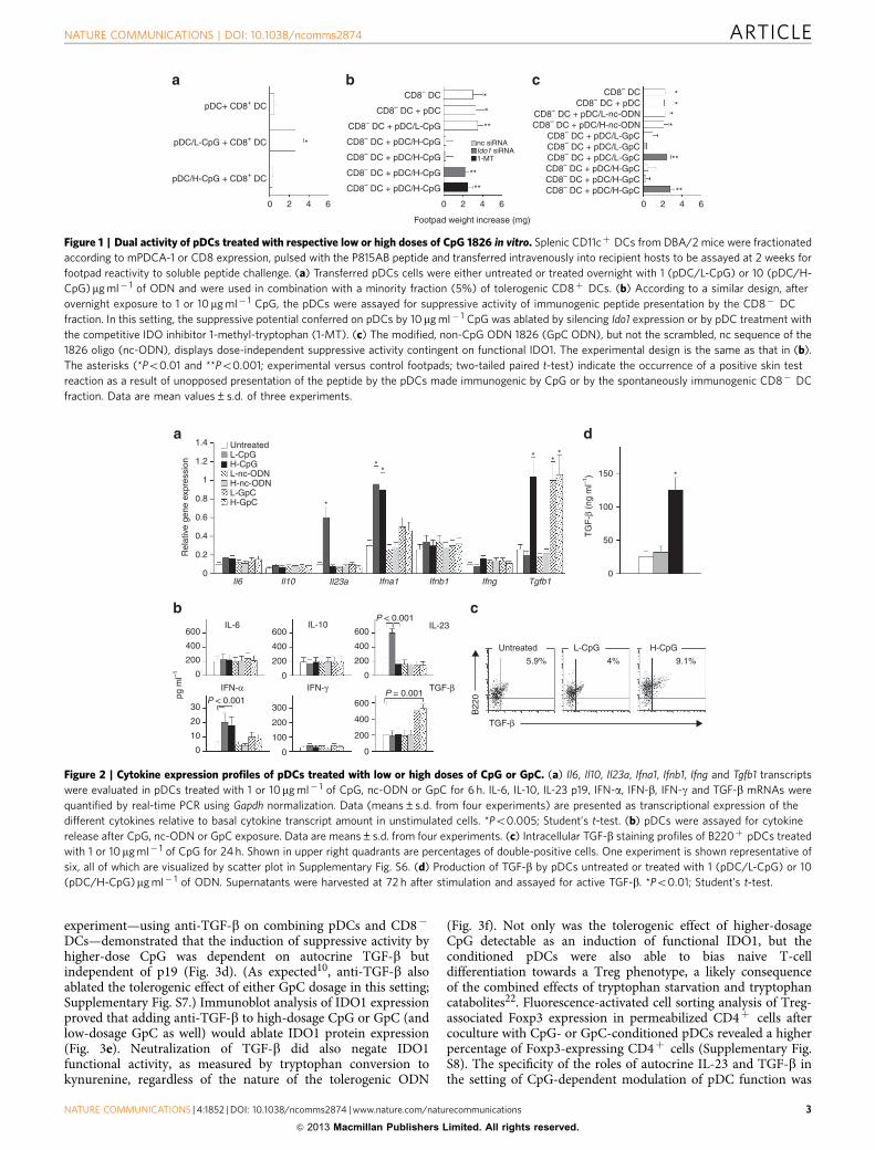
Figure 1 | Dual activity of pDCs treated with respective low or high doses of CpG 1826 in vitro. Splenic CD11cþ DCs from DBA/2 mice were fractionated
according to mPDCA-1 or CD8 expression, pulsed with the P815AB peptide and transferred intravenously into recipient hosts to be assayed at 2 weeks for
footpad reactivity to soluble peptide challenge. (a) Transferred pDCs cells were either untreated or treated overnight with 1 (pDC/L-CpG) or 10 (pDC/H-
CpG)mg ml� 1 of ODN and were used in combination with a minority fraction (5%) of tolerogenic CD8þ DCs. (b) According to a similar design, after
overnight exposure to 1 or 10 mg ml� 1 CpG, the pDCs were assayed for suppressive activity of immunogenic peptide presentation by the CD8� DC
fraction. In this setting, the suppressive potential conferred on pDCs by 10mg ml� 1 CpG was ablated by silencing Ido1 expression or by pDC treatment with
the competitive IDO inhibitor 1-methyl-tryptophan (1-MT). (c) The modified, non-CpG ODN 1826 (GpC ODN), but not the scrambled, nc sequence of the
1826 oligo (nc-ODN), displays dose-independent suppressive activity contingent on functional IDO1. The experimental design is the same as that in (b).
The asterisks (*Po0.01 and **Po0.001; experimental versus control footpads; two-tailed paired t-test) indicate the occurrence of a positive skin test
reaction as a result of unopposed presentation of the peptide by the pDCs made immunogenic by CpG or by the spontaneously immunogenic CD8� DC
fraction. Data are mean values±s.d. of three experiments.
1.4
1.2
0.8
Untreated
Untreated
L-CpG
L-CpG
H-CpG
H-CpG
L-nc-ODNH-nc-ODNL-GpCH-GpC
Il6 Il10 Il23a Ifna1 Ifnb1 Ifng Tgfb1
150
50
TG
F-β
(ng
ml–1
)
0
100
*
***
**
*
Rel
ativ
e ge
ne e
xpre
ssio
n
0.6
0.4
600
400
200
pg m
l–1 0
0
10
20
30
0
100
200
300
600
400
200
0
600
P < 0.001
P = 0.001P < 0.001
400
200
0
600
400
200
0
0.2
0
1
IFN-α IFN-γ TGF-β
TGF-β
B22
0
IL-6 IL-10 IL-23
5.9% 4% 9.1%
Figure 2 | Cytokine expression profiles of pDCs treated with low or high doses of CpG or GpC. (a) Il6, Il10, Il23a, Ifna1, Ifnb1, Ifng and Tgfb1 transcripts
were evaluated in pDCs treated with 1 or 10mg ml� 1 of CpG, nc-ODN or GpC for 6 h. IL-6, IL-10, IL-23 p19, IFN-a, IFN-b, IFN-g and TGF-b mRNAs were
quantified by real-time PCR using Gapdh normalization. Data (means±s.d. from four experiments) are presented as transcriptional expression of the
different cytokines relative to basal cytokine transcript amount in unstimulated cells. *Po0.005; Student’s t-test. (b) pDCs were assayed for cytokine
release after CpG, nc-ODN or GpC exposure. Data are means±s.d. from four experiments. (c) Intracellular TGF-b staining profiles of B220þ pDCs treated
with 1 or 10mg ml� 1 of CpG for 24 h. Shown in upper right quadrants are percentages of double-positive cells. One experiment is shown representative of
six, all of which are visualized by scatter plot in Supplementary Fig. S6. (d) Production of TGF-b by pDCs untreated or treated with 1 (pDC/L-CpG) or 10
(pDC/H-CpG)mg ml� 1 of ODN. Supernatants were harvested at 72 h after stimulation and assayed for active TGF-b. *Po0.01; Student’s t-test.
NATURE COMMUNICATIONS | DOI: 10.1038/ncomms2874 ARTICLE
NATURE COMMUNICATIONS | 4:1852 | DOI: 10.1038/ncomms2874 | www.nature.com/naturecommunications 3
& 2013 Macmillan Publishers Limited. All rights reserved.

demonstrated under conditions of in vitro manipulation of pDCfunction by manoeuvres other than CpG-ODN exposure(Supplementary Fig. S9a,b).
MyD88 is required for CpG adjuvant but not suppressiveeffects. Following preliminary experiments showing that neitherlower-dosage nor higher-dosage CpG affects Tlr9 transcription(nor does either GpC dosage; Supplementary Fig. S10), weexamined the role of the essential TLR adaptors MyD88 (myeloiddifferentiating factor 88) and TRIF (Toll/IL-1 receptor domain-containing adaptor inducing IFN-b) in pDC conditioning by low-or high-dose CpG. In a first set of experiments, by assaying skintest reactivity to the HY peptide and using genetically deficient(Tlr9� /� or Myd88� /� ) donors of pDCs, we confirmed that theimmunostimulant effect of a lower CpG dosage required TLR9and MyD88 signalling. We examined the effects of CpG-condi-tioned pDCs admixed with a minority fraction of tolerogenicCD8þ DCs (Fig. 4a). In the reciprocal combination (that is, onadmixing immunogenic CD8� DCs with 5% CpG-conditionedpDCs), the suppressive effect of the higher CpG dosage requiredTLR9 signalling but was independent of MyD88 (Fig. 4b). (Asexpected10, under the same experimental conditions, thetolerogenic effects of GpC required neither TLR9 nor MyD88,Fig. 4b.) IDO1 functional activity was initiated by high-dose CpG,in wild-type and Myd88� /� mice but not in Tlr9� /� pDCs
(Fig. 4c). In contrast, the GpC-ODN was effective in inducingfunctional IDO1 under conditions of either MyD88 or TLR9deficiency (Fig.4c). Again, in all of these experiments the nc-ODNlacked biological activity (Fig. 4a–c). p19-encoding Il23a wasinduced transcriptionally by low-dose CpG in wild-type but notTlr9� /� or Myd88� /� mice, whereas Tgfb1 was induced by thehigher CpG dosage in wild-type and Myd88� /� mice but not inTlr9� /� mice. This was in marked contrast with the pattern ofTGF-b production by GpC (Supplementary Fig. S11a,c). Directcomparison of IL-23 and TGF-b productions in wild-type andMyd88� /� mice treated with 1 or 10mg ml� 1 CpG-ODN 1826for 24 h confirmed that IL-23 production was selectively affectedby MyD88 deficiency (Supplementary Fig. S11b). In addition,using a different method of TGF-b induction, namely tolerogenicCTLA-4–Ig, cytokine production was unaffected by geneticdeficiency of TLR9 or MyD88 (Supplementary Fig. S12).
CpG-suppressive effects depend on TRIF but not TLR3 orTLR4. In a second set of experiments, by examining skin testreactivity to the HY peptide as modulated by pDCs lacking TRIF-encoding Ticam1, Tlr3 or Tlr4, we obtained evidence that thetolerogenic effect of a higher CpG dosage, and of GpC as well,required TRIF but neither TLR3 nor TLR4, with which TRIFtypically associates (Fig. 5a). Tgfb1 expression was inducedby higher-dose CpG (or GpC at either dosage) in Tlr3� /�
Figure 3 | Type I IFNs are determinant for the immunomodulatory
activities of pDCs treated with CpG or GpC. (a) Splenic CD11cþmPDCA-
1þ DCs from Ifnar�/� mice were combined with CD8þ DCs, pulsed with
the P815AB peptide and transferred intravenously into recipient hosts that
were assayed at 2 weeks for footpad reactivity to the peptide in saline.
Transferred pDCs were either untreated or treated overnight with 1 (pDC/L-
CpG)mg ml� 1 of CpG 1826 or with 1mg ml� 1 of the modified, non-CpG
ODN 1826 (pDC/L-GpC). Data are mean values±s.d. of four experiments.
(b) After overnight exposure to 10 (pDC/H-CpG) mg ml� 1 CpG or
10 mg ml� 1 of GpC (pDC/H-GpC), pDCs from Ifnar�/� mice were
assayed for suppressive activity of immunogenic peptide presentation by
the CD8� DC fraction. The asterisk (*Po0.001; experimental versus
control footpads; two-tailed paired t-test) indicates the occurrence of a
positive skin test reaction as a result of immunogenic peptide presentation.
Data are mean values±s.d. of four experiments. (c,d) IL-23 and TGF-bdependency of the immunostimulant and immunosuppressive activity of
pDCs treated with CpG. Splenic CD11cþ DCs were fractionated according
to mPDCA-1 or CD8 expression, pulsed with the P815AB peptide and
transferred into recipient DBA/2 hosts to be assayed at 2 weeks for skin
test reactivity to the eliciting peptide. (c) The pDCs were treated overnight
with 1mg ml� 1 CpG and cotransferred with a minority fraction of
suppressive CD8þ DCs. A portion of the pDCs was coexposed to
anti-IL-23 p19 (20mg ml� 1) or anti-TGF-b (10 mg ml� 1). (d) Alternatively,
the pDCs were treated with 10 mg ml� 1 CpG, with or without anti-TGF-bor anti-IL-23 p19, and cotransferred with a majority fraction of immunogenic
CD8� DCs. The asterisks indicate the occurrence of a positive skin test
reaction. Data are mean values±s.d. of three experiments. (*Po0.01;
experimental versus control footpads; two-tailed paired t-test). (e) IDO1
protein expression in pDCs treated with low- or high-dose CpG, low- or
high-dose GpC, and control or anti-TGF-b (þ ) antibody was assayed by
immunoblot analysis of whole-cell lysates. Blots were stripped and
reprobed with anti-b-tubulin. One of three experiments. (f) Functional
IDO1 activity was measured in terms of kynurenine levels in supernatants
from pDCs treated as above, with either GpC or high-dose CpG. The
reference IDO1 inhibitor, 1-methyl-tryptophan (1-MT), was also used in
place of anti-TGF-b. Data are means±s.d. of three experiments. Po0.01
(treatment versus control; Student’s t-test).
pDC treatment
pDC Ifnar –/– + DC CD8+
pDC/L-CpG + CD8+ DC
CD8– DC + pDC Ifnar –/–
CD8– DC + pDC/H-CpG
*
*
*
*
*
*
*None
None
Control Ab
Anti-p19
Anti-TGF-β
Anti-TGF-β
Anti-TGF-β
Contro
l
β-Tubulin
IDO:β-tubulin
Kyn
uren
ine
(μM
)
None
Control Ab
Anti-p19
Anti-TGF-β
L-CpG
L-GpC
0
0
1
4
2
2 0 42
Footpad weightincrease (mg)
Footpad weightincrease (mg)
Footpad weightincrease (mg)
Footpad weightincrease (mg)
3 0 1 2 3
None
H-CpG
H-GpC
IDO
L-CpG
0.2
15 1-MT*
**
5
10
0
0.2 0.2 0.2 0.2 0.20.50.7 0.7
H-CpG
H-CpG
– – + – + – + – +
L-GpC
L-GpCL-CpG
H-GpC
H-GpC
42 kDa
55 kDa
Control
ARTICLE NATURE COMMUNICATIONS | DOI: 10.1038/ncomms2874
4 NATURE COMMUNICATIONS | 4:1852 | DOI: 10.1038/ncomms2874 | www.nature.com/naturecommunications
& 2013 Macmillan Publishers Limited. All rights reserved.

or Tlr4� /� mice but not in Ticam1� /� pDCs, lacking TRIF(Supplementary Fig. S13). IDO1 functional activity, when trig-gered by high-dose CpG, or GpC, was demonstrable in pDCsfrom TLR3- or TLR4- but not TRIF-deficient mice (Fig. 5b).
Next, we examined cytokine (IL-6, IL-10, IL-23, IFN-a, IFN-g,TGF-b, IL-12 p40, IL-12 p70 and tumour-necrosis factor (TNF)-a) production in wild-type, Tlr9� /� , Myd88� /� or Ticam� /�
pDCs exposed to high-dose or low-dose CpG (SupplementaryFig. S14a). In response to high-dose CpG, the production ofIFN-a required TLR9- and TRIF-dependent signalling, whereaslow-dose CpG induced IL-23 and IFN-a via TLR9 andMyD88. Wild-type, Tlr9� /� , Myd88� /� or Ticam� /� pDCsexposed to high-dose or low-dose CpG were also assayed by
immunoblotting for IDO1 protein expression (SupplementaryFig. S14b), demonstrating the need for functional TLR9 andTRIF. Induction of IFN-a, TGF-b and functional IDO wasdependent on the TRIF competence of GpC-treated pDCs butrequired neither TLR9 nor MyD88 (Supplementary Fig. S14a,b).Direct association of TRIF with TLR9 could be demonstrated inan HEK293 cell line that stably expressed mouse TLR9 fused, atthe carboxyl terminus, with the influenza hemagglutinin tag(Supplementary Fig. S15).
TRAF proteins are required for both activities of CpG. TNF-receptor-associated factors (TRAFs) are intracellular proteins thatbind the cytoplasmic portion of TNF receptors and mediatedownstream signalling. The six known TRAF proteins (TRAF1–6) have overlapping yet distinct roles in controlling immuneresponses as well as cellular processes23. TRAF3 is one of themost enigmatic members in the TRAF family and has beenidentified to perform apparently distinct roles. Recent studiessuggest that nuclear factor kB-inducing kinase (crucial innoncanonical NF-kB signalling) is suppressed throughconstitutive proteasome-mediated degradation regulated byTRAF2 and TRAF3 (ref. 24). In contrast, TRAF6 contributes toCD40-mediated activation of NF-kB, stress-activated proteinkinases and other signalling molecules25. We investigated thepossible involvement of the two TRAF proteins in the P815ABskin test model system by siRNA technology. TRAF3-specificsiRNA—but not TRAF6-specific siRNA—added to the pDCcultures during CpG exposure blocked pDC ability to overcomesuppression by tolerogenic CD8þ DCs (Supplementary Fig.S16a). In the other setting—using siRNA on combining pDCsand CD8� DCs—we found that the induction of suppressiveactivity by higher-dose CpG was dependent on TRAF6 butindependent of TRAF3 (Supplementary Fig. S16b,c).
Dominance of canonical and noncanonical NF-jB signalling.IDO1 expression is contingent on the noncanonical pathway ofNF-kB activation19,26,27. Molecular dissection of NF-kBactivation has shown that NF-kB can be induced by the so-called canonical (classical; IkB kinase (IKK)b-dependent) andnoncanonical (alternative; IKKa-dependent) signalling pathways,leading to distinct patterns in the individual NF-kB subunits thatare activated and the downstream genetic responses that areinduced. Both the Tgfb1 and Ido1 promoters contain putativenoncanonical NF-kB-binding sites recognized by noncanonicalp52/RelB dimers17,21,26,27.
Much like CD40 ligation27, in IDO1þ pDCs, triggering of TLR9can activate both the IKKa- and IKKb-mediated pathways28. Usingimmunoblot analysis, we examined CpG at 1 or 10mg ml� 1 forpossible activation of canonical and noncanonical NF-kB pathways(Fig. 6a). Lower-dose CpG activated phosphorylation of both IKKaand IKKb, and 10mg ml� 1 CpG activated higher-levelphosphorylation of only IKKa. We used ELISA to determine therelative amounts of canonical p65 and noncanonical p52 in wild-type or genetically deficient pDCs treated with CpG. We found aprogressive increase of p65 in wild-type and Ticam1� /� , butneither Tlr9� /�nor Myd88� /� , pDCs (Fig. 6b). In parallel, p52—as induced by the higher CpG dosage—increased over time and tocomparable extents in control and MyD88-competent mice, but notin TLR9- or TRIF-deficient pDCs (Fig. 6c). In parallel gene-silencing experiments, we found that p65 activation by lower-doseCpG required TRAF3 but not TRAF6 (Fig. 6b), whereas p52activation by higher CpG treatment required TRAF6 but notTRAF3 (Fig. 6c). In gene-silencing experiments in vivo, IKKa butnot IKKb was required for high-dose CpG to confer tolerogenicproperties on pDCs in a skin test assay (Supplementary Fig. S17a,b).
WT
WT
Kyn
uren
ine
(μM
)
15
5
10
0
Tlr9 –/–
Tlr9 –/–
Myd88 –/–
Myd88 –/–
Footpad weight increase (mg)
* ** *
* * **
0 2 4 0 2 4
CD8– DC +pDCpDC + CD8+ DC
*
*
*
*
*
*
*
*
pDC treatment
NoneL-nc-ODN
H-nc-ODN
L-CpGL-GpC
H-CpGH-GpC
Figure 4 | TLR9–MyD88 dependency of CpG adjuvant effect.
(a) Induction of skin test reactivity by CpG requires TLR9 as well as MyD88
signalling in pDCs. Wild-type (WT) C57BL/6 pDCs were treated
overnight with 1 mg ml� 1 CpG (L-CpG), nc-ODN (L-nc-ODN) or GpC
(L-GpC), pulsed with the HY peptide, and cotransferred with a minority
fraction of suppressive CD8þ DCs. Parallel groups included pDCs from
Tlr9� /� or Myd88� /� mice. Skin test reactivity to the eliciting peptide
was measured at 2 weeks. (b) Suppression of skin test reactivity by CpG
requires TLR9 but not MyD88 signalling, whereas the suppressive effect
of GpC occurs independently of TLR9 and MyD88. pDCs from WT or
genetically deficient mice were treated with 10mg ml� 1 CpG (H-CpG),
nc-ODN (H-nc-ODN) or GpC (H-GpC), and cotransferred with a majority
fraction of immunogenic CD8� DCs. In both a and b, the asterisks indicate
the occurrence of a positive skin test reaction (*Po0.01; experimental
versus control footpads; two-tailed paired t-test), and data are mean
values±s.d. of three experiments. (c) IDO1 functional activity is induced by
high-dose CpG in Myd88� /� but not Tlr9� /� mice, and by either GpC
dosage in both Myd88� /� and Tlr9� /� mice. Functional IDO1 activity
was measured in terms of kynurenine production by pDCs treated with
either dosage of CpG, nc-ODN or GpC. Data are means±s.d. of three
experiments. Po0.01 (treatment versus none; Student’s t-test).
NATURE COMMUNICATIONS | DOI: 10.1038/ncomms2874 ARTICLE
NATURE COMMUNICATIONS | 4:1852 | DOI: 10.1038/ncomms2874 | www.nature.com/naturecommunications 5
& 2013 Macmillan Publishers Limited. All rights reserved.

Overall, it would appear that lower dosages of CpG-ODNwill result in limited yet coexistent activation of IKKb and IKKa,but higher dosages will selectively result in high-level noncanonical
activation (in the relative absence of canonical signalling, which is,in fact, actively suppressed by the former). This might explain thecoexistence of IL-23 and IFN-a under lower CpG-ODN treatmentconditions and the great preponderance of TGF-b found ontreating pDCs with higher CpG-ODN concentrations.
Stimulation and suppression depend on IRF7 and IRF3,respectively. Expression and function of type I IFNs can beregulated by IFN regulatory factor (IRF)3 and 7, typically acti-vated by the triggering of signalling cascades from TLRs thatrecognize pathogenic nucleic acids. TLR9 signalling in pDCsactivates the MyD88-dependent canonical NF-kB pathway andthe IRF7-dependent IFN pathway29. We investigated whether theeffects of lower and higher CpG dosages are contingent on IRFs.pDCs, either untreated or treated with lower-dose CpG, wereused in combination with a minority fraction of tolerogenicCD8þ DCs in a skin test assay. A portion of the pDCs had beentransfected with negative control, IRF3-specific, or IRF7-specificsiRNA (Supplementary Fig. S18a). In parallel, pDCs, treated withhigh-dose CpG, were cotransferred with a majority fraction ofimmunogenic CD8� DCs. A portion of the pDCs had likewisebeen transfected with negative control, or Irf3- or Irf7-targetingsiRNA (Supplementary Fig. S18b). The results provided evidencefor the dependence of the stimulant effect on IRF7, as well as forIRF3, as a required component of the tolerogenic circuit. Inaddition, high-dose CpG induced nuclear translocation of IRF3 inpDCs from wild-type and Myd88� /� , but not Ticam1� /� , mice(Supplementary Fig. S18c). Finally, high-dose CpG induced Tgfb1and IDO1 protein expression in the absence of functional IRF7,yet both effects required IRF3 (Supplementary Fig. S18d–f).Although the mechanisms whereby IRF3 contributes to Tgfb1transcription are unclear, this might be compatible with thenotion that TRIF-dependent signalling activates multipletranscription factors, including IRF3, NF-kB and AP-1, leadingto induction of type I IFNs and other cytokines30,31, likely via anIRF3–IFN-b–STAT1 pathway, in response to TLR activation32.This interpretation is consistent with the need for a functionaltype I IFN receptor in the higher-dose-suppressive effect of CpG(Fig. 3b). In addition, it should be noted that the Tgfb1 promotercontains, itself, a putative consensus motif (50-AAAGGAAA-30;from � 1,412 to � 1,405 bp, relative to transcription start site)for binding IRF3.
WT*
**
*
*
*
*
****
**
WT
Ticam1 –/–
Ticam1 –/–
Tlr3 –/–
Tlr3 –/– Tlr4 –/–
Tlr4 –/–
Footpad weightincrease (mg)
0 2 4
Kyn
uren
ine
(μM
)
15
5
10
20 NoneH-CpG * * * * * *
L-GpCH-GpC
0
pDC treatmentCD8– DC +pDC
Figure 5 | Tolerogenesis by CpG and GpC requires TRIF but not TLR3 or TLR4 signalling. (a) pDCs from WT or genetically deficient mice were treated
with 10mg ml� 1 CpG (H-CpG), or with 1 or 10mg ml� 1 GpC (L-GpC and H-GpC, respectively) and cotransferred with a majority fraction of spontaneously
immunogenic CD8� DCs. The asterisks indicate the occurrence of a positive skin test reaction (*Po0.01 and **Po0.001; experimental versus control
footpads; two-tailed paired t-test), and data are mean values±s.d. of three experiments. (b) IDO1 functional activity is induced by high-dose CpG and by
both low- and high-dose GpC administrations to Tlr3� /� or Tlr4� /� but not Ticam1� /� mice. Enzyme activity was measured in terms of kynurenine
production by the pDCs treated with CpG or GpC. Data are means±s.d. of three experiments. Po0.01 (treatment versus none; Student’s t-test).
L-CpG
0
0
0.2
0.4
0.6
0.8
5
*
*
*
*
*
*
**
*
*
15
p65p52
p65p52
30
L-CpG
H-CpG
*
*
*
***
*
Min 0
1
0Min
WT nc siRNA
0 30 60 0 30 60 0 30 60 0 30 60 0 30 60 0 30 60
Tlr9 –/– Myd88 –/– Ticam1 –/– Traf3 siRNA Traf6 siRNA
0.2
0.4
0.6
0.8
1
5 15 30
87 kDa
87 kDa
85 kDa
85 kDa
pIKKα/β
IKKα
IKKβ
H-CpGβα
Figure 6 | Respective dominance of MyD88/canonical and TRIF/
noncanonical NF-jB signalling pathways as induced by immunostimulant
and immunosuppressive CpG. (a) Low-dose CpG activates
phosphorylation of IKKa and IKKb whereas higher-dose CpG activates
phosphorylation of IKKa. pDCs were treated with 1 or 10 mg ml� 1 CpG
for different times and immunoblots of cell lysates were probed with
anti-pIKKa/b followed by anti-IKKa or anti-IKKb. One experiment
representative of three. ELISA measurements were used to determine the
relative amounts (A450) of p65 and p52 in pDCs treated with low-dose CpG
(b) or high-dose CpG (c) as above for different times. Nuclear extracts
were obtained from cells of WT or genetically deficient mice. Data are
means±s.d. of three experiments. *Po0.05–0.01 (treatment versus
respective time 0 control; Student’s t-test).
ARTICLE NATURE COMMUNICATIONS | DOI: 10.1038/ncomms2874
6 NATURE COMMUNICATIONS | 4:1852 | DOI: 10.1038/ncomms2874 | www.nature.com/naturecommunications
& 2013 Macmillan Publishers Limited. All rights reserved.
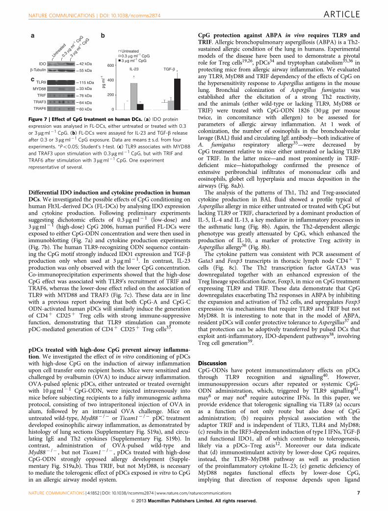
Differential IDO induction and cytokine production in humanDCs. We investigated the possible effects of CpG conditioning onhuman Flt3L-derived DCs (FL-DCs) by analysing IDO expressionand cytokine production. Following preliminary experimentssuggesting dichotomic effects of 0.3mg ml� 1 (low-dose) and3mg ml� 1 (high-dose) CpG 2006, human purified FL-DCs wereexposed to either CpG-ODN concentration and were then used inimmunoblotting (Fig. 7a) and cytokine production experiments(Fig. 7b). The human TLR9-recognizing ODN sequence contain-ing the CpG motif strongly induced IDO1 expression and TGF-bproduction only when used at 3mg ml� 1. In contrast, IL-23production was only observed with the lower CpG concentration.Co-immunoprecipitation experiments showed that the high-doseCpG effect was associated with TLR9’s recruitment of TRIF andTRAF6, whereas the lower-dose effect relied on the association ofTLR9 with MYD88 and TRAF3 (Fig. 7c). These data are in linewith a previous report showing that both CpG-A and CpG-CODN-activated human pDCs will similarly induce the generationof CD4þ CD25þ Treg cells with strong immune-suppressivefunction, demonstrating that TLR9 stimulation can promotepDC-mediated generation of CD4þ CD25þ Treg cells33.
pDCs treated with high-dose CpG prevent airway inflamma-tion. We investigated the effect of in vitro conditioning of pDCswith high-dose CpG on the induction of airway inflammationupon cell transfer onto recipient hosts. Mice were sensitized andchallenged by ovalbumin (OVA) to induce airway inflammation.OVA-pulsed splenic pDCs, either untreated or treated overnightwith 10 mg ml� 1 CpG-ODN, were injected intravenously intomice before subjecting recipients to a fully immunogenic asthmaprotocol, consisting of two intraperitoneal injection of OVA inalum, followed by an intranasal OVA challenge. Mice onuntreated wild-type, Myd88� /� or Ticam1� /� pDC treatmentdeveloped eosinophilic airway inflammation, as demonstrated byhistology of lung sections (Supplementary Fig. S19a), and circu-lating IgE and Th2 cytokines (Supplementary Fig. S19b). Incontrast, administration of OVA-pulsed wild-type andMyd88� /� , but not Ticam1� /� , pDCs treated with high-doseCpG-ODN strongly opposed allergy development (Supple-mentary Fig. S19a,b). Thus TRIF, but not MyD88, is necessaryto mediate the tolerogenic effect of pDCs exposed in vitro to CpGin an allergic airway model system.
CpG protection against ABPA in vivo requires TLR9 andTRIF. Allergic bronchopulmonary aspergillosis (ABPA) is a Th2-sustained allergic condition of the lung in humans. Experimentalmodels of the disease have been used to demonstrate a pivotalrole for Treg cells19,26, pDCs34 and tryptophan catabolism35,36 inprotecting mice from allergic airway inflammation. We evaluatedany TLR9, MyD88 and TRIF dependency of the effects of CpG onthe hypersensitivity response to Aspergillus antigens in the mouselung. Bronchial colonization of Aspergillus fumigatus wasestablished after the elicitation of a strong Th2 reactivity,and the animals (either wild-type or lacking TLR9, MyD88 orTRIF) were treated with CpG-ODN 1826 (30mg per mousetwice, in concomitance with allergen) to be assessed forparameters of allergic airway inflammation. At 1 week ofcolonization, the number of eosinophils in the bronchoalveolarlavage (BAL) fluid and circulating IgE antibody—both indicative ofA. fumigatus respiratory allergy35—were decreased byCpG treatment relative to mice either untreated or lacking TLR9or TRIF. In the latter mice—and most prominently in TRIF-deficient mice—histopathology confirmed the presence ofextensive peribronchial infiltrates of mononuclear cells andeosinophils, globet cell hyperplasia and mucus deposition in theairways (Fig. 8a,b).
The analysis of the patterns of Th1, Th2 and Treg-associatedcytokine production in BAL fluid showed a profile typical ofAspergillus allergy in mice either untreated or treated with CpG butlacking TLR9 or TRIF, characterized by a dominant production ofIL-5, IL-4 and IL-13, a key mediator in inflammatory processes inthe asthmatic lung (Fig. 8b). Again, the Th2-dependent allergicphenotype was greatly attenuated by CpG, which enhanced theproduction of IL-10, a marker of protective Treg activity inAspergillus allergy36 (Fig. 8b).
The cytokine pattern was consistent with PCR assessment ofGata3 and Foxp3 transcripts in thoracic lymph node CD4þ Tcells (Fig. 8c). The Th2 transcription factor GATA3 wasdownregulated together with an enhanced expression of theTreg lineage specification factor, Foxp3, in mice on CpG treatmentexpressing TLR9 and TRIF. These data demonstrate that CpGdownregulates exacerbating Th2 responses in ABPA by inhibitingthe expansion and activation of Th2 cells, and upregulates Foxp3expression via mechanisms that require TLR9 and TRIF but notMyD88. It is interesting to note that in the model of ABPA,resident pDCs will confer protective tolerance to Aspergillus37 andthat protection can be adoptively transferred by pulsed DCs thatexploit anti-inflammatory, IDO-dependent pathways38, involvingTreg cell generation39.
DiscussionCpG-ODNs have potent immunostimulatory effects on pDCsthrough TLR9 recognition and signalling40. However,immunosuppression occurs after repeated or systemic CpG-ODN administration, which, triggered by TLR9 signalling41,may6 or may not8 require autocrine IFNs. In this paper, weprovide evidence that tolerogenic signalling via TLR9 (a) occursas a function of not only route but also dose of CpGadministration; (b) requires physical association with theadaptor TRIF and is independent of TLR3, TLR4 and MyD88;(c) results in the IRF3-dependent induction of type I IFNs, TGF-band functional IDO1, all of which contribute to tolerogenesis,likely via a pDCs–Treg axis12. Moreover our data indicatethat (d) immunostimulant activity by lower-dose CpG requires,instead, the TLR9–MyD88 pathway as well as productionof the proinflammatory cytokine IL-23; (e) genetic deficiency ofMyD88 negates functional effects by lower-dose CpG,implying that direction of response depends upon ligand
600TGF-βIL-23
IDO
β-Tubulin
TLR9
MYD88
TRIF
TRAF3
TRAF6
42 kDa
Untre
ated
55 kDa
115 kDa
33 kDa
76 kDa
64 kDa
60 kDa
**
Untreated0.3 μg ml–1 CpG0.
3 μg
ml–1 C
pG
3 μg ml–1 CpG3
μg m
l–1 C
pG
400
200
0
pg m
l–1
Figure 7 | Effect of CpG treatment on human DCs. (a) IDO protein
expression was analysed in FL-DCs, either untreated or treated with 0.3
or 3mg ml� 1 CpG. (b) FL-DCs were assayed for IL-23 and TGF-b release
after 0.3 or 3mg ml� 1 CpG exposure. Data are means±s.d. from four
experiments. *Po0.05; Student’s t-test. (c) TLR9 associates with MYD88
and TRAF3 upon stimulation with 0.3mg ml� 1 CpG, but with TRIF and
TRAF6 after stimulation with 3mg ml� 1 CpG. One experiment
representative of several.
NATURE COMMUNICATIONS | DOI: 10.1038/ncomms2874 ARTICLE
NATURE COMMUNICATIONS | 4:1852 | DOI: 10.1038/ncomms2874 | www.nature.com/naturecommunications 7
& 2013 Macmillan Publishers Limited. All rights reserved.

concentration, which likely affects the affinity with whichTLR9 recruits downstream adaptor and signalling molecules,including TRAF3 and TRAF6; (f) the protective effects of CpG inexperimental allergy in vivo require a TLR9–TRIF circuit butnot MyD88-dependent events. Not secondarily, we alsoconfirmed that (g) a non-CpG ODN with two GpC motifs isexclusively immunosuppressive in the same setting, an effectwe have recently demonstrated not to require TLR9 and be, infact, contingent on TLR7- and TRIF-dependent signalling
events10. As a corollary, a role, per se, of the phosphorothioatebackbone in the TLR7 (GpC-dependent) and TLR9 (higher-doseCpG-dependent) pathways of tolerogenesis is ruled outby the stimulatory effect of lower-dose CpG on the samebackbone, which is in line with the dual effects both of CpGon a phosphodiester backbone and of bacterial DNA.Finally, (h) differential IDO induction and cytokine productionby low- versus high-dose CpG could also be documented inhuman DCs.
Control
C57
BL/
6Myd
88–/
–
Ticam
1–/
– Tlr9
–/–
ABPA ABPA + CpG
Control ABPA ABPA + CpG
Control ABPA ABPA + CpG
Control ABPA ABPA + CpG
100
EOS MON PMN LYM80
BA
L (%
)Ig
E (
ng m
l–1)
IL-1
3 (p
g m
l–1)
IL-4
(pg
ml)–1
IL-5
(pg
ml–1
)
60
40
20
0
0
1
2
3
0
0
0 0
20
40
60
60
30
90
80
100
10
Gata3
mR
NA
fold
cha
nge
Fox
p3 m
RN
A fo
ld c
hang
e
15p=0.016
p=0.016
p=0.05 p=0.04
ControlC57BL/6C57BL/6 + CpGTlr9 –/–
Tlr9 –/– + CpGMyd88 –/–
Myd88 –/– + CpGTicam1 –/–
Ticam1 –/– + CpG
5
0
10
15
5
200
300
**
* ** * * *
** *
**
IL-1
0 (p
g m
l–1)
0
40
120
80
*
*
Figure 8 | Effect of CpG treatment on allergic airway inflammation and requirement for TRIF but not MyD88 signalling. Bronchial colonization with
A. fumigatus was established in wild-type, Tlr9� /� , Myd88� /� and Ticam1� /� Th2-primed mice with or without CpG treatment, to be assayed for
A. fumigatus allergy at 1 week. Unsensitized, uncolonized wild-type mice (control) and genetically deficient mice (indicated) were present in all assays, and
so were sensitized and colonized mice of all genotypes on CpG treatment. Control littermates were included in individual experiments to check for
nonspecific effects of each genetic manipulation, and none were found. (a) Paraffin-embedded lung sections were stained with periodic acid-Schiff to
visualize mucine, goblet cells and inflammatory cells. Data are representative of three independent experiments. Upper panels, scale bar, 200mm; and
lower panels, scale bar, 100 mm. CpG treatment alone was indistinguishable from control. (b) The abundance of eosinophils (eos), monocytes (mon),
polymorphonucleates (pmn) and lymphocytes (lym) was assessed in BAL fluid. IgE antibodies were determined in sera, and secreted cytokines were
measured in BAL fluid. Control, naıve C57BL/6 mice. Data are mean±s.d. of three experiments. *Po0.05 and **Po0.005; Student’s t-test. (c) Gata3 and
Foxp3 transcripts were evaluated in CD4þ T cells from thoracic lymph nodes. Sorted CD4þ lymphocytes (5� 105 ml� 1) were activated with soluble
antibodies to CD3 for 24 h. Gata3 and Foxp3 mRNAs were quantified by real-time PCR using Gapdh normalization. Data (mean±s.d. of three (Foxp3) or four
(Gata3) experiments) are presented as normalized transcript expression in the samples relative to normalized transcript expression in the control culture
(cells from naıve mice of the respective genotype; that is, fold change ¼ 1, dotted line).
ARTICLE NATURE COMMUNICATIONS | DOI: 10.1038/ncomms2874
8 NATURE COMMUNICATIONS | 4:1852 | DOI: 10.1038/ncomms2874 | www.nature.com/naturecommunications
& 2013 Macmillan Publishers Limited. All rights reserved.
The TLR4 pathway employs signalling through two distinctadaptors, MyD88 and TRIF, and there is evidence that someCpG-ODNs may also require a second receptor or cofactor tosignal in addition to TLR9 (ref. 42). Although the molecularbasis of TLR4 signalling transition in response to endotoxin isunclear43, evidence suggests that the MyD88 and TRIF pathwaysare temporally segregated, nonetheless influencing each other44.Early signalling through MyD88 leads to phosphorylation-mediated degradation of IkBa, nuclear translocation of NF-kBp65/p50 heterodimers and transcription of proinflammatorygenes such as TNFA, IL1B, IL12B and IL6. Later in time, asendotoxin concentrations increase, and owing to a prevailingTRIF-dependent signalling and plasticity of NF-kB function,accumulating p50/p50 NF-kB homodimers trigger thetranscription of genes like IL10, TGFB1 and COX2(refs 43,45). This could be seen as a form of ‘protective’35 or‘infectious’21 tolerance, whereby exaggerated inflammatoryresponses are avoided via the rapid onset of cell refractorinessto sustained stimulation and the spread of a tolerant state fromone cell type to another. Seen in this light, both the selectiveinduction of immunosuppression in the spleen by systemicCpG-ODN7,8 and the duality of its effects as a function of localconcentrations in the microenvironment might reflect a negativefeedback mechanism in order to avoid hyperinflammatoryresponses27,45,46.
TLR9 modulation of IDO1 is effective not only in theprevention and treatment of animal models of autoimmune13
and allergic disorders4,6,8, but also in maintaining aphysiological state of protective tolerance to ubiquitous fungalaeroantigens36. ABPA is a Th2-dependent hypersensitivity lungdisease due to bronchial colonization of A. fumigatus that affects1–2% of asthmatic and 7–9% of cystic fibrosis patients.Although the organism can cause allergic disease in otherwisehealthy individuals and devastating disease in theimmunosuppressed, ABPA is a hypersensitivity response toAspergillus antigens in the lung and is distinct from other formsof Aspergillus pulmonary disease. In an experimental model ofABPA, in which the combined effects of Treg cells, pDCs andtryptophan catabolism mediate protection, we have previouslyfound that dexamethasone inhibited Th2 responses andallergy, and induced Foxp3 expression in CD4þ T cells viamechanisms dependent on tryptophan catabolism26. Here, usingthe same model system, we found that the beneficial activity ofCpG immunotherapy involves the TLR9–TRIF pathway,independently of MyD88.
In conclusion, our results demonstrate the occurrence of afunctional TLR9–IDO1 axis involving type I IFNs and TGF-b.Although the molecular basis of the switch in TLR9 signallingoutcomes from an inflammatory MyD88-dependent to animmunosuppressive TRIF-dependent effect remains elusive, ourstudy shows that CpG ODN-induced immune stimulation ischaracterized by a bell-shaped dose–response correlation, that is,only intermediate but not high doses of CpG induce effectstypically associated with immune activation. In contrast, higherdosages trigger TLR9- and IDO1-dependent tolerogenesis, whichabrogates the stimulatory effects and relies on TRIF and thenoncanonical NF-kB signalling pathway as well as on TGF-bproduction, which may further exploit IDO1 as a signallingprotein in a feedforward loop involving the stable conditioning ofpDCs towards a regulatory phenotype12. These data may have animpact on novel CpG-based therapeutic approaches in thecontext of antitumor and vaccination strategies, includingsiRNA delivery approaches targeting phagocytic cells throughan siRNA conjugated to a TLR9 ligand1. These studies may alsolead to innovative means of treating autoimmune and allergicinflammation.
MethodsMice. Eight- to 10-week-old female DBA/2J (H-2d), C57BL/6 (H-2b) and 129 Sv/Ev (H-2b) mice were obtained from Charles River Breeding Laboratories. Femalemice deficient for the IFN-a/b receptor (Ifnar� /� ), on a 129 Sv/Ev background,were generated as described26. Mice homozygous for the TLR3 (Tlr3� /� ), TLR4(Tlr4� /� ), TLR9 (Tlr9� /� ), MyD88 (Myd88� /� ) or TRIF (Ticam1� /� )-targeted mutation raised on the C57BL/6 background were as described13 and werebred at the breeding facilities of the University of Perugia. All in vivo studies werein compliance with National (Italian Parliament DL 116/92) and PerugiaUniversity Animal Care and Use Committee guidelines.
DC purification and treatments. These procedures have been described inprevious publications47,48. Briefly, splenic DCs were purified by magnetic-activatedsorting using CD11c MicroBeads and MidiMacs (Miltenyi Biotec, BergischGladbach, Germany), in the presence of EDTA to disrupt DC–T-cell complexes.Cells were 499% CD11cþ , 499% MHC I-Aþ , 498% B7-2þ , o0.1% CD3þ ,and appeared to consist of 90–95% CD8� , 5–10% CD8þ and B5% mPDCA-1þ
cells. For positive selection of mPDCA-1þ DCs (6), CD11cþ cells were fractionedusing mPDCA-1 MicroBeads (Miltenyi Biotec). More than 95% of the mPDCA-1þ
cells were stained by 120G8. For negative selection of CD8� DCs, CD11cþ cellswere fractioned by means of CD8a MicroBeads (Miltenyi Biotec). The CD8�
fraction was B45% CD4þ and typically contained o0.5% contaminating CD8þ
cells. Less than 5% CD8� DCs expressed the 120G8 marker. All endotoxin-freeCpG-ODNs either on a phosphorothioate (ODN 1826 (50-tccatgacgttcctgacgtt-30),ODN 1826 negative control (nc-ODN; 50-tccatgatattcctgatatt-30), ODN 1826non-CpG control (GpC; 50-tccataagcttcctgagctt-30), ODN 2006 (50-tcgtcgttttgtcgttttgtcgtt-30)) or on a phosphodiester backbone (CpG-A (5-TCCATGACGTTCCTGACGTT-3)) were purchased from Invitrogen Life Technologies. (Lowercaseletters in ODN sequences refer to nucleotides for which the 30 internucleotidelinkage is phosphorothioate-modified; endotoxin contamination, when measuredby the Limulus assay (Sigma), was negligible in all ODN preparations.) pDCs weretreated in vitro with 1 or 10 mg of CpG-ODN 1826 or the control ODNs.
siRNA synthesis and transfection. These procedures, performed according tostandard methodologies, are described in full in Supplementary Methods.
Immunization and skin test assay. The skin test assay we have been usingmeasures class I-restricted responses to synthetic peptides (H-2Ld–restrictedP815AB.35-43 [LPYLGWLVF] and H-2Db–restricted HY [WMHHNMDLI]).Following intravenous transfer of peptide-pulsed DCs, the response tointrafootpad challenge with the soluble peptide was measured, as describedpreviously in detail20,26 and also elucidated in Supplementary Methods.
Kynurenine assay. IDO1 functional activity was measured in vitro in terms of theability of DCs to metabolize tryptophan to kynurenine, whose concentrations weremeasured by HPLC, as described previously26,48.
Immunoblot and ELISA analyses. These procedures, performed according tostandard methodologies, are described in full in Supplementary Methods.
PCR assay. PCR assessment of Gata3 and Foxp3 transcripts in thoracic lymphnode CD4þ T cells was described26,35.
Human FL-DC preparation and immunoprecipitation. Human mature FL-DCswere obtained from peripheral blood mononuclear cells (PBMCs). Briefly, PBMCswere separated from whole blood of healthy donors by centrifugation througha density gradient of Ficoll–Paque (GE Healthcare). Whole blood was layeredonto a sterile aqueous medium containing Ficoll at a predetermined density of1.077 g ml� 1 at 25 �C. Gentle centrifugation at room temperature resulted in theseparation of PBMCs at the blood/Ficoll interface, with the other white blood cellsand red blood cells passing through the interface and accumulating at the bottomof the tube. FL-DCs were obtained from purified CD14þ monocytes by culturingcells in Iscove-modified medium for 7 days in the presence of 50 ng ml� 1 ofhuman FLT3L (Peprotech). The resulting FL-DCs were washed and culturedfor 18 h with 0.3 or 3 mg ml� 1 of CpG-ODN 2006 (50-TCGTCGTTTTGTCGTTTTGTCGTT-30). Supernatants were then analysed for cytokine production(IL-23 and TGF-b; Promega) and cell lysates for IDO expression by westernblotting using an anti-human IDO reagent (Millipore). FACS analysis revealed thatthe FL-DCs were CD123þ CD11clow BDCA2þ and BDCA4þ . For immunopre-cipitation, FL-DCs (100� 106 cells per sample) were stimulated for 30 min withlow-dose (0.3 mg ml� 1) or high-dose (3mg ml� 1) CpG 2006, recognizing humanTLR9, and lysed on ice in RIPA buffer (50 mM Tris-HCl, pH 7.4, 150 mM NaCl,1% Nonidet P-40, 0.25% Na-deoxycholate, 1 mM EDTA, 1.4 mM Na3VO4 andprotease inhibitors). Proteins were immunoprecipitated using anti-humanTLR9 (Santa Cruz Biotechnology). Western blot analyses were performedusing rabbit anti-human TLR9 (Cell Signaling), rabbit anti-human MYD88, rabbit
NATURE COMMUNICATIONS | DOI: 10.1038/ncomms2874 ARTICLE
NATURE COMMUNICATIONS | 4:1852 | DOI: 10.1038/ncomms2874 | www.nature.com/naturecommunications 9
& 2013 Macmillan Publishers Limited. All rights reserved.
anti-human TRIF (Alexis), rabbit anti-TRAF3 or rabbit anti-TRAF6 (CellSignaling Technology).
Allergic bronchopulmonary aspergillosis. The induction and evaluationof the Th2-driven hypersensitivity response to Aspergillus antigens in the mouselung has previously been described in detail26. Briefly, C57BL/6 or geneticallydeficient mice were sensitized by the concomitant intraperitoneal (100 mg)and subcutaneous (100 mg) administration of A. fumigatus culture filtrate extractfollowed 1 week later by the intranasal instillation of 20 mg of the extract.After an additional 7 days, bronchial colonization of A. fumigatus was induced byresting conidia administered intratracheally (1� 107), and the animals wereevaluated 1 week later for parameters of allergic airway inflammation.Treatments included CpG-ODN administered intraperitoneally (CpG-ODN1826; 30 mg per mouse twice, on the same days as the first and secondadministration of A. fumigatus culture filtrate extract). Analysis of respiratoryallergy phenotype was as described35.
Statistical analysis. Student’s t-test was used to analyse the results ofin vitro studies in which data are mean values (±s.d.). In the in vivo skin testassay, statistical analysis was performed using two-tailed paired t-test by comparingthe mean weight of experimental footpads with that of control, saline-injectedcounterparts26. Data are mean values (±s.d.) of three experiments with atleast six mice per group per experiment, as computed by power analysis so to yielda power of at least 80% with an a level of 0.05.
References1. Kortylewski, M. et al. In vivo delivery of siRNA to immune cells by conjugation
to a TLR9 agonist enhances antitumor immune responses. Nat. Biotechnol 27,925–932 (2009).
2. Klinman, D.M., Currie, D., Gursel, I. & Verthelyi, D. Use of CpGoligodeoxynucleotides as immune adjuvants. Immunol. Rev. 199, 201–216(2004).
3. Mutwiri, G., van Drunen Littel-van den Hurk, S. & Babiuk, L.A. Approaches toenhancing immune responses stimulated by CpG oligodeoxynucleotides. Adv.Drug Deliv. Rev. 61, 226–232 (2009).
4. Hayashi, T. et al. Inhibition of experimental asthma by indoleamine 2,3-dioxygenase. J. Clin. Invest. 114, 270–279 (2004).
5. Christensen, S.R. et al. Toll-like receptor 7 and TLR9 dictate autoantibodyspecificity and have opposing inflammatory and regulatory roles in a murinemodel of lupus. Immunity 25, 417–428 (2006).
6. Mellor, A.L. et al. Cutting Edge: CpG oligonucleotides induce splenic CD19þ
dendritic cells to acquire potent indoleamine 2,3-dioxygenase-dependent T cellregulatory functions via IFN type 1 signaling. J. Immunol 175, 5601–5605(2005).
7. Fallarino, F. & Puccetti, P. Toll-like receptor 9-mediated induction of theimmunosuppressive pathway of tryptophan catabolism. Eur. J. Immunol 36,8–11 (2006).
8. Wingender, G. et al. Systemic application of CpG-rich DNA suppressesadaptive T cell immunity via induction of IDO. Eur. J. Immunol 36, 12–20(2006).
9. Boor, P.P. et al. Human plasmacytoid dendritic cells induce CD8þ
LAG-3þ Foxp3þ CTLA-4þ regulatory T cells that suppress allo-reactivememory T cells. Eur. J. Immunol 41, 1663–1674 (2011).
10. Volpi, C. et al. A GpC-rich oligonucleotide acts on plasmacytoid dendritic cellsto promote immune suppression. J. Immunol 189, 2283–2289 (2012).
11. Puccetti, P. et al. Use of a skin test assay to determine tumor-specificCD8þ T cell reactivity. Eur. J. Immunol 24, 1446–1452 (1994).
12. Pallotta, M.T. et al. Indoleamine 2,3-dioxygenase is a signalingprotein in long-term tolerance by dendritic cells. Nat. Immunol 12,870–878 (2011).
13. Fallarino, F. et al. IDO mediates TLR9-driven protection from experimentalautoimmune diabetes. J. Immunol 183, 6303–6312 (2009).
14. Grohmann, U. et al. Functional plasticity of dendritic cell subsets as mediatedby CD40 versus B7 activation. J. Immunol 171, 2581–2587 (2003).
15. Belladonna, M.L. et al. IL-23 and IL-12 have overlapping, but distinct, effects onmurine dendritic cells. J. Immunol 168, 5448–5454 (2002).
16. Grohmann, U. et al. IL-6 inhibits the tolerogenic function of CD8aþ
dendritic cells expressing indoleamine 2,3-dioxygenase. J. Immunol 167,708–714 (2001).
17. Belladonna, M.L. et al. Cutting edge: autocrine TGF-b sustains defaulttolerogenesis by IDO-competent dendritic cells. J. Immunol 181, 5194–5198(2008).
18. Orabona, C. et al. Toward the identification of a tolerogenic signature in IDO-competent dendritic cells. Blood 107, 2846–2854 (2006).
19. Puccetti, P. & Grohmann, U. IDO and regulatory T cells: a role for reversesignalling and non-canonical NF-kB activation. Nat. Rev. Immunol 7,817–823 (2007).
20. Orabona, C. et al. SOCS3 drives proteasomal degradation of indoleamine 2,3-dioxygenase (IDO) and antagonizes IDO-dependent tolerogenesis. Proc. NatlAcad. Sci. USA 105, 20828–20833 (2008).
21. Belladonna, M.L., Orabona, C., Grohmann, U. & Puccetti, P. TGF-b andkynurenines as the key to infectious tolerance. Trends Mol. Med. 15,41–49 (2009).
22. Fallarino, F. et al. The combined effects of tryptophan starvation andtryptophan catabolites down-regulate T cell receptor z-chain andinduce a regulatory phenotype in naive T cells. J. Immunol 176, 6752–6761(2006).
23. Habelhah, H. Emerging complexity of protein ubiquitination in theNF-kB pathway. Genes Cancer 1, 735–747 (2010).
24. Zarnegar, B.J. et al. Noncanonical NF-kB activation requires coordinatedassembly of a regulatory complex of the adaptors cIAP1, cIAP2, TRAF2 andTRAF3 and the kinase NIK. Nat. Immunol 9, 1371–1378 (2008).
25. Inoue, J., Gohda, J. & Akiyama, T. Characteristics and biological functions ofTRAF6. Adv. Exp. Med. Biol. 597, 72–79 (2007).
26. Grohmann, U. et al. Reverse signaling through GITR ligand enablesdexamethasone to activate IDO in allergy. Nat. Med 13, 579–586 (2007).
27. Tas, S.W. et al. Noncanonical NF-kB signaling in dendritic cells is required forindoleamine 2,3-dioxygenase (IDO) induction and immune regulation. Blood110, 1540–1549 (2007).
28. Kaisho, T. & Tanaka, T. Turning NF-kB and IRFs on and off in DC. TrendsImmunol 29, 329–336 (2008).
29. O’Neill, L.A. & Bowie, A.G. The family of five: TIR-domain-containingadaptors in Toll-like receptor signalling. Nat. Rev. Immunol. 7, 353–364 (2007).
30. Tatematsu, M. et al. A molecular mechanism for Toll-IL-1 receptor domain-containing adaptor molecule-1-mediated IRF-3 activation. J. Biol. Chem 285,20128–20136 (2010).
31. Johnson, J. et al. A conventional protein kinase C inhibitor targetingIRF-3-dependent genes differentially regulates IL-12 family members. Mol.immunol 48, 1484–1493 (2011).
32. Bhattacharyya, S., Zhao, Y., Kay, T.W. & Muglia, L.J. Glucocorticoids targetsuppressor of cytokine signaling 1 (SOCS1) and type 1 interferons to regulateToll-like receptor-induced STAT1 activation. Proc. Natl Acad. Sci. USA 108,9554–9559 (2011).
33. Moseman, E.A. et al. Human plasmacytoid dendritic cells activated by CpGoligodeoxynucleotides induce the generation of CD4þCD25þ regulatoryT cells. J. Immunol 173, 4433–4442 (2004).
34. Romani, L. et al. Thymosin a1 activates dendritic cell tryptophan catabolismand establishes a regulatory environment for balance of inflammation andtolerance. Blood 108, 2265–2274 (2006).
35. Romani, L. et al. Defective tryptophan catabolism underlies inflammation inmouse chronic granulomatous disease. Nature 451, 211–215 (2008).
36. Romani, L. Immunity to fungal infections. Nat. Rev. Immunol. 11, 275–288(2011).
37. Ramirez-Ortiz, Z.G. et al. A nonredundant role for plasmacytoid dendritic cellsin host defense against the human fungal pathogen Aspergillus fumigatus. CellHost Microbe 9, 415–424 (2011).
38. Bonifazi, P. et al. Intranasally delivered siRNA targeting PI3K/Akt/mTORinflammatory pathways protects from aspergillosis. Mucosal Immunol 3,193–205 (2010).
39. Romani, L., Zelante, T., De Luca, A., Fallarino, F. & Puccetti, P. IL-17 andtherapeutic kynurenines in pathogenic inflammation to fungi. J. immunol 180,5157–5162 (2008).
40. Colonna, M., Trinchieri, G. & Liu, Y.J. Plasmacytoid dendritic cells inimmunity. Nat. Immunol. 5, 1219–1226 (2004).
41. Heikenwalder, M. et al. Lymphoid follicle destruction and immunosuppressionafter repeated CpG oligodeoxynucleotide administration. Nat. Med 10, 187–192(2004).
42. Verthelyi, D. & Zeuner, R.A. Differential signaling by CpG DNA in DCs and Bcells: not just TLR9. Trends Immunol 24, 519–522 (2003).
43. Biswas, S.K. & Lopez-Collazo, E. Endotoxin tolerance: new mechanisms,molecules and clinical significance. Trends Immunol. 30, 475–487 (2009).
44. Covert, M.W., Leung, T.H., Gaston, J.E. & Baltimore, D. Achievingstability of lipopolysaccharide-induced NF-kB activation. Science 309,1854–1857 (2005).
45. Lawrence, T., Bebien, M., Liu, G.Y., Nizet, V. & Karin, M. IKKa limitsmacrophage NF-kB activation and contributes to the resolution ofinflammation. Nature 434, 1138–1143 (2005).
46. Foligne, B. et al. A key role of dendritic cells in probiotic functionality. PLoSONE 2, e313–e324 (2007).
47. Fallarino, F. et al. Murine plasmacytoid dendritic cells initiate theimmunosuppressive pathway of tryptophan catabolism in response toCD200 receptor engagement. J. Immunol 173, 3748–3754 (2004).
48. Fallarino, F. et al. Ligand and cytokine dependence of the immunosuppressivepathway of tryptophan catabolism in plasmacytoid dendritic cells. Int. Immunol17, 1429–1438 (2005).
ARTICLE NATURE COMMUNICATIONS | DOI: 10.1038/ncomms2874
10 NATURE COMMUNICATIONS | 4:1852 | DOI: 10.1038/ncomms2874 | www.nature.com/naturecommunications
& 2013 Macmillan Publishers Limited. All rights reserved.

AcknowledgementsSupported by funding from the Italian Association for Cancer Research (to P.P.),the Fondazione Italiana Sclerosi Multipla (Project No. 2010/R/17; to F.F.) and MIUR(Ministero dell’Istruzione, Universita e Ricerca, Italy, FIRB RBAP11T3WB; to U.G.).We thank G. Andrielli for digital art and image editing.
Author contributionsC.Vo. designed and did experiments; M.T.P., R.B., C.Va., M.L.B., C.O. and A.D.L. didexperiments; L.R. and L.B. contributed reagents; F.F. and U.G. designed experiments andsupervised the research; and P.P. supervised the research and wrote the manuscript.
Additional informationSupplementary Information accompanies this paper at http://www.nature.com/naturecommunications
Competing financial interests: The authors declare no competing financial interests.
Reprints and permission information is available online at http://npg.nature.com/reprintsandpermissions/
How to cite this article: Volpi, C. et al. High doses of CpG oligodeoxynucleotidesstimulate a tolerogenic TLR9–TRIF pathway. Nat. Commun. 4:1852 doi: 10.1038/ncomms2874 (2013).
NATURE COMMUNICATIONS | DOI: 10.1038/ncomms2874 ARTICLE
NATURE COMMUNICATIONS | 4:1852 | DOI: 10.1038/ncomms2874 | www.nature.com/naturecommunications 11
& 2013 Macmillan Publishers Limited. All rights reserved.
Related Documents