IMMUNOBIOLOGY A novel role for HMGB1 in TLR9-mediated inflammatory responses to CpG-DNA Stanimir Ivanov, 1 Ana-Maria Dragoi, 1 Xin Wang, 1 Corrado Dallacosta, 2 Jennifer Louten, 1 Giovanna Musco, 2 Giovanni Sitia, 2 George S. Yap, 1 Yinsheng Wan, 3 Christine A. Biron, 1 Marco E. Bianchi, 4 Haichao Wang, 5 and Wen-Ming Chu 1 1 Department of Molecular Microbiology and Immunology, Brown University, Providence, RI; 2 San Raffaele Scientific Institute, Milano, Italy; 3 Department of Biology, Providence College, Providence, RI; 4 San Raffaele University, Faculty of Medicine, Milano, Italy; 5 Department of Emergency Medicine, North Shore-Long Island Jewish Research Institute, Manhasset, NY CpG-DNA or its synthetic analog CpG- ODN activates innate immunity through Toll-like receptor 9 (TLR9). However, the mechanism of TLR9 activation by CpG- DNA remains elusive. Here we have iden- tified HMGB1 as a CpG-ODN–binding pro- tein. HMGB1 interacts and preassociates with TLR9 in the endoplasmic reticulum- Golgi intermediate compartment (ER- GIC), and hastens TLR9’s redistribution to early endosomes in response to CpG- ODN. CpG-ODN stimulates macrophages and dendritic cells to secrete HMGB1; in turn, extracellular HMGB1 accelerates the delivery of CpG-ODNs to its receptor, leading to a TLR9-dependent augmenta- tion of IL-6, IL-12, and TNF secretion. Loss of HMGB1 leads to a defect in the IL-6, IL-12, TNF, and iNOS response to CpG-ODN. However, lack of intracellular TLR9-associated HMGB1 can be compen- sated by extracellular HMGB1. Thus, the DNA-binding protein HMGB1 shuttles in and out of immune cells and regulates inflammatory responses to CpG-DNA. (Blood. 2007;110:1970-1981) © 2007 by The American Society of Hematology Introduction Members of the TLR family mediate the innate immune response upon encounter with biochemically diverse pathogen molecules. 1,2 Among them is TLR9, which is essential for recognition of microbial CpG-DNA or its analog, synthetic oligonucleotides containing a CpG motif (CpG-ODNs). 3-6 CpG-DNA/CpG-ODNs activate macrophages, monocytes, and dendritic cells (DCs) to secrete proinflammatory cytokines, driving the Th1 response 3-6 and serving as attractive adjuvants in vaccine strategies for allergy, asthma, infectious disease, and cancer. TLR9 is confined primarily to cells of the immune system and is not present on the cell surface. 7-10 It is proposed that TLR9 is initially localized in the endoplasmic reticulum (ER), and redistributes to early endosomes upon stimulation with CpG-DNA. 9,11,12 TLR9 becomes activated and recruits MyD88, 9,11,12 leading to subsequent immune re- sponses. However, the mechanism by which TLR9 is activated remains elusive, and it is unknown whether CpG-DNA-binding proteins are involved in this activation process. HMGB1 is an abundant, highly conserved nuclear protein that modulates chromatin structure, facilitates interaction of proteins with DNA, regulates transcription, and assists in V(D)J recombina- tion. 13,14 Immune cells stimulated with IFN, IL-1, and TNF export nuclear HMGB1 to the cytoplasm and subsequently secrete it. 15 HMGB1 can also be passively released by necrotic cells, 16 serving as a signal for trauma and tissue damage. 17 Additionally, HMGB1 is released during bacterial or viral infection, 18,19 and extracellular HMGB1 can act as a chemoattractant for inflammatory cells, strongly suggesting its role as a modulator of the immune system. In this study, we demonstrate that CpG-ODN–treated macro- phages and DCs quickly secrete HMGB1; moreover, HMGB1 engages CpG-ODNs and enhances their immunostimulatory poten- tial in a TLR9-dependent manner. Confocal microscopy reveals that HMGB1 preassociates with TLR9 and colocalizes with markers of the ER, the ERGIC, and the Golgi in quiescent cells. Upon stimulation with CpG-ODN, HMGB1 and TLR9 colocalize with the early endosomal marker EEA1. Ablation or depletion of HMGB1 impaired redistribution of TLR9 to early endosomes in response to CpG-ODN. As a consequence, HMGB1-deficient cells exhibited substantially decreased responses to CpG-ODN, but these defects could be complemented by extracellular HMGB1. Materials and methods Animals Myd88 / , Tlr2 / , Tlr9 / , and their control littermates on B6/129 genetic background were gifts from Dr Akira and bred at Brown University (Providence, RI). Hmgb1 / mice were bred at San Raffaele, Italy. C.C3-Tlr4 lps-d/J and their control were purchased from the Jackson Labora- tory (Bar Harbor, ME). All institutions followed the respective national and local regulations on animal experimentation. Bone marrow–derived macro- phages (BMDMs) and bone marrow–derived DCs (BMDCs) were prepared as previously described. 7,9,10,20 Oligodeoxynucleotides and antibodies Endotoxin-free CpG-ODN 1018 and GpG-ODN 1019 were synthesized with a phosphorothioate backbone (Trilinker, San Diego, CA). For binding and microscopy studies, all ODNs (1018, 1019, 1668, and n1668) containing Cy5 (Sigma, St Louis, MO) or biotin (Invitrogen, San Diego, CA) at the 3 end were synthesized on the phosphodiester backbone. CpG-A (2216) was purchased from InvivoGen (San Diego, CA). Antibodies used were as follows: anti-HMGB1 (mouse monoclonal [MBL International, Nagoya, Japan]; rabbit polyclonal [PharMingen, San Submitted August 31, 2006; accepted May 28, 2007. Prepublished online as Blood First Edition paper, June 4, 2007; DOI 10.1182/blood-2006-09-044776. The online version of this article contains a data supplement. The publication costs of this article were defrayed in part by page charge payment. Therefore, and solely to indicate this fact, this article is hereby marked ‘‘advertisement’’ in accordance with 18 USC section 1734. © 2007 by The American Society of Hematology 1970 BLOOD, 15 SEPTEMBER 2007 VOLUME 110, NUMBER 6 For personal use only. at KANAZAWA UNIVERSITY on January 17, 2008. www.bloodjournal.org From

Welcome message from author
This document is posted to help you gain knowledge. Please leave a comment to let me know what you think about it! Share it to your friends and learn new things together.
Transcript

IMMUNOBIOLOGY
A novel role for HMGB1 in TLR9-mediated inflammatory responses to CpG-DNAStanimir Ivanov,1 Ana-Maria Dragoi,1 Xin Wang,1 Corrado Dallacosta,2 Jennifer Louten,1 Giovanna Musco,2 Giovanni Sitia,2
George S. Yap,1 Yinsheng Wan,3 Christine A. Biron,1 Marco E. Bianchi,4 Haichao Wang,5 and Wen-Ming Chu1
1Department of Molecular Microbiology and Immunology, Brown University, Providence, RI; 2San Raffaele Scientific Institute, Milano, Italy; 3Department ofBiology, Providence College, Providence, RI; 4San Raffaele University, Faculty of Medicine, Milano, Italy; 5Department of Emergency Medicine,North Shore-Long Island Jewish Research Institute, Manhasset, NY
CpG-DNA or its synthetic analog CpG-ODN activates innate immunity throughToll-like receptor 9 (TLR9). However, themechanism of TLR9 activation by CpG-DNA remains elusive. Here we have iden-tified HMGB1 as a CpG-ODN–binding pro-tein. HMGB1 interacts and preassociateswith TLR9 in the endoplasmic reticulum-Golgi intermediate compartment (ER-GIC), and hastens TLR9’s redistribution
to early endosomes in response to CpG-ODN. CpG-ODN stimulates macrophagesand dendritic cells to secrete HMGB1; inturn, extracellular HMGB1 acceleratesthe delivery of CpG-ODNs to its receptor,leading to a TLR9-dependent augmenta-tion of IL-6, IL-12, and TNF� secretion.Loss of HMGB1 leads to a defect in theIL-6, IL-12, TNF�, and iNOS response toCpG-ODN. However, lack of intracellular
TLR9-associated HMGB1 can be compen-sated by extracellular HMGB1. Thus, theDNA-binding protein HMGB1 shuttles inand out of immune cells and regulatesinflammatory responses to CpG-DNA.(Blood. 2007;110:1970-1981)
© 2007 by The American Society of Hematology
Introduction
Members of the TLR family mediate the innate immune responseupon encounter with biochemically diverse pathogen molecules.1,2
Among them is TLR9, which is essential for recognition ofmicrobial CpG-DNA or its analog, synthetic oligonucleotidescontaining a CpG motif (CpG-ODNs).3-6 CpG-DNA/CpG-ODNsactivate macrophages, monocytes, and dendritic cells (DCs) tosecrete proinflammatory cytokines, driving the Th1 response3-6 andserving as attractive adjuvants in vaccine strategies for allergy,asthma, infectious disease, and cancer. TLR9 is confined primarilyto cells of the immune system and is not present on the cellsurface.7-10 It is proposed that TLR9 is initially localized in theendoplasmic reticulum (ER), and redistributes to early endosomesupon stimulation with CpG-DNA.9,11,12 TLR9 becomes activatedand recruits MyD88,9,11,12 leading to subsequent immune re-sponses. However, the mechanism by which TLR9 is activatedremains elusive, and it is unknown whether CpG-DNA-bindingproteins are involved in this activation process.
HMGB1 is an abundant, highly conserved nuclear protein thatmodulates chromatin structure, facilitates interaction of proteinswith DNA, regulates transcription, and assists in V(D)J recombina-tion.13,14 Immune cells stimulated with IFN�, IL-1, and TNF�export nuclear HMGB1 to the cytoplasm and subsequently secreteit.15 HMGB1 can also be passively released by necrotic cells,16
serving as a signal for trauma and tissue damage.17 Additionally,HMGB1 is released during bacterial or viral infection,18,19 andextracellular HMGB1 can act as a chemoattractant for inflammatorycells, strongly suggesting its role as a modulator of the immune system.
In this study, we demonstrate that CpG-ODN–treated macro-phages and DCs quickly secrete HMGB1; moreover, HMGB1engages CpG-ODNs and enhances their immunostimulatory poten-
tial in a TLR9-dependent manner. Confocal microscopy revealsthat HMGB1 preassociates with TLR9 and colocalizes withmarkers of the ER, the ERGIC, and the Golgi in quiescent cells.Upon stimulation with CpG-ODN, HMGB1 and TLR9 colocalizewith the early endosomal marker EEA1. Ablation or depletion ofHMGB1 impaired redistribution of TLR9 to early endosomes inresponse to CpG-ODN. As a consequence, HMGB1-deficient cellsexhibited substantially decreased responses to CpG-ODN, butthese defects could be complemented by extracellular HMGB1.
Materials and methods
Animals
Myd88�/�, Tlr2�/�, Tlr9�/�, and their control littermates on B6/129 geneticbackground were gifts from Dr Akira and bred at Brown University(Providence, RI). Hmgb1�/� mice were bred at San Raffaele, Italy.C.C3-Tlr4lps-d/J and their control were purchased from the Jackson Labora-tory (Bar Harbor, ME). All institutions followed the respective national andlocal regulations on animal experimentation. Bone marrow–derived macro-phages (BMDMs) and bone marrow–derived DCs (BMDCs) were preparedas previously described.7,9,10,20
Oligodeoxynucleotides and antibodies
Endotoxin-free CpG-ODN 1018 and GpG-ODN 1019 were synthesizedwith a phosphorothioate backbone (Trilinker, San Diego, CA). For bindingand microscopy studies, all ODNs (1018, 1019, 1668, and n1668)containing Cy5 (Sigma, St Louis, MO) or biotin (Invitrogen, San Diego,CA) at the 3� end were synthesized on the phosphodiester backbone.CpG-A (2216) was purchased from InvivoGen (San Diego, CA).
Antibodies used were as follows: anti-HMGB1 (mouse monoclonal[MBL International, Nagoya, Japan]; rabbit polyclonal [PharMingen, San
Submitted August 31, 2006; accepted May 28, 2007. Prepublished online asBlood First Edition paper, June 4, 2007; DOI 10.1182/blood-2006-09-044776.
The online version of this article contains a data supplement.
The publication costs of this article were defrayed in part by page chargepayment. Therefore, and solely to indicate this fact, this article is herebymarked ‘‘advertisement’’ in accordance with 18 USC section 1734.
© 2007 by The American Society of Hematology
1970 BLOOD, 15 SEPTEMBER 2007 � VOLUME 110, NUMBER 6
For personal use only. at KANAZAWA UNIVERSITY on January 17, 2008. www.bloodjournal.orgFrom

Diego, CA]); anti-TLR9 (monoclonal and polyclonal; Imgenex, San Diego,CA); anti-EEA1, anti-ERGIC-53, anticalnexin and anti-GM130 (goatpolyclonal; Santa Cruz Biotech, Santa Cruz, CA); and AlexaFluore- andFITC/rhodamine-conjugated secondary antibodies (Molecular Probes [Eu-gene, OR] and Biosource [Camarillo, CA], respectively).
ELISA analysis
Macrophages and DCs were seeded (0.8-2.5 � 105/well) in triplicate in96-well plates and treated with endotoxin-free poly(I:C) (GE Healthcare,Piscataway, NJ), CpG-ODN (1018), CpG-A (2216), GpG-ODN (1019),LPS (Sigma), or PGN (Sigma) in the presence or absence of recombinantHMGB1 (rHMGB1), produced by HMGBiotech (Milan, Italy) and purifiedfrom E coli (LPS � 4 EU per mg). The LPS inhibitor polymyxin B (10�g/mL; Sigma) was added for at least 15 minutes prior to treatment. After 24hours in culture, supernatants were collected and assayed for IL-6, IL-12,and TNF� with enzyme-linked immunosorbent assay (ELISA) kits(PharMingen).
Confocal microscopy
Cells were seeded at 7.5 � 104 per chamber on culture slides. Followingtreatments, cells were fixed with 3% paraformaldehyde, permeabilized with0.2% Triton X-100 in PBS for 5 minutes, and stained with antibodies (1:300dilution for rabbit anti-HMGB1, mouse anti-TLR9, goat anticalnexin, andanti-ERGIC; 1:200 for goat anti-GM130). An inverted Leica TCS SP2AOBS confocal microscope (DMIRE2; Wetzlar, Germany) with a HC � PLAPO lbd. BL 63�/1.4 oil immersion objective was used. Fluorophoreswere sequentially excited at 488, 543, and 633 nm to prevent cross-excitation. Images were collected and raw data were quantified with LeicaImaging Software (CTRMIC; TCS SP2). Representative images wereprocessed in Photoshop (Adobe Systems, San Jose, CA) applying onlylinear corrections.13,21,22
Immunoprecipitation and immunoblotting
Following treatment, whole-cell lysates (WCLs) were prepared with a lysisbuffer.23 To examine protein interactions, WCLs (400 �g) were incubatedwith 0.5 �g anti-HMGB1 (polyclonal) or anti-TLR9 (monoclonal) antibod-ies and 20 �L protein A/G-beads at 4°C overnight. After several washingswith lysis buffer, proteins were boiled and separated on sodium dodecylsulfate-polyacrylamide gel electrophoresis (SDS-PAGE) and subsequentlyprobed with anti-TLR9 (monoclonal) or anti-HMGB1 (monoclonal) antibod-ies. Visualization was completed with enhanced chemiluminescence (ECL;GE Healthcare).
Derivation of HMGB1-deficient and wt cells
Hmgb1 heterozygotes were on pure BALB/C genetic background ( 10backcrosses). Hmgb1�/� embryos were significantly fewer (6%) thanexpected by Mendelian segregation, contrary to what is described for mixedgenetic backgrounds.24 Fetal livers were taken from sibling 14-dayembryos; they were dissociated with a cell strainer, and cells were eitherexpanded for 8 days to differentiate into DCs and macrophages25 or infectedwith a retrovirus coding for HoxB4 and puromycin resistance. Pools ofpuromycin-resistant cells were expanded in IMDM � 10% FBS in thepresence of IL-3 (10 ng/mL), IL-6 (10 ng/mL), and stem-cell factor (20 ng/mL),and further cultured in the presence of GM-CSF (5 ng/mL) for 7 days (DCs) orBMDM medium for 10 days (macrophages) prior to treatments.
Flow cytometry
Cell-surface staining on DCs derived from immortalized fetal liverprogenitor cells (IFLDCs), DCs derived from primary fetal liver progenitorcells (PFLDCs), macrophages derived from immortalized fetal liverprogenitor cells (IFLDMs), and macrophages derived from primary fetalliver progenitor cells (PFLDMs) was performed in PBS � 2% FBS withfluorescently conjugated antibodies against CD11c, CD11b, and F4/80(eBiosciences, San Diego, CA); for CpG-ODN/GpG-ODN endocytosis byIFLDCs, cells were treated with CpG-ODNs labeled with TOTO-3 (Molecu-lar Probes), or with Cy5-CpG-ODN/GpGp-ODN for the indicated time
points. Cells were extensively washed, trypsinized, washed again, and thenfixed in 2% paraformaldehyde. Data were immediately acquired using aFACSCalibur (Becton Dickinson, Mountain View, CA) and analyzed usingCell Quest Pro software, version 5.2 (Becton Dickinson).
Results
HMGB1 is a CpG-DNA–binding protein
IFN� can sensitize primary murine macrophages to producecytokines in response to CpG-DNA.7,9,10 Although IFN� is able toalter the level of TLR9 protein,9 the possibility that IFN� inducessecretion of other protein factors enhancing the CpG-DNA re-sponse has yet to be explored. To search for such factors,serum-free media from IFN�-treated Raw264.7 cells were fraction-ated on a single-stranded DNA column followed by incubationwith CpG-ODN-biotin and precipitation with streptavidin agarose.Bound proteins were separated by SDS-PAGE, silver stained, andsubjected to mass spectrometry. This identified HMGB1 as aCpG-ODN–binding protein (Figure 1A).
Generally, CpG-ODNs fall in 2 groups: class A and class B. Theformer, which mainly elicits IFN�/ production, contains a singleCpG motif and a poly-G tail at the 3� end on a mixed phosphorothio-ate-phosphodiester backbone (eg, D-19).26-28 The latter, whichactivates DCs and macrophages to produce inflammatory cyto-kines, contains single or multiple CpG motifs on a phosphorothio-ate backbone (eg, 1018, 1668).26 To address the binding specificityof HMGB1 to CpG-ODNs, we precipitated recombinant HMGB1(rHMGB1)17 or histone H2A using biotin-labeled ODNs. Interest-ingly, CpG-ODNs (D-19, 1018, and 1668) precipitated 3- to 8-foldhigher amounts of rHMGB1 than their control GpG/GpC-ODNs(c-405, 1019, and n-1668)29-31 (Figure 1B). In contrast, all thetested ODNs bound histone H2A equally well (Figure 1B). Tofurther confirm the binding preference of HMGB1 for CpG-ODNs,we determined circular dichroism (CD) spectra of ODNs in thepresence or absence of HMGB1. Incubation of CpG-ODN 1018,but not GpG-ODN 1019, with rHMGB1 led to a dramatic change inthe CD spectra of the ODN (Figure 1C), suggesting that HMGB1can bind to single-stranded DNA with some preference forCpG-ODNs over GpG-ODN.
HMGB1 augments inflammatory cytokine responses toCpG-ODN in a TLR9-dependent manner
The effect of CpG-ODN on HMGB1 secretion has beeninvestigated before, with conflicting results.32,33 When we testedwhether CpG-ODN 1018 (simply called CpG-ODN) inducessecretion of HMGB1 in bone marrow–derived DCs (BMDCs)and macrophages (BMDMs), we found that it triggered HMGB1release within minutes (Figure 2A), rather than after severalhours as reported for LPS, IFN�, or TNF�.21,22 This release wasnot due to cell death as indicated by a lack of LDH release, amarker for cell necrosis (Figure 2A), and also by an observedcell survival rate of 95% to 99%. Because HMGB1 canrecognize CpG-ODNs, its presence in the environment couldpotentially regulate the response to CpG-DNA. We treatedBMDMs with CpG-ODN together with the LPS inhibitorpolymyxin B in the presence or absence of rHMGB1. CpG-ODNtogether with rHMGB1 was more effective in eliciting IL-6secretion compared with CpG-ODN alone or HMGB1 alone(Figure 2B). The response to CpG-ODN plus HMGB1 wasgreater than the sum of the responses to CpG-ODN and HMGB1
MECHANISM OF TLR9 ACTIVATION 1971BLOOD, 15 SEPTEMBER 2007 � VOLUME 110, NUMBER 6
For personal use only. at KANAZAWA UNIVERSITY on January 17, 2008. www.bloodjournal.orgFrom

added separately. In contrast, the addition of rHMGB1 failed toaugment IL-6 production in BMDMs in response to PGN (2.5�g/mL) and LPS (0.01, 0.1, 0.2, or 1 �g/mL) (Figure 2B, anddata not shown). HMGB1 also enhanced the secretion of IL-6, IL-12,and TNF� in response to CpG-ODN in BMDCs (Figure 2C).
This phenomenon was strictly dependent on the TLR9/MyD88 pathway, which is essential for cytokine induction byCpG-ODN.7 TLR9 inhibitors chloroquine and quinacrine (datanot shown) and the loss of TLR9 or MyD88 severely impairedIL-6 production in BMDMs in response to CpG-ODN, regard-less of the presence or absence of rHMGB1 (Figure 2D upperpanel). Conversely, the lack of functional TLR4 or the deletionof TLR2 did not significantly affect the ability of HMGB1 toaugment cytokine secretion in response to CpG-ODNs (Figure2D lower panel). Together, our findings demonstrate thatHMGB1 specifically enhances CpG-DNA-triggered cytokineresponses that are TLR9 dependent.
Unexpectedly, the amount of endocytosed CpG-ODN wasmoderately decreased when CpG-ODN was added together with
rHMGB1 (Figure 2E). This suggests that the augmentation byHMGB1 of cytokine responses to CpG-ODN is not due toincreased CpG-DNA uptake by cells. We hypothesized thatHMGB1 enhances the association of CpG-ODN with TLR9intracellularly. Thus, we examined the kinetics of CpG-ODNassociation with TLR9. CpG-ODN was used to treat WEHI 231,a murine B-lymphoma cell line that responds to CpG-DNA byproducing cytokines.34 TLR9 was immunoprecipitated and itsassociation to CpG-ODN was determined by a specific polymer-ase chain reaction (PCR) analysis (Figure S1, available on theBlood website; see the Supplemental Figures link at the top ofthe online article). Following CpG-ODN addition to the me-dium, we detected a transient association between TLR9 andCpG-ODNs, which peaked at 60 minutes after treatment (Figure2F). In the presence of HMGB1, the maximum association ofCpG-ODN with TLR9 occurred within 15 to 30 minutes (Figure2G). These results suggest that extracellular HMGB1 acceler-ates the formation of the CpG-DNA/TLR9 complex.
Figure 1. The DNA-binding protein HMGB1 is a CpG-DNA engaging factor released by macrophages in response to IFN�. (A) Purification of HMGB1 as aCpG-DNA–binding protein. Raw264.7 cells (2.5 � 106/mL) were treated with IFN� (30 ng/mL) for 18 hours. Cell-free supernatants (50 mL) were slowly loaded (10 mL/hour)onto a single-stranded DNA-cellulose column pre-equilibrated with buffer A (20 mM Tris-Cl, pH 8.8, 50 mM NaCl, 1 mM EDTA, 1 mM DTT, 5% glycerol). After absorption, thecolumn was washed sequentially with 150 mL buffer A and rinsed with 150 mL 0.1 M NaCl in buffer A. Proteins were recovered from the column by sequential elution with0.5 and 2 M NaCl in buffer A. Fractions (1 mL) were dialyzed overnight against buffer B (5 mM KH2PO4/NaOH, pH 7.9, 10% glycerol). Of each fraction, 30 �L was loaded on a10% SDS-PAGE and subsequently silver stained. Fractions 2, 3, and 4 that had peak protein content were pooled, diluted with buffer A at 0.16 M NaCl, and incubated with 0.1mg biotinyl-1018 ODN for 30 minutes followed by incubation overnight at 4°C with 0.5 mL streptavidin-agarose beads. The beads were washed with buffer A containing 0.16 MNaCl, boiled, and loaded on a 10% SDS-PAGE, and proteins were detected by silver staining. All visible bands were excised and subjected to mass spectrometry. Severalrepresentative HMGB1 peptides are listed. (B) HMGB1 binds CpG-ODNs (1018, 1668, and D-19) preferentially over controls (1019, n-1668, and c-405). Mouse rHMGB1(25 ng) or histone H2A (25 ng) was incubated in the absence (control) or presence of CpG-ODN-biotin (5 �g) for 60 minutes. ODNs were immunoprecipitated withstreptavidin-agarose beads, washed, and subjected to immunoblot analysis (IB) with anti-HMGB1 antibody. The levels of unbound HMGB1 or H2A were estimated by collecting5% of the supernatant from each precipitation reaction. The gray value of the pixel intensity (range, 1 to 250) of the respective protein bands is listed. Results represent 1 of 6 or3 reproducible independent experiments for HMGB1 and H2A binding, respectively. (C) CD spectra of oligos 1018 and 1019 in the presence of increasing amounts of HMGB1.All spectra have been acquired at 20°C, 20 mM phosphate buffer, pH 7.0, 10 mM NaCl, with an initial DNA concentration of 10 �M. Traces from red to violet correspond to thespectra acquired by adding 0, 1, 2, 2.8, 4.5, or 10 �M protein to the oligo solutions. The spectra were corrected by subtracting the buffer and the protein, and compensating fordilution. The panel “HMGB1 alone” shows the spectra recorded for 0, 1, 2, 2.8, 4.5, or 10 �M protein (in the same buffer and in the absence of DNA), to show that correctionsapplied to the recorded spectra are neutral in the wavelength range considered here.
1972 IVANOV et al BLOOD, 15 SEPTEMBER 2007 � VOLUME 110, NUMBER 6
For personal use only. at KANAZAWA UNIVERSITY on January 17, 2008. www.bloodjournal.orgFrom

HMGB1 and TLR9 form a complex within specialized vesicles
Since HMGB1 engages CpG-ODNs, and CpG-ODNs engageTLR9, we examined the possible interaction of HMGB1 withTLR9. WEHI-231 cells were treated with CpG-ODN and theassociation of TLR9 with HMGB1 was assessed by immunopre-cipitation. Surprisingly, we found that HMGB1 preassociatedwith TLR9 prior to CpG-ODN treatment (Figure 3A left panel). Weexamined the specificity of this interaction in splenocytes, whichexpress the highest levels of TLR9 among cells and tissues.7 Asshown in Figure 3A (right panel), HMGB1 was detected in TLR9complexes immunoprecipitated from wt, but not TLR9-deficient,splenocytes.
The observed HMGB1/TLR9 association in quiescent cells waspuzzling because HMGB1 is predominantly a nuclear protein,whereas TLR9 has been reported to reside in the ER and translocateto endosomes after CpG-ODN stimulation.11,35 In order to reconcilewhere the TLR9/HMGB1 complex resides in BMDMs, we usedconfocal microscopy. Although dispersed TLR9 staining wasevident, high levels of TLR9 were detected in discrete vesicularstructures prior to CpG-ODN treatment (Figure 3B,C and FigureS2C,D). As previously described,11 in wt BMDMs, within 5 min-utes of stimulation, Cy5-labeled CpG-ODN distinctively colocal-ized with TLR9 in vesicular structures, most likely early endo-somes, followed by translocation into the tubular lysosomal
Figure 2. HMGB1 potentiates the cytokine response to CpG-ODNs. (A) CpG-ODN triggers the release of HMGB1. BMDCs (3 � 106 cells/mL) and BMDMs (2 �106 cells/mL) were treated with CpG-ODN (10 �g/mL) for the indicated time periods. The medium bathing the cells (40 �L) was subjected to SDS-PAGE and immunoblotted(IB) with anti-HMGB1 or anti-LDH antibody. As a positive control, 2 �g macrophage whole cell lysates were used. Cell viability was determined by trypan blue exclusion. (B-D)Extracellular HMGB1 enhances induction of cytokines by CpG-DNA in a TLR9-dependent manner. Cells were seeded at 1 to 2.5 � 105/well in a 96-well plate (in triplicate) andthen treated with CpG-ODN, PGN, or LPS, or left untreated. The LPS inhibitor polymyxin B (10 �g/mL) was used in all treatments except LPS. (B) BMDMs were treated withCpG-ODN (10 �g/mL), LPS (0.2 or 1 �g/mL), or PGN (2.5 �g/mL) in the presence or absence of rHMGB1 (50 ng/mL), or left untreated for 24 hours. IL-6 secretion wasassessed by ELISA (averages of triplicates � SD). Experiments were replicated 3 times. (C) BMDCs were treated with CpG-ODN (10 nM to 1000 nM) in the presence orabsence of rHMGB1 (1 �g/mL) or left untreated for 24 hours. Levels of IL-6, IL-12, and TNF� secretion were assessed by ELISA (averages of triplicates � SD). Experimentswere replicated 3 times. (D, upper panel) BMDMs from wild-type (wt), Tlr9�/�, or Myd88�/� mice were treated for 24 hours with LPS (0.2 �g/mL), CpG-ODN (10 �g/mL) plus orminus rHMGB1 (50 ng/mL), or rHMGB1 alone (50 ng/mL); IL-6 secretion was assessed by ELISA. Bars represent the average of 6 independent experiments done in triplicateplus or minus SD (**P � .001, Student t test). (D, lower panels) BMDCs from wt, Tlr4m, or Tlr2�/� mice were treated with LPS (0.1 �g/mL), PGN (10 �g/mL), or CpG-ODN (10�g/mL) plus or minus rHMGB1 (50 ng/mL). IL-6 secretion was assessed by ELISA. (E) rHMGB1 does not effect CpG-ODN uptake by BMDCs. Cells were treated withCpG-ODN-Cy5 in the presence or absence of rHMGB1 (250 ng/mL) for 1 hour as indicated. Cells were trypsinized and Cy5-positive cells were determined byfluorescence-activated cell sorting (FACS) analysis. (F) TLR9 was immunoprecipitated from the lysates of WEHI-231 cells that were treated with CpG-ODN (10 �g/mL). Thepresence of CpG-ODN in the TLR9 immunoprecipitate (IP) was detected by PCR. Levels of immunoprecipitated TLR9 for each reaction were assessed by immunoblotting (IB).(G) rHMGB1 speeds up the formation of the CpG-ODNs/TLR9 complex. WEHI-231 cells were treated with CpG-ODN (10 �g/mL) alone or preincubated for 1 hour with rHMGB1(50 ng). TLR9 was immunoprecipitated and the levels of CpG-ODN in the TLR9 complex were assessed by PCR.
MECHANISM OF TLR9 ACTIVATION 1973BLOOD, 15 SEPTEMBER 2007 � VOLUME 110, NUMBER 6
For personal use only. at KANAZAWA UNIVERSITY on January 17, 2008. www.bloodjournal.orgFrom
compartment (Figure 3C). We observed that these CpG-ODN–containing endosomes acquired both TLR9 and HMGB1 simulta-neously (Figure 3C). Approximately 29% of optical sections acrosscells contained TLR9/HMGB1 vesicles, and this percentage re-mained relatively unchanged following CpG-ODN exposure (Fig-
ure 3D). However, this number represents an underestimation as itaccounts for only a single plane of vision.
Taken together, immunoprecipitation and immunofluorescenceexperiments indicate that HMGB1 specifically associates withTLR9, before and during the association of CpG-ODN to TLR9.
Figure 3. BMDMs contain vesicles rich in HMGB1 and TLR9, which participate in CpG-ODN recognition. (A) HMGB1 is associated with TLR9 prior to CpG-ODNtreatment. Lysates from TLR9-deficient and wt splenocytes were used to identify the TLR9 band (left panel). WEHI-231 cells were treated with CpG-ODN (10 �g/mL),GpG-ODN (10 �g/mL), or peptidoglycan (PGN, 10 �g/mL) for the times indicated. HMGB1 was immunoprecipitated from cell lysates, and the presence of TLR9 or HMGB1 inthe precipitated materials was detected by IB (left panel). Wt and TLR9-deficient splenocytes were treated with CpG-ODN (10 �g/mL) for the indicated time points or leftuntreated (top right panel). TLR9 was immunoprecipitated, and coprecipitation of HMGB1 with TLR9 was determined by IB. Whole-cell lysates from wt, HMGB1-deficient, andTLR9-deficient cells were probed by anti-HMGB1 and anti-TLR9 antibodies (bottom right panel). (B) Confocal microscopy of quiescent BMDMs reveals that HMGB1 and TLR9colocalize in vesicular structures (indicated by triangles in z-stack images), which consistently appear close to the nucleus. Depth sections across 2 vesicles are shown in i-iv.Proteins were detected with anti-HMGB1/FITC and anti-TLR9/rhodamine. (C) BMDMs treated with CpG-ODN-Cy5 (1018, 5 �g/mL) as indicated were fixed, permeabilized, andstained with anti-TLR9/rhodamine and anti-HMGB1/FITC. ‚ indicate HMGB1/TLR9-containing vesicles; Œ indicate CpG-DNA–containing vesicles. Solid arrows point todispersed CpG-ODN-Cy5 staining in the tubular lysosomal compartment. (D) Quantification of the percentage of BMDMs containing at least one TLR9/HMGB1 vesicle. Cellswere treated or not with CpG-ODN (1018, 5 �g/mL) in at least 3 independent experiments. Vesicles clearly visible within a single plane were counted.
1974 IVANOV et al BLOOD, 15 SEPTEMBER 2007 � VOLUME 110, NUMBER 6
For personal use only. at KANAZAWA UNIVERSITY on January 17, 2008. www.bloodjournal.orgFrom

HMGB1/TLR9-containing vesicles contain calnexin, GM130,and ERGIC-53 but lack lysosomal resident proteins
The TLR9 inhibitors chloroquine and quinacrine are weak basesand preferentially partition to acidic vesicles,36,37 suggesting thatTLR9 activation requires an acidic environment. Thus, we soughtto determine when TLR9 first encounters such an acidic environ-ment. The acidotropic fluorescent probe LysoTracker (MolecularProbes, Eugene, OR) was allowed to accumulate in BMDMs priorto CpG-ODN stimulation. Unexpectedly, the vesicles containingTLR9 and HMGB1 appeared to be acidic prior to CpG-ODNstimulation (Figure 4A), suggesting that they might represent apopulation of secretory lysosomes,38 or could belong to acidicintracellular compartments such as the trans-Golgi network and theER-Golgi intermediate compartment (ERGIC).39 However, thelysosome-associated membrane protein 1 (LAMP-1), a hallmark ofboth regular and secretory lysosomes,38,40 did not colocalize withHMGB1 prior to or during early stages of CpG-ODN endocytosis,but only 15 minutes after stimulation (Figure 4B). This is consistentwith a previous report that CpG-ODNs at late stages accumulate in
the tubular lysosomal compartment.11 These observations suggestthat the vesicles containing HMGB1 and TLR9 are not lysosomes.
Next, we tested the possibility that TLR9- and HMGB1-contaning vesicles belong to the Golgi network. Both a cis-Golgiresident transmembrane protein GM130 and an ER marker cal-nexin colocalized with TLR9 and HMGB1 in quiescent macro-phages (Figure 4C,D). Five minutes after stimulation, a survey of 129cells showed that approximately 10% of the endocytosed CpG-ODNlocalized in vesicles that lacked TLR9/HMGB1, suggesting that theTLR9/HMGB1 vesicles do not participate in CpG-DNA endocyto-sis. About 87% of TLR9/HMGB1/GM130-containing vesiclescolocalized with CpG-ODNs (Figure 4F). However, only about40% of the TLR9/HMGB1/calnexin-containing vesicles colocal-ized with CpG-ODNs (Figure 4D,F). These results suggest thatacquisition of CpG-ODNs by the TLR9/HMGB1-containing vesiclesleads to remodeling of the organelle’s membrane, as GM130 isretained but calnexin is rapidly lost.
More specifically, we investigated whether HMGB1/TLR9 arelocated in the ERGIC. The transmembrane chaperone ERGIC-53,
Figure 4. The HMGB1/TLR9-containing vesicles colocalize with calnexin, GM130, and ERGIC-53, but lack lysosomal markers. (A) TLR9-containing vesiclesaccumulated the acidophilic fluorophore LysoTracker. BMDMs were treated with LysoTracker for 30 minutes prior to stimulation with CpG-ODNs (5 �g/mL), then fixed,permeabilized, and stained with anti-TLR9/FITC. Open triangles indicate representative vesicles. (B) BMDMs were stained with anti-HMGB1/FITC and anti-LAMP-1/rhodamine following stimulation with CpG-ODNs (5 �g/mL). ‚ indicate HMGB1-containing vesicles; Œ, CpG-DNA-containing vesicles; and1, tubular lysosomal compartment.(C-D) BMDMs were stained with anti-GM130/Alexa488 (C) or anti-calnexin/Alexa488 (D) and anti-TLR9/Alexa568 or anti-HMGB1/Alexa568, prior to or following stimulationwith CpG-ODN-Cy5 (5 �g/mL) for 10 minutes. Confocal images were acquired by indirect immunofluorescence. Solid arrows indicate colocalization between GM130/calnexinand TLR9 or GM130 and HMGB1. Solid triangles indicate vesicles containing CpG-ODN-Cy5 as well as GM130 and TLR9 or HMGB1. Open triangles indicate vesiclescontaining HMGB1/TLR9 and CpG-ODN but lack calnexin or GM130. (E) Control staining of BMDMs with antimouse-Alexa568/antirabbit-Alexa568 and antigoat-Alexa488. (F)Percentages of vesicles containing CpG-ODNs alone, CpG-ODN in the presence of TLR9/HMGB1, or CpG-ODN in the presence of TLR9/HMGB1 and GM130 or calnexin.Vesicles were analyzed from 2 independent experiments. (G,H) Quiescent (G) or treated (H) BMDMs were stained with anti-ERGIC-53/Alexa488 and eitheranti-TLR9/Alexa568 or anti-HMGB1/Alexa568. Rectangular regions showing representative colocalization between ERGIC-53/TLR9 and ERGIC/HMGB1 (solid arrows) aremagnified. Colocalization of CpG-ODNs with TLR9/HMGB1 in the presence or absence of ERGIC-53 is shown with solid or open triangles, respectively.
MECHANISM OF TLR9 ACTIVATION 1975BLOOD, 15 SEPTEMBER 2007 � VOLUME 110, NUMBER 6
For personal use only. at KANAZAWA UNIVERSITY on January 17, 2008. www.bloodjournal.orgFrom

which accumulates within the ERGIC and serves as its marker,41
colocalized with TLR9 and HMGB1 in assorted or clusters ofvesicles within quiescent BMDMs (Figure 4G). These vesicleswere observed in the perinuclear region and in the cell periphery,consistent with the dynamic nature of the ERGIC.42 FollowingCpG-ODN endocytosis, TLR9/HMGB1 vesicles devoid of DNAexhibited a more robust staining for ERGIC-53 compared withthose vesicles containing DNA (Figure 4H).
Taken together, these results indicate that HMGB1- and TLR9-containing vesicles are acidic and colocalize with markers of theER, the ERGIC, and Golgi in quiescent cells, and suggest that theymay belong to the ERGIC.
HMGB1 in the TLR9- and ERGIC-containing vesicles originatesfrom the nucleus and can be reacquiredfrom the extracellular milieu
Although HMGB1 is mainly nuclear, both active and passivetransport ferry it into the cytoplasm.43 Two nuclear export se-quences on HMGB1 govern its nuclear export via the chromosomeregion maintenance 1 protein (CRM1), also known as exportin-1.43
Leptomycin B (LMB) can bind directly to CRM1, thus blockinginteraction with its targets.44,45 To determine whether HMGB1 thatcolocalizes with TLR9 and ERGIC-53 originates from the nucleus,we treated BMDMs with LMB for 45 minutes. As shown in Figure5A,B, in the presence of LMB HMGB1 was not predominantlyassociated with vesicles that contained TLR9. As a result, theHMGB1/TLR9 complex failed to assemble as verified by animmunoprecipitation assay (Figure 5C). Interestingly, the TLR9-contaning vesicles depleted of HMGB1 by LMB regained HMGB1when rHMGB1 was added in the medium (compare Figure 5D with5A). Further, in HMGB1-deficient macrophages (described in one of thefollowing sections), addition of rHMGB1 to the medium dramaticallyincreased colocalization of HMGB1 with TLR9 (Figure 5E).
Similarly, the original source of HMGB1 that colocalized withERGIC-53 was found to be the nucleus, as 60 minutes of treatmentwith LMB led to an almost complete depletion of HMGB1 in thecytoplasm in the presence or absence of CpG-ODN, and nocolocalization of HMGB1 with ERGIC-53 was observed (Figure5F). Addition of exogenous HMGB1 reconstituted its colocaliza-tion with ERGIC-53 (Figure 5F). Therefore, HMGB1 can enter the
Figure 5. HMGB1 in vesicles is from the nucleus. (A) LMB inhibits HMGB1 translocation into TLR9-containing vesicles. BMDMs were incubated in the presence or absenceof 20 ng/mL LMB for 45 minutes, followed by treatment with CpG-ODN-Cy5 (1018, 5 �g/mL). ‚ indicate HMGB1-containing vesicles; Œ, CpG-DNA-containing vesicles; and1,early CpG-DNA-containing vesicle prior to acquisition by HMGB1/TLR9-containing vesicles. (B) Quantitative analysis of the fluorescence of HMGB1 and TLR9 within vesiclesover background fluorescence in BMDMs treated with or without 20 ng/mL LMB for 45 minutes (means � SEM, n � 35, **P � .001, Student t test). (C) LMB impairs theformation of the TLR9-HMGB1 complex. HMGB1 was immunoprecipitated from lysates of WEHI-231 cells that were treated with 20 ng/mL LMB for 45 minutes prior tostimulation with CpG-ODN (10 �g/mL) for 30 minutes. (D) Exogenously added rHMGB1 can restore the presence of HMGB1 in the vesicles. rHMGB1 (25 ng/mL) wasincubated with CpG-ODN-Cy5 (1018, 5 �g/mL) for 60 minutes and added to BMDMs treated with 20 ng/mL LMB for 45 minutes. Œ indicate TLR9 extensively colocalized withCpG-DNA/HMGB1-containing vesicles. (E) Addition of rHMGB1 increases colocalization of HMGB1 with TLR9. Different amounts of rHMGB1 as indicated were incubated withwt and HMGB1-deficient macrophages (IFLMDs) for 10 minutes. Colocalization of TLR9 with HMGB1 was determined. (F) Translocation of HMGB1 into ERGIC-53–containingvesicles can be blocked by LMB and restored by exogenous rHMGB1. BMDMs were starved for 3 hours and then treated with LMB or left untreated for 60 minutes. Cells wereincubated with CpG-ODN (10 �g/mL) or rHMBG1 (50 ng/mL) for 10 minutes in the presence or absence of LMB.
1976 IVANOV et al BLOOD, 15 SEPTEMBER 2007 � VOLUME 110, NUMBER 6
For personal use only. at KANAZAWA UNIVERSITY on January 17, 2008. www.bloodjournal.orgFrom
TLR9 vesicles by 2 routes: from the nuclear pool and from theextracellular milieu.
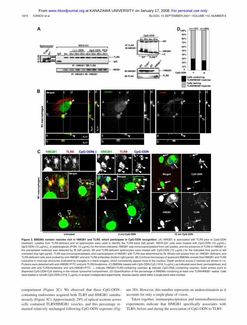
HMGB1 is involved in the translocation of TLR9 to earlyendosomes in response to CpG-ODN
The redistribution of TLR9 from the ER to early endosomes isessential for TLR9 activation by CpG-DNA.11 As our experimentalresults demonstrate that HMGB1 preassociates with TLR9 inquiescent cells, we postulated that HMGB1 is involved in the TLR9translocation process. Thus, we investigated the relationship be-tween HMGB1, TLR9, and the early endosome marker EEA111 inboth wt and HMGB1-deficient macrophages (characterized inFigure 7A) by surveying 200 cells in each condition (Figure 6).Consistent with previous results,11 most TLR9 and EEA1 did notcolocalize in quiescent wt and HMGB1-deficient cells (untreated,Figure 6A,B). Surprisingly, CpG-ODN uptake and colocalizationof CpG-ODN with EEA1 in both wt and HMGB1-deficient cellswere comparable (CpG vs CpG/EEA1, Figure 6A,B), suggestingthat the lack of HMGB1 has no effect on CpG-DNA endocytosis.Importantly, in wt macrophages most TLR9 was redistributed andcolocalized with EEA1 after 5 to 15 minutes of CpG-ODNtreatment, and then deassociated from early endosomes after30 minutes. There was little colocalization of TLR9 with EEA1 inHMGB1-deficient cells 5 to 15 minutes following CpG-ODNstimulation (Figure 6A,B), but there was increased colocalizationof TLR9 with EEA1 at 30 minutes. Thus, the kinetics of TLR9colocalization with EEA1 in HMGB1-deficient cells is delayed.This is further supported by our pharmacological results (Figure S3), asthe depletion of cytoplasmic HMGB1 in wt BMDMs by LMB largelyimpaired the association of HMGB1 with TLR9, retained TLR9 inER-like structures for up to 15 minutes following CpG-ODN stimula-tion, and postponed the colocalization of TLR9 with EEA1. Takentogether, our results suggest that HMGB1 does not affect the uptake of
CpG-ODN or its entry into early endosomes, but accelerates TLR9redistribution to early endosomes in response to CpG-ODN.
HMGB1-deficient cells show defective cytokine responsesto CpG-ODN
Because HMGB1-null mice die postnatally,24 we were unable touse them to assess cellular responses to CpG-ODN in vivo. Thus,we sought to create chimeras by transplanting fetal liver cells(FLCs) isolated from mouse embryos into mice irradiated lethally with137Cs. Unexpectedly, HMGB1-deficient FLCs failed to rescue theirradiated mice, due to a yet unidentified reason (Chu et al, unpublisheddata). Then we decided to derive DCs and macrophages from primaryFLCs (PFLCs) or FLCs immortalized by transfection with HoxB4(IFLCs). Both IFLCs and PFLCs were able to differentiate into DCs(IFLDCs and PFLDCs, respectively), as determined by cell surfaceexpression of CD11b and CD11c, or macrophages (IFLDMs andPFLDMs, respectively), as determined by CD11b and F4/80(Figure 7A).
Both wt and HMGB1-deficient IFLDCs expressed equal amountsof TLR9 (Figure 7B). Importantly, both wt and HMGB1-deficientIFLDCs demonstrated a comparable ability to uptake CpG-ODN ina dose-independent manner (Figure 7C). This is consistent withobservations described previously (Figure 6A,B).
Next, we determined the cytokine response to CpG-DNA inHMGB1-deficient cells. Wt IFLDMs showed a robust IL-6 induc-tion in response to CpG-ODN with a saturation threshold at100 nM CpG-ODN. Conversely, HMGB1-deficient IFLDMs pro-duced only minimal amounts of IL-6 even at 1 �M CpG-ODN(Figure 7D). Wt and HMGB1-deficient IFLDMs responded compa-rably with LPS and PGN.
HMGB1-deficient IFLDCs exhibited a reduced IL-6, TNF�,and IL-12 response to CpG-ODN (Figure 7E). The reduction ofcytokines in Hmgb1�/� IFLDCs was most visible at lower
Figure 6. HMGB1 accelerates early endosomal translocation of TLR9 in macrophages in response to CpG-ODN. (A,B) Wt (top panels) and HMGB1-deficient (bottompanels) IFLDMs (A) or PFLDMs (B) were treated with CpG-ODN-Cy5 (1018, 5 �g/mL) for 0, 5, 15, or 30 minutes. Cells were fixed, permeabilized, and stained withanti-TLR9/FITC and anti-EEA1/rhodamine. Confocal images were acquired by indirect immunofluorescence.
MECHANISM OF TLR9 ACTIVATION 1977BLOOD, 15 SEPTEMBER 2007 � VOLUME 110, NUMBER 6
For personal use only. at KANAZAWA UNIVERSITY on January 17, 2008. www.bloodjournal.orgFrom

Figure 7. Impaired CpG-ODN responses in Hmgb1�/� cells. (A) Wt and Hmgb1�/� immortalized or primary hematopoietic progenitor cells (HPCs) expressed the DCmarkers CD11b and CD11c following incubation with GM-CSF for 7 days, or expressed CD11b and F4/80 after culture with macrophage medium for 10 days. Although HPCsare uniformly round and nonadherent, following differentiation they become adherent and acquire a macrophage-like morphology. (IFLDCs indicates DCs derived fromimmortalized fetal liver. HPCs; PFLDCs, DCs derived from primary fetal liver HPCs.) (B) Protein levels of HMGB1, TLR9, and actin in wt and Hmgb1�/� IFLDCs. (C) Wt andHmgb1�/� IFLDCs endocytosed comparable levels of CpG-ODN or GpG-ODN. Cells were treated with CpG-ODN-Cy5 (0.1 or 1.0 �g/mL for 1 hour, left panel), CpG-ODN-Cy5(1018, 10 or 100 nM), or GpG-ODN-Cy5 (1019, 10 or 100 nM) for the indicated time points (right panel), and then subjected to FACS analysis. (D) Wild-type and Hmgb1�/�
IFLDMs were seeded at 1 � 105/well in a 96-well plate (in triplicate) and treated with CpG-ODN (1018, 10 to 1000 nM), LPS (0.1 �g/mL), or PGN (10 �g/mL). After 24 hours,IL-6 secretion was assessed by ELISA (bars represent the average of triplicates � SD). Experiments were replicated 3 times. (E) Wt and Hmgb1�/� IFLDCs were seeded at1 � 105/well in a 96-well plate (in triplicate) and treated with CpG-ODN (1018, 1 to 1000 nM), LPS (0.1 �g/mL), or PGN (10 �g/mL). After 24 hours, IL-6, IL-12, and TNF�secretion was assessed by ELISA (bars represent the average of triplicates � SD). Experiments were replicated 5 times. (F) Wt and Hmgb1�/� IFLDCs were seeded at1 � 105/well in a 96-well plate (in triplicate) and treated with CpG-A (2216, 1 to 1000 nM, top panel) or CpG-ODN (1018, 0.1 or 1.0 �g/mL, bottom panel) in the presence orabsence of DOTAP (10 �g/mL), or LPS (0.01 or 0.1 �g/mL, bottom panel), or left untreated. After 24 hours, IL-12 (top panel) or IL-6 (bottom panel) secretion was assessed byELISA (bars represent the average of triplicates � SD). (G) Wt and Hmgb1�/� PFLDCs were seeded at 0.8 � 105/well in a 96-well plate (in triplicate) and treated withCpG-ODN (1018, 100 or 1000 nM) or CpG-A (2216, 100 or 1000 nM) in the presence or absence of DOTAP (10 �g/mL), or LPS (0.1 �g/mL), or left untreated. After 24 hours,IL-6 and IL-12 secretion was assessed by ELISA (bars represent the average of triplicates � SD). ND: not detected. (H) Wt and Hmgb1�/� IFLDCs were treated with CpG-B(1018, 10 �g/mL), CpG-A (2216, 3.3 �g/mL), and poly(I:C) (10 �g/mL) in the presence or absence of DOTAP (10 �g/mL) for 24 hours. Type 1 IFN bioactivity in supernatantsamples was detected by a biologic assay against vesicular stomatitis virus.45 (I) Whole-cell lysates were prepared at 16 hours after treatment with CpG-ODN (1018, 10 �g/mL)or LPS (1 �g/mL) and the levels of iNOS, HMGB1, and actin were determined by IB. (n.s. � nonspecific band.) (J) Exogenous rHMGB1 restored cytokine production byHmgb1�/� IFLDCs in response to CpG-ODN. rHMGB1 (rH1, 0.2 �g/mL) was incubated in the presence or absence of CpG-ODN (1018) for 15 minutes. Wt and Hmgb1�/�
IFLDCs were treated with CpG-ODN (1018, 1 or 10 �g/mL) � rHMGB1, LPS (0.1 �g/mL), or rH1 alone, or left untreated for 24 hours, and the levels of secreted IL-6 weredetermined by ELISA (bars represent the average of triplicates � SD).
1978 IVANOV et al BLOOD, 15 SEPTEMBER 2007 � VOLUME 110, NUMBER 6
For personal use only. at KANAZAWA UNIVERSITY on January 17, 2008. www.bloodjournal.orgFrom

concentrations of CpG-ODN. Interestingly, the IL-12 responseto CpG-ODN at 1 �M in HMGB1-deficient IFLDCs was closeto, but still less than, that of wt controls. Both wt andHMGB1-deficient IFLDCs responded to LPS and PGN equally(Figure 7E). The defective response to CpG-ODN was reproduc-ible in 3 independently derived pools of HMGB1-deficientIFLCs (data not shown). Unlike class B CpG-ODN 1018(CpG-B), in response to class A CpG-ODN (CpG-A) wt, but notHmgb1�/�, IFLDCs secreted IL-12 (Figure 7F top panel). Lowerlevels of IL-12 were produced by Hmgb1�/� compared with wtIFLDCs when stimulated with CpG-A in the presence of thecationic lipid 1,2-dioleoyloxy-3-trimethylammonium-propane(DOTAP) (Figure 7F top panel). A similar phenomenon oc-curred for IL-6 when cells were treated with CpG-A andDOTAP, whereas CpG-A alone failed to induce IL-6 (Figure 7Fbottom panel). Cellular activation triggered by CpG-ODNs inthe presence of DOTAP was sensitive to quinacrine andtherefore dependent on TLR9 (data not shown).
Consistently, the defects in HMGB1-deficient IFLDCs werealso observed in HMGB1-deficient PFLDCs. HMGB1 deficiencyled to a severe defect in the IL-6 and IL-12 response to CpG-ODNeven at 1 �M (Figure 7G). In addition, regardless of the presence orabsence of DOTAP, CpG-A failed to induce a detectable level ofIL-6 and IL-12 production in HMGB1- deficient PFLDCs (Figure7G). These experimental results suggest a critical role for HMGB1in the cytokine response to CpG-DNA.
To further investigate a role of HMGB1 in the activation ofinnate immunity by CpG-DNA, we tested the ability of wt andHmgb1�/� IFLDCs to secrete IFN�/ in response to CpG-A andCpG-B.47 Wt IFLDCs produced low but significant levels ofIFN�/ when treated with CpG-B irrespective of DOTAP pres-ence, whereas CpG-A was stimulatory only in the presence ofDOTAP (Figure 7H). Wt IFLDCs produced 2- to 3-fold higherlevels of type I IFNs compared with Hmgb1�/� cells in response toCpG-DNAs. As a control, poly(I:C) triggered a potent release ofIFN�/ by both wt and HMGB1-deficient cells.
In addition, we assessed the expression of inducible nitric oxidesynthase (iNOS),48 which can be induced by CpG-DNA and otherTLR agonists.49-51 CpG-ODN completely failed to induce iNOSexpression in HMGB1-deficient IFLDCs (Figure 7I), in contrast towt IFLDCs. However, both wt and HMGB1-deficient IFLDCsexhibited a similar iNOS response to LPS (Figure 7I).
Finally, we determined whether exogenously supplied HMGB1could compensate for the lack of endogenous HMGB1 in thecytokine response to CpG-DNA. When HMGB1-deficient IFLDCswere provided with exogenous HMGB1, the IL-6, IL-12, and TNF�response to CpG-ODN was restored (Figure 7J and data not shown).
Taken together, these experiments indicate that HMGB1 isrequired for optimal cytokine and iNOS responses to CpG-DNA.
Discussion
TLR9 activation by microbial CpG-DNA and CpG-ODNs in innateimmune cells induces the secretion of cytokines, leading toelimination of microbial pathogens and activation of the adaptiveimmunity.1,2 It has been suggested that CpG-DNAs are endocy-tosed into endosomal compartments where they engage the intracel-lular TLR9, resulting in MyD88 recruitment and the subsequentimmune response.7-10 However, the precise physiological processleading to TLR9 activation by CpG-DNA remains unclear. In this
study, we describe a novel role for HMGB1 as an importantregulator in this process.
Our findings demonstrate that HMGB1, which binds CpG-ODNs, interacts with TLR9 within vesicles, probably belonging tothe ERGIC, of quiescent macrophages. This interaction precedesCpG-ODN uptake, and both intracellular HMGB1 from the nucleusand extracellular HMGB1 from the medium can be recruited tointeract with TLR9. Immune cells lacking HMGB1 show a delayedredistribution of TLR9 into the early endosomes and an impairedcytokine response to CpG-ODN. The latter defect was restoredwhen exogenous HMGB1 was supplied. Thus, our experimentaldata suggest that HMGB1 plays an important role in regulating theprocess of TLR9 activation by CpG-DNA. Recently, a similarconclusion was reached independently by Coyle’s group (Tian et al52).
Most cytokine responses to CpG-DNA are undetectable inTLR9-null cells7; however, HMGB1-deficient cells exhibited par-tial responses. Most notably, the IL-12 response to high doses ofCpG-ODN in HMGB1-deficient IFLDCs was lower than, but closeto, that of wt cells. Interestingly, when examined in primary fetalliver progenitor cell-derived DCs (PFLDCs), the IL-12 response toCpG-ODNs even at 1 �M remains largely impaired in HMGB1-deficient PFLDCs, underscoring a critical role for HMGB1 in thecytokine response to CpG-DNA. Nevertheless, the partial pen-etrance of the Hmgb1 mutation suggests a role of HMGB1 as acofactor that modulates TLR9 activation. This cofactor is not asessential as TLR9 in the activation of the CpG-DNA pathway, butlowers the effective concentration of CpG-DNA necessary fortriggering cellular responses by engaging CpG-DNA, interactingwith TLR9, and facilitating the recognition of CpG-DNA.
TLR9 resides in the ER and its activation requires CpG-ODNendocytosis and the subsequent early endosomal acidification.35
Our findings demonstrate that TLR9/HMGB1 vesicles not onlyreside in the ER, but also colocalize with markers of the ERGICand the Golgi in quiescent cells. We observed that the lumen ofTLR9/HMGB1 vesicles is acidic prior to CpG-DNA accumulation.This is consistent with the ERGIC environment where a vacuolarH�-ATPase pump is active.53,54
MD2, a coreceptor for TLR4, plays an important role in LPSrecognition and contributes to the cell surface localization ofTRL4.55,56 The loss of MD2 results in TLR4 accumulation in theGolgi compartment.56 Similarly, in HMGB1-deficient macro-phages or cytoplasmic HMGB1-depleted BMDMs, TLR9 is re-tained in the ER or the ERGIC following CpG-ODN treatment,whereas HMGB1 deficiency has no apparent effect on CpG-ODNuptake or its entry into early endosomes. Our results show thatpreassociation of HMGB1 with CpG-ODN leads to earlier detec-tion of CpG-ODNs in the TLR9 immune complex (Figure 2). Thus,our data further suggest that HMGB1 contributes to the redistribu-tion of TLR9 to early endosomes.
HMGB1 is considered a nonspecific single/double-strandedDNA-binding protein,13 whereas our data show that HMGB1 hassome preference for binding CpG-ODNs over GpC/GpG-ODNs. Itis known that a simple inversion of the “CpG” in CpG-ODN to the“GpC” or a subtle substitution of the “CpG” with the “GpG,” whichlargely impairs CpG-ODN�s biologic activity, also affects theability to form hairpins or oligo-dimer structures.31,57,58 However, itis unclear whether HMGB1 has some preferences to bind suchstructures. To resolve this, more detailed structure-activity studiesare needed in the future.
HMGB1 is secreted from immune cells upon challenge with avariety of stimuli. Whether CpG-DNA induces secretion of HMBG1from immune cells is controversial.32,33 Our data demonstrate that
MECHANISM OF TLR9 ACTIVATION 1979BLOOD, 15 SEPTEMBER 2007 � VOLUME 110, NUMBER 6
For personal use only. at KANAZAWA UNIVERSITY on January 17, 2008. www.bloodjournal.orgFrom

CpG-ODN triggers the release of HMGB1 from immune cells in amatter of minutes. Quick release of HMGB1 may accelerate therecognition of CpG-DNA and sensitize other immune cells toCpG-DNA, by both autocrine and paracrine signaling. Moreover,other stimuli that lead to HMGB1 secretion could also potentiallysensitize cells to CpG-ODNs.
How does secreted HMGB1 enhance cytokine production inresponse to CpG-ODN? One possibility is that HMGB1 increasesCpG-ODN uptake. Conversely, our experimental results indicatethat the uptake of CpG-ODNs by cells was somewhat decreased inthe presence of HMGB1, and was not apparently affected by thedepletion or deletion of HMGB1. Another possibility is thatHMGB1 uses its potential receptors such as TLR2 and TLR419,59,60
to synergize or cross-talk with TLR9. However, the HMGB1-enhanced cytokine response we have shown here is CpG-DNAspecific, as HMGB1 alone had minimal effects, and TLR2-deficientand TLR4 mutant cells responded comparably with wt cell whenstimulated with CpG-ODN and HMGB1 in producing IL-6.Moreover, coincubation of HMGB1 with LPS or PGN failed toenhance cytokine secretion. Thus, we suggest that HMGB1 acceler-ates and increases the recognition of CpG-ODN by TLR9. Weshowed that loss of HMGB1 delayed the early endosomal translo-cation of TLR9 (Figure 6) and preassociation of HMGB1 withCpG-ODNs led to the earlier detection of CpG-ODNs in the TLR9immune complex (Figure 2G).
CpG-ODN and HMGB1 can give rise to a reciprocal feedbackcycle: innate immune cells secrete HMGB1 in response to CpG-DNA, and HMGB1 sensitizes innate immune cells to CpG-DNA.Our data demonstrate that macrophages can prime this cycle in theabsence of preexisting extracellular HMGB1. They assemblevesicular organelles where a small pool of HMGB1 that is releasedfrom the nucleus preassociates with TLR9. Following stimulation,CpG-DNA-rich endosomes acquire HMGB1 and TLR9 from thesevesicles, leading to TLR9 activation. The defect in the CpG-DNAresponse due to the lack of HMGB1 is relieved by addition ofappropriate forms of exogenous HMGB1.
In conclusion, our identification of HMGB1 as an importantmodulator of TLR9 activation by CpG-DNA provides a linkbetween endogenous nuclear proteins and the principal innateimmune sensors of mammals.
Acknowledgments
This work was supported by grants from the NIH to W.-M.C. (AI54128) and C.A.B. (AI 55677), from COBRE and DOD (PR054819)to W.-M.C., and from Associazione Italiana Ricerca sul Cancro toM.E.B. J.L. was supported by the NDSE predoctoral fellowship.W.-M.C. is a scholar of Leukemia and Lymphoma Society.
We are grateful to Dr Shizuo Akira for providing TLR2-,TLR9-, and MyD88-deficient mice, Dr Dominic van Essen for theHox4 vector, and to Dr Lorenza Ronfani and the CFCM staff forproducing Hmgb1�/� embryos. We thank Drs Sankar Ghosh andAnlin Lin and Mr Matthew Riolo for their critical discussion, andDr Enrico Bucci for useful suggestions. We also thank Drs XiaoPeng, Seung-Hwan Lee, Charles Vaslet, Mingwei Zhu, andStephanie Beall for their technical assistance.
Authorship
Contribution: S.I., A.-M.D., and W.-M.C. executed all experimentsexcept those indicated; X.W. identified the relationship betweenHMGB1/TLR9, early endosomes, and the ERGIC; C.D. and G.M.studied HMGB1/ODN binding; G.S. generated Hmgb1�/� cells;J.L., C.A.B., and G.S.Y. characterized HPCs, CpG-ODN uptake,and IFN�/ production; Y.W. characterized Hmgb1�/� cells andCpG-ODN activation; W.-M.C., M.E.B, H.W., and G.S.Y. providedintellectual input.
Conflict-of-interest disclosure: The authors declare no compet-ing financial interests.
Correspondence: Wen-Ming Chu, Box G-B6, Brown Uni-versity, 171 Meeting St, Providence, RI 02912; email:[email protected].
References
1. Akira S, Takeda K, Kaisho T. Toll-like receptors:critical proteins linking innate and acquired immu-nity. Nat Immunol. 2001;2:675-680.
2. Takeda K, Kaisho T, Akira S. Toll-like receptors.Annu Rev Immunol. 2003;21:335-376.
3. Karlin S, Doerfler W, Cardon LR. Why is CpGsuppressed in the genomes of virtually all smalleukaryotic viruses but not in those of large eu-karyotic viruses? J Virol. 1994;68:2889-2897.
4. Cardon LR, Burge C, Clayton DA, Karlin S. Per-vasive CpG suppression in animal mitochondrialgenomes. Proc Natl Acad Sci U S A. 1994;91:3799-3803.
5. Razin A, Friedman J. DNA methylation and itspossible biological roles. Prog Nucleic Acid ResMol Biol. 1981;25:33-52.
6. Stacey KJ, Young GR, Clark F, et al. The molecu-lar basis for the lack of immunostimulatory activityof vertebrate DNA. J Immunol. 2003;170:3614-3620.
7. Hemmi H, Takeuchi O, Kawai T, et al. A Toll-likereceptor recognizes bacterial DNA. Nature. 2000;408:740-745.
8. Bauer M, Redecke V, Ellwart JW, et al. BacterialCpG-DNA triggers activation and maturation ofhuman CD11c-, CD123� dendritic cells. J Immu-nol. 2001;166:5000-5007.
9. Ahmad-Nejad P, Hacker H, Rutz M, Bauer S,
Vabulas RM, Wagner H. Bacterial CpG-DNA andlipopolysaccharides activate Toll-like receptors atdistinct cellular compartments. Eur J Immunol.2002;32:1958-1968.
10. Bauer S, Kirschning CJ, Hacker H, et al. HumanTLR9 confers responsiveness to bacterial DNAvia species-specific CpG motif recognition. ProcNatl Acad Sci U S A. 2001;98:9237-9242.
11. Latz E, Schoenemeyer A, Visintin A, et al. TLR9signals after translocating from the ER to CpGDNA in the lysosome. Nat Immunol. 2004;5:190-198.
12. Hacker H, Vabulas RM, Takeuchi O, Hoshino K,Akira S, Wagner H. Immune cell activation bybacterial CpG-DNA through myeloid differentia-tion marker 88 and tumor necrosis factor recep-tor-associated factor (TRAF)6. J Exp Med. 2000;192:595-600.
13. Agresti A, Bianchi ME. HMGB proteins and geneexpression. Curr Opin Genet Dev. 2003;13:170-178.
14. Bianchi ME, Agresti A. HMG proteins: dynamicplayers in gene regulation and differentiation.Curr Opin Genet Dev. 2005;15:496-506.
15. Dumitriu IE, Baruah P, Manfredi AA, Bianchi ME,Rovere-Querini P. HMGB1: guiding immunityfrom within. Trends Immunol. 2005;26:381-387.
16. Scaffidi P, Misteli T, Bianchi ME. Release of chro-
matin protein HMGB1 by necrotic cells triggersinflammation. Nature. 2002;418:191-195.
17. Palumbo R, Sampaolesi M, De Marchis F, et al.Extracellular HMGB1, a signal of tissue damage,induces mesoangioblast migration and prolifera-tion. J Cell Biol. 2004;164:441-449.
18. Park JS, Svetkauskaite D, He Q, et al. Involvementof toll-like receptors 2 and 4 in cellular activationby high mobility group box 1 protein. J Biol Chem.2004;279:7370-7377.
19. Tsung A, Sahai R, Tanaka H, et al. The nuclearfactor HMGB1 mediates hepatic injury after mu-rine liver ischemia-reperfusion. J Exp Med. 2005;201:1135-1143.
20. Chu W, Gong X, Li Z, et al. DNA-PKcs is requiredfor activation of innate immunity by immunostimu-latory DNA. Cell. 2000;103:909-918.
21. Wang H, Bloom O, Zhang M, et al. HMG-1 as alate mediator of endotoxin lethality in mice. Sci-ence. 1999;285:248-251.
22. Wang H, Yang H, Tracey KJ. Extracellular role ofHMGB1 in inflammation and sepsis. J Intern Med.2004;255:320-331.
23. Dragoi AM, Fu X, Ivanov S, et al. DNA-PKcs, butnot TLR9, is required for activation of Akt by CpG-DNA. EMBO J. 2005;24:779-789.
24. Calogero S, Grassi F, Aguzzi A, et al. The lack ofchromosomal protein Hmg1 does not disrupt cell
1980 IVANOV et al BLOOD, 15 SEPTEMBER 2007 � VOLUME 110, NUMBER 6
For personal use only. at KANAZAWA UNIVERSITY on January 17, 2008. www.bloodjournal.orgFrom

growth but causes lethal hypoglycaemia in new-born mice. Nat Genet. 1999;22:276-280.
25. Zhang Y, Wang Y, Ogata M, Hashimoto S, OnaiN, Matsushima K. Development of dendritic cellsin vitro from murine fetal liver-derived lineagephenotype-negative c-kit(�) hematopoietic pro-genitor cells. Blood. 2000;95:138-146.
26. Verthelyi D, Zeuner RA. Differential signaling byCpG DNA in DCs and B cells: not just TLR9.Trends Immunol. 2003;24:519-522.
27. Lee SW, Song MK, Baek KH, et al. Effects of ahexameric deoxyriboguanosine run conjugationinto CpG oligodeoxynucleotides on their immuno-stimulatory potentials. J Immunol. 2000;165:3631-3639.
28. Wu CCN, Lee J, Raz E, Corr M, Carson DA. Ne-cessity of oligonucleotide aggregation for Toll-likereceptor 9 activation. J Biol Chem. 2004;279:33071-33078.
29. Marshall JD, Fearon K, Abbate C, et al. Identifica-tion of a novel CpG DNA class and motif that opti-mally stimulate B cell and plasmacytoid dendriticcell functions. J Leukoc Biol. 2003;73:781-792.
30. Sato Y, Roman M, Tighe H, et al. Immunostimula-tory DNA sequences necessary for effective intra-dermal gene immunization. Science. 1996;273:352-354.
31. Krieg AM, Yi AK, Matson S, et al. CpG motifs inbacterial DNA trigger direct B-cell activation. Na-ture. 1995;374:546-549.
32. Dumitriu IE, Baruah P, Bianchi ME, Manfredi AA,Rovere-Querini P. Requirement of HMGB1 andRAGE for the maturation of human plasmacytoiddendritic cells. Eur J Immunol. 2005;35:2184-2190.
33. Jiang W, Li J, Gallowitsch-Puerta M, Tracey KJ,Pisetsky DS. The effects of CpG DNA on HMGB1release by murine macrophage cell lines. J Leu-koc Biol. 2005;78:930-936.
34. Yi AK, Hornbeck P, Lafrenz DE, Krieg AM. CpGDNA rescue of murine B lymphoma cells fromanti-IgM-induced growth arrest and programmedcell death is associated with increased expres-sion of c-myc and bcl-xL. J Immunol. 1996;157:4918-4925.
35. Leifer CA, Kennedy MN, Mazzoni A, Lee C, Kruh-lak MJ, Segal DM. TLR9 is localized in the endo-plasmic reticulum prior to stimulation. J Immunol.2004;173:1179-1183.
36. Macfarlane DE, Manzel L. Antagonism of immu-nostimulatory CpG-oligodeoxynucleotides byquinacrine, chloroquine, and structurally relatedcompounds. J Immunol. 1998;160:1122-1131.
37. Manzel L, Strekowski L, Ismail FM, Smith JC, Mac-farlane DE. Antagonism of immunostimulatory CpG-oligodeoxynucleotides by 4-aminoquinolines andother weak bases: mechanistic studies. J PharmacolExp Ther. 1999;291:1337-1347.
38. Blott EJ, Griffiths GM. Secretory lysosomes. NatRev Mol Cell Biol. 2002;3:122-131.
39. Appenzeller-Herzog C, Roche AC, Nufer O, HauriHP. pH-induced conversion of the transport lectinERGIC-53 triggers glycoprotein release. J BiolChem. 2004;279:12943-12950.
40. Eskelinen EL, Tanaka Y, Saftig P. At the acidicedge: emerging functions for lysosomal mem-brane proteins. Trends Cell Biol. 2003;13:137-145.
41. Schweizer A, Fransen JA, Bachi T, Ginsel L,Hauri HP. Identification, by a monoclonal anti-body, of a 53-kD protein associated with a tubulo-vesicular compartment at the cis-side of the Golgiapparatus. J Cell Biol. 1988;107:1643-1653.
42. Marra P, Maffucci T, Daniele T, et al. The GM130and GRASP65 Golgi proteins cycle through anddefine a subdomain of the intermediate compart-ment. 2001;3:1101-1113.
43. Bonaldi T, Talamo F, Scaffidi P, et al. Monocyticcells hyperacetylate chromatin protein HMGB1 toredirect it towards secretion. EMBO J. 2003;22:5551-5560.
44. Fornerod M, Ohno M, Yoshida M, Mattaj IW.CRM1 is an export receptor for leucine-richnuclear export signals. Cell. 1997;90:1051-1060.
45. Fukuda M, Asano S, Nakamura T, et al. CRM1 isresponsible for intracellular transport mediated bythe nuclear export signal. Nature. 1997;390:308-311.
46. Dalod M, Salazar-Mather TP, Malmgaard L, et al.Interferon alpha/beta and interleukin 12 re-sponses to viral infections: pathways regulatingdendritic cell cytokine expression in vivo. J ExpMed. 2002;195:517-528.
47. Kerkmann M, Rothenfusser S, Hornung V, et al.Activation with CpG-A and CpG-B oligonucleo-tides reveals two distinct regulatory pathways oftype I IFN synthesis in human plasmacytoid den-dritic cells. J Immunol. 2003;170:4465-4474.
48. Bogdan C. Nitric oxide and the immune re-sponse. Nat Immunol. 2001;2:907-916.
49. Ghosh DK, Misukonis MA, Reich C, Pisetsky DS,Weinberg JB. Host response to infection: the roleof CpG DNA in induction of cyclooxygenase 2and nitric oxide synthase 2 in murine macro-phages. Infect Immun. 2001;69:7703-7710.
50. Frances R, Munoz C, Zapater P, et al. Bacterial
DNA activates cell mediated immune responseand nitric oxide overproduction in peritoneal mac-rophages from patients with cirrhosis and ascites.Gut. 2004;53:860-864.
51. He H, Crippen TL, Farnell MB, Kogut MH. Identifi-cation of CpG oligodeoxynucleotide motifs thatstimulate nitric oxide and cytokine production inavian macrophage and peripheral blood mono-nuclear cells. Dev Comp Immunol. 2003;27:621-627.
52. Tian J, Avalos AM, Mao SY, et al. Toll-like recep-tor 9-dependent activation by DNA-containingimmune complexes is mediated by HMGB1 andRAGE. Nat Immunol. 2007;8:487-496.
53. Ying M, Flatmark T, Saraste J. The p58-positivepre-golgi intermediates consist of distinct sub-populations of particles that show differentialbinding of COPI and COPII coats and containvacuolar H(�)-ATPase. J Cell Sci. 2000;113:3623-3638.
54. Palokangas H, Ying M, Vaananen K, Saraste J.Retrograde transport from the pre-Golgi interme-diate compartment and the Golgi complex is af-fected by the vacuolar H�-ATPase inhibitorbafilomycin A1. Mol Biol Cell. 1998;9:3561-3578.
55. Shimura H, Nitahara A, Ito A, Tomiyama K, Ito M,Kawai K. Up-regulation of cell surface Toll-likereceptor 4-MD2 expression on dendritic epider-mal T cells after the emigration from epidermisduring cutaneous inflammation. J Dermatol Sci.2005;37:101-110.
56. Nagai Y, Akashi S, Nagafuku M, et al. Essentialrole of MD-2 in LPS responsiveness and TLR4distribution. Nat Immunol. 2002;3:667-672.
57. Yamamoto S, Yamamoto T, Kataoka T, KuramotoE, Yano O, Tokunaga T. Unique palindromic se-quences in synthetic oligonucleotides are re-quired to induce IFN [correction of INF] and aug-ment IFN-mediated [correction of INF] naturalkiller activity. J Immunol. 1992;148:4072-4076.
58. Sonehara K, Saito H, Kuramoto E, Yamamoto S,Yamamoto T, Tokunaga T. Hexamer palindromicoligonucleotides with 5�-CG-3� motif(s) induceproduction of interferon. J Interferon CytokineRes. 1996;16:799-803.
59. Lotze MT, Tracey KJ. High-mobility group box 1protein (HMGB1): nuclear weapon in the immunearsenal. Nat Rev Immunol. 2005;5:331-342.
60. Yu M, Wang H, Ding A, et al. HMGB1 signalsthrough toll-like receptor (TLR) 4 and TLR2.Shock. 2006;26:174-179.
MECHANISM OF TLR9 ACTIVATION 1981BLOOD, 15 SEPTEMBER 2007 � VOLUME 110, NUMBER 6
For personal use only. at KANAZAWA UNIVERSITY on January 17, 2008. www.bloodjournal.orgFrom
Related Documents