Entorhinal volume, aerobic fitness, and recognition memory in healthy young adults: a voxel-based morphometry study 1 Andrew S. Whiteman, B.A. a , Daniel E. Young, Sc.D. b , Andrew E. Budson, M.D. c , Chantal E. Stern, D.Phil. a , and Karin Schon, Ph.D. a,d Andrew S. Whiteman: [email protected]; Daniel E. Young: [email protected]; Andrew E. Budson: [email protected]; Chantal E. Stern: [email protected] a Dept. of Psychological and Brain Sciences and Center for Memory and Brain, Boston Univ., 2 Cummington Mall, Boston, MA 02215, U.S.A. b College of Nursing and Health Sciences, Exercise and Health Sciences Program, Univ. of Massachusetts Boston, 100 Morrissey Blvd, Boston, MA, 02125, U.S.A. 1 Abbreviatons: The following non-standard abbreviations are used throughout the text. ACSM American College of Sports Medicine BDNF Brain-Derived Neurotrophic Factor BMI Body Mass Index CBV cerebral blood volume DMS Delayed Matching-to-Sample EC entorhinal cortex ELISA Enzyme-Linked Immunosorbent Assay MTL Medial Temporal Lobes RER Respiratory Exchange Ratio RER max maximum observed Respiratory Exchange Ratio SMT Subsequent Memory Test VEGF vascular endothelial growth factor VBM voxel-based morphometry VO 2 max rate of maximal oxygen consumption in mL per kg of body weight per min VO 2 peak peak rate of oxygen consumption in mL per kg of body weight per min, measured during test Corresponding Author: Karin Schon, Ph.D., Dept. of Anatomy and Neurobiology, Boston University School of Medicine, 72 East Concord St., L-1004, Boston, MA, U.S.A., telephone number: +1-617- 414-2327, fax number: +1-617-638-4216, [email protected]. Publisher's Disclaimer: This is a PDF file of an unedited manuscript that has been accepted for publication. As a service to our customers we are providing this early version of the manuscript. The manuscript will undergo copyediting, typesetting, and review of the resulting proof before it is published in its final citable form. Please note that during the production process errors may be discovered which could affect the content, and all legal disclaimers that apply to the journal pertain. HHS Public Access Author manuscript Neuroimage. Author manuscript; available in PMC 2017 February 01. Published in final edited form as: Neuroimage. 2016 February 1; 126: 229–238. doi:10.1016/j.neuroimage.2015.11.049. Author Manuscript Author Manuscript Author Manuscript Author Manuscript

Welcome message from author
This document is posted to help you gain knowledge. Please leave a comment to let me know what you think about it! Share it to your friends and learn new things together.
Transcript
Entorhinal volume, aerobic fitness, and recognition memory in healthy young adults: a voxel-based morphometry study1
Andrew S. Whiteman, B.A.a, Daniel E. Young, Sc.D.b, Andrew E. Budson, M.D.c, Chantal E. Stern, D.Phil.a, and Karin Schon, Ph.D.a,d
Andrew S. Whiteman: [email protected]; Daniel E. Young: [email protected]; Andrew E. Budson: [email protected]; Chantal E. Stern: [email protected] Dept. of Psychological and Brain Sciences and Center for Memory and Brain, Boston Univ., 2 Cummington Mall, Boston, MA 02215, U.S.A.
b College of Nursing and Health Sciences, Exercise and Health Sciences Program, Univ. of Massachusetts Boston, 100 Morrissey Blvd, Boston, MA, 02125, U.S.A.
1Abbreviatons: The following non-standard abbreviations are used throughout the text.
ACSM American College of Sports Medicine
BDNF Brain-Derived Neurotrophic Factor
BMI Body Mass Index
CBV cerebral blood volume
DMS Delayed Matching-to-Sample
EC entorhinal cortex
ELISA Enzyme-Linked Immunosorbent Assay
MTL Medial Temporal Lobes
RER Respiratory Exchange Ratio
RERmax maximum observed Respiratory Exchange Ratio
SMT Subsequent Memory Test
VEGF vascular endothelial growth factor
VBM voxel-based morphometry
VO2 max rate of maximal oxygen consumption in mL per kg of body weight per min
VO2 peak peak rate of oxygen consumption in mL per kg of body weight per min, measured during test
Corresponding Author: Karin Schon, Ph.D., Dept. of Anatomy and Neurobiology, Boston University School of Medicine, 72 East Concord St., L-1004, Boston, MA, U.S.A., telephone number: +1-617- 414-2327, fax number: +1-617-638-4216, [email protected].
Publisher's Disclaimer: This is a PDF file of an unedited manuscript that has been accepted for publication. As a service to our customers we are providing this early version of the manuscript. The manuscript will undergo copyediting, typesetting, and review of the resulting proof before it is published in its final citable form. Please note that during the production process errors may be discovered which could affect the content, and all legal disclaimers that apply to the journal pertain.
HHS Public AccessAuthor manuscriptNeuroimage. Author manuscript; available in PMC 2017 February 01.
Published in final edited form as:Neuroimage. 2016 February 1; 126: 229–238. doi:10.1016/j.neuroimage.2015.11.049.
Author M
anuscriptA
uthor Manuscript
Author M
anuscriptA
uthor Manuscript

c Dept. of Neurology, Boston Univ. School of Medicine, and VA Boston Healthcare System, 150 South Huntington St., Boston, MA, 02130, U.S.A.
d Dept. of Anatomy and Neurobiology and Center for Biomedical Imaging, Boston Univ. School of Medicine, 72 East Concord St., Boston, MA, U.S.A.
Abstract
Converging evidence supports the hypothesis effects of aerobic exercise and environmental
enrichment are beneficial for cognition, in particular for hippocampus-supported learning and
memory. Recent work in humans suggests exercise training induces changes in hippocampal
volume, but it is not known if aerobic exercise and fitness also impact the entorhinal cortex. In
animal models, aerobic exercise increases expression of growth factors, including brain derived
neurotrophic factor (BDNF). This exercise-enhanced expression of growth hormones may boost
synaptic plasticity, and neuronal survival and differentiation, potentially supporting function and
structure in brain areas including but not limited to the hippocampus. Here, using voxel based
morphometry and a standard graded treadmill test to determine cardio-respiratory fitness (Bruce
protocol; VO2 max), we examined if entorhinal and hippocampal volumes were associated with
cardio-respiratory fitness in healthy young adults (N = 33). In addition, we examined if volumes
were modulated by recognition memory performance and by serum BDNF, a putative marker of
synaptic plasticity. Our results show a positive association between volume in right entorhinal
cortex and cardio-respiratory fitness. In addition, average gray matter volume in the entorhinal
cortex, bilaterally, was positively associated with memory performance. These data extend prior
work on the cerebral effects of aerobic exercise and fitness to the entorhinal cortex in healthy
young adults thus providing compelling evidence for a relationship between aerobic fitness and
structure of the medial temporal lobe memory system.
Keywords
medial temporal lobes; hippocampus; recognition memory; cardiovascular fitness; neurotrophins
1. Introduction
The beneficial effects of cardio-respiratory fitness, aerobic exercise, and environmental
enrichment on brain health and cognition are well documented (e.g. see van Praag et al.,
2000; Cotman and Berchtold, 2002; Cotman et al., 2007 for reviews). For example, aerobic
exercise and environmental enrichment are thought to improve learning and memory and to
induce changes in the morphology of many brain structures, notably the hippocampus,
through a variety of mechanisms. Most of this knowledge, however, is inferred from rodent
models, which have focused eminently on effects in the dentate gyrus (DG), a sub-region of
the hippocampus. Comparatively fewer direct observations have been made in humans. We
therefore take a translational approach considering putative physical and neural correlates of
exercise adaptation cross-sectionally in healthy young adults.
In rodents, both exercise and environmental enrichment have been shown to upregulate birth
and survival rates of adult born neuronal and glial cells in the DG of the hippocampus, as
Whiteman et al. Page 2
Neuroimage. Author manuscript; available in PMC 2017 February 01.
Author M
anuscriptA
uthor Manuscript
Author M
anuscriptA
uthor Manuscript

well as improve performance on hippocampal dependent memory tasks (Creer et al., 2010;
Falls et al., 2010; Fordyce and Farrar, 1991; Kempermann et al., 1997; O’Callaghan et al.,
2007; Uda et al., 2006; van Praag et al., 2005, 1999). More generally, environmental
enrichment has also been linked to increased cortical thickness across the brain, most
notably in posterior regions and the entorhinal cortex (EC) (Diamond et al., 1976, 1987;
Greer et al., 1982a, 1982b; reviewed in Mohammed et al., 2002). Exercise-induced brain
plasticity is thought to be regulated in part by the complex, pleiotropic actions of different
neurotrophins, namely brain-derived neurotrophic factor (BDNF) and insulin-like growth
factor-1 (IGF-1). These neurotrophins are associated with synaptic plasticity, neuronal
survival, and differentiation (Kang and Schuman, 1995; McAllister et al., 1999; Trejo et al.,
2001; see Cotman et al., 2007 for a review). In animal models BDNF mRNA expression,
while highest in the hippocampus, is also high in EC and perirhinal cortex (Conner et al.,
1997; Okuno et al., 1999).
Owing to the adult neurogenesis hypothesis, animal models have primarily targeted the DG
and the hippocampal memory system. Exercise not only affects the DG, however, but also
other regions of the medial temporal lobes (MTL), especially hippocampal subfield CA1 and
the EC (Neeper et al., 1996; Stranahan et al., 2007). Specifically, structural changes have
been observed in these regions in the form of increased dendritic spine density in basal
dendrites of pyramidal neurons in entorhinal layer III and in basal and apical CA1 neurons
after two months of voluntary wheel running (Stranahan et al., 2007). These findings stand
on their own, but also integrate well with the literature on neurogenesis, etc., given the EC
has direct projections to the DG and CA1 via layers II and III, respectively (Steward and
Scoville, 1976; Van Hoesen and Pandya, 1975; Witter et al., 1989, 1988), and entorhinal
input may be needed to integrate newborn DG neurons into existing functional networks
(Vivar et al., 2012). In addition, angiogenesis could also affect hippocampal and/or
entorhinal structure following exercise training. Angiogenesis and neurogenesis are
upregulated cooperatively (Palmer et al., 2000), resulting in enhanced formation of new
blood vessels that support newborn neurons. Together, these findings suggest aerobic
exercise and cardio-respiratory fitness may directly alter the structure of the MTL more
broadly.
It is plausible angiogenesis, adult neurogenesis, and neurotrophin-mediated plasticity may
underlie aerobic exercise-related changes in MTL function and structure in humans.
Although these hypotheses cannot be assessed directly in living individuals, evidence for
adult neurogenesis has been observed in postmortem human tissue (Eriksson et al., 1998). In
addition, increased cerebral blood volume (CBV) in the DG (and somewhat in the EC) has
been linked to exercise, providing a possible correlate of exercise-induced neurogenesis in
mice and by extension, perhaps in humans (Pereira et al., 2007). In support of these ideas,
recent human studies indicate aerobic exercise training and cardio-respiratory fitness may be
positively correlated with hippocampal volume (Erickson et al., 2011b, 2009) and
hippocampal cerebral blood flow in healthy older adults (Maass et al., 2015b). In turn,
changes in hippocampal volume following the exercise intervention were correlated with
changes in serum BDNF (Erickson et al., 2011b). Previous work from our lab suggests
effects of aerobic fitness and serum BDNF interact to support episodic recognition memory
(Whiteman et al., 2014) in a task we have shown to recruit the hippocampus and
Whiteman et al. Page 3
Neuroimage. Author manuscript; available in PMC 2017 February 01.
Author M
anuscriptA
uthor Manuscript
Author M
anuscriptA
uthor Manuscript
perirhinal/EC (Schon et al., 2005, 2004). Additionally, increased cardio-respiratory fitness is
associated with greater volume of the parahippocampal gyrus in Alzheimer’s disease
patients (Honea et al., 2009), and aerobic exercise consistently appears as one of the most
effective interventions to attenuate cognitive decline in geriatric populations (Barnes &
Yaffe, 2011; Burns et al., 2008). In younger cohorts, exercise-induced gains in cardio-
respiratory fitness have been linked to better relational memory in children (Chaddock et al.,
2010), and better learning of a virtual Morris Water Maze task in adolescents (Herting and
Nagel, 2012).
Given this background, it is likely entorhinal-dependent memory is associated with cardio-
respiratory fitness and related mechanisms, but a direct link has not yet been established
with entorhinal structure in humans. Establishing such a connection is of interest given the
EC provides the primary input to the hippocampus during episodic memory encoding. The
present study reports on a subsample of participants from Whiteman et al. (2014) that
participated in a magnetic resonance imaging (MRI) study to examine associations between
aerobic capacity and volumes of structures in the medial temporal lobe (MTL) memory
system. Healthy young participants underwent a standard graded treadmill test to measure
cardiorespiratory fitness (Bruce et al., 1963; Thompson et al., 2010), provided blood
samples to assay serum BDNF concentration, and performed an episodic recognition
memory task (Schon et al., 2004; Whiteman et al., 2014). We used region-of-interest (ROI)
based voxel-based morphometry (VBM; Ashburner & Friston, 2000) to analyze regional
gray matter volume in the EC and hippocampus in an unbiased manner. We predicted
volume in these structures would be positively associated with cardiorespiratory fitness. In
addition, based on our previous work (Whiteman et al., 2014), we hypothesized serum
BDNF would also predict MTL volumes. Here, we report evidence for a relationship
between aerobic fitness and gray matter volume in the EC. We also report performance on
our recognition memory task was correlated with average volume in both the hippocampus
and EC; we did not find relationships between gray matter volume and serum BDNF.
2. Materials and Methods
2.1.Participants
One hundred and fourteen healthy young participants were recruited from the Boston
University student community. A random sub-sample of this cohort (sixty-one individuals)
was recruited to participate in an MRI study; the full sample is described in Whiteman et al.
(2014). Of this sub-sample, 16 did not meet inclusion/exclusion criteria, and forty-five
participants were enrolled. Ten participants voluntarily withdrew or were lost to contact, and
two were excluded due to equipment malfunction, leaving a final sample size of N = 33
participants (20 female, 13 male).
All participants were native English speakers or bilingual, all had normal or corrected to
normal vision, and all gave signed, informed consent prior to the start of any study
procedures. All protocols were approved by the Boston University Charles River Campus
Institutional Review Board. Subject characteristics are described in Table 1.
Whiteman et al. Page 4
Neuroimage. Author manuscript; available in PMC 2017 February 01.
Author M
anuscriptA
uthor Manuscript
Author M
anuscriptA
uthor Manuscript
2.2.Procedure
For each participant, the experiment consisted of three visits: (i) informed consent and
screening; (ii) VO2 max aerobic capacity and body composition testing; and (iii) blood draw
and MRI (including functional MRI and cognitive testing). For each participant, all visits
were performed approximately within one month, and visit three (MRI and blood draw) took
place no later than one week after visit two (aerobic fitness testing).
Screening ensured participants could safely participate in an MRI experiment, and were
healthy and able to perform a strenuous treadmill test. Participants were screened for the
following contraindicators: history of neurological or psychiatric conditions; learning
disability; heart, lung, or musculoskeletal conditions or disorders; diabetes mellitus;
electrolyte disorder; high cholesterol; eating disorder or obesity; or current use of any
prescription or recreational cardioactive or psychoactive drugs, or recreational smoking.
2.3. Assessment of Aerobic Capacity
To assess participants’ aerobic capacity (VO2 max; the maximal rate of oxygen consumption
during exercise, measured in milliliters of oxygen per minute per kilogram body-mass), we
used a standard graded maximal exercise test performed on a treadmill (Bruce et al., 1963).
VO2 max is a widely used measurement of stable aerobic fitness, and can be improved with
training depending on exercise intensity and consistency (e.g. Huang et al., 2005).
Participants were asked not to engage in strenuous physical activity for at least 24 hours
prior their scheduled fitness test, and not to consume food or caffeine for at least 3 hours
prior. A physician was on call, and two study staff members with current cardio-pulmonary
resuscitation training were present for each exercise test. Each test began with the treadmill
at a speed of 0.8 m/s and an incline of 10% grade. Throughout a test, the speed and incline
of the treadmill increased by an average of 0.35 m/s and 2% grade every three minutes.
Participants were given a 3-5 minute warm-up at 0.5 m/s and 0% grade before testing;
following testing, participants were given a five-minute cool-down at 0.5 m/s and 0% grade.
Fitness tests were terminated when participants reached volitional exhaustion. Graded
exercise testing protocol and termination criteria followed the recommendations of the
American College of Sports Medicine (ACSM; Thompson et al., 2010).
All VO2 max tests were performed between the hours of 8:00 – 10:00 AM at Boston
University Sargent College of Health and Rehabilitation Sciences. The ParvoMedics True
One 2400 system (ParvoMedics, Sandy, UT, USA) was used during testing to analyze gas
exchange in participants’ breath. The True One 2400 system was calibrated against medical
grade gasses (ParvoMedics, Sandy, UT, USA), and VO2 values were averaged over 30 s
intervals. Respiratory exchange ratio (RER; volume expired CO2 divided by volume expired
O2) was also monitored with this system. An RER at or above 1.15 can be taken as a rough
indicator VO2 max has been reached (Issekutz et al., 1962). Because VO2 max varies by age
and gender, we converted VO2 max values into fitness percentiles using ACSM norms
(Thompson et al., 2010).
Whiteman et al. Page 5
Neuroimage. Author manuscript; available in PMC 2017 February 01.
Author M
anuscriptA
uthor Manuscript
Author M
anuscriptA
uthor Manuscript

2.4. Blood Draw, Cognitive Testing, and Hormone Assays
Procedures for the blood draw, memory test, and hormone assays have been described in
detail previously (Whiteman et al., 2014). Briefly, blood samples were drawn from the
median cubital vein by a trained nurse at the Boston University General Clinical Research
Unit between 8:00 – 9:45 AM. Serum aliquots were prepared from the obtained blood
samples and stored at −80° C. At the end of the study, serum concentrations of BDNF,
cortisol, IGF-1, and Vascular Endothelial Growth Factor (VEGF) were determined for all
blood samples in duplicate using Quantikine® quantitative sandwich ELISA kits (R&D
Systems Inc., Minneapolis, MN, USA).
Following their blood draw, participants were walked across the street to the Boston
University Center for Biomedical Imaging, where MRI data was collected. In addition to
structural MRI data collection, fMRI data was collected while participants performed a
delayed match-to-sample task with unfamiliar complex visual scenes (functional data to be
reported in another publication; Fig. 1a). About fifteen minutes after the delayed match-to-
sample task, participants performed a surprise subsequent memory test outside of the
scanner with all (144) “old” delayed match-to-sample scenes, plus an equal number of
content similar lures.
Participants were unaware of the ratio of old to new images, and were asked to rate their
recognition memory strength for each SMT image along a 5 point scale: 1 – sure, new; 2 –
unsure, new; 3 – unsure, old; 4 – sure, old; R – sure, old, plus accompanied by some
subjective association with the scene, e.g. memory for which run the scene came from or a
thought the scene prompted when it was first shown. Our previous work suggests this
paradigm recruits the hippocampus and MTL cortex (Schon et al., 2005, 2004). Stimulus
presentation and participant response data were displayed and recorded using EPrime 2.0
software (Psychology Software Tools, Pittsburgh, PA). As a measure of memory accuracy,
we used a standard corrected hit rate: the proportion of subsequent memory test scenes
correctly recognized as old, less the proportion of lure scenes misclassified as old.
Behavioral results are shown in Fig. 1b and 1c.
2.5. MR Image Acquisition and Preprocessing
High resolution T1-weighted structural scans (multi-planar rapidly acquired gradient echo
images) were acquired on a 3 T Phillips Achieva scanner using an 8-channel head coil (TR =
6.8 ms; TE = 3.1 ms; voxel size = 0.98 × 0.98 × 1.2 mm; flip angle = 9°; 150 slices; SENSE
reduction factor = 1.5). Structural images were preprocessed using SPM8 software
(Wellcome Department of Cognitive Neurology, London, UK). For each subject, structural
scans were preprocessed as follows: (i) posterior probability maps for gray and white matter
tissues, and cerebrospinal fluid, etc. were obtained using SPM8’s “new segment” option. (ii)
Gray matter posterior probability maps were spatially normalized to Montreal Neurological
Institute space using the diffeomorphic anatomical registration using exponentiated lie
algebra algorithm (DARTEL; Ashburner, 2007) for a high degree of inter-subject co-
registration. During normalization, images were Jacobian scaled for “modulated” VBM,
resampled to 1.5 mm3 isotropic voxels, and spatially smoothed with a 6 mm full-width at
Whiteman et al. Page 6
Neuroimage. Author manuscript; available in PMC 2017 February 01.
Author M
anuscriptA
uthor Manuscript
Author M
anuscriptA
uthor Manuscript

half-maximum Gaussian kernel to reduce noise. Resultant images show total regional gray
matter volume, accounting for differences in global brain anatomy between subjects.
2.6.Regions of Interest
Based on our a priori anatomical predictions, we created a region of interest (ROI) for
statistical analysis that included the hippocampus and EC. To define our ROI as accurately
as possible, we used ITK-SNAP 2.2 (Yushkevich et al., 2006; http://www.itksnap.org/) to
trace these structures manually on our group average normalized brain image based on the
work of Pruessner et al. (2000, 2002). Hippocampal segmentation was performed following
Pruessner et al. (2000), and entorhinal segmentation was performed following Pruessner et
al. (2002). Since we used VBM to analyze the data, our results are, to a point, free from the
usual segmentation biases (e.g. definition of boundaries between regions). We note, though,
that defining our ROIs manually may induce a small bias with regard to spatial extent
statistics. Briefly, borders for the EC were defined anteriorly with the appearance of the
collateral sulcus and posteriorly with the disappearance of the intralimbic gyrus. The medial
border of EC was amygdala if present or hippocampus if not; the lateral border was the
midpoint of the medial bank of the collateral sulcus in accordance with Pruessner et al.
(2002). We did not adjust the lateral border of the EC according to collateral sulcus depth,
etc. as in Insausti et al. (1998). We do not include fimbria/fornix in our hippocampal
tracings. A brief overview of our anatomical demarcations can be seen in Fig. 2d.
2.7.Statistical Analysis
Our analysis of the MR data was performed using R 3.0.1 (R Development Core Team,
2012), and in-house code written for MATLAB (The MathWorks Inc., Natick, MA, USA)
and SPM8. We fit standard least-squares regression models to log-scaled VBM data (natural
logarithm), predicting log-volume from variables fitness percentile (percentile score), SMT
corrected hit rate (percentage), and serum BDNF (ng · mL−1), controlling for effects of total
intra-cranial volume (L), and binary variables gender (0 female, 1 male) and RER ≥ 1.15 (0
RER < 1.15, 1 RER ≥ 1.15; RER see Methods: Assessment of Aerobic Capacity).
Continuous input variables were mean-centered, except for fitness percentile, which was
centered to the 50th percentile for better interpretability.
We chose a statistical approach whereby we began our analysis by testing our a priori
hypotheses under our most stringent statistical thresholds, and afterwards relaxed our
assumptions into an exploratory whole brain analysis. Because there are three variables of
interest in this analysis and either positive or negative contrasts could be declared significant
(although we are only interested in positive effects), we displayed our statistical parametric
maps at a threshold P < 0.008 ≈ 0.05 / 6. We corrected these P values for our main ROI
analysis using a permutation test (Nichols and Holmes, 2002) based on 1000 relabelings.
Following the permutation distribution, the corrected 2-tailed α < 0.05 theight thresholds
were 3.79 and 3.71 for fitness and memory, respectively. Single tailed α < 0.05 cluster
extent thresholds were 131 and 113 voxels for fitness and memory, respectively. In contrast,
“exploratory” whole brain analyses were collated using a rule-of-thumb cluster extent k ≥
100 contiguous voxels to visualize results, and are otherwise uncorrected for multiple
comparisons. Results from both ROI and exploratory analyses are reported in Table 2.
Whiteman et al. Page 7
Neuroimage. Author manuscript; available in PMC 2017 February 01.
Author M
anuscriptA
uthor Manuscript
Author M
anuscriptA
uthor Manuscript

Following up on the results from our classical VBM analysis, we wanted to provide an
estimate for the cross sectional effect of aerobic fitness on average volume in the whole EC.
To do this, we extracted the mean VBM intensity—the amount of gray matter in the average
entorhinal voxel—from subjects’ ECs judging that this should provide a reasonable correlate
of the size of the gross structure. We then fit a standard least squares regression model to the
extracted mean VBM data with independent variables fitness percentile, memory accuracy,
total intra-cranial volume, gender, and RER ≥ 1.15. For this analysis, we again opted to log-
transform the mean VBM estimates (natural logarithm). This has at least two benefits: (i)
since gray matter volume is all-positive it makes sense effects may occur on a multiplicative
scale (Keene, 1995) and, similarly, (ii) small regression coefficients can be interpreted as
percentage increases in y per change in x (e.g. βlog = 0.02 corresponds to an approximate 2%
increase in y on the native scale since e0.02 = 1.02). This helps us to connect the present
results to the extant literature.
3. Results
3.1. Fitness and entorhinal gray matter volume
Results from our primary VBM analysis showed gray matter volume in a region of the right
EC composed of 157 voxels was positively associated with aerobic fitness (Table 2; Fig.
2a). This was the only region that showed a correlation with aerobic fitness when controlling
for the effects of memory accuracy, BDNF, total intra-cranial volume, gender, and RER ≥
1.15 for our primary ROI-based VBM analysis. Based on the permutation distribution, the
observed result is significant in both height (P < 0.01), and extent (P < 0.05). Exploratory
whole brain results also suggested regions of gray matter may be associated with fitness in
the precuneus, lingual gyrus, inferior parietal lobule, middle occipital gyrus, visual areas BA
17 and BA 18, middle and inferior temporal gyrus, cerebellum, and superior frontal gyrus
(Table 2 and Fig. 3a).
Given that we observed a region in EC where gray matter volume was significantly
associated with cardio-respiratory fitness in our sample, we wanted to provide an estimate
for how aerobic fitness may be expected to relate to EC volume in the general population
and compared to previous literature. To do this, we determined the average VBM gray
matter volume estimate from each subjects’ ROIs and refit our model separately to the
vectors of means. Results of this analysis, reported in table 3, suggest positive effects of
fitness in the right EC, and a positive trend across both hemispheres (right hemisphere:
rpart(26) = 0.45; left hemisphere: rpart(26) = −0.02; average: rpart(26) = 0.28).
3.2. Memory accuracy and gray matter volume
Results from our primary ROI-based VBM analysis did not show any region within the
MTL where gray matter volume was associated with memory task performance. An
exploratory whole brain analysis suggested possible effects in the right EC, hippocampus,
amygdala, middle temporal gyrus, and cerebellum (Table 2 and Fig. 3b). In the follow-up
analysis of average ROI gray matter volume, large correlations between memory accuracy
and volume are apparent in the EC (right hemisphere: rpart(26) = 0.44; left hemisphere:
rpart(26) = 0.35; average: rpart(26) = 0.43; Table 3), and a positive trend is apparent in
Whiteman et al. Page 8
Neuroimage. Author manuscript; available in PMC 2017 February 01.
Author M
anuscriptA
uthor Manuscript
Author M
anuscriptA
uthor Manuscript
hippocampus (right hemisphere: rpart(26) = 0.27; left hemisphere: rpart(26) = 0.16; average:
rpart(26) = 0.23; Table 3).
3.3. Fitness and memory accuracy
In our previous work (Whiteman et al., 2014), we have shown that fitness and serum BDNF
interactively predicted recognition memory accuracy. In that study, fitness percentile alone
did not predict memory accuracy. Here, we reanalyzed this data in our subsample (N = 33).
As expected based on our previously reported results from the larger sample, fitness
percentile did not predict recognition memory performance (rpart(29) = −0.09; controlling
for gender and RER). When we refit our full model from our previous work to this
subsample (predictors: fitness, BDNF, the BDNF by fitness interaction, gender, and RER),
we find that the BDNF by fitness interaction showed a statistical trend predicting memory
accuracy in these data. The slope coefficient is comparable between full and subsample, but
the standard error is larger (full sample β ± SE = 0.59 ± 0.22; current subsample β ± SE =
0.52 ± 0.28). For a complete description of the association between serum BDNF, aerobic
fitness, and memory accuracy in the full sample, we refer the interested reader to Whiteman
et al. (2014).
3.3. BDNF and gray matter volume
Results from our primary ROI-based VBM analysis did not show any region within the
MTL where gray matter volume was associated with serum BDNF levels. An exploratory
whole brain analysis suggested a region of medial thalamus potentially associated with
BDNF (peak: coordinates = [2, −4, 4], t(26) = 4.88; cluster extent k = 299). For a description
of the association between serum BDNF, aerobic fitness, and memory accuracy, we refer the
interested reader to Whiteman et al. (2014). The follow-up analysis of effects at the average
ROI level did not suggest any correlation between serum BDNF and structure in the EC or
hippocampus.
3.4. Analysis of variability in structural VBM data
The fitness percentile effect we report was observed close to the border between entorhinal
and perirhinal cortex on the medial bank of the collateral sulcus. Without our ROI mask, the
effect extended into perirhinal cortex. Given that VBM as a technique relies heavily on the
anatomical coregistration algorithm(s) used, and because the collateral sulcus is an
especially variable region (e.g. Pruessner et al., 2002; Yushkevich et al., 2015a), we were
concerned our results could be explained by the possibility of anatomical registration
artifacts. To examine this question, we created a coefficient of variation map for our group
data. This map shows regions of high anatomical variability across subjects. Examination of
this data shows the anatomical variation in the EC and hippocampus is not unusually high
(Fig. 2c-d). The histogram in Fig. 2c shows the distribution of the coefficient of variation for
all gray matter voxels in the brain, including our regions of interest, the EC and the
hippocampus (shown additionally as boxplots overlaid on the histogram). Coefficient of
variation values for the EC and the hippocampus fall within the high density region on the
histogram (Fig. 2c), suggesting our results may be unlikely to be due to anatomical warping
artifacts.
Whiteman et al. Page 9
Neuroimage. Author manuscript; available in PMC 2017 February 01.
Author M
anuscriptA
uthor Manuscript
Author M
anuscriptA
uthor Manuscript
4. Discussion
With the present study, we examined the relationships between aerobic fitness, serum
BDNF, recognition memory, and gray matter volume in the entorhinal-hippocampal
memory system. Our data suggest volume in the EC may be positively associated with
aerobic fitness and recognition memory performance, but not with serum BDNF. These
results provide translational support for rodent models on exercise, neurogenesis, and the
entorhinal-hippocampal memory system.
Motivation for this general line of research ultimately comes from studies that showed
enriched environments could induce cortical thickening in rodents relative to standard living
condition controls (Diamond, 1988; Diamond et al., 1985; Malkasian and Diamond, 1971).
Early studies showed increases in cortical thickness were most pronounced in posterior
regions and in the EC (Greer et al., 1982; Diamond, 1988; reviewed in Mohammed et al.,
2002), which relays incoming information to the hippocampus via its direct projections to
the dentate gyrus and CA fields (Van Hoesen and Pandya, 1975; Witter et al., 1989, 2000).
In the hippocampus, however, changes in brain morphology related to enrichment were
absent or not very robust (Diamond, 1988; Diamond et al., 1976; Greer et al., 1982b; Walsh
et al., 1969) unless measurements were confined to the granular layer of the dentate gyrus
(Diamond, 1988; Juraska et al., 1985). It is now known the granular layer of the dentate
gyrus is the neurogenic zone of the hippocampus, and shows a strong response to aerobic
exercise in animal models (e.g. Pereira et al., 2007; van Praag et al., 2005, 1999).
Additionally, other regions of the hippocampal-entorhinal memory circuit show structural
changes in manipulations limited to aerobic exercise (Neeper et al., 1996; Stranahan et al.,
2007). Specifically, both the CA1 subfield of the hippocampus and the EC layer III show
increased dendritic spine density following two months of voluntary wheel running
(Stranahan et al., 2007). Aerobic exercise not only enhances adult neurogenesis in the
dentate gyrus, but also promotes angiogenesis (Clark et al., 2009; Palmer et al., 2000), the
formation of new blood vessels, which could also explain changes in thickness or volume.
Given the unique architecture of the entorhinal-hippocampal memory system, a causal
relationship between aerobic exercise training and increased entorhinal volume is plausible
and consistent with our data. In support of these findings, we report a positive association
between aerobic fitness and EC volume in healthy young adult humans.
This finding is not without its caveats. Given the nature of VBM, it is difficult to distinguish
between meaningful anatomical variation and anatomical warping artifacts since both are
induced by the chosen coregistration algorithm. Since our primary result in right EC lies
partially within the collateral sulcus, a region of high anatomical variability (Insausti et al.,
1998; Pruessner et al., 2002; Yushkevich et al., 2015a), we explored the possibility of
artifactual warping. To do this, we created and inspected a coefficient of variation map for
all gray matter voxels in the brain, and concluded that the variation in hippocampal and
entorhinal volume across subjects fell within a normal range. Note in Fig. 2c-d one can
appreciate the relatively modest variation in the collateral sulcus against the larger variation
in the fundus of the neighboring occipitotemporal sulcus, or even greater variation in some
dorsal regions.
Whiteman et al. Page 10
Neuroimage. Author manuscript; available in PMC 2017 February 01.
Author M
anuscriptA
uthor Manuscript
Author M
anuscriptA
uthor Manuscript
Additionally, it is of interest why the fitness effect might at first appear to be lateralized to
right EC. Under more careful consideration, however, the data do not warrant the
assumption that the effect is truly unilateral. For example, if instead of the peak voxel, we
look at the average of a 3 mm sphere surrounding the peak, we find t statistics of +4.51 in
the right EC, and +1.72 in the complimentary set of voxels in the left EC. Importantly, the t
statistic for the difference in these effects is only about 1.74, which is non-significant (P =
0.09). This calculation is by no means unbiased, and ignores any potential correlation
between the hemispheric estimates, but it suggests the hypothesis that effects in the right and
left hemispheres are different is not supported by the data. One can even apply the same
argument to the average effects for each ROI given in Table 3, though in this case it leads to
a weaker rejection. Indeed, based on the literature we discuss here, we conclude effects of
fitness are likely to be bilateral. Therefore, in terms of laterality, we believe our data should
be interpreted with caution. Convergent results from multiple complimentary methodologies
are necessary to support our findings and their interpretation.
Several published studies provide information on the relationship between fitness and
volume of MTL structures in healthy adult humans (Chaddock et al., 2010; Colcombe et al.,
2006, 2003; Erickson et al., 2011b; Killgore et al., 2013; Makizako et al., 2011; Ruscheweyh
et al., 2011; Schlaffke et al., 2014); perhaps the most relevant, with the most complete set of
descriptive statistics is the study by Erickson et al. (2011b; also see Coen et al., 2011;
Erickson et al., 2011a). Erickson et al. (2011b) conducted a year-long aerobic exercise
intervention in older adults and reported on average small increases in hippocampal volume
bilaterally for exercising subjects, and on average small decreases in hippocampal volume
bilaterally for participants of an exercise training control group that performed resistance
and stretching exercises. Averaging over both hemispheres, results from this study suggest
approximately a 3.1% ± 2.8% (mean ± SE) increase in hippocampal volume per
approximately a 4.1 point increase in fitness percentile in the exercise group (N = 60; data
from Table 2 in Erickson et al., 2011b). We computed the 4.1 percentile points based on the
average age and male to female ratio in the exercise group in this study. It seems reasonable
to assume changes in gross hippocampal volume over a year period fit within the range of
plus or minus a few percent in otherwise healthy older adults. Compared with the report
from Erickson et al. (2011b) of approximately a 0.76% increase in volume per point fitness
percentile (linear scale), our estimate of a 0.14% increase in right EC volume per point
fitness percentile (multiplicative scale) makes sense for healthy young adults. Although this
similarity is encouraging, there are considerable methodological differences between the
studies.
Among the most interesting differences between the results of Erickson et al. (2011b, 2009)
and the present report is that we have not replicated an effect in hippocampus. We are
inclined to believe the failure to replicate the hippocampal result may either be simply a
difference in the type of sample used (i.e. young vs. older adults), or it may be a
methodological difference. Our implementation of VBM relies on the DARTEL algorithm
(Ashburner, 2007), which is optimized for aligning global structures (Klein et al., 2009;
Yassa and Stark, 2009) but may be less adept at aligning hippocampal subregions (Yassa
and Stark, 2009) like the CA3/dentate gyrus. Our coefficient of variation map (Fig. 2c)
Whiteman et al. Page 11
Neuroimage. Author manuscript; available in PMC 2017 February 01.
Author M
anuscriptA
uthor Manuscript
Author M
anuscriptA
uthor Manuscript

shows very limited variation in the hippocampus; our analysis may not be sensitive enough
to detect small hippocampal differences. In contrast to the present study, Erickson et al.
(2011b) used an automated segmentation tool to compute overall hippocampal volume.
Future studies using different morphometric techniques such as assessment of cortical or
gray matter thickness (e.g. Burggren et al., 2008; Yushkevich et al., 2015), or high-field
MRI (e.g. Maass et al., 2015a, b) may have more success in this area and may map better
onto the earliest findings regarding enrichment and entorhinal and hippocampal morphology
(Diamond, 1988; Juraska et al., 1985).
Since we have observed a positive relationship between aerobic fitness and gray matter
volume in the EC, it may be important to understand the underlying neurobiological
mechanisms of this putative correlation. In this regard, Pereira and colleagues assessed
longitudinal changes in CBV in murine and human hippocampi and EC over a six week
exercise intervention (Pereira et al., 2007). In both species, the dentate gyrus and EC were
the only regions that exhibited increases in CBV over the course of the exercise intervention.
Although effects in EC were not statistically significant in that study, their result may
provide valuable insight into the biological basis of our observed relationship between
fitness and volume in the EC. Given their data, we speculate angiogenic mechanisms may
underlie observed CBV responses following exercise training. An angiogenic account seems
a parsimonious explanation if the effects of exercise extend beyond the dentate gyrus and to
the EC, as our data suggest. Moreover, a recent proof-of-concept study in older adults
suggests exercise related enhancement of object recognition may be modulated by
hippocampal cerebral blood flow and CBV (Maass et al., 2015b). This idea is further
supported by studies on CBV and metabolism (Ide and Secher, 2000; Vissing et al., 1996)
and by work suggesting angiogenesis and neurogenesis are upregulated cooperatively
(Palmer et al., 2000). Note this angiogenic account, while parsimonious, is speculative as we
cannot distinguish between a general increased CBF/CBV across the EC and one specific to
improved synaptic functioning during cognition.
We have also reported evidence suggestive of associations between average EC volume and
recognition memory performance. In our data, this result is most evident when looking at the
average effect over the whole EC (one would expect this to be the most robust level). At the
voxel level, however, the effect is just sub-threshold in the right EC in our ROI analysis due
to the strict multiple comparison correction, but it is present at the exploratory threshold.
Given the breadth of literature on the EC and episodic memory, we are inclined to consider
these results supporting evidence. Recent high-resolution fMRI studies from our group have
shown greater entorhinal activity for faster correct decisions during encoding of novel
spatial environments (Brown et al., 2014), and reported entorhinal activity when unfamiliar
stimuli with overlapping features or a greater stimulus load, respectively, need to be
maintained in a working memory buffer (Newmark et al., 2013; Schon et al. 2015). These
studies suggest a role for the EC in both episodic encoding and working memory
maintenance. For the present experiment, we used an adaptation of a delayed match-to-
sample task with outdoor scenes from our previous work (Schon et al., 2005, 2004). These
studies reported delay period activity in the perirhinal/entorhinal cortex and hippocampus
predicted whether a scene was later remembered with high confidence. Together with
Whiteman et al. Page 12
Neuroimage. Author manuscript; available in PMC 2017 February 01.
Author M
anuscriptA
uthor Manuscript
Author M
anuscriptA
uthor Manuscript

previous work (e.g. Bellgowan et al., 2009; Brown et al., 2014), these results suggest a role
for both the perirhinal/entorhinal region and the hippocampus in encoding.
In addition, we have described estimates for relationships between hippocampal volume and
memory performance on a subsequent memory test adapted from Schon et al. (2005, 2004),
which showed a positive trend. Although the hippocampal estimates are non-significant in
the present case, they are in the correct direction: previous work suggests hippocampal
volume is related to episodic learning of spatial contexts in humans of the same age range
(Brown et al., 2014), and others have suggested links between hippocampal volume and
other memory tasks (e.g. Maguire et al., 2006; Hartley & Harlow, 2012; Horner et al., 2012;
Rodrigue et al., 2013).
Finally we note VBM as a technique is somewhat limited in that it cannot provide
volumetric measures of gross anatomic structures as accurately as, for example, manual
segmentation methods can. Manual techniques, however, do not have the same spatial
sensitivity of voxel-based techniques like VBM, and may be biased with regard to regional
boundaries; the fundamental research questions that can be addressed by the two types of
methodologies are slightly different. Whereas VBM is optimized for analysis of structural
data collected with a fine isotropic resolution (like ours), manual segmentation methods are
best suited to data collected with a very fine in-plane resolution, and a larger slice thickness.
Further, VBM is ideally suited for investigating questions involving correlations between
relative brain volumes and external variables (especially in healthy, non-clinical subjects
where absolute volumes may be less informative than relative brain volume changes). For
these reasons converging evidence from complementary morphometric techniques and from
animal models are needed to support our findings.
5. Conclusions
We have successfully tested the hypothesis that effects of aerobic exercise on structure in
the hippocampal-entorhinal formation observed in rodents can be translated to healthy
young adult humans. Consistent with work on environmental enrichment in rodents, our data
demonstrated a positive association between fitness and volume in the right EC. Here we
have also reported evidence for correlations between volume in both entorhinal cortices and
performance on an episodic recognition memory task. Our data provide compelling evidence
for a relationship between aerobic fitness and structure of the MTL memory system in
healthy young adults. Our results may have theoretical implications for rodent models of
exercise-associated adult neurogenesis in the hippocampus, and suggest clinical exercise
trials in older adults at risk for developing Alzheimer’s disease should examine entorhinal
function and structure. Aerobic exercise such as running is by no means the only way to
improve aerobic capacity. For example, studies suggest circuit training or strenuous weight-
lifting may improve VO2 performance (Gettman et al., 1982; Stone et al., 1991). This may
prove an interesting avenue for future researchers to try to isolate effects of, for example,
aerobic fitness vs. running.
Whiteman et al. Page 13
Neuroimage. Author manuscript; available in PMC 2017 February 01.
Author M
anuscriptA
uthor Manuscript
Author M
anuscriptA
uthor Manuscript
Acknowledgements
This work was supported by a Pathway to Independence Award to K.S. (NIH K99AG036845 and NIH R00AG036845), the Boston University Clinical and Translational Science Institute (CTSI; UL1-TR000157), the Boston University Center for Biomedical Imaging (CBI), and a Student Research Award from the Boston University Undergraduate Research Opportunities Program to A.S.W. We would like to thank Drs. Xuemei He and Tai Chen for performing and overseeing the BDNF ELISAs, respectively, and the staff of the CTSI General Clinical Research Unit (GCRU) and of the CBI for their support. In addition, we would like to thank Dr. Neil Kowall for serving as the study physician for the GCRU, and Ms. Rachel Nauer for assistance with preparation of figures.
References
Adler, D.; Murdoch, D., et al. rgl: 3D visualization device system (OpenGL). 2014.
Ashburner J. A fast diffeomorphic image registration algorithm. Neuroimage. 2007:95–113. [PubMed: 17761438]
Ashburner J, Friston KJ. Voxel-based morphometry--the methods. Neuroimage. 2000; 11:805–21. doi:10.1006/nimg.2000.0582. [PubMed: 10860804]
Bellgowan PSF, Buffalo EA, Bodurka J, Martin A. Lateralized spatial and object memory encoding in entorhinal and perirhinal cortices. Learn. Mem. 2009; 16:433–8. doi:10.1101/lm.1357309. [PubMed: 19553381]
Brown TI, Whiteman AS, Aselcioglu I, Stern CE. Structural differences in hippocampal and prefrontal gray matter volume support flexible context-dependent navigation ability. J. Neurosci. 2014; 34:2314–20. doi:10.1523/JNEUROSCI.2202-13.2014. [PubMed: 24501370]
Bruce R, Blackmon J, University of W.D. of A.C. Jones J, Strait G. Exercise Testing in Adult Normal Subjects and Cardiac Patients. Pediatrics. 1963; 32:742–756. [PubMed: 14070531]
Burggren AC, Zeineh MM, Ekstrom AD, Braskie MN, Thompson PM, Small GW, Bookheimer SY. Reduced cortical thickness in hippocampal subregions among cognitively normal apolipoprotein E e4 carriers. Neuroimage. 2008; 41:1177–83. doi:10.1016/j.neuroimage.2008.03.039. [PubMed: 18486492]
Chaddock L, Erickson KI, Prakash RS, Kim JS, Voss MW, Vanpatter M, Pontifex MB, Raine LB, Konkel A, Hillman CH, Cohen NJ, Kramer AF. A neuroimaging investigation of the association between aerobic fitness, hippocampal volume, and memory performance in preadolescent children. Brain Res. 2010; 1358:172–83. doi:10.1016/j.brainres.2010.08.049. [PubMed: 20735996]
Clark PJ, Brzezinska WJ, Puchalski EK, Krone DA, Rhodes JS. Functional analysis of neurovascular adaptations to exercise in the dentate gyrus of young adult mice associated with cognitive gain. Hippocampus. 2009; 19:937–50. doi:10.1002/hipo.20543. [PubMed: 19132736]
Coen RF, Lawlor BA, Kenny R. Failure to demonstrate that memory improvement is due either to aerobic exercise or increased hippocampal volume. Proc. Natl. Acad. Sci. U. S. A. 2011; 108:E89–E90. author reply. doi:10.1073/pnas.1102593108. [PubMed: 21504947]
Colcombe SJ, Erickson KI, Raz N, Webb AG, Cohen NJ, McAuley E, Kramer AF. Aerobic fitness reduces brain tissue loss in aging humans. J. Gerontol. A. Biol. Sci. Med. Sci. 2003; 58:176–80. [PubMed: 12586857]
Colcombe SJ, Erickson KI, Scalf PE, Kim JS, Prakash R, McAuley E, Elavsky S, Marquez DX, Hu L, Kramer AF. Aerobic exercise training increases brain volume in aging humans. J. Gerontol. A. Biol. Sci. Med. Sci. 2006; 61:1166–70. [PubMed: 17167157]
Conner JM, Lauterborn JC, Yan Q, Gall CM, Varon S. Distribution of brain-derived neurotrophic factor (BDNF) protein and mRNA in the normal adult rat CNS: evidence for anterograde axonal transport. J. Neurosci. 1997; 17:2295–313. [PubMed: 9065491]
Cotman CW, Berchtold NC, Christie L-A. Exercise builds brain health: key roles of growth factor cascades and inflammation. Trends Neurosci. 2007; 30:464–72. doi:10.1016/j.tins.2007.06.011. [PubMed: 17765329]
Creer DJ, Romberg C, Saksida LM, van Praag H, Bussey TJ. Running enhances spatial pattern separation in mice. Proc. Natl. Acad. Sci. U. S. A. 2010; 107:2367–72. doi:10.1073/pnas.0911725107. [PubMed: 20133882]
Whiteman et al. Page 14
Neuroimage. Author manuscript; available in PMC 2017 February 01.
Author M
anuscriptA
uthor Manuscript
Author M
anuscriptA
uthor Manuscript
Diamond, M. Enriching Heredity: The Impact of the Environment on the Anatomy of the Brain. The Free Press: A Division of Macmillan, Inc.; New York: 1988.
Diamond MC, Greer ER, York A, Lewis D, Barton T, Lin J. Rat cortical morphology following crowded-enriched living conditions. Exp. Neurol. 1987; 96:241–7. [PubMed: 3569452]
Diamond MC, Ingham CA, Johnson RE, Bennett EL, Rosenzweig MR. Effects of environment on morphology of rat cerebral cortex and hippocampus. J. Neurobiol. 1976; 7:75–85. doi:10.1002/neu.480070108. [PubMed: 1249575]
Diamond MC, Johnson RE, Protti AM, Ott C, Kajisa L. Plasticity in the 904-day-old male rat cerebral cortex. Exp. Neurol. 1985; 87:309–17. [PubMed: 3967715]
Erickson KI, Prakash RS, Voss MW, Chaddock L, Hu L, Morris KS, White SM, Wójcicki TR, McAuley E, Kramer AF. Aerobic fitness is associated with hippocampal volume in elderly humans. Hippocampus. 2009; 19:1030–9. doi:10.1002/hipo.20547. [PubMed: 19123237]
Erickson KI, Voss M, Prakash R, Basak C, Szabo A, Chaddock L, White S, Wojcicki T, Mailey E, McAuley E, Kramer AF. Reply to Coen et al: Exercise, hippocampal volume, and memory. Proc. Natl. Acad. Sci. 2011a; 108:E90–E90. doi:10.1073/pnas.1103059108.
Erickson KI, Voss MW, Prakash RS, Basak C, Szabo A, Chaddock L, Kim JS, Heo S, Alves H, White SM, Wojcicki TR, Mailey E, Vieira VJ, Martin SA, Pence BD, Woods JA, McAuley E, Kramer AF. Exercise training increases size of hippocampus and improves memory. Proc. Natl. Acad. Sci. U. S. A. 2011b; 108:3017–22. doi:10.1073/pnas.1015950108. [PubMed: 21282661]
Eriksson PS, Perfilieva E, Björk-Eriksson T, Alborn AM, Nordborg C, Peterson DA, Gage FH. Neurogenesis in the adult human hippocampus. Nat. Med. 1998; 4:1313–7. doi:10.1038/3305. [PubMed: 9809557]
Falls WA, Fox JH, MacAulay CM. Voluntary exercise improves both learning and consolidation of cued conditioned fear in C57 mice. Behav. Brain Res. 2010; 207:321–31. doi:10.1016/j.bbr.2009.10.016. [PubMed: 19837115]
Feng D, Tierney L. Computing and displaying isosurfaces in R. J. Stat. Softw. 2008:28.
Fordyce DE, Farrar RP. Enhancement of spatial learning in F344 rats by physical activity and related learning-associated alterations in hippocampal and cortical cholinergic functioning. Behav. Brain Res. 1991; 46:123–33. [PubMed: 1664728]
Gettman LR, Ward P, Hagan RD. A comparison of combined running and weight training with circuit weight training. Med. Sci. Sports Exerc. 1982; 14:229–34. [PubMed: 7109892]
Greer ER, Diamond MC, Tang JM. Effect of age and enrichment on certain brain dimensions in Brattleboro rats deficient in vasopressin. Exp. Neurol. 1982a; 75:11–22. [PubMed: 7060670]
Greer ER, Diamond MC, Tang JM. Environmental enrichment in Brattleboro rats: brain morphology. Ann. N. Y. Acad. Sci. 1982b; 394:749–52. [PubMed: 6960793]
Hartley T, Harlow R. An association between human hippocampal volume and topographical memory in healthy young adults. Front. Hum. Neurosci. 2012; 6:338. doi:10.3389/fnhum.2012.00338. [PubMed: 23293595]
Herting MM, Nagel BJ. Aerobic fitness relates to learning on a virtual Morris Water Task and hippocampal volume in adolescents. Behav. Brain Res. 2012; 233:517–25. doi:10.1016/j.bbr.2012.05.012. [PubMed: 22610054]
Honea RA, Thomas GP, Harsha A, Anderson HS, Donnelly JE, Brooks WM, Burns JM. Cardiorespiratory fitness and preserved medial temporal lobe volume in Alzheimer disease. Alzheimer Dis. Assoc. Disord. 2009; 23:188–97. doi:10.1097/WAD.0b013e31819cb8a2. [PubMed: 19812458]
Horner AJ, Gadian DG, Fuentemilla L, Jentschke S, Vargha-Khadem F, Duzel E. A rapid, hippocampus-dependent, item-memory signal that initiates context memory in humans. Curr. Biol. 2012; 22:2369–74. doi:10.1016/j.cub.2012.10.055. [PubMed: 23177479]
Huang G, Gibson CA, Tran ZV, Osness WH. Controlled endurance exercise training and VO2max changes in older adults: a meta-analysis. Prev. Cardiol. 2005; 8:217–25. [PubMed: 16230876]
Ide K, Secher NH. Cerebral blood flow and metabolism during exercise. Prog. Neurobiol. 2000; 61:397–414. [PubMed: 10727781]
Whiteman et al. Page 15
Neuroimage. Author manuscript; available in PMC 2017 February 01.
Author M
anuscriptA
uthor Manuscript
Author M
anuscriptA
uthor Manuscript
Insausti R, Juottonen K, Soininen H, Insausti AM, Partanen K, Vainio P, Laakso MP, Pitkänen A. MR volumetric analysis of the human entorhinal, perirhinal, and temporopolar cortices. AJNR. Am. J. Neuroradiol. 1998; 19:659–71. [PubMed: 9576651]
Issekutz B, Birkhead NC, Rodahl K. Use of respiratory quotients in assessment of aerobic work capacity. J. Appl. Physiol. 1962; 17:47–50.
Juraska JM, Fitch JM, Henderson C, Rivers N. Sex differences in the dendritic branching of dentate granule cells following differential experience. Brain Res. 1985; 333:73–80. [PubMed: 3995290]
Kang H, Schuman EM. Long-lasting neurotrophin-induced enhancement of synaptic transmission in the adult hippocampus. Science. 1995; 267:1658–62. [PubMed: 7886457]
Keene ON. The log transformation is special. Stat. Med. 1995; 14:811–9. [PubMed: 7644861]
Kempermann G, Kuhn HG, Gage FH. More hippocampal neurons in adult mice living in an enriched environment. Nature. 1997; 386:493–5. doi:10.1038/386493a0. [PubMed: 9087407]
Killgore WDS, Olson EA, Weber M. Physical exercise habits correlate with gray matter volume of the hippocampus in healthy adult humans. Sci. Rep. 2013; 3:3457. doi:10.1038/srep03457. [PubMed: 24336512]
Klein A, Andersson J, Ardekani BA, Ashburner J, Avants B, Chiang M-C, Christensen GE, Collins DL, Gee J, Hellier P, Song JH, Jenkinson M, Lepage C, Rueckert D, Thompson P, Vercauteren T, Woods RP, Mann JJ, Parsey RV. Evaluation of 14 nonlinear deformation algorithms applied to human brain MRI registration. Neuroimage. 2009; 46:786–802. doi:10.1016/j.neuroimage.2008.12.037. [PubMed: 19195496]
Maass A, Düzel S, Brigadski T, Goerke M, Becke A, Sobieray U, Neumann K, Lövdön M, Lindenberger U, Bäckman L, Braun-Dullaeus R, Ahrens D, Heinze H-J, Müller NG, Lessmann V, Sendtner M, Düzel E. Relationships of peripheral IGF-1, VEGF and BDNF levels to exercise-related changes in memory, hippocampal perfusion and volumes in older adults. Neuroimage. 2015a doi:10.1016/j.neuroimage.2015.10.084.
Maass A, Düzel S, Goerke M, Becke A, Sobieray U, Neumann K, Lövden M, Lindenberger U, Bäckman L, Braun-Dullaeus R, Ahrens D, Heinze H-J, Müller NG, Düzel E. Vascular hippocampal plasticity after aerobic exercise in older adults. Mol. Psychiatry. 2015b; 20:585–93. doi:10.1038/mp.2014.114. [PubMed: 25311366]
Maguire EA, Woollett K, Spiers HJ. London taxi drivers and bus drivers: a structural MRI and neuropsychological analysis. Hippocampus. 2006; 16:1091–101. doi:10.1002/hipo.20233. [PubMed: 17024677]
Makizako H, Shimada H, Doi T, Yoshida D, Ito K, Kato T, Shimokata H, Washimi Y, Endo H, Suzuki T. The association between decline in physical functioning and atrophy of medial temporal areas in community-dwelling older adults with amnestic and nonamnestic mild cognitive impairment. Arch. Phys. Med. Rehabil. 2011; 92:1992–9. doi:10.1016/j.apmr.2011.07.195. [PubMed: 22133247]
Malkasian DR, Diamond MC. The effects of environmental manipulation on the morphology of the neonate rat brain. Int. J. Neurosci. 1971; 2:161–9. [PubMed: 5161309]
McAllister AK, Katz LC, Lo DC. Neurotrophins and synaptic plasticity. Annu. Rev. Neurosci. 1999; 22:295–318. doi:10.1146/annurev.neuro.22.1.295. [PubMed: 10202541]
Mohammed AH, Zhu SW, Darmopil S, Hjerling-Leffler J, Ernfors P, Winblad B, Diamond MC, Eriksson PS, Bogdanovic N. Environmental enrichment and the brain. Prog. Brain Res. 2002; 138:109–33. doi:10.1016/S0079-6123(02)38074-9. [PubMed: 12432766]
Neeper SA, Gómez-Pinilla F, Choi J, Cotman CW. Physical activity increases mRNA for brain-derived neurotrophic factor and nerve growth factor in rat brain. Brain Res. 1996; 726:49–56. [PubMed: 8836544]
Newmark RE, Schon K, Ross RS, Stern CE. Contributions of the hippocampal subfields and entorhinal cortex to disambiguation during working memory. Hippocampus. 2013; 23:467–75. doi:10.1002/hipo.22106. [PubMed: 23504938]
Nichols TE, Holmes AP. Nonparametric permutation tests for functional neuroimaging: a primer with examples. Hum. Brain Mapp. 2002; 15:1–25. [PubMed: 11747097]
Whiteman et al. Page 16
Neuroimage. Author manuscript; available in PMC 2017 February 01.
Author M
anuscriptA
uthor Manuscript
Author M
anuscriptA
uthor Manuscript

O’Callaghan RM, Ohle R, Kelly AM. The effects of forced exercise on hippocampal plasticity in the rat: A comparison of LTP, spatial- and non-spatial learning. Behav. Brain Res. 2007; 176:362–6. doi:10.1016/j.bbr.2006.10.018. [PubMed: 17113656]
Okuno H, Tokuyama W, Li YX, Hashimoto T, Miyashita Y. Quantitative evaluation of neurotrophin and trk mRNA expression in visual and limbic areas along the occipito-temporo-hippocampal pathway in adult macaque monkeys. J. Comp. Neurol. 1999; 408:378–98. [PubMed: 10340513]
Palmer TD, Willhoite AR, Gage FH. Vascular niche for adult hippocampal neurogenesis. J. Comp. Neurol. 2000; 425:479–94. [PubMed: 10975875]
Pereira AC, Huddleston DE, Brickman AM, Sosunov AA, Hen R, McKhann GM, Sloan R, Gage FH, Brown TR, Small SA. An in vivo correlate of exercise-induced neurogenesis in the adult dentate gyrus. Proc. Natl. Acad. Sci. 2007; 104:5638–5643. doi:10.1073/pnas.0611721104. [PubMed: 17374720]
Pruessner JC, Köhler S, Crane J, Pruessner M, Lord C, Byrne A, Kabani N, Collins DL, Evans AC. Volumetry of temporopolar, perirhinal, entorhinal and parahippocampal cortex from high-resolution MR images: considering the variability of the collateral sulcus. Cereb. Cortex. 2002; 12:1342–53. [PubMed: 12427684]
Pruessner JC, Li LM, Serles W, Pruessner M, Collins DL, Kabani N, Lupien S, Evans a C. Volumetry of hippocampus and amygdala with high-resolution MRI and three-dimensional analysis software: minimizing the discrepancies between laboratories. Cereb. Cortex. 2000; 10:433–42. [PubMed: 10769253]
R Development Core Team. R: A Language and Environment for Statistical Computing. 2012.
Rodrigue KM, Daugherty AM, Haacke EM, Raz N. The role of hippocampal iron concentration and hippocampal volume in age-related differences in memory. Cereb. Cortex. 2013; 23:1533–41. doi:10.1093/cercor/bhs139. [PubMed: 22645251]
Ruscheweyh R, Willemer C, Krüger K, Duning T, Warnecke T, Sommer J, Völker K, Ho HV, Mooren F, Knecht S, Flöel A. Physical activity and memory functions: an interventional study. Neurobiol. Aging. 2011; 32:1304–19. doi:10.1016/j.neurobiolaging.2009.08.001. [PubMed: 19716631]
Schlaffke L, Lissek S, Lenz M, Brüne M, Juckel G, Hinrichs T, Platen P, Tegenthoff M, Schmidt-Wilcke T. Sports and brain morphology - a voxel-based morphometry study with endurance athletes and martial artists. Neuroscience. 2014; 259:35–42. doi:10.1016/j.neuroscience.2013.11.046. [PubMed: 24291669]
Schon K, Atri A, Hasselmo ME, Tricarico MD, LoPresti ML, Stern CE. Scopolamine reduces persistent activity related to long-term encoding in the parahippocampal gyrus during delayed matching in humans. J. Neurosci. 2005; 25:9112–23. doi:10.1523/JNEUROSCI.1982-05.2005. [PubMed: 16207870]
Schon K, Hasselmo ME, Lopresti ML, Tricarico MD, Stern CE. Persistence of parahippocampal representation in the absence of stimulus input enhances long-term encoding: a functional magnetic resonance imaging study of subsequent memory after a delayed match-to-sample task. J. Neurosci. 2004; 24:11088–97. doi:10.1523/JNEUROSCI.3807-04.2004. [PubMed: 15590925]
Schon K, Newmark RE, Ross RS, Stern CE. A Working Memory Buffer in Parahippocampal Regions: Evidence from a Load Effect during the Delay Period. Cereb. Cortex. 2015 doi:10.1093/cercor/bhv013.
Steward O, Scoville SA. Cells of origin of entorhinal cortical afferents to the hippocampus and fascia dentata of the rat. J. Comp. Neurol. 1976; 169:347–70. doi:10.1002/cne.901690306. [PubMed: 972204]
Stone MH, Fleck SJ, Triplett NT, Kraemer WJ. Health- and performance-related potential of resistance training. Sports Med. 1991; 11:210–31. [PubMed: 2014369]
Stranahan AM, Khalil D, Gould E. Running induces widespread structural alterations in the hippocampus and entorhinal cortex. Hippocampus. 2007; 17:1017–22. doi:10.1002/hipo.20348. [PubMed: 17636549]
Thompson, WR.; Gordon, NF.; Pescatello, LS., editors. ACSM’s Guidelines for Exercise Testing and Prescription. 8th. Lippincott Williams & Wilkins; Baltimore, MD: 2010.
Whiteman et al. Page 17
Neuroimage. Author manuscript; available in PMC 2017 February 01.
Author M
anuscriptA
uthor Manuscript
Author M
anuscriptA
uthor Manuscript
Trejo L, Carro E, Torres-alema I. Circulating Insulin-Like Growth Factor I Mediates Exercise-Induced Increases in the Number of New Neurons in the Adult Hippocampus. J. Neurosci. 2001; 21:1628–1634. [PubMed: 11222653]
Uda M, Ishido M, Kami K, Masuhara M. Effects of chronic treadmill running on neurogenesis in the dentate gyrus of the hippocampus of adult rat. Brain Res. 2006; 1104:64–72. doi:10.1016/j.brainres.2006.05.066. [PubMed: 16824490]
Van Hoesen GW, Pandya DN. Some connections of the entorhinal (area 28) and perirhinal (area 35) cortices of the rhesus monkey. III. efferent connections. Brain Res. 1975; 95:39–59. [PubMed: 1156868]
Van Praag H, Kempermann G, Gage FH. Neural consequences of environmental enrichment. Nat. Rev. Neurosci. 2000; 1:191–8. doi:10.1038/35044558. [PubMed: 11257907]
Van Praag H, Kempermann G, Gage FH. Running increases cell proliferation and neurogenesis in the adult mouse dentate gyrus. Nat. Neurosci. 1999; 2:266–70. doi:10.1038/6368. [PubMed: 10195220]
Van Praag H, Shubert T, Zhao C, Gage FH. Exercise enhances learning and hippocampal neurogenesis in aged mice. J. Neurosci. 2005; 25:8680–5. doi:10.1523/JNEUROSCI.1731-05.2005. [PubMed: 16177036]
Vissing J, Andersen M, Diemer NH. Exercise-induced changes in local cerebral glucose utilization in the rat. J. Cereb. Blood Flow Metab. 1996; 16:729–36. doi:10.1097/00004647-199607000-00025. [PubMed: 8964814]
Vivar C, Potter MC, Choi J, Lee J-Y, Stringer TP, Callaway EM, Gage FH, Suh H, van Praag H. Monosynaptic inputs to new neurons in the dentate gyrus. Nat. Commun. 2012; 3:1107. doi:10.1038/ncomms2101. [PubMed: 23033083]
Walsh RN, Budtz-Olsen OE, Penny JE, Cummins RA. The effects of environmental complexity on the histology of the rat hippocampus. J. Comp. Neurol. 1969; 137:361–6. doi:10.1002/cne.901370309. [PubMed: 5353668]
Whiteman AS, Young DE, He X, Chen TC, Wagenaar RC, Stern CE, Schon K. Interaction between serum BDNF and aerobic fitness predicts recognition memory in healthy young adults. Behav. Brain Res. 2014; 259:302–12. doi:10.1016/j.bbr.2013.11.023. [PubMed: 24269495]
Witter M, Van Hoesen GW, Amaral DG. Topographical organization of the entorhinal projection to the dentate gyrus of the monkey. J. Neurosci. 1989; 9:216–28. [PubMed: 2913203]
Witter MP, Griffioen AW, Jorritsma-Byham B, Krijnen JL. Entorhinal projections to the hippocampal CA1 region in the rat: an underestimated pathway. Neurosci. Lett. 1988; 85:193–8. [PubMed: 3374835]
Witter MP, Wouterlood FG, Naber P. a, Van Haeften T. Anatomical organization of the parahippocampal-hippocampal network. Ann. N. Y. Acad. Sci. 2000; 911:1–24. [PubMed: 10911864]
Yassa MA, Stark CEL. A quantitative evaluation of cross-participant registration techniques for MRI studies of the medial temporal lobe. Neuroimage. 2009; 44:319–27. doi:10.1016/j.neuroimage.2008.09.016. [PubMed: 18929669]
Yushkevich PA, Amaral RSC, Augustinack JC, Bender AR, Bernstein JD, Boccardi M, Bocchetta M, Burggren AC, Carr VA, Chakravarty MM, Chötelat G, Daugherty AM, Davachi L, Ding S-L, Ekstrom A, Geerlings MI, Hassan A, Huang Y, Iglesias JE, La Joie R, Kerchner GA, LaRocque KF, Libby LA, Malykhin N, Mueller SG, Olsen RK, Palombo DJ, Parekh MB, Pluta JB, Preston AR, Pruessner JC, Ranganath C, Raz N, Schlichting ML, Schoemaker D, Singh S, Stark CEL, Suthana N, Tompary A, Turowski MM, Van Leemput K, Wagner AD, Wang L, Winterburn JL, Wisse LEM, Yassa MA, Zeineh MM. Quantitative comparison of 21 protocols for labeling hippocampal subfields and parahippocampal subregions in in vivo MRI: towards a harmonized segmentation protocol. Neuroimage. 2015a; 111:526–41. doi:10.1016/j.neuroimage.2015.01.004. [PubMed: 25596463]
Yushkevich PA, Piven J, Hazlett HC, Smith RG, Ho S, Gee JC, Gerig G. User-guided 3D active contour segmentation of anatomical structures: significantly improved efficiency and reliability. Neuroimage. 2006; 31:1116–28. doi:10.1016/j.neuroimage.2006.01.015. [PubMed: 16545965]
Whiteman et al. Page 18
Neuroimage. Author manuscript; available in PMC 2017 February 01.
Author M
anuscriptA
uthor Manuscript
Author M
anuscriptA
uthor Manuscript

Yushkevich PA, Pluta JB, Wang H, Xie L, Ding S-L, Gertje EC, Mancuso L, Kliot D, Das SR, Wolk DA. Automated volumetry and regional thickness analysis of hippocampal subfields and medial temporal cortical structures in mild cognitive impairment. Hum. Brain Mapp. 2015b; 36:258–87. doi:10.1002/hbm.22627. [PubMed: 25181316]
Whiteman et al. Page 19
Neuroimage. Author manuscript; available in PMC 2017 February 01.
Author M
anuscriptA
uthor Manuscript
Author M
anuscriptA
uthor Manuscript

HIGHLIGHTS
• Assessed MTL volume, aerobic fitness, recognition memory, and peripheral
BDNF
• Sub-structurally, observed strong association between fitness and entorhinal
volume
• Gross-structurally, strong association between entorhinal volume and memory
• Supports studies showing exercise impacts MTL structure; extends work to
humans
Whiteman et al. Page 20
Neuroimage. Author manuscript; available in PMC 2017 February 01.
Author M
anuscriptA
uthor Manuscript
Author M
anuscriptA
uthor Manuscript
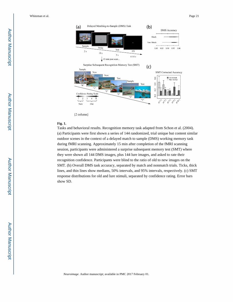
Fig. 1. Tasks and behavioral results. Recognition memory task adapted from Schon et al. (2004).
(a) Participants were first shown a series of 144 randomized, trial unique but content similar
outdoor scenes in the context of a delayed match to sample (DMS) working memory task
during fMRI scanning. Approximately 15 min after completion of the fMRI scanning
session, participants were administered a surprise subsequent memory test (SMT) where
they were shown all 144 DMS images, plus 144 lure images, and asked to rate their
recognition confidence. Participants were blind to the ratio of old to new images on the
SMT. (b) Overall DMS task accuracy, separated by match and nonmatch trials. Ticks, thick
lines, and thin lines show medians, 50% intervals, and 95% intervals, respectively. (c) SMT
response distributions for old and lure stimuli, separated by confidence rating. Error bars
show SD.
Whiteman et al. Page 21
Neuroimage. Author manuscript; available in PMC 2017 February 01.
Author M
anuscriptA
uthor Manuscript
Author M
anuscriptA
uthor Manuscript

Fig. 2. Results of ROI-based VBM analysis. (a) shows the region of right entorhinal cortex (EC)
where we find gray matter volume is associated with aerobic fitness percentile. Results (red)
are shown within a 3D rendering of our anatomical regions of interest, the hippocampus
(gold) and EC (cyan). The gray plane coincides with the black box drawn on the MR slice in
(c) and corresponds to slice y = 10.5 cm caudal to the anterior commissure. The
relationships between volume in the right EC (averaged over the whole structure) and fitness
percentile and recognition memory accuracy is portrayed in (b). Gray lines illustrate the
uncertainty in the regression fits. (c) shows the spatial layout and distribution of the
coefficient of variation (CV) for gray matter volume across participants. Values for
hippocampal and EC voxels are shown as horizontal boxplots overlaid on the histogram,
indicating that variation is not unusually large for these regions. Boxplots show absolute
range, interquartile range, and median. The histogram depicts the 95% high-density region
(white bars) and the extreme tails (gray bars). Exploration of gray matter variability is
extended throughout our ROI in (d). Sections run caudally from the anterior EC through
posterior EC and hippocampal head. The top images show the borders of the EC,
hippocampus, and labeled gray matter; bottom images show corresponding CV maps. All
results are depicted on the group average brain. Figures were made using functionality from
the R rgl and misc3d packages (Adler et al., 2014; Feng and Tierney, 2008).
Whiteman et al. Page 22
Neuroimage. Author manuscript; available in PMC 2017 February 01.
Author M
anuscriptA
uthor Manuscript
Author M
anuscriptA
uthor Manuscript

Fig. 3. Results of exploratory whole-brain analysis. Parts (a) and (b) illustrate the results of an
exploratory whole brain analysis, showing regions (red) where gray matter volume may be
associated with fitness percentile or memory accuracy, respectively. Results are depicted
within the group average brain.
Whiteman et al. Page 23
Neuroimage. Author manuscript; available in PMC 2017 February 01.
Author M
anuscriptA
uthor Manuscript
Author M
anuscriptA
uthor Manuscript

Author M
anuscriptA
uthor Manuscript
Author M
anuscriptA
uthor Manuscript
Whiteman et al. Page 24
Table 1
Participant demographics. Data are presented as mean ± sd. Asterisks in the Meanmalw column indicate
differences in the gender goup means
Range Mean Meanfemale Meanmale
Age (yrs) 18.0 – 30.0 21.1 ± 2.8 20.9 ± 2.8 21.6 ± 2.9
Education (yrs) 12.0 – 22.0 15.3 ± 2.2 14.9 ± 1.6 15.7 ± 2.9
Fitness percentile 17.9 – 100.0 65.3 ± 27.0 55.9 ± 23.7 78.7 ± 26.4*
Memory accuracy (%) 26.3 – 68.2 46.6 ± 9.8 46.8 ± 11.5 46.4 ± 7.4
BDNF (ng·ml−1) 4.6 – 30.5 18.0 ± 6.4 17.7 ± 6.1 18.5 ± 7.1
VO2 peak (ml·kg−1) 31.3 – 66.5 45.5 ± 10.3 39.8 ± 6.9 53.6 ± 8.7***
RERmax 1.0 – 1.6 1.3 ± 0.2 1.2 ± 0.1 1.3 ± 0.2
Intra-cranial Volume (L) 1.2 – 1.8 1.5 ± 0.1 1.4 ± 0.1 1.6 ± 0.1**
BMI 19.2 – 28.9 23.4 ± 2.8 23.2 ± 3.0 23.7 ± 2.4
Body Fat (%) 5.0 – 29.2 18.7 ± 8.3 24.5 ± 3.9 10.3 ± 5.2***
Height (m) 1.5 – 1.9 1.7 ± 0.1 1.6 ± 0.1 1.7 ± 0.1***
Weight (kg) 49.2 – 86.1 66.2 ± 11.4 61.4 ± 10.5 73.0 ± 9.1**
Waist circumference (cm) 62.0 – 93.4 74.9 ± 7.5 72.5 ± 6.6 78.4 ± 7.6*
Hip circumference (cm) 81.0 – 112.5 93.4 ± 8.2 93.2 ± 9.0 93.7 ± 7.2
N = 33 (20 female)
Neuroimage. Author manuscript; available in PMC 2017 February 01.

Author M
anuscriptA
uthor Manuscript
Author M
anuscriptA
uthor Manuscript
Whiteman et al. Page 25
Table 2
Summary of main (ROI-based), and exploratory (whole brain) VBM analyses
Main (ROI-based) Analysis – fitness percentile
Region Peak coordinates Peak t(26) Cluster size k
R. Entorhinal (28.50, −10.50, −34.50) 4.57 157
Exploratory Analysis – fitness percentile
R. Precuneus (19.50, −63.00, 25.50) 7.16 1768
L. Lingual Gyr. (−25.50, −81.00, −16.50) 4.73 1162
R. Inf. Parietal Lobule (57.00, −49.50, 37.50) 4.30 725
R. Lingual Gyr. (13.50, −67.50, −10.50) 4.90 654
L. Mid. Occipital Gyr. (−25.50, −93.00, 6.00) 4.26 385
R. Perirhinal/Entorhinal (30.00, −9.00, −34.50) 4.64 348
L. Calcarine Gyr. (BA 18) (−6.00, −88.50, −13.50) 3.58 186
L. Precuneus (−6.00, −78.00, 42.00) 4.18 185
L. Calcarine Gyr. (BA 17) (−10.50, −67.50, 12.00) 3.46 163
R. Mid. Temporal Gyr. (54.00, −60.00, −1.50) 3.60 140
R. Inf. Temporal Gyr. (69.00, −33.00, −18.00) 3.40 133
Cerebellum (−15.00, −43.50, −60.00) 3.63 114
R. Mid. Temporal Gyr. (49.50, −39.00, −1.50) 3.44 112
R. Sup. Frontal Gyr. (21.00, 61.50, 13.50) 3.14 102
Exploratory Analysis – memory accuracy
R. Entorhinal (31.50, −6.00, −37.50) 3.29 252
Cerebellum (−22.50, −58.50, −27.00) 3.30 245
R. Mid. Temporal Gyr. (54.00, −36.00, −3.00) 3.71 203
R. Amygdala (25.50, −12.00, −9.00) 3.28 118
Neuroimage. Author manuscript; available in PMC 2017 February 01.
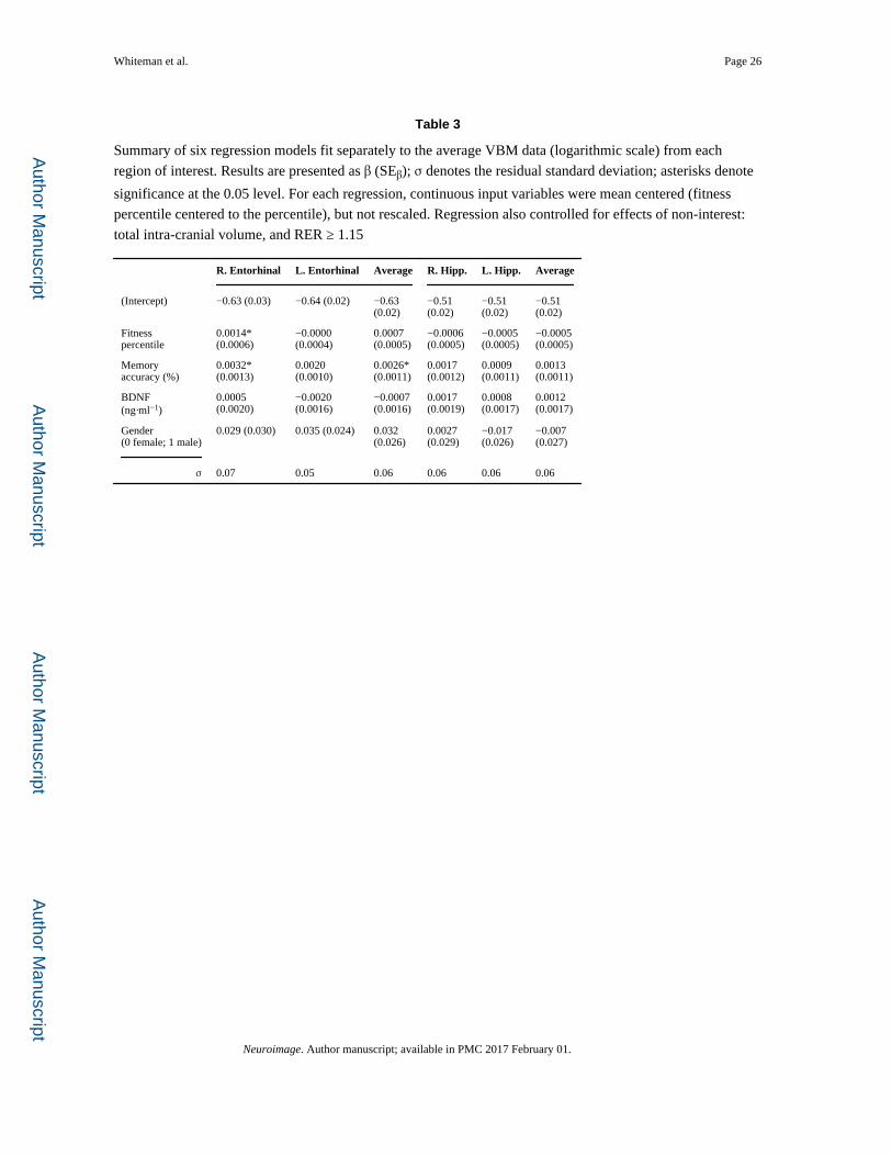
Author M
anuscriptA
uthor Manuscript
Author M
anuscriptA
uthor Manuscript
Whiteman et al. Page 26
Table 3
Summary of six regression models fit separately to the average VBM data (logarithmic scale) from each
region of interest. Results are presented as β (SEβ); σ denotes the residual standard deviation; asterisks denote
significance at the 0.05 level. For each regression, continuous input variables were mean centered (fitness
percentile centered to the percentile), but not rescaled. Regression also controlled for effects of non-interest:
total intra-cranial volume, and RER ≥ 1.15
R. Entorhinal L. Entorhinal Average R. Hipp. L. Hipp. Average
(Intercept) −0.63 (0.03) −0.64 (0.02) −0.63(0.02)
−0.51(0.02)
−0.51(0.02)
−0.51(0.02)
Fitnesspercentile
0.0014*(0.0006)
−0.0000(0.0004)
0.0007(0.0005)
−0.0006(0.0005)
−0.0005(0.0005)
−0.0005(0.0005)
Memoryaccuracy (%)
0.0032*(0.0013)
0.0020(0.0010)
0.0026*(0.0011)
0.0017(0.0012)
0.0009(0.0011)
0.0013(0.0011)
BDNF(ng·ml−1)
0.0005(0.0020)
−0.0020(0.0016)
−0.0007(0.0016)
0.0017(0.0019)
0.0008(0.0017)
0.0012(0.0017)
Gender(0 female; 1 male)
0.029 (0.030) 0.035 (0.024) 0.032(0.026)
0.0027(0.029)
−0.017(0.026)
−0.007(0.027)
σ 0.07 0.05 0.06 0.06 0.06 0.06
Neuroimage. Author manuscript; available in PMC 2017 February 01.
Related Documents