Journal of General Microbiology (1991), 137, 1193-1 199. Printed in Great Britain 1193 Heterogeneity of proteinases from the hyperthermophilic archaeobacterium Pyrococcus furiosus HELEN CONNARIS,' DON A. COWAN,'* and RICHARD J. SHARP^ Department of Biochemistry, University College London, Gower Street, London W C l E 6BT Division of Biotechnology, Centre for Applied Microbiological Research, Porton Down, Wiltshire SP4 OJG (Received 4 September 1990; revised 31 December 1990; accepted 9 January 1991) ~~ ~ Intracellular and extracellular samples from the extremely thermophilic archaeobacterium Pyrococcus furiosus showed the presence of multiple active proteinases. Using gelatin-containing SDS-PAGE, up to 13 activity bands were visualized with apparent molecular masses of between 66 and 135 kDa. Characterizationstudies revealed these bands to be due to discrete polypeptides, and not artefacts. Results from gel permeation chromatography, sucrose density gradient centrifugation and non-denaturing PAGE suggested that some of these proteolytic polypeptides may exist as active aggregates either in uivo or in uitro before being dissociated by SDS to active monomers. Introduction The ability for extreme thermophiles, most of which belong to the Third Primary Kingdom of the Archaeo- bacteria, to survive temperatures of around 80°C and above is critically dependent on the structure and function of cellular components. Enzymes from these organisms are generally capable of withstanding high temperatures and exhibit a high degree of resistance to denaturing reagents, organic solvents and other deleteri- ous influences (Veronese et al., 1984; Owusu & Cowan, 1989). It is generally accepted that protein stability in these extreme thermophiles results from modifications to protein structure, such as the addition of hydrogen bonds and ionic bonds, generated by subtle differences in the amino acid sequence. Enhancement of thermal stability can also occur through improved rigidity of the more thermolabile regions of the protein structure, due to an extrinsic influence of metal ions, as with Co2+ for Suljolobus solfataricus aminopeptidase (Hanner et al., 1990)and Ca2+ for Thermus aquaticus proteinase (Cowan & Daniel, 1982). Despite these examples of extrinsic stabilization, most proteins from extreme thermophiles are of high intrinsic molecular stability [e.g. the extracellular proteinase archaelysin from the archaeo- bacterium Desulfurococcus (Cowan et al., 1987), the acid proteinase from Sulfolobus acidocaldarius (Lin & Tang, 1990) and the hydrogenase from the hyperthermophilic archaeobacterium Pyrococcusfuriosus (Bryant & Adams, 1989)l. Proteinases from extremely thermophilic archaeobacteria are all highly thermostable. In other respects, these enzymes show a diversity of characteris- tics typical of eubacterial proteinases (Cowan et al., 1987). The extremely thermophilic archaeobacterium Pyro- coccus furiosus, isolated by Fiala & Stetter (1986), is a heterotrophic organism which grows optimally at 100 "C under strictly anaerobic conditions. Five intracellular proteinases have been previously identified from this hyperthermophile (Blumentals et al., 1990; Eggen et al., 1990), with an SDS-resistant proteinase (66 kDa) puri- fied to homogeneity (Blumentals et al., 1990). In this paper, we present further data on the heterogeneity of the proteinases. Methods Growth conditions. Pyrococcus furiosus (DSM 3638) was grown anaerobically in 1 I Duran bottles at 98 "C as described by Fiala & Stetter (1986). For the production of intracellular proteinases, cells were harvested after a 17 h incubation. Cell pellets were collected by centrifugation (50000 g for 15 min), washed, and resuspended in 0.01 M-N~H~PO,/N~,HPO, buffer, pH 7.5. Cell-free extracts for gel permeation chromatography and sucrose density gradients were prepared by sonicating cells for three 1 min bursts with 30 s intervals using a Soniprep Sonicator. The cell lysate was then centrifuged for 30 min at 30000g to remove cell debris. Otherwise, aliquots of culture (1 ml) were centrifuged at high speed in an MSE Microcentaur centrifuge and cell pellets were washed, resuspended in phosphate buffer (100 pl) and lysed by the addition of an equal volume of SDS sample buffer (4% (w/v) SDS/20% (w/v) glycerol/ 10% (w/v) mercapto- ethanol with bromophenol blue). For extracellular proteinases, cell-free culture supernatants were obtained after harvesting and used directly. 0001-6484 0 1991 SGM

Welcome message from author
This document is posted to help you gain knowledge. Please leave a comment to let me know what you think about it! Share it to your friends and learn new things together.
Transcript

Journal of General Microbiology (1991), 137, 1193-1 199. Printed in Great Britain 1193
Heterogeneity of proteinases from the hyperthermophilic archaeobacterium Pyrococcus furiosus
HELEN CONNARIS,' DON A. COWAN,'* and RICHARD J. SHARP^
Department of Biochemistry, University College London, Gower Street, London W C l E 6BT Division of Biotechnology, Centre for Applied Microbiological Research, Porton Down, Wiltshire SP4 OJG
(Received 4 September 1990; revised 31 December 1990; accepted 9 January 1991)
~~ ~
Intracellular and extracellular samples from the extremely thermophilic archaeobacterium Pyrococcus furiosus showed the presence of multiple active proteinases. Using gelatin-containing SDS-PAGE, up to 13 activity bands were visualized with apparent molecular masses of between 66 and 135 kDa. Characterization studies revealed these bands to be due to discrete polypeptides, and not artefacts. Results from gel permeation chromatography, sucrose density gradient centrifugation and non-denaturing PAGE suggested that some of these proteolytic polypeptides may exist as active aggregates either in uivo or in uitro before being dissociated by SDS to active monomers.
Introduction
The ability for extreme thermophiles, most of which belong to the Third Primary Kingdom of the Archaeo- bacteria, to survive temperatures of around 80°C and above is critically dependent on the structure and function of cellular components. Enzymes from these organisms are generally capable of withstanding high temperatures and exhibit a high degree of resistance to denaturing reagents, organic solvents and other deleteri- ous influences (Veronese et al., 1984; Owusu & Cowan, 1989). It is generally accepted that protein stability in these extreme thermophiles results from modifications to protein structure, such as the addition of hydrogen bonds and ionic bonds, generated by subtle differences in the amino acid sequence. Enhancement of thermal stability can also occur through improved rigidity of the more thermolabile regions of the protein structure, due to an extrinsic influence of metal ions, as with Co2+ for Suljolobus solfataricus aminopeptidase (Hanner et al., 1990) and Ca2+ for Thermus aquaticus proteinase (Cowan & Daniel, 1982). Despite these examples of extrinsic stabilization, most proteins from extreme thermophiles are of high intrinsic molecular stability [e.g. the extracellular proteinase arc haelysin from the archaeo- bacterium Desulfurococcus (Cowan et al., 1987), the acid proteinase from Sulfolobus acidocaldarius (Lin & Tang, 1990) and the hydrogenase from the hyperthermophilic archaeobacterium Pyrococcus furiosus (Bryant & Adams, 1989)l. Proteinases from extremely thermophilic
archaeobacteria are all highly thermostable. In other respects, these enzymes show a diversity of characteris- tics typical of eubacterial proteinases (Cowan et al., 1987).
The extremely thermophilic archaeobacterium Pyro- coccus furiosus, isolated by Fiala & Stetter (1986), is a heterotrophic organism which grows optimally at 100 "C under strictly anaerobic conditions. Five intracellular proteinases have been previously identified from this hyperthermophile (Blumentals et al., 1990; Eggen et al., 1990), with an SDS-resistant proteinase (66 kDa) puri- fied to homogeneity (Blumentals et al., 1990). In this paper, we present further data on the heterogeneity of the proteinases.
Methods
Growth conditions. Pyrococcus furiosus (DSM 3638) was grown anaerobically in 1 I Duran bottles at 98 "C as described by Fiala & Stetter (1986). For the production of intracellular proteinases, cells were harvested after a 17 h incubation. Cell pellets were collected by centrifugation (50000 g for 15 min), washed, and resuspended in 0.01 M-N~H~PO, /N~ ,HPO, buffer, pH 7.5. Cell-free extracts for gel permeation chromatography and sucrose density gradients were prepared by sonicating cells for three 1 min bursts with 30 s intervals using a Soniprep Sonicator. The cell lysate was then centrifuged for 30 min at 30000g to remove cell debris. Otherwise, aliquots of culture (1 ml) were centrifuged at high speed in an MSE Microcentaur centrifuge and cell pellets were washed, resuspended in phosphate buffer (100 pl) and lysed by the addition of an equal volume of SDS sample buffer (4% (w/v) SDS/20% (w/v) glycerol/ 10% (w/v) mercapto- ethanol with bromophenol blue). For extracellular proteinases, cell-free culture supernatants were obtained after harvesting and used directly.
0001-6484 0 1991 SGM

1 194 H . Connaris, D . A . Cowan and R . J . Sharp
Cells and supernatants were either used immediately or stored at - 70 "C.
Proteinase assay. Proteinase activity was determined by the hydrolysis of azocasein (Sigma) as described by Cowan et al. (1987).
Substrate-containing SDS-PAGE. Discontinuous SDS-PAGE was performed according to Laemmli (1970), except that the separation gel (10%) contained 0.01% gelatin (Type I, porcine skin) or casein (purified; Sigma) and 0.1 % SDS. The stacking gel (5%) also contained 0.1 % SDS. Protein samples (75 pl) and known molecular mass markers (50p1) were mixed with an equal volume of SDS sample buffer (Laemmli, 1970) and boiled for 5-10 min before loading on to slab gels (20 x 16 x 1-5 mm). Electrophoresis was performed at 45 mA constant current using an LKB Vertical Electrophoresis Unit with an LKB 2197 Power Supply.
After electrophoresis, gels were gently shaken at room temperature for 2 h in two changes of aqueous 2.5% (w/v) Triton X-100 (Horie et al . , 1984), then incubated for 1 h at 90°C in 0.1 M-HEPES (N-2- hydroxyethylpiperazine-N'-2-ethanesulphonic acid) buffer, pH 7-5 (90 "C). After incubation, gels were immediately fixed with 50% (w/v) TCA for 30-60 mins before staining with Coomassie Blue G250. Gels were then destained overnight in methanol :acetic acid :water ( 3 : 2 : 35, by vol.). Unstained bands, indicating proteolytic activity, were recorded by photography.
Quantitatitie analysis of data. Negatives of Polaroid photographs of substrate-containing gels were scanned using an Ultrascan XL Laser Densitometer (LKB Instruments). Proteinase activity was determined by integration of peak area for each band.
Native gels. Non-denaturing PAGE was performed on slab gels of different acrylamide concentrations (up to 10%) to determine the molecular masses of the proteinases. Native gels were prepared (Clarke, 1964) and electrophoresed as before. After electrophoresis, gels were immersed in a solution of 1 %gelatin in 0.1 M-HEPES, pH 8.3, and gently shaken for 1 h. The gels were then incubated for 1-3 h at 90 "C before being fixed with 50% (w/v) TCA. Gels were stained and destained as before.
Sucrose gradient centrifugation. Sucrose gradients [6-30% (w/v) sucrose in distilled water] were prepared as described (Martin & Ames, 1961). Cell extract and culture supernatant samples (500 pl) were layered on top of the gradients together with known molecular mass markers (0-25 mg of P-amylase and bovine serum albumin in 50 p1 of distilled water). The samples were centrifuged at 208000g for 24 h at 20 "C in an L-7 Beckman Ultracentrifuge. The contents were removed by inserting a capillary tube to the bottom of each tube and withdrawing the contents using a peristaltic pump. Fractions (0.5 ml) were collected. Protein was monitored spectrophotometrically at 280 nm and proteinase activity was determined using substrate PAGE. Molecular masses were calculated using the equation sl/sz x (mm,/mm2)2'3, where s l / s t is the partial specific volume, mm, is the molecular mass of the unknown protein and mm2 is the molecular mass of the known standard (Martin & Ames, 1961).
Fast protein liquid gel permeation chromatography. Samples of P . furiosus cell extracts (200 pl) were applied to a Superose 12 HRlO/30 column (Pharmacia). The column was pre-equilibrated with 20 mM- triethanolamine buffer, 50 mM-NaC1, pH 7.5, before samples were loaded. Proteins were then eluted with the same buffer, and 1 ml fractions were collected. Throughout the chromatography, protein content was monitored spectrophotometrically at 280 nm. All fractions collected were assayed for proteinase activity using substrate PAGE.
Eflect of inhibitors. Proteinase inhibitors (final concentrations 10- 100 mM) were added to 100 pl of cell extracts and culture supernatants and incubated at room temperature for 1 h before analysis by substrate
PAGE. Non-covalent inhibitors were added to the proteinase assay buffer during incubation of gels at 90 "C, at a final concentration of 10 mM.
Thermostability ofproteases. Cell-free culture supernatants samples (100 pl) were sealed in capillary tubes (Barach & Adams, 1977), and immersed in a paraffin oil bath at temperatures between 95 "C and 125 "C. At appropriate time intervals, tubes were removed and immediately placed on ice. Residual proteinase activity was deter- mined using substrate PAGE.
Results and Discussion
Static cultures of the hyperthermophilic archaeobacter- ium Pyrococcus .furiosus were sampled for proteinase activity using azocasein as the proteolytic substrate. Proteinase activity was detected in both cell extracts and culture supernatants, reaching a peak at around 17-18 h after inoculation (Fig. 1 a and b). Proteinase production did not appear to be growth-phase dependent, as specific proteinase production (expressed as units of activity per g of protein) was observed to be linear throughout growth (Fig. lc). Since the azocasein assay yields only the total titre of proteinase activity and provides no information on enzyme multiplicity, substrate-containing PAGE was used subsequently for more detailed analysis.
Proteinase heterogeneity
Using substrate-containing SDS-PAGE, a multiple band pattern of up to 13 distinct active proteinase monomers was observed in both cell extracts and culture super- natants, where apparent molecular masses ranged from 66 kDa to 135 kDa (Fig. 2). Levels of these active hydrolases were seen to increase until late exponential phase in a manner consistent with the analysis of total proteinase titres (Fig. 1 b).
The observation of multiple proteinase bands from P . furiosus confirmed data obtained by Blumentals et al. (1 990) and Eggen et al. (1990) who both report a multiple band pattern with the lowest molecular weight band being around 66 kDa. However, both groups describe the presence of only 5 proteinase bands.
The difference between our observations and those of Blumentals et al. (1 990) and Eggen et al. (1 990) lies in the sensitivity and resolution of the detection procedures used. Blumentals et al. (1 990) reported extensive streak- ing and poor resolution of bands with gelatin-SDS- PAGE and were thus forced to use a substrate gel overlay, from which only 5 bands were detected. On the other hand, Eggen et al. (1990) who also reported 5 bands, did not appear to have the problem of streaking in substrate gels. We can only conclude that the sensitivity and resolution of the substrate-containing SDS-PAGE technique may be dependent on details of its operation.

Proteinase heterogeneity in Pyrococcus furiosus 1 195
I I I I 1 I
Fig. 2. Proteinase multiplicity from P. furiosus. Lane 1, cell extract fraction; lane 2, culture supernatant fraction. Numbers on RHS and LHS of photograph indicate band numbers in ascending molecular mass (indicated in brackets), whereas numbers on the far LHS denote molecular masses of standard markers (66 kDa, bovine serum albumin; 97 kDa, phosphorylase b; 116 kDa, P-galactosidase).
10 20 30 Incubation time (h)
Fig. 1. Growth, proteinase activity and specific proteinase pro- ductivity of Pyrococcusfuriosus. (a) Cell density; (b) proteinase activity in cell extract (0) and culture supernatant (0) fractions; and (c) specific proteinase production in cell extract (0) and culture super- natant (0) fractions.
Also, the reliance of the gel overlay detection method on diffusion of the enzyme will inevitably result in substantial loss of resolution and detection. We note that the substrate concentration used by us, Blumentals et al. (1990) and Eggen et al. (1990) was found to differ as much as 50-fold.
There are a number of possible origins of a multiplicity of discrete activity bands in substrate-containing SDS- PAGE gels. These might include: (i) active processing products from larger species, such as proteolytic or autolytic degradation products; (ii) artefacts resulting from incomplete unfolding ; (iii) artefacts resulting from heterogeneous associations with the substrate during electrophoresis; and (iv) discrete genetically distinct polypeptides. We shall consider each of these possible mechanisms in turn.
The generation of heterogeneous active proteinase fragments by proteolytic or autolytic degradation is known to occur (Fontana, 1988). However, preincuba- tion of Pyrococcus cell-free culture supernatants and cell extract samples at different temperatures (4 "C, 20 "C, 60°C and 95 "C) for up to 24 h before analysis by substrate PAGE, showed no activity bands of <66 kDa (data not shown). Also, banding patterns identical to those in Fig. 2 were observed with negligible loss of apparent activity for any proteinase. If significant autolysis did occur, then a gradual decrease in the higher- molecular-mass bands, possibly corresponding to an increase in the lower-molecular-mass bands, might be observed over time. Under no conditions did we observe this behaviour, suggesting that the activity bands were unlikely to be degradation products.
1196 H . Connaris, D . A . Cowan and R . J . Sharp
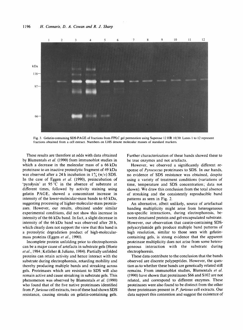
Fig. 3. Gelatin-containing SDS-PAGE of fractions from FPLC gel permeation using Superose 12 HR 10/30. Lanes 1 to 12 represent fractions obtained from a cell extract. Numbers on LHS denote molecular masses of standard markers.
These results are therefore at odds with data obtained by Blumentals et al. (1990) from immunoblot studies in which a decrease in the molecular mass of a 66 kDa proteinase to an inactive proteolytic fragment of 49 kDa was observed after a 24 h incubation in 1 % (w/v) SDS. In the case of Eggen et al. (1990), preincubation of ‘pyrolysin’ at 95 “C in the absence of substrate at different times, followed by activity staining using gelatin PAGE, showed a concomitant increase in intensity of the lower-molecular-mass bands to 65 kDa, suggesting processing of higher-molecular-mass protein- ases. However, our results, obtained under similar experimental conditions, did not show this increase in intensity of the 66 kDa band. In fact, a slight decrease in intensity of the 66 kDa band was observed after 20 h, which clearly does not support the view that this band is a proteolytic degradation product of high-molecular- mass proteins (Eggen et al., 1990).
Incomplete protein unfolding prior to electrophoresis can be a major cause of artefacts in substrate gels (Horie et al., 1984; Kelleher & Juliano, 1984). Partially unfolded proteins can retain activity and hence interact with the substrate during electrophoresis, retarding mobility and thereby producing multiple bands and streaking across gels. Proteinases which are resistant to SDS will also remain active and cause streaking in substrate gels. This phenomenon was observed by Blumentals et al. (1990) who found that of the five native proteinases identified from P. furiosus cell extracts, two of these had shown SDS resistance, causing streaks on gelatin-containing gels.
Further characterization of these bands showed these to be true enzymes and not artefacts.
However, we observed a significantly different re- sponse of Pyrococcus proteinases to SDS. In our hands, no evidence of SDS resistance was obtained, despite using a variety of treatment conditions (variations of time, temperature and SDS concentration ; data not shown). We draw this conclusion from the total absence of streaking and the consistently reproducible band patterns as seen in Fig. 2.
An alternative, albeit unlikely, source of artefactual banding multiplicity might arise from heterogeneous non-specific interactions, during electrophoresis, be- tween denatured protein and gel-encapsulated substrate. However, our observation that casein-containing SDS- polyacrylamide gels produce multiple band patterns of high resolution, similar to those seen with gelatin- containing gels, is strong evidence that the apparent proteinase multiplicity does not arise from some hetero- geneous interaction with the substrate during electrophoresis.
These data contribute to the conclusion that the bands observed are discrete polypeptides. However, the ques- tion as to whether these bands are genetically related still remains. From immunoblot studies, Blumentals et al. (1990) have shown that proteinases S66 and S102 are not related, and correspond to different enzymes. These proteinases were also found to be distinct from the other three proteinases present in P. furiosus cell extracts. Our data support this contention and suggest the existence of

Proteinase heterogenqity in Pyrococcus furiosus 1 197
Fig. 4. Sucrose density gradient fractions analysed using gelatin-SDS-PAGE. Portions of cell extract (500 pl) were loaded and run on a 6 3 0 % (w/v) sucrose gradient (see Methods section). Fractions collected were run alongside standard markers on gelatin-SDS-PAGE. Gels were renatured and incubated for 3 h at 90 “C, before staining with Coomassie Blue. Lanes 1 to 8 represent fractions from sucrose density gradient runs.
additional discrete peptides. Eggen et al. (1990), on the other hand, contend that the multiple protease bands from ‘pyrolysin’ are processed to give a final proteolyti- cally active 66 kDa band. Neither our data, nor that of Blumentals et al. (1 990), support this conclusion.
In vivo state of proteinase monomers
Non-denaturing (native) PAGE indicated the presence of more than 7 proteinases from cell extracts and culture supernatants. Analysis of native gels of varying acryl- amide concentrations (5-10% acrylamide) gave appar- ent molecular masses of between 95 and 280 kDa (data not shown).
Separation of cell extracts by FPLC gel permeation chromatography and sucrose density centrifugation produced 2 higher-molecular-mass proteinases of ap- proximately 185 to 200 kDa (from sucrose gradients, sedimentation coefficients of 9.5 S and 10 S respectively) and 200 to 280 kDa (FPLC) from cell extracts and culture supernatants. When subsequently analysed by subs trate-con taining SDS-P AG E, these hig h-molecular- mass active proteinases showed an identical pattern to that observed previously (Figs 3 and 4). The most
probable explanation for these results is that the individual proteinases interact (either in vivo or in vitro) to form high-molecular-mass active aggregates. We have no evidence to confirm the existence of these aggregates in vivo, although the appearance of multiple bands on native PAGE might argue for an in vitro aggregation phenomenon (Figs 3 and 4).
Inhibitors
All but one of the 13 proteinase bands from Pyrococcus showed complete inhibition by 10 mhl-phenylmethylsul- phony1 fluoride (PMSF) suggesting predominance of serine-type proteinases (data not shown). There was no significant change in activity after treatment with the thiol proteinase inhibitor iodoacetamide. Partial inhibi- tion by the metal chelator EDTA (10 mM) was observed with the PMSF-insensitive band (1 30 kDa), suggesting a requirement of metal ions for activity. The results of inhibition studies obtained for the additional bands support the data obtained by Blumentals et al. (1990) and Eggen et al. (1990), who both classify the 5 bands reported in their work as serine-type proteinases.

1198 H . Connaris, D . A . Cowan and R . J . Sharp
60
40
20
-
-
-
5 10 15 20 25 Time (h)
- .I
C U Time (rnin)
10 20 Time (min)
1 2 3
Time (min)
Fig. 5. Thermostability profiles for Pyrococcus 66 kDa proteinase at various temperatures. Culture supernatant samples (100 PI) were incubated at (a) 95 "C, (b) 105 "C, (c) I15 "C, and (d ) 125 "C, with (0) and without (0) the addition of 10 mM-CaC1,. Samples were removed at appropriate intervals, cooled and assayed for activity using gelatin- SDS-PAGE (see Methods).
Table I . Apparent half life values of the 66 kDa proteinase from P . furiosus with and without I 0 mM-CaCI,
Half life (t,,2)
Temperature ("C) Without calcium With calcium
95 105 115 125
>20 h 15 min 52 s 22 s
>20 h 117 rnin 6-3 rnin 20 s
Thermostability
At temperatures between 95 "C and 105 "C, all 13 proteinases demonstrated a high degree of stability, with some proteinases showing enhanced stability in the presence of 10 mM-CaC1, (Fig. 5). Using gelatin-SDS- PAGE as a semi-quantitative assay, the apparent half life values of the 66 kDa protein (selected as this appeared to be one of the most stable) calculated ii-om first order constants (Moore, 1962) were considerably increased when calcium was added, suggesting that this metal ion is required to stabilize the protein structure and prevent unfolding at higher temperatures (Table 1).
The value obtained for the 66 kDa proteinase exceeds the half life values of other known archaeobacterial proteinases such as archaelysin, t,/* (95 "C), 70-90 minutes (Cowan et al., 1987), and thermopsin, tl/, (80 "C), 48 hours (Lin & Tang, 1990). Blumentals et al. (1990) have reported that the 66 kDa proteinase from Pyrococcus was found to have a half life of about 33 h at 98 "C, making this the most stable proteinase to date.
Concluding remarks
In this study, up to 13 active proteolytic polypeptides from P . furiosus have been identified. We believe that the heterogeneity of these enzymes is valid from the evidence presented, despite conflicting evidence of 5 active bands obtained from studies by Blumentals et al. (1990) and Eggen et al. (1990). We also suggest that these active proteinases are not artefacts, but arise from the dissociation of larger multimeric proteinases.
The question that is automatically raised is why so many proteinases are produced from a hyperthermo- phile. It is known that multiple heterogeneous intra- cellular proteinases serve a wide variety of functions including protein turnover, nutritional and specific post- translational modification events. When information is available on the peptide bond specificity of individual proteinases, it may be possible to imply one or other function. However, at present the specific function for these P. furiosus proteinases remains unknown.

Proteinase heterogeneity in Pyrococcus juriosus I 1 99
References
BARACH, J . T. & ADAMS, D. M . (1977). Thermostability at ultrahigh temperatures of Thermolysin and a protease from a psychrotrophic Pseudomonus. Biuchimicu et Biuphjxica Acta 485, 4 I 7.
BLUMENTALS, I . I . , ROBINSON, A. S. & KELLY, R . M . (1990). Characterization of SDS-resistant proteolytic activity in the hyper- the r mop h i 1 i c arch a e b a c te r i u m Pj wcuccus .fur iosus . Applied uritl Eniironnienttil Micrubiologj~ 56, 1992- 1998.
BRYANT, F. 0. & ADAMS, M. W. W . (1989). Characterization of hydrogenase from the hyperthermophilic archaebacterium, Pj'rucoc- cus furiusus. Journul uf' Bicilogical Chemistrj, 264, 5070 -5079.
CLARKE, J . T. (1964). Simplified 'disc' (polyacrylamide gel) electro- phoresis. Anriu1.s o f t h e Ncw York A c u t i m j - yf'Scicwcc~s 121, 428 435.
COWAN, D. A. & DANIEL, R. M . (1982). Purification and some properties of an extracellular protease (caldolysin) from an extreme thermophile. Biochimica et Biophj-sicu Acta 705, 293 -305.
COWAN, D. A., SMOLENSKI, K . A., DANIEL, R. M . & MORGAN, H . W . (1987). An extremely thermostable extracellular proteinase from the strain of the archaebacterium Desulfurococcus growing at 88 "C. Biochcwiicul Jouriiul 247, I 2 1 ~ 1 33.
DAVIS, €3. J . (1964). Disc electrophoresis-11. Method and application to human serum proteins. Annuls i?f the New York Acucic.nij. c?f'Scicwccv 121, 404 427.
EGGEN. R . , GEERLING, A., WATTS, J . & M . DE Vos, W . (1990). Characterization of pyrol ysin, a h ypert hermoacti ve serine protease from the archaebacterium Pyrococcus juriusus. F E M S Microhio1o.g~~ Lxttrrs 71. 17 -20.
FIALA, G . & STETTER, K . 0. (1986). Pj-rococcus ,furiosu.s sp. nov., represents a novel genus of marine heterotrophic archaebacteria growing optimally at 100 "C. Archires of' Microhiologj, 145, 56-61.
FONTANA. A. (1988). Structure and stability of thermophilic enzymes. Studies on thermolysin. Biophj-sical Chemistrj. 29, I8 1-193.
HANNER, M . , REDL, B. & STOFFLER, G. (1990). Isolation and character i z.a t ion of an i n t race1 1 u 1 a r am i nope p t i d ase from the extreme the rmo p h i I ic arc hae bac t e r i um Su!folo bus .so!firt ciricus . Biochiniicu ct Biuphj'sicu Actu 1033, 148 153.
HORIE, N . , FUKUYAMA. K . . ITO, Y . & EPSTEIN, W . L. (1984). Detection and characterization of epidermal proteinases by polyacrylamide gel electrophoresis. Conipurutii*r Bioc~heniistrj~ mid Phj*.siolugj, 77B, 349- 353.
KELLEHER, P. J . & JULIANO. R . L. (1984). Detection of proteascs in polyacrylamide gels containing covalently bound substrates. Auu1j.- tical Biochemi.strj~ 136, 470 -475.
LAEMMLI, U. K . (1970). Cleavage of structural proteins during the assembly of the head of bacteriophage T4. Nature, Lmidoii 227,
LIN. X.-L. & TANG, J . (1990). Purification, characterization and genc cloning of Thermopsin. a thermostable acid protease from Su/folohu.s acidocaldurius. Journal c?f ' Biologicul Chtwiistrj! 265, 1490 - 1495.
MARTIN, R . G . & AMES, B. N . (1961). A method for determining the sedimentation behaviour of enzymes: Applications to protein mixtures. Journul (?f'Biologicul Chmiistr!. 236, 1372 1379.
MOORE. W. J . (1962). Phj*sical Chemi.strJ*, 4th edn, pp. 260-263. London : Longman.
OWUSC. R . K . & COWAN, D. A . (1989). Correlation between microbial protein thermostability and resistance to denaturation in aqueous organic solvent ?-phase systems. Enzjmcj untl Microbial Tcc~hnologj.
VERONESE. F. M., B o c c u , E., SCHIAVON, C. , GRANDI, C. & FONTANA, A. (1984). General stability of thermophilic enzymes. Studies on 6- phosphogluconate dehydrogenase from Baci1lu.s .stearothermc~philu.s and yeast. Journal ?/'Applied Biochemistrj. 6 , 39 47.
680--685.
11. 568-574.
Related Documents