Heart outline • pulmonary & systemic circulatio • location of heart in thorax • pericardium • external anatomy of heart • internal anatomy of heart • coronary circulation • CAD, angina and heart attacks • heart valves • conduction system of heart • EKG’s

Welcome message from author
This document is posted to help you gain knowledge. Please leave a comment to let me know what you think about it! Share it to your friends and learn new things together.
Transcript
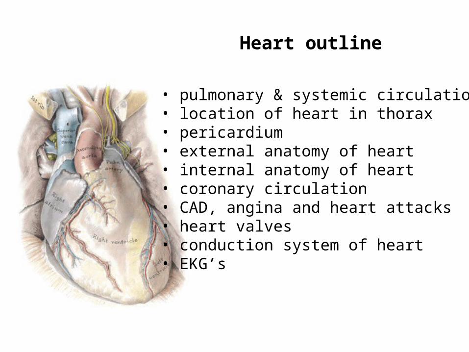
Heart outline
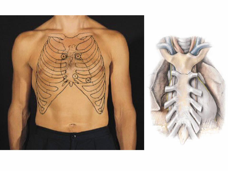
• pulmonary & systemic circulation• location of heart in thorax• pericardium• external anatomy of heart• internal anatomy of heart• coronary circulation• CAD, angina and heart attacks• heart valves• conduction system of heart• EKG’s
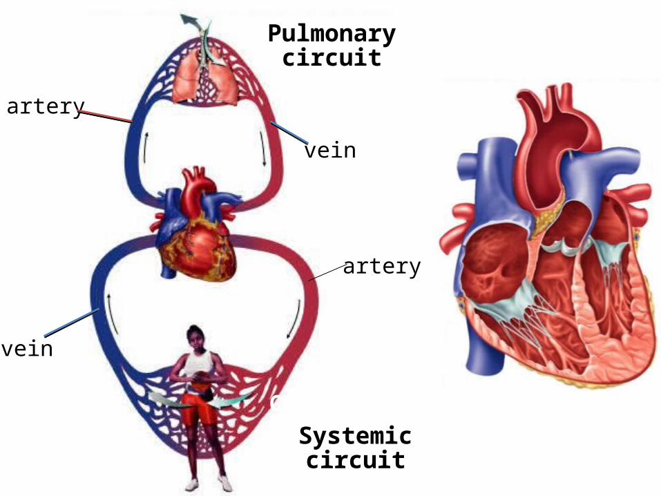
Systemiccircuit
Pulmonarycircuit
CO2 O2
O2artery
vein
vein
artery
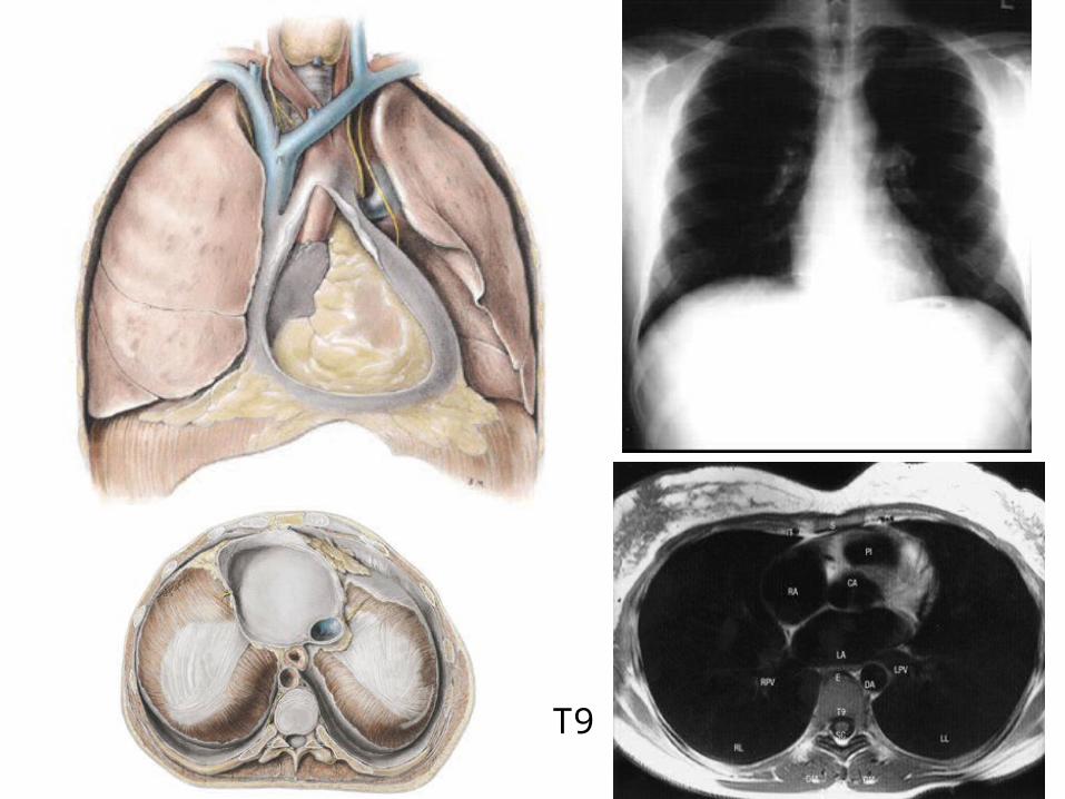
T9
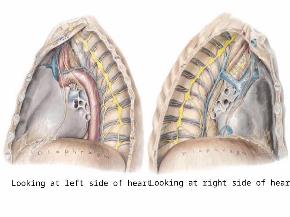
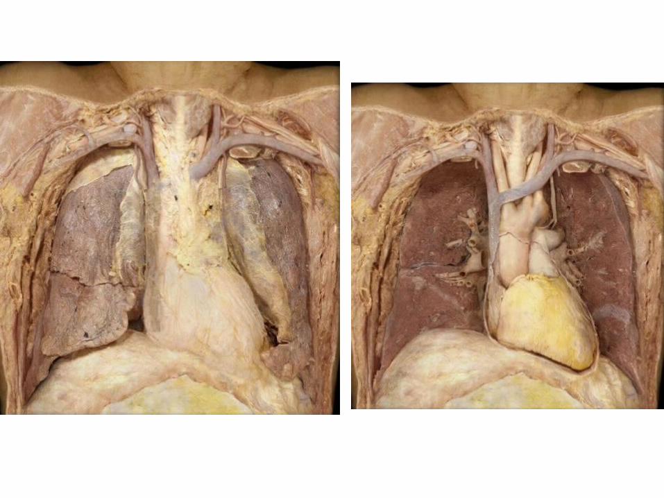
Looking at left side of heart Looking at right side of heart
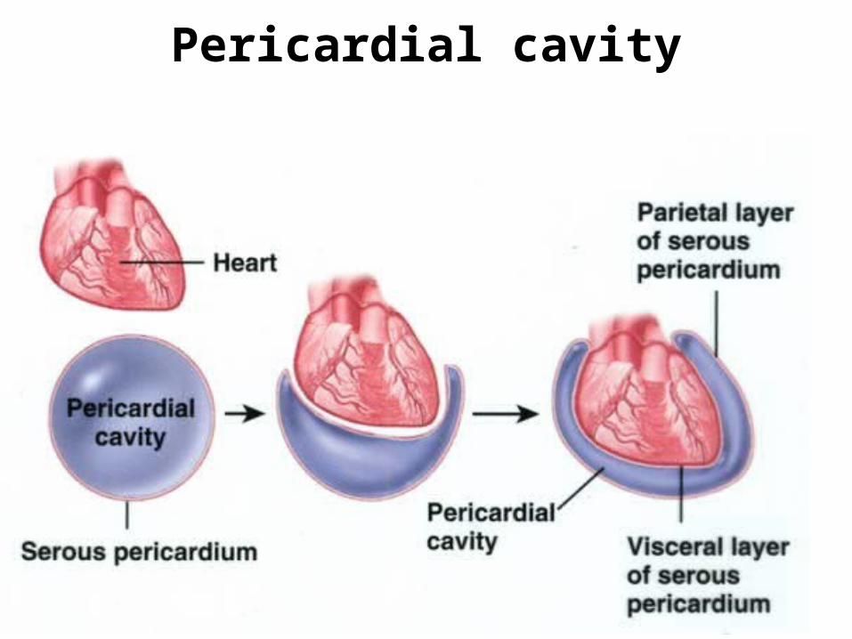
Pericardial cavity

Fibrous pericardium
Myocardium
Endocardium
Parietal pericardium
Visceral pericardium
Epicardium = visceral pericardium + adipose
cardiomyopathy
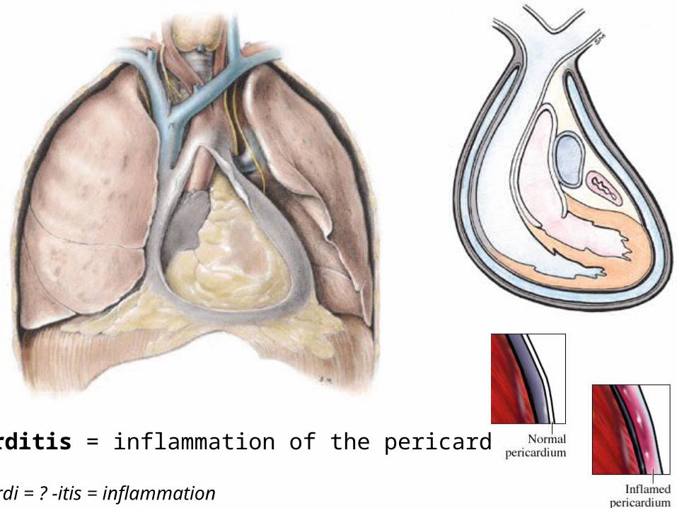
Pericarditis = inflammation of the pericardium
peri = ? cardi = ? -itis = inflammation
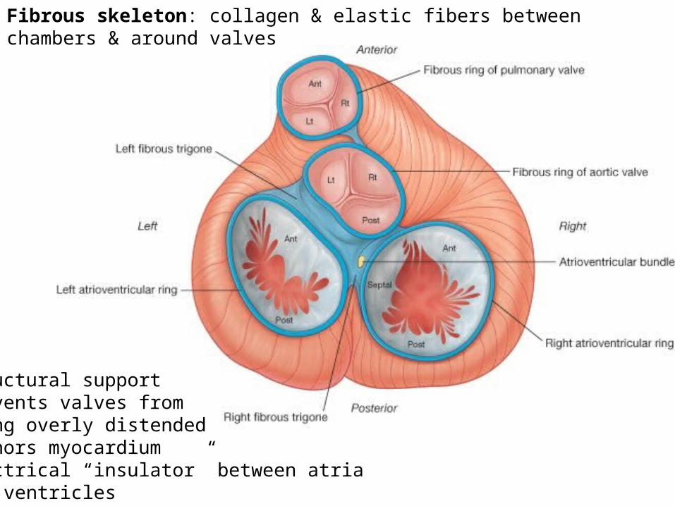
Fibrous skeleton: collagen & elastic fibers between chambers & around valves
• structural support • prevents valves from being overly distended• anchors myocardium• electrical “insulator” between atria and ventricles
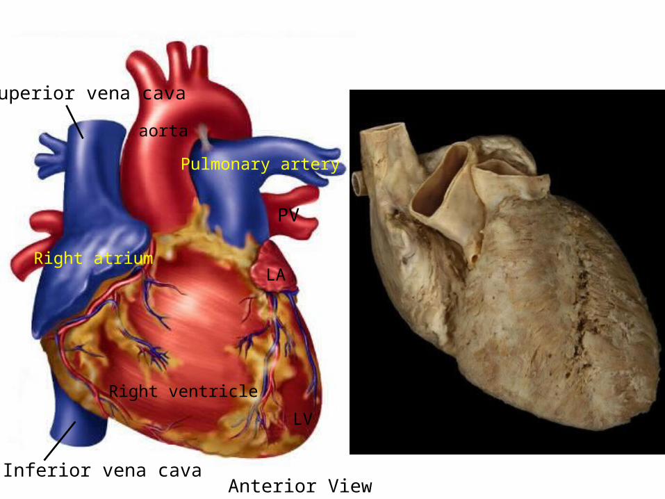
aorta
Superior vena cava
Inferior vena cava
Right atrium
Right ventricle
Pulmonary artery
Anterior View
PV
LV
LA
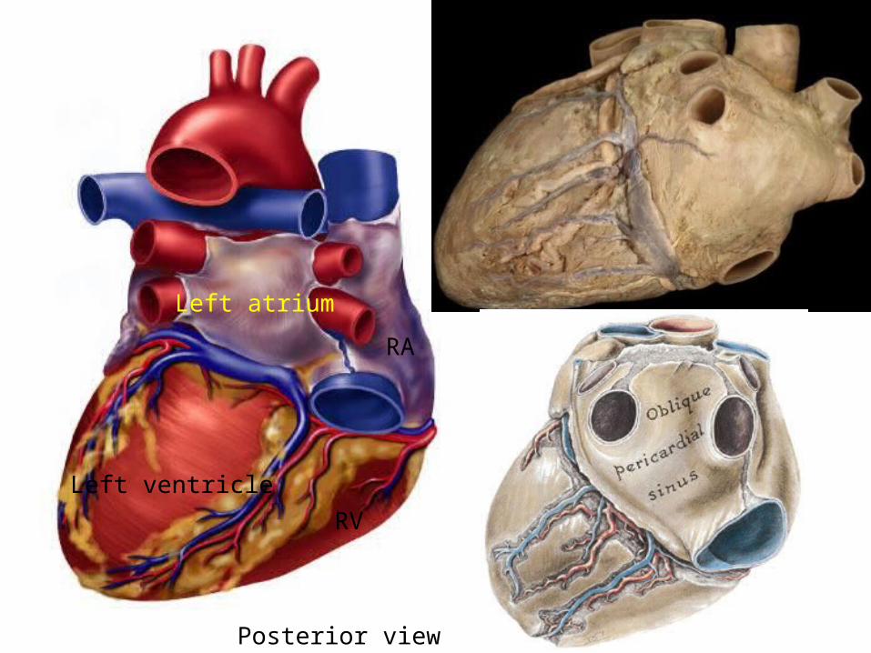
Left atrium
Left ventricleRV
RA
Posterior view
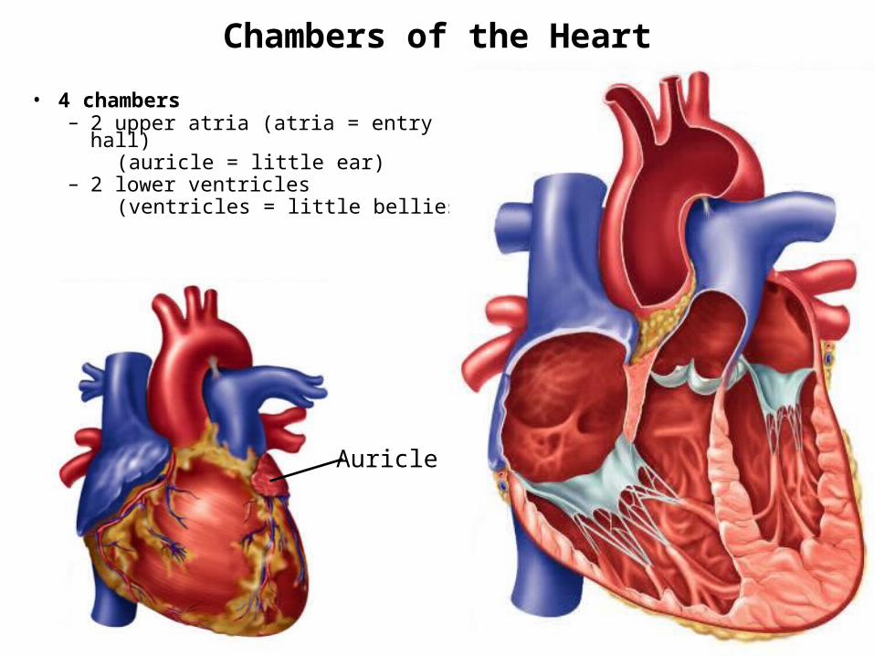
Chambers of the Heart
• 4 chambers – 2 upper atria (atria = entry hall) (auricle = little ear)– 2 lower ventricles (ventricles = little bellies)
Auricle
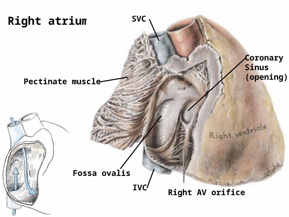
Right atrium SVC
IVC
CoronarySinus(opening)
Fossa ovalis
Pectinate muscle
Right AV orifice
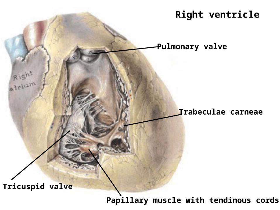
Right ventricle
Pulmonary valve
Tricuspid valve
Trabeculae carneae
Papillary muscle with tendinous cords
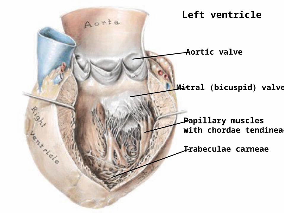
Left ventricle
Aortic valve
Mitral (bicuspid) valve
Papillary muscleswith chordae tendineae
Trabeculae carneae
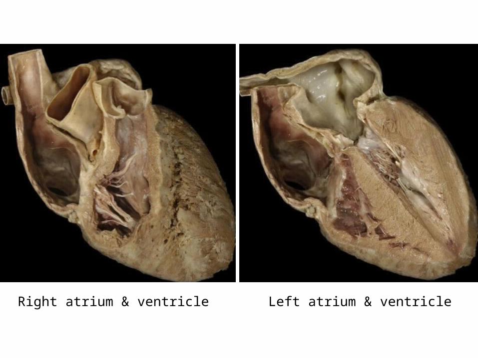
Right atrium & ventricle Left atrium & ventricle
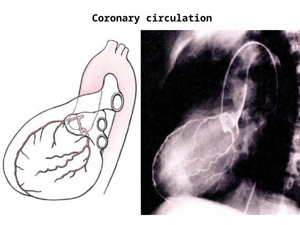
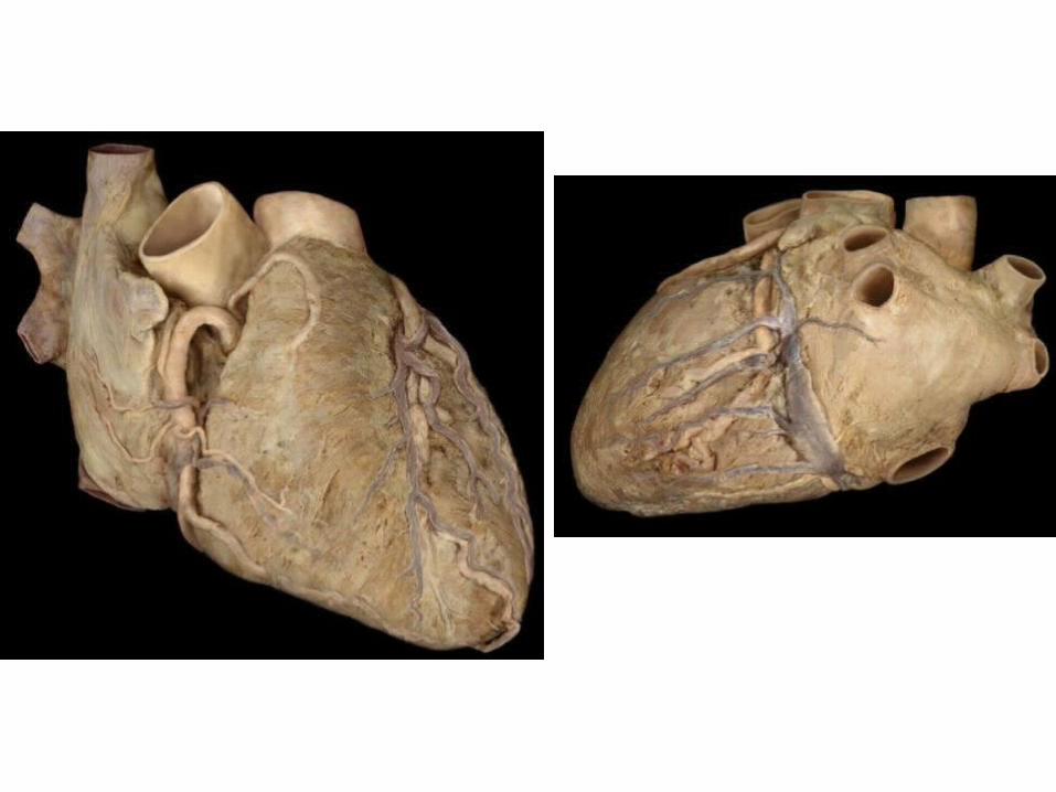
Coronary circulation
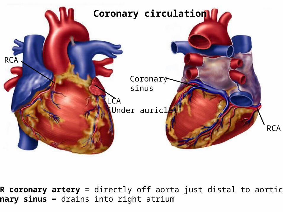
Coronarysinus
Coronary circulation
L & R coronary artery = directly off aorta just distal to aortic valvesCoronary sinus = drains into right atrium
RCA
RCA
LCA (Under auricle)
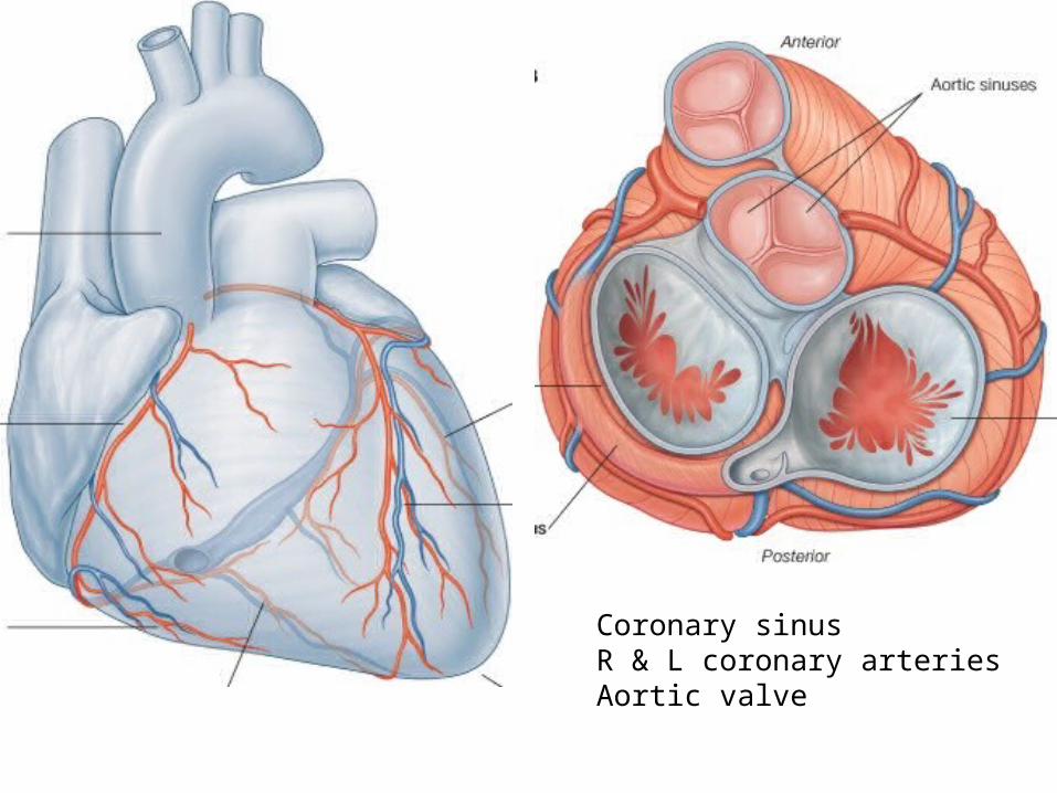
Coronary sinusR & L coronary arteriesAortic valve
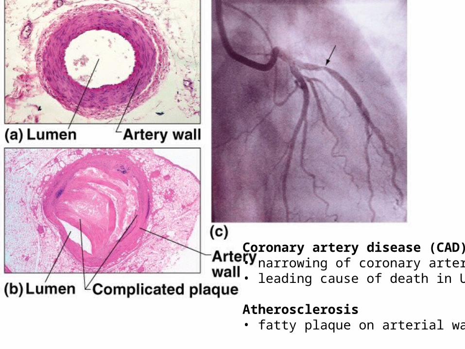
Fig. 20.12
Coronary artery disease (CAD)• narrowing of coronary arteries• leading cause of death in US
Atherosclerosis• fatty plaque on arterial walls
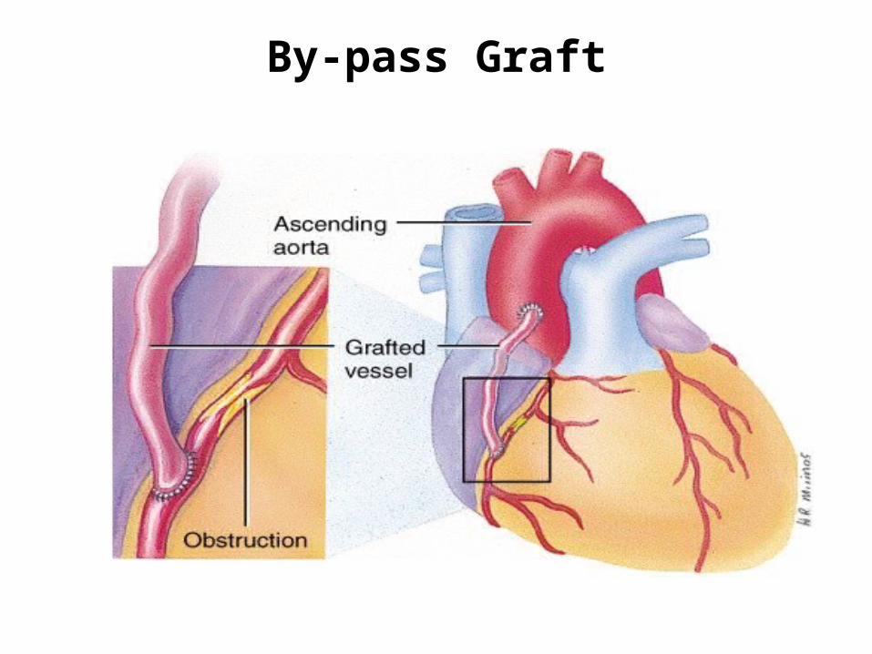
By-pass Graft

Myocardial infarction (MI)
• dead tissue areas in myocardium• caused by interruption of blood flow• cardiac muscles cells don’t regenerate• replaced by scar tissue• scarred or ischemic cardiac muscle can’t pump or conduct electrical impulses• arrhythmias (ventricular fibrillation)• Angina pectoris (chest pain)
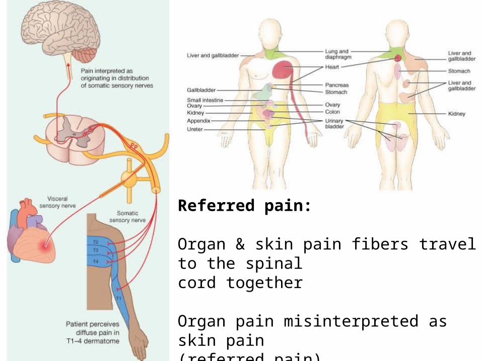
Referred pain:
Organ & skin pain fibers travel to the spinal cord together
Organ pain misinterpreted as skin pain (referred pain)
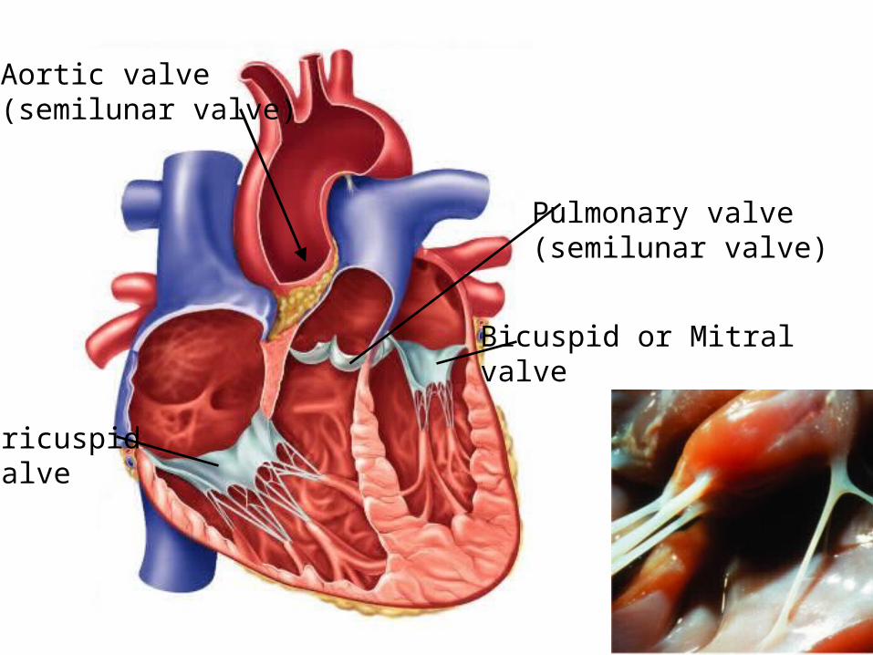
Tricuspidvalve
Bicuspid or Mitralvalve
Pulmonary valve(semilunar valve)
Aortic valve(semilunar valve)
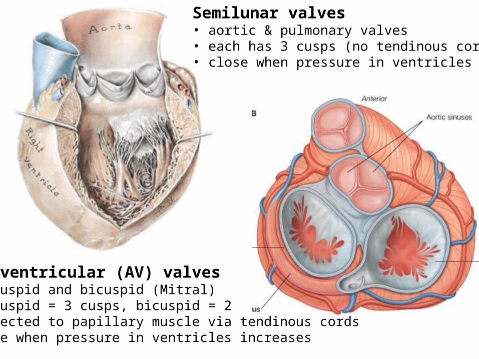
Semilunar valves• aortic & pulmonary valves• each has 3 cusps (no tendinous cords)• close when pressure in ventricles falls
Atrioventricular (AV) valves• tricuspid and bicuspid (Mitral)• tricuspid = 3 cusps, bicuspid = 2• connected to papillary muscle via tendinous cords• close when pressure in ventricles increases
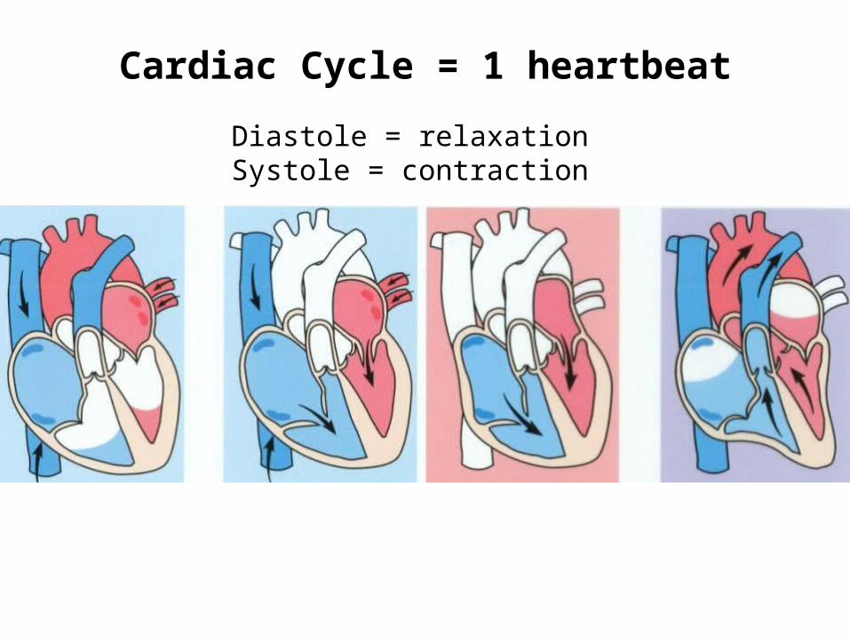
Cardiac Cycle = 1 heartbeat
Diastole = relaxationSystole = contraction
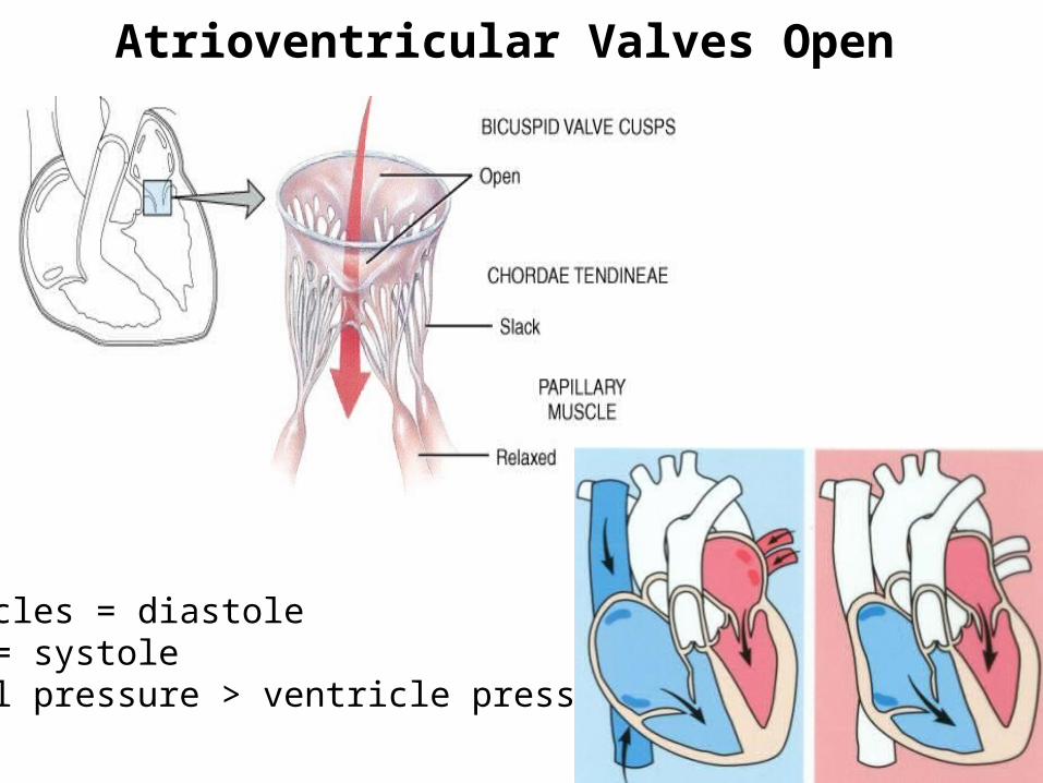
Atrioventricular Valves Open
Ventricles = diastoleAtria = systole(atrial pressure > ventricle pressure)
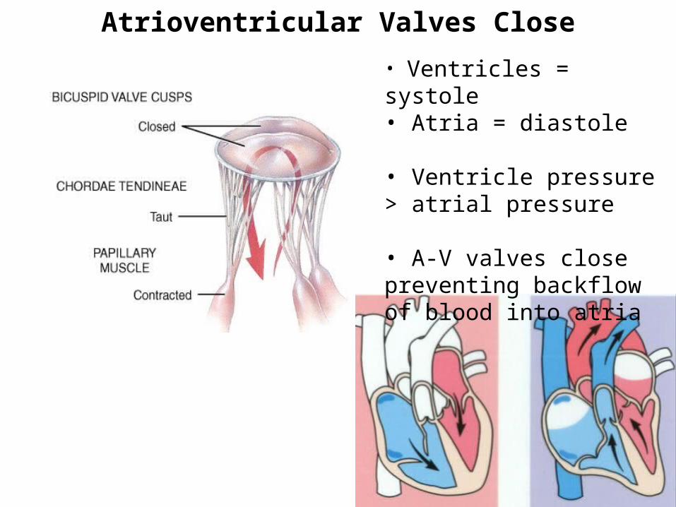
Atrioventricular Valves Close
• Ventricles = systole• Atria = diastole
• Ventricle pressure > atrial pressure
• A-V valves close preventing backflow of blood into atria
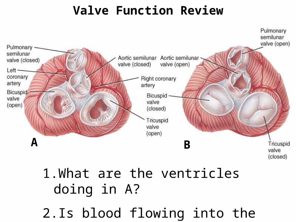
1.What are the ventricles doing in A?
2.Is blood flowing into the coronary arteries in A or B?
Valve Function Review
A B

Murmurs – any abnormal heart sound
Stenosis – narrowing of valve orifice• Congenital, rheumatic fever• Mitral most common (aortic)
Insufficiency or regurgitation – incomplete closure of valve• Many causes (MI, RF, HF, prolapse)• Aortic and mitral most common
Prolapse: most common valve disease• CT diseases or genetic
Rheumatic fever - strepA
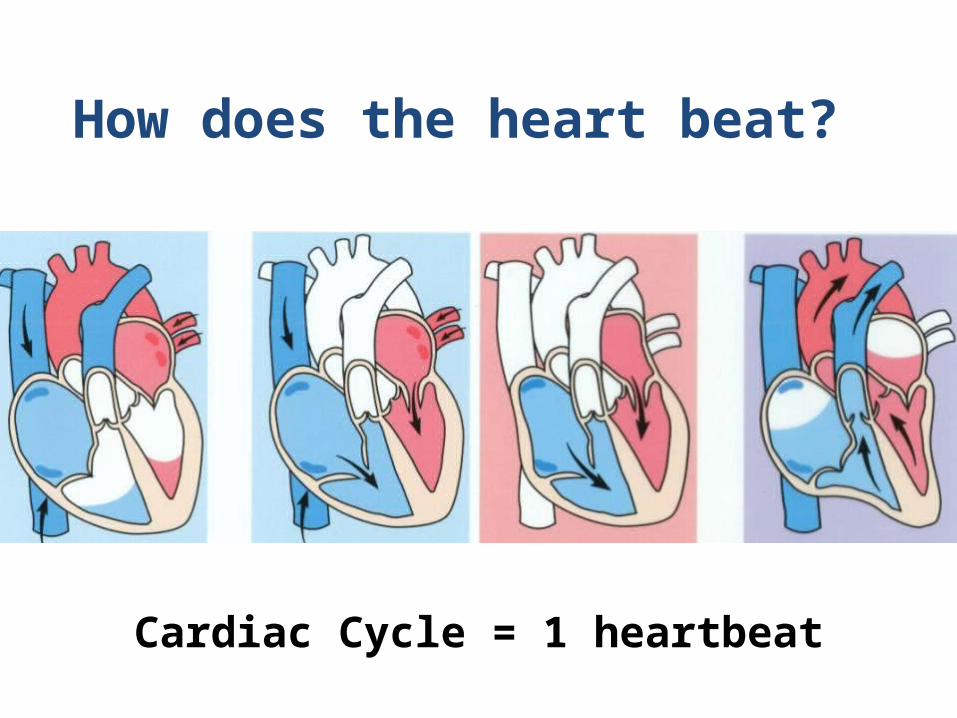
Cardiac Cycle = 1 heartbeat
How does the heart beat?
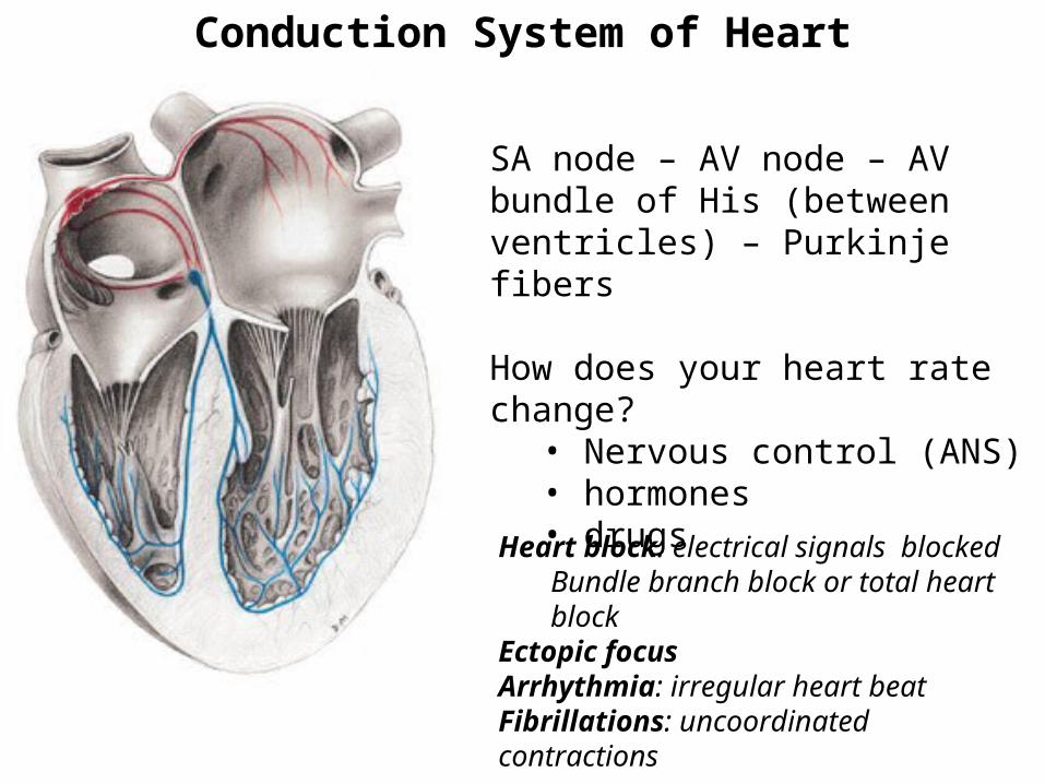
Conduction System of Heart
SA node – AV node – AV bundle of His (between ventricles) – Purkinje fibers
How does your heart rate change?• Nervous control (ANS)• hormones• drugs
Heart block: electrical signals blockedBundle branch block or total heart block
Ectopic focusArrhythmia: irregular heart beatFibrillations: uncoordinated contractions
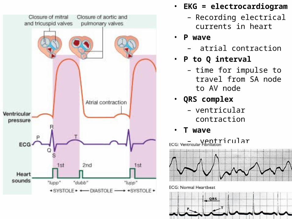
• EKG = electrocardiogram– Recording electrical
currents in heart • P wave
– atrial contraction • P to Q interval
– time for impulse to travel from SA node to AV node
• QRS complex – ventricular contraction
• T wave– ventricular relaxation
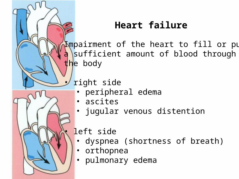
Impairment of the heart to fill or pump a sufficient amount of blood through the body
• right side• peripheral edema• ascites• jugular venous distention
• left side• dyspnea (shortness of breath)• orthopnea• pulmonary edema
Heart failure
Related Documents


