STATE-OF-THE-ART PAPER Heart Failure Eugene Braunwald, MD Boston, Massachusetts JACC: HEART FAILURE CME This article has been selected as the month’s JACC: Heart Failure CME activity. Accreditation and Designation Statement The American College of Cardiology Foundation (ACCF) is accredited by the Accreditation Council for Continuing Medical Education (ACCME) to provide continuing medical education for physicians. The ACCF designates this Journal-based CME activity for a maximum of 1 AMA PRA Category 1 Credit(s). Physicians should only claim credit commensurate with the extent of their participation in the activity. Method of Participation and Receipt of CME Certificate To obtain credit for JACC: Heart Failure CME, you must: 1. Be an ACC member or JACC: Heart Failure subscriber. 2. Carefully read the CME-designated article available online and in this issue of the journal. 3. Answer the post-test questions. At least 2 out of the 3 questions provided must be answered correctly to obtain CME credit. 4. Complete a brief evaluation. 5. Claim your CME credit and receive your certificate electroni- cally by following the instructions given at the conclusion of the activity. CME Objective for This Article: After reading this article, the reader should understand: 1) the epidemiology of heart failure including risk factors, associated morbidity and mortality, and costs to the healthcare system; 2) heart failure pathophysiology models; and 3) recent developments in the management of heart failure. CME Editor Disclosure: Deputy Managing Editor Mona Fiuzat, PharmD, FACC, reports that she has equity interest or stock options in ARCA Biopharma, consults for CCA, and receives research support from ResMed, GE Healthcare, Gilead, Critical Diagnostics, BG Medicine, Otsuka, Astellas, and Roche Diagnostics. Author Disclosures: Dr. Braunwald has received research support from Johnson & Johnson. Medium of Participation: Print (article only); online (article and quiz) CME Term of Approval: Issue date: February 2013 Expiration date: January 31, 2014 From the TIMI Study Group, Cardiovascular Division, Department of Medicine, Brigham and Women’s Hospital; and the Department of Medicine, Harvard Medical School, Boston, Massachusetts. Dr. Braunwald has received research support from Johnson & Johnson. Manuscript received September 27, 2012; accepted October 5, 2012. JACC: Heart Failure Vol. 1, No. 1, 2013 Ó 2013 by the American College of Cardiology Foundation ISSN 2213-1779/$36.00 Published by Elsevier Inc. http://dx.doi.org/10.1016/j.jchf.2012.10.002

Welcome message from author
This document is posted to help you gain knowledge. Please leave a comment to let me know what you think about it! Share it to your friends and learn new things together.
Transcript
JACC: Heart Failure Vol. 1, No. 1, 2013� 2013 by the American College of Cardiology Foundation ISSN 2213-1779/$36.00Published by Elsevier Inc. http://dx.doi.org/10.1016/j.jchf.2012.10.002
STATE-OF-THE-ART PAPER
Heart Failure
Eugene Braunwald, MD
Boston, Massachusetts
JACC: HEART FAILURE CME
This article has been selected as the month’s JACC: Heart FailureCME activity.
Accreditation and Designation Statement
The American College of Cardiology Foundation (ACCF) isaccredited by the Accreditation Council for Continuing MedicalEducation (ACCME) to provide continuing medical education forphysicians.
The ACCF designates this Journal-based CME activity fora maximum of 1 AMA PRA Category 1 Credit(s). Physicians shouldonly claim credit commensurate with the extent of their participationin the activity.
Method of Participation and Receipt of CME Certificate
To obtain credit for JACC: Heart Failure CME, you must:1. Be an ACC member or JACC: Heart Failure subscriber.2. Carefully read the CME-designated article available online and
in this issue of the journal.3. Answer the post-test questions. At least 2 out of the 3 questions
provided must be answered correctly to obtain CME credit.4. Complete a brief evaluation.
From the TIMI Study Group, Cardiovascular Division, Department of Medicine,
Brigham and Women’s Hospital; and the Department of Medicine, Harvard Medical
School, Boston, Massachusetts. Dr. Braunwald has received research support from
Johnson & Johnson.
Manuscript received September 27, 2012; accepted October 5, 2012.
5. Claim your CME credit and receive your certificate electroni-cally by following the instructions given at the conclusion ofthe activity.
CME Objective for This Article: After reading this article, thereader should understand: 1) the epidemiology of heart failureincluding risk factors, associated morbidity and mortality, and coststo the healthcare system; 2) heart failure pathophysiology models;and 3) recent developments in the management of heart failure.
CME Editor Disclosure: Deputy Managing Editor Mona Fiuzat,PharmD, FACC, reports that she has equity interest or stock optionsin ARCA Biopharma, consults for CCA, and receives researchsupport fromResMed, GEHealthcare, Gilead, Critical Diagnostics,BG Medicine, Otsuka, Astellas, and Roche Diagnostics.
Author Disclosures: Dr. Braunwald has received research supportfrom Johnson & Johnson.
MediumofParticipation: Print (article only); online (article andquiz)
CME Term of Approval:Issue date: February 2013Expiration date: January 31, 2014
Braunwald JACC: Heart Failure Vol. 1, No. 1, 2013Heart Failure February 2013:1–20
2
Heart Failure
D
espite major improvements in the treatment of virtually all cardiac disorders, heart failure (HF) is an exception, inthat its prevalence is rising, and only small prolongations in survival are occurring. An increasing fraction, especiallyolder women with diabetes, obesity, and atrial fibrillation exhibit HF with preserved systolic function. Severalpathogenetic mechanisms appear to be operative in HF. These include increased hemodynamic overload, ischemia-related dysfunction, ventricular remodeling, excessive neurohumoral stimulation, abnormal myocyte calciumcycling, excessive or inadequate proliferation of the extracellular matrix, accelerated apoptosis, and geneticmutations. Biomarkers released as a consequence of myocardial stretch, imbalance between formation andbreakdown of extracellular matrix, inflammation, and renal failure are useful in the identification of the pathogeneticmechanism and, when used in combination, may become helpful in estimating prognosis and selecting appropriatetherapy. Promising new therapies that are now undergoing intensive investigation include an angiotensin receptorneprilysin inhibitor, a naturally-occurring vasodilator peptide, a myofilament sensitizer and several drugs thatenhance Caþþ uptake by the sarcoplasmic reticulum. Cell therapy, using autologous bone marrow and cardiacprogenitor cells, appears to be promising, as does gene therapy. Chronic left ventricular assistance with continuousflow pumps is being applied more frequently and successfully as destination therapy, as a bridge to transplantation,and even as a bridge to recovery and explantation. While many of these therapies will improve the care of patientswith HF, significant reductions in prevalence will require vigorous, multifaceted, preventive approaches. (J Am CollCardiol HF 2013;1:1–20) ª 2013 by the American College of Cardiology FoundationEpidemiology
During the past half-century, the advances in the prevention,diagnosis, and management of cardiovascular disease (CVD)have been nothing short of spectacular. Age-adjusted CVD-related deaths have declined by about two-thirds in indus-trialized nations (1). Mortality rates associated with the acutecoronary syndromes (ACS), valvular and congenital heartdisease, uncontrolled hypertension, and many arrhythmiasall have fallen dramatically.
Heart failure (HF) is a notable exception to these encour-aging trends. Indeed, after normal delivery, it is the mostcommoncause of hospitalization.Annualhospital discharges inpatientswith a primary diagnosis ofHFhave risen steadily since1975, and now exceed 1 million discharges per year, althoughthey may at last be leveling off (2,3) (Fig. 1), or actuallydecreasing, in the United States (4,5). In Europe, hospitaliza-tions forHFare clearly declining (6,7).HF is primarily a diseaseof the elderly that affects about 10% of men and 8% of womenover the age of 60 years, and its prevalence rises with age (Fig. 2)and has risen overall. In the United States, patients witha primary diagnosis of HF now make >3 million physicianvisits per year.The direct and indirect costs ofHF in theUnitedStates are staggering; in 2010 they were estimated to be US$39.2 billion (8). The estimated lifetime cost of HF per indi-vidual patient is $110,000/year (-2008 US dollars), with morethan three-fourths of this cost consumedby in-hospital care (9).
Survival after a diagnosis of HF has improved during thepast 30 years; the age-adjusted death rate has declined (4–6),and themean age at death fromHFhas risen (7,10). However,despite these modest improvements, the 5-year mortality isstill approximately 50%dworse than that of many cancers(11). Among Medicare patients, 30-day mortality is 10% to12% (12), and the 30-day readmission rate after hospitaldischarge is 20% to 25% (13).
How can this so-called “HF paradox”dthat is, the strikingimprovements in the prognosis of individual cardiac conditions,such asACS, severe hypertension, valvular and congenital heartdiseases, but a growing prevalence ofHFdbe explained?Threepossibilities warrant consideration. The first is that, while therisk for mortality in each of these disorders has been reduced,the patients are not “cured” (with the exception of those withcertain congenital malformations). For example, while earlymortality in patients with acute myocardial infarction may havedeclined by 75%during the past half-century (14), survivors stillhave coronary artery disease (CAD) and remain at risk forsubsequent episodes of ischemic myocardial damage withfurther loss of myocardium and possiblyHF. A second possibleexplanation may be related to the increased frequency ofmyocyte death with aging and with the adverse cardiac conse-quences of comorbid conditions, the prevalences of which risewith age. These comorbid conditions include hyper-tension; type 2 diabetes mellitus; chronic renal disease; chronicobstructive pulmonary disease; and dysrhythmias, especiallyatrial fibrillation (15). The third possible explanation is that theslow but progressive improvement in HF prognosis mentionedpreviously simply increases the prevalence of this condition.Whatever the explanation(s), one might conclude that with thecontinued aging of the population, HF will remain a majorhealth problem,not only in industrialized nations but also in thedeveloping world.
Given this magnitude, attention is being directed,appropriately, to identifying individuals at higher risk forHF. Risk factors include increased body mass index,abdominal fat accumulation, elevated fasting blood glucose,elevated systolic blood pressure, elevated apolipoprotein B/apolipoprotein A ratio, and cigarette smoking. In a large-scale (1 million person-years) study that included bothoutpatients and inpatients from all age groups in an insured

Abbreviationsand Acronyms
ACE = angiotensin-
converting enzyme
ACS = acute coronary
syndromes
ADHF = acute
decompensation heart
failure
ANP = atrial natriuretic
peptide
ARB = angiotensin II
receptor blocker
BNP = brain natriuretic
peptide
CAD = coronary artery
disease
CPC = cardiac stem/
progenitor cell
CRP = C-reactive protein
cTn = cardiac-specific
troponin
CVD = cardiovascular
disease
ECM = extracellular matrix
EF = ejection fraction
HF = heart failure
HFPEF = heart failure and
a preserved ejection fraction
HFREF = heart failure and
a reduced ejection fraction
hs = high-sensitivity
LV = left-ventricular
LVAD = left-ventricular
assist device
JACC: Heart Failure Vol. 1, No. 1, 2013 BraunwaldFebruary 2013:1–20 Heart Failure
3
population in Georgia, CAD, hypertension, diabetes, andvalvular heart disease most frequently preceded the diagnosisof HF (16).
The clinical–hemodynamic profile of patients with HFappears to be changing (10). In a registry of >110,000patients hospitalized with HF, the proportion with heartfailure and a preserved ejection fraction (HFPEF), usuallydefined as an EF >50%, was approximately 40%, and in-hospital mortality was only slightly lower than that inpatients with HF and reduced EF (HFREF) (17). Also,a smaller percentage of patients with HFPEF than ofpatients with HFREF die from CVD-related causes (18).As a group, HFPEF patients are older and more commonlyfemale, with greater hypertension, obesity, anemia, and atrialfibrillation compared to those with HFREF (19). Diastolicdysfunction may remain asymptomatic for years, but age,renal dysfunction, hypertension, and progression of thisdysfunction all appear to be associated with the developmentof overt HF in this population (20,21).
Acute decompensation heart failure (ADHF), that is, thenew onset of severe HF or the sudden intensification ofchronic HF, is a life-threatening condition that usuallyrequires hospitalization and is, in fact, the most commoncause of hospital admission among patients with HF.ADHF may result from 1 or more precipitating events,including the development of a variety of dysrhythmias;ACS; a rapid increase in the need for an increased cardiacoutput of the failing heart by conditions such as infection,anemia, and pulmonary thromboembolism superimposed onchronic HF (22); discontinuation of treatment of chronicHF; and progression of the underlying disease. Based ondata from >100,000 hospitalizations in ADHERE (AcuteDecompensated Heart Failure National Registry), a simpleprognostic tool was established with findings that can beobtained easily at presentation. In a multivariate analysis,
Male
Years
Female
Dis
ch
arg
es
in
T
ho
us
an
ds
700
600
400
300
200
100
0
500
1979 1980 1985 1990 1995 2000 2005 2009
Figure 1Discharges From Hospitalization Due to Heart Failure,by Sex (United States, 1979–2009)
Reprinted with permission from: Roger VL, Go AS, Lloyd-Jones DM et al. Heart
disease and stroke statisticsd2012 update. Circulation 2012;125:e12–30.
Source: National Hospital Discharge Survey/National Center for Health Statistics
and National Heart, Lung, and Blood Institute.
miR = micro RNA
MMP = matrix
metalloproteinases
NP = natriuretic peptide
NT-proBNP = N-terminal pro-
B-type natriuretic peptide
PINP = pro collagen type I
aminoterminal propeptide
PIIINP = collagen III N-
terminal propeptide
RyR2 = ryanodine receptor
SR = sarcoplasmic reticulum
SERCA2a = sarcoplasmic
reticulum Ca2þ adenosine
triphosphatase
elevations in age, blood ureanitrogen, creatinine, and heartrate; lower systolic pressure andserum sodium; the presence ofdyspnea at rest; and the lack oflong-term treatment with ab-blocker were identified asindependent predictors ofmortality (23).
Mechanisms
The mechanisms involved in HFhave been investigated froma variety of perspectives duringthe past half-century. These per-spectives have sometimes beenreferred to as “models” (24).Hemodynamic model. In 1967,the author and his colleaguesdefined HF as “a clinical syn-drome characterized by wellknown symptoms and physicalsigns. . . . [It is] the pathologicalstate in which an abnormality ofmyocardial function is respon-sible for the failure of the heart topump blood at a rate commen-surate with the requirements ofthe metabolizing tissues duringordinary activity” (25). Supportfor this hemodynamic model ofHF came from the observationthat, in HF resulting from abso-lute or relative increases in hemo-dynamic load, there is actually areduction in the intrinsic contrac-tility of cardiac muscle. This wasreflected in a reduction in forcedevelopment of isolated cardiacmuscle obtained from the failinghearts of experimental animalpreparations with pressure over-load (26) and then from isolatedmyocytes obtained from patientswith HFREF (27).
Importanthemodynamic changesin HF result from ventricular re-modeling, which is common inpatients with chronic dysfunctionof the ventricular pump, andwhich
varies byHF type (28). In patients withHFPEF, the volume ofthe left-ventricular (LV) cavity is typically normal, but the wallis thickened, and the ratios of LV mass/end-diastolic volumeand themyocardial stiffnessmodulus are both increased (29). Incontrast, in patients with HFREF, the LV cavity is typically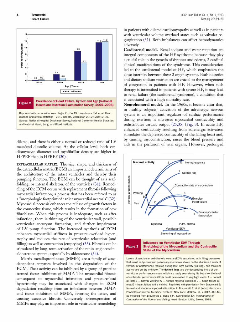
Figure 3Influences on Ventricular EDV ThroughStretching of the Myocardium and the ContractileState of the Myocardium
Levels of ventricular end-diastolic volume (EDV) associated with filling pressures
that result in dyspnea and pulmonary edema are shown on the abscissa. Levels of
ventricular performance required during rest, light activity (walking), and maximal
activity are on the ordinate. The dashed lines are the descending limbs of the
ventricular performance curves, which are rarely seen during life but show the level
of ventricular performance if EDV could be elevated to very high levels. A ¼ normal
at rest; B ¼ normal walking; C ¼ normal maximal exercise; D ¼ heart failure at
rest; E ¼ heart failure while walking. Reprinted with permission from Braunwald E:
Normal and abnormal myocardial function. In Braunwald E, et al. [eds]: Harrison’s
Principles of Internal Medicine, 15th ed. New York: McGraw-Hill, 2001:1309–18,
as modified from Braunwald E, Ross J Jr., Sonnenblick EH: Mechanisms ofContraction of the Normal and Failing Heart. Boston: Little, Brown; 1978.
Pe
rc
en
t o
f P
op
ula
tio
n
Male
Age ( Years)
20-39
0.2 0.3
1.9
9.0
5.4
11.5 11.8
0.8
40-59 60-79 80+
14
12
8
6
4
2
0
10
Female
Figure 2Prevalence of Heart Failure, by Sex and Age (NationalHealth and Nutrition Examination Survey, 2005–2008)
Reprinted with permission from: Roger VL, Go AS, Lloyd-Jones DM, et al. Heart
disease and stroke statisticsd2012 update. Circulation 2012;125:e12–30.
Source: National Hospital Discharge Survey/National Center for Health Statistics
and National Heart, Lung, and Blood Institute.
Braunwald JACC: Heart Failure Vol. 1, No. 1, 2013Heart Failure February 2013:1–20
4
dilated, and there is either a normal or reduced ratio of LVmass/end-diastolic volume. At the cellular level, both car-diomyocyte diameter and myofibrillar density are higher inHFPEF than in HFREF (30).
EXTRACELLULAR MATRIX. The size, shape, and thickness ofthe extracellular matrix (ECM) are important determinants ofthe architecture of the intact ventricles and thereby theirpumping function. The ECM can be thought of as a scaf-folding, or internal skeleton, of the ventricles (31). Remod-eling of the ECM occurs with replacement fibrosis followingmyocardial infarction, a process that has been referred to asa “morphologic footprint of earlier myocardial necrosis” (32).Myocardial necrosis enhances the release of growth factors inthe connective tissue, which results in the formation of newfibroblasts. When this process is inadequate, such as afterinfarction, there is thinning of the ventricular wall, possibleventricular aneurysm formation, and further impairmentof LV pump function. The increased synthesis of ECMenhances myocardial stiffness in pressure overload hyper-trophy and reduces the rate of ventricular relaxation (andfilling) as well as contraction (emptying) (33). Fibrosis can bestimulated by long-term activation of the renin-angiotensin-aldosterone system, especially by aldosterone (34).
Matrix metalloproteinases (MMPs) are a family of zinc-dependent enzymes involved in the degradation of theECM. Their activity can be inhibited by a group of proteinstermed tissue inhibitors of MMP. The myocardial fibrosisconsequent to myocardial infarction and pressure-loadhypertrophy may be associated with changes in ECMdegradation resulting from an imbalance between MMPsand tissue inhibitors of MMPs, favoring the latter, andcausing excessive fibrosis. Conversely, overexpression ofMMPs may play an important role in ventricular remodeling
in patients with dilated cardiomyopathy as well as in patientswith ventricular volume overload states such as valvular re-gurgitation (31). Both imbalances can affect hemodynamicsadversely.Cardiorenal model. Renal sodium and water retention areintegral components of the HF syndrome because they playa crucial role in the genesis of dyspnea and edema, 2 cardinalclinical manifestations of the syndrome. This considerationled to the cardiorenal model of HF, which emphasizes theclose interplay between these 2 organ systems. Both diureticsand dietary sodium restriction are crucial to the managementof congestion in patients with HF. However, when suchtherapy is intensified in patients with severe HF, it may leadto renal failure (the cardiorenal syndrome), a condition thatis associated with a high mortality rate.Neurohumoral model. In the 1960s, it became clear that,in healthy subjects, activation of the adrenergic nervoussystem is an important regulator of cardiac performanceduring exertion; it increases myocardial contractility andredistributes cardiac output (25,35) (Fig. 3). In acute HF,enhanced contractility resulting from adrenergic activationstimulates the depressed contractility of the failing heart and,by causing vasoconstriction, raises the blood pressure andaids in the perfusion of vital organs. However, prolonged
JACC: Heart Failure Vol. 1, No. 1, 2013 BraunwaldFebruary 2013:1–20 Heart Failure
5
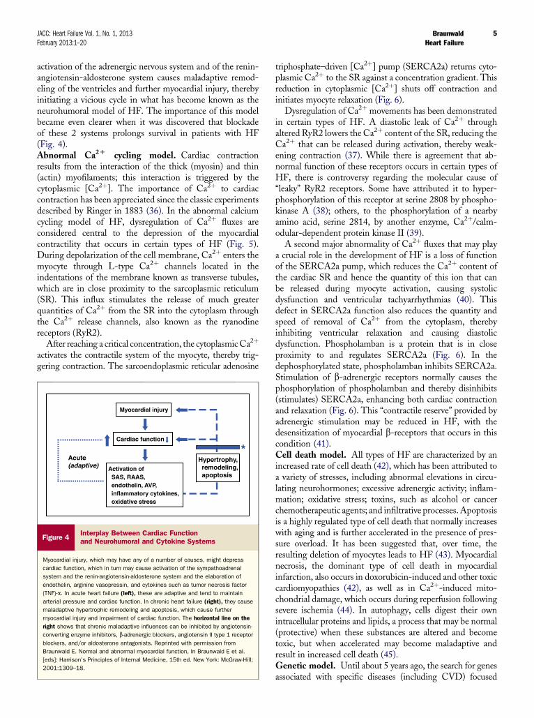
activation of the adrenergic nervous system and of the renin-angiotensin-aldosterone system causes maladaptive remod-eling of the ventricles and further myocardial injury, therebyinitiating a vicious cycle in what has become known as theneurohumoral model of HF. The importance of this modelbecame even clearer when it was discovered that blockadeof these 2 systems prolongs survival in patients with HF(Fig. 4).Abnormal Ca2D cycling model. Cardiac contractionresults from the interaction of the thick (myosin) and thin(actin) myofilaments; this interaction is triggered by thecytoplasmic [Ca2þ]. The importance of Ca2þ to cardiaccontraction has been appreciated since the classic experimentsdescribed by Ringer in 1883 (36). In the abnormal calciumcycling model of HF, dysregulation of Ca2þ fluxes areconsidered central to the depression of the myocardialcontractility that occurs in certain types of HF (Fig. 5).During depolarization of the cell membrane, Ca2þ enters themyocyte through L-type Ca2þ channels located in theindentations of the membrane known as transverse tubules,which are in close proximity to the sarcoplasmic reticulum(SR). This influx stimulates the release of much greaterquantities of Ca2þ from the SR into the cytoplasm throughthe Ca2þ release channels, also known as the ryanodinereceptors (RyR2).
After reaching a critical concentration, the cytoplasmicCa2þ
activates the contractile system of the myocyte, thereby trig-gering contraction. The sarcoendoplasmic reticular adenosine
Figure 4Interplay Between Cardiac Functionand Neurohumoral and Cytokine Systems
Myocardial injury, which may have any of a number of causes, might depress
cardiac function, which in turn may cause activation of the sympathoadrenal
system and the renin-angiotensin-aldosterone system and the elaboration of
endothelin, arginine vasopressin, and cytokines such as tumor necrosis factor
(TNF)-a. In acute heart failure (left), these are adaptive and tend to maintain
arterial pressure and cardiac function. In chronic heart failure (right), they cause
maladaptive hypertrophic remodeling and apoptosis, which cause further
myocardial injury and impairment of cardiac function. The horizontal line on the
right shows that chronic maladaptive influences can be inhibited by angiotensin-
converting enzyme inhibitors, b-adrenergic blockers, angiotensin II type 1 receptor
blockers, and/or aldosterone antagonists. Reprinted with permission from
Braunwald E. Normal and abnormal myocardial function, In Braunwald E et al.
[eds]: Harrison’s Principles of Internal Medicine, 15th ed. New York: McGraw-Hill;
2001:1309–18.
triphosphate–driven [Ca2þ] pump (SERCA2a) returns cyto-plasmic Ca2þ to the SR against a concentration gradient. Thisreduction in cytoplasmic [Ca2þ] shuts off contraction andinitiates myocyte relaxation (Fig. 6).
Dysregulation of Ca2þ movements has been demonstratedin certain types of HF. A diastolic leak of Ca2þ throughaltered RyR2 lowers the Ca2þ content of the SR, reducing theCa2þ that can be released during activation, thereby weak-ening contraction (37). While there is agreement that ab-normal function of these receptors occurs in certain types ofHF, there is controversy regarding the molecular cause of“leaky” RyR2 receptors. Some have attributed it to hyper-phosphorylation of this receptor at serine 2808 by phospho-kinase A (38); others, to the phosphorylation of a nearbyamino acid, serine 2814, by another enzyme, Ca2þ/calm-odular-dependent protein kinase II (39).
A second major abnormality of Ca2þ fluxes that may playa crucial role in the development of HF is a loss of functionof the SERCA2a pump, which reduces the Ca2þ content ofthe cardiac SR and hence the quantity of this ion that canbe released during myocyte activation, causing systolicdysfunction and ventricular tachyarrhythmias (40). Thisdefect in SERCA2a function also reduces the quantity andspeed of removal of Ca2þ from the cytoplasm, therebyinhibiting ventricular relaxation and causing diastolicdysfunction. Phospholamban is a protein that is in closeproximity to and regulates SERCA2a (Fig. 6). In thedephosphorylated state, phospholamban inhibits SERCA2a.Stimulation of b-adrenergic receptors normally causes thephosphorylation of phospholamban and thereby disinhibits(stimulates) SERCA2a, enhancing both cardiac contractionand relaxation (Fig. 6). This “contractile reserve” provided byadrenergic stimulation may be reduced in HF, with thedesensitization of myocardial b-receptors that occurs in thiscondition (41).Cell death model. All types of HF are characterized by anincreased rate of cell death (42), which has been attributed toa variety of stresses, including abnormal elevations in circu-lating neurohormones; excessive adrenergic activity; inflam-mation; oxidative stress; toxins, such as alcohol or cancerchemotherapeutic agents; and infiltrative processes.Apoptosisis a highly regulated type of cell death that normally increaseswith aging and is further accelerated in the presence of pres-sure overload. It has been suggested that, over time, theresulting deletion of myocytes leads to HF (43). Myocardialnecrosis, the dominant type of cell death in myocardialinfarction, also occurs in doxorubicin-induced and other toxiccardiomyopathies (42), as well as in Ca2þ-induced mito-chondrial damage, which occurs during reperfusion followingsevere ischemia (44). In autophagy, cells digest their ownintracellular proteins and lipids, a process that may be normal(protective) when these substances are altered and becometoxic, but when accelerated may become maladaptive andresult in increased cell death (45).Genetic model. Until about 5 years ago, the search for genesassociated with specific diseases (including CVD) focused
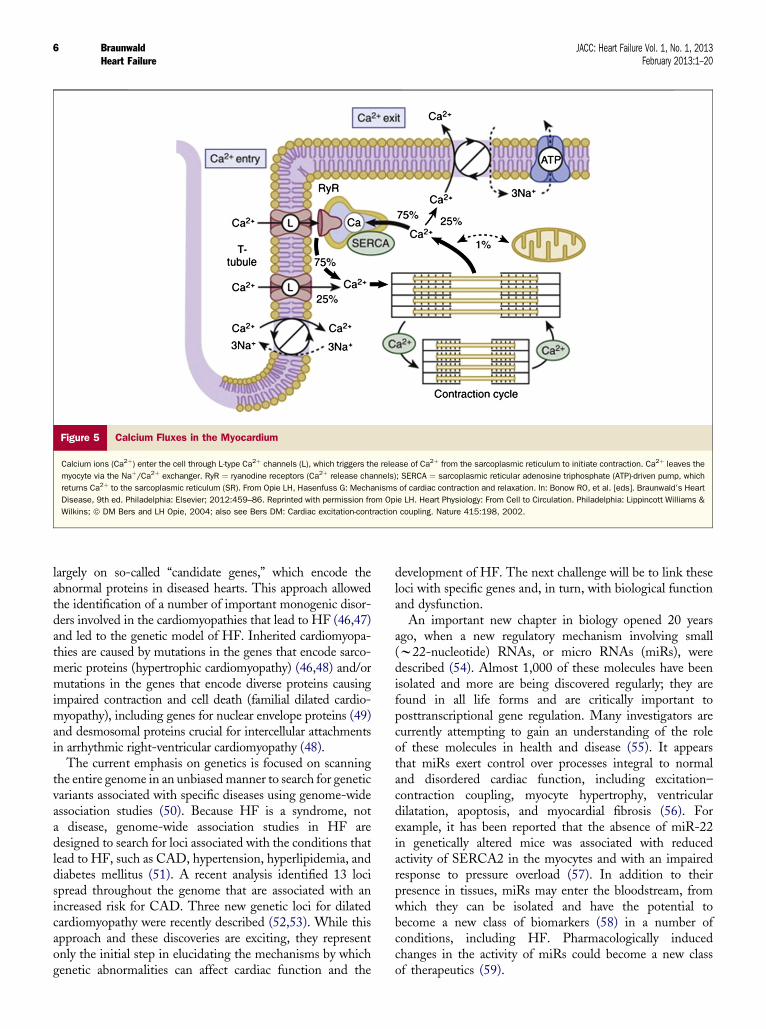
Figure 5 Calcium Fluxes in the Myocardium
Calcium ions (Ca2þ) enter the cell through L-type Ca2þ channels (L), which triggers the release of Ca2þ from the sarcoplasmic reticulum to initiate contraction. Ca2þ leaves the
myocyte via the Naþ/Ca2þ exchanger. RyR ¼ ryanodine receptors (Ca2þ release channels); SERCA ¼ sarcoplasmic reticular adenosine triphosphate (ATP)-driven pump, which
returns Ca2þ to the sarcoplasmic reticulum (SR). From Opie LH, Hasenfuss G: Mechanisms of cardiac contraction and relaxation. In: Bonow RO, et al. [eds]. Braunwald’s Heart
Disease, 9th ed. Philadelphia: Elsevier; 2012:459–86. Reprinted with permission from Opie LH. Heart Physiology: From Cell to Circulation. Philadelphia: Lippincott Williams &
Wilkins; ª DM Bers and LH Opie, 2004; also see Bers DM: Cardiac excitation-contraction coupling. Nature 415:198, 2002.
Braunwald JACC: Heart Failure Vol. 1, No. 1, 2013Heart Failure February 2013:1–20
6
largely on so-called “candidate genes,” which encode theabnormal proteins in diseased hearts. This approach allowedthe identification of a number of important monogenic disor-ders involved in the cardiomyopathies that lead to HF (46,47)and led to the genetic model of HF. Inherited cardiomyopa-thies are caused by mutations in the genes that encode sarco-meric proteins (hypertrophic cardiomyopathy) (46,48) and/ormutations in the genes that encode diverse proteins causingimpaired contraction and cell death (familial dilated cardio-myopathy), including genes for nuclear envelope proteins (49)and desmosomal proteins crucial for intercellular attachmentsin arrhythmic right-ventricular cardiomyopathy (48).
The current emphasis on genetics is focused on scanningthe entire genome in an unbiased manner to search for geneticvariants associated with specific diseases using genome-wideassociation studies (50). Because HF is a syndrome, nota disease, genome-wide association studies in HF aredesigned to search for loci associated with the conditions thatlead to HF, such as CAD, hypertension, hyperlipidemia, anddiabetes mellitus (51). A recent analysis identified 13 locispread throughout the genome that are associated with anincreased risk for CAD. Three new genetic loci for dilatedcardiomyopathy were recently described (52,53). While thisapproach and these discoveries are exciting, they representonly the initial step in elucidating the mechanisms by whichgenetic abnormalities can affect cardiac function and the
development of HF. The next challenge will be to link theseloci with specific genes and, in turn, with biological functionand dysfunction.
An important new chapter in biology opened 20 yearsago, when a new regulatory mechanism involving small(w22-nucleotide) RNAs, or micro RNAs (miRs), weredescribed (54). Almost 1,000 of these molecules have beenisolated and more are being discovered regularly; they arefound in all life forms and are critically important toposttranscriptional gene regulation. Many investigators arecurrently attempting to gain an understanding of the roleof these molecules in health and disease (55). It appearsthat miRs exert control over processes integral to normaland disordered cardiac function, including excitation–contraction coupling, myocyte hypertrophy, ventriculardilatation, apoptosis, and myocardial fibrosis (56). Forexample, it has been reported that the absence of miR-22in genetically altered mice was associated with reducedactivity of SERCA2 in the myocytes and with an impairedresponse to pressure overload (57). In addition to theirpresence in tissues, miRs may enter the bloodstream, fromwhich they can be isolated and have the potential tobecome a new class of biomarkers (58) in a number ofconditions, including HF. Pharmacologically inducedchanges in the activity of miRs could become a new classof therapeutics (59).

Figure 6 Mechanism of Ca2þ Uptake Into the SR by the ATP-Driven Ca2þ Uptake Pump (SERCA2a)
Within the SR, Ca2þ is attached to the protein calsequestrin. An increased rate of uptake of Ca2þ into the SR enhances the rate of relaxation (lusitropic effect). Phospholamban
(PL), when phosphorylated (P), removes the inhibition exerted on the Ca2þ pump by its dephosphorylated form. Thereby, Ca2þ uptake is increased either in response to an
enhanced cytosolic Ca2þ or in response to b-adrenergic stimulation, which activates cyclic adenosine monophosphate (cAMP) kinase. Calmodulin kinase may be a second
messenger of the b-adrenergic system. RyR ¼ ryanodine receptor in the Ca2þ release channel; other abbreviations as in Figure 5. From Opie LH, Hasenfuss G: Mechanisms of
cardiac contraction and relaxation. In: Bonow RO, et al. [eds]. Braunwald’s Heart Disease, 9th ed. Philadelphia: Elsevier; 2012, as modified with permission from Opie LH. Heart
Physiology, from Cell to Circulation. Philadelphia: Williams & Wilkins; 2004.
Figure 7 Seven Major Classes of Biomarkers Contributing tothe Biomarker Profile in HF
Adapted with permission from Braunwald E. Preface. Heart Fail Clin 2009;5:xiii–xiv.
JACC: Heart Failure Vol. 1, No. 1, 2013 BraunwaldFebruary 2013:1–20 Heart Failure
7
Biomarkers
Although the definition of a biomarker includes virtually anymeasurement that can be made on a biological system, thisdiscussion is restricted to substances measured in the bloodother than genetic markers, electrolytes, and commonly usedmarkers of hepatic or renal function. These biomarkers aid inthe diagnosis of HF, provide an estimate of prognosis, andhelp in the identification of apparently healthy people who areat excessive risk for HF (60). Biomarkers that are currentlyavailable reflect at least 7 pathobiological processes operativein HF (Fig. 7), help to identify the specific ones involved inindividual patients, and aid in guiding management plans.The increased availability of point-of-care and rapid-turnaround methodologies, and the declining costs of anal-ysis of several of the most frequently used biomarkers, arefacilitating their widespread use.
In the assessment of the clinical value of any individualbiomarker, it is important to determine whether it providesindependent incremental information when added to previ-ously available information, which can be estimated by deter-mining whether it increases the c statistic (61), as well as bycalculating the net reclassification improvement index and theintegrated discrimination improvement index (62).Despite theimportance of these rigorous statistical tests, measurements ofbiomarkers, even those that are not independent predictors ofrisk on multivariate analysis, may nonetheless be of clinicalimportance because they provide information on the
pathogenesis of HF and can help to direct treatment. Forexample, in patients with abnormally elevated levels of a natri-uretic peptide (NP) and troponin, an abnormally elevatedconcentration of a marker for ECM remodelingmight not adddiscriminatory diagnostic power but might suggest that a drugthat reduces collagen deposition may be beneficial (see subse-quent discussion).Markers of myocyte stretch. Atrial NP (ANP) was thefirst NP elaborated by stretched cardiac tissue to be identi-fied and studied in patients with HF (63). However, because
Braunwald JACC: Heart Failure Vol. 1, No. 1, 2013Heart Failure February 2013:1–20
8
of its instability and other analytic problems, it was soonreplaced by B-type NP (BNP) and its prohormone frag-ment, N-terminal pro B–type natriuretic peptide (NT-proBNP), peptides derived largely from the ventricles. These2 peptides are now the most widely used biomarkers in thecare of patients with known or suspected HF (64,65). Inaddition, mid-regional (MR) proANP, a precursor of ANP,does not pose the same analytical problems as does ANP andhas been reported to have been as accurate as BNP andNT-proBNP in diagnosing HF (66) and in estimating theprognosis of patients with HF (67). In patients with chronicCAD and normal EF, MR-proANP has also been reportedto predict CVD-related death or hospitalization for HFand to identify patients who might benefit from theadministration of an angiotensin-converting enzyme (ACE)inhibitor (68).
NPs are of enormous clinical value in diagnosing HF inpatients with dyspnea of unknown etiology (64–66,69,70); inpatients with heart disease without clinical manifestations of,but who are at risk for, HF; and in apparently healthy patientswho are at higher risk for HF (71). However, like any labo-ratory measurement, NP levels must be interpreted in thecontext of a patient’s characteristics, such as age and bodymass, and laboratory tests, including cardiac imaging.
There is some controversy regarding the clinical value ofNP-guided therapy for HF (72). However, 2 meta-analyseshave reported significant advantages of NP-guided therapyin terms of survival (73,74). In the larger of these (1,726patients), NP-guided therapy was compared with usual carein 8 moderate-scale trials, and a significantly lower mortalitywas reported with NP-guided therapy (hazard ratio ¼ 0.76).However, in these trials, NP-guided therapy was of littlevalue in 2 subgroupsdpatients age >75 years and those withHFPEF. Like all meta-analyses, these were limited bydifferences in the characteristics of the populations studied,the peptide concentrations targeted, and the treatmentalgorithms employed. Fortunately, a robust, adequately sizedmulticenter trial of a single-target NP level and the use ofguideline-approved therapies in both treatment arms of pre-specified subgroups is now underway (GUIDE-IT [GuidingEvidence Based Therapy Using Biomarker IntensifiedTreatment]; NCT01685840).
ST2 is a protein that exists in soluble and membrane-bound forms, the latter being the receptor for interleukin33. When the myocardium is stretched, the ST2 gene isupregulated, and the concentration of circulating solubleST2 rises rapidly. The level of circulating ST2 has beenreported to be a predictor of HF and death in patients withST-segment elevation myocardial infarction (75), ADHF,and/or chronic HF (76). This biomarker provides prognosticinformation that is independent of and in addition to thatoffered by NT-proBNP (77), although the release of bothseems to occur in response to the same stimulusdcardiacstretch.Myocyte injury. The 2 cardiac-specific troponins (cTn)dIand T (cTnI and cTnT, respectively)dexist in 2 pools in
myocytes. The larger is an integral constituent of themyofibrillar protein apparatus and is released slowly overseveral days after cell death; the second, smaller source ofcTn resides in a cytoplasmic pool this is released relativelyrapidly, within 1 to 2 hours of myocyte injury. It is not yetclear whether irreversible injury is required or whetherreversibly injured cells whose membranes have transientlybecome more permeable can also release this pool (78).
cTnI and cTnT have become the most accurate andwidely used markers of myocardial necrosis in patients withACS. However, in 1997, it was reported that cTnI is alsopresent in the serum of patients with severe HF withoutischemia (79), and it was then observed that levels of cTnIand cTnT were predictive of adverse clinical outcomes inthese patients (80). This observation has been amply con-firmed, particularly as progressively more sensitive assaysfor cTn have become available (81,82). The release oftroponin from the heart in HF has been considered to bedue to myocyte injury, apparently irrespective of themechanism involved, that is, ischemia, necrosis, apoptosis,or autophagy.
Using standard assays, abnormal elevations of circulatingcTn have been reported in about one-quarter of patientswith HF and denoted a poor prognosis, generally definedas death or early readmission for HF (60). Using high-sensitivity assays (hsTn), abnormal elevations in circulatingtroponins have been detected in virtually all patients withADHF (83–86), in a majority of a population with chronicHF (87), in some patients with stable CAD and normal EF(88), as well as in a minority of general populations ofapparently healthy elderly (89) and middle-aged persons(90). Serial measurements of hsTn in populations withADHF (86) and chronic HF (87) have been reported toprovide additional prognostic information; cTn levels thatrose during hospitalization portended a poorer outcome thandid stable or declining levels.ECM markers. The importance of the ECMs in ventric-ular remodeling is discussed in the Mechanisms andManagement sections. Serum peptides derived fromcollagen metabolism reflect both the synthesis and degra-dation of collagen and thus constitute a “window” on theECM (91,92). The ratio of pro collagen type I amino-terminal propeptide (PINP), a marker of collagen synthesis,to collagen type I cross-linked carboxyterminal telopeptide,a marker of collagen breakdown, is a useful serum marker ofcollagen accumulation (93). A multimarker panel consistingof increased levels of MMP-2, tissue inhibitor of MMP-4,and collagen III N-terminal propeptide (PIIINP), accom-panied by decreased levels of MMP-8, has been reported tobe characteristic of HFPEF (92). Elevated ECM turnoverhas also been reported in patients with ADHF (91).
Aldosterone is a stimulant of collagen synthesis andenhances cardiac fibrosis in HF and in ventricular hyper-trophy secondary to pressure overload. The administrationof the aldosterone receptor antagonist spironolactone inpatients with chronic HF in RALES (Randomized
JACC: Heart Failure Vol. 1, No. 1, 2013 BraunwaldFebruary 2013:1–20 Heart Failure
9
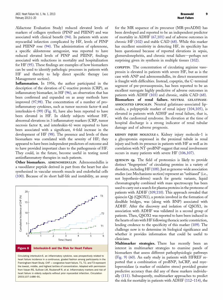
Aldactone Evaluation Study) reduced elevated levels ofmarkers of collagen synthesis (PINP and PIIINP) and wasassociated with clinical benefit (94). In patients with acutemyocardial infarction complicated by HF, levels of PINPand PIIINP rose (94). The administration of eplerenone,a specific aldosterone antagonist, was reported to havereduced elevated levels of PINP and PIIINP, findingsassociated with reductions in mortality and hospitalizationfor HF (95). These findings are examples of how biomarkerscan be used to identify pathologic processes in patients withHF and thereby to help direct specific therapy (seeManagement section).Inflammation. In 1956, the author participated in thedescription of the elevation of C-reactive protein (CRP), aninflammatory biomarker, in HF (96), an observation that hasbeen confirmed and expanded on as assay methods haveimproved (97,98). The concentration of a number of pro-inflammatory cytokines, such as tumor necrosis factor-a andinterleukin-6 (99) (Fig. 8), have also been reported to havebeen elevated in HF. In elderly subjects without HF,abnormal elevations in 3 inflammatory markers (CRP, tumornecrosis factor a, and interleukin-6) were reported to havebeen associated with a significant, 4-fold increase in thedevelopment of HF (98). The presence and levels of thesebiomarkers was correlated with the severity of HF; theyappeared to have been independent predictors of outcome andto have provided important clues to the pathogenesis of HF.They could, in the future, become useful in testing novelantiinflammatory therapies in such patients.Other biomarkers. ADRENOMEDULLIN. Adrenomedullin isa vasodilator peptide derived in part from the heart but alsosynthesized in vascular smooth muscle and endothelial cells(100). Because of its short half-life and instability, an assay
Figure 8 Interleukin-6 and the Risk for Heart Failure
Circulating interleukin-6, an inflammatory cytokine, was prospectively related to
heart failure incidence in a continuous, graded fashion among participants in the
Framingham Heart Study. CHF ¼ congestive heart failure. T1, T2, and T3 represent
the lowest, middle, and highest tertiles of concentration. Adapted with permission
from Vasan RS, Sullivan LM, Roubenoff R, et al. Inflammatory markers and risk of
heart failure in elderly subjects without prior myocardial infarction. Circulation
2003;107:1486–91.
for the MR sequence of its precursor (MR-proADM) hasbeen developed and reported to be an independent predictorof mortality in ADHF (67,101) and of adverse outcomes inchronic HF (102) and stable CAD (68). While this markerhas excellent sensitivity in detecting HF, its specificity hasbeen questioned because of reported elevations in sepsis,glomerulonephritis, and chronic renal failuredperhaps notsurprising given its synthesis in multiple tissues (102).
COPEPTIN. The concentration of circulating arginine vaso-pressin is elevated in patients with severe HF, but as is thecase with ANP and adrenomedullin, its direct measurementis fraught with difficulties. Instead, copeptin, the C-terminalsegment of pre-provasopressin, has been reported to be anexcellent surrogate highly predictive of adverse outcomes inpatients with ADHF (103) and chronic, stable CAD (68).Biomarkers of renal failure. NEUTRAL GELATINASE-
ASSOCIATED LIPOCALIN. Neutral gelatinase-associated lip-ocalin, a polypeptide marker of renal injury (104,105), iselevated in patients with ADHF and renal failure, that is,with the cardiorenal syndrome. Its elevation at the time ofhospital discharge is a strong indicator of renal tubulardamage and of adverse prognosis.
KIDNEY INJURY MOLECULE-1. Kidney injury molecule-1 isa glycoprotein expressed in the proximal tubule in renalinjury and both its presence in patients with HF as well as itscorrelation with NT-proBNP suggest that renal involvementoccurs in many patients with severe HF (106,107).
QUIESCIN Q6. The field of proteomics is likely to providedistinct “fingerprints” of circulating proteins in a variety ofdisorders, includingHF (108). Just as genome-wide associationstudies (see Mechanisms section) represent an “unbiased” (i.e.,not hypothesis-driven) search for genetic variants, liquidchromatography combined with mass spectroscopy has beenused to carry out a search for plasma proteins in the proteome ofpatients with ADHF (109,110). This approach revealed thatquiescin Q6 (QSOX1), a protein involved in the formation ofdisulfide bridges, was (along with BNP) associated withADHF. After the discovery and isolation of QSOX1, itsassociation with ADHF was validated in a second group ofpatients. Then, QSOX1 was reported to have been induced inthe hearts of ratswithHF following thoracic aortic constriction,lending credence to the specificity of this marker (109). Thechallenge now is to determine its biological significance andwhether it provides information that could be useful toclinicians.Multimarker strategies. There has recently been aninterest in multimarker strategies to examine panels ofbiomarkers that assess different pathophysiologic pathways(Fig. 9) (60). An early study in patients with HFREF re-ported that a combination of proBNP, hsCRP, and mye-loperoxidase (a marker of oxidative stress) provided greaterpredictive accuracy than did any of these markers individu-ally (111). Subsequently, multimarker approaches to predictthe risk for mortality in patients with ADHF (112–114), the
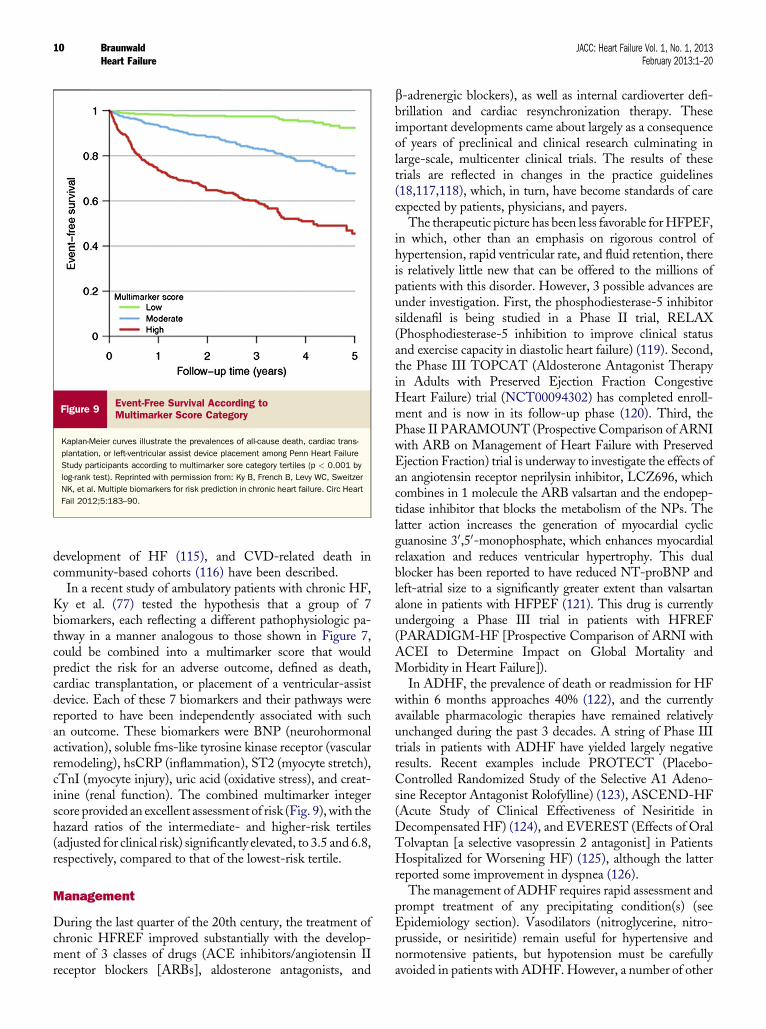
Figure 9Event-Free Survival According toMultimarker Score Category
Kaplan-Meier curves illustrate the prevalences of all-cause death, cardiac trans-
plantation, or left-ventricular assist device placement among Penn Heart Failure
Study participants according to multimarker sore category tertiles (p < 0.001 by
log-rank test). Reprinted with permission from: Ky B, French B, Levy WC, Sweitzer
NK, et al. Multiple biomarkers for risk prediction in chronic heart failure. Circ Heart
Fail 2012;5:183–90.
Braunwald JACC: Heart Failure Vol. 1, No. 1, 2013Heart Failure February 2013:1–20
10
development of HF (115), and CVD-related death incommunity-based cohorts (116) have been described.
In a recent study of ambulatory patients with chronic HF,Ky et al. (77) tested the hypothesis that a group of 7biomarkers, each reflecting a different pathophysiologic pa-thway in a manner analogous to those shown in Figure 7,could be combined into a multimarker score that wouldpredict the risk for an adverse outcome, defined as death,cardiac transplantation, or placement of a ventricular-assistdevice. Each of these 7 biomarkers and their pathways werereported to have been independently associated with suchan outcome. These biomarkers were BNP (neurohormonalactivation), soluble fms-like tyrosine kinase receptor (vascularremodeling), hsCRP (inflammation), ST2 (myocyte stretch),cTnI (myocyte injury), uric acid (oxidative stress), and creat-inine (renal function). The combined multimarker integerscore provided an excellent assessment of risk (Fig. 9), with thehazard ratios of the intermediate- and higher-risk tertiles(adjusted for clinical risk) significantly elevated, to 3.5 and 6.8,respectively, compared to that of the lowest-risk tertile.
Management
During the last quarter of the 20th century, the treatment ofchronic HFREF improved substantially with the develop-ment of 3 classes of drugs (ACE inhibitors/angiotensin IIreceptor blockers [ARBs], aldosterone antagonists, and
b-adrenergic blockers), as well as internal cardioverter defi-brillation and cardiac resynchronization therapy. Theseimportant developments came about largely as a consequenceof years of preclinical and clinical research culminating inlarge-scale, multicenter clinical trials. The results of thesetrials are reflected in changes in the practice guidelines(18,117,118), which, in turn, have become standards of careexpected by patients, physicians, and payers.
The therapeutic picture has been less favorable forHFPEF,in which, other than an emphasis on rigorous control ofhypertension, rapid ventricular rate, and fluid retention, thereis relatively little new that can be offered to the millions ofpatients with this disorder. However, 3 possible advances areunder investigation. First, the phosphodiesterase-5 inhibitorsildenafil is being studied in a Phase II trial, RELAX(Phosphodiesterase-5 inhibition to improve clinical statusand exercise capacity in diastolic heart failure) (119). Second,the Phase III TOPCAT (Aldosterone Antagonist Therapyin Adults with Preserved Ejection Fraction CongestiveHeart Failure) trial (NCT00094302) has completed enroll-ment and is now in its follow-up phase (120). Third, thePhase II PARAMOUNT (Prospective Comparison of ARNIwith ARB on Management of Heart Failure with PreservedEjection Fraction) trial is underway to investigate the effects ofan angiotensin receptor neprilysin inhibitor, LCZ696, whichcombines in 1 molecule the ARB valsartan and the endopep-tidase inhibitor that blocks the metabolism of the NPs. Thelatter action increases the generation of myocardial cyclicguanosine 30,50-monophosphate, which enhances myocardialrelaxation and reduces ventricular hypertrophy. This dualblocker has been reported to have reduced NT-proBNP andleft-atrial size to a significantly greater extent than valsartanalone in patients with HFPEF (121). This drug is currentlyundergoing a Phase III trial in patients with HFREF(PARADIGM-HF [Prospective Comparison of ARNI withACEI to Determine Impact on Global Mortality andMorbidity in Heart Failure]).
In ADHF, the prevalence of death or readmission for HFwithin 6 months approaches 40% (122), and the currentlyavailable pharmacologic therapies have remained relativelyunchanged during the past 3 decades. A string of Phase IIItrials in patients with ADHF have yielded largely negativeresults. Recent examples include PROTECT (Placebo-Controlled Randomized Study of the Selective A1 Adeno-sine Receptor Antagonist Rolofylline) (123), ASCEND-HF(Acute Study of Clinical Effectiveness of Nesiritide inDecompensated HF) (124), and EVEREST (Effects of OralTolvaptan [a selective vasopressin 2 antagonist] in PatientsHospitalized for Worsening HF) (125), although the latterreported some improvement in dyspnea (126).
The management of ADHF requires rapid assessment andprompt treatment of any precipitating condition(s) (seeEpidemiology section). Vasodilators (nitroglycerine, nitro-prusside, or nesiritide) remain useful for hypertensive andnormotensive patients, but hypotension must be carefullyavoided in patients with ADHF. However, a number of other

JACC: Heart Failure Vol. 1, No. 1, 2013 BraunwaldFebruary 2013:1–20 Heart Failure
11
vasodilators are being investigated. One compound, serelaxin,or recombinant human relaxin-2, is a naturally occurringpeptide that is upregulated in normal pregnancy and that hasundergone a positive phase II trial in patients with a normal orelevated blood pressure (127). In the RELAX-AHF trial,serelaxin or placebo was added to a regimen of standardtherapy in 1,161 patients hospitalized with ADHF, evidenceof congestion, and systolic pressure >125 mm Hg. Serelaxinwas associated with improved dyspnea, less early worsening ofHF, and greater early reductions in signs and symptoms ofcongestion. CVD-related mortality and all-cause mortality at6 months (both exploratory endpoints) were each reduced.There were no significant reductions inCVD-related death orreadmission for HF or renal failure (128). This agent is ex-pected to undergo further study.Positive inotropic agents. Impaired myocardial contrac-tility represents a core problem in many patients with HF,both acute and chronic, and the search for tolerable andeffective positive inotropic agents has gone on for decades.Drugs that increase the intracellular concentration of cyclicadenosine monophosphate, such as sympathomimetic aminesand phosphodiesterase-3 inhibitors, are powerful positiveinotropic agents whose activity results from an increase incytoplasmic [Ca2þ]. This increase is accompanied by in-creases in myocardial oxygen demands, which lead to thedevelopment, exacerbation, or intensification of ischemiaand/or life-threatening dysrhythmias. Although these agentstypically improve hemodynamics and reduce symptoms ofHFwhen infused intravenously for short periods (129,130), theyoften shorten survival (131).MYOFILAMENT CA2D SENSITIZERS. Attention is now focusedon the development of inotropic agents that do not raiseintracellular [Ca2þ] but instead increase myofilament sensi-tivity to Ca2þ (129). Levosimendan is a Ca2þ sensitizer withboth inotropic and vasodilatory activity, the latter related tophosphodiesterase-3 inhibition (132,133). The guidelines forthe treatment of HF published by the European Society ofCardiology recommend levosimendan in patients withsymptomatic HFREF without hypotension (18). The vaso-dilatory activity of levosimendan makes it unsuitable inpatients with hypotension. The effects of this drug on survivalare not clear, and it is not available in the United States.
The small-molecule selective myosin activator mecamtivmecarbil (134) raises stroke volume by prolonging theejection period and increasing fractional shortening.Importantly, it does not elevate the velocity of shortening orof force development and therefore may not be “oxygenwasting,” as are drugs that raise intracellular cyclic adenosinemonophosphate. It has undergone Phase I and II testing andappears to be well-tolerated (in the absence of tachycardia)(135,136). Mecamtiv mecarbil is currently being studied ina 600-patient, Phase IIb trial (ATOMIC-AHF [Study toEvaluate the Safety and Efficacy of IV Infusion TreatmentWith Omecamtiv Mecarbil in Subjects With Left Ventric-ular Systolic Dysfunction Hospitalized for Acute HeartFailure]; NCT01300013). Other positive inotropic agents at
earlier stages of development include SR33805, a potentCa2þ channel blocker that also increases myofilamentsensitivity to Ca2þ by inhibiting the activity of protein kinaseA and by reducing the phosphorylation of cTnI (137).
An approach to enhancing myocardial contractility is toreduce the diastolic leak of Ca2þ from the SR via RyR2(138,139) (seeMechanisms section). S100A1 is a protein thatinteracts with RyR2 and SERCA2a but requires adminis-tration by gene transfer (140). JTV519 (FKBP12.6), a 1,4-benzothiazepine, has been reported to have improvedcardiac performance in experimental HF and in myocardiumisolated from patients with HF (141,142). RyR2 stabilizers,termed rycals, are currently under development and have beenreported to have reduced dysrhythmias and to have enhancedcontractility in animal models (143). S107 is an orally avail-able, more specific rycal (144). A number of positive inotropicagents that act on the SERCA2a/phospholamban system arealso being investigated (145).Pharmacologic treatment of chronic HF. Ivabradine is aninhibitor of the If current in the sinoatrial node (146) andthereby slows the heart rate. SHIFT (Systolic Heart FailureTreatment with Ivabradine Compared with Placebo Trial)(147) was conducted in patients with Class II or III HFREF,a heart rate>70 beats/min, and hospitalization for HF duringthe previous year. Ivabradine was associated with a significantreduction in the primary endpoint (CVD-related death orhospitalization forHF), driven by a decrease in hospitalization.Two-thirds of the patientswere enrolled in easternEurope and,with a few exceptions, did not receive internal cardioverterdefibrillation or cardiac resynchronization therapy. Althoughthe patients were appropriately treated with diuretics and ACEinhibitors (or ARBs), 40% did not receive a mineralocorticoidreceptor antagonist. While 90% received b-blockers, only 26%were on full doses, and it has been questioned how effectiveivabradine would have been in patients receiving robust,guideline-recommended therapy for HF (148). In the 2012European Society of Cardiology guidelines for the treatment ofHF (18), ivabradine was given a IIa/B indication.
Sildenafil is a selective inhibitor of phosphodiesterase-5Aand is an effective pulmonary vasodilator. It has been re-ported to have improved exercise performance, exerciseoxygen uptake (149), exercise capacity (150), and diastolicfunction (151) in patients with HFREF. In addition, silde-nafil has been reported to have improved pulmonary andsystemic hemodynamics in patients with severe aortic stenosis(152) and, as already mentioned, is currently being studied inHFPEF (119).Cardiac progenitor/stem cell therapy. The observationsthat some cardiomyocyte renewal occurs normally in mam-malian hearts (153,154) and accelerates following myocardialinjury or infarction and in HF (155) have served as stimuli tostudies on autologous cardiac stem/progenitor cell (CPC)therapy. A number of small- and moderate-scale trials of suchtherapy have focused on post–myocardial infarction patientsand have employed autologous bone marrow–derivedprogenitor or stem cells.
Braunwald JACC: Heart Failure Vol. 1, No. 1, 2013Heart Failure February 2013:1–20
12
Jeevanantham et al. (156) reported a meta-analysis of 50such studies involving 2,615 patients with ischemic heartdiseasedboth early post-myocardial infarction and chronicCAD. Thirty-six of these were randomized controlled trials(n ¼ 1,751) and 14 were cohort studies (n ¼ 874). Althoughmost of the individual studies failed to show significant benefitof treatment with autologous bone marrow-derived CPCscompared to standard therapy, most favored cell therapynumerically. Statistically significant benefits of cell therapywere achieved by pooling the results in the meta-analysis.Treatment with CPCs was associated with significantreductions in all-cause mortality and cardiac mortality, prev-alence of recurrent myocardial infarction, infarct size, stentthrombosis, and LV end-systolic and end-diastolic volumes,while LVEF rose. Subgroup analysis revealed that thereductions in both end-systolic and end-diastolic volumeswere significantly greater in patients with baseline EF <43%than in those with baseline EF >43%.
An alternative to autologous bone marrow–derivedprogenitor cells is autologous cardiac-derived stem cells. Theresults of 2 trials of therapy with such cells have beenreported. In one, autologous c-kit–positive cells isolated fromthe atria obtained from patients undergoing coronary arterybypass grafting were cultured, processed, and reinfused(157,158). In the other, cardiosphere-derived cells grownfrom endomyocardial biopsy specimens were employed(159). Both trials were conducted in post–myocardialinfarction patients with LV dysfunction, and the cells wereadministered by intracoronary infusion. In both trials, LVfunction was improved.
From the work carried out on cell therapy during the pastdecade, it appears that treatment with both autologous bonemarrow-derived progenitor cells as well as cardiac-derivedstem cells may be beneficial in the management of LVdysfunction in patients after acute myocardial infarction andin those with chronic ischemic heart disease. However,several important questions are raised by these observations:Because the studies were not blinded, was cotherapycomparable in control and cell-treated patients?; What is thepreferred source of cells (bone marrow, cardiac, mesen-chymal)?; How should they be processed?; and What is theoptimum timing and route of administration? These andrelated questions can be answered only by adequately sizedtrials in which eligibility criteria; the nature and intensity ofcotherapies; and the number, type, and pre-injection treat-ment of the cells are pre-specified. The first such large-scale(n ¼ 3,000) trial, BAMI (Effect of Intracoronary Reinfu-sion of Bone Marrow-Derived Mononuclear Cells onAll-Cause Mortality in Acute Myocardial Infarction;NCT01569178) has commenced in Europe. It seems likelythat cell therapy will, within the next decade, occupya significant place in the treatment of HF secondary to bothacute and chronic ischemic heart disease. Its role in non-ischemic HF is more difficult to predict.Left ventricular assist devices. In 1994, the U.S. Food andDrug Administration approved the use of a pneumatically
driven pulsatile-flow left ventricular assist device (LVAD) incritically ill patients as a bridge to cardiac transplantation. Thetechnology evolved rapidly to an electrical device. TheREMATCH (Randomized Evaluation of MechanicalAssistance for the Treatment of Congestive Heart Failure)trial reported that survival in patients with near-terminal HFwhowere not candidates for transplantation was prolonged bysuch a device, suggesting that chronic mechanical assistancecould provide destination therapy (160). However, theavailable pulsatile-flow LVADs were large and requiredinsertion of the pumping chamber within the peritoneal cavityor abdominal wall, and there were significant risks for infec-tion; thrombosis; bleeding; perioperative death; and, later,device malfunction.
The development of nonpulsatile, continuous-flowLVADs represents a major step forward because thesedevices are smaller, have only a single moving part (the rotor),are more energy efficient, impose a lower perioperativemortality, and result in more favorable long-term survivalthan their more cumbersome predecessors (161). Although,with growing experience, thrombotic and hemorrhagiccomplications and device-related infections are diminishing(162), they have not been resolved (163), and long-termanticoagulation is still required. The increased shear stressassociated with the use of the continuous-flow devicessometimes causes von Willebrand disease, resulting inexcessive bleeding. Despite the great advantages of thecontinuous-flow devices compared to the pulsatile pumps,the former are associated with less mechanical unloading thanthe latter (164).
With the FDA-approved HeartMate II (ThoratecCorporation, Pleasanton, California) continuous-flowLVAD (161), the outcomes in patients with advanced HFeligible for cardiac transplantation have been reported to havebeen similar between patients who received an allograft orwere treated with an LVAD and those who underwentdestination therapy (165,166). The most recent report fromINTERMACS (Interagency Registry for MechanicallyAssistedCirculatory Support) describes an annual growth rateof about 50% in the number of continuous-flow devicesinserted during the past several years. Moreover, there hasbeen a steady increase in the fraction of patients who receivesuch implants as destination therapy and therefore a reciprocalreduction in the fraction who receive implants inserted asa bridge to transplantation (167). The majority of patientswho receive chronic LVAD support still have advanced HFand are inotrope dependent (167).
Two groups of critically ill patients still pose special chal-lenges. They are in INTERMACS levels 1 or 2, in whomemergent LVAD implantation is required and in whoma 1-year survival of 65% has been reported (168). In criticallyill patients who require biventricular support, a variety ofdevices have been employed. Survival at 6 months in 206 suchpatients was 56% (169). Although it may not be possible tocarry out controlled trials in these 2 groups, based onhistorical controls, a much smaller percentage of patients

JACC: Heart Failure Vol. 1, No. 1, 2013 BraunwaldFebruary 2013:1–20 Heart Failure
13
would have been expected to have survived before thecurrent generation of devices became available.
Progress in the development of continuous-flow LVADsis continuing. The HeartWare LVAD (HeartWare Inter-national, Inc., Framingham, Massachusetts) is smaller thanthe HeartMate II. It is implanted directly into the leftventricle, is within the pericardial space, andhas nomechanicalbearings. The results of early trials have been encouraging(ADVANCE [Evaluation of the HeartWare Left VentricularAssist Device for the Treatment of Advanced Heart Failure];NCT00751972) (170).Cardiac remodeling and recovery. A pleasant surprise hasbeen the substantial reverse remodeling of the heart that occurswith chronic LVAD support (171,172). LV size and mass arereduced, EF rises, and there is regression of LV myocytehypertrophy (173). Reductions in blood levels of catechol-amines, renin, angiotensin II, arginine vasopressin, and tumornecrosis factor-a occur (174). Myocardial contractility isincreased (175), as is the density of b-adrenergic receptors(176). Of great importance, Ca2þ cycling is improved (172),sarcolemmal Ca2þ entry is more rapid, and SR Ca2þ contentand SERCA2a abundance are both increased (171).
It is important to distinguish the reverse remodeling changessummarized above, which are quite common in patients withadvanced HF who have received long-term LVAD support,from myocardial recovery, in which patients can be successfullyweaned from the device (177,178).The percentage of patients inwhom sustained myocardial recovery has occurred varies widelyamong trials and has ranged from 1% to 40%. The lowerresponse rates were seen most frequently in trials that did notsystematically attempt weaning of patients from the device. Thehighest percentages came from 2 series of patients withadvanced HF secondary to dilated cardiomyopathy from theHarefield hospital inMiddlesex, England and the University ofLouisville, Kentucky, an effort led by Birks and Yacoub(179,180).The long-termoutcomes in the 40patientswhowerebridged to recovery were comparable to those in patients whowere bridged to transplantation (181). The Harefield protocol,which was begun when the device was implanted, involved anaggressive use of pharmacotherapy with high doses of an ACEinhibitor, ARB, carvedilol, and an aldosterone antagonist.When maximal regression of LV diameter occurred, carvediloluse was discontinued and replaced by clenbuterol, a b2-adren-ergic agonist that has been reported to induce physiologichypertrophy in animal models (182). Echocardiographicchanges following pre-explantation reduction of pump flow canbe used to predict whether a patient will tolerate explantation(183). Confirmation of these salutary findings is awaited.Gene therapy. The idea of replacing a faulty gene witha normal one has been a dream of biologists and clinicalinvestigators for decades. However, the clinical application ofgene transfer has faced a series of obstacles, forcing itssuspension for about 2 decades. During this interval, extensivepreclinical research has provided the theoretical infrastructurefor this therapy, while effective and well-tolerated techniqueshave been developed. These efforts have paved the way for
clinical trials of gene therapy. Advanced HF is the first majorCVD in which gene transfer is being studied.
A variety of viral vectors (carriers of the gene that istransferred) have been explored, with adeno-associatedviruses appearing to be optimal because they arenonpathogenic and exhibit low immunogenicity (184). Afteradministration, the vector of the gene binds to a receptor onthe target celldthe cardiomyocyte in the case ofHFdundergoes endocytosis; traverses the cytoplasm; andpenetrates the nucleus, where the gene is incorporated intothe genome and transcription occurs (Fig. 10).
Several methods of gene delivery have been employed,including coronary artery infusion, direct intramyocardialinjection, retrograde coronary venous infusion, and injectioninto the pericardial space (184). A number of molecular targetsare under investigation in animal models, including b2-adrenergic receptors (185), inhibitors of G protein–coupledreceptor kinase, and a variety of Ca2þ cycling proteins (seeMechanisms section). The latter include S100A, a Ca2þ
modulating protein (140), as well as an inhibitor of phospho-lamban (186). Furthest along in clinical trials is SERCA2a,which reloads SR with Ca2þ during diastole (40) (see Mech-anisms section) and has been reported to be deficient in patientswithHF. After extensive preclinical studies (187) and a Phase Itrial in patients withHF that demonstrated tolerability, a PhaseII randomized, double-blind, placebo-controlled trial, CUPID(Efficacy and Safety Study of Genetically Targeted EnzymeReplacement Therapy for Advanced Heart Failure;NCT00454818) (188), was conducted. The coronary arterialinfusion of adeno-associated virus type 1 carrying the gene forSERCA2a resulted in reverse remodeling, a reduction incirculating NPs, and symptomatic improvement, and thisexperimental treatment was well tolerated.
Other gene therapies for HF being investigated include theuse of adenovirus-5 virus as the vector of the gene for humanadenyl cyclase 6. Another therapy involves the administrationof the “naked” gene for stromal-derived factor 1 by directmyocardial injection; stromal-derived factor 1 enhancesmyocardial repair by improving the “homing” of stem cells tothe site of tissue injury (184).
The possibility of treating HF by gene transfer is notlimited to delivering the gene to the myocardium directly(189). There is evidence that CPCs that are treated to over-express Akt (190) and Pim-1, a prosurvival and pro-proliferation kinase (191), increase long-term cell survivaland enhancemyocardial regeneration inmice. If this approachwere developed further, it has the potential to increase enor-mously the benefits that can be achieved from treatment withautologous CPCs.
Future Directions
Demographic realities are leading to an inescapableconclusion: If the incidence of new cases of HF were toremain at the present level of 1% per year in persons age >65years, with the expected aging of the population, by 2050

Figure 10 Gene Transfer
Viral vectors bind to cell surface receptors, initiating endocytosis. Once internalized, the viral particles avoid degradation and travel to “dock” on the nuclear envelope membrane
pores, and the genome is delivered into the cell nucleus. Created by the U.S. National Library of Medicine. Reprinted with permission from Lyon AR, Sato M, Hajjar RJ, Samulski
RJ, Harding SE. Gene therapy targeting the myocardium. Heart 2008;94:89–99.
Braunwald JACC: Heart Failure Vol. 1, No. 1, 2013Heart Failure February 2013:1–20
14
there would be >1 million new cases per year in the UnitedStates. While the war to reduce the toll of CVD, broadlyconsidered, must continue, the battle to control HF has nowmoved to the center stage of this war. This battle will be longand difficult, and because the “enemy has many faces,” it willhave to be fought simultaneously on multiple fronts and withmany different weapons.Prevention. In 2001, the authors of the American Collegeof Cardiology/American Heart Association guidelines forthe treatment of HF (192) were prescient when they definedthe first stage of HF (stage A) as occurring in patients whohave no symptoms of HF and no structural disease of theheart, but who are at high risk for HF. In so doing, theyemphasized the importance of preventing HF long before itbecomes apparent clinically. A wide variety of approaches tothe prevention of CVD are now available. They begin witheducation and setting goals for lifestyle changes (193).
Perhaps most important is the prevention of the disordersthat give rise toHF in the first place.Measures for the primaryprevention of CAD, the most common cause of HF inindustrialized nations, are well established, but they are notapplied with sufficient vigor and consistency. Of course, itremains of critical importance to reduceCVD risk factors suchas elevated cholesterol and blood pressure once they develop.However, because these risk factors may begin to exert theireffects long before they are clinically apparent, interventionsshould commence earlier to prevent their development, anapproach referred to as primordial prevention. To accomplish
this, it may be desirable to intervene in childhood or adoles-cence in 2 populations. The first is those with genomicprofiles associated with the risk for hypercholesterolemia andcoronary arterial calcifications (194) and who have additionalrisk factors, such as diabetes and hypertension, as the geno-mics of these conditions become better defined. The secondgroup is children and adolescents who are in the top quartilesof their age and sex in levels of cholesterol and blood pre-ssure. Because these rankings usually track into adulthood,aggressive management of these two risk factors should bebegun as early as possible, perhaps in the second decade of life.
There are 2 additional groups of asymptomatic individualsin whom the risk for HF is elevated. These include osten-sibly healthy persons with elevated biomarkers such as NT-proBNP (71) and cardiac-specific hsTn (89,90). Screeningfor these and subsequent biomarkers discovered by evolvingproteomic techniques (108–110) should become routine andbegun in early adulthood. The second group is individualswho have exhibited structural disorders of the heart, butwithout overt clinical manifestations of HF, that is, stage Bin the American College of Cardiology/American HeartAssociation classification (192). For these latter 2 groups,modern sensitive imaging techniques (preferably those thatdo not require radiation) can be of enormous value becausethey can identify persons in whom very intensive preventionof HF is mandatory and allow the assessment of theprogression, halting, and possibly regression of structuralchanges that are the forerunners of clinical HF.
JACC: Heart Failure Vol. 1, No. 1, 2013 BraunwaldFebruary 2013:1–20 Heart Failure
15
In patients with symptomatic HF (stage C [192]), a batteryof biomarkers such as those shown in Figure 7 and employedsuccessfully by Ky et al. (77) (Fig. 9) will be useful. It is likelythat such multimarker panels will be expanded as the variouspathobiological causes of HF become better understood.When such multimarker approaches are combined withgenomics and advanced imaging (110), the resultant profilesare likely to aid in the selection of personalized therapy, and byremeasuring the biomarkers at regular intervals, the effec-tiveness of therapy can be evaluated.Treatment. Insofar as the treatment of overt HF is con-cerned, as discussed in the section on pharmacologicmanagement, drugs tailored to prevent or reverse the specificmolecular abnormalities present in the various types and stagesof HF are in active development. Patient education is ofcritical importance to prevention, as already mentioned, but itmust be carried out in patients with overt HF as well. Becausemost patients with HF are elderly and likely require manymedications, their adherence to HF therapies is oftensuboptimal (195). Also, it is increasingly appreciated thatthere are genetic variations in responses to drugs commonlyused in the treatment of HF, including diuretics, neurohor-monal antagonists, b-blockers, and ACE inhibitors (196).There is growing interest in the pharmacogenetic targeting ofdrugs for the treatment of HF (197,198). It is anticipated thatwhen genetic differences in responses to drugs are taken intoaccount, the efficacy–tolerability relation will improve andcontribute to the personalized treatment of patients with HF.An illustrative example of this approach comes from a study ofa variant in the gene that encodes the enzyme G-proteincoupled receptor kinase 5, which downregulates b-adrenergicreceptors. A single amino acid substitution of this enzymeresults in what has been termed “genetic b-blockade”(198,199). Patients with HF who were carriers of this single-nucleotide polymorphism exhibited improved survival.
The use of additional drugs in the experimental stage (seeManagement section) is expected to become routine in thetreatment of HF. Gene therapy will be applied in patients inwhom pharmacotherapy has not halted the progression ofHF. Like pharmacotherapy, gene therapy will be individu-alized to target patient-specific molecular abnormalitiesinvolved in HF.
Cell death that is localized, as in myocardial infarction, orgeneralized, as in a variety of cardiomyopathies as well as inchronic hypertension, is the direct cause of HF in manypatients. Therapy with autologous progenitor or stem cells islikely to play a progressively more important role in themanagement of patients in whom a substantial loss of myo-cytes has occurred and who are therefore at a high risk for, orwho have already developed, overt HF. The possibility oftransferring genes that enhance the survival of autologousprogenitor cells could represent a “happy marriage” of the 2techniques (190,191) and be mutually reinforcing in thetreatment of HF.Advanced HF. We are currently in the midst of a rapidevolution in the management of advanced HF that is
reminiscent of the enormous progress made in the treatmentof chronic, moderately severe HFREF in the 1980s and1990s. The prolonged survival and the improvement in thequality of life in patients with advanced HF by the use ofchronic LVAD support are leading to a “sea change” in themanagement of these gravely ill patients.
Until a few years ago, such patients either died or, ifcandidates for cardiac transplantation, were hospitalized,sometimes for many months, while on inotropic support,before a donor heart became available; many did not survivethis waiting period. With the advent of continuous-flowLVADs, such support is now begun in some of thesepatients as soon as they are listed for transplantation, toreduce the risk for death and to maintain quality of lifeduring the waiting period. As LVADs continue to improve,as the mortality and complications associated with theirimplantation decline further, both the absolute number andthe fraction of patients who receive these devices as desti-nation therapy, as opposed to as a bridge to transplantation,will continue to rise. Hopefully, with increasing use, the costof the devices, the duration of the hospitalization requiredfor implantation, and the need for subsequent rehospitali-zations for HF or complications of the device will all decline,and making this approach more cost-effective.
Perhaps even more remarkable is how the aforementionedobservations on cardiac reverse remodeling and, in a fewselected instances, actual myocardial recovery (see Manage-ment section), are altering the concepts of HF. Until recently,advanced HF was generally considered to be an irreversible,end-stage condition. Gross examination of the hearts ofpatients dying from HF and examined at autopsy, or thoseremoved from transplant recipients, usually revealed flabby,dilated, scarred organs. Histologically, they showed hyper-trophied muscle bundles, often fragmented and in disarray,with excessive fibrous tissue; when viewed on electronmicroscopy, the myocytes were misshapen, with disturbedintracellular structures. From such inspection, it seemedunlikely that such damaged organs could ever resumesuccessful support of the circulation. However, it is nowapparent that, just as a fractured bone can heal after it hasbeen properly treated and immobilized, so can a failing heartexhibit substantial reverse remodeling if it can be “rested” bylong-term LVAD support. In some patients with dilatedcardiomyopathy (179–182), the native heart can resume itsfunction, thereby allowing explantation of the LVAD. Theseobservations, if confirmed, would constitute a paradigm shift,with profound implications. They would suggest that LVADsupport can be commenced earlier in the course of HF, withthe goal of using LVAD support as a bridge to recovery.
An important step would be the extension of this approachfrom patients with dilated cardiomyopathy, in some of whomcardiac recovery after LVAD support has been demonstrated,to those with ischemic cardiomyopathy, a much morecommon condition. Such a goal would be greatly facilitated bythe next generation of LVADs, which will be totallyimplantable and will not require a transcutaneous line. It is
Braunwald JACC: Heart Failure Vol. 1, No. 1, 2013Heart Failure February 2013:1–20
16
now possible to envision a time when treatment with long-term LVAD support might be extended from patients inNew York Heart Association functional class IV to the many,many more patients who remain in class III despite aggressivetreatment (including the use of drugs still in the develop-mental stage, as well as gene and cell therapy and theircombination). These patients would receive LVAD supportuntil myocardial recovery has been achieved, at which time thedevice might be safely explanted.
Conclusions
Substantial improvements in the prevention and managementof HF present formidable challenges, but these challengesmay be met because much of the necessary groundwork hasalready been carried out. The pages of JACC: Heart Failureare expected to report the important skirmishes and victoriesof this last great battle in the war on CVD. In so doing,JACC: Heart Failure will accelerate the translation of scientificdiscoveries and technical developments to clinical care, willhelp to educate health care professionals, and will therebymake an important contribution to improving quality andduration of life in patients worldwide.
Reprint requests and correspondence: Dr. Eugene Braunwald,TIMI Study Group, 350 Longwood Avenue, Boston, Massachu-setts 02115. E-mail: [email protected].
REFERENCES
1. Nabel EG, Braunwald E. A tale of coronary artery disease andmyocardial infarction. N Engl J Med 2012;366:54–63.
2. Roger VL, Go AS, Lloyd-Jones DM, et al. Heart disease and strokestatisticsd2012 update: a report from the American Heart Associa-tion. Circulation 2012;125:e2–220.
3. National Heart, Lung and Blood Institute. Morbidity and Mortality:2012 Chart Book on Cardiovascular, Lung, and Blood Diseases.February 2012.
4. Chen J, Normand S-LT, Wang Y, Krumholz HM. National andregional trends in hear failure hospitalization and mortality rates forMedicare beneficiaries, 1998-2008. JAMA 2011;306:1669–78.
5. Levy D, Jenchaiah S, Larson MG, et al. Longterm trends in theincidence of and survival with heart failure. N Engl J Med 2002;347:1397–402.
6. Jhund PS, MacIntyre K, Simpson CR, et al. Long-term trends in firsthospitalization for heart failure and subsequent survival between 1986and 2003: a population study of 5.1 million people. Circulation 2009;119:515–23.
7. Laribi S, Aouba A, Nikolaou M, et al. Trends in death attributed toheart failure over the past two decades in Europe. Eur J Heart Fail2012;14:234–9.
8. Lloyd-Jones D, Adams RJ, Brown TM, et al. Heart disease and strokestatisticsd2010 Update. Circulation 2010;121:e46–215.
9. Dunlay SM, Shah ND, Shi Q, et al. Lifetime costs of medical careafter heart failure diagnosis. Circ Cardiovasc Qual Outcomes 2011;4:68–75.
10. Roger VL. The heart failure epidemic. Int J Environ Res PublicHealth 2010;7:1807–30.
11. Askoxylakis V, Thieke C, Pleger ST, et al. Long-term survival ofcancer patients compared to heart failure and stroke: a systematicreview. BMC Cancer 2010;10:105.
12. Hummel SL, Pauli NP, Krumholz HM, et al. Thirty-day outcomes inMedicare patients with heart failure at heart transplant centers. CircHeart Fail 2010;3:244–52.
13. Ross JS, Chen J, Lin Z, et al. Recent national trends in readmissionrates after heart failure hospitalizations. Circ Heart Fail 2010;3:97–103.
14. Braunwald E. Shattuck lecturedcardiovascular medicine at the turnof the millennium. Triumphs, concerns and opportunities. N Engl JMed 1997;337:1360–9.
15. Mountanonakis SE, Grau-Sepulveda MV, Bhatt DL, Hernandez AF,Peterson ED, Fonarow GC. Presence of atrial fibrillation is inde-pendently associated with adverse outcomes in patients hospitalizedwith heart failure. Circ Heart Fail 2012;5:191–201.
16. Goyal A, Norton CR, Thomas TN, et al. Predictors of incident heartfailure in a large insured population. A one million person-yearfollow-up study. Circ Heart Fail 2010;3:698–705.
17. Owan TE, Hodge DO, Herges RM, Jacobsen SJ, Roger VL,Redfield MM. Trends in prevalence and outcome of heart failure withpreserved ejection fraction. N Engl J Med 2006;355:251–9.
18. Task Force for the Diagnosis and Treatment of Acute andChronic Heart Failure. ESC guidelines for the diagnosis andtreatment of acute and chronic heart failure 2012. Eur Heart J2012;33:1787–847.
19. Steinberg BA, Zhao X, Heidenreich PA, et al. Trends in patientshospitalized with heart failure and preserved left ventricular ejectionfraction. Circulation 2012;126:65–75.
20. Vogel MW, Slusser JP, Hodge DO, Chen HH. The natural history ofpreclinical diastolic dysfunction. A population-based study. CircHeart Fail 2012;5:144–51.
21. Kane GC, Karon BL, Mahoney DW, et al. Progression of leftventricular diastolic dysfunction and risk of heart failure. JAMA 2011;306:856–63.
22. Peacock WF, Braunwald E, Abraham W, et al., for the NHLBIWorking Group on Emergency Department Management of AcuteHeart Failure. Research challenges and opportunities. J Am CollCardiol 2010;56:343–51.
23. Yancy CW, Lopatin M, Stevenson LW, et al. Clinical presentation,management, and in-hospital outcomes of patients admitted withacute decompensated heart failure with preserved systolic function:a report from the National Registry (ADHERE) Database. J Am CollCardiol 2006;47:76–84.
24. Katz AM. Definition, historical aspects. In: Katz AM, Konstam MA,editors. Heart Failure: Pathophysiology, Molecular Biology, andClinical Management. 2nd ed. Baltimore, MD: Lippincott Williams& Wilkins; 2008.
25. Braunwald E, Ross J Jr., Sonnenblick EH. Medical progress.Mechanisms of contraction of the normal and failing heart. N Engl JMed 1967;277:794–800, 853–63, 910–20, 962–71, 1012–22.
26. Spann JF Jr, Buccino RA, Sonnenblick EH, Braunwald E. Contractilestate of cardiac muscle obtained from cats with experimentallyproduced ventricular hypertrophy and heart failure. Circulation Res1967;21:341–54.
27. van der Velden J, Klein LJ, Zaremba R, et al. Effects of calcium,inorganic phosphate, and pH on isometric force in single skinnedcardiomyocytes from donor and failing human hearts. Circulation2001;104:1140–6.
28. van Heerebeck L, Borbely A, Niessen HWM, et al. Myocardialstructure and function differ in systolic and diastolic heart failure.Circulation 2006;113:1966–73.
29. Ohtani T, Mohammed SF, Yamamoto K, et al. Diastolic stiffness asassessed by diastolic wall strain is associated with adverse remodelingand poor outcomes in heart failure with preserved ejection fraction.Eur Heart J 2012;33:1742–9.
30. Borbely A, van der Velden J, Papp Z, et al. Cardiomyocyte stiffness indiastolic heart failure. Circulation 2005;111:774–81.
31. Owen AA, Spinale FG. Myocardial basis for heart failure: role of thecardiac interstitium. In:MannDL, editor.Heart Failure:ACompanionto Braunwald’s Heart Disease. 2nd ed. Philadelphia, PA: Elsevier,2010:73–84.
32. Gandhi MS, Kamalov G, Shahbaz AU, et al. Cellular and molecularpathways to myocardial necrosis and replacement fibrosis. Heart FailRev 2011;16:23–4.
33. Katz AM, Zile MR. New molecular mechanism in diastolic heartfailure. Circulation 2006;113:1922–5.
34. Creemers EE, Pinto YM. Molecular mechanisms that control inter-stitial fibrosis in the pressure-overloaded heart. Cardiovasc Res 2011;89:265–72.

JACC: Heart Failure Vol. 1, No. 1, 2013 BraunwaldFebruary 2013:1–20 Heart Failure
17
35. Braunwald E, Chidsey CA, Harrison DC, Gaffney TE, Kahler RL.Studies on the function of the adrenergic nerve endings in the heart.Circulation 1963;28:958–69.
36. Ringer S. A further contribution regarding the influence of thedifferent constituents of the blood on the contraction of the heart.J Physiol 1883;4:29–42.
37. Belevych AE, Terentyev D, Terentyeva R, et al. The relationshipbetween arrhythmogenesis and impaired contractility in heart failure:Role of altered ryanodine receptor function. Cardiovasc Res 2011;90:493–502.
38. Shan J, Betzenhauser MJ, Kushnir A, et al. Role of chronic ryanodinereceptor phosphorylation in heart failure and beta-adrenergic receptorblockade in mice. J Clin Invest 2010;120:4375–87.
39. Respress JL, van Oort RJ, Li N, et al. Role of RyR2 phosphorylationat S2814 during heart failure progression. Circ Res 2012;110:1474–83.
40. Chen Y, Escoubet B, Prunier F, et al. Constitutive cardiac over-expression of sarcoplasmic/endoplasmic reticulum Ca2þATPasedelays myocardial failure after myocardial infarction in rats at a cost ofincreased acute arrhythmias. Circulation 2004;109:1898–903.
41. El-Armouche A, Eschenhagen T. b-adrenergic stimulation andmyocardial function in the failing heart. Heart Fail Rev 2009;14:225–41.
42. Konstantinidis K, Whelan RS, Kitsis RN. Mechanisms of cell deathin heart disease. Arterioscler Thromb Vasc Biol 2012;32:1552–62.
43. Olivetti G, Abbi R, Quaini F, et al. Apoptosis in the failing humanheart. N Engl J Med 1997;336:1131–41.
44. Mudd JO, Kass DA. Tackling heart failure in the twenty-first century.Nature 2008;451:919–28.
45. de Meyer GRY, de Keulenaer GW. Role of autophagy in heart failureassociated with aging. Heart Fail Rev 2010;15:423–30.
46. Morita H, Seidman J, Seidman CE. Genetic causes of human heartfailure. J Clin Invest 2005;115:518.
47. Herman DS, Lam L, Taylor MR, et al. Truncations of titin causingdilated cardiomyopathy. N Engl J Med 2012;366:619–28.
48. Watkins H, Ashafian H, Redwood C. Inherited cardiomyopathies.N Engl J Med 2011;364:1643–56.
49. Chander S, Yeo LS, Leimena C, et al. Effects of mechanical stressand carvedilol in lamin A/C-deficient dilated cardiomyopathy. CircRes 2010;106:573–82.
50. Zeller T, Blankenberg S, Diemert P. Genomewide association studiesin cardiovascular diseasedan update 2011. Clin Chem 2012;58:92–103.
51. Schunkert H, Konig IR, Kathiresan S, et al. Large-scale association-analysis identifies 134 new susceptibility loci for coronary arterydisease. Nat Genet 2011;43:333–8.
52. Stark K, Esslinger UB, Reinhard W, et al. Genetic association studyidentifies HSPB7 as a risk gene for idiopathic dilated cardiomyopathy.PLoS Genet 2010;6:e1001167.
53. Villard E, Perret C, Gary F, et al. A genome-wide association studyidentifies to loci associated with heart failure due to dilated cardio-myopathy. Eur Heart J 2011;32:1065–76.
54. Almeida MI, Reis RM, Calin GA. MicroRNA history: discovery,recent applications, and next frontiers. Mutat Res 2011;717:1–8.
55. Pritchard CC, Cheng HH, Tewari M. MicroRNA profiling:approaches and considerations. Nat Rev Genet 2012;13:358–69.
56. Care A, Catalucci CA, Felicetti F, et al. MicroRNA-133 controlscardiac hypertrophy. Nat Med 2007;13:613–8.
57. Gurha P, Abreu-Goodger C, Wang T, et al. Targeted deletion ofmicroRNA-22 promotes stress-induced cardiac dilation and con-tractile dysfunction. Circulation 2012;125:2751–61.
58. McManus DD, Ambros V. Circulating microRNAs in cardiovasculardisease. Circulation 2011;124:1908–10.
59. Montgomery RL, Hullinger TG, Semus HM, et al. Therapeuticinhibition of miR-208a improves cardiac function and survival duringheart failure. Circulation 2011;124:1537–47.
60. Braunwald E. Biomarkers in heart failure. N Engl J Med 2008;358:2148–59.
61. Pencina MJ, D’Agostino RB. Overall C as a measure of discrimina-tion in survival analysis: model specific population value and confi-dence interval estimation. Strat Med 2004;23:2109–23.
62. Pencina MJ, D’Agostino RB Sr, D’Agostino RB Jr, Vasan RS.Evaluating the added predictive ability of a new marker: from areaunder the ROC curve to reclassification and beyond. Stat Med 2008;27:157–72.
63. Burnett JC Jr, Kao PC, Hu DC, et al. Atrial natriuretic peptideelevation in congestive heart failure in the human. Science 1986;231:1145–7.
64. van Kimmenade RRJ, Januzzi JL Jr. Emerging biomarkers in heartfailure. Clin Chem 2012;58:127–38.
65. Maisel AS, Daniels LB. Breathing not properly 10 years later. J AmColl Cardiol 2012;60:277–82.
66. Maisel A, Mueller C, Nowak R, et al. Mid-region pro-hormonemarkers for diagnosis and prognosis in acute dyspnea: results fromthe BACH (Biomarkers in Acute Heart Failure) trial. J Am CollCardiol 2010;55:2062–76.
67. Shah RV, Truong QA, Gaggin HK, et al. Mid-regional pro-atrialnatriuretic peptide and pro-adrenomedullin testing for the diagnosticand prognostic evaluation of patients with acute dyspnoea. Eur Heart J2012;33:2197–205.
68. Sabatine MS, Morrow DA, de Lemos JA, et al. Evaluation ofmultiple biomarkers of cardiovascular stress for risk prediction andguiding medical therapy in patients with stable coronary disease.Circulation 2012;125:233–40.
69. Januzzi JL Jr, Camargo CA, Anwaruddin S, et al. The N-terminal ProBNP Investigation of Dyspnea in the Emergency Department(PRIDE) study. Am J Cardiol 2005;95:948–54.
70. Di Angelantonio E, Chowdhury R, Sarwar N, et al. B-type natriureticpeptides and cardiovascular risk: systematic review and meta-analysisof 40 prospective studies. Circulation 2009;120:2177–87.
71. Wang TJ, Larson MG, Levy D, et al. Plasma natriuretic peptide levelsand the risk of cardiovascular events and death. N Engl J Med 2004;350:655–63.
72. Owens AT, Jessup M. The year in heart failure. J Am Coll Cardiol2012;60:359–68.
73. Felker GM, Hasselblad V, Hernandez AF, O’Connor CM.Biomarker-guided therapy in chronic heart failure: a meta-analysis orrandomized controlled trials associated with a significant reduction inall-cause mortality compared to usual care in patients with chronicheart failure. Am Heart J 2009;158:422–30.
74. Porpakkham P, Porpakkham P, Zimmet H, Billah B, Krum H. B-type natriuretic peptide-guided heart failure therapy: a meta-analysis.Arch Intern Med 2010;170:507–14.
75. Sabatine MS, Morrow DA, Higgins LJ, et al. Complementary rolesfor biomarkers of biomechanical strain ST2 and N-terminal pro-hormone B-type natriuretic peptide in patients with ST-elevationmyocardial infarction. Circulation 2008;117:1936–44.
76. Ky B, French B, McCloskey K, et al. High-sensitivity ST2 forprediction of adverse outcomes in chronic heart failure. Circ HeartFail 2011;4:180–7.
77. Ky B, French B, Levy WC, Sweitzer NK. Multiple biomarkers forrisk prediction in chronic heart failure. Circ Heart Fail 2012;5:183–90.
78. Apple FS, Collinson PO. Analytical characteristics of high-sensitivitycardiac troponin assays. Clin Chem 2012;58:54–61.
79. Missov E, Calzolari C, Pau B. Circulating cardiac troponin I in severecongestive heart failure. Circulation 1997;96:2953.
80. La Vecchia L, Mezzena G, Zanolla L, et al. Cardiac troponin I asdiagnostic and prognostic marker in severe heart failure. J Heart LungTransplant 2000;19:644–52.
81. Latini R, Masson S, Anand IS, et al. Prognostic value of very lowplasma concentrations of troponin T in patients with stable chronicheart failure. Circulation 2007;116:1242–9.
82. Tentzeris I, Jarai R, Farhan, et al. Complementary role ofcopeptin and high-sensitivity troponin in predicting outcome inpatients with stable chronic heart failure. Eur J Heart Fail 2011;13:726–33.
83. Pascual-Figal DA, Manzano-Fernandez S, Boronat M, et al.Soluble ST2, high sensitivity troponin T and N-terminal pro-B-type natriuretic peptide: complementary role for risk stratificationin acutely decompensated heart failure. Eur J Heart Fail 2011;13:718–25.
84. Januzzi JL, Filippatos G, Nieminen M, Gheorghiade M, for theThird Universal Definition of Myocardial Infarction Global TaskForce, Heart Failure Section. Troponin elevation in patients withheart failure. Eur Heart J 2012;33:2265–71.
85. Felker GM, Hasselblad V, Tang WHW, et al. Troponin I in acutedecompensated heart failure: insights from the ASCEND-HF study.Eur J Heart Fail 2012;14:1257–64.
Braunwald JACC: Heart Failure Vol. 1, No. 1, 2013Heart Failure February 2013:1–20
18
86. O’Connor CM, FiuzatM, Lombardi C, et al. Impact of serial troponinrelease on outcomes in patients with acute heart failure. Analysis fromthe PROTECT pilot study. Circ Heart Fail 2011;4:724–32.
87. Masson S, Anand I, Favero C, et al. Serial measurement of cardiactroponin T using a highly sensitive assay in patients with chronic heartfailure. Circulation 2012;125:280–8.
88. Omland T, de Lemos JA, Sabatine MS, et al. A sensitive cardiactroponin T assay in stable coronary artery disease. N Engl J Med2009;361:2538–47.
89. deFilippi CR, de Lemos JA, Christenson RH, et al. Association ofserial measures of cardiac troponin T using a sensitive assay withincident heart failure and cardiovascular mortality in older adults.JAMA 2010;304:2494–502.
90. Saunders JT, Nambi V, de Lemos JA, et al. Cardiac troponin Tmeasured by a highly sensitive assay predicts coronary heart disease,heart failure, and mortality in the Atherosclerosis Risk In Commu-nities study. Circulation 2011;123:1367–76.
91. Zannad F, Rossignol P, Iraqi W. Extracellular matrix fibrotic markersin heart failure. Heart Fail Rev 2010;15:319–29.
92. Zile MR, DeSantis SM, Baicu CF, et al. Plasma biomarkers thatreflect determinants of matrix composition identify the presence of leftventricular hypertrophy and diastolic heart failure. Circ Heart Fail2011;4:246–56.
93. Biolo A, Fisch M, Balog J, et al. Episodes of acute heart failuresyndrome are associated with increased levels of troponin and extra-cellular matrix markers. Circ Heart Fail 2010;3:44–50.
94. Zannad F, Alla F, Doucett B, Perez A, Pitt B. Limitation of excessiveextracellular matrix turnover may contribute to survival benefit ofspironolactone therapy in patients with congestive heart failure:insights from the Randomized Aldactone Evaluation Study(RALES). Circulation 2000;102:2700–6.
95. Iraqi W, Rossignol P, Angioi M, et al. Extracellular cardiac matrixbiomarkers in patients with acute myocardial infarction complicatedby left ventricular dysfunction and heart failure. Circulation 2009;119:2471–9.
96. Elster SK, Braunwald E, Wood HF. A study of C-reactive protein inthe serum of patients with congestive heart failure. Am Heart J 1956;51:533–41.
97. Bozkurt B, Mann DL, Deswal A. Biomarkers of inflammation inheart failure. Heart Fail Rev 2010;15:331–41.
98. Cesari M, Penninx BW, Newman AB, et al. Inflammatory markersand onset of cardiovascular events: results from the Health ABCstudy. Circulation 2003;108:2317–22.
99. Vasan RS, Sullivan LM, Roubenoff R, et al. Inflammatory markersand risk of heart failure in elderly subjects without prior myocardialinfarction. Circulation 2003;107:1486–91.
100. Jougasaki M, Burnett JC Jr. Adrenomedullin: potential in physiologyand pathophysiology. Life Sci 2000;66:855–72.
101. Maisel A, Mueller C, Nowak RM, et al. Midregion prohormoneadrenomedullin and prognosis in patients presenting with acutedyspnea. J Am Coll Cardiol 2011;58:1057–67.
102. von Haehling S, Filippatos GS, Papassotiriou J, et al. Mid-regionalpro-adrenomedullin as a novel predictor of mortality in patients withchronic heart failure. Eur J Heart Fail 2010;12:484–91.
103. Maisel A, Xue Y, Shah K, et al. Increased 90-day mortality in patientswith acute heart failure with elevated copeptin. Circ Heart Fail 2011;4:613–20.
104. Maisel AS, Mueller C, Fitzgerald R, et al. Prognostic utility of plasmaneutrophil gelatinase-associated lipcalin in patients with acute heartfailure: the NGAL EvaLuation Along with B-type NaTriureticPeptide in acutely decompensated heart failure (GALLANT) trial.Eur J Heart Fail 2011;13:846–51.
105. Cruz DN, Fard A, Clementi A, et al. Role of biomarkers in thediagnosis and management of cardio-renal syndromes. Semin Nephrol2012;32:79–92.
106. Damman K, Van Veldhuisen DJ, Navis G, et al. Tubular damagein chronic systolic heart failure is associated with reduced survivalindependent of glomerular filtration rate. Heart 2010;96:1297–302.
107. Connick M, Ishani A. Renal biomarkers of kidney injury in car-diorenal syndrome. Circ Heart Fail Rep 2011;8:99–105.
108. Arab S, Gramolini AO, Ping P, et al. Cardiovascular proteomics:tools to develop novel biomarkers and potential applications. J AmColl Cardiol 2006;48:1733–41.
109. Mebazaa A, Vanpoucke G, Thomas G, et al. Unbiased plasma pro-teomics for novel diagnostic biomarkers in cardiovascular disease:identification of quiescin Q6 as a candidate biomarker of acutelydecompensated heart failure. Eur J Heart Fail 2012 June 24 [E-pubahead of print].
110. Piran S, Liu P, Morales A, Hershberger RE. Where genome meetsphenome: rationale for integrating genetic and protein biomarkers inthe diagnosis and management of dilated cardiomyopathy and heartfailure. J Am Coll Cardiol 2012;60:283–9.
111. Ng LL, Pathik B, Loke IW, et al. Myeloperoxidase and C-reactiveprotein augment the specificity of B-type natriuretic peptide incommunity screening for systolic heart failure. Am Heart J 2006;152:94–101.
112. Pascual-Figal DA, Manzano-Fernandez S, Boronat M, et al. SolubleST2, high-sensitivity troponin T- and N-terminal pro-B-type natri-uretic peptide: complementary role for risk stratification in acutelydecompensated heart failure. Eur Heart J 2011;13:718–25.
113. Rehman SU, Martinez-Rumayan A, Mueller T, Januzzi JL Jr.Independent and incremental prognostic value of multimarker testingin acute dyspnea: results from the ProBNP Investigation of Dyspneain the Emergency Department (PRIDE) study. Clinica Chimica Acta2008;392:41–5.
114. van Kimmenade RR, Januzzi JL Jr., Ellinor PT, et al. Utility ofamino-terminal pro-brain natriuretic peptide, galectin 3, and apelinfor the evaluation of patients with acute heart failure. J Am CollCardiol 2006;48:1217–24.
115. Velagaleti RS, Goma P, Larson MG, et al. Multimarker approach forthe prediction of heart failure incidence in the community. Circulation2010;122:1700–6.
116. Zethulius B, Berglund L, Sundstrom J, et al. Use of multiplebiomarkers to improve the prediction of death from cardiovascularcauses. N Engl J Med 2008;358:2107–16.
117. Lindenfeld J, Albert NM, Boehmer JP, et al. HFSA 2010 compre-hensive heart failure practice guideline. J Cardiac Fail 2010;16:e1–194.
118. Jessup M, Abraham WT, Caset DE, et al. 2009 focused update:ACCF/AHA guidelines for the diagnosis and management of heartfailure in adults. Circulation 2009;119:1977–2016.
119. Redfield MM, Borlaug BA, Lewis GD, et al. Phosphodiesterase-5inhibition in diastolic heart failure: the RELAX trial rationale anddesign. Circ Heart Fail 2012;5:653–9.
120. Desai AS, Lewis EF, Li R, et al. Rationale and design of the treat-ment of preserved cardiac function heart failure with an aldosteroneantagonist trial: a randomized, controlled study of spironolactone inpatients with symptomatic heart failure and preserved ejection frac-tion. Am Heart J 2011;162:966–72.
121. Solomon SD, Zile M, Pieske B, et al. The angiotensin receptorneprilysin inhibitor LCZ696 in heart failure with preserved ejectionfraction: a phase 2 double-blind randomised controlled trial. Lancet2012;380:1387–95.
122. Felker G, Pang PS, Adams KF, et al. Clinical trials of pharmaco-logical therapies in acute heart failure syndromes. Lessons learned anddirections forward. Circ Heart Fail 2010;3:314–25.
123. Massie BM, O’Connor CM, Metra M, et al. Rolofylline, an adeno-sine A1-receptor antagonist, in acute heart failure. N Engl J Med2010;363:1419–28.
124. O’Connor CM, Starling RC, Hernandez AF, et al. Effect of nesiritidein patients with acute decompensated heart failure. N Engl J Med2011;365:32–43.
125. Konstam MA, Gheorghiade M, Burnett JC Jr, et al. Effects of oraltolvaptan in patients hospitalized for worsening heart failure. TheEVEREST outcome trial. JAMA 2007;297:1319–31.
126. Pang PS, Konstam MA, Krasa HB, et al. Effects of tolvaptan ondyspnoea relief from the EVEREST trials. Eur Heart J 2009;30:2233–40.
127. Teerlink JR, Metra M, Felker GM, et al. Relaxin for the treatment ofpatients with acute heart failure (Pre-RELAX-AHF): a multicentre,randomised, placebo-controlled, parallel-group, dose-finding phaseIIb study. Lancet 2009;373:1429–39.
128. Teerlink JR, Cotter G, Davison BA, et al. Serelaxin, recombinanthuman relaxin-2, for treatment of acute heart failure (RELAX-AHF):a randomised, placebo-controlled trial. Lancet 2013;381:29–39.
129. Hasenfuss G, Teerlink JR. Cardiac inotropes: current agents andfuture directions. Eur Heart J 2011;32:1838–45.

JACC: Heart Failure Vol. 1, No. 1, 2013 BraunwaldFebruary 2013:1–20 Heart Failure
19
130. Movessian M, Stehlik J, Vandeput F, Bristow MR. Phosphodiesteraseinhibition in heart failure. Heart Fail Rev 2009;14:255–63.
131. Aronson D, Krum H. Novel therapies in acute and chronic heartfailure. Pharmacol Ther 2012;135:1–17.
132. Parissis JT, Rafouli-Stergiou P, Parakevaidis I, Mebazaa A. Levosi-mendan: from basic science to clinical practice. Heart Fail Rev 2009;14:165–75.
133. Delaney A, Bradford C, McCaffrey J, Bagshaw SM, Lee R. Levosi-mendan for the treatment of acute severe heart failure: a meta-analysis ofrandomized controlled trials. Int J Cardiol 2010;138:281–9.
134. Malik FI, Hartman JJ, Elias KA, et al. Cardiac myosin activation:a potential therapeutic approach for systolic heart failure. Science2011;331:1439–43.
135. Teerlink JR, Clarke CP, Saikali KG, et al. Dose-dependentaugmentation of cardiac systolic function with the selective cardiacmyosin activator, omecamtiv mecarbil: a first-in-man study. Lancet2011;378:667–75.
136. Cleland JGF, Teerlink JR, Senior R, et al. The effects of the cardiacmyosin activator, omecamtiv mecarbil, on cardiac function in systolicheart failure: a double-blind, placebo-controlled, crossover, dose-ranging phase 2 trial. Lancet 2011;378:676–83.
137. Ait MY, Toth A, Cassan C, et al. Beneficial effects of SR33805 infailing myocardium. Cardiovasc Res 2011;91:412–9.
138. Toischer K, Lahnart SE, Tenderich G, et al. K201 improves aspectsof the contractile performance of human failing myocardium viareduction in Ca2þ leak from the sarcoplasmic reticulum. Basic ResCardiol 2010;105:279–87.
139. Kushnir A, Marks AR. The ryanodine receptor in cardiac physiologyand disease. Adv Pharmacol 2010;59:1–30.
140. Brinks H, Rohde D, Voelkers M, et al. S100A1 genetically targetedtherapy reverses dysfunction of human failing cardiomyocytes. J AmColl Cardiol 2011;58:966–73.
141. Lehnart SE, Wehrens XH, Reiken S, et al. Phosphodiesterase 4Ddeficiency in the ryanodine-receptor complex promotes heart failureand arrhythmias. Cell 2005;123:535–6.
142. Wehrens XH, Lehnart SE, Huang F, et al. FKBP12.6 deficiency anddefective calcium release channel (ryanodine receptor) function linkedto exercise-induced sudden cardiac death. Cell 2003;113:829–40.
143. Shan J, Betzenhauser MJ, Kishnir A, et al. Role of chronic ryanodinereceptor phosphorylation in heart failure and b-adrenergic receptorblockade in mice. J Clin Invest 2010;120:4375–87.
144. Lehnart SE, Mongillo M, Bellinger A, et al. Leaky Ca2þ releasechannel/ryanodine receptor 2 causes seizures and sudden cardiac deathin mice. J Clin Invest 2008;118:2230–45.
145. Kranias EG, Hajjar RJ. Modulation of cardiac contractility bythe phospholamban/SERCA2a regulatome.CircRes 2012;110:1646–60.
146. DiFrancesco D. Funny channels in the control of cardiac rhythm andmode of action of selective blockers. Pharmacol Res 2006;53:399–406.
147. Swedberg K, Komajda M, BöhmM, et al. Ivabradine and outcomes inchronic heart failure (SHIFT): a randomised placebo-controlledstudy. Lancet 2010;376:875–85.
148. Teerlink JR. Ivabradine in heart failuredno paradigm SHIFT.yet.Lancet 2010;376:847–9.
149. Lewis GD, Lachmann J, Camuso J, et al. Sildenafil improves exercisehemodynamics and oxygen uptake in patients with systolic heartfailure. Circulation 2007;115:59–66.
150. Lewis GD, Shah R, Shahzad K, et al. Sildenafil improves exercisecapacity and quality of life in patients with systolic heart failure andsecondary pulmonary hypertension. Circulation 2007;116:1555–62.
151. Guazzi M, Vicenzi M, Arena R, Guazzi MD. PDE5 inhibition withsildenafil improves left ventricular diastolic function, cardiac geometry,and clinical status in patients with stable systolic heart failure. Resultsof a 1-year, prospective, randomized, placebo-controlled study. CircHeart Fail 2011;4:8–17.
152. Lindman BR, Zajarias A, Madrazo JA, et al. Effects of phosphodi-esterase type 5 inhibition on systemic and pulmonary hemodynamicsand ventricular function in patients with severe symptomatic aorticstenosis. Circulation 2012;125:2353–62.
153. Leri A, Kajstura J, Anversa P. Cardiac stem cells and mechanisms ofmyocardial regeneration. Physiol Rev 2005;85:1373–416.
154. Bergmann O, Bhardwaj RD, Bernard S, et al. Evidence for car-diomyocyte renewal in humans. Science 2009;324:98–102.
155. Kajstura J, Leri A, Finato N, et al. Myocyte proliferation in end-stagecardiac failure in humans. Proc Natl Acad Sci USA 1998;95:8801–5.
156. Jeevanantham V, Butler M, Saad A, Abdel-Latif A, Zuba-Surma EK,Dawn B. Adult bone marrow cell therapy improves survival andinduces long-term improvement in cardiac parameters. A systematicreview and meta-analysis. Circulation 2012;126:551–68.
157. Bolli R, Chucgh AR, D’Amario D, et al. Cardiac stem cells in patientswith ischaemic cardiomyopathy (SCIPIO): initial results of a rando-mised phase 1 trial. Lancet 2011;378:1847–57.
158. Chugh AR, Beache GM, Loughran JH, et al. Administration ofcardiac stem cells in patients with ischemic cardiomyopathy: theSCIPIO trial. Circulation 2012;126 Suppl 1:S54–64.
159. Makkar RR, Smith RR, Cheng K, et al. Intracoronary cardiosphere-derived cells for heart regeneration after myocardial infarction(CADUCEUS): a prospective, randomised phase 1 trial. Lancet 2012;379:895–904.
160. Rose EA, Gellins AC, Moskowitz AJ, et al. Long-term use of a leftventricular assist device for end-stage heart failure. N Engl J Med2001;345:1435–43.
161. Slaughter MS, Rogers JG, Milano CA, et al. Advanced heart failuretreated with continuous-flow left ventricular assist device. N Engl JMed 2009;361:2241–51.
162. Park SJ, Milano CA, Tatooles A, et al. Outcomes in advanced heartfailure patients with left ventricular assist devices for destinationtherapy. Circ Heart Fail 2012;5:241–8.
163. Eckman PM, John R. Bleeding and thrombosis in patients withcontinuous-flow ventricular assist devices. Circulation 2012;125:3038–47.
164. Kato TS, Chokshi A, Singh P, et al. Effects of continuous-flow versuspulsatile-flow left ventricular assist devices on myocardial unloadingand remodeling. Circ Heart Fail 2011;4:546–53.
165. Williams ML, Trivedi JR, McCants KC, et al. Heart transplant vs leftventricular assist device in heart transplant-eligible patients. AnnThorac Surg 2011;91:1330–4.
166. Kirklin JK, Naftel DC, Pagani FD, et al. Long-term mechanicalcirculatory support (destination therapy): on track to compete withheart transplantation? J Thorac Cardiovasc Surg 2012;144:584–603.
167. Kirklin JK, Naftel DC, Kormos RL, et al. The fourth INTERMACSannual report: 4000 implants and counting. J Heart Lung Transplant2012;31:117–26.
168. Attisani M, Centofanti P, Torre ML, et al. Advanced heart failure incritical patients (INTERMACS 1 and 2 levels): ventricular assistdevices or emergency transplantation? Interact Cardiovasc ThoracSurg 2012;15:678–84.
169. Cleveland JC Jr, Naftel DC, Reece TB, et al. Survival after biven-tricular assist device implantation: an analysis of the InteragencyRegistry for Mechanically Assisted Circulatory Support database.J Heart Lung Transplant 2011;30:862–9.
170. Aaronson KD, Slaughter MS, Miiller LW, et al. Use of an intra-pericardial, continuous-flow, centrifugal pump in patients awaitingheart transplantation. Circulation 2012;125:3191–200.
171. Ambardekar AV, Buttrick PM. Reverse remodeling with leftventricular assist devices. A review of clinical, cellular, and moleculareffects. Circ Heart Fail 2011;4:224–33.
172. Hall JL, Fermin DR, Birks EJ, et al. Clinical, molecular, and genomicchanges in response to a left ventricular assist device. J Am CollCardiol 2011;57:641–52.
173. Zafeiridis A, Jeevanandam V, Houser SR, Margulies KB. Regressionof cellular hypertrophy after left ventricular assist device support.Circulation 1998;98:656–62.
174. Torre-Amione G, Stetson SJ, Youker KA, et al. Decreased expressionof tumor necrosis factor-a in failing human myocardium aftermechanical circulatory support: a potential mechanisms for cardiacrecovery. Circulation 1999;100:1189–93.
175. Dipla K, Mattiello JA, Jeevanandam V, Houser SR, Margulies KB.Myocyte recovery after mechanical circulatory support in humans withend-stage heart failure. Circulation 1998;97:2316–22.
176. Klotz S, Barbone A, Reiken S, et al. Left ventricular assist devicesupport normalizes left and right ventricular beta-adrenergic pathwayproperties. J Am Coll Cardiol 2005;45:668–76.
177. Mann DL, Burkhoff D. Is myocardial recovery possible and how doyou measure it? Curr Cardiol Rev 2012;14:293–8.
178. Maybaum S. Cardiac recovery during continuous-flow left ventricularassist device support. Some good news from across the Atlantic.Circulation 2011;123:355–7.
Braunwald JACC: Heart Failure Vol. 1, No. 1, 2013Heart Failure February 2013:1–20
20
179. Birks EJ, Tansley PD, Hardy J, et al. Left ventricular assist device anddrug therapy for the reversal of heart failure. N Engl J Med 2006;355:1873–84.
180. Birks EJ, George RS, Hedger M, et al. Reversal of severe heart failurewith a continuous-flow left ventricular assist device and pharmacologictherapy: a prospective study. Circulation 2011;123:381–90.
181. Birks EJ, George RS, Firouzi A, et al. Long-term outcomes ofpatients bridged to recovery versus patients bridged to transplantation.J Thorac Cardiovasc Surg 2012;144:190–6.
182. Yacoub MH, Terracciano CM. Bridge to recovery and the search fordecision nodes. Circ Heart Fail 2011;4:393–5.
183. Dandel M, Weng Y, Siniawski H, et al. Pre-explant stability ofunloading-promoted cardiac improvement predicts outcome afterweaning from ventricular assist devices. Circulation 2012;126 Suppl 1:S9–19.
184. Tilemann L, Ishikawa K, Weber T, Hajjar RJ. Gene therapy for heartfailure. Circ Res 2012;110:777–93.
185. Shah AS, Lilly RE, Kypson AP, et al. Intracoronary adenovirus-mediated delivery and overexpression of the beta(s)-adrenergicreceptor in the heart: prospects for molecular ventricular assistance.Circulation 2000;101:408–14.
186. del Monte F, Harding SE, Dec GW, Gwathmey JK, Hajjar RJ.Targeting phospholamban by gene transfer in human heart failure.Circulation 2002;105:904–7.
187. Kawase Y, Ly H, Prunier F, et al. Reversal of cardiac dysfunction afterlong-term expression of SERCA2a by gene transfer in a pre-clinicalmodel of heart failure. J Am Coll Cardiol 2008;51:1112–9.
188. Jessup M, Greenberg B, Mancini D, et al. Calcium upregulation bypercutaneous administration of gene therapy in cardiac disease(CUPID). Circulation 2011;124:304–13.
189. Lan F, Liu J, Narsinh KH, et al. Safe genetic modification of cardiacstem cells using a site-specific integration technique. Circulation2012;126 Suppl 1:S20–8.
190. Noiseux N, Gnecchi M, Lopez-Ilasaca M, et al. Mesenchymal stemcells overexpressing Akt dramatically repair infarcted myocardium andimprove cardiac function despite infrequent cellular fusion or differ-entiation. Mol Ther 2006;14:840–50.
191. Fischer KM, Cottage CT, Wu W, et al. Enhancement of myocardialregeneration through genetic engineering of cardiac progenitor cellsexpressing Pim-1 kinase. Circulation 2009;120:2077–87.
192. Hunt SA, Baker DW, Chin MH, et al. ACC/AHA guidelines for theevaluation and management of chronic heart failure in the adult:a report of the American College of Cardiology/American HeartAssociation Task Force on Practice Guidelines (Committee to revisethe 1995 Guidelines for the Evaluation and Management of HeartFailure). J Am Coll Cardiol 2001;38:2101–13.
193. Lloyd-Jones DM, Hong Y, Labarthe D, et al. Defining andsetting national goals for cardiovascular health promotion anddisease reduction. The American Heart Association’s StrategicImpact Goal through 2020 and Beyond. Circulation 2010;121:586–613.
194. Thanassoulis G, Peloso GM, Pencina MJ, et al. A genetic risk score isassociated with incident cardiovascular disease and coronary arterycalciumdthe Framingham Heart Study. Circ Cardiovasc Genet2012;5:113–21.
195. Dunlay SM, Eveleth JM, Shah ND, McNallan SM, Roger VL.Medication adherence among community-dwelling patients withheart failure. Mayo Clinic Proc 2011;86:273–81.
196. Voora D, Ginsburg GS. Clinical application of cardiovascular phar-macogenetics. J Am Coll Cardiol 2012;60:9–20.
197. Musunuru K, Roden DM, Boineau R, et al. Cardiovascular phar-macogenomics: current status and future directionsdreport ofa National Heart, Lung, and Blood Institute Working Group. J AmHeart Assoc 2012;1:e000554.
198. Damani SB, Topol EJ. Emerging clinical applications in cardiovas-cular pharmacogenomics. Wiley Interdiscip Rev Syst Biol Med 2011;3:206–15.
199. Ligett SB, Cresci S, Kelly RJ, et al. A GRK5 polymorphism thatinhibits beta-adrenergic receptor signaling is protective in heartfailure. Nat Med 2008;14:510–7.
Key Words: biomarkers - calcium cycling - cardiovascular disease - heartfailure - left ventricular assist device - review.
Related Documents