HBIO: PHOTOSYNTHESIS

HBIO: PHOTOSYNTHESIS. Leaf Anatomy Cuticle – waxy coating to prevent desiccation Epidermis – outer layer of protective cells Vascular Bundle - vein.
Dec 30, 2015
Welcome message from author
This document is posted to help you gain knowledge. Please leave a comment to let me know what you think about it! Share it to your friends and learn new things together.
Transcript

HBIO: PHOTOSYNTHESIS

Leaf Anatomy Cuticle – waxy coating to
prevent desiccation Epidermis – outer layer of
protective cells Vascular Bundle - vein
Xylem – delivers water from roots
Phloem – delivers sugars from leaves
Mesophyll Palisade – tightly packed
photosynthetic cells Spongy – loose arrangement of
photosynthetic0 cells w/air spaces
Stoma – openings allow for gas exchange
Guard Cells – regulate stoma
Homework:
Draw or describe the carbon cycle
Explain how it relates to photosynthesis

Do Now:
Homework on Desk Grab your clickers!

Which leaf structure is responsible for gas exchange?
1 2 3 4 5 6
0% 0% 0%
7%
50%
43%
1. Epidermis2. Cuticle3. Mesophyll4. Guard cells5. Xylem 6. Phloem

Which leaf structure is responsible for transport of water?
1 2 3 4 5 6
0% 0%7%
93%
0%0%
1. Epidermis2. Cuticle3. Mesophyll4. Guard cells5. Xylem 6. Phloem

Which leaf structure is responsible for synthesis of sugars?
1 2 3 4 5 6
0% 0%
40%
0%
7%
53%
1. Epidermis2. Cuticle3. Mesophyll4. Guard cells5. Xylem 6. Phloem


Which process increases C in the atmosphere?
1 2 3
7%
93%
0%
1. Reforestation2. Coal formation3. Burning fossil
fuels
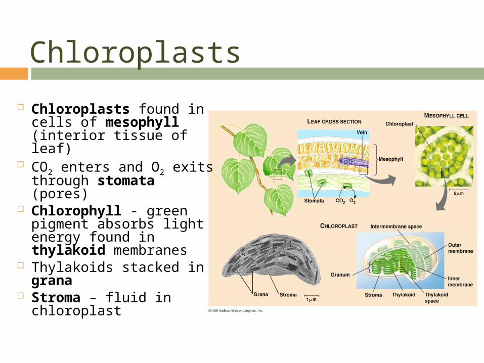
Chloroplasts
Chloroplasts found in cells of mesophyll (interior tissue of leaf)
CO2 enters and O2 exits through stomata (pores)
Chlorophyll - green pigment absorbs light energy found in thylakoid membranes
Thylakoids stacked in grana
Stroma – fluid in chloroplast

Electromagnetic Spectrum

Spectrophotometer Measures the ability for a
pigment to absorb various wavelengths of light
Directs a beam of light of different wavelengths through a solution of the pigment and measures the fraction of the light transmitted at each wavelength
Absorption spectrum plots a pigment’s light absorption vs. wavelength

ALL wavelengths are equally effective for photosynthesis
1 2 3
33%
7%
60%1. True2. False3. Cannot be
determined

The colors of light most useful in photosynthesis are
1 2 3 4
47%
0%
7%
47%1. green, yellow,
and orange2. red, violet, and
blue3. infrared, red,
and yellow4. red, white, and
blue

Big Picture: Energy Cycles All energy ultimately
comes from sun Light reactions of
photosynthesis Take place in thylakoid
Carbon fixation -all C in living things ultimately from CO2 Dark reactions of
photosynthesis Take place in stroma
Photosynthesis and cellular respiration are reverse reactions
Redox
Redox Rxns w/ transfer of e Oxidation – loss of e Reduction – gain of e Both photosynthesis and cellular respiration
use redox rxns in a series of steps Electron transport chain (ETC) breaks the
fall of electrons into several Energy releasing steps using proteins in the cell membrane

Photosystem
Photosystem – reaction center surrounded by light harvesting complexes called pigments (chlorophyll a, chlorophyll b, xanthrophyll, carotenoids, etc) Pigment – absorbs photon E transferred from
pigment to chlorophyll a rxn center Rxn Center – protein complex w/ primary e
acceptor 2 photosystems used in light reactions
Photosystem II – p680 chlorophyll a rxn center Photossystem I – p700 chlorophyll a rxn center

Light Reactions: Non-Cyclic e flow
Photons absorbed by pigments and funneled to p680 in PSII excites e to higher energy state
E is captured by primary e acceptor
Enzyme splits H2O 2e + 2 H+ + O (will form O2)
Excited e passes thru etc to rxn center in PSI
Exergonic rxns in etc provide E for synthesis of ATP
Photons excite e in p700 in PSI excites e to higher energy state
E is captured by primary e acceptor and passed down 2nd etc
NADP+ transfers 2e from etc to form NADPH

OR Light Reactions: Cyclic e flow
Uses PSI only! (not PSII) Excited e from primary e
acceptor to 1st etc produces ATP and e falls back to replace lost e from p700 No water splitting No production of NADPH Increases production of
ATP for Calvin Cycle (req. more ATP then NADPH)

Chemiosmosis
etc passes e thru carrier proteins in thylakoid membrane creating a H+ gradient (pumps H+ from stroma (pH8) into thylakoid space pH5)
Chloroplasts – photophosphorylation
ATP synthase embedded in same membrane (as H+ diffuse down gradient ADP is phosphorylated into ATP on stroma side of thylakoid)
NADPH also made on stroma side of membrane

Classwork:
Draw a flowchart (or diagram) of the light dependent reactions

Do Now
Good Morning! Grab your Clicker Take out your photosyntehsis pogil Have out your homework ws (light
reactions)

The light dependent reactions take place in the
1 2 3 4
0% 0%7%
93%1. Cloroplast
membrane2. Thylakoid3. Stroma4. Cytoplasm

NADP+ NADPH is an example of
1 2 3 4 5
40%
0%
13%13%
33%
1. Reduction2. Oxidation3. Chemiosmosis4. Photophosphorylat
ion5. None of the above

During what stage of photosynthesis is O2 produced?
1 2 3 4 5
20%
80%
0%0%0%
1. Photosystem I2. Photosystem II3. ETC4. Photophosphoryla
tion5. O2 is not
produced

The photophosphorylation of ATP is due to
1 2 3 4 5
13%
47%
0%
27%
13%
1. Chemiosmosis of H+
2. ATP synthase3. Electron
Transport Chain4. All of the above5. None of the
above

Dark Rxns: Calvin Cycle Anabolic rxns – consuming E to build
sugar C enters Calvin cycle as CO2 C exits Calvin cycle as 3C sugar called
G3P Uses ATP as E source (ATP) from light
rxns Uses NADPH as reducing agent to add
high E e (NADPH from light rxns) Phase I: Carbon Fixation
CO2 + RuBP (5C sugar) 2 3-phosphoglycerate
Phase II: Making Sugar Each 3-phosphoglycerate + P (from
ATP) 1,3-biphosphoglycerate + 2e (from NADPH) G3P
Phase III: Regenerating RuBP (5) G3P + 3ATP 3 RuBP (ready for C
fixation phase I) 3 molecules CO2 1 molecule G3P 6 molecules CO2 2 G3P (Glucose) Must go thru Calvin Cycle 6x to
make 1 molecule of glucose!

Alternate Methods of C Fixation Hot/Dry day – stomata close to prevent
water loss Stomata close – limits CO2 from entering
leaves, O2 builds up from light rxns
Photorespiration occurs on hot/dry days No ATP produced nor sugar Due to excess O2 instead of CO2 a 2C compound
is produced and rearranged and released as CO2 No advantage

Alternate Methods: C4 Plants
Minimizes Photorespiration Forms a 4C compound as 1st product
(instead of 3) Ex. Sugar cane, corn, grass Photosynthesis occurs between 2
separate cells Mesophyll cells
CO2 reacts with Phophonolpyruvate (PEP) Oxaloacetate (4C cmpd)
Bundle Sheath Cells 4C cpmd CO2 Calvin Cycle

Alternate Methods: CAM Plants
Succulent plants ex. Cacti and pinapple
Photosynthesis occurs in same cell, but at different times
Stomata open at night and close during day
Crassulacean Acid Metabolism (CAM) Calvin cycle produces organic
acids at night and mesophyll cell store in vacuoles until morning
During day light rxns supply ATP and NADPH for Calvin cycle, then CO2 is released from organic acids stored in vacuoles
Related Documents