REVIEW ARTICLE published: 28 November 2013 doi: 10.3389/fpls.2013.00482 Desiccation tolerance in resurrection plants: new insights from transcriptome, proteome, and metabolome analysis Challabathula Dinakar 1,2 and Dorothea Bartels 1 * 1 Institute of Molecular Physiology and Biotechnology of Plants, University of Bonn, Bonn, Germany 2 Department of Life Sciences, School of Basic and Applied Sciences, Central University ofTamil Nadu,Thiruvarur, India Edited by: John Moore, Stellenbosch University, South Africa Reviewed by: Tsanko Savov Gechev, University of Plovdiv, Bulgaria Xin Deng, Institute of Botany, Chinese Academy of Sciences, China *Correspondence: Dorothea Bartels, Institute of Molecular Physiology and Biotechnology of Plants, University of Bonn, Kirschallee 1, D-53115 Bonn, Germany e-mail: [email protected] Most higher plants are unable to survive desiccation to an air-dried state. An exception is a small group of vascular angiosperm plants, termed resurrection plants.They have evolved unique mechanisms of desiccation tolerance and thus can tolerate severe water loss, and mostly adjust their water content with the relative humidity in the environment. Desiccation tolerance is a complex phenomenon and depends on the regulated expression of numerous genes during dehydration and subsequent rehydration. Most of the resurrection plants have a large genome and are difficult to transform which makes them unsuitable for genetic approaches. However, technical advances have made it possible to analyze changes in gene expression on a large-scale. These approaches together with comparative studies with non-desiccation tolerant plants provide novel insights into the molecular processes required for desiccation tolerance and will shed light on identification of orphan genes with unknown functions. Here, we review large-scale recent transcriptomic, proteomic, and metabolomic studies that have been performed in desiccation tolerant plants and discuss how these studies contribute to understanding the molecular basis of desiccation tolerance. Keywords: transcriptomics, proteomics, metabolomics, resurrection plants, desiccation tolerance INTRODUCTION The sessile nature of plants has endowed them with a wide spec- trum of adaptations to combat environmental perturbations. The mechanisms to survive under various environmental fluctuations are complex and vary widely. Drought, a physiological form of water stress or deficit, affects the performance of plants and leads to low crop yield. Most of the flowering angiosperm plants are drought sensitive and have relative water contents of around 85–100% under actively growing conditions and do not survive, if the water content falls below 59–30% (Höfler et al., 1941). In this context, Arabidopsis thaliana is considered as a model to study the responses of plants toward tolerating moderate water stress and to study the genes involved in this response. Although desiccation tol- erance in seeds is common in higher plants, desiccation tolerance in vegetative tissues is restricted to the unique group of resur- rection plants (Bartels and Hussain, 2011). Several resurrection species have been extensively studied for understanding the molec- ular basis of desiccation tolerance: the bryophyte Tortula ruralis, the clubmosses Selaginella lepidophylla and Selaginella tamariscina, the dicots Craterostigma plantagineum, C. wilmsii, Boea hygromet- rica, and Myrothamnus flabellifolia, and the monocots Xerophyta viscosa, X. humilis, and Sporobolus stapfianus (Ingram and Bar- tels, 1996; Alpert and Oliver, 2002; Moore et al., 2009; Cushman and Oliver, 2011; Oliver et al., 2011a, b). The survival strate- gies often involve the re-activation of existing protection systems (Moore et al., 2009; Dinakar et al., 2012; Gechev et al., 2013). The importance of orphan genes/proteins/metabolites in the context of desiccation tolerance also needs to be considered. Most res- urrection plants are polyploid with large genomes, difficult to transform and their genome sequences are not available; due to these features a mutational approach is at present not possible for functional analysis. Desiccation tolerance is controlled by many genes or proteins, therefore, a systems biology approach com- bining transcriptomics, proteomics, and metabolomics should be informative to understand the mechanism of desiccation tol- erance, and to determine at which level of control the changes are affected. Here recent progress is reviewed on transcriptomic, proteomic, and metabolomic analyzes in resurrection plants and future prospects are discussed. “OMICS” APPROACHES TO UNDERSTAND DESICCATION TOLERANCE Recent advances in “omics” technologies have enabled quanti- tative monitoring of the abundance of biological molecules in a high-throughput manner, thus making it possible to com- pare their levels between desiccation tolerant and sensitive species. Transcriptomic, proteomic, and metabolomic approaches attempt to capture complete information on the changes in tran- scripts/proteins/metabolites that take place during desiccation and subsequent rehydration thereby giving an outline of the metabolic situation. The identification of the abundant transcripts gives an indication which metabolic processes may be important at different physiological conditions. TRANSCRIPTOME ANALYSIS Transcriptomics or mRNA expression profiling captures spatial and temporal gene expression and quantifies RNAs under dif- ferent conditions. While quantitative analysis of gene expression www.frontiersin.org November 2013 | Volume 4 | Article 482 | 1

Welcome message from author
This document is posted to help you gain knowledge. Please leave a comment to let me know what you think about it! Share it to your friends and learn new things together.
Transcript
“fpls-04-00482” — 2013/11/28 — 12:27 — page 1 — #1
REVIEW ARTICLEpublished: 28 November 2013doi: 10.3389/fpls.2013.00482
Desiccation tolerance in resurrection plants: new insightsfrom transcriptome, proteome, and metabolome analysisChallabathula Dinakar 1,2 and Dorothea Bartels 1*
1 Institute of Molecular Physiology and Biotechnology of Plants, University of Bonn, Bonn, Germany2 Department of Life Sciences, School of Basic and Applied Sciences, Central University of Tamil Nadu, Thiruvarur, India
Edited by:
John Moore, Stellenbosch University,South Africa
Reviewed by:
Tsanko Savov Gechev, University ofPlovdiv, BulgariaXin Deng, Institute of Botany, ChineseAcademy of Sciences, China
*Correspondence:
Dorothea Bartels, Institute ofMolecular Physiology andBiotechnology of Plants, University ofBonn, Kirschallee 1, D-53115 Bonn,Germanye-mail: [email protected]
Most higher plants are unable to survive desiccation to an air-dried state. An exception is asmall group of vascular angiosperm plants, termed resurrection plants. They have evolvedunique mechanisms of desiccation tolerance and thus can tolerate severe water loss, andmostly adjust their water content with the relative humidity in the environment. Desiccationtolerance is a complex phenomenon and depends on the regulated expression of numerousgenes during dehydration and subsequent rehydration. Most of the resurrection plants havea large genome and are difficult to transform which makes them unsuitable for geneticapproaches. However, technical advances have made it possible to analyze changes ingene expression on a large-scale. These approaches together with comparative studieswith non-desiccation tolerant plants provide novel insights into the molecular processesrequired for desiccation tolerance and will shed light on identification of orphan geneswith unknown functions. Here, we review large-scale recent transcriptomic, proteomic,and metabolomic studies that have been performed in desiccation tolerant plants anddiscuss how these studies contribute to understanding the molecular basis of desiccationtolerance.
Keywords: transcriptomics, proteomics, metabolomics, resurrection plants, desiccation tolerance
INTRODUCTIONThe sessile nature of plants has endowed them with a wide spec-trum of adaptations to combat environmental perturbations. Themechanisms to survive under various environmental fluctuationsare complex and vary widely. Drought, a physiological form ofwater stress or deficit, affects the performance of plants andleads to low crop yield. Most of the flowering angiosperm plantsare drought sensitive and have relative water contents of around85–100% under actively growing conditions and do not survive, ifthe water content falls below 59–30% (Höfler et al., 1941). In thiscontext, Arabidopsis thaliana is considered as a model to study theresponses of plants toward tolerating moderate water stress and tostudy the genes involved in this response. Although desiccation tol-erance in seeds is common in higher plants, desiccation tolerancein vegetative tissues is restricted to the unique group of resur-rection plants (Bartels and Hussain, 2011). Several resurrectionspecies have been extensively studied for understanding the molec-ular basis of desiccation tolerance: the bryophyte Tortula ruralis,the clubmosses Selaginella lepidophylla and Selaginella tamariscina,the dicots Craterostigma plantagineum, C. wilmsii, Boea hygromet-rica, and Myrothamnus flabellifolia, and the monocots Xerophytaviscosa, X. humilis, and Sporobolus stapfianus (Ingram and Bar-tels, 1996; Alpert and Oliver, 2002; Moore et al., 2009; Cushmanand Oliver, 2011; Oliver et al., 2011a,b). The survival strate-gies often involve the re-activation of existing protection systems(Moore et al., 2009; Dinakar et al., 2012; Gechev et al., 2013). Theimportance of orphan genes/proteins/metabolites in the contextof desiccation tolerance also needs to be considered. Most res-urrection plants are polyploid with large genomes, difficult to
transform and their genome sequences are not available; due tothese features a mutational approach is at present not possible forfunctional analysis. Desiccation tolerance is controlled by manygenes or proteins, therefore, a systems biology approach com-bining transcriptomics, proteomics, and metabolomics shouldbe informative to understand the mechanism of desiccation tol-erance, and to determine at which level of control the changesare affected. Here recent progress is reviewed on transcriptomic,proteomic, and metabolomic analyzes in resurrection plants andfuture prospects are discussed.
“OMICS” APPROACHES TO UNDERSTAND DESICCATIONTOLERANCERecent advances in “omics” technologies have enabled quanti-tative monitoring of the abundance of biological molecules ina high-throughput manner, thus making it possible to com-pare their levels between desiccation tolerant and sensitivespecies. Transcriptomic, proteomic, and metabolomic approachesattempt to capture complete information on the changes in tran-scripts/proteins/metabolites that take place during desiccation andsubsequent rehydration thereby giving an outline of the metabolicsituation. The identification of the abundant transcripts givesan indication which metabolic processes may be important atdifferent physiological conditions.
TRANSCRIPTOME ANALYSISTranscriptomics or mRNA expression profiling captures spatialand temporal gene expression and quantifies RNAs under dif-ferent conditions. While quantitative analysis of gene expression
www.frontiersin.org November 2013 | Volume 4 | Article 482 | 1
“fpls-04-00482” — 2013/11/28 — 12:27 — page 2 — #2
Dinakar and Bartels Omics studies in resurrection plants
can be done by either qRT-PCR (quantitative reverse transcrip-tase polymerase chain reaction) or by gene microarray, the mostwidely used early approach toward transcriptome analysis was thecollection of expressed sequence tags (ESTs) which is limited toa few hundred or thousand sequenced cDNAs. Recent advancesin sequencing technologies and assembly algorithms have facili-tated the reconstruction of the entire transcriptome by deep RNAsequencing (RNA-seq), even without a reference genome, there-fore, this is also applicable to resurrection plants (Table 1). Geneexpression studies and EST sequencing have been performed insome resurrection species, such as the moss T. ruralis (Scott andOliver, 1994; Wood and Oliver, 1999; Zeng et al., 2002; Oliveret al., 2004), the clubmosses Selaginella lepidophylla and Selaginellatamariscina (Zentella et al., 1999; Iturriaga et al., 2006; Liu et al.,2008), the monocot species Sporobolus stapfianus (Neale et al.,2000; Le et al., 2007), X. viscosa (Mundree et al., 2000; Mowlaet al., 2002; Lehner et al., 2008), X. humilis (Collett et al., 2003,2004; Illing et al., 2005; Mulako et al., 2008), and X. villosa (Collettet al., 2004), and the dicot species C. plantagineum (Bockel et al.,1998). In these studies, the cDNA libraries for EST sequencing wereeither generated from one or two physiological conditions (dehy-drated and rehydrated gametophytes/fronds/roots or leaves) andrestricted in number thereby not always reflecting global transcriptchanges. Comprehensive transcriptome analysis have so far beenreported for C. plantagineum and Haberlea rhodopensis (Rodriguezet al., 2010; Gechev et al., 2013).
Transcriptome sequencing of C. plantagineum and H. rhodopen-sis at four different physiological stages (control, partially dehy-drated, desiccated, and rehydrated) revealed that the overall iden-tified transcripts have highest similarity to genes of Vitis vinifera,Ricinus communis, and Populus trichocarpa. In C. plantagineum,182 MB of transcript sequences were assembled into 29,000 con-tigs which yielded more than 15,000 uniprot identities and inH. rhodopensis 96,353 expressed transcript contigs were identi-fied (Rodriguez et al., 2010; Gechev et al., 2013). EST sequencingof a cDNA library from the rehydrated moss T. ruralis resultedin the characterization of around 10,368 ESTs representing 5,563
genes (Oliver et al., 2004). An interesting feature that resultedfrom these studies is that about one-third of the contigs from C.plantagineum and around 40% of the sequences from H. rhodopen-sis and T. ruralis did not map to uniprot identities and encodeunknown transcripts which are potential sources for gene dis-covery. The transcripts can be divided into two main groupsaccording to expression patterns observed for C. plantagineum andH. rhodopensis. The first group consists of transcripts abundantin control and rehydrated tissues and the second group con-sists of transcripts abundant in dehydrated and desiccated tissues(Rodriguez et al., 2010; Gechev et al., 2013).
TRANSCRIPTS ABUNDANTLY EXPRESSED IN FULLY HYDRATEDTISSUES AND UNDER REHYDRATION CONDITIONSThe most abundant transcripts in fully hydrated and rehydratedconditions encode proteins involved in photosynthesis and car-bohydrate metabolism. In C. plantagineum and H. rhodopensis,the transcripts encoding RuBisCO activase, carbonic anhydrases,fructose bisphosphatases, chlorophyll a/b binding protein, lightharvesting complex, and RuBisCO small subunit are highlyabundant in fully hydrated conditions. The transcripts relatedto photosynthesis decline gradually upon dehydration suggestingdecreasing photosynthetic activity as the primary target duringdehydration (Rodriguez et al., 2010; Gechev et al., 2013). A similartrend in the gene expression related to photosynthesis was alsoobserved in X. viscosa (Collett et al., 2003).
Comparison of genes abundantly expressed in fully hydratedconditions indicates the presence of different pathways inC. plantagineum and H. rhodopensis. Galactinol synthases whichcatalyze the first step in the synthesis of raffinose oligosaccharidesare abundantly expressed in fully hydrated leaves of C. plan-tagineum, but in H. rhodopensis their expression is high duringdehydration and desiccation suggesting that raffinose synthe-sis is induced in H. rhodopensis at later stages of dehydration(Rodriguez et al., 2010; Gechev et al., 2013). Other abundanttranscripts in C. plantagineum encode acid phosphatases thatare required for the maintenance of cellular inorganic phosphate
Table 1 | Omics studies carried out in resurrection plants and sister group comparisons between desiccation tolerant and sensitive plants.
Approach Desiccation tolerant species Desiccation sensitive species Reference
Resurrection plants
Transcriptome analysis Craterostigma plantagineum Rodriguez et al. (2010)
Transcriptome and metabolomic analysis Haberlea rhodopensis Gechev et al. (2013)
Proteome analysis Selaginella tamariscina Wang et al. (2010)
Proteome analysis Xerophyta viscosa Ingle et al. (2007)
Proteome analysis Boea hygrometrica Jiang et al. (2007)
Proteome analysis Sporobolus stapfianus Oliver et al. (2011a)
Metabolomic analysis Selaginella lepidophylla Yobi et al. (2013)
Sister group comparisons
Metabolomic comparison Sporobolus stapfianus Sporobolus pyramidalis Oliver et al. (2011b)
EST sequencing and comparison Selaginella lepidophylla Selaginella moellendorffii Iturriaga et al. (2006)
Metabolomic comparison Selaginella lepidophylla Selaginella moellendorffii Yobi et al. (2012)
Frontiers in Plant Science | Plant Physiology November 2013 | Volume 4 | Article 482 | 2

“fpls-04-00482” — 2013/11/28 — 12:27 — page 3 — #3
Dinakar and Bartels Omics studies in resurrection plants
levels. The transcripts encoding proteins involved in ion trans-port such as membrane-associated carriers together with proteinsinvolved in cell wall plasticity and membrane integrity such asxyloglucan endotransglucosylases and members of the expansingene family are abundant in fully hydrated conditions in C. plan-tagineum (Rodriguez et al., 2010). In H. rhodopensis two of themost abundant transcripts observed in hydrated conditions arecatalase encoding genes that are not observed in C. plantagineum(Rodriguez et al., 2010; Gechev et al., 2013).
A feature that was observed in rehydrated H. rhodopensis plantsis the abundance of genes encoding an auxin efflux carrier, hypo-thetical proteins and genes with unknown functions (Gechevet al., 2013). In C. plantagineum, the abundant transcripts inrehydrated tissues are related to defense, oxidative stress, andmetabolism of vitamin K-related compounds (Rodriguez et al.,2010). Several of the transcripts expressed under rehydration con-ditions in C. plantagineum encode pathogen responsive proteins,carbohydrate metabolism associated enzymes like transketolases,enzymes related to phylloquinone metabolism which are elec-tron transfer cofactors in photosystems, and peroxidase transcriptswhich function in detoxification of reactive oxygen species (ROS;Rodriguez et al., 2010).
TRANSCRIPTS INDUCED IN MILDLY DEHYDRATED AND DESICCATEDTISSUESGenerally genes induced during dehydration code for proteinsthat prevent stress-related cellular damage and participate inantioxidant defense. Dehydration-induced transcripts encodeproteins with protective properties, enzymes related to carbo-hydrate metabolism, regulatory proteins such as transcriptionfactors, kinases, and signaling molecules as well as unknown pro-teins (Iturriaga et al., 2006; Rodriguez et al., 2010; Gechev et al.,2013).
TRANSCRIPTS ENCODING LATE EMBRYOGENESIS ABUNDANTPROTEINSLate embryogenesis abundant (LEA) genes comprise the mostabundantly up-regulated group of genes in response to dehy-dration. In H. rhodopensis some LEA genes are constitutivelyexpressed in hydrated conditions and their expression is inducedto higher levels upon drought and desiccation suggesting thatthe transcriptome of Haberlea is primed already for dehy-dration and desiccation tolerance (Gechev et al., 2013). Genesencoding LEA proteins are abundant in dehydrated leaves ofC. plantagineum but unlike Haberlea not in fully hydrated tis-sues (Piatkowski et al., 1990; Michel et al., 1994; Rodriguez et al.,2010). LEA proteins are localized in different cellular compart-ments such as cytosol, chloroplasts, mitochondria, or nuclei.Dehydration-induced expression of LEA transcripts is a commonresponse in resurrection plants as well as in desiccation sensitiveplants (Bartels et al., 1990; Collett et al., 2004; Oliver et al., 2004;Rodriguez et al., 2010; Gechev et al., 2012, 2013). The major dif-ference between desiccation tolerant and sensitive plants seemsto be in the abundance of the transcripts. As an example, theLEA-like CDeT11-24 transcript is highly expressed during desic-cation in C. plantagineum, whereas the transcript is expressed ata low level in desiccation sensitive Lindernia subracemosa plants.
This suggests that the CDeT11-24 transcript in L. subracemosa isunstable or induced at a lower rate during dehydration. Com-parative promoter analysis of the CDeT11-24 gene between C.plantagineum and L. brevidens showed different promoter activi-ties and the absence of dehydration specific promoter cis elementsin L. brevidens, which correlates with the transcript expressionlevel (van den Dries et al., 2011). These results lead to the hypoth-esis that high level gene expression under dehydration is evolvedby selection of promoter cis elements.
TRANSCRIPTS ENCODING PROTEINS RELATED TO DETOXIFICATIONAND ANTIOXIDANT DEFENSEReactive oxygen species such as superoxide, hydrogen peroxide,and hydroxyl radicals are unavoidable by-products of aerobicmetabolism and are commonly generated during dehydrationstress. Since ROS can potentially damage proteins, lipids, andnucleic acids, genes encoding antioxidant enzymes are supposedto be up-regulated in resurrection plants. Transcriptome analy-sis confirmed an up-regulation of genes involved in antioxidativedefense. In hydrated conditions resurrection plants maintain highlevels of antioxidants, which increase upon stress. This feature isobserved in all resurrection species studied so far (Sherwin andFarrant, 1998; Farrant, 2000; Kranner et al., 2002). The transcrip-tome of H. rhodopensis contained an extensive antioxidant genenetwork in the fully hydrated state. The number of expressed genesencoding superoxide dismutases, catalases, monodehydroascor-bate reductases, and glutathione (GSH) reductases are higher in H.rhodopensis than in C. plantagineum in the hydrated state. Expres-sion of these genes is even further up-regulated during dehydrationin H. rhodopensis (Gechev et al., 2013). Induction of genes relatedto the antioxidant pathway during desiccation has also beenreported in the desiccation tolerant plant Sporobolus stapfianus(Neale et al., 2000). In X. humilis, a plant that looses chlorophyllduring desiccation, also a large number of antioxidant defensegenes are up-regulated during dehydration (Collett et al., 2004).Besides conserved antioxidant genes some resurrection plantsacquired expression of genes from other pathways. An examplewas reported for X. viscosa, in which the desiccation-inducedantioxidant gene encoding 1-Cys peroxiredoxin (XvPer1) shows70% sequence identity to Arabidopsis seed specific dormancyrelated 1-Cys peroxiredoxin (AtPer1; Haslekás et al., 1998; Ndimaet al., 2001; Mowla et al., 2002). In the C. plantagineum tran-scriptome analysis thiamine biosynthesis transcripts and aldehydedehydrogenase (CpALDH) transcripts are up-regulated duringdehydration and contribute to antioxidant defense (Rodriguezet al., 2010). In T. ruralis the transcript levels of an aldehyde dehy-drogenase (ALDH21A1) were also increased during dehydrationsuggesting ALDH as a stress regulated enzyme with the potential todetoxify excess amounts of aldehydes (Chen et al., 2002; Stiti et al.,2011). Transgenic Arabidopsis plants over-expressing AtALDH3showed tolerance to dehydration stress and low accumulation ofmalondialdehyde thereby emphasizing the importance of aldehydedehydrogenase in conferring tolerance to oxidative stress (Sunkaret al., 2003).
Another group of transcripts that are induced during dehy-dration/desiccation encode early light induced proteins (ELIPs).These ELIPs are nuclear-encoded proteins associated with
www.frontiersin.org November 2013 | Volume 4 | Article 482 | 3
“fpls-04-00482” — 2013/11/28 — 12:27 — page 4 — #4
Dinakar and Bartels Omics studies in resurrection plants
thylakoid membranes in the chloroplast and are believed tobind to chlorophyll thus preventing ROS-induced photooxida-tive damage. Several desiccation-related genes encoding ELIPshave been isolated from C. plantagineum. DSP-22 a homologof a class of ELIPs is thought to stabilize photosynthetic struc-tures within the chloroplasts of C. plantagineum and amelioraterehydration-induced damage (Bartels et al., 1992; Ingram and Bar-tels, 1996). In C. plantagineum, the dsp-22 transcript is undetectedin unstressed plants but abundantly expressed during desiccationin light (Alamillo and Bartels, 1996). ELIPs are also abundantlyexpressed during dehydration in H. rhodopensis (Gechev et al.,2013). In Sporobolus stapfianus induction of transcripts encod-ing ELIPs was observed only in desiccation tolerant tissues but notin desiccation sensitive tissues, which supports the role for ELIPsin desiccation tolerance (Neale et al., 2000).
TRANSCRIPTS ENCODING ENZYMES RELATED TO CARBOHYDRATEMETABOLISMAccumulation of sucrose and raffinose oligosaccharides is com-monly observed in resurrection plants (Bartels and Hussain,2011). Their accumulation correlates with the up-regulationof transcripts encoding enzymes of carbohydrate metabolism.In C. plantagineum and H. rhodopensis, several genes encod-ing galactinol synthases and a stachyose synthase are present inhydrated leaves and are additionally up-regulated during dehy-dration (Rodriguez et al., 2010; Gechev et al., 2013) suggestingthe importance of oligosaccharides in protecting cells during des-iccation. Similarly, the cDNAs encoding enzymes of the polyolbiosynthesis and raffinose family oligosaccharides are abundantlyexpressed upon dehydration in X. humilis (Illing et al., 2005). Ahigh induction is observed for several sucrose synthases, sucrose6-phosphate synthases, a sucrose transporter, and a sucroseresponsive element-binding protein in H. rhodopensis duringdehydration and desiccation (Gechev et al., 2013). Induction oftranscripts encoding sucrose synthases and sucrose-phosphatesynthases is also observed in C. plantagineum during dehydra-tion (Ingram and Bartels, 1996; Kleines et al., 1999). The roleof sugar metabolism for the adaptation of C. plantagineum andH. rhodopensis to desiccation is further substantiated by the pres-ence of transketolase transcripts (tkt). Transketolases are keyenzymes of the reductive and oxidative pentose phosphate path-ways that are responsible for the synthesis of sugar phosphateintermediates, which can flow into different pathways. C. plan-tagineum has three transketolase isoforms (tkt3, tkt7, tkt10) andit has been suggested that a transketolase isoform is involvedin octulose synthesis (Willige et al., 2009). While tkt7 is moreabundant in rehydrating tissues of C. plantagineum, tkt10 is pref-erentially expressed in fully hydrated tissues. Tkt3 is constitutivelyexpressed and is involved in the Calvin cycle (Bernacchia et al.,1995; Rodriguez et al., 2010). Compared to C. plantagineum,two tkt are observed in H. rhodopensis, which differs in itscarbohydrate metabolism from C. plantagineum (Gechev et al.,2013).
TRANSCRIPTS RELATED TO CELL WALL MODIFICATIONSeveral reversible modifications occur to stabilize cell wallarchitecture in resurrection plants, while some of these changes are
constitutive some are inducible. Cell wall folding plays an impor-tant role in mechanical stabilization during desiccation (Farrantet al., 2007). The modifications in cell wall properties to resist themechanical stress during dehydration in resurrection plants havebeen discussed by Vicré et al. (2004) and Moore et al. (2006, 2008,2013). Plasticity of cell walls is particularly important to avoiddamage due to mechanical stress imposed during desiccation andrehydration (Moore et al., 2013). Transcripts encoding proteinsinvolved in the maintenance of cell wall plasticity such as xyloglu-can endotransglucosylases are abundantly expressed in unstressedtissues of C. plantagineum (Rodriguez et al., 2010). Concomitantwith the increased cell wall extensibility transcripts encoding alphaexpansins are up-regulated during desiccation in C. plantagineum(Jones and McQueen-Mason, 2004). In H. rhodopensis, genesencoding xyloglucan endotransglucosylases, pectin esterases, andpectate lyases are abundantly expressed in hydrated conditions andare switched off during dehydration. Concomitantly, laccase genesinvolved in lignin biosynthesis are only expressed in desiccatedtissues pointing toward cell wall remodeling during desiccation(Gechev et al., 2013).
TRANSCRIPTS ENCODING REGULATORY MOLECULES ANDPARTICIPATING IN GENERAL METABOLISM ARE UP-REGULATEDDURING DEHYDRATIONThe transcriptome of desiccated tissues also includes genes thatare either involved in general metabolism or that are relatedto environmental stresses other than dehydration. Examples aretemperature-induced lipocalins, aquaporins, tonoplast intrin-sic proteins, cation transporters, and rare cold inducible 2Aprotein. Lipocalins are membrane-associated proteins with alow-temperature response element, dehydration-responsive ele-ments, and heat shock elements in their promoters suggest-ing their involvement in various abiotic stress conditions. AYSIRK signaling peptide, an alcohol oxidase, and genes encodingheat shock proteins are expressed in desiccated H. rhodopen-sis leaves. The occurrence of these types of transcripts impliesthat these genes have become dehydration-responsive duringevolution.
The massive expression of transcripts during dehydrationand the re-synthesis during rehydration requires a fine-tunedregulatory network. This is reflected by the fact that tran-scripts with a regulatory function comprise a large heterogenousgroup. Transcripts of desiccated C. plantagineum are domi-nated by those encoding DNA binding proteins, cysteine pro-teases, and proteins of amino acid metabolism. These tran-scripts are mostly members of gene families which participatein diverse pathways. During evolution some family membershave acquired regulatory cis elements which trigger their expres-sion in response to dehydration. In H. rhodopensis induc-tion in gene expression upon dehydration was observed for agene encoding a putative protein phosphatase/hydrolase whichwas not detected in hydrated/rehydrated samples indicating theimportance of phosphorylation and dephosphorylation duringdehydration (Gechev et al., 2013). Genes coding for transcrip-tion factors, heat shock proteins, and components of signalingcascades are among the transcripts expressed in response todehydration in all resurrection plants. In the transcriptome of
Frontiers in Plant Science | Plant Physiology November 2013 | Volume 4 | Article 482 | 4
“fpls-04-00482” — 2013/11/28 — 12:27 — page 5 — #5
Dinakar and Bartels Omics studies in resurrection plants
H. rhodopensis, a broad range of transcription factors have beenidentified such as MYB, NAC, WRKY, GRAS family members,DREB2, NF-YA, MADS-box transcription factors, and severalheat shock transcription factors (Gechev et al., 2013). Some ofthe regulatory transcripts are exclusively expressed in desiccatedsamples, e.g., a receptor like protein kinase, kinases, phos-phatases, a Ca2+ antiporter cation exchanger, and a phospholipaseD isoform.
The few examples cited above show that the transcriptome anal-ysis provides a catalog of the regulatory genes that are up-regulatedduring dehydration, but at present it is not understood how thesegenes interact with other pathways and which target genes theyregulate.
PROTEOME ANALYSISTranslational regulation of mRNA is an important step in thecontrol of gene expression. Changes in gene expression atthe transcript level need not always correspond to changes inthe protein level due to either transcript instability or post-transcriptional modifications. Post-translational modificationsand protein degradation modulate the quality and quantity ofexpressed proteins and thus affect the correlation of transcriptand protein levels. The main limitation of proteomics is the iden-tification of the proteins due to absence of genome sequenceinformation in resurrection plants. Therefore, functions haveto be attributed according to homologies. Reports on proteomeanalysis in resurrection plants are restricted to a few species.A direct correlation between transcript and protein abundancewas observed for many of the dehydration-induced gene prod-ucts in particular for gene products with protective functions(Ingle et al., 2007; Jiang et al., 2007; Wang et al., 2010; Oliver et al.,2011a).
Qualitatively proteome data correlate with transcript data andconfirm that the abundant proteins in the hydrated tissues arerelated to photosynthesis and carbohydrate metabolism. Proteomedata demonstrated that the deactivation of photosynthetic activ-ity and subsequent re-activation are major responses observedupon sensing dehydration and after rewatering, respectively (Ingleet al., 2007; Rodriguez et al., 2010; Wang et al., 2010; Oliver et al.,2011a). The decline of photosynthesis coincided with the decreasein chloroplast-localized photosynthetic proteins such as psbO,psbP (the two components of luminal oxygen evolving complexof PSII), the PSII stability factor HCF136, the α subunit of theF-ATPase, and the Calvin cycle enzyme transketolase in X. vis-cosa during dehydration at 35% relative water content (Ingle et al.,2007). Similarly photosynthesis-related proteins that decreased inabundance in Selaginella tamariscina during dehydration includedRuBisCO large sub unit, chlorophyll a/b binding protein, andoxygen evolving complex protein (Wang et al., 2010).
During dehydration, LEA proteins accumulate abundantly inresurrection plants supporting their protective roles (Michel et al.,1994; Velasco et al., 1994; Alamillo and Bartels, 1996; Ingram andBartels, 1996; Ndima et al., 2001). Using two dimensional SDS-PAGE coupled with a phosphoprotein specific stain at least twoLEA proteins CDeT11-24 and CDeT6-19 were shown to be phos-phorylated in C. plantagineum (Röhrig et al., 2006). Although therole of the protein phosphorylation in these two proteins is still
unclear, phosphorylation may increase the hydrophilic residuesnecessary for interaction with other macromolecules or phos-phorylation may be required for correct subcellular localization,as it was shown for maize embryo LEA proteins (Goday et al.,1988).
Proteome analysis also revealed the expression of unknownproteins in resurrection plants. In Selaginella tamariscina, 138dehydration-responsive protein spots representing 103 uniqueproteins with unknown functions were identified (Wang et al.,2010). The proteins down-regulated in Selaginella tamariscinaduring dehydration included proteins involved in photosynthesis,carbohydrate and energy metabolism, stress and defense proteins,signaling, membrane transport, cell structure, and cell division.The protein abundance increased for antioxidant enzymes (Wanget al., 2010). From B. hygrometrica leaves more than 200 proteinswere analyzed out of which 78 (35%) increased in expressionin response to dehydration and 5% were induced in rehydratedleaves and 60% showed decreased or unchanged levels (Jianget al., 2007). Several of the proteins related to antioxidant andenergy metabolism are constitutively expressed, indicating thatprotective mechanisms exist constitutively which emphasize thepreparedness of the plant for stress. Dehydration-induced pro-teins in B. hygrometrica are associated with energy metabolism,GSH and polyphenol metabolism. This indicates that GSH mayserve as a major antioxidant in B. hygrometrica. Protein analysisalso indicated degradation of photosynthesis-related proteins. A20-kDa fragment of the RuBisCO large subunit (RbcL) and a 23-kDa polypeptide of the oxygen evolving complex of photosystemII were identified in dehydrated leaf proteins. The appearance ofthe 20-kDa RbcL protein fragment in B. hygrometrica is thoughtto be the result of stress-induced proteolysis mediated throughROS-induced chloroplast-localized protease activity (Jiang et al.,2007). ABC transporters that mediate ATP-dependent transportof solutes were also induced during dehydration in B. hygro-metrica (Jiang et al., 2007). The induction of putative ATPasesubunits matching a vacuolar H+-ATPase A subunit during dehy-dration may help in preparation for rehydration. Desiccated leavesof Sporobolus stapfianus and X. viscosa showed similar proteinprofiles as B. hygrometrica (Blomstedt et al., 1998; Marais et al.,2004). Enzymes related to sugar metabolism, such as sucrosesynthase, ADP-glucose pyrophosphorylase, and GDP-mannose3,5-epimerase were up-regulated during dehydration confirmingthe importance of sugar metabolism.
The protein expression patterns observed in different res-urrection plants lead to the conclusion that stress protectiveproteins are rapidly and massively induced upon dehydration andpresent throughout desiccation. The induced proteins are involvedin diverse functions such as scavenging ROS, accumulation ofsucrose, protective proteins, cell wall remodeling proteins, andproteins with unknown functions.
There are examples in which mRNA levels do not correlatewith protein expression patterns. Although the transcript levelsof tkt3 are constitutively expressed in C. plantagineum vegetativetissues, the protein levels are higher in hydrated tissues, whichsuggest a high translation rate or slower protein turnover duringhydrated conditions. Similarly, the abundance of tkt7 mRNA dur-ing late phases of rehydration in C. plantagineum does not match
www.frontiersin.org November 2013 | Volume 4 | Article 482 | 5
“fpls-04-00482” — 2013/11/28 — 12:27 — page 6 — #6
Dinakar and Bartels Omics studies in resurrection plants
the protein abundance (Bernacchia et al., 1995). This may alsobe true for regulatory genes like transcription factors, which areoften difficult to investigate due to the low abundance of theseproteins.
ENZYME ACTIVITIESMany of the stress-induced proteins are enzymes and thus mea-suring their enzyme activities indicates whether their activities aremaintained despite dehydration. Enzymes such as those involvedin antioxidant synthesis, carbohydrate and nitrogen metabolismshowed high enzymatic activities during dehydration/desiccation.This confirms that the protein activities are not affected by dehy-dration and are protected, e.g., by LEA proteins (Illing et al.,2005; Farrant et al., 2007; Petersen et al., 2012). In X. viscosa, theactivities of ascorbate peroxidase, GSH reductase, and superoxidedismutase increased during dehydration and declined during rehy-dration whereas in C. wilmsii the activities of GSH reductase andsuperoxide dismutase increased upon rehydration (Sherwin andFarrant, 1998) indicating diversity of defense pathways in the poik-ilochlorophyllous monocot plant and the homoiochlorophyllousdicot plant.
Sucrose accumulation correlates with the up-regulation ofcarbohydrate metabolic enzymes in desiccated tissues. Duringdehydration, increased hexokinase activity is correlated withsucrose accumulation and the decline in glucose and fructoselevels in both Sporobolus stapfianus and X. viscosa (Whittakeret al., 2001). Sucrose-phosphate synthase activity in leaves ofC. plantagineum and Sporobolus stapfianus increases along withsucrose accumulation during dehydration indicating the redirec-tion of carbon flow to sucrose from reserve substances such asstarch and octulose (Whittaker et al., 2007). In Sporobolus stap-fianus, increases in hexose sugars, sucrose, and amino acidsare associated with concomitant increases in sucrose-phosphatesynthase and pyruvate kinase (PK) activities, and maximalactivity levels of phosphoenolpyruvate carboxylase (PEPCase),NADP-dependent isocitrate dehydrogenase (NADP-ICDH),and NADH-dependent glutamate synthase (NADH-GOGAT;Whittaker et al., 2007).
METABOLOMIC ANALYSISMetabolomic approaches deal with quantitative analysis of smallmolecules giving a detailed analysis of the plant’s metabolicstate. Because of the large variety of chemical structures andthe different biochemical properties of molecules, no singletechnique and no single metabolite extraction protocol willidentify all metabolites in a plant cell. Several approaches includ-ing gas chromatography–mass spectrometry (GC–MS), liquidchromatography (LC)–MS, capillary electrophoresis (CE)–MS,and nuclear magnetic resonance (NMR) spectroscopy are com-monly used in plant metabolomics research. Although large-scalemetabolomic studies have been reported for Arabidopsis plantsundergoing dehydration (Urano et al., 2009), metabolomic studieshave so far only been reported for two closely related Sporobo-lus and Selaginella species which differ in desiccation tolerance(Oliver et al., 2011b; Yobi et al., 2012). Metabolite changes dur-ing dehydration/rehydration include carbohydrates, amino acids,nucleotide derivatives, lipids, polyamines, antioxidants, and
defense compounds. Several key metabolites have been identi-fied from the metabolome studies in resurrection plants and aredescribed below.
The metabolic profile of fully hydrated Selaginella lepido-phylla is distinct from the dehydration state. The mapping of theknown metabolites (66.5%) into the general biochemical path-ways revealed that the more prevalent metabolites are amino acids(19%) followed by carbohydrates, lipids, cofactors, nucleotides,peptides, and secondary metabolites. While the amino acids,peptides, and nucleotide metabolites are overrepresented duringdesiccation, carbohydrates such as 4C–6C containing sugars, sugaralcohols, lipids, or lipid metabolites (with the exception of cholinephosphate), and cofactors are overrepresented in the hydratedstate (Yobi et al., 2013). Besides the known 251 metabolites thatwere identified in Selaginella lepidophylla, 33.4% (84) representedunknown metabolites. Seven of the unknown metabolites showedgreater abundance under dehydration conditions than in hydratedconditions suggesting their role in desiccation tolerance. Sporobo-lus stapfianus is metabolically primed for dehydration as it containshigh concentrations of osmolytes and nitrogen metabolites alongwith low levels of metabolites associated with energy metabolismin the hydrated state. Upon dehydration, the metabolism isshifted toward antioxidant production, carbohydrate produc-tion, nitrogen remobilization, and ammonia detoxification (Oliveret al., 2011b). This appears to be different in C. plantagineumwhere major metabolite changes are induced by dehydration(see below).
CARBOHYDRATESCarbohydrate metabolism plays a critical role in cellular pro-tection in resurrection plants. Some carbohydrate metaboliteschange drastically in abundance during dehydration and rehy-dration. Table 2 displays a summary of the main carbohydratechanges observed in various resurrection plants. Accumulationof sucrose, trehalose, and short-chain oligosaccharides such asraffinose has been observed during dehydration (Bianchi et al.,1991b; Drennan et al., 1993; Norwood et al., 2000; Peters et al.,2007; Table 2). Sucrose is the major carbohydrate in tissues ofangiosperm resurrection plants upon desiccation (Bianchi et al.,1991b; Ghasempour et al., 1998; Norwood et al., 2000; Scott, 2000;Whittaker et al., 2001; Cooper and Farrant, 2002; Zivkovic et al.,2005). The importance of sucrose is demonstrated in a compara-tive metabolic analysis of desiccation tolerant Eragrostis nindensisand desiccation sensitive Eragrostis species. While E. nindensisaccumulates sucrose in desiccated leaves, desiccation sensitivespecies of Eragrostis do not accumulate sucrose (Illing et al., 2005).Although trehalose is a critical key compound of some lower orderdesiccation tolerant organisms and yeast, it is a minor componentin a few desiccation tolerant angiosperm species (Ghasempouret al., 1998).
In C. plantagineum and C. wilmsii an extremely high con-centration of the unusual C8 sugar, 2-octulose accumulates inwell watered leaves (Bianchi et al., 1992; Norwood et al., 2000;Cooper and Farrant, 2002) and upon dehydration, the level ofthis sugar declines and the concentration of sucrose increases(Bianchi et al., 1991b). This conversion accounts for up to 80% ofthe carbohydrates in desiccated leaves of Craterostigma. According
Frontiers in Plant Science | Plant Physiology November 2013 | Volume 4 | Article 482 | 6
“fpls-04-00482” — 2013/11/28 — 12:27 — page 7 — #7
Dinakar and Bartels Omics studies in resurrection plants
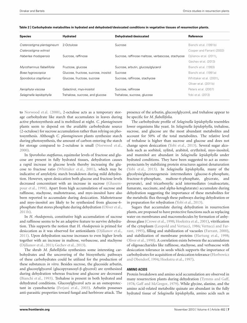
Table 2 | Carbohydrate metabolites in hydrated and dehydrated/desiccated conditions in vegetative tissues of resurrection plants.
Species Hydrated Dehydrated/desiccated Reference
Craterostigma plantagineum 2-Octulose Sucrose Bianchi et al. (1991b)
Craterostigma wilmsii Cooper and Farrant (2002)
Haberlea rhodopensis Sucrose, raffinose Sucrose, raffinose maltose, verbascose, stachyose Djilianov et al. (2011),
Gechev et al. (2013)
Myrothamnus flabellifolia Fructose, glucose Sucrose, arbutin, glucosylglycerol Bianchi et al. (1993)
Boea hygroscopica Glucose, fructose, sucrose, inositol Sucrose Bianchi et al. (1991a)
Sporobolus stapfianus Glucose, fructose, sucrose Sucrose, raffinose, stachyose Whittaker et al. (2001),
Oliver et al. (2011b)
Xerophyta viscosa Galactinol, myo-inositol Sucrose, raffinose Peters et al. (2007)
Selaginella lepidophylla Trehalose, sucrose, and glucose Trehalose, sucrose, glucose Yobi et al. (2013)
to Norwood et al. (2000), 2-octulose acts as a temporary stor-age carbohydrate like starch that accumulates in leaves duringactive photosynthesis and is mobilized at night. C. plantagineumplants seem to depend on the available carbohydrate source(2-octulose) for sucrose accumulation rather than relying on pho-tosynthesis. Although C. plantagineum plants synthesize starchduring photosynthesis, the amount of carbon entering the starchfor storage compared to 2-octulose is small (Norwood et al.,2000).
In Sporobolus stapfianus, equimolar levels of fructose and glu-cose are present in fully hydrated tissues, dehydration causesa rapid increase in glucose levels thereby increasing the glu-cose to fructose ratio (Whittaker et al., 2001), which may beindicative of amylolytic starch breakdown during mild dehydra-tion. However, upon desiccation both glucose and fructose levelsdecreased concomitant with an increase in sucrose (Ghasem-pour et al., 1998). Apart from high accumulation of sucrose andraffinose, stachyose, maltotetraose, and myo-inositol have alsobeen reported to accumulate during desiccation. Maltotetraoseand myo-inositol are likely to be synthesized from glucose-6-phosphate that stores phosphate during dehydration (Oliver et al.,2011b).
In H. rhodopensis, constitutive high accumulation of sucroseand raffinose seems to be an adaptive feature to survive dehydra-tion. This supports the notion that H. rhodopensis is primed fordesiccation as it was observed for antioxidants (Djilianov et al.,2011). Upon dehydration sucrose increases to even higher levelstogether with an increase in maltose, verbascose, and stachyose(Djilianov et al., 2011; Gechev et al., 2013).
The shrub M. flabellifolia synthesizes some interesting car-bohydrates and the uncovering of the biosynthetic pathwaysof these carbohydrates could be utilized for the production ofthese substances in vitro. Besides sucrose, the glucoside arbutin,and glucosylglycerol (glucopyranosyl-β-glycerol) are synthesizedduring dehydration whereas fructose and glucose are decreased(Bianchi et al., 1993). Trehalose is present in both hydrated anddehydrated conditions. Glucosylglycerol acts as an osmoprotec-tant in cyanobacteria (Ferjani et al., 2003). Arbutin possessesanti-parasitic properties toward fungal and herbivore attack. The
presence of the arbutin, glucosylglycerol, and trehalose appear tobe specific for M. flabellifolia.
The carbohydrate profile of Selaginella lepidophylla resembleslower organisms like yeast. In Selaginella lepidophylla, trehalose,sucrose, and glucose are the most abundant metabolites andaccount for 50% of the total metabolites. The relative levelof trehalose is higher than sucrose and glucose and does notchange upon desiccation (Yobi et al., 2013). Several sugar alco-hols such as sorbitol, xylitol, arabitol, erythritol, myo-inositol,and mannitol are abundant in Selaginella lepidophylla underhydrated conditions. They have been suggested to act as osmo-protectants by stabilizing protein structures against denaturation(Yobi et al., 2013). In Selaginella lepidophylla, many of theglycolysis/gluconeogenesis intermediates (glucose-6-phosphate,fructose-6-phosphate, maltose-6-phosphate, glycerate, andpyruvate), and tricarboxylic acid intermediates (oxaloacetate,fumarate, succinate, and alpha-ketoglutarate) accumulate duringdehydration suggesting the importance of these metabolites forthe metabolic flux through these pathways during dehydration orin preparation for rehydration (Yobi et al., 2013).
Sugars that accumulate during dehydration in resurrectionplants, are proposed to have protective functions such as replacingwater on membranes and macromolecules by formation of anhy-drous glass (Crowe et al., 1998; Hoekstra et al., 2001), vitrificationof the cytoplasm (Leopold and Vertucci, 1986; Vertucci and Far-rant, 1995), filling and stabilization of vacuoles (Farrant, 2000),and stabilization of membrane proteins (Hartung et al., 1998;Oliver et al., 1998). A correlation exists between the accumulationof oligosaccharides like raffinose, stachyose, and verbascose withdesiccation tolerance in seeds which supports the importance ofcarbohydrates for acquisition of desiccation tolerance (Horbowiczand Obendorf, 1994; Hoekstra et al., 1997).
AMINO ACIDSProtein breakdown and amino acid accumulation are observed inmany resurrection plants during dehydration (Tymms and Gaff,1978; Gaff and McGregor, 1979). While glycine, alanine, and theamino acid-related metabolite quinate are abundant in the fullyhydrated tissue of Selaginella lepidophylla, amino acids such as
www.frontiersin.org November 2013 | Volume 4 | Article 482 | 7
“fpls-04-00482” — 2013/11/28 — 12:27 — page 8 — #8
Dinakar and Bartels Omics studies in resurrection plants
glutamine, glutamate, arginine, aspartate, citrulline, asparagine,N-6-trimethyllysine, trans-4-hydroxyproline, as well as the inter-mediate metabolites 3-(3-hydroxyphenyl)propionate, the tripep-tide ophthalmate (L-Y-glutamyl-L-α-aminobutyrylglycine) areprominent in desiccated tissues (Yobi et al., 2013). Similarly inSporobolus stapfianus, amino acids such as asparagine, arginine,glutamate, glutamine, and the amino acid precursor quinate accu-mulated during desiccation (Martinelli et al., 2007; Oliver et al.,2011b). These amino acids could function as compatible solutesor as mobile nitrogen reserves for the rehydrating tissues. InSelaginella lepidophylla citrulline, a non-proteinogenic amino acidand a structural analog of arginine, is the only amino acid that ismore abundant in desiccated tissues than in hydrated tissues. InC. plantagineum, amino acid composition did not change signif-icantly during dehydration/rehydration (Bianchi et al., 1992). Ina number of species, γ-glutamyl amino acids accumulate duringdesiccation (Oliver et al., 2011b; Yobi et al., 2013). Addition ofglutamyl residues to amino acids prevents their degradation. Theamino acid derivative 3-(3-hydroxyphenyl) propionate is moreabundant in dehydration and desiccation states in Selaginellalepidophylla than in the hydrated tissue. Several aromatic aminoacids such as tryptophan and the derivatives acetyltryptophan orphenylalanine which serve as biosynthetic precursors for primaryand secondary metabolites accumulate in Sporobolus stapfianusand Selaginella lepidophylla during dehydration (Oliver et al.,2011b; Yobi et al., 2012). The amino acids phenylalanine andtyrosine accumulate during dehydration in vegetative tissues ofH. rhodopensis suggesting the activation of the shikimate path-way (Gechev et al., 2013) which can result in the synthesis ofantioxidants.
NUCLEOTIDE METABOLITESBesides amino acids, the relative abundance of other nitrogen-richmetabolites within the purine and pyrimidine nucleotide pathwaysis altered during the dehydration/rehydration cycle in Selaginellalepidophylla (Yobi et al., 2013). Some nucleotides [e.g., allan-toin, 1-methyladenosine, and uridine 5′-monophosphate (UMP)]were more abundant in desiccated tissues than in hydrated tis-sues. Inosine, a purine nucleoside containing the purine-basehypoxanthine and the sugar D-ribose, accumulated during desic-cation in Selaginella lepidophylla, whereas 2′-deoxyadenosine wasmore abundant in fully hydrated Selaginella lepidophylla. Theseresults are similar to those observed for Sporobolus stapfianus,in which allantoin increased during dehydration (Oliver et al.,2011b).
LIPIDSMaintenance of membrane integrity plays a pivotal role in des-iccation tolerance (Hoekstra et al., 2001) and therefore, changesin lipid compositions are essential for desiccation tolerance. Adetailed lipid analysis was recently reported for C. plantagineumand some closely related species (Gasulla et al., 2013). Althoughthe total lipid content remained constant during desiccation,remarkable changes in lipid composition were observed in C.plantagineum and L. brevidens (Gasulla et al., 2013). An interest-ing observation was the removal of monogalactosyldiacylglycerol(MGDG) from the thylakoid membranes during desiccation.
MGDG was either converted to digalactosyldiacylglycerol or it washydrolyzed and the resulting diacylglycerol was used for phospho-lipid and triacylglycerol production (Gasulla et al., 2013). Accu-mulation of phosphatidylinositol is a specific response observedin desiccation tolerant C. plantagineum and L. brevidens but not indesiccation sensitive plants suggesting its importance for desicca-tion tolerance (Gasulla et al., 2013). A common response observedin plants is the increase of phosphatidic acid upon dehydration.Phosphatidic acid is considered to be a signaling molecule whichmay regulate down-stream processes. The increase in phosphaticacid is due to the activation of phospholipase D under desicca-tion in C. plantagineum (Frank et al., 2000). Lysolipids and fattyacids which are natural products formed by the hydrolysis ofphospholipids are accumulated during dehydration. In Sporobolusstapfianus, accumulation of lysolipids suggests the scope for mini-mal damage to lipid membranes during dehydration (Oliver et al.,2011b). In Selaginella lepidophylla out of the 32 lipid metabolites,only choline phosphate accumulated in response to dehydration.Membranes are protected during dehydration in Selaginella lepido-phylla as polyunsaturated fatty acids and markers of lipoxygenaseactivity increased (Yobi et al., 2013). A decrease in the relativeamounts of various unsaturated lipids upon dehydration has beenreported for the desiccation tolerant Sporobolus stapfianus (Quar-tacci et al., 1997) and Ramonda serbica (Quartacci et al., 2002).These alterations in unsaturated fatty acid concentrations are sup-posed to contribute to membrane fluidity to allow for recoveryafter dehydration (Upchurch, 2008).
POLYAMINESPolyamines are low molecular weight polycationic compoundsinvolved in cellular processes, such as membrane stabilization,enzyme activity modulation, plant growth and development,nitrogen assimilation, and respiratory metabolism (Alcázar et al.,2010). Polyamines are able to bind to negatively charged moleculeslike DNA, proteins, or membrane phospholipids and thus areable to protect these macromolecules. A positive correlationexists between the abundance of polyamine levels and stresstolerance in plants (Alcázar et al., 2010). In agreement withthis, putrescine, spermidine, and spermine were found to beat higher levels after drought treatment in C. plantagineumthan in Arabidopsis putrescine levels along with spermidineand spermine increased after 96 h of dehydration in C. plan-tagineum (Alcázar et al., 2011). This implies an early droughtresponse phenomenon in C. plantagineum and indicates that stim-ulation in spermidine and spermine biosynthesis occurs withconcomitant reduction of putrescine precursor levels. It sug-gests a metabolic canalization of putrescine to spermine in C.plantagineum. In drought sensitive Arabidopsis, although theputrescine to spermine canalization occurs under dehydration,the spermidine and spermine levels do not increase and this isdue to conversion of spermine to putrescine thereby forming apolyamine recycling-loop during drought acclimation (Alcázaret al., 2011).
ANTIOXIDANTSMost of the cellular damage occurs through the activity ofROS (Smirnoff, 1993). ROS are particularly prevalent during
Frontiers in Plant Science | Plant Physiology November 2013 | Volume 4 | Article 482 | 8
“fpls-04-00482” — 2013/11/28 — 12:27 — page 9 — #9
Dinakar and Bartels Omics studies in resurrection plants
desiccation of photosynthetic tissues because chlorophyll retainsthe ability to transfer electrons while carbon fixation does nottake place. Under light conditions a flow of electrons and energyis passed from chlorophyll molecules to ground state oxygenthus generating singlet oxygen. To detoxify the ROS plant cellscontain a wide array of reactive oxygen scavenging antioxidantenzymes (superoxide dismutase, catalase, ascorbate peroxidase,etc.) along with low molecular weight antioxidants. Both desicca-tion sensitive and tolerant plants up-regulate antioxidant synthesisto detoxify ROS upon dehydration. A major difference betweenresurrection plants and desiccation sensitive plants appears tobe in the ability to maintain the antioxidant levels during thelater stages of desiccation where oxidative stress prevails (Mowlaet al., 2002; Illing et al., 2005). The importance of low molecularweight antioxidants (ascorbate and GSH), tocopherols, pheno-lic acids, or polyphenols (galloylquinic acid) has been deducedfrom metabolic analyzes (Kranner et al., 2002; Moore et al., 2005).Ascorbate–GSH cycle metabolites are often elevated during des-iccation in resurrection plants (Navari-Izzo et al., 1997; Jianget al., 2007). M. flabellifolia contains large amounts of antioxi-dants in the hydrated state suggesting that the antioxidant-basedprotection mechanisms are constitutively expressed. However,desiccation still leads to increased levels of reduced GSH andoxidized GSH (GSSG; Kranner et al., 2002). Unlike GSH, bothascorbate and dehydroascorbate levels decreased during desic-cation in M. flabellifolia, and were completely depleted afterfour months of desiccation, which compromised the survivalmechanism (Kranner et al., 2002). A similar pattern is observedin H. rhodopensis leaves where the total GSH levels and theGSSG/GSH ratio increased upon desiccation (Djilianov et al.,2011). In Sporobolus stapfianus, γ-glutamyl dipeptides that areinvolved in GSH recycling via the γ-glutamyl cycle and GSSG wereincreased during desiccation (Oliver et al., 2011b). In T. ruralis, themost abundant dehydration-responsive γ-glutamyl dipeptides areγ-glutamylisoleucine, γ-glutamylleucine, γ-glutamylvaline, andγ-glutamylphenylalanine.
Apart from common antioxidants like GSH or ascorbate,polyphenols such as 3,4,5-tri-O-galloylquinic acid accumulated inM. flabellifolia, and seed-associated antioxidants, e.g., 1-Cys perox-iredoxin and metallothioneins occurred in X. viscosa (Ndima et al.,2001; Mowla et al., 2002; Moore et al., 2005). Polyphenols are pow-erful detoxifiers of ROS and are present in some resurrection plants(Veljovic-Jovanovic et al., 2008). In Selaginella lepidophylla, phe-nolics (e.g., caffeate), flavonols (e.g., apigenin and naringenin),and phenylpropanoids (e.g., coniferyl alcohol) accumulated indesiccated tissues (Yobi et al., 2013). In H. rhodopensis, phenolsreached around 15–20% of the total dry weight of the desiccatedplant. In contrast, the phenolic acids in Ramonda serbica decreasedunder desiccation and increased upon rehydration (Sgherri et al.,2004; Veljovic-Jovanovic et al., 2008) suggesting the operation ofdifferent mechanisms. In Sporobolus stapfianus, both alpha andbeta-tocopherols are induced during desiccation whereas they areat low levels in M. flabellifolia and Selaginella lepidophylla (Kran-ner et al., 2002; Oliver et al., 2011b; Yobi et al., 2013). The examplesdescribed demonstrate diversity in the antioxidants in resurrectionplants. This might be determined by the environmental factors atthe habitat of the species.
COMPARATIVE OMICS STUDIES BETWEEN DESICCATIONTOLERANT AND SENSITIVE SPECIESTwo types of comparisons can be performed to analyze the changesin gene expression, protein, or metabolite levels across species. Theancestor-descendent comparison depends on the reconstructionof the evolutionary history of the gene and its association withdesiccation tolerance. The sister group comparison can be madefor two closely related species that differ in desiccation tolerance.Both approaches have been applied. The aim of these compara-tive approaches is to identify components which are linked withdesiccation tolerance. Illing et al. (2005) compared the expres-sion profiles of different genes in vegetative tissues of desiccationtolerant X. viscosa with Arabidopsis seed specific genes from avail-able expression data. The expression profiles of various LEA genesand antioxidant genes that are induced during desiccation inX. viscosa vegetative tissues are also induced during seed develop-ment in Arabidopsis suggesting similarities between the responseto desiccation in the resurrection plant and in seeds. In anotherstudy, a comparative analysis of antioxidant gene expression wasperformed between the two desiccation tolerant species C. plan-tagineum and L. brevidens and the desiccation sensitive speciesL. subracemosa, all belonging to the same family. Antioxidantgenes were either constitutively expressed or up-regulated dur-ing desiccation and rehydration in desiccation tolerant speciesbut down-regulated in the desiccation sensitive L. subracemosa(Dinakar and Bartels, 2012). Comparative lipid profiles in thesame species identified phosphoinositol as a compound associatedwith desiccation tolerance (Gasulla et al., 2013; see also Section“Lipids”). Phosphoinositol could replace water due to the hydroxylgroups and therefore could contribute to maintain structures ofmacromolecules. Species specific sequencing of EST clones fromthe desiccation tolerant Selaginella lepidophylla and the desicca-tion sensitive Selaginella moellendorffii identified ESTs associatedwith desiccation tolerance (Iturriaga et al., 2006). ESTs for trans-porters, cell structure, molecular chaperones, and LEA geneswere more abundant in Selaginella lepidophylla than in Selaginellamoellendorffii.
Comparative metabolomic analysis between desiccation tol-erant and sensitive species has been performed in Sporobolusstapfianus vs Sporobolus pyramidalis and Selaginella lepidophyllavs Selaginella moellendorffii respectively (Oliver et al., 2011b; Yobiet al., 2012). Metabolomic comparison between desiccation tol-erant Sporobolus stapfianus and desiccation sensitive Sporoboluspyramidalis demonstrated that Sporobolus stapfianus is metabol-ically primed for desiccation by accumulating osmoregulatorymetabolites (Oliver et al., 2011b). Sporobolus stapfianus has higherlevels of osmolytes, nitrogen source compounds and lower con-centrations of compounds related to energy metabolism andgrowth than Sporobolus pyramidalis (Oliver et al., 2011b). Sev-eral of the polyols are also more abundant in Sporobolus stapfianusunder hydrated conditions than in Sporobolus pyramidalis sug-gesting that constitutive expression of these polyols is a strategy tocombat oxidative stress (Oliver et al., 2011b). Like Sporobolus stap-fianus, desiccation tolerant Selaginella lepidophylla retained higheramounts of sucrose, mono- and polysaccharides, and sugar alco-hols (sorbitol, xylitol) than desiccation sensitive Selaginella moel-lendorffii (Yobi et al., 2012). Another common feature between
www.frontiersin.org November 2013 | Volume 4 | Article 482 | 9
“fpls-04-00482” — 2013/11/28 — 12:27 — page 10 — #10
Dinakar and Bartels Omics studies in resurrection plants
Sporobolus stapfianus and Selaginella lepidophylla is the abundancyof the amino acids alanine and leucine in hydrated conditions andtheir increase during dehydration (Oliver et al., 2011b; Yobi et al.,2012). However, both species differ in the accumulation of otheramino acids (asparagine, aspartate, arginine, and glutamate),which are higher in Sporobolus stapfianus than in Sporobolus pyra-midalis under hydrated conditions, and vice versa in Selaginella(Oliver et al., 2011b; Yobi et al., 2013). The desiccation tolerantSelaginella lepidophylla accumulates more γ-glutamyl amino acidsduring dehydration than Selaginella moellendorffii (Yobi et al.,2013). This feature is also observed in desiccating T. ruralis andSporobolus stapfianus which suggests that it has been conservedduring evolution (Oliver et al., 2011b). Some differences betweenSporobolus and Selaginella could also be related to the differencein the life cycle of the plants.
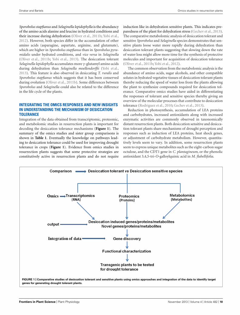
INTEGRATING THE OMICS RESPONSES AND NEW INSIGHTSIN UNDERSTANDING THE MECHANISM OF DESICCATIONTOLERANCEIntegration of the data obtained from transcriptomic, proteomic,and metabolomic studies in resurrection plants is important fordecoding the desiccation tolerance mechanisms (Figure 1). Thesummary of the omics studies and sister group comparisons isshown in Table 1. Eventually the knowledge on pathways lead-ing to desiccation tolerance could be used for improving droughttolerance in crops (Figure 1). Evidence from omics studies inresurrection plants suggests that some protective strategies areconstitutively active in resurrection plants and do not require
induction like in dehydration sensitive plants. This indicates pre-paredness of the plant for dehydration stress (Gechev et al., 2013).The comparative metabolomic analysis of desiccation tolerant andsensitive Sporobolus and Selaginella species demonstrates that sen-sitive plants loose water more rapidly during dehydration thandesiccation tolerant plants suggesting that slowing down the rateof water loss might allow more time for the synthesis of protectivemolecules and important for acquisition of desiccation tolerance(Oliver et al., 2011b; Yobi et al., 2012).
The common observation from the metabolomic analysis is theabundance of amino acids, sugar alcohols, and other compatiblesolutes in hydrated vegetative tissues of desiccation tolerant plantsthereby reducing the speed of water loss from the plants allowingthe plant to synthesize compounds required for desiccation tol-erance. Comparative omics studies have aided in differentiatingthe responses of tolerant and sensitive species thereby giving anoverview of the molecular processes that contribute to desiccationtolerance (Rodriguez et al., 2010; Gechev et al., 2013).
Reduction in photosynthesis, accumulation of LEA proteinsand carbohydrates, increased antioxidants along with increasedenzymatic activities are commonly observed in taxonomicallydistant resurrection plants. Both desiccation sensitive and desicca-tion tolerant plants share mechanisms of drought perception andresponses such as induction of LEA proteins, heat shock genes,or adjustment of carbohydrate metabolism. However, quantita-tively levels seem to vary. In addition, some resurrection plantsseem to express unique metabolites such as the eight-carbon sugaroctulose, and the CDT1 gene in C. plantagineum, or the phenolicantioxidant 3,4,5-tri-O-galloylquinic acid in M. flabellifolia.
FIGURE 1 | Comparative studies of desiccation tolerant and sensitive plants using omics approaches and integration of the data to identify target
genes for generating drought tolerant plants.
Frontiers in Plant Science | Plant Physiology November 2013 | Volume 4 | Article 482 | 10
“fpls-04-00482” — 2013/11/28 — 12:27 — page 11 — #11
Dinakar and Bartels Omics studies in resurrection plants
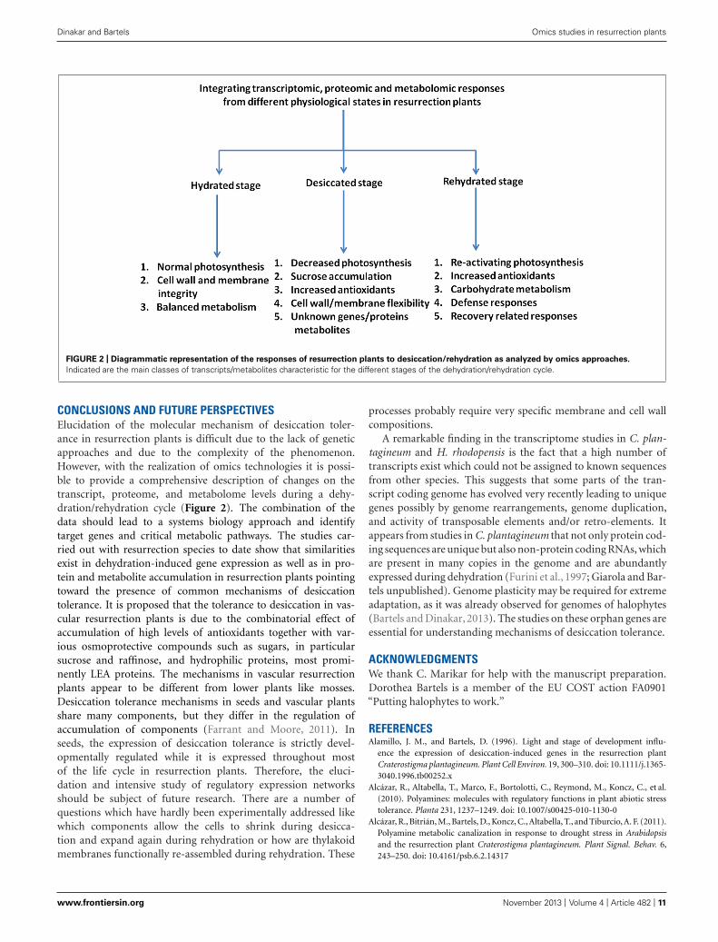
FIGURE 2 | Diagrammatic representation of the responses of resurrection plants to desiccation/rehydration as analyzed by omics approaches.
Indicated are the main classes of transcripts/metabolites characteristic for the different stages of the dehydration/rehydration cycle.
CONCLUSIONS AND FUTURE PERSPECTIVESElucidation of the molecular mechanism of desiccation toler-ance in resurrection plants is difficult due to the lack of geneticapproaches and due to the complexity of the phenomenon.However, with the realization of omics technologies it is possi-ble to provide a comprehensive description of changes on thetranscript, proteome, and metabolome levels during a dehy-dration/rehydration cycle (Figure 2). The combination of thedata should lead to a systems biology approach and identifytarget genes and critical metabolic pathways. The studies car-ried out with resurrection species to date show that similaritiesexist in dehydration-induced gene expression as well as in pro-tein and metabolite accumulation in resurrection plants pointingtoward the presence of common mechanisms of desiccationtolerance. It is proposed that the tolerance to desiccation in vas-cular resurrection plants is due to the combinatorial effect ofaccumulation of high levels of antioxidants together with var-ious osmoprotective compounds such as sugars, in particularsucrose and raffinose, and hydrophilic proteins, most promi-nently LEA proteins. The mechanisms in vascular resurrectionplants appear to be different from lower plants like mosses.Desiccation tolerance mechanisms in seeds and vascular plantsshare many components, but they differ in the regulation ofaccumulation of components (Farrant and Moore, 2011). Inseeds, the expression of desiccation tolerance is strictly devel-opmentally regulated while it is expressed throughout mostof the life cycle in resurrection plants. Therefore, the eluci-dation and intensive study of regulatory expression networksshould be subject of future research. There are a number ofquestions which have hardly been experimentally addressed likewhich components allow the cells to shrink during desicca-tion and expand again during rehydration or how are thylakoidmembranes functionally re-assembled during rehydration. These
processes probably require very specific membrane and cell wallcompositions.
A remarkable finding in the transcriptome studies in C. plan-tagineum and H. rhodopensis is the fact that a high number oftranscripts exist which could not be assigned to known sequencesfrom other species. This suggests that some parts of the tran-script coding genome has evolved very recently leading to uniquegenes possibly by genome rearrangements, genome duplication,and activity of transposable elements and/or retro-elements. Itappears from studies in C. plantagineum that not only protein cod-ing sequences are unique but also non-protein coding RNAs, whichare present in many copies in the genome and are abundantlyexpressed during dehydration (Furini et al., 1997; Giarola and Bar-tels unpublished). Genome plasticity may be required for extremeadaptation, as it was already observed for genomes of halophytes(Bartels and Dinakar, 2013). The studies on these orphan genes areessential for understanding mechanisms of desiccation tolerance.
ACKNOWLEDGMENTSWe thank C. Marikar for help with the manuscript preparation.Dorothea Bartels is a member of the EU COST action FA0901“Putting halophytes to work.”
REFERENCESAlamillo, J. M., and Bartels, D. (1996). Light and stage of development influ-
ence the expression of desiccation-induced genes in the resurrection plantCraterostigma plantagineum. Plant Cell Environ. 19, 300–310. doi: 10.1111/j.1365-3040.1996.tb00252.x
Alcázar, R., Altabella, T., Marco, F., Bortolotti, C., Reymond, M., Koncz, C., et al.(2010). Polyamines: molecules with regulatory functions in plant abiotic stresstolerance. Planta 231, 1237–1249. doi: 10.1007/s00425-010-1130-0
Alcázar, R., Bitrián, M., Bartels, D., Koncz, C., Altabella, T., and Tiburcio, A. F. (2011).Polyamine metabolic canalization in response to drought stress in Arabidopsisand the resurrection plant Craterostigma plantagineum. Plant Signal. Behav. 6,243–250. doi: 10.4161/psb.6.2.14317
www.frontiersin.org November 2013 | Volume 4 | Article 482 | 11
“fpls-04-00482” — 2013/11/28 — 12:27 — page 12 — #12
Dinakar and Bartels Omics studies in resurrection plants
Alpert, P., and Oliver, M. (2002). “Drying without dying,” in Desiccation and Survivalin Plants: Drying without Dying, eds M. Black and H. Pritchard (New York, NY:CABI Publishing), 1–43.
Bartels, D., and Dinakar, C. (2013). Balancing salinity stress responses in halo-phytes and non-halophytes: a comparison between Thellungiella and Arabidopsisthaliana. Funct. Plant Biol. 40, 819–831. doi: 10.1071/FP12299
Bartels, D., Hanke, C., Schneider, K., Michel, D., and Salamini, F. (1992). Adesiccation-related ELIP-like gene from the resurrection plant Craterostigmaplantagineum is regulated by light and ABA. EMBO J. 11, 2771–2778.
Bartels, D., and Hussain, S. S. (2011). “Resurrection plants: physiology and molec-ular biology,” in Ecological Studies: Desiccation Tolerance in Plants, eds U. Lüttge,E. Beck, and D. Bartels (Heidelberg: Springer), 339–364.
Bartels, D., Schneider, K., Terstappen, G., Piatkowski, D., and Salamini, F. (1990).Molecular cloning of abscisic acid modulated genes which are induced duringdesiccation of the resurrection plant Craterostigma plantagineum. Planta 181,27–34. doi: 10.1007/BF00202321
Bernacchia, G., Schwall, G., Lottspeich, F., Salamini, F., and Bartels, D. (1995). Thetransketolase gene family of the resurrection plant Craterostigma plantagineum:differential expression during the rehydration phase. EMBO J. 14, 610–618.
Bianchi, G., Gamba, A., Limiroli, R., Pozzi, N., Elster, R., Salamini, F., et al. (1993).The unusual sugar composition in leaves of the resurrection plant Myrothamnusflabellifolia. Physiol. Plant. 87, 223–226. doi: 10.1111/j.1399-3054.1993.tb00146.x
Bianchi, G., Gamba, A., Murelli, C., Salamini, F., and Bartels, D. (1992). Low molec-ular weight solutes in desiccated and ABA-treated calli and leaves of Craterostigmaplantagineum. Phytochemistry 31, 1917–1922. doi: 10.1016/0031-9422(92)80334-B
Bianchi, G., Murelli, C., Bochicchio, A., and Vazzana, C. (1991a). Changes in lowmolecular weight substances in Boea hygroscopica in response to desiccation andrehydration. Phytochemistry 30, 461–466. doi: 10.1016/0031-9422(91)83705-P
Bianchi, G., Gamba, A., Murelli, C., Salamini, F., and Bartels, D. (1991b). Novelcarbohydrate metabolism in the resurrection plant Craterostigma plantagineum.Plant J. 1, 355–359. doi: 10.1046/j.1365-313X.1991.t01-11-00999.x
Blomstedt, C. K., Gianello, R. D., Gaff, D. F., Hamill, J. D., and Neale, A. D. (1998).Differential gene expression in desiccation-tolerant and desiccation sensitive tis-sue of the resurrection grass, Sporobolus stapfianus. Aust. J. Plant Physiol. 25,937–946. doi: 10.1071/PP98113
Bockel, C., Salamini, F., and Bartels, D. (1998). Isolation and characterization ofgenes expressed during early events of the dehydration process in the resur-rection plant Craterostigma plantagineum. J. Plant Physiol. 152, 158–166. doi:10.1016/S0176-1617(98)80127-2
Chen, X., Zeng, Q., and Wood, A. J. (2002). The stress-responsive Tortula ruralisgene ALDH21A1 describes a novel eukaryotic aldehyde dehydrogenase proteinfamily. J. Plant Physiol. 159, 677–684. doi: 10.1078/0176-1617-0813
Collett, H., Shen, A., Gardner, M., Farrant, J. M., Denby, K. J., and Illing, N.(2004). Towards transcript profiling of desiccation tolerance in Xerophyta humilis:construction of a normalized 11 k X. humilis cDNA set and microarray expres-sion analysis of 424 cDNAs in response to dehydration. Physiol. Plant. 122,39–53. doi: 10.1111/j.1399-3054.2004.00381.x
Collett, H. M., Butowt, R., Smith, J., Farrant, J., and Illing, N. (2003). Photo-synthetic genes are differentially transcribed during the dehydration-rehydrationcycle in the resurrection plant, Xerophyta humilis. J. Exp. Bot. 54, 2543–2595. doi:10.1093/jxb/erg285
Cooper, K., and Farrant, J. M. (2002). Recovery of the resurrection plantCraterostigma wilmsii from desiccation: protection versus repair. J. Exp. Bot. 53,1805–1813. doi: 10.1093/jxb/erf028
Crowe, J. H., Carpenter, J. F., and Crowe, L. M. (1998). The role of vit-rification in anhydrobiosis. Annu. Rev. Physiol. 60, 73–103. doi: 10.1146/annurev.physiol.60.1.73
Cushman, J. C., and Oliver, M. J. (2011). “Understanding vegetative desiccationtolerance using integrated functional genomics approaches within a comparativeevolutionary framework,” in Ecological Studies: Desiccation Tolerance in Plants,eds E. B. Ulrich Luttge and D. Bartels (Heidelberg: Springer), 307–338.
Dinakar, C., and Bartels, D. (2012). Light response, oxidative stress managementand nucleic acid stability in closely related Linderniaceae species differing indesiccation tolerance. Planta 236, 541–555. doi: 10.1007/s00425-012-1628-8
Dinakar, C., Djilianov, D., and Bartels, D. (2012). Photosynthesis in desiccation-tolerant plants: energy metabolism and antioxidative stress defense. Plant Sci.182, 29–41. doi: 10.1016/j.plantsci.2011.01.018
Djilianov, D., Ivanov, S., Moyankova, D., Miteva, L., Kirova, E., Alexieva, V.,et al. (2011). Sugar ratios, glutathione redox status and phenols in the resur-rection species Haberlea rhodopensis and the closely related non-resurrectionspecies Chirita eberhardtii. Plant Biol. 13, 767–776. doi: 10.1111/j.1438-8677.2010.00436.x
Drennan, P. M., Smith, M. T., Goldsworth, D., and Van Staden, J. (1993). The occur-rence of trehalose in the leaves of the desiccation tolerant angiosperm Myrotham-nus flabellifolia Welw. J. Plant Physiol. 142, 493–496. doi: 10.1016/S0176-1617(11)81257-5
Farrant, J. M. (2000). A comparison of mechanisms of desiccation toleranceamong three angiosperm resurrection plant species. Plant Ecol. 151, 29–39. doi:10.1023/A:1026534305831
Farrant, J. M., Brandt, W., and Lindsey, G. G. (2007). An overview of mechanismsof desiccation tolerance in selected angiosperm resurrection plants. Plant Stress1, 72–84.
Farrant, J. M., and Moore, J. P. (2011). Programming desiccation-tolerance: fromplants to seeds to resurrection plants. Curr. Opin. Plant Biol. 14, 340–345. doi:10.1016/j.pbi.2011.03.018
Ferjani, A., Mustardy, L., Sulpice, R., Marin, K., Suzuki, I., Hagemann, M., et al.(2003). Glucosylglycerol, a compatible solute, sustains cell division under saltstress. Plant Physiol. 131, 1628–1637. doi: 10.1104/pp.102.017277
Frank, W., Munnik, T., Kerkmann, K., Salamini, F., and Bartels, D. (2000). Waterdeficit triggers phospholipase D activity in the resurrection plant Craterostigmaplantagineum. Plant Cell 12, 111–123. doi: 10.1105/tpc.12.1.111
Furini, A., Koncz, C., Salamini, F., and Bartels, D. (1997). High level transcrip-tion of a member of a repeated gene family confers dehydration toleranceto callus tissue of Craterostigma plantagineum. EMBO J. 16, 3599–3608. doi:10.1093/emboj/16.12.3599
Gaff, D. F., and McGregor, G. R. (1979). The effect of dehydration and rehydrationon the nitrogen content of various fractions from resurrection plants. Biol. Plant.22, 92–99. doi: 10.1007/BF02909453
Gasulla, F., Vom Dorp, K., Dombrink, I., Zahringer, U., Gisch, N., Dormann, P., et al.(2013). The role of lipid metabolism in the acquisition of desiccation tolerance inCraterostigma plantagineum: a comparative approach. Plant J. 75, 726–741. doi:10.1111/tpj.12241
Gechev, T., Dinakar, C., Benina, M., Toneva, V., and Bartels, D. (2012). Molecularmechanisms of desiccation tolerance in resurrection plants. Cell. Mol. Life Sci. 69,3175–3186. doi: 10.1007/s00018-012-1088-0
Gechev, T. S., Benina, M., Obata, T., Tohge, T., Sujeeth, N., Minkov, I., et al. (2013).Molecular mechanisms of desiccation tolerance in the resurrection glacial relicHaberlea rhodopensis. Cell. Mol. Life Sci. 70, 689–709. doi: 10.1007/s00018-012-1155-6
Ghasempour, H. R., Gaff, D. F., Williams, R. P. W., and Gianello, R. D. (1998). Con-tents of sugars in leaves of drying desiccation tolerant flowering plants particularlygrasses. Plant Growth Regul. 24, 185–191. doi: 10.1023/A:1005927629018
Goday, A., Sánchez-Martinez, D., Gómez, J., Puigdomènech, P., and Pagès, M.(1988). Gene expression in developing Zea mays embryos. Regulation by abscisicacid of a highly phosphorylated 23- to 25-kD group of proteins. Plant Physiol. 88,564–569. doi: 10.1104/pp.88.3.564
Hartung, W., Schiller, P., and Karl-Josef, D. (1998). Physiology of poikilohydricplants. Prog. Bot. 59, 299–327. doi: 10.1007/978-3-642-80446-5_11
Haslekás, C., Stacy, R. A., Nygaard, V., Culianez-Marcia, F. A., and Aalen, R. B.(1998). The expression of a peroxiredoxin antioxidant gene, AtPerl, in Arabidopsisthaliana is seed-specific and related to dormancy. Plant Mol. Biol. 36, 833–845.doi: 10.1023/A:1005900832440
Hoekstra, F. A., Golovina, E. A., and Buitink, J. (2001). Mechanisms of plant des-iccation tolerance. Trends Plant Sci. 6, 431–438. doi: 10.1016/S1360-1385(01)02052-0
Hoekstra, F. A., Wolkers, W. F., Buitink, J., Golovina, E. A., Crowe, J. H., and Crowe,L. M. (1997). Membrane stabilization in the dry state. Comp. Biochem. Physiol.117, 335–341. doi: 10.1016/S0300-9629(96)00272-1
Höfler, K., Migsch, H., and Rottenburg, W. (1941). Über die Austrocknungresistenzlandwirtschaftlicher Kulturpflanzen. Forschungsdienst 12, 50–61.
Horbowicz, M., and Obendorf, R. L. (1994). Seed desiccation tolerance and stora-bility: dependence of flatulence-producing oligosaccharides and cyclitols. SeedSci. Res. 4, 385–405. doi: 10.1017/S0960258500002440
Illing, N., Denby, K., Collett, H., Shen, A., and Farrant, J. (2005). The signa-ture of seeds in resurrection plants: a molecular and physiological comparison
Frontiers in Plant Science | Plant Physiology November 2013 | Volume 4 | Article 482 | 12
“fpls-04-00482” — 2013/11/28 — 12:27 — page 13 — #13
Dinakar and Bartels Omics studies in resurrection plants
of desiccation tolerance in seeds and vegetative tissues. Integr. Comp. Biol. 45,771–787. doi: 10.1093/icb/45.5.771
Ingle, R., Schmidt, U., Farrant, J., Thomson, J., and Mundree, S. (2007). Proteomicanalysis of leaf proteins during dehydration of the resurrection plant Xerophytaviscosa. Plant Cell Environ. 30, 435–446. doi: 10.1111/j.1365-3040.2006.01631.x
Ingram, J., and Bartels, D. (1996). The molecular basis of dehydration toler-ance in plants. Annu. Rev. Plant Physiol. Plant Mol. Biol. 47, 377–403. doi:10.1146/annurev.arplant.47.1.377
Iturriaga, G., Cushman, M., and Cushman, J. (2006). An EST catalogue from theresurrection plant Selaginella lepidophylla reveals abiotic stress-adaptive genes.Plant Biol. 170, 1173–1184.
Jiang, G., Wang, Z., Shang, H., Yang, W., Hu, Z., Phillips, J., et al. (2007). Proteomeanalysis of leaves from the resurrection plant Boea hygrometrica in response todehydration and rehydration. Planta 225, 1405–1420. doi: 10.1007/s00425-006-0449-z
Jones, L., and McQueen-Mason, S. (2004). A role for expansins in the dehydrationand rehydration of the resurrection plant Craterostigma plantagineum. FEBS Lett.559, 61–65. doi: 10.1016/S0014-5793(04)00023-7
Kleines, M., Elster, R. C., Rodrigo, M. J., Blervacq, A. S., Salamini, F., and Bartels,D. (1999). Isolation and expression analysis of two stress-responsive sucrose-synthase genes from the resurrection plant Craterostigma plantagineum (Hochst.).Planta 209, 13–24. doi: 10.1007/s004250050602
Kranner, I., Beckett, R. P., Wornik, S., Zorn, M., and Pfeifhofer, H. W. (2002). Revivalof a resurrection plant correlates with its antioxidant status. Plant J. 31, 13–24.doi: 10.1046/j.1365-313X.2002.01329.x
Le, T. N., Blomstedt, C. K., Kuang, J., Tenlen, J., Gaff, D. F., Hamill, J. D., et al. (2007).Genes differentially expressed in desiccation tolerant leaf tissue of the resurrec-tion grass Sporobolus stapfianus. Funct. Plant Biol. 34, 589–600. doi: 10.1071/FP06231
Lehner, A., Chopera, D., Peters, S., Keller, F., Mundree, S., Thomson, J., et al. (2008).Protection mechanisms in the resurrection plant Xerophyta viscosa: cloning,expression, characterisation and role of XvINO1, a gene coding for a myo-inositol1-phosphate synthase. Funct. Plant Biol. 35, 26–39. doi: 10.1071/FP07142
Leopold, A. C., and Vertucci, C. W. (1986). “Physical attributes of desiccated seeds,”in Membranes, metabolism and Dry Organisms, ed. A. C. Leopold (New York:Cornell University Press), 22–34.
Liu, M. S., Chien, C. T., and Lin, T. P. (2008). Constitutive components andinduced gene expression are involved in the desiccation tolerance of Selaginellatamariscina. Plant Cell Physiol. 49, 653–663. doi: 10.1093/pcp/pcn040
Marais, S., Thomson, J. A., Farrant, J. M., and Mundree, S. G. (2004). VATP1XV –a novel stress responsive V-ATPase subunit c homologue isolated from theresurrection plant Xerophyta viscosa Baker. Physiol. Plant 122, 54–61. doi:10.1111/j.1399-3054.2004.00389.x
Martinelli, T., Whittaker, A., Bochicchio, A., Vazzana, C., Suzuki, A., and MasclauxDaubresse, C. (2007). Amino acid pattern and glutamate metabolism duringdehydration stress in the ‘resurrection’ plant Sporobolus stapfianus: a comparisonbetween desiccation-sensitive and desiccation tolerant leaves. J. Exp. Bot. 58,3037–3046. doi: 10.1093/jxb/erm161
Michel, D., Furini, A., Salamini, F., and Bartels, D. (1994). Structure andregulation of an ABA-and desiccation-responsive gene from the resurrec-tion plant Craterostigma plantagineum. Plant Mol. Biol. 24, 549–560. doi:10.1007/BF00023553
Moore, J. P., Nguema-Ona, E. E., Chevalier, L., Lindsey, G. G., Brandt, W. F.,Lerouge, P., et al. (2006). Response of the leaf cell wall to desiccation in theresurrection plant Myrothamnus flabellifolia. Plant Physiol. 141, 651–662. doi:10.1104/pp.106.077701
Moore, J. P., Nguema-Ona, E. E., Vicre-Gibouin, M., Sorensen, I., Willats, W. G.,Driouich, A., et al. (2013). Arabinose-rich polymers as an evolutionary strategyto plasticize resurrection plant cell walls against desiccation. Planta 237, 739–754.doi: 10.1007/s00425-012-1785-9
Moore, J. P., Tuan Le, N., Brandt, W. F., Driouch, A., and Farrant, J. M. (2009).Towards a systems-based understanding of plant desiccation tolerance. TrendsPlant Sci. 14, 110–117. doi: 10.1016/j.tplants.2008.11.007
Moore, J. P., Vicre-Gibouin, M., Farrant, J. M., and Driouich, A. (2008). Adaptationsof higher plant cell walls to water loss: drought vs desiccation. Physiol. Plant. 134,237–245. doi: 10.1111/j.1399-3054.2008.01134.x
Moore, J. P., Westall, K., Ravenscroft, N., Farrant, J., Lindsey, G., and Brandt,W. (2005). The predominant polyphenol in the leaves of the resurrection plant
Myrothamnus flabellifolius, 3,4,5 tri-O-galloylquinic acid, protects membranesagainst desiccation and free radical-induced oxidation. Biochem. J. 385, 301–308.doi: 10.1042/BJ20040499
Mowla, S. B., Thomson, J. A., Farrant, J. M., and Mundree, S. G. (2002). A novel stressinducible antioxidant enzyme identified from the resurrection plant Xerophytaviscosa Baker. Planta 215, 716–726. doi: 10.1007/s00425-002-0819-0
Mulako, I., Farrant, J., Collett, H., and Illing, N. (2008). Expression of Xhdsi-1VOC,a novel member of the vicinal oxygen chelate (VOC) metalloenzyme superfam-ily, is up-regulated in leaves and roots during desiccation in the resurrectionplant Xerophyta humilis (Bak) Dur and Schinz. J. Exp. Bot. 59, 3885–3901. doi:10.1093/jxb/ern226
Mundree, S. G., Whittaker, A., Thomson, J. A., and Farrant, J. M. (2000). An aldosereductase homolog from the resurrection plant Xerophyta viscosa Baker. Planta211, 693–700. doi: 10.1007/s004250000331
Navari-Izzo, F., Meneguzzo, S., Loggini, B., Vazzana, C., and Sgherri, C. (1997). Therole of the glutathione system during dehydration of Boea hygroscopica. Physiol.Plant. 99, 23–30. doi: 10.1111/j.1399-3054.1997.tb03426.x
Ndima, T., Farrant, J., Thomson, J., and Mundree, S. (2001). Molecular character-ization of XVT8, a stress-responsive gene from the resurrection plant Xerophytaviscosa Baker. Plant Growth Regul. 35, 137–145. doi: 10.1023/A:1014433821730
Neale, A. D., Blomstedt, T., Bronson, T. N., Guthridge, L. K., Evans, J., Gaff, D.F., et al. (2000). The isolation of genes from the resurrection grass Sporobolusstapfianus which are induced during severe drought stress. Plant Cell Environ. 23,265–277. doi: 10.1046/j.1365-3040.2000.00548.x
Norwood, M., Truesdale, M., Richter, A., and Scott, P. (2000). Photosyntheticcarbohydrate metabolism in the resurrection plant Craterostigma plantagineum.J. Exp. Bot. 51, 159–165. doi: 10.1093/jexbot/51.343.159
Oliver, M. J., Dowd, S. E., Zaragoza, J., Mauget, S. A., and Payton, P. R. (2004). Therehydration transcriptome of the desiccation-tolerant bryophyte Tortula ruralis:transcript classification and analysis. BMC Genomics 5:89. doi: 10.1186/1471-2164-5-89
Oliver, M. J., Jain, R., Balbuena, T. S., Agrawal, G., Gasulla, F., and Thelen, J. J.(2011a). Proteome analysis of leaves of the desiccation-tolerant grass, Sporobo-lus stapfianus, in response to dehydration. Phytochemistry 72, 1273–1284. doi:10.1016/j.phytochem.2010.10.020
Oliver, M. J., Guo, L., Alexander, D., Ryals, J., Wone, B., and Cushman, J. (2011b).A sister group metabolomic contrast using untargeted global metabolomic anal-ysis delineates the biochemical regulation underlying desiccation tolerance inSporobolus stapfianus. Plant Cell 23, 1231–1248. doi: 10.1105/tpc.110.082800
Oliver, M. J., Wood, A. J., and O’Mahony, P. (1998). To dryness and beyond prepara-tion for the dried state and rehydration in vegetative desiccation-tolerant plants.Plant Growth Regul. 24, 193–201. doi: 10.1023/A:1005863015130
Peters, S., Mundree, S., Thomson, J., Farrant, J., and Keller, F. (2007). Protectionmechanisms in the resurrection plant Xerophyta viscosa (Baker): both sucroseand raffinose family oligosaccharides (RFOs) accumulate in leaves in response towater deficit. J. Exp. Bot. 58, 1947–1956. doi: 10.1093/jxb/erm056
Petersen, J., Eriksson, S. K., Harryson, P., Pierog, S., Colby, T., Bartels, D., et al.(2012). The lysine-rich motif of intrinsically disordered stress protein CDeT11-24 from Craterostigma plantagineum is responsible for phosphatidic acid bindingand protection of enzymes from damaging effects caused by desiccation. J. Exp.Bot. 63, 4919–4929. doi: 10.1093/jxb/ers173
Piatkowski, D., Schneider, K., Salamini, F., and Bartels, D. (1990). Char-acterization of five abscisic acid-responsive cDNA clones isolated from thedesiccation-tolerant plant Craterostigma plantagineum and their relationship toother water-stress genes. Plant Physiol. 94, 1682–1688. doi: 10.1104/pp.94.4.1682
Quartacci, M. F., Forli, M., Dalla-Vecchia, F., Bochicchio, A., and Navari-Izzo, F.(1997). Desiccation-tolerant Sporobolus stapfianus: lipid composition and cellularultrastructure during dehydration and rehydration. J. Exp. Bot. 48, 1269–1279.doi: 10.1093/jxb/48.6.1269
Quartacci, M. F., Glišic, O., Stevanovic, B., and Navari-Izzo, F. (2002). Plasmamembrane lipids in the resurrection plant Ramonda serbica following dehydrationand rehydration. J. Exp. Bot. 53, 2159–2166. doi: 10.1093/jxb/erf076
Rodriguez, M. C. S., Edsgärd, D., Hussain, S. S., Alquezar, D., Rasmussen, M.,Gilbert, T., et al. (2010). Transcriptomes of the desiccation-tolerant resurrectionplant Craterostigma plantagineum. Plant J. 63, 212–228. doi: 10.1111/j.1365-313X.2010.04243.x
Röhrig, H., Schmidt, J., Colby, T., Bräutigam, A., Hufnagel, P., and Bartels, D.(2006). Desiccation of the resurrection plant Craterostigma plantagineum induces
www.frontiersin.org November 2013 | Volume 4 | Article 482 | 13
“fpls-04-00482” — 2013/11/28 — 12:27 — page 14 — #14
Dinakar and Bartels Omics studies in resurrection plants
dynamic changes in protein phosphorylation. Plant Cell Environ. 29, 1606–1617.doi: 10.1111/j.1365-3040.2006.01537.x
Scott, P. (2000). Resurrection plants and the secrets of eternal leaf. Ann. Bot. 85,159–166. doi: 10.1006/anbo.1999.1006
Scott, H. B., and Oliver, M. J. (1994). Accumulation and polysomal recruitment oftranscripts in response to desiccation and dehydration of the moss Tortula ruralis.J. Exp. Bot. 45, 577–583. doi: 10.1093/jxb/45.5.577
Sgherri, C., Stevanovic, B., and Navari-Izzo, F. (2004). Role of phenolics inthe antioxidative status of the resurrection plant Ramonda serbica duringdehydration and rehydration. Physiol. Plant. 22, 478–485. doi: 10.1111/j.1399-3054.2004.00428.x
Sherwin, H., and Farrant, J. (1998). Protection mechanisms against excess light inthe resurrection plants Craterostigma wilmsii and Xerophyta viscosa. Plant GrowthRegul. 24, 203–210. doi: 10.1023/A:1005801610891
Smirnoff, N. (1993). The role of active oxygen in the response of plants towater deficit and desiccation, New Phytol. 125, 27–58. doi: 10.1111/j.1469-8137.1993.tb03863.x
Stiti, N., Missihoun, T. D., Kotchoni, S. O., and Kirch, H. H. (2011).Aldehyde dehydrogenases in Arabidopsis thaliana: biochemical requirements,metabolic pathways, and functional analysis. Front. Plant Sci. 2:1–11. doi:10.3389/fpls.2011.00065
Sunkar, R., Bartels, D., and Kirch, H. H. (2003). Overexpression of a stress-inducible aldehyde dehydrogenase gene from Arabidopsis thaliana in transgenicplants improves stress tolerance. Plant J. 35, 452–464. doi: 10.1046/j.1365-313X.2003.01819.x
Tymms, M. J., and Gaff, D. F. (1978). Proline accumulation during water stress inresurrection plants. J. Exp. Bot. 30, 165–168. doi: 10.1093/jxb/30.1.165
Upchurch, R. (2008). Fatty acid unsaturation, mobilization, and regulation in theresponse of plants to stress. Biotechnol. Lett. 30, 967–977. doi: 10.1007/s10529-008-9639-z
Urano, K., Maruyama, K., Ogata, Y., Morishita, Y., Takeda, M., Sakurai, N., et al.(2009). Characterization of the ABA-regulated global responses to dehydrationin Arabidopsis by metabolomics. Plant J. 57, 1065–1078. doi: 10.1111/j.1365-313X.2008.03748.x
van den Dries, N., Facchinelli, F., Giarola, V., Phillips, J. R., and Bartels, D. (2011).Comparative analysis of LEA-like 11-24 gene expression and regulation in relatedplant species within the Linderniaceae that differ in desiccation tolerance. NewPhytol. 190, 75–88. doi: 10.1111/j.1469-8137.2010.03595.x
Velasco, R., Salamini, F., and Bartels, D. (1994). Dehydration and ABA increasemRNA levels and enzyme activity of cytosolic GAPDH in the resurrec-tion plant Craterostigma plantagineum. Plant Mol. Biol. 26, 541–546. doi:10.1007/BF00039567
Veljovic-Jovanovic, S., Kukavica, B., and Navari-Izzo, F. (2008). Characterization ofpolyphenol oxidase changes induced by desiccation of Ramonda serbica leaves.Physiol. Plant. 132, 407–416. doi: 10.1111/j.1399-3054.2007.01040.x
Vertucci, C. W., and Farrant, J. M. (1995). “Acquisition and loss of desiccation-tolerance,” in Seed Development and Germination, eds J. Kigel and G. Galili (NewYork: Marcel Dekker), 237–271.
Vicré, M., Lerouxel, O., Farrant, J., Lerouge, P., and Driouich, A. (2004).Composition and desiccation-induced alterations of the cell wall in the res-urrection plant Craterostigma wilmsii. Physiol. Plant. 120, 229–239. doi:10.1111/j.0031-9317.2004.0234.x
Wang, X., Chen, S., Zhang, H., Shi, L., Cao, F., Guo, L., et al. (2010). Desiccationtolerance mechanism in resurrection fern-ally Selaginella tamariscina revealedby physiological and proteomic analysis. J. Proteome Res. 9, 6561–6577. doi:10.1021/pr100767k
Whittaker, A., Bochicchio, A., Vazzana, C., Lindsey, G., and Farrant,J. (2001). Changes in leaf hexokinase activity and metabolite levels inresponse to drying in the desiccation-tolerant species Sporobolus stapfianusand Xerophyta viscosa. J. Exp. Bot. 52, 961–969. doi: 10.1093/jexbot/52.358.961
Whittaker, A., Martinelli, T., Farrant, J. M., Bochicchio, A., and Vazzana, C. (2007).Sucrose phosphate synthase activity and the co-ordination of carbon partitioningduring sucrose and amino acid accumulation in desiccation-tolerant leaf materialof the C4 resurrection plant Sporobolus stapfianus during dehydration. J. Exp. Bot.58, 3775–3787. doi: 10.1093/jxb/erm228
Willige, B. C., Kutzer, M., Tebartz, F., and Bartels, D. (2009). Subcellular localizationand enzymatic properties of differentially expressed transketolase genes isolatedfrom the desiccation tolerant resurrection plant Craterostigma plantagineum.Planta 229, 659–666. doi: 10.1007/s00425-008-0863-5
Wood, A. J., and Oliver, M. J. (1999). Translational control in plant stress:formation of messenger ribonucleoprotein complexes (mRNPs) in Tortularuralis in response to desiccation. Plant J. 18, 359–370. doi: 10.1046/j.1365-313X.1999.00458.x
Yobi, A., Wone, B. W. M., Xu, W., Alexander, D. C., Guo, L., Ryals, J. A., et al. (2012).Comparative metabolic profiling between desiccation-sensitive and desiccationtolerant species of Selaginella reveals insights into the resurrection trait. Plant J.72, 983–999. doi: 10.1111/tpj.12008
Yobi, A., Wone, B. W. M., Xu, W., Alexander, D. C., Guo, L., Ryals, J. A., et al.(2013). Metabolic profiling in Selaginella lepidophylla at various hydration statesprovides new insights into the mechanistic basis of desiccation tolerance. Mol.Plant 6, 369–385. doi: 10.1093/mp/sss155
Zeng, O., Chen, X. B., and Wood, A. J. (2002). Two early light-inducible pro-tein (ELIP) cDNAs from the resurrection plant Tortula ruralis are differentiallyexpressed in response to desiccation, rehydration, salinity, and high light. J. Exp.Bot. 371, 1197–1205. doi: 10.1093/jexbot/53.371.1197
Zentella, R., Gallardo, J. O., van Dijck, P., Mallol, J., Bonini, B., van Vaeck, C., et al.(1999). A Selaginella lepidophylla trehalose-6-phosphate synthase complementsgrowth and stress-tolerance defects in a yeast tsp1 mutant. Plant Physiol. 119,1473–1482. doi: 10.1104/pp.119.4.1473
Zivkovic, T., Quartacci, M. F., Stevanovic, B., Marinone, F., and Navari-Izzo, F.(2005). Low-molecular weight substances in the poikilohydric plant Ramondaserbica during dehydration and rehydration. Plant Sci. 168, 105–111. doi:10.1016/j.plantsci.2004.07.018
Conflict of Interest Statement: The authors declare that the research was conductedin the absence of any commercial or financial relationships that could be construedas a potential conflict of interest.
Received: 19 August 2013; paper pending published: 27 September 2013; accepted: 06November 2013; published online: 28 November 2013.Citation: Dinakar C and Bartels D (2013) Desiccation tolerance in resurrection plants:new insights from transcriptome, proteome, and metabolome analysis. Front. Plant Sci.4:482. doi: 10.3389/fpls.2013.00482This article was submitted to Plant Physiology, a section of the journal Frontiers inPlant Science.Copyright © 2013 Dinakar and Bartels. This is an open-access article distributed underthe terms of the Creative Commons Attribution License (CC BY). The use, distributionor reproduction in other forums is permitted, provided the original author(s) or licensorare credited and that the original publication in this journal is cited, in accordance withaccepted academic practice. No use, distribution or reproduction is permitted whichdoes not comply with these terms.
Frontiers in Plant Science | Plant Physiology November 2013 | Volume 4 | Article 482 | 14
Related Documents