MACROSCOPIC HEMATURIA| Tutorial B-1 GUS 130110110177|Gabriella Chafrina| 19/05/14 Definition Hematuria is the presence of blood in the urine Types - Macroscopic hematuria or “gross”: visible to the naked eye, called this when there is blood >1cc/liter Even one episode of “gross” hematuria warrants a visit to a health care provider’s office for further investigation. Unfortunately, many patients do not seek medical attention when they see o r think they see blood in their urine. A full medical history and assessment of the nature of an individual’s complaint are the first steps in determining the cause of blood in the urine Has a high diagnostic y ield for urological malignancy - Microscopic hematuri a or “microscopic ”: visible only under a microscope, called this when there is RBC >3 hpf Etiology - Malignancy: urological malignancies (most commonly transitional cell carc inoma of the bladder, but potentially anywhere along the urinary tract – that is, renal (kidney and adrenal pelvis), ureteric, prostatic and urethral malignancies) Macroscopic hematuria has been found to be a pre senting feature in >66% of patients confirmed as having a urological malignancy The sensitivity of macroscopic hematuria in identifying malignancies is relatively high: bladder carcinoma 0.83, ureteric carcinoma 0.66 and renal carcinoma 0.48 - Benign: benign prostatic hyperplasia, urinary tract calculi, urinary tract infections (UTIs) and nephrological problems, whereas others include trauma Clinical Examination - During the physical examination of the patient, it is important to elicit the following signs: Cardiovascular status Presence of a palpable bladder, which may be indicative of acute or imminent urinary retention Careful palpation for a tumour mass (potential sites include renal, bladder or gynaecological origin) In females, a vaginal examination should be performed to ensure that the blood comes from the urethra rather than the vagina. In males, the external genitalia should be examined In both sexes, a digital rectal examination should be performed to exclude tumour masses either in the prostate or in the pouch of Douglas - Urine tests In making the diagnosis of macroscopic hematuria, it is most important that true ‘‘frank hematuria’’ is seen in the urine sample The degree of blood staining in the urine has traditionally been described in relation to shades of red wine namely, Rose´, Claret A dipstick urine test should be performed in t he ED. It is quick and cost effect ive, and may indicate the presence of infection. Even in apparently clear urine, microscopic hematuria may be detected —this is important as macroscopic hematuria may clear over the course of the day as t he patient drinks. It is also important to recognise that a negative dipstick result does not exclude UTI, so t he microscopy, culture and sensitivities results should be checked Patients with incidental microscopic hematuria will also require follow-up, but this can be safely arranged via their general practitioner o β-Human chorionic gonadotrophin dipstick testing should be performed in all women of childbearing age o A sample of midstream urine should also be sent for microbiological testing. Any infection should be treated according to local antibiotic guidelines o Urine cytology is of little use in the ED setting. Despite being highly specific, it has an estimated sensitivity of only 25%. Its use is probably more appropriate as a second-line investigation

Welcome message from author
This document is posted to help you gain knowledge. Please leave a comment to let me know what you think about it! Share it to your friends and learn new things together.
Transcript
8/11/2019 GUS6 Macroscopic Hematuria
http://slidepdf.com/reader/full/gus6-macroscopic-hematuria 1/3
MACROSCOPIC HEMATURIA| Tutorial B-1 GUS
130110110177|Gabriella Chafrina| 19/05/14
Definition
Hematuria is the presence of blood in the urine
Types
- Macroscopic hematuria or “gross”: visible to the naked eye, called this when there is blood >1cc/liter
Even one episode of “gross” hematuria warrants a visit to a health care provider’s office for further
investigation. Unfortunately, many patients do not seek medical attention when they see or think they see
blood in their urine. A full medical history and assessment of the nature of an individual’s complaint are the
first steps in determining the cause of blood in the urine
Has a high diagnostic yield for urological malignancy
- Microscopic hematuria or “microscopic”: visible only under a microscope, called this when there is RBC >3 hpf
Etiology
- Malignancy: urological malignancies (most commonly transitional cell carcinoma of the bladder, but potentially
anywhere along the urinary tract – that is, renal (kidney and adrenal pelvis), ureteric, prostatic and urethral
malignancies)
Macroscopic hematuria has been found to be a presenting feature in >66% of patients confirmed as having a
urological malignancy
The sensitivity of macroscopic hematuria in identifying malignancies is relatively high: bladder carcinoma0.83, ureteric carcinoma 0.66 and renal carcinoma 0.48
- Benign: benign prostatic hyperplasia, urinary tract calculi, urinary tract infections (UTIs) and nephrological
problems, whereas others include trauma
Clinical Examination
- During the physical examination of the patient, it is important to elicit the following signs:
Cardiovascular status
Presence of a palpable bladder, which may be indicative of acute or imminent urinary retention
Careful palpation for a tumour mass (potential sites include renal, bladder or gynaecological origin)
In females, a vaginal examination should be performed to ensure that the blood comes from the urethra
rather than the vagina. In males, the external genitalia should be examined In both sexes, a digital rectal examination should be performed to exclude tumour masses either in the
prostate or in the pouch of Douglas
- Urine tests
In making the diagnosis of macroscopic hematuria, it is most important that true ‘‘frank hematuria’’ is seen
in the urine sample
The degree of blood staining in the urine has traditionally been described in relation to shades of red wine
namely, Rose´, Claret
A dipstick urine test should be performed in the ED. It is quick and cost effective, and may indicate the
presence of infection. Even in apparently clear urine, microscopic hematuria may be detected—this is
important as macroscopic hematuria may clear over the course of the day as the patient drinks. It is also
important to recognise that a negative dipstick result does not exclude UTI, so the microscopy, culture andsensitivities results should be checked
Patients with incidental microscopic hematuria will also require follow-up, but this can be safely arranged
via their general practitioner
o β-Human chorionic gonadotrophin dipstick testing should be performed in all women of childbearing
age
o A sample of midstream urine should also be sent for microbiological testing. Any infection should be
treated according to local antibiotic guidelines
o Urine cytology is of little use in the ED setting. Despite being highly specific, it has an estimated
sensitivity of only 25%. Its use is probably more appropriate as a second-line investigation
8/11/2019 GUS6 Macroscopic Hematuria
http://slidepdf.com/reader/full/gus6-macroscopic-hematuria 2/3
MACROSCOPIC HEMATURIA| Tutorial B-1 GUS
130110110177|Gabriella Chafrina| 19/05/14
- Blood tests
Macroscopic hematuria is not associated with any specific diagnostic tests; however, it is important to know a
number of indices in order to decide whether a patient will be suitable for outpatient management
Full blood count—A full blood count indicates a baseline haemoglobin level and ensures that there is no
underlying thrombocytopenia that may need to be addressed
Urea and electrolytes -Urea and electrolyte levels indicate whether the patient has acute renal failure.
Again, this may necessitate admission for investigation to exclude urinary outflow-tract obstruction
Clotting—There is some debate as to whether a clotting screen should be a standard investigation in these
patients, as it has a low yield for new diagnoses of bleeding diatheses. A pragmatic approach is advised. For
patients with known clotting disorders, those taking anticoagulant drugs, those having hepatic disease or
very heavy bleeding, a clotting screen should be done; for those without risk factors for bleeding and with
mild hematuria, it may be omitted.
Group and save (with or without cross-match)—In patients with large amount of macroscopic hematuria,
group and save should be considered, and in these patients admission is warranted. In those with
haemodynamic compromise, crossmatching should be requested as a priority
- Radiological imaging
Kidney, ureters, bladder (KUB) x ray is easily available and simple to perform. Although there is limited
diagnostic information, it may be a useful first-line test in patients for whom contrast radiography is
contraindicated. However, in cases where renal calculi are suspected, a KUB x ray may prove to be of value,as 70 –80% of urinary tract calculi are radioopaque
Further investigation with complete intravenous urography (IVU)/ultrasound scanning (USS)/CT-KUB should
be performed in this group of patients
AFTER REFERRAL TO THE UROLOGIST
- Urine cytology
Despite being highly specific, urine cytology has an estimated sensitivity of only 25%. There is some debate on
its more appropriate use as a second-line investigation. However, the current European Association of Urology
guidelines continue to advocate its use in the diagnostic pathway
- Radiological imaging
Some of the radiological imaging techniques may be instituted in the ED, depending on the local policy and the
availability of resources. In all, 25% of urologists use USS imaging of the urinary tract as their first-lineinvestigation for hematuria, 24% use IVU and 51% use a combination of both of these
USS is a non-invasive test, but may be limited by its operator dependence. In combination with plain KUB
radiographs, USS by a radiologist has been shown to have a diagnostic accuracy comparable to that of IVU
IVU has a number of contraindications—asthma, use Of metformin, renal impairment, seafood/contrast
allergy, hepatic impairment and pregnancy. This may limit the usefulness of this investigation as a first-line
choice.
CT-KUB may be a useful second-line investigation in those patients with normal first-line tests and persistent
hematuria. The ‘‘pick-up rate’’ for current first-line radiological investigations is high, particularly when the
radiological tests are reported by specialist uroradiologists
- Flexible cystoscopy
This is a minimally invasive, outpatient procedure to directly visualize the urethra and bladder. Most
urologists would agree that all patients with macroscopic hematuria should undergo this procedure on at
least one occasion during the diagnostic process. It is possible to take biopsy specimens to obtain a tissue
diagnosis at the time of this procedure
Diagnosis
- Careful history and examination may give some indications as to the origin of the hematuria:
Painless hematuria is classically associated with the underlying malignancies, whereas hematuria associated
with pain may be more suggestive of stone disease (urinary calculi) or UTI.
8/11/2019 GUS6 Macroscopic Hematuria
http://slidepdf.com/reader/full/gus6-macroscopic-hematuria 3/3
MACROSCOPIC HEMATURIA| Tutorial B-1 GUS
130110110177|Gabriella Chafrina| 19/05/14
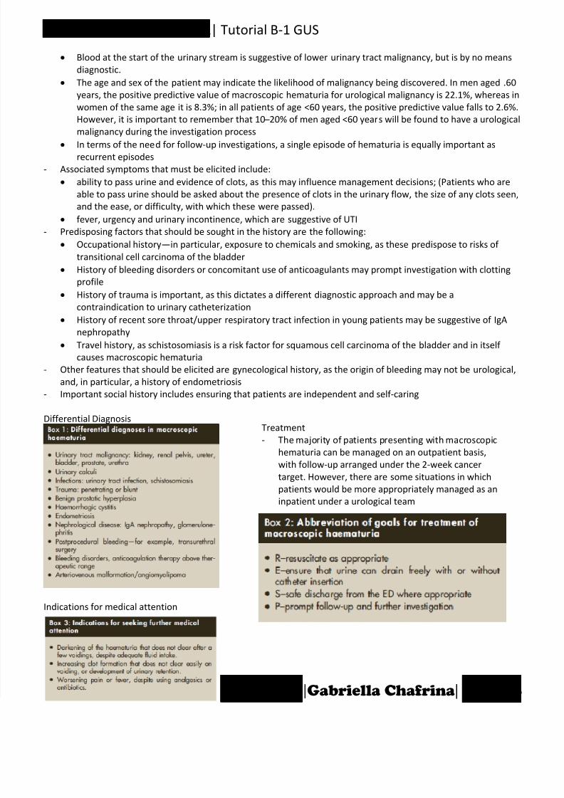
Treatment
- The majority of patients presenting with macroscopic
hematuria can be managed on an outpatient basis,
with follow-up arranged under the 2-week cancer
target. However, there are some situations in which
patients would be more appropriately managed as an
inpatient under a urological team
Blood at the start of the urinary stream is suggestive of lower urinary tract malignancy, but is by no means
diagnostic.
The age and sex of the patient may indicate the likelihood of malignancy being discovered. In men aged .60
years, the positive predictive value of macroscopic hematuria for urological malignancy is 22.1%, whereas in
women of the same age it is 8.3%; in all patients of age <60 years, the positive predictive value falls to 2.6%.
However, it is important to remember that 10 –20% of men aged <60 years will be found to have a urological
malignancy during the investigation process
In terms of the need for follow-up investigations, a single episode of hematuria is equally important as
recurrent episodes
- Associated symptoms that must be elicited include:
ability to pass urine and evidence of clots, as this may influence management decisions; (Patients who are
able to pass urine should be asked about the presence of clots in the urinary flow, the size of any clots seen,
and the ease, or difficulty, with which these were passed).
fever, urgency and urinary incontinence, which are suggestive of UTI
- Predisposing factors that should be sought in the history are the following:
Occupational history—in particular, exposure to chemicals and smoking, as these predispose to risks of
transitional cell carcinoma of the bladder
History of bleeding disorders or concomitant use of anticoagulants may prompt investigation with clotting
profile History of trauma is important, as this dictates a different diagnostic approach and may be a
contraindication to urinary catheterization
History of recent sore throat/upper respiratory tract infection in young patients may be suggestive of IgA
nephropathy
Travel history, as schistosomiasis is a risk factor for squamous cell carcinoma of the bladder and in itself
causes macroscopic hematuria
- Other features that should be elicited are gynecological history, as the origin of bleeding may not be urological,
and, in particular, a history of endometriosis
- Important social history includes ensuring that patients are independent and self-caring
Differential Diagnosis
Indications for medical attention
Related Documents