Granulomatous diseases Dr Shaesta Naseem 13-10-12

Granulomatous diseases
Jan 01, 2016
Granulomatous diseases. Dr Shaesta Naseem 13-10-12. Gross and histopathology. 1- Tuberculosis of the lung. Ghon’s Complex. Organ: lung Dx : Caseous necrosis (tuberculosis). Tuberculous Granulomas. Caseation Necrosis. Epitheloid cells in Granuloma. - PowerPoint PPT Presentation
Welcome message from author
This document is posted to help you gain knowledge. Please leave a comment to let me know what you think about it! Share it to your friends and learn new things together.
Transcript

Granulomatous diseases
Dr Shaesta Naseem13-10-12

Gross and histopathology

1- Tuberculosis of the lung

Ghon’s Complex
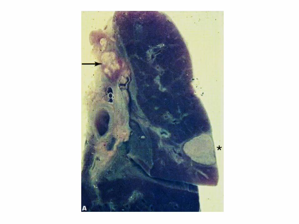

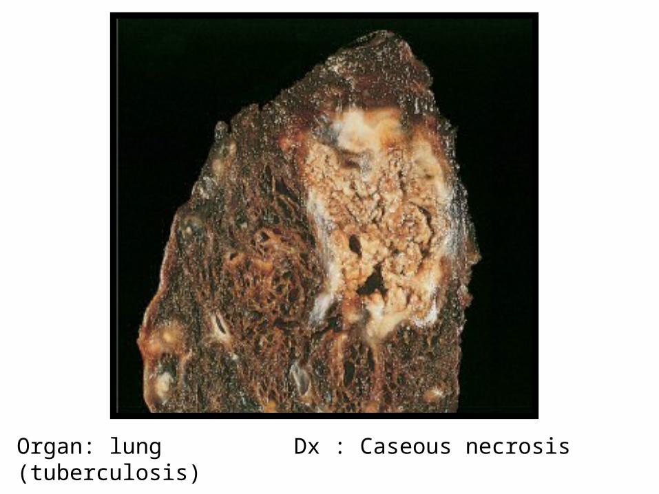
Organ: lung Dx : Caseous necrosis (tuberculosis)

Tuberculous Granulomas

Caseation Necrosis

Epitheloid cells in Granuloma

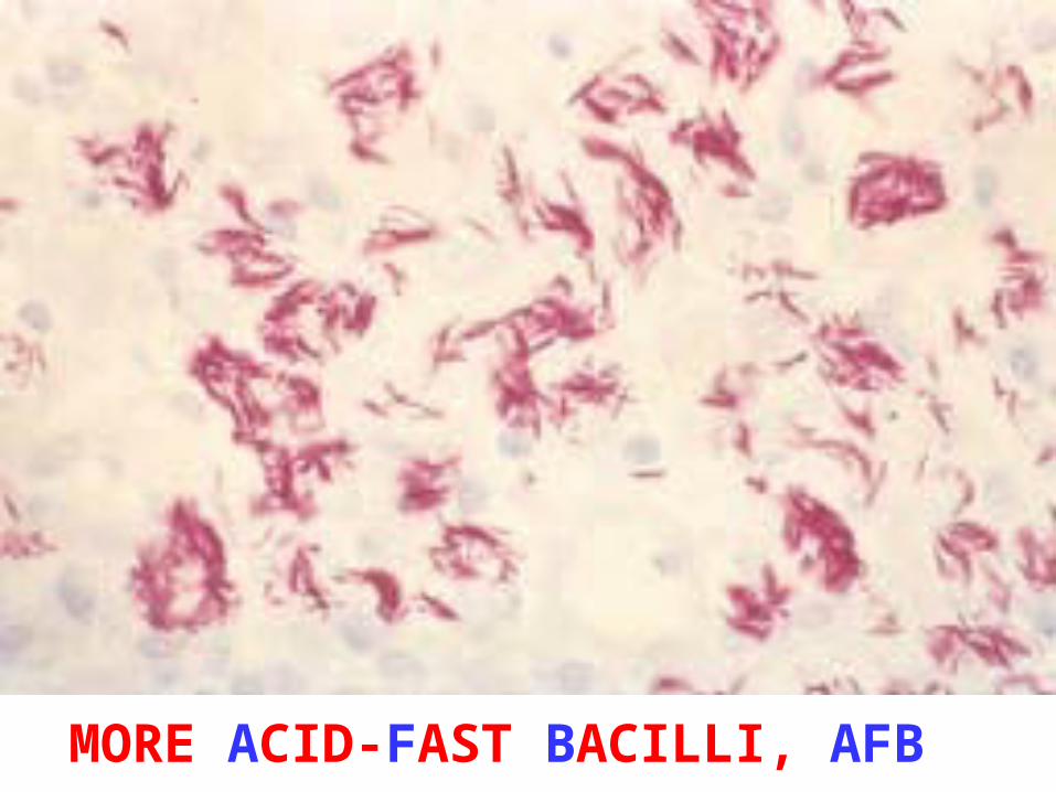
MORE ACID-FAST BACILLI, AFB

Miliary tuberculosis of the lung :
• Section of the lung shows : The alveolar septae contain many tubercles which consist of epithelioid cells , few langhan’s giant cells and peripheral rim of lymphocytes with or without caseation
2- Tuberculous lymphadenitis


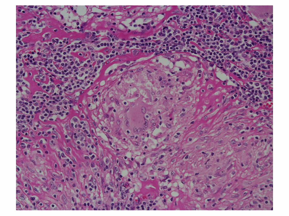

Tuberculous lymphadenitis :Section of a lymph node with connective tissue
capsule and lymphoid tissue shows:
Many round and oval tubercles/ granulomas with or without central caseation that appears structureless, homogenous and pink in colour.
The granulomas consists of epithelioid cells, few langhan’s giant cells (large cell with multiple peripheral nuclei) and peripheral rim of lymphocytes.

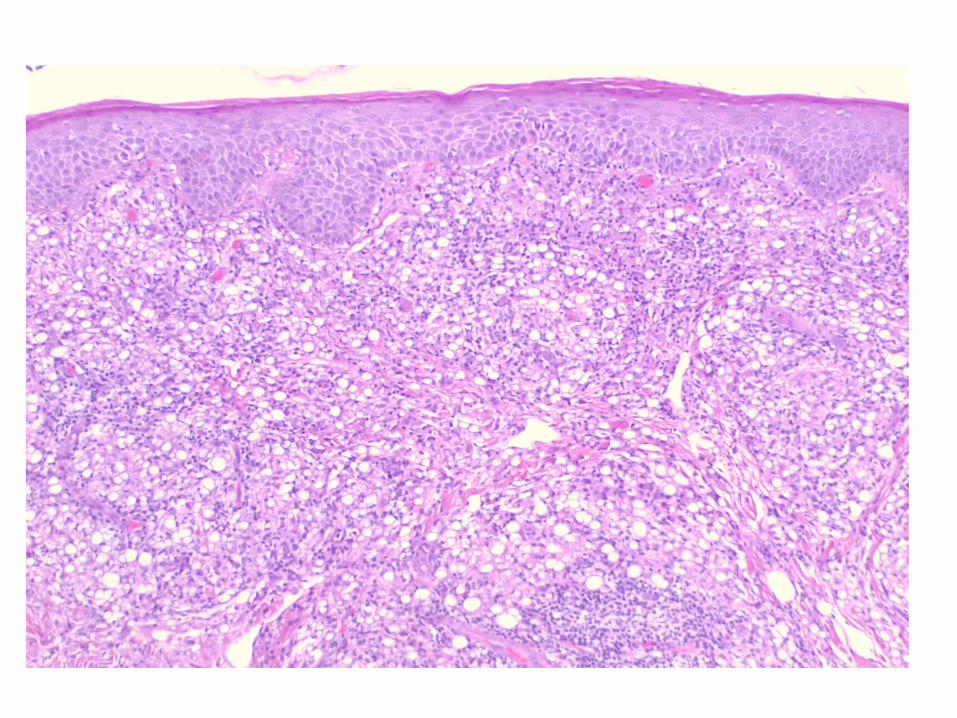
3- Bilharziasis of the colon

COLONIC BILHARZIASIS
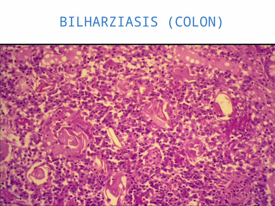
BILHARZIASIS (COLON)

SCHISTOSOMIASIS OF THE URINARY BLADDER

Bilharziasis of the rectum\urinary bladder:Section of fragments of rectal\urinary bladder mucosa shows:
Many Bilharzial ova with yellow brown shells in mucosa and submucosa surrounded by fibrosis and chronic inflammatory cells consisting of lymphocytes, plasma cells and many eosinophils.
Few granulomas are seen around the ova.

5- Cutaneous leishmaniasis

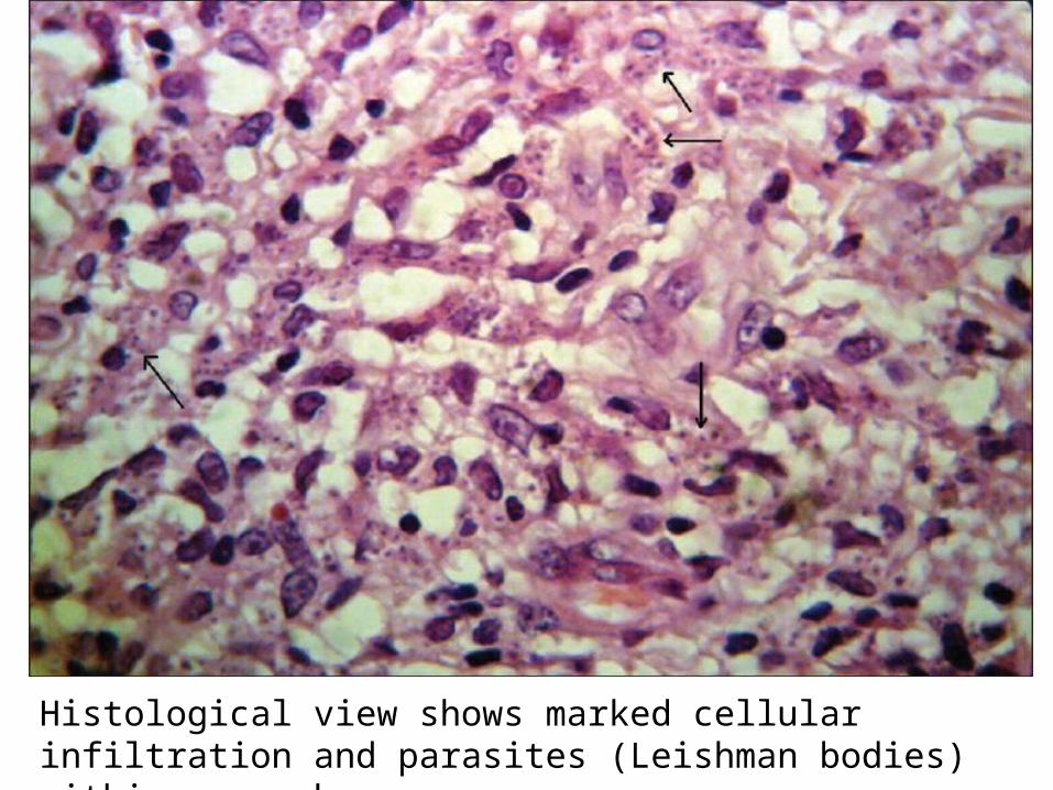
Histological view shows marked cellular infiltration and parasites (Leishman bodies) within macrophages

Picture shows a macrophage containing Leishmania amastigotes with characteristic features (round nucleus, rod-shaped kinetoplast; arrows) .
Related Documents