doi:10.1182/blood-2013-12-541698 Prepublished online March 20, 2014; Laurent Renia, Francois Nosten and Bruce Russell Rossarin Suwanarusk, Rou Zhang, Letusa Albrecht, Fabio T. M. Costa, Peter Preiser, Rose McGready, Wenn-Chyau Lee, Benoit Malleret, Yee-Ling Lau, Marjorie Mauduit, Mun-Yik Fong, Jee Sun Cho, normocytes Glycophorin C (CD236R) mediates vivax malaria parasite rosetting to http://bloodjournal.hematologylibrary.org/site/misc/rights.xhtml#repub_requests Information about reproducing this article in parts or in its entirety may be found online at: http://bloodjournal.hematologylibrary.org/site/misc/rights.xhtml#reprints Information about ordering reprints may be found online at: http://bloodjournal.hematologylibrary.org/site/subscriptions/index.xhtml Information about subscriptions and ASH membership may be found online at: digital object identifier (DOIs) and date of initial publication. the indexed by PubMed from initial publication. Citations to Advance online articles must include final publication). Advance online articles are citable and establish publication priority; they are appeared in the paper journal (edited, typeset versions may be posted when available prior to Advance online articles have been peer reviewed and accepted for publication but have not yet Copyright 2011 by The American Society of Hematology; all rights reserved. 20036. the American Society of Hematology, 2021 L St, NW, Suite 900, Washington DC Blood (print ISSN 0006-4971, online ISSN 1528-0020), is published weekly by For personal use only. at CAPES CONSORTIUM on March 24, 2014. bloodjournal.hematologylibrary.org From For personal use only. at CAPES CONSORTIUM on March 24, 2014. bloodjournal.hematologylibrary.org From

Welcome message from author
This document is posted to help you gain knowledge. Please leave a comment to let me know what you think about it! Share it to your friends and learn new things together.
Transcript

doi:10.1182/blood-2013-12-541698Prepublished online March 20, 2014;
Laurent Renia, Francois Nosten and Bruce RussellRossarin Suwanarusk, Rou Zhang, Letusa Albrecht, Fabio T. M. Costa, Peter Preiser, Rose McGready, Wenn-Chyau Lee, Benoit Malleret, Yee-Ling Lau, Marjorie Mauduit, Mun-Yik Fong, Jee Sun Cho, normocytesGlycophorin C (CD236R) mediates vivax malaria parasite rosetting to
http://bloodjournal.hematologylibrary.org/site/misc/rights.xhtml#repub_requestsInformation about reproducing this article in parts or in its entirety may be found online at:
http://bloodjournal.hematologylibrary.org/site/misc/rights.xhtml#reprintsInformation about ordering reprints may be found online at:
http://bloodjournal.hematologylibrary.org/site/subscriptions/index.xhtmlInformation about subscriptions and ASH membership may be found online at:
digital object identifier (DOIs) and date of initial publication. theindexed by PubMed from initial publication. Citations to Advance online articles must include
final publication). Advance online articles are citable and establish publication priority; they areappeared in the paper journal (edited, typeset versions may be posted when available prior to Advance online articles have been peer reviewed and accepted for publication but have not yet
Copyright 2011 by The American Society of Hematology; all rights reserved.20036.the American Society of Hematology, 2021 L St, NW, Suite 900, Washington DC Blood (print ISSN 0006-4971, online ISSN 1528-0020), is published weekly by
For personal use only. at CAPES CONSORTIUM on March 24, 2014. bloodjournal.hematologylibrary.orgFrom For personal use only. at CAPES CONSORTIUM on March 24, 2014. bloodjournal.hematologylibrary.orgFrom

- 1 -
Article Title: Glycophorin C (CD236R) mediates vivax malaria parasite rosetting to
normocytes
Running head: LEE et al CD236R and Plasmodium vivax rosetting
Section designation: E-Blood
Scientific Designation: Red Cells, Iron and Erythropoiesis
Authors: Wenn-Chyau Lee,1 Benoit Malleret,2,3 Yee-Ling Lau,1 Marjorie Mauduit,3 Mun-
Yik Fong,1 Jee Sun Cho,2 Rossarin Suwanarusk,3 Rou Zhang,2 Letusa Albrecht,4 Fabio T. M.
Costa,4 Peter Preiser,5 Rose McGready,6,7 Laurent Renia,3 Francois Nosten,6,7 and Bruce
Russell 2
Affiliations: 1Department of Parasitology, Faculty of Medicine, University of Malaya, Kuala
Lumpur, Malaysia; 2Department of Microbiology, Yong Loo Lin School of Medicine,
National University of Singapore, National University Health System, Singapore; 3Singapore
Immunology network (SIgN), A*STAR, Singapore; 4Instituto de Biologia, Universidade
Estadual de Campinas (UNICAMP), São Paulo, Brazil; 5School of Biological Sciences,
Nanyang Technological University, Singapore; 6Shoklo Malaria Research Unit, Mahidol-
Oxford Tropical Medicine Research Unit, Faculty of Tropical Medicine, Mahidol University,
Mae Sot, Thailand and 7Centre for Tropical Medicine, Nuffield Department of Medicine,
University of Oxford, Oxford, UK..
Correspondence: Bruce Russell, Vivax Malaria Laboratory, Department of Microbiology,
Yong Loo Lin School of Medicine, National University of Singapore. 5 Science Drive 2, Blk
MD4, Level 3, Singapore. 117597, Tel: (65) 6516 3517; Fax: (65) 6464 2056 E-Mail:
Blood First Edition Paper, prepublished online March 20, 2014; DOI 10.1182/blood-2013-12-541698
Copyright © 2014 American Society of Hematology
For personal use only. at CAPES CONSORTIUM on March 24, 2014. bloodjournal.hematologylibrary.orgFrom

- 2 -
Key words: Glycophorin C (CD236R), Plasmodium vivax, Vivax malaria, Rosetting,
Reticulocyte, Normocytes, CD235a, CD35, Lentivirus knock-down.
Key Points:
• Plasmodium vivax infected cells rosette exclusively to normocytes. Thus, rosetting
does not directly facilitate P. vivax merozoite invasion.
• Glycophorin C (CD236R) mediates vivax malaria parasite rosetting. This finding will
help in the search for the P. vivax rosette ligand.
Abstract:
Rosetting phenomenon has been linked to malaria pathogenesis. While rosetting occurs in all
causes of human malaria, most data on this subject has been derived from Plasmodium
falciparum. Here we investigate the function and factors affecting rosette formation in
Plasmodium vivax. To achieve this, we utilised a range of novel ex vivo protocols to study
fresh and cryopreserved P. vivax (n=135) and P. falciparum (n=77) isolates from Thailand.
Rosetting is more common in vivax than falciparum malaria, both in terms of incidence in
patient samples and percentage of infected erythrocytes forming rosettes. Rosetting to P.
vivax asexual and sexual stages was evident 20 hrs post reticulocyte invasion, reaching a
plateau after 30 hrs. Host ABO blood group, reticulocyte count and parasitemia were not
correlated with P. vivax rosetting. Importantly, mature erythrocytes (normocytes) rather than
reticulocytes preferentially form rosetting complexes, indicating this process is unlikely to
directly facilitate merozoite invasion. While antibodies against host erythrocyte receptors
CD235a and CD35 had no effect; Fab against the BRIC 4 region of CD236R significantly
inhibited rosette formation. Rosetting assays using CD236R knock down normocytes derived
For personal use only. at CAPES CONSORTIUM on March 24, 2014. bloodjournal.hematologylibrary.orgFrom

- 3 -
from hematopoietic stem cells, further supports the role of Glycophorin C as a receptor in P.
vivax rosette formation.
Introduction:
In malariology, rosetting is defined by the adherence of uninfected erythrocytes to a
Plasmodium spp. -infected erythrocyte. While the role of rosetting phenomenon remains
unknown, two major hypotheses have been proposed to explain its importance to the survival
and fitness of the malaria parasite. Firstly the ‘rosette assisted invasion’ hypothesis, which
supposes that rosetting facilitates the encounter of newly emergent merozoites with receptive
uninfected red cells bound to the schizont1-3. The second major hypothesis proposes that
uninfected cells rosetting shield the infected red blood cell from the host immune system. 2,4
Since its discovery in the 1980s5,6, rosetting phenomenon has been observed in the
four major causes of human malaria 1,7-10. However, almost all rosetting studies have focused
on P. falciparum and its possible role in the pathogenesis of severe disease11-17. A renewed
interest in vivax malaria and a better appreciation of its importance to public health has led to
an increased number of studies examining particular aspects of P. vivax pathogenesis18-24,25.
Certainly in the case of P. vivax, apart from some initial descriptions on P. vivax rosetting 8,10
and its association with anaemia26, little has been done to investigate the importance of
rosetting to P. vivax’s survival within human host and the molecular mechanisms associated
with the formation of rosettes in this species.
Due to the technical challenges associated with P. vivax research, the properties, as
well as the postulated roles of rosetting in vivax malaria have been extrapolated from
experiments conducted on P. falciparum 3,4. However, comparisons between these two
species are problematic, especially when considering the receptor ligand interactions
involved in the formation of rosettes. This is because one of the ligand associated with rosette
For personal use only. at CAPES CONSORTIUM on March 24, 2014. bloodjournal.hematologylibrary.orgFrom
- 4 -
formation in P. falciparum (P. falciparum erythrocyte membrane protein 1(PfEMP1)), has no
orthologs in P. vivax. Thus all of receptors on the host red cell corresponding to PfEMP1
such as complement receptor 1 (CD35)27, Blood group antigens (A and B) 28-30, heparan
sulphate 31 and thrombospondin (CD36) 32,33 may not be relevant to P. vivax rosette
formation.
Recent advances in our ability to manipulate ex vivo isolates of P. vivax 25 have
enabled us to conduct detailed investigations on the rosetting phenomenon in this species
allowing us conduct side by side comparisons with rosetting in P. falciparum. Specifically
this study has two major objectives. First, to evaluate the postulated roles of rosette formation
in P. vivax and second, to elucidate erythrocytic receptors that are involved in P. vivax rosette
formation.
Methodology:
A summary of the methodology applied, number of isolates utilised and corresponding figure
index is shown in a flow chart (Figure 1).
Blood Sample Collection
A total of 87 fresh isolates of P. vivax and 77 fresh isolates of P. falciparum were used in this
study. Another 48 cryopreserved P. vivax isolates were also used. All isolates were obtained
from malaria patients presenting at the clinics of the Shoklo Malaria Research Unit (SMRU)
in North-western Thailand. The clinical samples were collected and tested in accordance to
protocols approved by The Center for Clinical Vaccinology and Tropical Medicine at
University of Oxford (OXTREC 58-09 and OXTREC 04-10) in consultation with the Ethics
Committee of the Faculty of Tropical Medicine at Mahidol University. The study was
conducted in accordance with the Declaration of Helsinki. Blood samples were collected
For personal use only. at CAPES CONSORTIUM on March 24, 2014. bloodjournal.hematologylibrary.orgFrom

- 5 -
using BD Vacutainer® with lithium heparin anticoagulant. ABO blood group of each sample
was determined via standard hemagglutination with TransClone® Anti-A and Anti-B
antibodies (Bio-Rad, California, USA). A thick and thin blood smear were prepared from
each blood sample to determine species of malaria parasites involved, parasitaemia and the
predominant erythrocytic stage of the parasite. Reticulocyte concentrations were prepared
from human cord blood using the method outlined by Russell et al. 2011 24 .
Rosetting Assay on Fresh Samples
Plasmodium sp. infected blood samples with at least 70% of parasite population in “ring”
forms were cultivated at 3% hematocrit using McCoy’s 5A medium enriched with 20%
homologous serum, using the method described by Russell et al. 24. Samples were checked
frequently and sampled at ring, early trophozoite, late trophozoite and schizont stages. The
presence of rosettes and living parasites were detected and quantified using a novel Giemsa
subvital staining methodology 34 modified from techniques applied in previous study 8.
Briefly, the sampled culture suspension was stained with Giemsa (the final stain
concentration was 5%) for 15 minutes. A small volume of this suspension (7.5 μl) was used
to make a wet mount with 22 x 32 mm (0.17 mm thickness) glass cover slip. The wet mount
was examined immediately with light microscope under oil immersion magnification.
Rosetting rate was then determined by examining 200 infected erythrocytes (in McCoy’s 5A
medium enriched with 20% homologous serum).
Rosetting Assay on Cryopreserved Samples
Vivax malaria blood samples with at least 70% of parasite population in “ring” forms were
cryopreserved using the glycerolyte method as described previously22,35. Prior to
cryopreservation, vivax malaria samples with parasitaemia lower than 0.1% were subjected to
For personal use only. at CAPES CONSORTIUM on March 24, 2014. bloodjournal.hematologylibrary.orgFrom

- 6 -
75% Percoll density gradient centrifugation to concentrate the parasitized erythrocytes. The
cryopreserved samples were then thawed using the sodium chloride method 36. The thawed
samples were then matured and rosetting assay was conducted as described above. Rosetting
prevalence of each cryopreserved isolate was determined.
Erythrocyte subset preference study
Blood samples infected with either P. vivax or P. falciparum were cultivated and matured
until at least 60% of the parasite population reached schizogony. One hundred µl of each
culture suspension was used as a control where its rosetting rate was determined. Matured
parasites from the remaining culture suspension were then concentrated with Magnetic
Activated Cell Sorting Column (MACS, Miltenyi Biotec). The concentrated infected packed
erythrocytes (volume = x) were then mixed with 0.5 X volume of uninfected packed
normocytes and 0.5X volume of uninfected reticulocytes. The cell mixtures were suspended
in enriched McCoy’s 5A medium as described above. The cell mixtures were mixed well by
vortexing to disrupt rosettes. Importantly in a preliminary study (data not shown), we found
that rosettes were disrupted even after one minute of vortexing (we used two minutes to be
absolutely sure of total disruption). The free infected IRBCs started reforming rosettes within
seconds, and the vortexed isolate returned to the original rosetting frequency within five
minutes. To ensure maximal rosette formation we incubated the vortexed blood mixtures at
37°C for 30 minutes. After that, rosetting assay was conducted as described above. Types of
erythrocytes involved in each rosette were studied, and the erythrocyte subset preference for
rosetting in P. vivax and P. falciparum were then determined.
For personal use only. at CAPES CONSORTIUM on March 24, 2014. bloodjournal.hematologylibrary.orgFrom

- 7 -
Erythrocyte receptor blocking study using Fabs and Antibodies
Preliminary trials clearly showed that the intact anti-glycophorin C and A antibodies caused
considerable hemagglutination (not observed with CD35 antibodies). Therefore it was
necessary to use Fab fragments of anti-CD236R and CD235a monoclonal antibodies.
Mouse anti-human CD236R clone BRIC 4 IgG (Thermo Scientific Pierce, USA), mouse anti-
human CD236R clone BRIC 10 IgG (Abcam®, Cambridge, UK) and mouse anti-human
glycophorin A IgG (Abcam®, Cambridge, UK) were used for this experiment. Fab fragments
of the respective antibodies were prepared using the Pierce® Fab Micro Preparation Kit-
Resin Kit (Thermo Scientific Pierce, USA). Plasmodium sp. infected blood samples were
cultured and matured as described above. After that, the culture suspensions were vortexed
vigorously for two minutes to mechanically dissociate any preformed rosettes. Each culture
suspension were divided into four 1.5 ml microcentrifuge tubes, where each tube of culture
suspension was added with one type of the prepared antibody or Fab fragments (1 mg/ml
final concentration), and one treatment free tube was the negative control for the experiment.
The mixtures were incubated for 30 minutes at 37°C. After that, rosetting assay was
conducted on each tube of suspension as described above. In addition to the Fab treatments
we also used mouse anti-human CD 35 antibody (BD Pharmigen™) as a positive control.
Hematopoietic cell culture and CD236R knock down experiments.
Cryopreserved human umbilical Cord Blood Mononuclear Cells (HUMC) were expanded
using a method described by Giarratana et al. 37 with few modifications. Briefly, the CD34+
hematopoietic progenitors were isolated by immunomagnetic selection using CD34
MicroBead kit (Miltenyi Biotec, Singapore). The CD34+ cells were harvested during 3 weeks
in IMDM Glutamax (Gibco) supplemented with 330 μg/mL of human holotransferin (Sigma,
Singapore), 10 μg/mL recombinant human insulin (SAFC Biosciences), 2 IU/mL heparin
For personal use only. at CAPES CONSORTIUM on March 24, 2014. bloodjournal.hematologylibrary.orgFrom

- 8 -
(Sigma) and 5% AB serum. Between day 0 and 7, the following additional supplementation
were incorporated: 10-6 M of hydrocortisone (Stemcell Technologies™), 100 ng/mL of SCF
(Pepro Tech®), 5ng/mL of IL-3 (Pepro Tech®) and 3 UI/mL of erythropoietin (Epo)
(StemCell Technologies™), between day 7 and day 11, 100 ng/mL of SCF and 3 UI/mL of
Epo, and after day 11 only Epo was added.
One day after the immunomagnetic sorting, the CD34+ cells were transduced (MOI of 5)
with shRNA lentitvector MISSION™, shRNA Target Set NM_002101 (Glycophorin C
(CD236R)) (Sigma®) in the presence of 8 μg/mL of Polybrene (Sigma). Fifteen days after
the transduction, GFP positive and negative cells were sorted using Influx BD flow cytometer
(BD Biosciences). The day after, GFP positive and negative fraction were incubated with P.
vivax late stages isolated by magnetic sorting as described previously38. The level of CD236R
expression in each subset was measured by flow cytometry using anti-human CD236R, clone
BRIC -4 (Thermo Scientific Pierce, USA) antibody as primary staining and anti-mouse e660
(eBioscience) as secondary staining. The cells were acquired on LSRII flow cytometer (BD
Biosciences) and analysed with FlowJo software (Tree Star).
Statistical Analyses
In most cases rosetting rate was defined as the percentage of 200 infected erythrocytes that
formed stable adhesion with at least one uninfected erythrocyte after 15 minutes of post
vortex incubation (as observed under 100 X oil immersion). Giant rosette is defined as a
rosetting complex with the participation of at least 10 uninfected erythrocytes 9-11. As most
of the data was not normally distributed, non-parametric analysis such as Mann-Wittney U,
Kruskal Wallis (more than 2 groups) and Friedman’s test (repeated measures)) were utilised.
In the latter two tests Dunns’ post hoc analysis was used. All statistical analysis used Prism 5
for Windows (version 5.01), Software MackievTM.
For personal use only. at CAPES CONSORTIUM on March 24, 2014. bloodjournal.hematologylibrary.orgFrom

- 9 -
Results:
Cryopreservation and Rosetting
At the beginning of this study, for purposes of practicality, we utilised cryopreserved isolates
of P. vivax. However, it soon became evident that the rates of rosetting in these
cryopreserved-then-thawed samples were considerably lower than those previously reported
from fresh isolates 10. Subsequently, we decided to focus our work on fresh isolates rather
than cryopreserved (with the exception of the knock down experiment). Certainly, when we
compared our data on rosetting prevalence from fresh isolates with cryopreserved isolates, we
showed that cryopreservation was associated with a lower incidence of rosette formation;
where the median rosetting rate for fresh isolates being 23.80% (Inter quartile range (IQR)
[10.00-31.00%]) while median rosetting rate for cryopreserved isolates being 0.00% (IQR
[0.00-14.85%]) on P (two-tailed) < 0.0001, Mann-Whitney test, (Supplemental Figure 1).
Rosetting properties of P. vivax
Rosette formation of P. vivax increased with the parasite erythrocytic maturation (Figure 2).
In this study, no early stage (ring form) was found to be involved in rosette formation.
Rosette formation of P. vivax was noted 24 hours post cultivation, represented by early
trophozoite stage. The rosetting rate increased markedly between early trophozoite stage and
the late trophozoite stage. After 30 hours the rosetting development continued, albeit with
much lower rate, until the parasite reached schizogony at hour 44.
Giant rosettes were found occasionally in P. vivax isolates. 10% of the rosettes found
in P. vivax isolates were giant rosettes. On the other hand, only 0.5% of the rosettes found in
P. falciparum isolates were giant rosettes. Besides the asexual stages, gametocytes of P. vivax
were found to be involved in rosette formation (Figure 2B). Involvement of gametocyte-
For personal use only. at CAPES CONSORTIUM on March 24, 2014. bloodjournal.hematologylibrary.orgFrom

- 10 -
infected erythrocytes in rosette formation was found in P. falciparum isolates as well (Figure
2B).
Rosetting rate and rosetting prevalence
Rosetting occurs as frequently in isolates of P. vivax (91.5%, 43/47) as it does in P.
falciparum (79.5%, 31/39) confirming earlier studies by Udomsangpetch et al. 10.
Interestingly, the median rate of rosetting was significantly higher in P. vivax (24.5%) when
compared to P. falciparum (9.0%) isolates from the same area in Thailand, P<0.001 (Figure
3A). No significant correlation was found between rosetting rate and parasitemia or ABO
blood groups in both vivax and falciparum malaria patients (Figure 3B and C). The
parasitaemia range for P. vivax and P. falciparum was; 0.01 - 1.70% and 0.10 – 11.00%
respectively.
Erythrocyte subset preference study
The availability of reticulocytes in the peripheral blood of vivax or falciparum malaria
patients had no bearing on the incidence of rosettes (Figure 3D). Close inspection of rosettes
from 9 vivax malaria (562 rosettes) and 11 falciparum (391 rosettes) malaria isolates revealed
that only ~10% of rosettes involved attachment of reticulocytes (and in these cases only one
reticulocyte per rosette could be observed); the majority of rosettes are exclusively composed
of mature erythrocytes (Figure 4A).
As reticulocytes only make up a small portion of the peripheral blood (<2%), we
wanted to ensure that the lack of reticulocyte involvement in rosette formation wasn’t only a
function of the lower probability of parasitized cells encountering the reticulocyte (rare)
versus the commonly found normocyte. To test this we added MACS-concentrated P. vivax
and P. falciparum IRBCs to blood samples containing enriched reticulocytes (50%+/-10).
For personal use only. at CAPES CONSORTIUM on March 24, 2014. bloodjournal.hematologylibrary.orgFrom

- 11 -
When the reticulocyte enriched experiment group were compared with the paired rosetting
data from the control group (the non-enriched blood) using Wilcoxon signed rank test;
rosetting rates for P. vivax, was unaffected whereas P. falciparum rosetting rates actually
decreased in the enriched reticulocyte treatments P < 0.01 (one-tailed, Mann-Whitney test)
(Figure 4B, C). While we did observe the occasional P. vivax rosette with reticulocytes
attached in the enriched treatment, the formation of rosettes almost exclusively involved
normocytes, despite the even chance to encounter reticulocytes. As there may be concerns
regarding the difference between cord blood reticulocytes and adult reticulocytes, we
repeated the reticulocyte enrichment experiment using three more fresh isolates of P.vivax
and reticulocytes isolated from two adult donors. We found no significant difference between
the adult and cord blood derived reticulocytes in their lack of involvement in P. vivax rosette
formation (Supplemental Figure 2).
Rosetting Inhibition Assay
Fab and Antibody blocking experiment
In vivax malaria isolates tested, Fab of mouse anti-human CD236R IgG (Clone BRIC 4)
(Figure 5A shows where this Fab clone binds) significantly reduced rosetting rate by 27% (P
< 0.05, Friedman analysis with Dunn’s multiple comparison test) (Figure 5B). Interestingly
BRIC 4 Fabs reduced rosette formation in falciparum malaria isolates by at least half (Figure
5C). Importantly, the Fab fragment of another anti-CD236R antibody had no effect on rosette
formation in the same paired experiments demonstrating the specificity of the inhibition of
BRIC4 antibodies (Figure 5B). Anti- Glycophorin A Fab fragments had no effect on rosette
formation for both species (Figure 5D).
For personal use only. at CAPES CONSORTIUM on March 24, 2014. bloodjournal.hematologylibrary.orgFrom

- 12 -
CD35 blocking experiment
Complement receptor 1 or CD35 is one of the erythrocytic receptors involved in rosetting of
P. falciparum27. Certainly in the P. falciparum isolates we tested, the rosetting rate was
significantly inhibited (33.3%) by the mouse anti-human CD35 antibody, P <0.0001, R =
0.8236, 95% CI 3.026 to 6.504 However, CD35 had no effect on rosette formation in the P.
vivax isolates tested (Figure 5E).
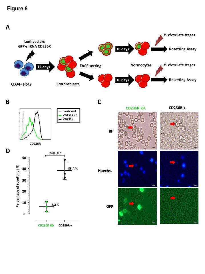
Hematopoietic cell culture and CD236R knock down
Although the above Fab mediated inhibition assays provide useful clues to the identity of the
host rosetting receptor, such an approach is limited by potential off target effects (such as
steric hindrance or changes to membrane rigidity) 39. To limit these unwanted ‘off target
effects’ we employed a transgenic method to knock down the expression of CD236R to
produce normocytes mostly deficient in this receptor (Figure 6A). Phenotyping of GFP
positive and GFP negative cells (according to BRIC 4 region recognition (Figure 5A))
showed a knock down of 81.5% ± 2.5% in CD236R expression (Figure 6B). It should be
noted that the method we employed to knock down the expression of Glycophorin A was
different to one recently used by Bei et al.40 (where puromycin and neomycin rather than
GFP was used for selection ) but with the same knock down efficiency. It is important to note
that the erythrocytes generated from the CD236R knockdown had a normal phenotype in
terms of seven other characteristic erythrocytic receptors (Supplemental Figure 3)
Initially this work needed to be conducted in Singapore rather than Mae Sod, thus we
had to rely on cryopreserved isolates of P. vivax to conduct this experiment. Of the three P.
vivax cryopreserved isolates used only one showed very high levels of rosetting (>50%).
Despite this limitation, differences in rosetting between the control normocytes (CD236R
positive) and CD236R knockdown normocytes was immediately evident. After examination
For personal use only. at CAPES CONSORTIUM on March 24, 2014. bloodjournal.hematologylibrary.orgFrom

- 13 -
of 200 fields (x1000) and counting 50 P. vivax IRBCs per treatment, only 4 of 50 were
rosetting with CD236R knockdown erythrocytes compared to 27 out of 50 in the control
(Chi-square = 22.6, d.f =1, P<0.0001 (Chi-square with Yates’ correction)). As we were
concerned about possible confounders associated with the use of cryopreserved isolates
(Supplemental Figure 1) we repeated this experiment again this time using 3 fresh isolates.
As per the original experiment, we found that the CD236R knockdown erythrocytes
significantly reduced the formation of rosettes (P=0.007) (Figure 6D). It is important to
understand that although the cells were sorted on GFP, this protein has about 26 hrs half-life
41, Therefore in mammalian cells the signal gradually diminishes as the erythrocyte matures.
Consequently the normoblasts (Heilmeyer stage 0) and early reticulocytes (Heilmeyer stage I,
II and III) have bright signals. Mature reticulocytes (Heilmeyer stage IV) and normocytes
have little or no discernable signal (figure 6C). An unintended benefit of this age related GFP
signal output was that in the four rosettes observed in the CD236R knockdown, no rosette
was associated with GFP positive cells (reticulocytes), thus further supporting our data on P.
vivax rosette normocytic preference.
Discussion:
The biological role of P. vivax and P. falciparum rosetting
While the role of rosetting in the pathogenesis of malaria remains controversial, the real
mystery surrounding this phenomenon is what advantage it brings to the intra erythrocytic
parasite. While not providing a definitive answer to this question; our data provides
compelling evidence that rosetting does not facilitate the invasion of merozoites. We have
two lines of evidence disputing the ‘rosette assisted invasion’ hypothesis.
Foremost and most convincingly our data shows that P. vivax preferentially forms
rosettes with mature red cells and not reticulocytes. If P. vivax rosettes were to truly assist in
For personal use only. at CAPES CONSORTIUM on March 24, 2014. bloodjournal.hematologylibrary.orgFrom
- 14 -
merozoite invasion, one would hypothesize that its schizonts would preferentially adhere to
uninfected reticulocytes; thereby improving invasion success by the provision of immediate
access to proximal reticulocytes (which are generally scarce in the peripheral circulation).
However, the observations of naturally occurring rosettes from fresh isolates and our
reticulocyte enrichment experiment clearly show rosettes favour normocytes rather than
reticulocyte attachment. As P. vivax merozoites exclusively invade reticulocytes, there would
be little advantage for the parasite species to have its schizonts binding with normocytes
which are not receptive to invasion. As P. falciparum merozoites can invade both
reticulocytes and normocytes, its preferential rosette formation with normocytes does not
discount the possibility that rosetting may improve the invasion success of merozoites in this
species. However, convincing data from Clough et al. 1998 shows that rosette forming strains
of P. falciparum have no invasion advantage over non-rosetting strains 42. Unpublished data
(Supplemental Figure 4) derived from an earlier study on P. vivax invasion24, clearly shows
that the rate of rosetting within an isolate bears no correlation with invasion success.
A second line of evidence against the ‘rosette assisted invasion’ hypothesis is that P.
vivax and P. falciparum start rosetting at early trophozoite stage, at least 24 hrs prior to the
expulsion of merozoites from the segmenting schizont. While some may suggest that the
developing parasite is getting an ‘early start’ on the collection of receptive cells, such an
effort would be wasted as the shear forces present in the host circulation and physical barriers
such as the splenic red pulp would undoubtedly disrupt the rosette numerous times within the
24 hrs time interval between the initiation of rosette formation and schizont rupture. Perhaps
even more convincingly is our observation that P. vivax and P. falciparum gametocytes also
form rosettes, a new finding contrary to the earlier report from Lowe et al. 1998 1.
Clearly data from this and earlier studies 24,42 suggest that rosetting does not directly assist in
the targeting or invasion of malaria parasites into uninfected host erythrocytes. While we
For personal use only. at CAPES CONSORTIUM on March 24, 2014. bloodjournal.hematologylibrary.orgFrom

- 15 -
don’t have data to support the alternative hypothesis which proposes that rosetting shields the
infected red blood cell from the host immune system (first postulated by Mats Wahlgren in
1989)3; we agree that this is the most likely reason for such an adaptation in Plasmodium spp.
It certainly seems intuitive that infected red blood cell containing mature stages (and the
associated plethora of parasite antigens expressed on the red cell membrane) would greatly
benefit from a cloak of host red blood cells. Again this hypothesis is relatively speculative,
and we hope that future studies will focus on the role of rosetting in the protection of
Plasmodium spp. from the host immune system.
The observation that rosetting occurs as frequently in P. vivax as it does in P.
falciparum, raises a number of important questions regarding the clinical significance of
rosetting in vivax malaria. While our data is from de-identified samples (thus not allowing
direct clinical correlates with the rosetting rate), we do know that all our samples were
collected from uncomplicated cases of vivax malaria. As such, it is difficult to speculate on
the direct role of rosetting on P. vivax pathogenesis. We certainly hope future clinical
investigations into severe vivax malaria, includes rosetting as an important parasitological
parameter.
Erythrocyte receptors mediating rosetting in fresh isolates of P. vivax.
While numerous studies have shown that the human ABO blood-group and CR1 (CD35) are
key determinants on P. falciparum rosetting 7,27-30, this does not seem to be the case with P.
vivax. The absence of association between P. vivax rosetting and ABO blood group agrees
with the findings from an earlier study by Chotivanich et al. 1998 8. The lack of association
between ABO blood-group and CR1 in P. vivax should not be surprising as the ligand
responsible for this interaction in P. falciparum (PfEMP1) is absent from P. vivax. Our
efforts to shortlist novel receptors mediating rosetting in P. vivax were aided by unpublished
For personal use only. at CAPES CONSORTIUM on March 24, 2014. bloodjournal.hematologylibrary.orgFrom

- 16 -
data from one of the authors of this study, which indicated that CD236R modulates P.
falciparum rosetting in PfEMP1-independent manner. Certainly our results show that
specifically blocking the BRIC 4 region (amino acid residues 2-21) of CD236R significantly
inhibits rosette formation in P. vivax and P. falciparum. As most of the BRIC 4 region is
located in segment one of CD236R, it is unlikely that the Gerbich and Yus mutations (which
cause major deletions to regions 2 and 3) will have any effect on its ability to form rosettes
(Figure 5A). While the Leach mutation would indeed remove the residues of interest, we did
not attempt to source erythrocytes from this rare phenotype, as the mutant cells are elliptical
(not biconcave) in shape and have a profoundly modified biomechanical profile that would
confound any conclusions regarding their ability to rosette (irrespective of the complete
absence of CD236R in the Leach type cells). It is interesting to note that trypsin treatment of
erythrocytes completely removes two of extracellular domains of CD236R (1 and 2) (Figure
5A) resulting in the complete abrogation of rosetting in P. vivax 24.
To better understand the importance of CD236R as a key host receptor for P. vivax
rosetting we knocked down its expression in normocytes derived from hematopoietic stem
cells. Rosetting assays using these CD236R knock down erythrocytes clearly show the
significant reduction of rosetting phenomena in P. vivax compared to normocytes (from the
same culture) expressing this receptor.
One problem with the idea ‘CD236R is important to rosetting’, is that presentation
this receptor is decreasing during reticulocyte maturation to normocyte, however is still
highly expressed in both subsets43. As P. vivax does not rosette well with reticulocytes, one
has to question what prevents the interaction between reticulocyte CD236R and the yet to be
discovered P. vivax rosetting ligands. We postulate that the rigid biomechanical properties of
reticulocytes (reticulocytes are significantly stiffer than normocytes)43 inhibit formation of
close bonds with the P. vivax IRBC. Close examination of images of P. vivax rosettes
For personal use only. at CAPES CONSORTIUM on March 24, 2014. bloodjournal.hematologylibrary.orgFrom

- 17 -
especially in figure 4C show the tight association of the normocyte and infected cell (the flat
face of the biconcave disk to the IRBC). The globular and stiff reticulocyte inhibits intimate
contact with the P. vivax IRBC. Certainly our efforts to manipulate reticulocytes into direct
contact with P. vivax cells (using a micropipette) failed to promote rosetting.
Conclusions:
Rosetting is a common property of mature intra-erythrocytic malaria parasites, occurring as
frequently in P. vivax as it does in P. falciparum. The high frequency of rosetting in patient
isolates suggests that it provides P. vivax with a survival advantage. Our ex vivo experiments
indicate that in the case of P. vivax, this advantage is most likely to be associated with
immune invasion rather than facilitating the invasion of reticulocytes. While ligands and
receptors primarily involved in formation of rosettes in P. vivax still remain a mystery, we
provide strong evidence that CD236R is involved whereas receptors traditionally involved in
P. falciparum rosettes, such as ABO antigens and CD35 are not. We also wish to stress that
as with P. falciparium, there is likely to be more than one host receptor involved in P. vivax
rosetting. Future work should focus on the discovery of P. vivax ligands that modulate the
attachment of normocytic CD236R and other possible receptors involved in the rosetting
process.
Conflict of interest disclosure:
The authors declare that they have no competing interests.
Authors' contributions:
WCL, BR, BM, MM, RZ, RS, JSC, LA and FTMC carried out laboratory work, collected and
analyzed the data. RM, FN, YLL and MYF for clinical management of patients, ethical
For personal use only. at CAPES CONSORTIUM on March 24, 2014. bloodjournal.hematologylibrary.orgFrom

- 18 -
clearance and collection and processing of the blood samples. WCL, BR, PP, YLL and MYF
BM and LR participated in data interpretation and helped to draft the manuscript. All authors
read and approved the final manuscript.
Acknowledgements:
We would like to express our gratitude to the staff of SMRU who assisted in the management
of this research study. BR and LR are adjunct Professors at the Department of Parasitology,
University of Malaya, Kuala Lumpur, Malaysia. We would like to thank the SIgN flow
cytometry core lead by Dr. Anis Larbi and his team members, Ivy Low Nurhidaya Shadan,
Seri Mustafahi and Nurhidaya Shadan.
Funding Support: This study received financial supports from the following funds: WCL
was supported by University of Malaya Research Grant (UMRG) (PV044-2012-A), YLL and
MYF were supported by University of Malaya, High Impact Research Fund UM-MOHE
(UM.C/625/1/HIR/MOHE/CHAN/14/3) from the Ministry of Higher Education Malaysia and
LR was supported by funding from SIgN and from the Horizontal Programme on Infectious
Diseases and BM from a Young Investigator Grant (BMRC YIG Grant No:13/1/16/YA/009)
under the Agency for Science, Technology and Research (A*STAR, Singapore). SMRU is
part of the Mahidol Oxford University Research Unit, supported by the Wellcome Trust of
Great Britain. BR and BM were funded by National University of Singapore Faculty Start-Up
Grant and the Singapore NMRC, (NMRC/CBRG/0047/2013).
Presented in Part: International Malaria Symposium, Sabah, Malaysia, 16-17th April 2013
and the Gordon Research Conference (Malaria), 4-9th Lucca, Italy. 2013.
For personal use only. at CAPES CONSORTIUM on March 24, 2014. bloodjournal.hematologylibrary.orgFrom

- 19 -
References
1. Lowe BS, Mosobo M, Bull PC. All four species of human malaria parasites form
rosettes. Trans R Soc Trop Med Hyg.1998;92(5):526-526.
2. Rowe JA, Obiero J, Marsh K, Raza A. Short report: Positive correlation between
rosetting and parasitemia in Plasmodium falciparum clinical isolates. Am J Trop Med Hyg.
2002;66(5):458-460.
3. Wahlgren M, Carlson J, Udomsangpetch R, Perlmann P. Why do Plasmodium
falciparumm-infected erythrocytes form spontaneous erythrocyte rosettes? Parasitol Today.
1989;5(6):183-185.
4. Rowe JA. Rosetting. In: Sherman IW, ed. Molecular Approaches to Malaria.
Washington DC: ASM Press; 2005:416-426.
5. David PH, Handunnetti SM, Leech JH, Gamage P, Mendis KN. Rosetting: a new
cytoadherence property of malaria-infected erythrocytes. Am J Trop Med Hyg.
1988;38(2):289-297.
6. Handunnetti SM, David PH, Perera KL, Mendis KN. Uninfected erythrocytes form
"rosettes" around Plasmodium falciparum infected erythrocytes. Am J Trop Med Hyg.
1989;40(2):115-118.
7. Angus BJ, Thanikkul K, Silamut K, White NJ, Udomsangpetch R. Short report:
Rosette formation in Plasmodium ovale infection. Am J Trop Med Hyg. 1996;55(5):560-561.
8. Chotivanich KT, Pukrittayakamee S, Simpson JA, White NJ, Udomsangpetch R.
Characteristics of Plasmodium vivax-infected erythrocyte rosettes. Am J Trop Med Hyg.
1998;59(1):73-76.
9. Udomsangpetch R, Wahlin B, Carlson J, et al. Plasmodium falciparum-infected
erythrocytes form spontaneous erythrocyte rosettes. J Exp Med. 1989;169(5):1835-1840.
For personal use only. at CAPES CONSORTIUM on March 24, 2014. bloodjournal.hematologylibrary.orgFrom

- 20 -
10. Udomsanpetch R, Thanikkul K, Pukrittayakamee S, White NJ. Rosette formation by
Plasmodium vivax. Trans R Soc Trop Med Hyg. 1995;89(6):635-637.
11. al-Yaman F, Genton B, Mokela D, et al. Human cerebral malaria: lack of significant
association between erythrocyte rosetting and disease severity. Trans R Soc Trop Med Hyg.
1995;89(1):55-58.
12. Carlson J, Holmquist G, Taylor DW, Perlmann P, Wahlgren M. Antibodies to a
histidine-rich protein (PfHRP1) disrupt spontaneously formed Plasmodium falciparum
erythrocyte rosettes. Proc Natl Acad Sci U S A. 1990;87(7):2511-2515.
13. Ho M, Davis TM, Silamut K, Bunnag D, White NJ. Rosette formation of Plasmodium
falciparum-infected erythrocytes from patients with acute malaria. Infect Immun.
1991;59(6):2135-2139.
14. Treutiger CJ, Hedlund I, Helmby H, et al. Rosette formation in Plasmodium
falciparum isolates and anti-rosette activity of sera from Gambians with cerebral or
uncomplicated malaria. Am J Trop Med Hyg. 1992;46(5):503-510.
15. Udomsangpetch R, Todd J, Carlson J, Greenwood BM. The effects of hemoglobin
genotype and ABO blood group on the formation of rosettes by Plasmodium falciparum-
infected red blood cells. Am J Trop Med Hyg. 1993;48(2):149-153.
16. Udomsangpetch R, Webster HK, Pattanapanyasat K, Pitchayangkul S, Thaithong S.
Cytoadherence characteristics of rosette-forming Plasmodium falciparum. Infect Immun.
1992;60(11):4483-4490.
17. Wahlgren M, Carlson J, Helmby H, Hedlund I, Treutiger CJ. Molecular mechanisms
and biological importance of Plasmodium falciparum erythrocyte rosetting. Mem Inst
Oswaldo Cruz. 1992;87 Suppl 3:323-329.
For personal use only. at CAPES CONSORTIUM on March 24, 2014. bloodjournal.hematologylibrary.orgFrom

- 21 -
18. Anstey NM, Handojo T, Pain MC, et al. Lung injury in vivax malaria:
pathophysiological evidence for pulmonary vascular sequestration and posttreatment
alveolar-capillary inflammation. J Infect Dis. 2007;195(4):589-596.
19. Anstey NM, Russell B, Yeo TW, Price RN. The pathophysiology of vivax malaria.
Trends Parasitol. 2009;25(5):220-227.
20. Baird JK. Severe and fatal vivax malaria challenges 'benign tertian malaria' dogma.
Ann Trop Paediatr. 2009;29(4):251-252.
21. Bammigatti C, Shetty S, Shetty S, Kumar A. Benign tertian malaria - a misnomer?
Trop Doct. 2011;41(3):168-169.
22. Borlon C, Russell B, Sriprawat K, et al. Cryopreserved Plasmodium vivax and cord
blood reticulocytes can be used for invasion and short term culture. Int J Parasitol.
2012;42(2):155-160.
23. Kitchen SF. The infection of reticulocytes by Plasmodium vivax. Am J Trop Med Hyg.
1938;S1-18:13.
24. Russell B, Suwanarusk R, Borlon C, et al. A reliable ex vivo invasion assay of human
reticulocytes by Plasmodium vivax. Blood. 2011;118(13):e74-81.
25. Russell B, Suwanarusk R, Malleret B, et al. Human ex vivo studies on asexual
Plasmodium vivax: the best way forward. Int J Parasitol. 2012;42(12):1063-1070.
26. Marin-Menendez A, Bardaji A, Martinez-Espinosa FE, et al. Rosetting in Plasmodium
vivax: a cytoadhesion phenotype associated with anaemia. PLoS Negl Trop Dis.
2013;7(4):e2155.
27. Rowe JA, Moulds JM, Newbold CI, Miller LH. Plasmodium falciparum rosetting
mediated by a parasite-variant erythrocyte membrane protein and complement-receptor 1.
Nature. 1997;388(6639):292-295.
For personal use only. at CAPES CONSORTIUM on March 24, 2014. bloodjournal.hematologylibrary.orgFrom
- 22 -
28. Barragan A, Kremsner PG, Wahlgren M, Carlson J. Blood group A antigen is a
coreceptor in Plasmodium falciparum rosetting. Infect Immun. 2000;68(5):2971-2975.
29. Rowe JA, Handel IG, Thera MA, et al. Blood group O protects against severe
Plasmodium falciparum malaria through the mechanism of reduced rosetting. Proc Natl Acad
Sci U S A. 2007;104(44):17471-17476.
30. Vigan-Womas I, Guillotte M, Juillerat A, et al. Structural basis for the ABO blood-
group dependence of Plasmodium falciparum rosetting. PLoS Pathog. 2012;8(7):e1002781.
31. Leitgeb AM, Blomqvist K, Cho-Ngwa F, et al. Low anticoagulant heparin disrupts
Plasmodium falciparum rosettes in fresh clinical isolates. Am J Trop Med Hyg.
2011;84(3):390-396.
32. Chotivanich K, Sritabal J, Udomsangpetch R, et al. Platelet-induced autoagglutination
of Plasmodium falciparum-infected red blood cells and disease severity in Thailand. J Infect
Dis. 2004;189(6):1052-1055.
33. Janes JH, Wang CP, Levin-Edens E, et al. Investigating the host binding signature on
the Plasmodium falciparum PfEMP1 protein family. PLoS Pathog. 2011;7(5):e1002032.
34. Lee WC, Russell B, Lau YL, et al. Giemsa-stained wet mount based method for
reticulocyte quantification: a viable alternative in resource limited or malaria endemic
settings. PLoS One. 2013;8(4):e60303.
35. Christofinis GJ, Miller H. A simplified method for cryopreservation of Plasmodium
falciparum from continuous in vitro cultures. Ann Trop Med Parasitol. 1983;77(2):123-126.
36. Blomqvist K. Thawing of glycerolyte-frozen parasites with NaCl. In: Moll KL, I.;
Perlmann, H.; Scherf, A.; Wahlgren, M., ed. Methods in malaria research 5th Ed. Manassas
VA: MR4/ATCC; 2008:15.
37. Giarratana MC, Rouard H, Dumont A, et al. Proof of principle for transfusion of in
vitro-generated red blood cells. Blood. 2011;118(19):5071-5079.
For personal use only. at CAPES CONSORTIUM on March 24, 2014. bloodjournal.hematologylibrary.orgFrom

- 23 -
38. Malleret B, Claser C, Ong AS, et al. A rapid and robust tri-color flow cytometry assay
for monitoring malaria parasite development. Sci Rep. 2011;1:118.
39. Bei AK, Duraisingh MT. Functional analysis of erythrocyte determinants of
Plasmodium infection. Int J Parasitol. 2012;42(6):575-582.
40. Bei AK, Brugnara C, Duraisingh MT. In vitro genetic analysis of an erythrocyte
determinant of malaria infection. J Infect Dis. 2010;202(11):1722-1727.
41. Corish P, Tyler-Smith C. Attenuation of green fluorescent protein half-life in
mammalian cells. Protein Eng. 1999;12(12):1035-1040.
42. Clough B, Atilola FA, Pasvoi G. The role of rosetting in the multiplication of
Plasmodium falciparum: rosette formation neither enhances nor targets parasite invasion into
uninfected red cells. Br J Haematol. 1998;100(1):99-104.
43. Malleret B, Xu F, Mohandas N, et al. Significant biochemical, biophysical and
metabolic diversity in circulating human cord blood reticulocytes. PLoS One.
2013;8(10):e76062.
For personal use only. at CAPES CONSORTIUM on March 24, 2014. bloodjournal.hematologylibrary.orgFrom

- 24 -
Figure Legends
Figure 1. Experimental overview. Flow chart showing the summary of methodology
applied in this study; and the respective results (figures) are shown in boxes with dotted lines.
Note that * and ** indicate these isolates are the same, and used for more than one
experiment.
Figure 2. Rosetting kinetics. (A) Plot showing the kinetics of rosetting development in 47 P.
vivax isolates matured ex vivo. (B) Representative images of rosettes formed by different
stages of P. vivax and P. falciparum are shown after Giemsa subvital staining process 34 (2B).
Figure 3. Factors affecting rosetting formation in P. vivax and P. falciparum. (A)
Comparison of rosetting rate between P. vivax and P. falciparum isolates from the Thai-
Myanmar border. The median percentage of P. vivax IRBCs significantly high than P.
falciparum; P (two tailed) <0.001. (B) Rosetting rate of P. vivax and P. falciparum isolates
found in different human ABO blood groups. No significant association was found between
ABO phenotype of the malaria and occurrence of P. vivax (P = 0.28) or P. falciparum (P =
0.20). (C) Plot of rosetting rate of P. vivax and P. falciparum isolates against the original
parasitaemia of malaria patients presenting for treatment. No significant correlation was
observed between patient parasitemia and rosetting for P. vivax (Spearman r = -0.10; 95% CI
(-0.39 to 0.20); P = 0.50) or P. falciparum (Spearman r = -0.06; 95% CI (-0.38 to 0.27; P =
0.71). (D) Rosetting rate of P. vivax and P. falciparum isolates against peripheral reticulocyte
counts in malaria patients. No significant correlation was observed between rosetting and
patient reticulocyte counts for either P. vivax (Spearman r = 0.33; 95% CI (-0.04 to 0.62), P
(two tailed) = 0.07) or P. falciparum (Spearman r = -0.13; 95% CI (-0.49 to 0.28), P = 0.52).
For personal use only. at CAPES CONSORTIUM on March 24, 2014. bloodjournal.hematologylibrary.orgFrom

- 25 -
Figure 4. Plasmodium vivax rosetting preference. (A) Percentage of rosettes with
associated with at least one reticulocyte in P. vivax and P. falciparum isolates in patient
isolates already containing late stage parasites (no ex vivo maturation). (B) Differences in
rosetting rate of P. vivax and P. falciparum isolates in environment with less than 1%
reticulocytes and environment with approximately 50% reticulocytes (achieved by
concentrating host reticulocytes on a 75% percoll gradient). Lines connect paired
observations. Altering the reticulocyte concentration had little effect on P. vivax rosette
formation; however the number of normocytes attached to the P. vivax IRBC was notably
reduced in the treatment of enriched reticulocytes. Interestingly an increase in the available
reticulocytes reduced the ability of P. falciparum to rosette (P < 0.01). (C) Reticulocytes were
rarely associated with rosettes irrespective of the group or species observed. Here a single
normocyte is rosetting on a P. vivax schizont (red arrow). The yellow arrows point to
Heilmeyer stage IV reticulocytes subvitally stained with Giemsa. The tip of a glass
micropipette with an internal diameter of 6um is shown for scale.
Figure 5. Antibody blocking rosette formation. (A) Schematic diagram of the target sites
of anti-CD236R antibody clone BRIC 10 and anti-CD236R antibody clone BRIC 4 on human
CD236R structure. The target site of trypsin on this Sialoglycoprotein is shown by the black
arrow. (B and C) Rosetting inhibition in P. vivax and P. falciparum caused by Fab fragments
specifically targeting the BRIC 10 and BRIC 4 locations on CD236R. BRIC 4 showed
significant reduction in rosetting of P. falciparum isolates (P< 0.0001), and P. vivax isolates
(P< 0.0001) studied Paired t test was conducted. (D) Rosetting inhibition by mouse anti-
human CD35 antibody. Unlike in P. vivax, this antibody significantly reduced rosetting rate
of P. falciparum isolates tested (P < 0.0001). (E) Comparison of rosetting rates between the
control and cells incubated with Fab fragments of mouse anti-human glycophorin A antibody
For personal use only. at CAPES CONSORTIUM on March 24, 2014. bloodjournal.hematologylibrary.orgFrom
- 26 -
from P. falciparum and P. vivax isolates recruited. There is no significant difference between
the control group and the “anti-Glycophorin A” group in P. vivax and P. falciparum isolates
studied.
Figure 6: Transgenic approach to investigate the role of CD236R in P. vivax rosetting.
(A) Experimental design of schizont P. vivax rosetting assay with cultured red blood cells
(cRBCs) generated from CD34+ hematopoietic stem cells (HSCs). The stable knock down of
CD236R (Glycophorin C) is obtained using Sigma lentivector with GFP and short hairpin
RNA (shRNA) against CD236R cassette expression. The 2 subsets of cells: CD236R knock
down and CD236R+ cells were separated by flow cytometry using GFP expression one day
before performing the rosetting assay with P. vivax schizonts isolated by magnetic sorting.
(B) Flow cytometry histograms showing CD236R expression in GFP positive cells (CD236R
knock down cells in green line) and GFP negative (CD236R+ cells in black line) and
unstained cells in grey line. (C) Plots showing schizont P. vivax rosetting with cRBC with
CD236R knock down (CD236R KD) and without knock down (CD236+) with bright field
(BF), Hoechst and GFP detection. (D) Frequency of schizonts with or without rosetting in
presence of CD236R knock down cultured RBCs (cRBCs) or of CD236+ cRBCs showed a
significant difference in proportion of schizonts able to form rosettes between the 2 different
types of cRBCs (CD236R KD and CD236+) .
For personal use only. at CAPES CONSORTIUM on March 24, 2014. bloodjournal.hematologylibrary.orgFrom

For personal use only. at CAPES CONSORTIUM on March 24, 2014. bloodjournal.hematologylibrary.orgFrom

For personal use only. at CAPES CONSORTIUM on March 24, 2014. bloodjournal.hematologylibrary.orgFrom

For personal use only. at CAPES CONSORTIUM on March 24, 2014. bloodjournal.hematologylibrary.orgFrom

For personal use only. at CAPES CONSORTIUM on March 24, 2014. bloodjournal.hematologylibrary.orgFrom

For personal use only. at CAPES CONSORTIUM on March 24, 2014. bloodjournal.hematologylibrary.orgFrom
For personal use only. at CAPES CONSORTIUM on March 24, 2014. bloodjournal.hematologylibrary.orgFrom
Related Documents