Published: January 31, 2011 r2011 American Chemical Society 1962 dx.doi.org/10.1021/es103538b | Environ. Sci. Technol. 2011, 45, 1962–1969 ARTICLE pubs.acs.org/est Global Gene Expression Profiling in Larval Zebrafish Exposed to Microcystin-LR and Microcystis Reveals Endocrine Disrupting Effects of Cyanobacteria Emily D. Rogers, †,‡ Theodore B. Henry,* ,†,‡,§ Michael J. Twiner, || Julia S. Gouffon, ^ Jackson T. McPherson, † Gregory L. Boyer, # Gary S. Sayler, †, 3, O and Steven W. Wilhelm †, 3 † Center for Environmental Biotechnology, ‡ Department of Forestry, Wildlife and Fisheries, ^ Affymetrix Core Facility, 3 Department of Microbiology, and O Department of Ecology and Evolutionary Biology, University of Tennessee, Knoxville, Tennessee 37996, United States § School of Biomedical and Biological Sciences, University of Plymouth, Plymouth PL4 8AA, United Kingdom ) Department of Natural Sciences, University of Michigan-Dearborn, Dearborn, Michigan 48128, United States # Department of Chemistry, State University of New York, College of Environmental Science and Forestry, Syracuse, New York, 13210 b S Supporting Information ABSTRACT: Microcystis blooms occur worldwide and threaten aquatic ecosystems and human health. Sublethal effects on early developmental stages of fish are largely unknown, and research has mainly focused on microcystin toxins (such as MC-LR) rather than Microcystis cells. We exposed (96 h) zebrafish larvae to purified MC-LR (0-1000 μg/L) or lyophilized Microcystis aeruginosa containing 4.5 μg/L MC-LR and evaluated changes in global gene expression (Affymetrix GeneChip zebrafish genome arrays). Significant changes in gene expression (g1.7-fold change, p < 0.0001) were determined with Rosetta Resolver 7.0, and ontology analysis was conducted with the DAVID bioinformatics tool. The number of differentially expressed genes relative to control increased with MC-LR concentration and included genes related to known mechanisms of action for MC-LR in mammals and older life stages of fish, as well as genes unique to larval zebrafish. Up-regulation of vitellogenin genes (vtg) (19.2-fold to >100-fold on arrays; 619.3-fold confirmed by quantitative PCR) was observed in Microcystis-exposed larvae but not in larvae exposed to MC-LR. Up-regulation of vtg indicates exposure to estrogenic substance(s) and suggests that Microcystis may be a natural source of environmental estrogens. Concerns about effects of Microcystis blooms may extend beyond those associated with the microcystin toxin. ’ INTRODUCTION Toxin-producing harmful algal blooms occur throughout the world and are a major public health and ecological concern. While there are many different types of harmful algal blooms, cyano- bacterial blooms are especially important because their occur- rence is widespread and toxin concentrations regularly exceed levels considered safe for humans and wildlife. 1 Microcystins (MC) are a diverse group of toxins mainly produced by cyano- bacteria of the genus Microcystis and are cyclic heptapeptides, with more than 80 variants described. 2,3 Microcystin-LR (MC-LR) is generally recognized as being the most toxic microcystin variant, and concentrations in surface waters often exceed the World Health Organization advisory level of 1 μg/L. 4 The mechanisms of MC toxicity and detoxification in fish are believed to be similar to those reported in mammals. The liver is the major target organ, 5 and a primary mechanism of MC toxicity is inhibition of protein phosphatases (PPs) 1 and 2A. 6 Inhibition of PPs results in hyperphosphorylation of cytokeratins, 7,8 resulting in cytoskeletal rearrangement and compromised liver function, including necrosis, apoptosis, and intrahepatic hemorrhage. 5 The binding affinity of various MC variants to PPs is believed to govern variant potency. 9 Disruption of Na þ /K þ ATPase pumps and subsequent dysfunction of ion regulation has been reported to be a consequence of inhibition of PPs by MC and a secondary mechanism of MC-induced toxicity in fish. 10,11 The degree to which MC disrupts ion homeostasis in situ is not well understood and may be an effect of fatty acids associated with Microcystis rather than the microcystin toxins themselves. 12,13 Detoxification of MC occurs in the liver via glutathione conjugation catalyzed by glutathione S-transferase, 14 subsequently resulting in biliary excretion. 15 To date the major focus of research on MC effects in fish early life stages has been directed toward developing an understanding of toxicity during the embryo stage (before hatching), and in most cases where effects on larvae have been assessed, the exposure was terminated before hatching and downstream effects were observed while larvae were grown in clean water. 16 In experiments where larvae were assessed following acute embryonic exposure to microcystin, effects included decreased survival, 17-20 Received: June 2, 2010 Accepted: December 31, 2010 Revised: December 23, 2010

Welcome message from author
This document is posted to help you gain knowledge. Please leave a comment to let me know what you think about it! Share it to your friends and learn new things together.
Transcript

Published: January 31, 2011
r 2011 American Chemical Society 1962 dx.doi.org/10.1021/es103538b | Environ. Sci. Technol. 2011, 45, 1962–1969
ARTICLE
pubs.acs.org/est
Global Gene Expression Profiling in Larval Zebrafish Exposed toMicrocystin-LR andMicrocystis Reveals EndocrineDisrupting Effects ofCyanobacteriaEmily D. Rogers,†,‡ Theodore B. Henry,*,†,‡,§Michael J. Twiner,|| Julia S. Gouffon,^ Jackson T. McPherson,†
Gregory L. Boyer,# Gary S. Sayler,†,3,O and Steven W. Wilhelm†,3
†Center for Environmental Biotechnology, ‡Department of Forestry, Wildlife and Fisheries, ^Affymetrix Core Facility,3Department of Microbiology, and ODepartment of Ecology and Evolutionary Biology, University of Tennessee, Knoxville,Tennessee 37996, United States§School of Biomedical and Biological Sciences, University of Plymouth, Plymouth PL4 8AA, United Kingdom
)Department of Natural Sciences, University of Michigan-Dearborn, Dearborn, Michigan 48128, United States#Department of Chemistry, State University of New York, College of Environmental Science and Forestry, Syracuse, New York, 13210
bS Supporting Information
ABSTRACT: Microcystis blooms occur worldwide and threaten aquatic ecosystems and human health. Sublethal effects on earlydevelopmental stages of fish are largely unknown, and research has mainly focused on microcystin toxins (such as MC-LR) ratherthanMicrocystis cells. We exposed (96 h) zebrafish larvae to purified MC-LR (0-1000 μg/L) or lyophilizedMicrocystis aeruginosacontaining 4.5 μg/L MC-LR and evaluated changes in global gene expression (Affymetrix GeneChip zebrafish genome arrays).Significant changes in gene expression (g1.7-fold change, p < 0.0001) were determined with Rosetta Resolver 7.0, and ontologyanalysis was conducted with the DAVID bioinformatics tool. The number of differentially expressed genes relative to controlincreased withMC-LR concentration and included genes related to knownmechanisms of action forMC-LR in mammals and olderlife stages of fish, as well as genes unique to larval zebrafish. Up-regulation of vitellogenin genes (vtg) (19.2-fold to >100-fold onarrays; 619.3-fold confirmed by quantitative PCR) was observed inMicrocystis-exposed larvae but not in larvae exposed to MC-LR.Up-regulation of vtg indicates exposure to estrogenic substance(s) and suggests that Microcystis may be a natural source ofenvironmental estrogens. Concerns about effects of Microcystis blooms may extend beyond those associated with the microcystintoxin.
’ INTRODUCTION
Toxin-producing harmful algal blooms occur throughout theworld and are a major public health and ecological concern.Whilethere are many different types of harmful algal blooms, cyano-bacterial blooms are especially important because their occur-rence is widespread and toxin concentrations regularly exceedlevels considered safe for humans and wildlife.1 Microcystins(MC) are a diverse group of toxins mainly produced by cyano-bacteria of the genusMicrocystis and are cyclic heptapeptides, withmore than 80 variants described.2,3 Microcystin-LR (MC-LR) isgenerally recognized as being the most toxic microcystin variant,and concentrations in surface waters often exceed the WorldHealth Organization advisory level of 1 μg/L.4
The mechanisms of MC toxicity and detoxification in fish arebelieved to be similar to those reported in mammals. The liver isthe major target organ,5 and a primary mechanism ofMC toxicityis inhibition of protein phosphatases (PPs) 1 and 2A.6 Inhibitionof PPs results in hyperphosphorylation of cytokeratins,7,8 resultingin cytoskeletal rearrangement and compromised liver function,including necrosis, apoptosis, and intrahepatic hemorrhage.5 Thebinding affinity of variousMC variants to PPs is believed to governvariant potency.9 Disruption of Naþ/Kþ ATPase pumps and
subsequent dysfunction of ion regulation has been reported tobe a consequence of inhibition of PPs by MC and a secondarymechanism of MC-induced toxicity in fish.10,11 The degree towhich MC disrupts ion homeostasis in situ is not well understoodandmay be an effect of fatty acids associated withMicrocystis ratherthan the microcystin toxins themselves.12,13 Detoxification of MCoccurs in the liver via glutathione conjugation catalyzed byglutathione S-transferase,14 subsequently resulting in biliaryexcretion.15
To date the major focus of research onMC effects in fish earlylife stages has been directed toward developing an understandingof toxicity during the embryo stage (before hatching), and inmost cases where effects on larvae have been assessed, theexposure was terminated before hatching and downstream effectswere observed while larvae were grown in clean water.16 Inexperiments where larvae were assessed following acute embryonicexposure to microcystin, effects included decreased survival,17-20
Received: June 2, 2010Accepted: December 31, 2010Revised: December 23, 2010
1963 dx.doi.org/10.1021/es103538b |Environ. Sci. Technol. 2011, 45, 1962–1969
Environmental Science & Technology ARTICLE
severe skeletal malformations,20 and hepatobiliary hypertrophy.19
In the few cases where fish were exposed to microcystin duringthe larval stage, effects were more severe than embryo exposure.Wiegand et al.21 demonstrated that uptake of MC-LR in larvalzebrafish was greater than that of embryos. Loach larvae exposed toMC-LR had significantly lower survival rates than embryos, andmalformations, including alterations of hepatocytic organelles, heartmuscle, and erythrocytes, were alsomore severe.22 This informationsuggests that larval fish may be especially susceptible to MC-LRexposure, and a better understanding of mechanisms of toxicityduring this stage is needed.
Recently, other investigators have applied gene expressionanalyses in an effort to better characterize biochemical pathwaysinfluenced by MC-LR in fish. Microarray investigations withadult zebrafish injected (intraperitoneal) with MC-LR revealedthat numerous immune-related genes, in addition to genesinvolved in tumorigenesis and cell cycling, were differentiallyregulated in liver tissue.23 In larval zebrafish, immune-relatedgenes and heat-shock proteins were also differentially expressedin targeted analyses as assessed by quantitative PCR.24 Thesestudies have just begun detailing sublethal effects of MC-LRin fish.
Exposure of larval fish to Microcystis blooms in the environ-ment is a complex issue because larval fish are not exposed toMC-LR alone but rather to Microcystis cells and lysates thatcontain other substances in addition to MC-LR. Microcystis canproduce numerous peptides classified as aeruginosins,25 micro-peptins,26 and microviridins27 that putatively have some type ofbiological function.28,29 In addition,Microcystis cell walls containlipopolysaccharides that can be toxic.30 In several cases where fishwere exposed to Microcystis and MC-LR during early develop-ment, the toxicity ofMicrocystis was greater than that of purifiedMC-LR.17,18,31,32 As such, it is important to consider the effectsof both Microcystis and MC-LR in larval fish.
The objective of this study was to compare the responses oflarval zebrafish exposed to the purified MC-LR toxin with thoseexposed to Microcystis. A global gene expression approach wasused to distinguish the biochemical pathways affected byMC-LRfrom those pathways influenced by exposure toMicrocystis, whichcontains MC-LR and numerous other bioactive compounds. Wepredicted that MC-LR and Microcystis exposure to larval fishwould result in distinctive sets of differentially expressed genesrelated to toxicity mechanisms and pathways, potentially impact-ing larval development and survival.
’EXPERIMENTAL SECTION
Experimental Fish. Zebrafish (Danio rerio) were obtainedfrom the Zebrafish Research Facility in the Center for Environ-mental Biotechnology at the University of Tennessee. Fishhusbandry, spawning, and experimental procedures were con-ducted with approval from the University of Tennessee Institu-tional Animal Care and Use Committee (protocol 1690-1007).Water for holding fish and conducting experiments (hereafterreferred to as fish water) consisted of Milli-Q water (Millipore,Bedford, MA) with ions added: 19 mg/L NaHCO3, 1 mg/L seasalt (Instant Ocean Synthetic Sea Salt, Mentor, OH), 10 mg/LCaSO4, 10 mg/L MgSO4, and 2 mg/L KCl. Embryos wereobtained by spawning adult fish with no history of contaminantexposure. Fertilization of embryos took place at the same time((15 min), such that larvae used in experiments were of similarage at the time of exposure. All activities (maintenance of adult
fish, spawning, and experiments) were conducted in an environ-mental chamber with a temperature of 27 ( 1 �C and 14:10 hlight:dark photoperiod.Preparation of Exposure Solutions. The Microcystis treat-
ment was prepared from lyophilized cells of Microcystis aerugi-nosa. M. aeruginosa PCC-7806 was obtained from the PasteurCulture Collection of Cyanobacteria and cultured in BG-11medium according to the same methods described for M.aeruginosa LE-3 isolates in Rinta-Kanto and Wilhelm.33 Livecultures of M. aeruginosa were centrifuged in 250 mL batches at3500 rpm for 1 h to concentrate cells into a pellet, and pelletsobtained after centrifugation of 6 L of culture were combined.Cells were lyophilized for 48 h in a freeze-dry system (Labconco,Kansas City, MO) and the total dry weight mass of algal cellsobtained was 300 mg. For exposure of larval zebrafish, lyophi-lized Microcystis was reconstituted back to its original nominalconcentration of 50 mg of lyophilized cells/L.Solutions for MC-LR treatments were prepared by dissolving
1 mg of purified microcystin-LR (Alexis Biochemicals, San Diego,CA) in 0.5 mL of ethanol and dilution to 100 and 1000 μg/Lin fish water. The concentration of ethanol in all treatmentswas e0.05%, and a treatment of 0.05% ethanol was used as avehicle control. Fish water served as the negative control.Experimental Design. At 72 h postfertilization, larvae were
exposed to lyophilized Microcystis and purified MC-LR at con-centrations of 100 and 1000 μg/L. Controls consisted of zebra-fish system water (negative control) and zebrafish system watercontaining 0.05% ethanol (vehicle control). Larvae from bothcontrol groups as well as 100 μg/L MC-LR, 1000 μg/L MC-LR,and lyophilized Microcystis were exposed in groups of 50 withthree replicates per treatment (independent biological rep-licates) and were sacrificed after 96 h for RNA extraction andsubsequent microarray analysis (one array was used for eachreplicate and three independent arrays were used for eachtreatment). All larvae were exposed in beakers containing 100mL of solution. Water samples for water quality measurementsand microcystin analysis were taken during the experiment, andmortality and behavioral observations were recorded at 24-hintervals.WaterQuality andChemical Analyses. Water quality param-
eters measured following 96-h exposure included dissolvedoxygen (6.7 mg/L), pH (6.9), total alkalinity (36 mg/L asCaCO3), total hardness (18 mg/L as CaCO3), and ammonia(<0.2 mg/L). Analysis of MC-LR in samples was conducted atthe State University of New York College of EnvironmentalScience and Forestry (Syracuse, NY) by protein phosphataseinhibition assay following the methods of Carmichael and An.34
LyophilizedM. aeruginosa pellets were extracted in 50% acidifiedmethanol by use of ultrasound (25 W; three 20 s bursts with 20 sof cooling on ice between bursts), and water samples wereanalyzed directly without concentration. Measured MC-LRconcentrations (mean ( SD) were 140 ( 12 μg/L (in the 100μg/LMC-LR solution), 1703( 71 μg/L (in the 1000 μg/LMC-LR solution), and 4.5 μg of MC-LR equiv/L (in the lyophilizedMicrocystis). LC-MS analysis35 of the MC-LR standards andPCC7806 Microcystis cell material indicated that microcystin-LR was the only toxin variant present in these samples.Total RNA Extraction. Larvae were centrifuged for 10 min
at 13 000 rpm to separate larvae from exposure water, andpellets containing larvae were stored at -80 �C until RNAextraction was performed the following week. Total larval RNAwas extracted by use of the RNeasy mini extraction kit for

1964 dx.doi.org/10.1021/es103538b |Environ. Sci. Technol. 2011, 45, 1962–1969
Environmental Science & Technology ARTICLE
animal tissues (Qiagen, Valencia, CA) and quantified on a UV-spectrophotometer (Nanodrop, Wilmington, DE) as previouslydescribed.36
Microarray Methods. Microarray analysis was conducted atthe Affymetrix Core Facility located on the University ofTennessee campus, with which our lab has previously conductedmicroarray experiments with zebrafish.37 Equal amounts of RNAfrom controls and treatments were used for cDNA synthesis andsubsequent biotin labeling for microarray analysis (Message AmpII biotin enhanced kit, Ambion, Austin, TX). Samples were thenapplied to GeneChip zebrafish genome arrays (≈15 000 genetranscripts), and hybridization and scanning procedures wereconducted according to Affymetrix GeneChip expression anal-ysis technical manual.38
Statistical Analysis of Microarray Data. Analysis of arraydata was conducted with Rosetta Resolver 7.0 gene expressiondata analysis system (Rosetta Informatics, Seattle, WA) viamethods similar to those of Twiner et al.39 By use of a rankconsistency filter, features were subjected to a combination linearand Lowness normalization algorithm. On the basis of theRosetta error model, a composite array was generated for eachtreatment and control, in which the data underwent a weightedaveraging based on feature quality in the triplicate arrays makingup the composite. A list of “signature” gene features was thengenerated for each time point from the composite array byp-value sorting and absolute differential expression (g1.7-fold,p < 0.0001). The software does not assign an absolute value toexpression ratios >100-fold or to p-values <10-45. Signature genelists for each treatment were further characterized by ontology byuse of the Database for Annotation Visualization and IntegratedDiscovery (DAVID). DAVID calculates a modified Fishers Exactp-value to demonstrate GO enrichment, where p-values less than0.05 after Benjamini multiple test correction are considered to bestrongly enriched in the annotation category. This global correc-tion minimizes the family-wide false discovery rate to less than5%.40,41
Quantitative Reverse Transcriptase Polymerase ChainReaction. Aliquots of the same RNA samples used for arrayswere analyzed to confirm expression of vitellogenin type 1(vtg1A/B) by quantitative reverse transcriptase PCR (qRT-PCR). The qRT-PCR protocol and primer/probe sets aredescribed in Henry et al.,36 and zebrafish β-actin gene was usedas the internal control. Briefly, amplicons for qPCR standards forzebrafish vtg1A/B and β-actin were generated by use of TaqDNA polymerase (Fisher Scientific, Pittsburgh, PA) and T/Acloned into pCR2.1 (Invitrogen, Carlsbad, CA) for propagationand generation of template for qPCR. Plasmid DNA waslinearized with BamHI (Promega), enzyme was removed withQIAquick PCR purification kit (Qiagen), and reverse transcrip-tion was performed with T7 RiboMAX Express large-scale RNAproduction system (Promega). Taqman qPCR was performedwith an MJ Research PTC-200 (GMI, Inc. Ramsey, MN) and aQuantiTect Probe RT-PCR Kit (Qiagen). Each reaction con-tained 1 μg of total RNA, 7.5 pM primers, and 5 pM TaqManprobe, and each sample was run in triplicate. Reverse transcrip-tion was carried out at 50 �C for 30 min followed by 95 �C for15min, then followed by 40 cycles of 95 �C for 15 s and 60 �C for1 min for qPCR. Each plate contained a standard curve of 10-folddilutions from 108 to 101 transcript copies and the efficiencies ofthe PCR reaction were >93% for both vtg1A/B and β-actin.Relative quantification of vtg1A/B expression (CT) was obtainedby the Δ-ΔCT method.36
’RESULTS
Differential Gene Expression. The selected doses of MC-LR andMicrocystis caused no significant mortality (<2%) and noobservable behavioral changes in larval zebrafish during the 96-hexposure. However, changes in gene expression were observedafter fish were exposed to MC-LR and Microcystis. Out of∼15 000 gene transcripts assessed on the zebrafish arrays, therewas greater than 99.9% similarity in gene expression between thetwo controls (ethanol vs fish water). As such, all treatmentcomparisons were made relative to the ethanol control. Thetotal number of genes with significant changes in expression(g1.7-fold change, p < 0.0001) relative to the control increasedwith concentration of MC-LR, with 167 significant genes in fishexposed to 100 μg/L MC-LR and 916 significant genes in the1000 μg/LMC-LR treatment. Of the 916 differentially regulatedgenes in the 1000 μg/L MC-LR treatment, 69 were also diffe-rentially expressed in the 100 μg/LMC-LR treatment (Figure 1).In larval fish exposed toMicrocystis, 371 genes were significantlyaltered compared to control, and 79 of these genes were alsodifferentially regulated in the MC-LR treatments. Of the 371genes identified in the Microcystis treatment, 126 were notdifferentially expressed in either MC-LR treatment. All data arepublicly available at Gene Expression Omnibus (www.ncbi.nlm.nih.gov/geo/query/acc.cgi?acc=GSE12214).Functional Classification of Genes. Differentially expressed
genes were functionally categorized by use of DAVID. Genesresponding to exposure to MC-LR at 100 and 1000 μg/L (n =69) (Figure 1) were related to detoxification and metabolism,lipid binding and transport, cell signaling and development,blood clotting and oxygen transport, ion transport, liver function,maintenance of tight junctions, and thermoregulation (see TableS1, Supporting Information). Almost all genes in this group weredown-regulated, and x-fold change values of fatty acid bindingprotein 1B (-19.9), type IV antifreeze (-14.6), phospholipaseA2 (-11.2), fibrinogen B (-6.1), serpin peptidase inhibitor A7(-4.8), and selenoprotein p1B (-4.5), showed the greatestdecrease in expression in the 1000 μg/L treatment compared tocontrol.The majority of differentially expressed genes common to all
treatments (n = 79) (Figure 1) were cell signaling and develop-mental genes including calpain genes, CD9 and CD81 antigens,and an apoptosis inducing factor (see Table S2, Supporting
Figure 1. Numbers of genes in each treatment that differ significantlyfrom controls (g1.7-fold change, p < 0.0001). Numbers in overlappingregions represent genes common to multiple treatments, and numbersin nonoverlapping regions represent genes expressed only in thattreatment.
1965 dx.doi.org/10.1021/es103538b |Environ. Sci. Technol. 2011, 45, 1962–1969
Environmental Science & Technology ARTICLE
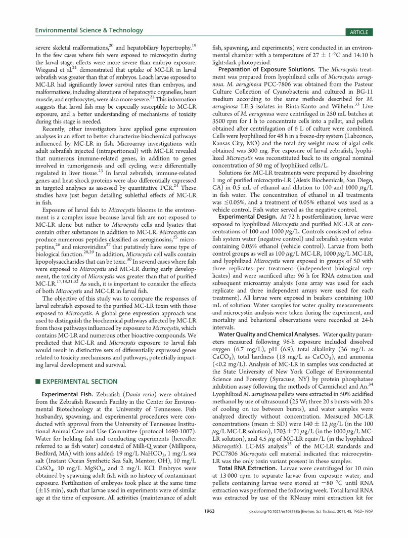
Table 1. Genes Unique to Microcystis Treatmenta
sequence code accession number description gene symbol x-fold changea p-value
Cell Signaling and Development
Dr.15260.1.S1_at BI429195 nuclear receptor subfamily 1, group D,
member 2B
nr1d2b -3.4 6.03 � 10-7
Dr.20567.1.S1_at AW567115 calcium binding protein 39-like cab39l -2.9 1.48 � 10-6
Dr.26399.1.A1_at AL719041 PAR-6 γ proteinb pard6gb -2.8 4.43 � 10-6
Dr.25140.8.A1_at AL730238 CD81 antigen cd81 -2.8 1.36 � 10-9
Dr.12138.2.A1_at AI957874 exostoses 1A ext1a -2.7 2.00 � 10-5
Dr.18414.1.S1_at AW165356 programmed cell death 4A pdcd4a -2.7 2.00 � 10-5
Dr.19421.1.A1_at AL725987 PHD finger protein 8 phf8 -2.4 6.54 � 10-6
Dr.11726.1.S1_at NM_131877.1 caspase 3 casp3 -2.4 3.00 � 10-5
Dr.7424.2.S1_at BI839632 COP9 constitutive photomorphogenic
homologue subunit 5
cops5 -2.3 4.44 � 10-6
Dr.3282.1.S1_at NM_131691.1 endothelial differentiation sphingolipid
G-protein-coupled receptor 1
edg1 -2.3 8.63 � 10-6
Dr.25322.1.S1_at AL726472 lin-7 homologue C lin7c -2.2 2.71 � 10-8
Dr.13009.2.S1_at BE556991 sprouty homologue 2 spry2 -2.2 4.48 � 10-6
Dr.1710.2.S1_at AI794095 eukaryotic translation initiation factor 4e family 3 eif4e3 -2.1 7.67 � 10-8
Dr.7804.1.S1_at AB069858.1 antizyme inhibitor 1 azin1 -2.1 3.00 � 10-5
Dr.6932.3.S1_at AL730217 high-mobility group box 3A hmgb3a -2.0 2.07 � 10-11
Dr.15361.1.S1_at AL926178 ras-related protein rab-22Ac rab22a -2.0 2.60 � 10-6
Dr.11764.1.S1_at NM_131774.1 annexin A13 anxa13 -2.0 1.00 � 10-5
Dr.7100.1.A1_at BQ091992 immediate early response 2b ier2 -1.9 9.07 � 10-10
Dr.11698.1.S1_at AL725759 transcription factor AP-2 γ tfap2c -1.9 1.51 � 10-9
Dr.16550.1.A1_at BI709565 TM2 domain-containing 2 tm2d2 -1.9 1.82 � 10-9
Dr.24242.2.S1_at BQ131454 mitochondrial processing peptidase Rc pmpca -1.9 3.11 � 10-8
Dr.578.2.S1_a_at U96848.1 thyrotroph embryonic factor tef -1.9 7.04 � 10-8
Dr.5572.1.S1_at NM_131101.1 homeo box B5A hoxb5a -1.8 6.73 � 10-10
Dr.3238.1.A1_at AI793363 Kruppel-like factor 11A klf11a -1.8 9.45 � 10-9
Dr.7225.1.S1_at BC045952.1 MOB1, Mps one binder kinase activator-like
1A (yeast)
mobkl1a -1.7 4.88 � 10-6
Dr.23293.1.A1_at BE016153 tubulin polymerization-promoting protein
family member 3
tppp3 -1.7 4.00 � 10-5
Dr.26344.2.S1_a_at AL717083 cell division cycle 42 cdc42 -1.7 6.00 � 10-5
Dr.3472.1.A1_at AI545021 spectrin, β, nonerythrocytic 1 sptbn1 1.7 7.95 � 10-13
Dr.15630.1.S1_at CD594794 similar to tubulin folding cofactor E-like tbcel 1.7 2.77 � 10-6
DrAffx.1.10.S1_at AY151045.1 cysteine-rich transmembrane BMP regulator 1 crim1 1.8 1.82 � 10-7
Dr.3421.1.A1_at AW342746 ribosome binding protein 1 homologue (dog) rrbp1 1.8 6.00 � 10-5
Dr.12403.1.S1_at NM_131633.1 roundabout homologue 2 robo2 1.9 2.46 � 10-7
Dr.6210.1.S1_at BM184670 cartilage acidic protein 1b crtac1 2.0 1.91 � 10-11
Dr.15261.1.A1_at BI710394 cytoplasmic polyadenylation element binding
protein 4
cpeb4 2.0 6.00 � 10-5
Dr.52.1.A1_at AA495026 phosphatidylinositol binding clathrin assembly
protein, like
picalml 2.2 3.51 � 10-6
Neurological Function
Dr.17557.1.S1_at AL730871 neurocalcin δd ncald -2.7 1.53 � 10-14
Dr.24196.1.S1_at NM_131452.1 embryonic lethal abnormal vision-like 1 elavl1 -2.1 4.11 � 10-10
Dr.4230.1.S1_a_at NM_130909.1 HuG hug -2.0 5.79 � 10-17
Dr.1968.1.S1_at BC049308.1 fusion involved in malignant liposarcoma fus -1.8 6.11 � 10-9
Dr.25322.2.S1_at BI888421 lin-7 homologue C lin7c -1.7 1.94 � 10-13
DrAffx.1.10.S1_at AY151045.1 cysteine-rich transmembrane BMP regulator 1 crim1 1.8 1.82 � 10-7
Dr.12617.1.A1_at NM_131806.1 ephrin B3 efnb3 1.8 1.00 � 10-5
Dr.12403.1.S1_at NM_131633.1 roundabout homologue 2 robo2 1.9 2.46 � 10-7
Dr.23350.1.S1_at AF425739.1 parvalbumin 8 pvalb8 4.7 2.00 � 10-5

1966 dx.doi.org/10.1021/es103538b |Environ. Sci. Technol. 2011, 45, 1962–1969
Environmental Science & Technology ARTICLE
Information). Changes in expression of these genes may indicatealtered cell cycle progression, carcinogenesis, cell death, ordisruption of cellular differentiation during development. Genesshowing the greatest degree of fold change were nephrosin(-4.4-fold change, p = 2.0 � 10-5) and mucin 2 (-4.7-foldchange, p = 2.0 � 10-5). Immune-related genes were also
affected, as well as those associated with cytoskeletal disruption,ion regulation, oxidative stress, and maintenance of tight junc-tions. Because these genes were affected by exposure to purifiedMC-LR and by the cyanobacterium responsible for productionof this toxin, this group of genes may represent a toxin responsethat more closely mimics a natural exposure situation than
Table 1. Continued
sequence code accession number description gene symbol x-fold changea p-value
Visual Perception
AFFX-Dr-NM_
131175-1_s_at
NM_131175-1 opsin 1, long-wavelength-sensitive 1 opn1lw1 -3.0 4.92 � 10-13
Dr.26436.1.S1_at AF210644.1 retinal degradation slow 4 rds4 -2.6 1.83 � 10-11
Dr.12204.1.S1_at BI706778 retinal degradation slow 4 rds4 -2.3 3.00 � 10-5
Dr.8102.1.S1_at NM_131253.1 opsin, medium-wavelength-sensitive 1 opn1mw1 -2.0 1.38 � 10-8
Dr.9899.1.S1_at NM_131868.1 guanine nucleotide binding protein,
R transducing activity 1
gnat1 -1.9 1.08 � 10-9
Dr.10433.1.S1_at NM_131791.1 cryptochrome 2A cry2a -1.9 5.00 � 10-5
Dr.8194.1.S1_at BI879950 opsin, short-wavelength-sensitive 1 opn1sw1 -1.8 1.20 � 10-8
Dr.8097.1.S1_at NM_131192.1 opsin, short-wavelength-sensitive 2 opn1sw2 -1.7 1.65 � 10-7
Endocrine Activity
Dr.10788.1.S1_at NM_131804.1 nothepsin nots 4.0 1.02 � 10-18
Dr.10461.1.S1_at NM_131642.1 cytochrome P450, family 19, subfamily A,
polypeptide 1B
cyp19a1b 4.0 6.68 � 10-8
Dr.25009.6.A1_at BI878405 vitellogenind vtg >100 <1.00 � 10-45
Dr.25009.1.S1_a_at NM_170767.1 vitellogenin 1 vtg1 >100 <1.00 � 10-45
Dr.25009.6.A1_a_at BI878405 vitellogenin 1 vtg1 >100 <1.00 � 10-45
Dr.25009.4.A1_at BG303658 vitellogenin 2 vtg2 >100 <1.00 � 10-45
Dr.2978.1.S1_at AI477604 vitellogenin 3 vtg3 19.2 <1.00 � 10-45
Ion Regulation
Dr.3.1.S1_at AF469651.1 ATPase, Naþ/Kþ transporting, β 3A atp1b3a -2.2 4.44 � 10-11
Dr.26437.1.S1_at NM_131669.1 ATPase, Naþ/Kþ transporting, β 2A atp1b2a -2.0 5.40 � 10-7
Dr.3613.1.S1_at BC048037.1 ceruloplasmin cp 2.4 1.73 � 10-9
Apoptosis
Dr.18414.1.S1_at AW165356 programmed cell death 4A pdcd4a -2.7 2.00 � 10-5
Dr.11726.1.S1_at NM_131877.1 caspase 3 casp3 -2.4 3.00 � 10-5
Glucose Transport
Dr.17415.1.S1_at BC050518.1 insulin receptor substrate 1Bd irs1b -2.4 1.86 � 10-7
Amino Acid Transport
Dr.7516.1.A1_at BM095174 solute carrier family 38, member 4 slc38a4 2.1 3.45 � 10-12
Muscle Contraction
Dr.23350.1.S1_at AF425739.1 parvalbumin 8 pvalb8 4.7 2.00 � 10-5
DNA Synthesis/Degradation
Dr.1691.1.S1_at NM_131450.1 ribonucleotide reductase protein R2 class I rrm2 -2.3 1.98 � 10-6
Dr.1668.1.S1_at BC046003.1 hypoxanthine phosphoribosyltransferase 1 hprt1 -1.8 2.90 � 10-6
Intestinal Function
Dr.18599.1.S1_at BQ479899 fatty acid binding protein 6 fabp6 1.9 3.81 � 10-6
Metabolism
Dr.14021.3.A1_at BM095392 3-hydroxyisobutyrate dehydrogenase B hibadhb -2.0 2.55 � 10-16
Protein Targeting
Dr.3075.1.S1_at BC049337.1 translocase of outer mitochondrial membrane 34 tomm34 -1.8 1.00 � 10-5
ag1.7-fold change, p < 0.0001 bWeak similarity to gene indicated. cModerate similarity to gene indicated. d Similar to gene indicated.
1967 dx.doi.org/10.1021/es103538b |Environ. Sci. Technol. 2011, 45, 1962–1969
Environmental Science & Technology ARTICLE
genes differentially expressed upon exposure to purified MC-LRalone.Also of interest were genes that responded when larval
zebrafish were exposed to Microcystis but not the purified MC-LR treatments. The majority of genes expressed only in theMicrocystis treatment (n = 126) (Figure 1) were genes involved incell signaling and development (28%), neurological function(7%), visual perception (6%), and endocrine activity (6%)(Table 1). Other genes included those affecting ion regulation,apoptosis, glucose and amino acid transport, muscle contraction,DNA synthesis and degradation, intestinal function, metabolism,and protein targeting. The majority of genes were down-regu-lated, but some genes related to cell signaling development,neurological function, and visual perception were up-regulated.Vitellogenin Expression and Validation by qRT-PCR.
Affymetrix probe sets designed for zebrafish vitellogenin genes indi-cated high up-regulation (19-fold to >100-fold change) of thesegenes in larval zebrafish exposed to Microcystis (Table 1). In larvalzebrafish exposed to MC-LR, there was no significant effect onexpression of any of the vitellogenin genes relative to the control.Up-regulation of vitellogenin (accession number NM_170767.1)was confirmed by qRT-PCR of vtg1A/B [mean x-fold-change( SD = 619.3 ( 130.2 (n = 9)].
’DISCUSSION
Gene expression results confirmed known mechanisms ofaction for MC-LR and also identified effects not previouslyassociated with this toxin. When larval fish were exposed to bothMC-LR andMicrocystis treatments, the top functional categoriesaffected (cell signaling and development, cytoskeleton, immunefunction, ion regulation, oxidative stress, tight junction, livereffects) were associated with effects shown previously for purifiedMC-LR exposure in fish and mammals5,10,11,16,20,42-45 (seeTable S2, Supporting Information). Because these genes werealso affected in theMicrocystis treatment in the present study, thisis consistent with the presence of MC-LR toxin in theMicrocystistreatment (i.e., MC-LR was generated by the Microcystis cells).
We did not test the effects of 4.5 μg/L purified MC-LR andtherefore are not able to distinguish the effects on gene expres-sion within the Microcystis treatment that were the result of thetoxin at the concentration (4.5 μg/L MC-LR) associated withMicrocystis cultures used in this study. It is likely that some of thedifferentially expressed genes within the Microcystis treatmentwere affected by the 4.5 μg/L MC-LR. When differentiallyexpressed genes from the Microcystis treatment were comparedto purifiedMC-LR treatment (100μg/L) only 22% (83 genes) ofall genes (371 genes) altered byMicrocystis were consistent withexposure to MC-LR. In this study however, it is the numerousgenes that were affected byMicrocystis, but not MC-LR, that areof most interest. Our objective was to see if there were effectsfrom theMicrocystis cells that were not due to the presence of thetoxin alone, and our observations strongly suggest that there arebiological effects beyond just the toxin MC-LR.
MC-LR is known to affect immune response; however, therewere few similarities between our work and previous geneexpression studies that mainly focused on the response ofimmune-related genes in zebrafish exposed to purified MC-LR.Of the 11 genes tested by qPCR and found to be up-regulated inzebrafish by Li et al.,24 none were differentially expressed in our100 μg/LMC-LR orMicrocystis treatments, but three heat-shockprotein genes were significantly down-regulated (-2.2-fold to
-1.9-fold change) at 1000 μg/LMC-LR.When compared to theresults of a microarray study by Wei et al.,23 in which adultzebrafish were exposed to MC-LR by intraperitoneal injection,five genes reported by Wei et al. to be up-regulated weresignificantly down-regulated (-4.2-fold to -1.8-fold change)in the 1000 μg/LMC-LR treatment, and three of these same fivegenes were down-regulated (-2.2-fold to -1.9-fold change) intheMicrocystis treatment. Variation between our work and thesestudies may be attributed to differences in life stage of the fish,MC-LR concentration, and/or exposure route.
Also when genes differentially expressed in both 100 and 1000μg/LMC-LR (but not theMicrocystis treatment) were examined,different effects emerge other than those typically associated withMC-LR. There were additional cell signaling effects not affectedby Microcystis (five genes) (see Table S1, Supporting In-formation); however the majority of genes affected by bothconcentrations of MC-LR tested were related to detoxification/metabolism and lipid binding/transport. Detoxification/meta-bolism genes included glutathione S-transferase, an enzymeinvolved in detoxification of MC-LR,14 as well as cytochromeP450 enzymes, which are typically not associated with micro-cystin exposure. Lipid transport metabolism genes mainly in-cluded apolipoproteins, which are known to influence nutritionand immune response in fish.46 In addition, strong down-regulation of type IV antifreeze protein (-14.6-fold) mayindicate that microcystin-LR may inhibit thermoregulation infish, which is an effect that has not previously been associatedwith this compound. It is also interesting that significant expres-sion of PP genes occurred only in exposures of 1000 μg/L andnot 100 μg/L, and these genes were also not differentiallyexpressed in larval zebrafish exposed to Microcystis. This implieseither that larval fish were able to compensate when exposed tolow concentrations of MC-LR or that PP enzymes may not be asresponsive at this stage in development and a higher dose may berequired for inhibition to occur. The absence of this majorbiomarker of MC-LR toxicity in larval fish exposed toMicrocystisis also interesting, because it implies that MC-LR may not be themost important compound affecting fish when they are exposedto the cyanobacteria.
Most of the gene function categories affected exclusively byMicrocystis exposure were different from those associated withMC-LR. Several genes related to neurological development and/or function were differentially expressed, including ephrin B3(1.8-fold change), a gene in which overexpression has beenassociated with notochord defects in zebrafish,47 and neurocalcinδ (-2.7-fold change), a neuronal calcium binding protein invol-ved in visual transduction in the retina.48 Alteration in theregulation of these genes suggests that Microcystis may interferewith proper nervous system development in larval fish. Similarly,several genes involved in visual perception, including opsingenes, were down-regulated, suggesting that development ofeye structures may not proceed correctly; an effect that mayaffect fish at later life stages and interfere with the ability toregulate circadian rhythm, locate food, and avoid predation.
The most highly up-regulated genes were those coding forvitellogenin, and this change in gene expression was observed onlyafter zebrafish were exposed to the Microcystis treatment. Vitello-genins are a group of lipoproteins produced in the liver in responseto estrogen and are transported through the blood and deposited inthe developing oocytes of female fish.49 Expression of vtg in maleand immature fish has become a biomarker of exposure to environ-mental estrogens,50 and the present study is the first to report

1968 dx.doi.org/10.1021/es103538b |Environ. Sci. Technol. 2011, 45, 1962–1969
Environmental Science & Technology ARTICLE
estrogenic effects associated with Microcystis exposure in anyorganism. Recently, a low-level estrogenic response was observedin a human breast carcinoma cell line when cells were exposed topurified MC-LR.51 Our results, conversely, did not show vitello-genin induction in fish exposed to purifiedMC-LR, which indicatesthat the estrogen receptor-mediated induction of vtg in zebrafishwas not activated by this toxin. Phytoestrogens are compoundsidentified in plants that can induce vitellogenin;52,53 and it ispossible that the substances(s) produced byMicrocystis that causedinduction of vtg in this study are similar to phytoestrogens.
The possibility that Microcystis blooms may release estrogenicsubstances (aka “phycoestrogens”) that can interfere with repro-duction is of considerable environmental interest. Because com-pounds that induce vitellogenin in fish are generally able to do so inother species,50 endocrine disruption fromMicrocystis could extendthroughout aquatic ecosystems and also impact the terrestrialenvironment, including birds and mammals. Human exposure toMicrocystis has long been a concern due to the microcystin toxin,and most monitoring programs are designed to evaluate presenceof this toxin; however, the potential for endocrine disruption tooccur that is unrelated to the presence of the toxin suggests thatmonitoring programsmay need to be re-evaluated. Projected globalincrease in the frequency ofMicrocystis blooms54 and the potentialfor estrogenic effects adds to the environmental and public healthconcerns related to bloom events.
’ASSOCIATED CONTENT
bS Supporting Information. Two tables listing genes com-mon to 100 and 1000 μg/L MC-LR treatments and genescommon to all treatments. This information is available free ofcharge via the Internet at http://pubs.acs.org.
’AUTHOR INFORMATION
Corresponding Author*Phone:þ44 (0)1752 584658; e-mail: [email protected].
’ACKNOWLEDGMENT
We thank P. Heah for laboratory assistance, D. Truitt for helpwithMicrocystis culturing, M. Satchwell for microcystin-LR analysis,and J. Ryan at the Oceans and Human Health Initiative (OHHI)GenomicsCoreFacility forRosettaResolver guidance.This researchwas supported by NOAA ECOHAB Grant NA06NOS4780234awarded to T.B.H. and S.W.W.
’REFERENCES
(1) Carmichael, W. W. Health effects of toxin-producing cyanobac-teria: The cyanoHABs. Hum. Ecol. Risk Assess. 2001, 7 (5), 1393–1407.(2) Dittmann, E.; Wiegand, C. Cyanobacterial toxins - occurrence,
biosynthesis and impact on human affairs. Mol. Nutr. Food Res. 2006,50 (1), 7–17.(3) Tanabe, Y.; Kaya, K.; Watanabe, M. M. Evidence for recombina-
tion in the microcystin synthetase (mcy) genes of toxic cyanobacteriaMicrocystis spp. J. Mol. Evol. 2004, 58 (6), 633–641.(4) Chorus, I.; Falconer, I. R.; Salas, H. J.; Bartram, J. Health risks
caused by freshwater cyanobacteria in recreational waters. J. Toxicol.Environ. Health, Part B 2000, 3 (4), 323–347.(5) Fischer,W. J.; Hitzfeld, B. C.; Tencalla, F.; Eriksson, J. E.;Mikhailov,
A.; Dietrich, D. R. Microcystin-LR toxicodynamics, induced pathology, andimmunohistochemical localization in livers of blue-green algae exposedrainbow trout (Oncorhynchus mykiss). Toxicol. Sci. 2000, 54 (2), 365–373.
(6) Fujiki, H.; Suganuma, M. Tumor promotion by inhibitors ofprotein phosphatases 1 and 2A: The okadaic acid class of compounds.Adv. Cancer Res. 1993, 61, 143–194.
(7) Eriksson, J. E.; Toivola, D.; Meriluoto, J. A. O.; Karaki, H.; Han,Y. G.; Hartshorne, D. Hepatocyte deformation induced by cyanobacte-rial toxins reflects inhibition of protein phosphatases. Biochem. Biophys.Res. Commun. 1990, 173 (3), 1347–1353.
(8) Ohta, T.; Nishiwaki, R.; Yatsunami, J.; Komori, A.; Suganuma,M.; Fujiki, H. Hyperphosphorylation of cytokeratins 8 and 18 bymicrocystin-LR, a new liver-tumor promoter, in primary cultured rathepatocytes. Carcinogenesis 1992, 13 (12), 2443–2447.
(9) Matsushima, R.; Yoshizawa, S.; Watanabe, M. F.; Harada, K.;Furusawa, M.; Carmichael, W. W.; Fujiki, H. In vitro and in vivo effects ofprotein phosphatase inhibitors, microcystins and nodularin, on mouse skinand fibroblasts. Biochem. Biophys. Res. Commun. 1990, 171 (2), 867–874.
(10) Gaete, V.; Canelo, E.; Lagos, N.; Zambrano, F. Inhibitoryeffects of Microcystis aeruginosa toxin on ion pumps of the gill offreshwater fish. Toxicon 1994, 32 (1), 121–127.
(11) Zambrano, F.; Canelo, E. Effects of microcystin-LR on thepartial reactions of the Naþ/Kþ pump of the gill of carp (Cyprinus carpioLinneo). Toxicon 1996, 34 (4), 451–458.
(12) Bury, N. R.; Flik, G.; Eddy, F. B.; Codd, G. A. The effects ofcyanobacteria and the cyanobacterial toxin microcystin-LR on Ca2þ
transport and Naþ/Kþ -ATPase in tilapia gills. J. Exp. Biol. 1996, 199 (6),1319–1326.
(13) Bury, N. R.; Codd, G. A.; Bonga, S. E. W.; Flik, G. Fatty acidsfrom the cyanobacterium Microcystis aeruginosa with potent inhibitoryeffects on fish gill Naþ/Kþ-ATPase activity. J. Exp. Biol. 1998, 201 (1),81–89.
(14) Pflugmacher, S.; Wiegand, C.; Oberemm, A.; Beattie, K. A.;Krause, E.; Codd, G. A.; Steinberg, C. E. W. Identification of anenzymatically formed glutathione conjugate of the cyanobacterial hepa-totoxin microcystin-LR: the first step of detoxication. Biochim. Biophys.Acta 1998, 1425 (3), 527–533.
(15) Sahin, A.; Tencalla, F. G.; Dietrich, D. R.; Naegeli, H. Biliaryexcretion of biochemically active cyanobacteria (blue-green algae)hepatotoxins in fish. Toxicology 1996, 106 (1-3), 123–130.
(16) Malbrouck, C.; Kestemont, P. Effects of microcystins on fish.Environ. Toxicol. Chem. 2006, 25 (1), 72–86.
(17) Oberemm, A.; Fastner, J.; Steinberg, C. E. W. Effects ofmicrocystin-LR and cyanobacterial crude extracts on embryo-larvaldevelopment of zebrafish (Danio rerio). Water Res. 1997, 31 (11),2918–2921.
(18) Oberemm, A.; Becker, J.; Codd, G. A.; Steinberg, C. Effects ofcyanobacterial toxins and aqueous crude extracts of cyanobacteria on thedevelopment of fish and amphibians. Environ. Toxicol. 1999, 14 (1),77–88.
(19) Jacquet, C.; Thermes, V.; de Luze, A.; Puiseux-Dao, S.; Bernard,C.; Joly, J. S.; Bourrat, F.; Edery, M. Effects of microcystin-LR ondevelopment of medaka fish embryos (Oryzias latipes). Toxicon 2004,43 (2), 141–147.
(20) Wang, P. J.; Chien, M. S.; Wu, F. J.; Chou, H. N.; Lee, S. J.Inhibition of embryonic development by microcystin-LR in zebrafish.Danio rerio. Toxicon 2005, 45 (3), 303–308.
(21) Wiegand, C.; Pflugmacher, S.; Oberemm, A.; Meems, N.;Beattie, K. A.; Steinberg, C. E. W.; Codd, G. A. Uptake and effects ofmicrocystin-LR on detoxication enzymes of early life stages of the zebrafish (Danio rerio). Environ. Toxicol. 1999, 14 (1), 89–95.
(22) Liu, Y.; Song, L; Li, X.; Liu, T. The toxic effects of microcystin-LR on embryo-larval and juvenile development of loach, Misgurunsmizolepis Gunthe. Toxicon 2002, 40 (4), 395–399.
(23) Wei, L.; Sun, B.; Song, L; Nie, P. Gene expression profiles inliver of zebrafish treated with microcystin-LR. Environ. Toxicol. Pharma-col. 2008, 26 (1), 6–12.
(24) Li, Y.; Sun, B.; Wu, H.; Nie, P. Effects of pure microcystin-LRon the transcription of immune related genes and heat shock proteins inlarval stage of zebrafish (Danio rerio). Aquaculture 2009, 289 (1-2),154–160.
1969 dx.doi.org/10.1021/es103538b |Environ. Sci. Technol. 2011, 45, 1962–1969
Environmental Science & Technology ARTICLE
(25) Ishida, K.; Okita, Y.; Matsuda, H.; Okino, T.; Murakami, M.Aeruginosins, protease inhibitors from the cyanobacterium Microcystisaeruginosa. Tetrahedron 1999, 55 (36), 10971–10988.(26) Yamaki, H.; Sitachitta, N.; Sano, T.; Kaya, K. Two new
chymotrypsin inhibitors isolated from the cyanobacterium Microcystisaeruginosa NIES-88. J. Nat. Prod. 2005, 68 (1), 14–18.(27) Rohrlack, T.; Christoffersen, K.; Hansen, P. E.; Zhang, W.;
Czarnecki, O.; Henning, M.; Fastner, J.; Erhard, M.; Neilan, B. A.;Kaebernick, M. Isolation, characterization, and quantitative analysis ofmicroviridin J, a new Microcystis metabolite toxic to Daphnia. J. Chem.Ecol. 2003, 29 (8), 1757–1770.(28) Harada, K. Production of secondary metabolites by freshwater
cyanobacteria. Chem. Pharm. Bull. 2004, 52 (8), 889–899.(29) Smith, J. L.; Boyer, G. L.; Zimba, P. V. A review of cyanobacter-
ial odorous and bioactive metabolites: Impacts and management alter-natives in aquaculture. Aquaculture 2008, 280 (1-4), 5–20.(30) Raziuddin, S.; Siegelman, H. W.; Tornabene, T. G. Lipopoly-
saccharides of the cyanobacteriumMicrocystis aeruginosa. Eur. J. Biochem.1983, 137 (1-2), 333–336.(31) Best, J. H.; Eddy, F. B.; Codd, G. A. Effects of purified
microcystin-LR and cell extracts of Microcystis strains PCC 7813 andCYA 43 on cardiac function in brown trout (Salmo trutta) alevins. FishPhysiol. Biochem. 2001, 24 (3), 171–178.(32) Palikova, M.; Krejci, R.; Hilscherova, K.; Babica, P.; Navratil,
S.; Kopp, R.; Blaha, L. Effect of different cyanobacterial biomasses andtheir fractions with variable microcystin content on embryonal devel-opment of carp (Cyprinus carpio L.). Aquat. Toxicol. 2007, 81 (3), 312–318.(33) Rinta-Kanto, J. M.; Wilhelm, S. W. Diversity of microcystin-
producing cyanobacteria in spatially isolated regions of Lake Erie. Appl.Environ. Microbiol. 2006, 72 (7), 5083–5085.(34) Carmichael, W. W.; An, J. S. Using an enzyme linked immu-
nosorbent assay (ELISA) and a protein phosphatase inhibition assay(PPIA) for the detection of microcystins and nodularins. Nat. Toxins1999, 7 (6), 377–385.(35) Boyer, G. L. The occurrence of cyanobacterial toxins in NewYork
lakes: Lessons from the MERHAB-Lower Great Lakes program. LakeReserv. Manage. 2007, 23 (2), 153–160.(36) Henry, T. B.; McPherson, J. T.; Rogers, E. D.; Heah, T. P.;
Hawkins, S. A.; Layton, A. C.; Sayler, G. S. Changes in the relativeexpression pattern of multiple vitellogenin genes in adult male and larvalzebrafish exposed to exogenous estrogens. Comp. Biochem. Physiol., PartA: Mol. Integr. Physiol. 2009, 154 (1), 119–126.(37) Henry, T. B.; Menn, F. M.; Fleming, J. T.; Wilgus, J.; Compton,
R. N.; Sayler, G. S. Attributing effects of aqueous C-60 nano-aggregatesto tetrahydrofuran decomposition products in larval zebrafish by assess-ment of gene expression. Environ. Health Perspect. 2007, 115 (7), 1059–1065.(38) Affymetrix. Affymetrix GeneChip Expression Analysis Technical
Manual; Affymetrix, Inc: Santa Clara, CA, 2004.(39) Twiner, M. J.; Ryan, J. C.; Morey, J. S.; Smith, K. J.;
Hammad, S. M.; Van Dolah, F. M.; Hess, P.; McMahon, T.;Satake, M.; Yasumoto, T.; Doucette, G. J. Transcriptional profilingand inhibition of cholesterol biosynthesis in human T lymphocytecells by the marine toxin azaspiracid. Genomics 2008, 91 (3), 289–300.(40) Dennis, G.; Sherman, B. T.; Hosack, D. A.; Yang, J.; Gao, W.;
Lane, H. C.; Lempicki, R. A. DAVID: Database for annotation,visualization, and integrated discovery. Genome Biol. 2003, 4, P3.(41) Huang, D. W.; Sherman, B. T.; Lempicki, R. A. Systematic and
integrative analysis of large gene lists using DAVID bioinformaticsresources. Nat. Protoc. 2009, 4 (1), 44–57.(42) Guzman, R. E.; Solter, P. F. Hepatic oxidative stress following
prolonged sublethal microcystin-LR exposure. Toxicol. Pathol. 1999,27 (5), 582–588.(43) Janssens, V.; Goris, J. Protein phosphatase 2A: a highly
regulated family of serine/threonine phosphatases implicated in cellgrowth and signalling. Biochem. J. 2001, 353, 417–439.
(44) Rymuszka, A.; Sieroslawska, A.; Bownik, A.; Skowronski, T.Immunotoxic potential of cyanotoxins on the immune system of fish.Cent. Eur. J. Immunol. 2008, 33 (3), 150–152.
(45) Toivola, D. M.; Eriksson, J. E. Toxins affecting cell signallingand alteration of cytoskeletal structure. Toxicol. in Vitro 1999, 13 (4-5),521–530.
(46) Concha, M. I.; Smith, V. J.; Castro, K.; Bastias, A.; Romero, A.;Amthauer, R. J.; Apolipoproteins, A-I and A-II are potentially importanteffectors of innate immunity in the teleost fish Cyprinus carpio. Eur.J. Biochem. 2004, 271 (14), 2984–2990.
(47) Chan, J.; Mably, J. D.; Serluca, F. C.; Chen, J. N.; Goldstein,N. B.; Thomas, M. C.; Cleary, J. A.; Brennan, C.; Fishman, M. C.;Roberts, T. M. Morphogenesis of prechordal plate and notochordrequires intact Eph/ephrin B signaling. Dev. Biol. 2001, 234 (2), 470–482.
(48) Krishnan, A.; Venkataraman, V.; Fik-Rymarkiewicz, E.; Duda,T.; Sharma, R. K. Structural, biochemical, and functional characteriza-tion of the calcium sensor neurocalcin delta in the inner retinal neuronsand its linkage with the rod outer segment membrane guanylate cyclasetransduction system. Biochemistry 2004, 43 (10), 2708–2723.
(49) Routledge, E. J.; Sheahan, D.; Desbrow, C.; Brighty, G. C.;Waldock, M.; Sumpter, J. P. Identification of estrogenic chemicals inSTW effluent. 2. In vivo responses in trout and roach. Environ. Sci.Technol. 1998, 32 (11), 1559–1565.
(50) Sumpter, J. P.; Jobling, S. Vitellogenesis as a biomarker forestrogenic contamination of the aquatic environment. Environ. HealthPerspect 1995, 103 (Suppl 7), 173–178.
(51) Oziol, L.; Bouaïcha, N. First evidence of estrogenic potential ofthe cyanobacterial heptotoxins the nodularin-R and the microcystin-LRin cultured mammalian cells. J. Hazard. Mater. 2010, 174 (1-3), 610–615.
(52) Latonnelle, K.; Fostier, A.; Le Menn, F.; Bennetau-Pelissero, C.Binding affinities of hepatic nuclear estrogen receptors for phytoestro-gens in rainbow trout (Oncorhynchus mykiss) and Siberian sturgeon(Acipenser baeri). Gen. Comp. Endrocrinol. 2002, 129 (2), 69–79.
(53) Inudo, M.; Ishibashi, H.; Matsumura, N.; Matsuoka, M.; Mori,T.; Taniyama, S.; Kadokami, K.; Koga, M.; Shinohara, R.; Hutchinson,T. H.; Iguchi, T.; Arizono, K. Effect of estrogenic activity, and phytoes-trogen and organochlorine pesticide contents in an experimental fishdiet on reproduction and hepatic vitellogenin production in medaka(Oryzias latipes). Comp. Med. 2004, 54 (6), 673–680.
(54) Paerl, H. W.; Huisman, J. Climate - Blooms like it hot. Science2008, 320 (5872), 57–58.
1
Online Supporting Information
Global gene expression profiling in larval zebrafish exposed to microcystin-LR and Microcystis
reveals endocrine disrupting effects of cyanobacteria
Emily D. Rogers,1,2
Theodore B. Henry,1,2,3*
Michael J. Twiner,4 Julia S. Gouffon,
5 Jackson T.
McPherson,1 Gregory L. Boyer,
6 Gary S. Sayler
1,7,8, and Steven W. Wilhelm,
1,7
1Center for Environmental Biotechnology, University of Tennessee, Knoxville, Tennessee, 37996;
2Department of Forestry, Wildlife and Fisheries, University of Tennessee, Knoxville, Tennessee, 37996;
3School of Biomedical and Biological Sciences, University of Plymouth, Plymouth, UK, PL4 8AA;
4Department of Natural Sciences, University of Michigan-Dearborn, Dearborn, Michigan, 48128;
5Affymetrix Core Facility, University of Tennessee, Knoxville, Tennessee, 37996;
6Department of
Chemistry, State University of New York College of Environmental Science and Forestry, Syracuse,
New York, 13210; 7
Department of Microbiology, University of Tennessee, Knoxville, Tennessee,
37996; 8Department of Ecology and Evolutionary Biology, University of Tennessee, Knoxville,
Tennessee, 37996
Corresponding author address: *School of Biomedical and Biological Sciences, The University of
Plymouth, 401 Davy Building, Drake Circus, Plymouth, United Kingdom, PL4 8AA; phone: +44
(0)1752 584658; email: [email protected]

2
Table of Contents
Table S1. Genes common to 100 and 1,000 µg/L MC-LR treatments relative to control. Pg. S3
Table S2. Genes common to all treatments relative to control. Pg. S7

3
Table S1. Genes common to 100 and 1,000 µg/L MC-LR treatments relative to control
a
Fold change
MC-LR vs.
controla
Sequence code Accession number Description Gene symbol
100
µg/L
1,000
µg/L
Detoxification/ metabolism
Dr.11596.1.S1_at AI545537 tryptophan 2,3-dioxygenase tdo2a -3.4 -3.4
Dr.3498.1.S1_at BC045343.1 methionine adenosyltransferase I, alpha mat1a -2.4 -3.6
Dr.18453.1.S1_at BC044525.1 uridine phosphorylase 2 upp2 -2.4 -2.6
Dr.9478.1.S1_at AB078927.1 cytochrome P450 1A cyp1a -2.4 -3.0
Dr.14058.1.A1_at CD015351 glutathione s-transferase, theta 1Ac gstt1a -2.3 -2.4
Dr.11729.1.S1_at NM_152954.1 cytochrome P450 2J28 cyp2j28 -2.2 -2.2
Dr.2132.1.A1_at BQ262149 hydroxyacid oxidase 1 hao1 -2.1 -3.4
Dr.4189.1.S1_at BI891596 nad(P)H dehydrogenase quinone 1 nqo1 -1.9 -2.6
Dr.7977.1.S1_at AW232474 glutathione peroxidase 1A gpx1a -1.9 -2.6
Dr.25191.1.S1_at BC046894.1 isocitrate dehyrodgenase 1 idh1 -1.8 -2.5
Dr.7520.1.A1_at AW019023 aconitase 1 aco1 -1.8 -2.3
Dr.1041.1.S1_at BC050158.1 alpha-L-fucosidase 1 fuca1 -1.7 -2.6
Lipid binding/transport
Dr.24953.1.S1_at CD014348 apolipoprotein C-II apoc2 -4.2 -7.8
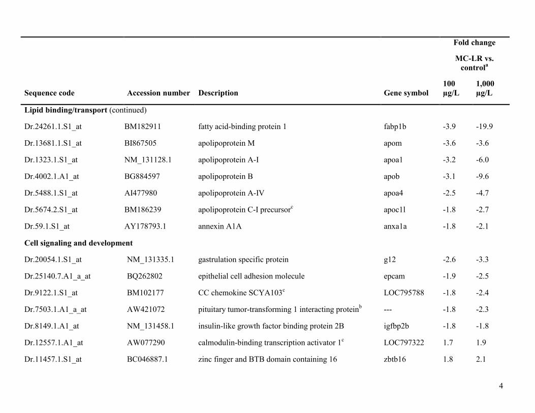
4
Fold change
MC-LR vs.
controla
Sequence code Accession number Description Gene symbol
100
µg/L
1,000
µg/L
Lipid binding/transport (continued)
Dr.24261.1.S1_at BM182911 fatty acid-binding protein 1 fabp1b -3.9 -19.9
Dr.13681.1.S1_at BI867505 apolipoprotein M apom -3.6 -3.6
Dr.1323.1.S1_at NM_131128.1 apolipoprotein A-I apoa1 -3.2 -6.0
Dr.4002.1.A1_at BG884597 apolipoprotein B apob -3.1 -9.6
Dr.5488.1.S1_at AI477980 apolipoprotein A-IV apoa4 -2.5 -4.7
Dr.5674.2.S1_at BM186239 apolipoprotein C-I precursorc apoc1l -1.8 -2.7
Dr.59.1.S1_at AY178793.1 annexin A1A anxa1a -1.8 -2.1
Cell signaling and development
Dr.20054.1.S1_at NM_131335.1 gastrulation specific protein g12 -2.6 -3.3
Dr.25140.7.A1_a_at BQ262802 epithelial cell adhesion molecule epcam -1.9 -2.5
Dr.9122.1.S1_at BM102177 CC chemokine SCYA103c LOC795788 -1.8 -2.4
Dr.7503.1.A1_a_at AW421072 pituitary tumor-transforming 1 interacting proteinb --- -1.8 -2.3
Dr.8149.1.A1_at NM_131458.1 insulin-like growth factor binding protein 2B igfbp2b -1.8 -1.8
Dr.12557.1.A1_at AW077290 calmodulin-binding transcription activator 1c LOC797322 1.7 1.9
Dr.11457.1.S1_at BC046887.1 zinc finger and BTB domain containing 16 zbtb16 1.8 2.1

5
Fold change
MC-LR vs.
controla
Sequence code Accession number Description Gene symbol
100
µg/L
1,000
µg/L
Blood clotting, oxygen transport
Dr.5462.1.S1_at BI878927 fibrinogen B fgb -2.9 -6.1
Dr.1450.1.S1_s_at BI896310 hemoglobin alpha embryonic-3 hbae3 -1.9 -2.9
Dr.845.1.A1_at BG729013 fibrinogen alpha chain fga -2.1 -2.7
Liver function
Dr.8516.1.S1_at NM_178298.2 selenoprotein P, plasma, 1B sepp1b -2.3 -4.5
Tight junction
Dr.994.1.S1_at NM_131763.1 claudin B cldnb -1.9 -3.0
Thermoregulation
Dr.696.1.S1_at AI496864 type IV antifreeze protein zgc:161979 -4.8 -14.6
Cytoskeletal function
Dr.9252.1.A1_at BE605502 sciellin scel -1.8 -2.0
Inflammation
Dr.15332.1.S1_at AL917567 phospholipase A2, group IB pla2g1b -11.1 -11.2
Muscle contraction
Dr.20153.1.S1_a_at AF210639.1 myosin light polypeptide 9 like myl9l -1.9 -2.2

6
Fold change
MC-LR vs.
controla
Sequence code Accession number Description Gene symbol
100
µg/L
1,000
µg/L
Integral to membrane
Dr.23357.1.A1_at BE201798 transmembrane protein 90Ac LOC569467 -2.2 -4.1
RNA processing
Dr.24764.1.S1_at AL727764 cleavage and polyadenylation specific factor 3d hbae1 -1.7 -2.1
Actin binding
Dr.20115.1.S1_at BC049463.1 cofilin 1 (non-muscle) cfl1 -1.8 -2.1
Dr.17687.1.A1_at BQ078227 IQ motif containing GTPase activating protein 1 iqgap1 -1.8 -2.4
Translation
Dr.20386.3.S1_at BM141602 eukaryotic translation initiation factor 1A, X-linked, B zgc:110087 -1.7 -1.8
a ≥ 1.7-fold change, p<0.0001
bweak similarity to gene indicated
csimilar to gene indicated
dstrongly similar to gene indicated

7
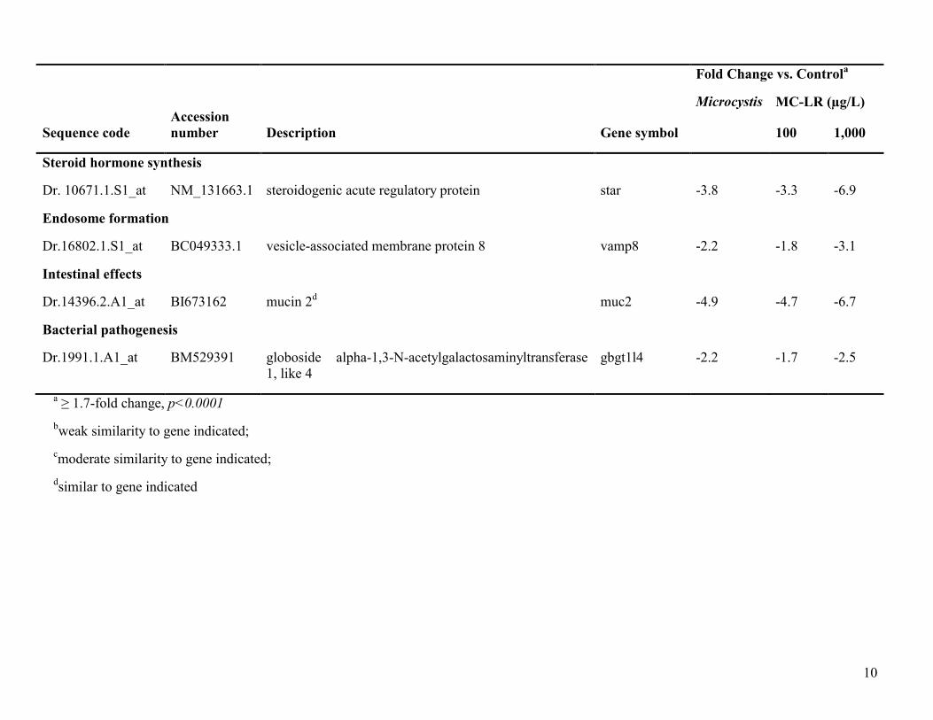
Table S2. Genes common to all treatments relative to control.
Fold Change vs. Control
a
Accession
number
Microcystis MC-LR (µg/L)
Sequence code Description Gene symbol 100 1,000
Cell signaling and development
Dr.17470.1.S1_at AF498291.1 nephrosin npsn -4.6 -4.4 -7.4
Dr.914.1.A1_a_at BE556864 WH2 domain-containing protein 1d -1.8 -2.6 -5.0
Dr.18186.1.S1_at BQ093694 S100 calcium-binding protein A1d s100a1 -2.1 -2.5 -3.7
Dr.4236.1.S1_at BQ092511 calpain 9 capn9 -2.2 -2.4 -3.6
Dr.13076.1.S1_at BC053138.1 pleckstrin homology containing, family F, member 1 plekhf1 -3.0 -2.2 -5.3
Dr.17116.1.S1_at AF282675.1 calpain 1 capn1 -2.5 -2.2 -3.0
Dr.11420.1.S1_at BC050238.1 BAI1-associated protein 2-like 1A baiap2l1a -1.9 -2.0 -2.6
Dr.2251.1.A1_at AI793815 golgi integral membrane protein 4A golim4a -2.5 -2.0 -3.7
Dr.1945.1.A1_at CD015541 calpain 2, large subunit like capn2l -2.3 -2.0 -3.7
Dr.1116.1.S1_at BQ092087 S100 calcium binding protein V2 s100v2 -1.8 -1.9 -2.1
Dr.4409.1.S1_at BC049036.1 CD9 antigen like cd9l -1.9 -1.8 -3.1
Dr.11692.1.S1_at BG727434 vasodilator-stimulated phosphoproteinb vasp -2.0 -1.8 -2.4
Dr.23066.1.S1_at AW019779 apoptosis-inducing factor, mitochondrion-associated 2 aifm2 -1.8 -1.7 -2.6
Dr.10664.1.S1_at NM_131518.1 cd81 antigen cd81 -2.2 -1.7 -2.3
Dr.13857.1.A1_at BM185945 similar to cyclin 1 ccn1 1.9 1.7 2.1
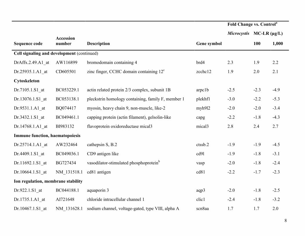
8
Fold Change vs. Control
a
Accession
number
Microcystis MC-LR (µg/L)
Sequence code Description Gene symbol 100 1,000
Cell signaling and development (continued)
DrAffx.2.49.A1_at AW116899 bromodomain containing 4 brd4 2.3 1.9 2.2
Dr.25935.1.A1_at CD605501 zinc finger, CCHC domain containing 12c zcchc12 1.9 2.0 2.1
Cytoskeleton
Dr.7105.1.S1_at BC053229.1 actin related protein 2/3 complex, subunit 1B arpc1b -2.5 -2.3 -4.9
Dr.13076.1.S1_at BC053138.1 pleckstrin homology containing, family F, member 1 plekhf1 -3.0 -2.2 -5.3
Dr.9531.1.A1_at BQ074417 myosin, heavy chain 9, non-muscle, like-2 myh9l2 -2.0 -2.0 -3.4
Dr.3432.1.S1_at BC049461.1 capping protein (actin filament), gelsolin-like capg -2.2 -1.8 -4.3
Dr.14768.1.A1_at BI983132 flavoprotein oxidoreductase mical3 mical3 2.8 2.4 2.7
Immune function, haematopoiesis
Dr.25714.1.A1_at AW232464 cathepsin S, B.2 ctssb.2 -1.9 -1.9 -4.5
Dr.4409.1.S1_at BC049036.1 CD9 antigen like cd9l -1.9 -1.8 -3.1
Dr.11692.1.S1_at BG727434 vasodilator-stimulated phosphoproteinb vasp -2.0 -1.8 -2.4
Dr.10664.1.S1_at NM_131518.1 cd81 antigen cd81 -2.2 -1.7 -2.3
Ion regulation, membrane stability
Dr.922.1.S1_at BC044188.1 aquaporin 3 aqp3 -2.0 -1.8 -2.5
Dr.1735.1.A1_at AI721648 chloride intracellular channel 1 clic1 -2.4 -1.8 -3.2
Dr.10467.1.S1_at NM_131628.1 sodium channel, voltage-gated, type VIII, alpha A scn8aa 1.7 1.7 2.0

9
Fold Change vs. Control
a
Accession
number
Microcystis MC-LR (µg/L)
Sequence code Description Gene symbol 100 1,000
Oxidative stress
DrAffx.1.74.S1_at AY216583.1 selenoprotein W2B sepw2b -3.0 -2.5 -5.8
Dr.7379.1.A1_at AW232459 selenoprotein W2B sepw2b -1.8 -1.8 -2.6
Dr.17468.1.A1_at BM956969 glutathione reductased gsr -1.7 -1.7 -3.1
Tight junction
Dr.7692.1.A1_at BC049304.1 occludin ocln -2.2 -1.9 -3.3
Dr. 20610.1.S1_at NM_131637.1 claudin 7 cldn7 -2.0 -1.9 -2.9
Liver effects
Dr.8947.2.S1_at CD594735 Kunitz-type serine protease inhibitor 2d spint2 -3.1 -2.9 -5.6
Dr.2408.2.S1_at BM571242 matrix metalloproteinase 2c mmp2 -2.4 -1.9 -2.6
Endopeptidase inhibitor
Dr.8947.2.S1_at CD594735 Kunitz-type protease inhibitor 2d spint2 -3.1 -2.9 -5.6
Dr.3073.1.A1_at AI585030 serpin peptidase inhibitor clade A, member 7 serpina7 -2.0 -2.5 -6.5
Detoxification
Dr.16014.1.S1_at BM024109 glutathione transferase omega 1b gsto1 -2.6 -2.4 -4.9
Thyroid hormone availability
Dr.3073.1.A1_at AI585030 serpin peptidase inhibitor, clade A, member 7 serpina7 -2.0 -2.5 -6.5
10
Fold Change vs. Control
a
Accession
number
Microcystis MC-LR (µg/L)
Sequence code Description Gene symbol 100 1,000
Steroid hormone synthesis
Dr. 10671.1.S1_at NM_131663.1 steroidogenic acute regulatory protein star -3.8 -3.3 -6.9
Endosome formation
Dr.16802.1.S1_at BC049333.1 vesicle-associated membrane protein 8 vamp8 -2.2 -1.8 -3.1
Intestinal effects
Dr.14396.2.A1_at BI673162 mucin 2d muc2 -4.9 -4.7 -6.7
Bacterial pathogenesis
Dr.1991.1.A1_at BM529391 globoside alpha-1,3-N-acetylgalactosaminyltransferase
1, like 4
gbgt1l4 -2.2 -1.7 -2.5
a ≥ 1.7-fold change, p<0.0001
bweak similarity to gene indicated;
cmoderate similarity to gene indicated;
dsimilar to gene indicated
Related Documents