GIANT INTRACA4NALICULAR FIBRO-ADENOMYXOhfA OF THE BREAST. THE 80-CiILLED CYSTOSARCOMA PHYLLODES MAMMAE OF JOHANNES MULLER BURTON J. LEE, M.D., AND GEORGE T. PACK, M.D. (From the Memorial Hospilnl for Concrr and A llicd Diseases, New Yorlc Cily) In the January 1931 issue of the Annals of Surgery, we pre- sented four case reports of giant intracanalicular myxomn of the breast and summarized the salient characteristics of this tumor. In the present article we will include a complete bibliography of the literature to date on this subject, a concise summary of one hundred and five case reports culled from this literature, and n report of two additional cases which we have observed. These two personal cases illustrate clearly the early and the late stages in the evolution of this curious neophsm. In 1835 Johnnnes Muller described this tumor as rz neoplastic entity, and gave it8 the name of cystosarcoma phyllodes. His classical description is worthy of quotation in detail: Three forms of cystosnrcomn have come under the author’s notice; the simple cyst os:trcoma, cyst osarcoma proliferum, and cyst 0s:ircoma with foliated warty excrescences from its cysts. In cystosnrcorna simplex, the cysts contained in the fibrous sarcomatous texture have each their distinct membmne, the inner wall of which is simple, smooth or at most beset with :L few vnscular nodules. “In the second form, the sarcomtttous mass is the same, but the cysts within it contain youiigcr cysts in their interior, which are attached to their walls by pedicles. This form of morbid growth is a repetition of the proliferating cysts, but inibeddetl in a sarcomatous mass, which constitutes the chief part of the tumor; and it may, therefore, be termed with propriety cystosnrcorna proliferum. The pedunculnted offsets from the cysts are hollow. “The third form, cystos:trcorn:~ phyllodes, differs greatly from the other two. The tumor forms a hrge firm mass, with :t more or less uneven surface. The fibrous substance which constitutes the greater part of it is of a greyish white color, extremely hard, and as firm as fibro-c:trtilage. Large portions of the tumor are made up entirely of this mass, but in some parts :ire cavities or clefts not lined with a distinct membrane. These cavities contnin but little fluid; for either their parictes, which are hard like fibro-cart il:ige, and finely polished, lie in ose apposition with each other, or :t number of firm, irregular 1amin:te 2583

Welcome message from author
This document is posted to help you gain knowledge. Please leave a comment to let me know what you think about it! Share it to your friends and learn new things together.
Transcript
GIANT INTRACA4NALICULAR FIBRO-ADENOMYXOhfA OF T H E BREAST. THE 80-CiILLED CYSTOSARCOMA
PHYLLODES MAMMAE OF JOHANNES MULLER
BURTON J. LEE, M.D., AND GEORGE T. PACK, M.D.
(From the Memorial Hospilnl f o r Concrr and A llicd Diseases, New Yorlc Cily)
In the January 1931 issue of the Annals of Surgery, we pre- sented four case reports of giant intracanalicular myxomn of the breast and summarized the salient characteristics of this tumor. In the present article we will include a complete bibliography of the literature to date on this subject, a concise summary of one hundred and five case reports culled from this literature, and n report of two additional cases which we have observed. These two personal cases illustrate clearly the early and the late stages in the evolution of this curious neophsm.
In 1835 Johnnnes Muller described this tumor as rz neoplastic entity, and gave it8 the name of cystosarcoma phyllodes. His classical description is worthy of quotation in detail:
Three forms of cystosnrcomn have come under the author’s notice; the simple cyst os:trcoma, cyst osarcoma proliferum, and cyst 0s:ircoma with foliated warty excrescences from its cysts. In cystosnrcorna simplex, the cysts contained in the fibrous sarcomatous texture have each their distinct membmne, the inner wall of which is simple, smooth or at most beset with :L few vnscular nodules.
“ I n the second form, the sarcomtttous mass is the same, but the cysts within it contain youiigcr cysts in their interior, which are attached to their walls by pedicles. This form of morbid growth is a repetition of the proliferating cysts, but inibeddetl in a sarcomatous mass, which constitutes the chief part of the tumor; and it may, therefore, be termed with propriety cystosnrcorna proliferum. The pedunculnted offsets from the cysts are hollow.
“The third form, cystos:trcorn:~ phyllodes, differs greatly from the other two. The tumor forms a hrge firm mass, with :t more or less uneven surface. The fibrous substance which constitutes the greater part of i t is of a greyish white color, extremely hard, and as firm as fibro-c:trtilage. Large portions of the tumor are made up entirely of this mass, but in some parts :ire cavities or clefts not lined with a distinct membrane. These cavities contnin but little fluid; for either their parictes, which are hard like fibro-cart il:ige, and finely polished, lie in ose apposition with each other, or :t number of firm, irregular 1amin:te
2583

2584 BURTON J. LEE AND GEORGE T. PACK
sprout from the mass, and form the walls of the fissures; or excrescences of a foliated or wartlike form sprout from the bottom of the cavities and fill up their interior. These excrescences are perfectly smooth on their surface, and never contain cysts or cells. The laminae lie very irregularly, and project into the cavities and fissures like the folds of the psalterium in the interior of the third stornnch of ruminant animals. In one instance the author saw these lamina here and there regularly notched or crenated like a cock’s comb. Sometimes the 1amin:te are but small, snd the warty excrescences from the cysts very largr, while in other instances both are greatly developed. Occasionally t hrse warty excrescences arc broad, sessile, and much indented; others have a more slender base, and somewhat resemble cauliflower condylomat,a.
“Tumors of this kind attain an enormous size; hitherto the nuthor has seen them only in the female breast, nor are they even there of frequent occurrence. They are decidedly innocent, occur earlier than i t is usual for cancer in the mamma to develop itsrlf, and sometimes they appear even in youth; they have but little tendency to grow to the skin or to the subjacent muscles, nnd are not attrnded with retraction of the nipple. They are not disposed to soften internally, but continue to grow slowly until they have attained an enormous size whrn they at length burst, and a very ill-looking suppurating fungus forms upon their surface. Even in this state, however, the operation has been performed with a successful result.
“Swelling of the axillnry glands is not a common occurrence, :in(!, when it is met with, is the consequence of simple irritation, :md suhsides after the operation. The extr:mrdinnry forms which cystosarconia phyllodes assumes, a t once suggest the notion of its cancerous nature; and yet, the disease is perfectly innocent, and as far removed from carcinoma as are those non-suppurating cauliflower condylomata of thc penis, and of the female genitals, which have so often been mist:tken for cancerous structures.”
This tumor of the breast had been reported in literature several times prior to Muller’s classification. Chelius had previously given an accurate gross description, which may be translated as follows :
“The sarcomatous or steatomatous degeneration of the mammary gland is one of the most benignant diseases to which that organ is subject, and it is with great impropriety that many have spoken of it as carcinoma mammae hydatides. It is characterized by the large size and great prominence of the tumor, which is not globular, but four-cornered, and projecting more a t one part then another; it does not cause retraction of the nipple, but that part projects and retains its natural appc:trance. The greatest diameter of the tumor is not a t its base, but a t a point some distance from the walls of the chest. This disease may be distinguished from scirrhus and fungus medullaris of the mammary gland, partly by the above mentioned signs; but other circumstances which servc still

FIBRO-ADENOMYXOMA OF THE BREAST 2585
further to distinguish it, are its different consistence a t different parts, i t being hard a t one spot, elastic and tense a t another, and even distinctly fluctuating a t a third; its mobility in d l directions, notwithstanding the great size it attains; the slight influence it exerts on thc general health, even after it has continued for a considerable time; and, lastly, the absence of swelling of the :ixillary glands. Although this tumor is inconvenient to the patient from its size, and painful from its dragging at surrounding parts, yet it does not affect the health.”
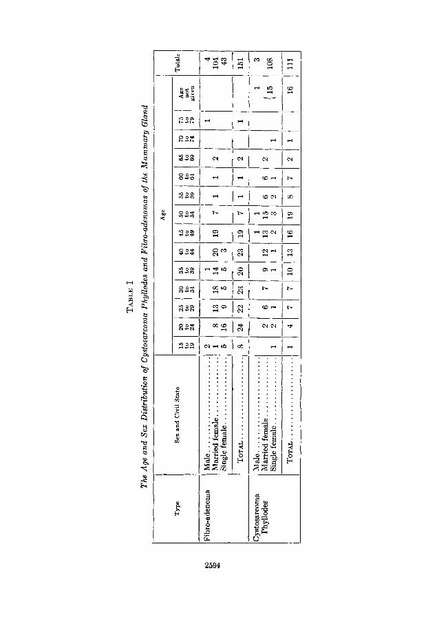
The characteristic features of this tumor have been summarized
1. Greatness in size, averaging 7.6 pounds. Frequently the
2. The lobulation and delimitation of the tumor with variable
3. Encapsulation of the tumor and non-invasion of the breast. 4. Mobility and usual non-adherence t o skin and fascia. 5 . No retraction of the nipple and no involvement of axillary
6. Possible occurrence in males (3 per cent). 7. Development from pre-existent fibro-adenomas, probably
intracanalicular fibro-adenomas. The transition occurs at the time when a gelatinous metamorphosis of the stroma takes place.
8. Long initial period of quiescence or slow growth, followed by sudden rapid acceleration.
9. Long duration-averaging seven years in 111 cases. 10. Important r81e of lactation and nursing difficulties in the
metamorphosis of simple fibro-adenoma to giant intracanalicular myxoma.
11. Intracystic polypoid excrescences moulded by apposition with each other.
12. Narrow sinuous distorted clefts between polyps. These epithelial-lined spaces contain variable quantities of clear yellow fluid.
13. Myxomatous stroma with cellular pseudosarcomatous regions.
14. Benignity; good prognosis with freedom from recurrence. 15. Successful treatment by wide local excision or simple
mastectomy. In addition to the metaplasia of the stroma into myxomatous
tissue, which is the distinguishing and essential feature of this variety of tumor, the cuboidal or columnar epithelium of the clefts
by us as follows:
tumor is as large as an adult head.
regions of fluctuation and resistance.
lymph nodes.

2586 BURTON J. LEE AND GEORGE T. PACK
may undergo metaplasin, to form pavement epithelium, even true epithelial pearls, indicating functiond instability. Such pearls were found by Groh6, Gorham, Kursteiner, G. €3. Schmidt, Stumpf and Beneke. In the layers of cyliridricnl epithelium of the adenomatous clefts these groups of pavement epithelial cells show all the characteristics of cutaneous epithelium, such as corni- fication. These pearls never occur unless the stroma rextion or proliferation has been really enormous, which is the reason that these tumors have been designated :is mixed tumors.
The tendency found in intracnnalicu1:tr fibro-adenomas to form true glmds by the cxtension of ductal epithelium down into the connective tissue is likewise present in cystos:trcoma phyllodes. These glandulnr productions in the tlumor are probably tinalogous t o the g1:inds in the mammary tissue. The acini in the adjoining breast are frequently cystic.
The ordinary intr:tcnnaliculitr fibro-adenoma of the breast is formed as follows. The inner or subepithelial layers of the peri- canaliculnr connective tissue participate more actively in the growth of the tumor than do the outermost connective-tissue layers, in consequence of which buckling or folding of these inner layers occurs, and rounded or p:ipillnry projections of the peri- ductal tissue are invagimtted into the lumina of the tubules. The same txp1:m:ttion may be valid for cystosarcoma phyllodes.
Rcinhnrdt w:~s the first t o demonstrate thzt cystosarcomn could develop from fibro-ndenomrt of the breast. In describing this transition, Billroth said that, if the stroma enlarges intracnnalic- uhrly, then either obliteration of the lumen occurs or these 1umin:t enlarge to cover the stromous proliferation. Another factor to which enlargement of the ductal lumina was attributed was secre- tion, which c:tused the iritraductular projectioris to resemble polypoid tufts. Frangenheim and Ribbert independently as- serted that a particular type of intr:tcanalicular fibroma was pre- cursory, because in this variety the periduct:il and periacinar connective tissue seem quite embryonic. Beneke also attributed the origin of cystosarcoma phyllodes to pre-existing fetal adenomas ; on this account, they were said to retain their embryonal capacity to proliferate. Frangenheim explains that cyst:tdenomas are transitional stages between fibro-adenomas and cystosarcomas. Schimmelbusch said that cystos:trcoma phyllodes was merely an advanced developmental phase of fibro-adenoma and, therefore, should not be called sarcoma, since it was a benign tumor. True

FIBRO-ADENOMYXOMA OF THE BREAST 2587
malignant tumors may originate, however, in benign mammary fibro-adenomas. In such fibro-epithelial tumors the epithelium may become carcinomatous, or both fibrous and epithelial tissue may become malignant (sarco-carcinoma), as described by Dorsch, Coenen, Wehner, and Schlagenhaufer. Virchow knew that sar- coma could originate in the stroma of an epithelial tumor. Ehrlich showed, when transplanting carcinoma of a mouse into different animals, that the stroma may change so as eventually to replace the carcinoma and finally become sarcomatous. The mechanism of stimulation in these instances, as in cystosarcoma phyllodes, may be similar in kind if not in quantity.
FIG. 1. CASE 1: CLINICAL PHOTOGRAPH OF PATIENT. Kote the size of the affected breast, the livid intact skin, and the protuberant
elastic eminence in the inner segment.
Lukowsky, Krompecher, and Sasse believed that cystosarcoma phyllodes may originate in the fibro-adenomas developing in breasts which are involved by diffuse fibromatosis, but the majority of pathologists accept the congenital origin (theory of Cohnheim). These authors, notiibly Wilms, insist that the anlagen of these tumors are embryonal tissue anomalies included in the region of the breast. The delimitation of the tumor by capsulation and the lack of infiltration indicate the separate entity of the tumor and contradict the theory of its origin from adult breast tissue.

2588 BURTON J. LEE AND GEORGE T. PACK
CASE REPORTS CASE I: E. R., a white, married wornan, aged forty-four years,
applied to the Rllemorinl Hospital, Oct,. 21, 1930, complaining of a painless lump in her right breast.
Ptrst History: Nine years previously a cyst of Bartholin’s gland was excised. The patient h:id been niarried only two years and had never been pregnant.
FIG. 2. CASE I: GROSS SPECIMEN ILLUSTRATING THE LARGE, INTRACYSTIC hIuxoh1- ATOUB 1’oLws SEPARATED BY N A I ~ I ~ O W . TORTUOUS CLEVTS.
Present Illness: One month prior to applicat,ion the patient chanced to feel a lump in her right breast. There was no mastalgia, and no discharge from the nipple. The tumor grew rapidly and soon produced a red mound-like eminence on t,he contour of the breast.
Physictrl Exatiiiriution: In the lower inner quadrant of the right breast was a firm, globular, freely movable tumor. The superficial part of the turnor seemed to be fluctuant. Both breasts were large. The nipples were erect. There were no palpable axillary 1ymphadenop:ithies. The immediate provisional diagnosis was cyst of the breast; but since aspiration by needle puncture did not yield fluid, the tentative diagnosis was changed to fetal adenoma.

FIBRO-ADENOMYXOMA OF T H E BREAST 2589
Treatment: On Oct. 22, 1930, the tumor was simply excised, without the removal of mammary tissue. This was accomplished by an elliptical skin incision surrounding the protuberant part of the tumor. Conva- lescence was uneventful.
Gross Pathologic Anatomy: The encapsulated tumor measured 8 x 5 x 4 cm. On hemisection numerous intracanalicular polypoid projections were found closely packed within the capsule. The polyps were sepa- rated by intercommunicating clefts, which contained thin, clear, straw-
There has been no recurrence to date.
FIQ. 3. CASE I: CROSS-SECTION OF A TYPICAL INTRACANALICULAR FIBRO-ADENOMYXOMA
colored fluid. The stroma was smooth, loose, and gelatinous in certain regions, especially within the polyps.
Pnlhnlogic Histolog!j: Myxomntous t,issue was found in many of the polypi. The spindle cells of the stroma tend to run parallel to the e1ong:tted clefts. Some parts of the stromn were composed of fusiform cells with bizarre nuclei, which were n:irrow and rod-like, resembling smooth muscle nuclei. Some hyulin degeneration had occurred in the polypoid tissue. The clefts were lined by cylindrical and cuboidal epithelium, which projected into the stroma to form pseudo-glandular lacunae.

FIQ. 4. CASE I: COMPLETE HYALINE METAMORPHOSIS OF NIJMEROUR POI~YPFI, WHICH HAVE RETAINED T H E I I ~ EPITHELIAL INVE~TMENTS.
I?IQ. 5. C A S E 1: SINUOUS, DI~~TORTED, ANA6TOMOTIC CLEFTS, h N E D BY CUBOIDAL 14:I’ITHELIUM AND FORMINQ SECONDARY I’SEUDOACINAR C R Y P T S .
The spindle cells of the stroma run parallel to these ductal clefts.
2590

FIBRO-ADENOMYXOMA OF THE BREAST 259 1
CASE 11: M. E., a white, married woman, aged fifty-two years, applied to the Memorial Hospital, June 20, 1927, complaining of large tumor masses in each breast.
Past History: The patient’s only child, born in 1895, had nursed both breasts without difficulties in lactation. The menopause had been passed without complica tions.
Present Illness: Forty years earlier, when the patient entered puberty, a t the age of twelve years, she discovered a small lump in the lower segment of the right breast. This lump gradually increased in size until 1922, when it grew wit>h greater rapidity. Five years before the patient was seen (1922), another lump appeared in the upper half of the left breast; this tumor gradually grew to involve the skin and became
FIG. 6. CASE 11: CLINICAL PHOTOQRAPH OF PATIENT WITH GIANT INTRACANALICULAR FIBRO-ADENOMYXOMA OF RIQHT BREAST; BULKY ADENOCARCINOMA OF
LEFT BREAST.
the size of an orange, The only discomfort experienced was the weight of the right breast.
Physical Excmintition: The right breast was completely replaced by a bulky, lobulated, elastic, semi-fluctunnt tumor. The nipple was obliterated. The tumor was encapsulated and freely movable. The breast was pedunculated, due to the traction exerted by the weight of this tumor, which rested against the upper abdomen. The right breast measured 21 inches in circumference. In the upper segment of the left breast was an ovoid movable tumor S x 5 cm. in diameter. The super- jacent skin was purplish-red and firmly adherent. There were no palpable axillary lymph nodes. A radiograph of the chest was negative except for obliteration of the left costophrenic angle by adhesions. There were no discernible regions of *calcification in the tumor of the
There never was any cough or pain.
27

FIG 7. CASE 11: GROSS SPECINEN OF A GIANT INTI~ACANALICULAR MYXONA OF THI
There is marked overgrowth of the stroma in lobular arrangement; some of these BREAST.
lobulea appear myxosarcomatous when examined microscopically.
2692
FIBRO-ADENOMYXOMA OF THE BREAST 2593
right breast. The provisional clinical diagnosis was sarcoma of both breasts,
Treatment and Progress: In June 1924 a simple mastectomy of the right breast was performed. At the same time 62; millicuries of radon in 25 gold seeds were implanted in the tumor of the left breast,. By December 1927 this tumor had disappeared completely, but the patient complained of pain in the left shoulder girdle. A radiograph of the left shoulder revealed early metastasis to bone. In November 1928 a left radical mastectomy was performed, using a transverse Stewart incision;
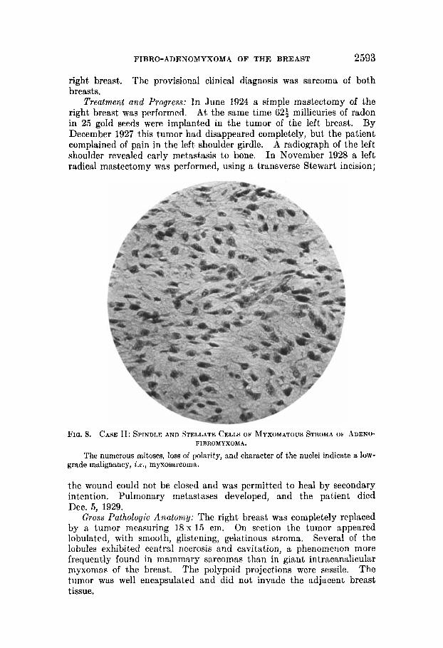
FIQ. 8. CASE 11: SPINDLE AND STELLATE CELLS OF MYXOMATOUS STROMA OF ADENO-
The numerous mitoses, loss of polarity, and character of the nuclei indicate a low- FIRROMYXOMA.
grade malignancy, i . e . , myxosarcomn.
the wound could not be closed and was permitted to heal by secondary intention. Pulmonary metastases developed, and the patient died Dec. 5, 1929.
Gross Pathologic Anatomy: The right breast was completely replaced by a tumor measuring 18 x 15 cm. On section the tumor appeared lobulated, with smooth, glistening, gelatinous stroma. Several of the lobules exhibited central necrosis and cavitation, a phenomenon more frequently found in mammary sarcomas than in giant intracanalicular myxomas of the breast. The polypoid projections were sessile. The tumor was well encapsulated and did not invade the adjacent breast tissue.
TA
BL
E I
The
Age
and
Sex D
istr
ibut
ion
of C
ysto
sarc
onia
Phy
llode
s an
d F
ibro
-ade
nom
as o
f th
e M
amm
ary
Gla
nd
Typ
e Se
x an
d C
ivil
Sta
te
Fibr
o-ad
enom
a M
ale.
. ...
......
......
......
. hl
arri
edfe
mal
e.. ..
....
....
...
Sing
le f
emal
e.. ..
....
....
....
. TOTAL..
....
....
....
....
...
Cys
tosa
rcom
a M
ale.
....
....
....
....
....
...
Sing
le f
emal
e.. ..
....
....
....
. Ph
yUod
es
Mar
ried
fem
ale.
....
....
....
..
Age
15
20
25
3
0
35
40
45
50
5
5
60
65
70
75
to
to
to
to
to
to
to
to
to
to
to
to
to
19
24
29
34
39
44
49
54
59
64
69
74
79
~
-----------~
2
1
1
1
8 13
18
14
20
19
7
1
1
2 5
16
9 5
5 3
8 24
22
23
20
23
19
7
1
1
2 1
11
2
6 7
91
21
31
56
6
2 1
2 1
11
23
21
1
__
__
__
-_
_ ------ ~
__
I I
Age
n
ot
give
n
Tot
ah
TOTAL ......
......
......
...
(1
I 151
I
4 7
7 10
13
16
19
8
7 2
1
~-
1
I 3
1
__
_-
{ 15
1 108 1
4 10
4 43
__
_-
16
111
1

FIBRO-ADENOMYXOMA OF THE BREAST 2595
The left breast contained a small, hard tumor measuring 2 x 13 x 1 cm., situated in the upper medial portion. The skin over the tumor was slightly ulcerated.
Pathologic Histology: The right mammary tumor consisted almost entirely of myxomatous tissue which had completely replaced the original stroma. The interlobular clefts were lined by cuboidal epi- thelium. The spider-like cells of the myxomatous stroma were very large but not closely packed. Microscopically one obtains the impression that the tumor is of low-grade malignancy, i e . , myxosarcoma.
The tumor in the left breast was obviously a necrotic carcinoma, probably of the sweat gland type. No metastases were found on micro- scopic examination of the lymph nodes in each axilla.
Comment: The tumor in the left breast was a bulky adenocarcinoma, which caused the patient’s death, but was histologically and histo- genetically not related to the giant intracanalicular fibro-adenomyxoma of the right breast.
[over]
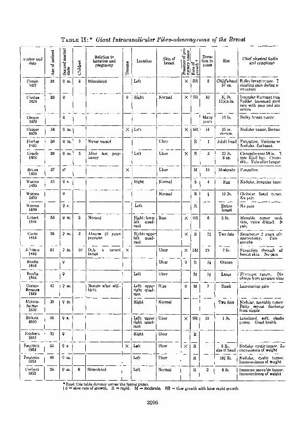
TABLE 11: * Giant Intracanalicular Fibro-adenomysoma of the Breaat 4a CI
Author and 2 date
0
4
Relation tn lactation and
pregnancy
Dura- tion in yenra
__ 5
__ 10
_- Many ycars
Skin of breast
Chief physirnl findin and symptoms Location Size
:hild’s hra 57 01.
8: lb: 1 5 x 8 in.
-__
13 lb.
Rulkv breast tiimor. I cilialing pain during n struation
Left Stimulated
Right Chclius 30 1828
Normal
0 m.
0 m. __
Left I4 3” in. eircum.
Addt heal -__
Nodular tumor, Buctuo
-____ Fkcher 30
Orucft: 31)
I835 -____
1838
Never nursed Ulcer 1
__ 2
Funcation. Varicose vc Nodular, tluctuant.
0 m. After lalit prep. nancy
Left Ulcer 22 Ib. 8 02.
Circumference 58 in. 1 mor filled lap. Cyanti skin. I’ainafterfungat
Ulcer 16 Moderate
1x39 Itight Normal 4
__ I
Nodular, irregular turn(
(:lobul:rr fused tunioi No p i n
- - E66
12 Ib. Warren 1 1839
0 Nnrmal
0 1.
0 m. -
Lcft No p i n Entire brenst
2 Ib.
Two fista
__ 6 Lcbert 50
I814 1 Nornl:il Iliaht: lower left quad- rant
Biue Movable tumor undt skjn. veins dilated. N pain
Coote 36 1816
0 m. Ridit: uppei left quad- rant
Recurrener 2 sears aft( mastectomy. I’irin moiitlis
P m.
0 ~
Ulcer 7 I\). Fungntion throulrh ad herent skin. No pain
Oiily 4 nurscd bread
Ulcer Urodie 1816
Orawe
Large Urodie 1846 1 Left Ulcer Fluetuaat tumor. Di.i
charge from pressure ulcei -_____ Gorup- 41 Hcsnnes
1849
hlettcn- 35 hrimer
1850
-_____
Ulue 3 Small Lanrinating pain Nodule after still. birth
Left: upper right quad- rant
Right Q m. Normal TWO fiats
1 Ih.
Nodular, movable tunior Fatty t w o w dischnrgr from nipple
Lobulateil. aoft. elastic tumor. Good health
__ 10 0 I). Left: upper
right quad- rant
Ulrer Birkett 51 1850
--___ ~
Erichscn 35 1852
Right Ulcer
0 8 . Ulcer 5 Ib., ize of head
Left Nodular cystic tumor. In- convenience of weight
Pcrauson 1 40 1853
0 m. Ulcer Left
Left
Nodular. cystic tumor. Inconvenience of weight
Immense movable tumor. Incoiivenience of weiglit
1st Ib.
8 Ib. Corlrtt 38 1853
P m. Stimulated Vormal
Road this table direotly across the facing paues. t S = slow rate of growth. R = rapid. M = moderate. SR = nlow growth with later rnpid growth
2596

Cases Reported i n the Literatwe
Nomenclature Treatment Gross patholosic anatomy Histopathologic anatomy
___-_.
Firm, cystic with intracystic fungois growths
Extirpation
- Vascular intact capsule Extirpation I’endulou~ intn-cystic tumors ystic hydalids
ystic liydatid~ Extirpation
Constriction amputation b! ligature
Simple mastectomy Lobulated, also cystic Fibrous t i w e of lobules very cellular like warcoma
ystossrcoma
teatomntoua and sarcomatous dc- cnoration
Simple mastectomy Five distinct portions. Resembles eoty kdolls or arbor vitae cereklli
:ystosarcoma phyllodes Simplc mastectomy Diagnosed by J. Miillcr
Cauliflower excrescences in cysts -
Simple mastectomy ~~ ~
Simple mastectomy Globular nodules in dairy fluid jydntid tumor of hrmst
Hydatid tumor of breast Cystic with globular growths; fluid red dish
Simple mastectomy
Mastectomy, death, wouni irifcctioii
~~
Lobulated; cauliflower growth into cys
~
hlastectomy, rccurrcnce, exci sion
Simple mastectomy Firm, irregular tumor adhercnt to skin fluctuant, cystic
Plicated. fimbriatcd intracystic ex crcsccnce
___- Simple mastectomy Sero-cystic tumor
Sero-cystic tumor Simple mastcctorny Many intracystic excresccnees. pize a to oran@
Lamcllated structure of tumor (intra tumoral strurture)
__-- Radical mastcetoniy
~~ ~
Brownish-rcd paiiillary intracysti vegetations, looked like cock’s comb
Not all vegetations vested with epi- thelium
Calcarcoua deponita in tumor ________
Simple mastectomy
~
Extirpation I n addition to eystosarcoma phyllode: aiiotlier fibroaderioma was present
____ __- Ilistolonically, similar to Paget’s de- scri~!tion
Cystic mrcomn.. . . . . . . . . . . . . . . . Simple mastectomy. recurrenc~
Simple mastectomy ~
Cyatie cavities with fungous indusioiir Cystic sarcoma
Cystic earcoma Cystic cavities with funroue inclusions Puncture, Inter mastectomy
Simple mastectomy Cauliflower excrescence in c3vit.y Vsscular tissue; no invasion; gelatinous Cystic sarcoma
2597
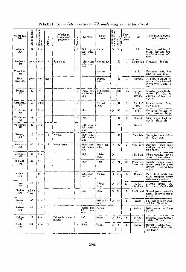
TABLE 11: Giant Zntracannlicular Fibro-adenomy.roma of the Breast
Relation to lartation and
pregnanry
____-~
Dura- tion in years
-
__ I
__
Skin of breast
Chief physical findinl and symptoms
Author and datn Locntian Size
2 Ih.
Largcappli
11 Ill
-- Velpeau
1854
~ _ _ - Irregular. nodular. 0 tuant, movable turn painful at mcnstrual riods
I~luctuant. Painlrss _____
_______ Right: upper Normal right quad- rant
Left: upper Normal, rei right quad- rant
_ _ _ _ ~
______- Normal
24
__ young Stimulatcd
~~
Benjamin 1855
Cruveilhier 1858
Enormous soft, 110s latcd, Huciuant tumor
young
- 20
~
52
1 Enormous Nadulnr, fluctuant cn nenees. Sanguineous d c h q e from iiipple. 1 pain
Bryant 1861
a: i in. ilinm 22 in.
clrcum.
Movable. clastic. Burtila tumor. No Inin. Sa siiincoua discliarse fro rlipplc
RiKht: Sub- Red. dilate1 areolar locn- vcin8 tion
Castresana 1861
2 16X13X7 cm.
Skin adherence Coiiti ually painful
Normal,
Riglit Weinlcchnci 1866
50
2 i ~
5 14 Ib. Enormous Huctunnt ti mor. Heaviness. No pai
Clear yellow fluid fro1 nwlc . Slight pain
-__ I___
Weiiileehna 1875
3 wnlnllt Right
Left: uppcr right quad- rant
Right: lower right quad- rant
______
_ _ _ ~ Weinlcchnei
1875 22 Walnut
__- Two lists
Vcry largc
Normal Bvrstre 1SG8
Demnrquay 1808
Tumor hoth solid and c y tic. Slight pain
Bosselated tumor, partlj solid. part1 cystic. Pair for 4 moi i t t
Niiiple retracted. Mobilc tumor. Nn symptoms
~ _ _ _ _ _ _
Never nursed Right: lower Tcnse, cya rinht quad- notic rant
hshhurst 1871
2 in. diam. Right Di!ated
Right Ulcer
vc1119 ___-.
Ilclmuth 1811
20 From clav- icle to thigl
Nodular. bluish, mnliile tuinor extending a Imc clavicle. Distress from weislit --__ ___
Sutiareolnr Normal lllcatiolr
I Amndo
1872 I +
15
Partly hard. partly Huc- tuant. Erysipchs followed prcliminnry puncture
Orange
4 1 I!., 6 in. ham.
Gross 1874
Di!atcd vellls
Movnble, elastic, hbu- lated tumor. Good health
niddle age
5 Adult h a d Non-adherent. movable tumor. Inconvcniencc of weight
Lcft Lllccr
___ Skin udher cncn
Ivcrscn 11177
52
49 ~
8 Lnrge Fluctuant with peripheral nodulcs. MastalJa
Well circumscribed lump. No win
- Watson
1878 I3ipht: uppc eft quad- .silt
ILrft
Normal Walnut
-~ Normal Watson
1878 48
36 ~
8 lox20 em.
Enlargcd a t time of menses
Irrcgulnr. tense. fluctunnt nodule. No pain
Movalde, nodular tumor. Traumntism, then pain, (hen tumor
Itidit Normal 3 IIcn's cgg Finrher 1x112
2598

Cases Reported in &he Literature (continued)
Extirpation. crysipelm. death
h'amenchtura
Edematoua polypid projections in to cystic clelta
Treatment
- Encarmulaled, Ruetuant. aolt ingrowths in cysts
~ _ _ _ _ _ _ - ~ __-- - ~ _ _ _
___ Extirpation
I G r m mtholopjc anatomy I
---i Hemorrhage; much elastic tisauc
-
.~
?stosarcvma
istosarcoma - ~ - - ~
Histopathologir anatomy '
__ .. . - - . . - . - - Inrision; then rxtirpation lntr~eystic polypoid crowths I
I I
- .. . - . . - --
! 1 - - - -
- . -- - Extirpation, no nnrmsi~
__ - - - .. . . - - . .. - - . . .. . . . . - - .-
Simple msstcctomy I I I
!ystasarcoma phyllodm
.~
Extirpalion - I I . . . - -. - - __ - - . - -. I - . .
Simple mastectomy I I
. _______-_---- . - - - I
_ - _ I yetosurcoma pbyllodea IGi rpn t ion
I
Simple mmtectomy 300 m. liquid; iiitracystic vegetation as 1 large as egg I --
:ystoplrcoma phyllodea Vewtntion. covered by flat and cyliii- drical cclle !
I -____ ~. I'ultncrous, friahle polyp in cyst con-
Intratumoml injection alphcl Marked s ~ontnneaue degeneration: nol. death enscapulatea
--ltainiiiy honey-colored fluid 2 y h c earconla I Extirpation
Cystoearcomn phyllodea .__.___ -
-i
Intracanalieular earcoma
I I
Large nuclei in connective-tisauc cells
__ Cyst fillcd by urapelike tumors Icaving Sl)i!idlc c e l h parallel to delta lined by c i r r t y cyliridricnl cells
Simple mastectomy
I _ _ _ I
TelaiiKiwtatie cystosarcoma I Puncture. then cxtirpation large telaneicctatic papillary projce- ~cirrlious tissue with many spindle tioils iota cysts
_~___ -- 1 - ~ ____-.
Proliferow cystosnrcoma Extirpation
Cyetofi brosareoma I ~lastcetomy. rcciirrencc -I-_-
Smooth. clistciiing, striated. gelnlinous. ycllow. tissue
Verified microscopimlly .~
hlyxofibmmn. intracanaliculnr Simirle nustcctnmy I-%-cystie nodular projections: i thin nedielra -.I. lnlraeanaliclrlar papillary Libro- Sinililc mastectomy myxoma I
\\'all8 and clcfta lined by cylindrical cpit helium
hf yromntous slroma _..___-
I I
2599

TABLE 11: Giant Tnfrnctrnolicrrlnr Pibro-ndenoInlJ.~omri of the Brefrst
Dlira- tion i n years
__
Relation to lacfatinn and
prcgnancy Chief physicnl findir
and symptoms 4ut hor and
datu Skin of breast Locat ion Rizc
Enormous Itai n :.Pard 1x42
No cliiiiczl liistory S l V C l l
Jungst 1x84
Right Nornial I? Mail’s fi8t H a d nodular. non-:irl eiit turnnr. I’aii~fiil tncr~atrual pcricrd
Imalized ilur.tu:uit t i i l?iin i i i t iiinor
~. . . - Schmidt
18XI Adhcrcnt to tumor
7 Man‘s fist
Schmidt 1884
Aillicrcrit to tumor
Man’s fist Loealjzcd, fluctuallt tu I’tii i i i n trunnr
Srhmidt 1884
Stiniiilated by lac. taiiun
Glurtiuint tumor. 1’: 1111 hrrast
Schmidt 1880
Lcscr 1 888
Normal 3 10 X5 X 5 cnr.
Rielit. whol breast
Norm:il 1 li 7XliXB mi.
Nailulnr tumor, painle!
I m e r 1884
1,eeer 1888
.- -_ I4 1oxti
cm.
DXSXl rm.
Ciiild’n lirail
Marl’s t is t ..-
Fluctuiint tumor
Smrotli, i~odular tumi p:iiiili.ss
____ l’lccr bee r
1888
?I Solt, rooildcd. Ructrin tumor
IPscr 1888
Lescr 1888
Noetael 1892
.___
Nortzel 1x92
Ntietzel 18Y2
Noetzcl 1x92
Noetzcl
I’ i l l iet 1693
liursteincr 1804
HaPC kel 1804
Hawkel lXY4
Hacckel IS94
Pcurcc 1895
.-__
ino2 __
____
-___
_-_ ___
___
~
40 clinical history
Nouo Itidit: lowe lcf t quad rant
t 13XllX8 em.
11x11 nn.
-__ Noiw 9 Nodular. painful tumor i
I)ri.:ist Itiicht: lef half
I l ix l i t ~ _ _ Tense cya- notic
~
Mnsti,tis iit eacl lactation
Noildar tumor i n brmst
1)eliniitr.d nodule Non-adhcr- c n t
IOXIO cm.
No cliriiwl history
No rlinical history 15X 15x9 rm.
Orunae __-
Right: uppe riglit quad. rant
28 ~ ~~
lrreyular. firm, clnstic, tnd soft tumnr
Rirht: u p p half
Left: upper r i d i t quad. rant
N o r i d 12XOX7 ern.
Walnut
I I em. d im.
t
-_ 7
Small puiiiful lum” in )rcaqt
Vodule i n brcast ~ _ _ _ _ _
Left Cyanotic, rdherent
Inflamed, .cd, adherent
1 Child’s head
Large tumor in breast
Punfiition thrniich skin. -’?clieiia. 1,:inciintirig
-
I i l l l l Y
Hight Ulcer 15 Vursed 5 cliildren in left b ras t only
___~ Veins dilated Snow
l800 11, 4 t Ib. Mobile; irregular; ii ipplu
ib!itcrated. Slight dull inin
2600

Cases Reported in the Literature (continued)
Nomenclature Gross pathologic anatomy Histopathologic anatomy Treatment
coma with lacunar cysts Simp!e mastectomy Encysted; lobulatod Great lacunar slits. Stroma has long fusiform cells. Myxomatous aspect
Star-shaped stroma cells racannlicular mysoma Simjile mastectomy I'olypoid projcctions into cysts; narrou pedicles
Hndicnl mastectomy Many intracystic polyps Myxomatous stroma stosarcoms
'atmrcoma Simple mastcetomy Many intracystic polypn Myxomatous stroma
Extirpation
Simple mastectomy -___
istosarcoma myxomatoidea Mammary ciarid atrophied; tumor rcd- dish gray
Extirpation Multiple cysts, some with pnlypoid pro- jectioiis
Encnpsulated. Uyalinization of stroma .olileratinp cystic tumor
roliferating cystic tumor
roliferating cystic tumor
rolilerating cystic tumor
'rolilerating cystic tumor
'rooliferating cystic tumor
-
-~
Extirpation Cholcstentoma in onc cyst Polypoid excrescence in cyst
Many cysts: a few having cauliflowcr projections
Cdcilicd connective-tiesue stroma
Extirpation Many cysts. Papillary excrescences in cysts
Intracystic proliferations in many cysts
Myxomatous stroma
Myxomatous stroma; cpithelium, cylin- drical -
Extirpation
Extirpation !My developing polypoid excrescences Ill cysts
Radical mqtectomy Smooth cauliflower growths in cysts. Clefts between polypi
Myxomatous and spindle-cell stromn
htomrcoma phyllodea Cauliflower excreecences in cysts eepa- rated by clefts
Myxomatous stroma and cylindrical cpit hclium
Extirpation
Extirpation Warty excrescences between ananasto- motic clefts
Cauliflower excrescences; aiiastomotic clefts
Stroma seems almost sarcomatous
Myxomatous stroma and cylindrical epithelium
Extirpation 'ystosarcoma pliyllodes
Cgstosarcoma mammae proliferun Extirpation Cauliflower polyps into cyst Myxomatous stroma
Fibrous tissue has sarcomatous aspect __
Cystosarcoma papillare Simplc mastectomy Arborescent vegetations
- Myxomatous stroma; qnamous meta- plasia
Cleft-like lumina. Ifihulated. Encag sulated. Plump cubical polype
Simple mastectomy Papillama intracanalicular
Cystomrcomn phyllodes Extirpation Proliferations filled cyst; thin pedicles Myxomatous stroma; few nuclei
Cystosarcoma phyllodea Leaf-shaped vegetations in cyst, packed closely
Myxomatous stroma, cubical epithelium Simple mastectomy
S h ple masfLe tom y __ ~ ~~ ~
5 cysts. Iirtra-cystic proliferations with hroad base
Cystosarcoma phyllodes Myxom:itous stroma
Iiitracanaliculer fibroma with gelatinous metamorphosis
- _- latracannlicular fihroma with selat , inous mctamorphnsis
Simple mastectomy
Emhryoid spindle cells and adult fibrnua tissue
1 Intracystic maminary sarcoma White fibrous masscs in cyst containing fluid
2601

TABLE 11: Giant Intracanalicular Fibro-adenomyroma of the Breast
Author and dste
Relation to lactation and
pregnancy Skin nf hrcnst
Chief physical Endinl and symptom Location P c
E3
0 Left ~~
Siae
48x40 em.
Infant head
Dcsplnta 1900
Enormous tumor. Sma one in right breast. Shooting pnins
Noduliu movalile turn No pain
______
Veim dilate(
Qrohe 1000
27 v m.1 1 Wet nurse for : more children I Right Veins dilate(
-___ Right: lowei
rant left qud.
43
__
Fist Tumor, partly tluctun and partly solid
\Vilms 1001
Beneke 1902
Ulcer __- Ulcer Bcnekc
1902
Schmuckcrt IOU4
~~
50
34 -
__ 46
Mnn's head
Large tumor in brenst
Did r iot nurse Teosr. di- lnted veins
Lyge tuniirr i l l brexr nipple iioriiiiil Amciir rhea for 2 years; no ma talgin
Lnrgr 1i:ird tumor in rig1 brenst
______._
Child's head
Two fists 0 Right: sub. areolnr loen- tion
x Lcft -____
Finsterer 1907
28 Tense, cya- ootic
Man's l1eXI
Large thrice rccurrcr tumor
Latcr married, stimulated recur rencee
54
__ Enormous Fiiist,ercr
1907
Finsterer ID07
Also reported 16 oilier cliildreu: 4 had noije;
but 'agc and parit. 3: 1 had 4; 1 hac
~~
Ages 17. 2, '; 2 had 8; 81
!, 41, 45, 4t 17, 48, 50, 51. 01. No. c BRCB givinR no d e b liad 1; 2 had 2; 2 t
Stimulated by each pregnancy
- 42 Moriinrd nni
Maason 1900
6 20x25 em.
Veins form euput u i ~ d i i s 3 Fluctiration. Iiiroi~ven ienre of weidit
Risht: upper Ulcer, hem- left quad- orrhaacs rant
Right: upper Dilated vciii left quad- rant
~ _ _ _ ~
~ ~ _ _ Ulcer
Prirnd 1YU0
54
__ 80
4 Adult's hund
Larrre tumor in breast Weight, no Inin
Girard 1910 Enormous
Hen's egg
Part of tumor ~ a n ~ r c n o u s Seroflanguineous dischargi
Small tumor (movable) No pain
Gorliam 1911
63 fight: sub- nrcular loca- tion
67 ~
60
Seeousse 1912
Prym 1912
Child's head
Hilohed tumor partly firm. partly tiuctuarit
Fist
Large
Virm round tumor in breast
l?lnstic, nodular. Ructunnt tumor
restermark 1918
63
59 -
- 36
4 Lactationnl diffi- :ulties
Right Normal
Right lower left quad- rant
-___ Silvaa 1910 4 Ih.
1 2 x 8 om.
Orange -_
Hard tumor in breast. No pain
Wulfung 1Y23
Right
Lcft ~~
Wulfung 1923
44 -
Orange
Ch ild'fl head
Wulfung 1923 Nodular. movable tumor
-__ Wulfun:: 1923 1 Red 68 Movable tumor
Large confluent nodules. Wcakncss: emaciation
_______- A P P ~ ~
1885 Rrnms 13 X23 X 14
cm.
Rinkert 1924
39 1
2602

Cases Reported i n the Literature (continued)
Simple mastectomy
Simple mastectomy
-~
Nomenclature
Encapsulated. Small clefta between proliferations
White pearls of aquamous meteplasia
Simpla mastectomy
Excision. then maatectomy for recurrence
_ _ ~ Edematous polypoid intracystic nodules
Hadienl masteetomy
Rad ia l mastectomy
Simple mnatectomy
Elongated clefts between polypoid nodules
Mastectomy; death from me- tastasea to lurras
Extirption
Typical cystosatcoma phyllodee
Extirpation
Extirpation
Intraeystic cauliflower maeaca
Bulbous cauliflower intracystic tumor
Extirpation
Iladical plr?stectomy. recur- rence. excision
Necrotic nodule in cyst
Deudritic arborizations; polypoid
Histopathologic anatomy
Radical mastectomy I Simple mastectomy 1 Cysts Wed with proliferations istofibrosarcomo
vstoearcom
Myxomatous stroma; some snrcomatou~
Myxomatous stroma: some compact embryonal connective tissue
areas
Sarcomatous proliferation of cnnnwtivc tissue
Pseudo-sareomatous proliferation ~ - _ _ _ ~ _ I _
wly cystoearcoma phyllodes
denoma pseudosarcomatodes Extirpation 1 Ducts distorted to form clefte
ihroadenoma rntrmcanaliculnre apillare
Extirpation & ~ i n d ~ e - c e ~ ~ and myxomntous are39 in stroma
2 tumors. intracystic polypoid growth
Smaller tumor is typical
True Bbrosarcoma in stroma-verified by recurrences
:ystossrcoma phyllodea
I Maatcctomy by severance of pedicle
Myxomatous stroma; giant cells
- Radical mastectomy Cptic, with nodules and gelatinous
structure Stroma contains spindle cells and e m bryonal cells
Intracannlicular fibroma with sar- comatous change
Cyatoearcoma
Adenomyxofihroma intracanalicu- lare papillare
Star ceib in etroma; squamoue met* plasia
~~
Fibrosarcomatous myxomatous stroma
~ ~
Cyetofibroma intracanaliculare Recurrent tumor; myxomrcomatoun
Stroma earcornatnus -
CY6tOsarcoma
Fusiform cellular elements
Cystosarcoma phyllodea -
Polywid n d d w into clefb
Special tumor; intracanalicular polypoid
Extirpation
Extirpation
Very cellular stroma CyStO8arcoma phyllodes
Cystosarcoma phyllodes __
Diffuse Bbromatosis
Hyalinized stroma; bizarre nuclei; pig ment
CYstosarcoma phyllodes
Fihroadenoma intrncanaliculnre earcornatodes
~- Xanthomatous and sarcomatous rum

2604 BURTON J. LEE AND GEORGE T. PACK
+
f f
SR 2 8
- SR
TABLE I1 : Giant Intracanalicular Fibro-adenomgxoma of the Breast
Dura- tion in years
-- 8
-_ 37
Author and date
- _ _ ~ Hart 1937
Lee niid Pnrk 1930
Lecand Pack 1830
Leeand Pack 1930
I.ernnd Pack 1930
Lee and Pack 1931
Ler and Pack 1031
___
____-
_____
_____
_____
___-
-__
..7
'i %
26
4:
-- 5ti
40
34
28
44
53
SR
SR
SR
- "
- Sit
10
___ 8
_- 3:
-- One
month
40 years
__
Relotion to lactation and
pregnancy Chief physical findini
and symptoms Location Skin of breaat
Sinus
Ulcer
Size
Orange Large movable tun Rapid growth during pi nnocy
2
~
7
Never nursed this breast; st,imulated
Started after child- birth
Bulky cyntir !obulatcd mor. No pain
Richt
Both breasta Normal 13x11 em.
Bulky lohulated tumors both breasts. Oecasioi dull bilaternl pain
1 Right: uppe half
IfiX1OXQ cm.
9X7X5 em.
8x5 cm.
___
__--
Tarae cystic lobulatcd t rnor muas. Occasional di pain
Movable non-adlierent t mor. Dragginr pain
Glolmlar, discrete. mo able, large tumor. No pa
Blue
Normal
-___- Adherent
Abscess in right breast
-
2
__ 0
Right: lower right quad- rant
Rirht: lower rirht quad- rnnt
-_F
1 -
Right Adherent 18x15 em. size of melon
Pedunculnted., baselate b r e d containing turn04 Nipple retrncted Bulk ndenocarcinorna in oppc site breast. No discomfor except weight
BIBLIOGRAPHY
ALEXANDER, G. : Cysto-fibrosarcoma phyllodes mammae, Objazat. pat.- anat. izslied. stud. med. imp. Charkov Univ., 1890, pp. 173-178.
AMADO, S. : Un caso de kisto-sarcoma telangiectasico a papillar da glandula mamaria do homcm, J. SOC. d. sci. m6d. de Lisboa 36: 56, 1872.
ARATA, P. : Cisto-sarcoma della mammella sinistra, Nuova Liguria med., Genova, 19: 401-403, 1874.
ASIIHURST, S.: Cystic sarcom;i of the breast, Am. J. M. Sc. 61: 154, 1871. BKNEKE: uber die Adenofibrome der Mamma, Verhandl. d. deutsche
path. Gesellsch. 4: 205, 1902. BENJAMIN, L. : Beitrng zur genaueren Kenntniss des Cystosarcoms der
weiblichen brust, Virchow's Arch. f . path. Anat. 9: 299-301, 1855. BERGERET AND BOTELHO : 13pith61-sarcome de la glande mammaire,
Gyn6c. et obst. 1: 139-147, 1920. BERKA, F. : Zur histologischen Charakteristik der fibroepithelialen
Mammatumoren, Beitr. z. path. Anat. u. z. allg. Path. 53: 284-323, 1912.
BERTOLET, R. M. : Cystosarcoma proliferum of the mammary gland, with metastasis in the liver and spleen (dog), Trans. Path. SOC. Phile. 4: 241, 1871.
BIEBL, M.: Das Mammasarkom und seine Beziehungen zur Fibrosis mammae wie zu den gutartigen Mammageschwulsten, Beitr. I . klin. Chir. 140: 52-74, 1927.

Nomenclature
wanalicular myxoma
ot intracanalicular myxoma stowmma phyllodes)
st intracanalicular myxoma Btosar~oma phyllodes)
mt intracannlicular myxoma atrmrmma phyllodes)
mt intramnalicular myxoma stcsarwma phyllodes)
ant intracanaliculsr myxoma of rvlt (cystmucoma phyllodes)
ant intracanalicular myxoma of wt (cystmarcoma phvllodes) th m osarcomatoua &an es. w bul$ adenocarcinoma of feft east
FIBRO-ADENOMYXOMA OF THE BREAST 2605
Cases Reported i n the Literature (continued)
Treatment
Simple mastectomy
S i p I e mastectomy
Extirpation
Radical mastectomy
Simple mnstectomy
Locnl excision of tumor
Simple mastectomy of righ breast: interstitial irradiatioi and simple mastectomy of lef breast
Grow pathologic anatomy
Large intracystic polyp
Intracystic polyp
Intracanalicular polypoid masses
Typical cystosarcoma phyllodes
Soft polypoid intracystic nodules
Intracanalieular papillary excrescences
Lobulated, smooth glistening tumo nodules. Some necrotic cavitation Slight preservation of intracanalicula arrangement
Histopathologic anatomy
Myxomatous stroma
Myxomatous stroms; calcification
Myxomatous stroma
~- Myxomatoue and xnntbomntou s t rom
Myxomatous stroma
Myxomntous stroma; hyalinization ir regions; secondary pseudoscinar crypt:
Myxosarcomatous metamarphoais oi stroma of this tumor: also adenocarci noma of opposite hresst
BIGELOW : Tumor of the breast ; intra-canalicular papillary fibro-myxoma Boston M. & S. J. 94: 581, 1876.
BILLROTH, T.: Untersuchungen uber den feineren Bau und die Ent- wicklung der Brustdrusengeschwulste, Virchow’s Arch, f. path. Anat.
BINKERT, M. : Fibrolipoadenoma intracanaliculare sarcomatodes xantho- matodes mammae, Frankfurt. Ztschr. f . Path. 30: 498-511, 1924.
BIRKETT, J. : (6) Diseases of the Breast and Their Treatment, Longmans Green & Co., London, 1850. ( b ) Case of cystic sarcoma of the breast, Lancet 1 : 161, 1852. (c) Trans. Path. SOC., London 20:
BLOCH, J.: uber cystische Tumoren der Mamma, Inaug. Diss. Freiburg,
BLOODGOOD, J. C.: Diseases of the Breast, in Kelly and Noble’s Gyne- cology and Abdominal Surgery, W. B. Saunders, Philadelphia, vol. 2, pp. 18CF275, 1908.
BORST, M.: Die Lehre von den Geschwulsten, J. F. Bergmann, Wies- baden, 1902.
BRODIE, B. C.: Lectures of Various Subjects in Pathology and Surgery, Longmans Green & Co., London, 1846, p. 148.
BRUCH, C. : Zur Entwickelunggeschichte der pathologischen Cysten- bildungen, Ztschr, f . rationelle Med. 8: 91-145, 1849.
BRYANT, T. : Very large cysto-sarcomatous tumor of the breast; successful removal, Lancet 2: 111, 1861.
BUSCH, W.: Quoted by Benjamin, p. 301.
18: 51-81, 1860.
357-359, 1869.
1910, pp. 28-31.

2606 BURTON J. LEE AND GEORGE T. PACK
CASTRESANA, F. : Cysto-sarcoma ; extirpation en una religiosa, Espafia
CHELIUS, M. J. : Teleangiektasie, Heidelberger klin. Ann. 4: 499, 517,
COENEN, H. : Uber Mutationsgeschwulste und ihre Stellung im onko-
COOPER, A. P.: 1llustrat)ions of the Diseases of the Breast, Longmans
COOTE, H.: (a) Sero-cystic sarcoma of the mammary gland, Lancet 1 : ( b ) Extirpation of the mammary gland for sero-cystic
CORBETT, R. T.: Cystic sarcoma of the mamma removed by operation,
CRUVEILHIER, J. : Trait6 d’anatomie pathologique g6n@mle, J. B. BailliGre
CUMIN, W.: Diseases of the mamma, Edinburgh M. J. 27: 229-231, 1827. DEAVER, J. B., AND MCFARLAND, J.: The Breast; I ts Anomalies, Its
Diseases and Their Treatment, P. Blakiston’s Son & Co., Phila- delphia, 1917, p. 402.
DEMARQUAY AND SFWESTRE: Kystes multiples de la mamelle, avec productions papillaires ti leur interieur (kystes prologbres de Paget. Cysto-sarcoma phyllode de Muller), Gaz. de h8p. 41: 466, 1868.
DE QUERVAIN: uber fibro-epitheliftle Neubildungen der Mamma und ihre maligne Entartung, Verhandl. d. Gesellsch. f . Chir. 1908,
DESPLATS, R.: Myxosarcome kystique de la mamelle, J. d. sci. m6d. de
DIETRICH, A., AND FRANGENHEIM, P. : Die Erkrankungen der Brustdruse,
ERICHSEN: Recurrence of a cystic sarcoma of the breast, Lancet 1: 120,
EWING, J. : Neoplastic Diseases, W. B. Saunders Co., Philadelphia,
FERGUSON: Two cases of enormous cystic sarcoma of the breast, Lancet
FINSTERER, J. : ffber das Sarkom der weiblichen Brustdruse, Deutsche
FISCHER: Ablation d’une mamelle indur6e ou moyen de la ligature,
FISCHER: Cystosarkom der rechten Mamma; Extirpation; Heilung,
FRANGENHEIM, P. : Die Chirurgie der Brustdrusen, in Handbuch der
FRIEND, E. : Intracanalicular fibroma of the female breast undergoing
GEBELE, H. : Zur Statistik der Brustdrusengeschwulste, Beitr. z. klin.
m6d. 6: 500, 1861.
1828. (Quoted by Muller.)
logischen System, Beitr. z. klin. Chir. 68: 605, 1910.
Green & Co., London, 1829, pt. 1, pp. 32-45.
393, 1846. sarcoma, Lancet 1 : 57, 1847.
Glasgow M. J. 1: 32-47, 1853-54.
et fils, Paris, 1856, vol. 3, p. 745.
pp. 135-137.
Lille 23: 273-276, 1900.
Neue deutsche Ztschr. 1926, vol. 35.
1852.
1919, p. 480.
1: 224-226, 1853; Med. Times & Gas. n.8. 1: 293, 1863.
Ztschr. f. Chir. 86: 352-381, 1907.
Gaz. m6d. de Paris 3: 729, 1835.
Ztschr. f . Wundarzte u. Geburtsch. 33: 223, 1882.
praktischen Chirurgie, 6th Ed., 1930, vol. 2, pp. 689-797.
sarcomatous change, J. A. M. A. 53: 1485, 1909.
Chir. 29: 167-190, 1901.
FIBRO-ADENOMYXOMA OF THE BREAST 2607
GIRARD: Cystosarcome du sein, Rev. m6d. de la Suisse Rom. No. 7: 652, 1910.
GORHAM, W. : Adenomyxofibroma papillare intracanaliculare mammae mit Sarkom und Epidermiscysten, Strassb. med. Ztg. 8: 121-123, 1911.
GORUP-BESANEZ, E. : Geschwtilste in der Brust und Achselhohle; Extir- pation. Cystosarcoma? Arch. f. physiol. Heilk. 8: 735-737, 1849.
GRAEFE, E. : Heilung eines ungewohnlich grossen Cysts-Sarcoma der weiblichen Brust, J. d. Chir. u. Augenh. 27: 576-590, 1838.
GROHE, B. : uber Cystofibrosarkome der Mamma mit epidermoidaler Metaplasie, Deutsche Ztschr. 55 : 67-90, 1900.
GROSS, S. W.: Proliferous cysto-sarcoma of the breast, Trans. Path. SOC. Philadelphia 4: 216, 1874.
HAECKEL, H. : Beitrage zur Kenntnis der Brustdrusengeschwulste, Arch. f. klin. Chir. 47: 294-299, 1894.
HARPECK: ffber die anatomischen Verhaltnisse des Cystosarcoma mammae in Beziehung zu denen der normalen Brustdriise, Jahresb. d. schles. Gesellsch. f. vaterl. Kult., Breslau 4: 358, 1858.
HART, D. : Intracystic papillomatous tumors of breast, benign and malignant; analysis of 124 cases, Arch. Surg. 14: 793-835, 1927.
HELMUTH, W. T. : A case of cysto-sarcoma phyllodes, Hahnemann Hosp. Rep. 1: 91-95, 1871.
IVERSEN, A. : Cysto-sarcoma mammae; amputatio mammae, Hospitalstid. 4: 331, 1877.
JOHNSON, H. J. : Descriptive Catalogue of the Pathological Specimens of the Museum, Royal College of Surgeons, England 1: 77, 1846.
J~~NGST, C. : Ein intracanaliculares Myxom der Mamma mit hyaliner Degeneration, Virchow’s Arch. f. path. Anat. 95: 195-210, 1884.
KREIBIG, W. : Zur Kenntnis seltener Geschwulstformen der weiblichen Brustdriise, Virchow’s Arch. f. path. Anat. 256: 649-665, 1925.
KURSTEINER, W. : Adenoma der Milchdriise mit cylindrischem und geschichtetem zum Theil verhorntem Epithel, Virchow’s Arch. f. path. Anat. 136: 302-311, 1894.
KURU, H. : Beitrage zur Pathologie der Mammageschwiilste, Deutsche Ztschr. f. Chir. 98: 415, 1909.
LEBERT: Anatomie pathologique 2: 198, 1844. LEC~NE, F.: Les tumeurs mixtes du sein, Rev, de chir. 33: 434449,1906. LEE, B. J., AND PACK, G. T.: Giant Intracanalicular Myxoma of the
Breast, Ann. Surg. 93: 250-268, 1931. LESER, E. : Beitrage zur pathologischen Anatomie der Geschwulste der
Brustdriise, Beitr. z. path. Anat. u. z. allg. Path. 2: 379410, 1888. LOEB, L.: The Cytology of the Mammary Gland in Special Cytology,
Edited by E. V. Cowdry, P. B. Hoeber, Inc., New York, 1928, vol. 2, pp. 1175-1204.
MCJUNKIN, F. A.: A case of so-called adeno-sarcoma of mammary gland, Phys. & Surg. 29: 21-24, 1907.
METTENHEIMER, C. : Beschreibung eines Cystosarcoma phyllodes mam- mae, Arch. f. Anat., Physiol. u. wissensch. Med. 1850, 207-224.
28

2608 BURTON J. L E E AND GEORGE T. PACK
MORNARD AND MASSON: Un volumineux sarcome du sein succedant B, une tumeur bknigne, Rev. de gyn6c. et chir. 13: 297-305, 1909.
M~~LLER, J.: (a) Arch. f . Anat. Physiol. u. wissensch. Med. 1836, vol. 3. (b) Uber den feinern Bau und die Formen der krankhaften Ge- schwiilste, G. Reimer, Berlin, 1838.
NOETZEL, W. : Ein Beitrag zur Kenntnis der Fibroadenome der weiblichen Brustdruse, Inaug. Diss. Berlin, 1892, 60 p.
ORTH, J. : Pathologisch-anatomische Diagnostik, A. Hirschwald, Berlin, 1893, vol. 2, p. 667.
PEARCE, F. S.: Further report of an intercanalicular fibroma of right mammary gland, Trans. Path. SOC. Philadelphia 17 : 157, 1895.
PILLIET, A,-H. : Cysto-sarcome papillaire du sein, Bull. SOC. anat. de Paris 7: 506, 1893.
POULSEN, K.: Die Geschwiilste der Mamma, Arch. f. klin. Chir. 42:
PRYM, P. : Fibrocystadenoma sarcomatosum der Mamma mit Metastasen,
RAINGEARD: Tumeur du sein (sarcome avec kystes lacunaires) , Bull
RIBBERT, H.: Geschwulstlehre, F. Cohn, Bonn, 2nd Ed. 1914, p. 520. S.usE, F.: tfber Cysten und cystische Tumoren der Mamma, Arch. f.
SCHMIDT, G. B. : (a) Ein Fall von Cystosarcom mit Epithelperlenbildung ( b ) Die Ge-
SCHMTJCKERT, K. : Adenofibrom der Mamma ubergehend in Adeno-
SCHUH: Oesterr. med. Jahrb. 55: 321, 1860. SEcoussE: Sur un cas de fibro-sarcome kystique de la mammelle s’accom-
pagnant de points carcinomateux et B, contenu gdatineux, J. de m6d. de Bordeaux 42: 791, 1912.
SEMB, C. : Fibro-adenomatosis Cystica Mammae, Acta Chir. Scandinav. (Supp. 10) 64: 130-133, 1929.
SILVAN, C.: Intorno ai tumori fibro-epitheliali della mammella con lo studio di un caso degenerato in sarcoma, Gaz. d. osp. 40: 793-796, 1919.
SNOW, H. : Large intra-cystic mammary sarcoma removed by operation, Brit. Gynaec. J. 15: 157-159, 1899.
STUMPF, F. P. F.: Uber cine Mischgeschwulst der Mamma, ein Cysto- sarkom mit Plattenepitheleinspregungen, B. Georgi, Leipdg, 1903, 31 p.
THEILE, P. : Zur Kenntnis der fibroepithelialen Veranderungen der Brustdruse, Arch. f . klin. Chir. 88: 261-300, 1909.
VELPEAU, A.: Trait6 des maladies du sein et de la region mammaire, V. Masson, Paris, 1854, p. 338.
VIGNARD: FibrBme du sein transform6 en ad6no-sarcome, Gaz. m6d. de Nantes 24: 370, 1906.
593-644, 1891.
Frankfurt. Ztschr. f. Path. 10: 60-77, 1912.
SOC. Anat. de Nantes 6: 39, 1882.
klin, Chir. 54: 1, 1897.
in der Mamma, Arch. f. Gyniik. 23: 93-106, 1884. schwulste der Brustdruse, Beitr. z. klin. Chir. 4: 79, 1889.
sarcoma, C. Wolf & Son, Munchen, 1904, 24 p.

FIBRO-ADENOMYXOMA OF THE BREAST 2609
VIRCHOW, R. : Die krankhaften Geschwulste, A. Hirschwald, Berlin, 1863, vol. 1, p. 426.
WARREN, J. C. : Surgical Observations on Tumors, J. Churchill, London, 1839, pp. 205-210.
WATSON, A.: uber das Fibro-Adenoma der Mamma, Inaug. Diss. Gottingen, 1878, 34 pp.
WEHNER, E. : Ein Beitrag zur Frage der Carcinosarkome unter Mitteilung eines Mammatumors, Frankfurt. Ztschr. f. Path. 16: 167, 1915.
WEINLECHNER, J. : (a) Cysto-Sarkom der Brustdruse, Wien. med. Wchnschr. 16: 1375, 1866; also in Wien. med. Press 7: 1035, 1866; Allg. Wien. med. Ztg. 11: 360, 1866. (b) Ber. d. k. k. Krankenanst, Rudolph Stiftung, in Wien (1875), 1876, p. 349.
WESTERMARK, H.: Ett fall av cystosarkom i mamma med verklig sar- komvavnad, Hygiea 80: 352-357, 1918.
WILMS, M.: Die Mischgeschwiilste, A. Georgie, Leipzig, 1901, vol. 3, p. 183.
WOHLSECKER, F.: ffber einen Fall von Adenofibroma periet intra- canaliculare obkterans mammae, Inaug. Diss. 1900, 27 pp.
WULFUNG, M. : Das Cyatosarcoma phyllodes der Mamma, Virchow’s Arch. f. path. Anat. 247: 613-622, 1923.
Related Documents