www.landesbioscience.com Gut Microbes 1 Gut Microbes 3:6, 1-9; November/December 2012; © 2012 Landes Bioscience RESEARCH PAPER RESEARCH PAPER *Correspondence to: Maïwenn Olier and Eric Oswald; Email: [email protected] and [email protected] Submitted: 05/24/12; Revised: 07/25/12; Accepted: 08/05/12 http://dx.doi.org/10.4161/gutmicrobes.21737 Introduction Probiotics, when administered alive and in adequate amounts, are supposed to be safe and confer health benefits to the host. 1 As they are generally marketed as “natural” cures, probiotics benefit also from a positive public image among patients. Nonetheless, physicians need scientific guidance and additional investigations to definitively state on efficacy of probiotics before they can be routinely recommended in clinical practice. 2 Several probiotic bacteria have been identified as promising in the management of inflammatory bowel disease (IBD), 3-5 but only few studies identified the mode of action of these bacteria. 6-9 A better under- standing of the mechanisms by which probiotics promote health remains critical to fully optimize their safety assessment for human use. 10 Escherichia coli Nissle 1917 (Mutaflor ® ) has been commercially available for almost one century and successfully used in humans as an oral treatment for a number of intestinal disorders. 11 A number of studies have shown positive results when this strain is used in conditions such as Crohn Disease, pouchitis, irritable bowel syndrome or necrotizing enterocolitis 12 but is especially Oral administration of the probiotic bacterium Escherichia coli Nissle 1917 improves chronic inflammatory bowel diseases, but the molecular basis for this therapeutic efficacy is unknown. E. coli Nissle 1917 harbors a cluster of genes coding for the biosynthesis of hybrid nonribosomal peptide-polyketide(s). This biosynthetic pathway confers the ability for bacteria to induce DNA double strand breaks in eukaryotic cells. Here we reveal that inactivation of the clbA gene within this genomic island abrogated the ability for the strain to induce DNA damage and chromosomal abnormalities in non-transformed cultured rat intestinal epithelial cells but is required for the probiotic activity of E. coli Nissle 1917. Thus, evaluation of colitis severity induced in rodent fed with E. coli Nissle 1917 or an isogenic non-genotoxic mutant demonstrated the need for a functional biosynthetic pathway both in the amelioration of the disease and in the modulation of cytokine expression. Feeding rodents with a complemented strain for which genotoxicity was restored confirmed that this biosynthetic pathway contributes to the health benefits of the probiotic by modulating its immunomodulatory properties. Our data provide additional evidence for the benefit of this currently used probiotic in colitis but remind us that an efficient probiotic may also have side effects as any other medication. Genotoxicity of Escherichia coli Nissle 1917 strain cannot be dissociated from its probiotic activity Maïwenn Olier, 1,2,3,4,5, * Ingrid Marcq, 2,3,4,5 Christel Salvador-Cartier, 1 Thomas Secher, 2,3,4,5 Ulrich Dobrindt, 6 Michèle Boury, 2,3,4,5 Valérie Bacquié, 1 Marie Penary, 2,3,4,5 Eric Gaultier, 1 Jean-Philippe Nougayrède, 2,3,4,5 Jean Fioramonti 1 and Eric Oswald 2,3,4,5,7, * 1 Neuro-gastroenterologie et Nutrition; UMR INRA/ENVT 1331; Toulouse, France; 2 INRA; USC 1360; Toulouse, France; 3 Inserm; UMR1043; Toulouse, France; 4 CNRS; UMR5282; Toulouse, France; 5 Université de Toulouse; UPS, Centre de Physiopathologie de Toulouse Purpan (CPTP); Toulouse, France; 6 Universität Münster; Institut für Hygiene; Münster, Germany; 7 CHU Toulouse; Hôpital Purpan; Service de bactériologie-Hygiène; Toulouse, France Keywords: Colibactin, colitis, Escherichia coli, probiotic, genotoxin, inflammation, DSS, CD4 + CD45RB high T-cell transfer model Abbreviations: ANOVA, analysis of variance; cfu, colony forming unit; DMEM, Dulbecco’s modified Eagle’s medium; DSS, dextran sodium sulfate; FACS, fluorescence activated cell sorting; FBS, fetal bovine serum; IBD, inflammatory bowel diseases; IEC, intestinal epithelial cells; IL, interleukin; MPO, myeloperoxidase; UC, ulcerative colitis; WT, wild-type This manuscript has been published online, prior to printing. Once the issue is complete and page numbers have been assigned, the citation will change accordingly. used in the prevention of relapse in patients with ulcerative coli- tis (UC). Double blind randomized controlled trials comparing the efficacy of E. coli Nissle 1917 to that of mesalazine, which is part of the first-line treatment in patients with UC, have shown that E. coli Nissle 1917 is as efficient as mesalazine to prevent relapse of UC. 13-15 In addition, the probiotic treatment is associ- ated with a prolonged remission without any reported adverse effects. However, although E. coli Nissle 1917 is one of the best studied probiotic bacterial strains, the genetic determinants gov- erning the probiotic nature of E. coli Nissle 1917 are yet to be elucidated. 16,17 We have previously identified in the genome of E. coli Nissle 1917 a cluster of genes named “pks island” that allow production of a hybrid peptide polyketide genotoxin, called Colibactin. 18 Up to date, purification of Colibactin has failed and its structure remains unsolved but we previously reported that E. coli strains harboring this pks island are able to induce DNA damage in vivo and trigger genomic instability and gene mutations in mammalian cells. 19 Given a possible contribution of Colibactin to the development of sporadic colorectal cancer, we wished to explore whether inactivation of this biosynthetic © 2012 Landes Bioscience. Do not distribute.

Welcome message from author
This document is posted to help you gain knowledge. Please leave a comment to let me know what you think about it! Share it to your friends and learn new things together.
Transcript
www.landesbioscience.com Gut Microbes 1
Gut Microbes 3:6, 1-9; November/December 2012; © 2012 Landes Bioscience
ReseaRch papeR ReseaRch papeR
*Correspondence to: Maïwenn Olier and Eric Oswald; Email: [email protected] and [email protected]: 05/24/12; Revised: 07/25/12; Accepted: 08/05/12http://dx.doi.org/10.4161/gutmicrobes.21737
Introduction
Probiotics, when administered alive and in adequate amounts, are supposed to be safe and confer health benefits to the host.1 As they are generally marketed as “natural” cures, probiotics benefit also from a positive public image among patients. Nonetheless, physicians need scientific guidance and additional investigations to definitively state on efficacy of probiotics before they can be routinely recommended in clinical practice.2 Several probiotic bacteria have been identified as promising in the management of inflammatory bowel disease (IBD),3-5 but only few studies identified the mode of action of these bacteria.6-9 A better under-standing of the mechanisms by which probiotics promote health remains critical to fully optimize their safety assessment for human use.10
Escherichia coli Nissle 1917 (Mutaflor®) has been commercially available for almost one century and successfully used in humans as an oral treatment for a number of intestinal disorders.11 A number of studies have shown positive results when this strain is used in conditions such as Crohn Disease, pouchitis, irritable bowel syndrome or necrotizing enterocolitis12 but is especially
Oral administration of the probiotic bacterium Escherichia coli Nissle 1917 improves chronic inflammatory bowel diseases, but the molecular basis for this therapeutic efficacy is unknown. E. coli Nissle 1917 harbors a cluster of genes coding for the biosynthesis of hybrid nonribosomal peptide-polyketide(s). This biosynthetic pathway confers the ability for bacteria to induce DNa double strand breaks in eukaryotic cells. here we reveal that inactivation of the clbA gene within this genomic island abrogated the ability for the strain to induce DNa damage and chromosomal abnormalities in non-transformed cultured rat intestinal epithelial cells but is required for the probiotic activity of E. coli Nissle 1917. Thus, evaluation of colitis severity induced in rodent fed with E. coli Nissle 1917 or an isogenic non-genotoxic mutant demonstrated the need for a functional biosynthetic pathway both in the amelioration of the disease and in the modulation of cytokine expression. Feeding rodents with a complemented strain for which genotoxicity was restored confirmed that this biosynthetic pathway contributes to the health benefits of the probiotic by modulating its immunomodulatory properties. Our data provide additional evidence for the benefit of this currently used probiotic in colitis but remind us that an efficient probiotic may also have side effects as any other medication.
Genotoxicity of Escherichia coli Nissle 1917 strain cannot be dissociated from its probiotic activity
Maïwenn Olier,1,2,3,4,5,* Ingrid Marcq,2,3,4,5 christel salvador-cartier,1 Thomas secher,2,3,4,5 Ulrich Dobrindt,6 Michèle Boury,2,3,4,5 Valérie Bacquié,1 Marie penary,2,3,4,5 eric Gaultier,1 Jean-philippe Nougayrède,2,3,4,5 Jean Fioramonti1 and eric Oswald2,3,4,5,7,*
1Neuro-gastroenterologie et Nutrition; UMR INRa/eNVT 1331; Toulouse, France; 2INRa; Usc 1360; Toulouse, France; 3Inserm; UMR1043; Toulouse, France; 4cNRs; UMR5282; Toulouse, France; 5Université de Toulouse; Ups, centre de physiopathologie de Toulouse purpan (cpTp); Toulouse, France; 6Universität Münster; Institut für hygiene; Münster,
Germany; 7chU Toulouse; hôpital purpan; service de bactériologie-hygiène; Toulouse, France
Keywords: Colibactin, colitis, Escherichia coli, probiotic, genotoxin, inflammation, DSS, CD4+ CD45RBhigh T-cell transfer model
Abbreviations: ANOVA, analysis of variance; cfu, colony forming unit; DMEM, Dulbecco’s modified Eagle’s medium; DSS, dextran sodium sulfate; FACS, fluorescence activated cell sorting; FBS, fetal bovine serum; IBD, inflammatory bowel diseases;
IEC, intestinal epithelial cells; IL, interleukin; MPO, myeloperoxidase; UC, ulcerative colitis; WT, wild-type
Thi
s m
anus
crip
t ha
s be
en p
ublis
hed
onlin
e, p
rior
to
prin
ting
. Onc
e th
e is
sue
is c
ompl
ete
and
page
num
bers
hav
e be
en a
ssig
ned,
the
cit
atio
n w
ill c
hang
e ac
cord
ingl
y.
used in the prevention of relapse in patients with ulcerative coli-tis (UC). Double blind randomized controlled trials comparing the efficacy of E. coli Nissle 1917 to that of mesalazine, which is part of the first-line treatment in patients with UC, have shown that E. coli Nissle 1917 is as efficient as mesalazine to prevent relapse of UC.13-15 In addition, the probiotic treatment is associ-ated with a prolonged remission without any reported adverse effects. However, although E. coli Nissle 1917 is one of the best studied probiotic bacterial strains, the genetic determinants gov-erning the probiotic nature of E. coli Nissle 1917 are yet to be elucidated.16,17
We have previously identified in the genome of E. coli Nissle 1917 a cluster of genes named “pks island” that allow production of a hybrid peptide polyketide genotoxin, called Colibactin.18 Up to date, purification of Colibactin has failed and its structure remains unsolved but we previously reported that E. coli strains harboring this pks island are able to induce DNA damage in vivo and trigger genomic instability and gene mutations in mammalian cells.19 Given a possible contribution of Colibactin to the development of sporadic colorectal cancer, we wished to explore whether inactivation of this biosynthetic
© 2
012
Land
es B
iosc
ienc
e. D
o no
t dis
tribu
te.
2 Gut Microbes Volume 3 Issue 6
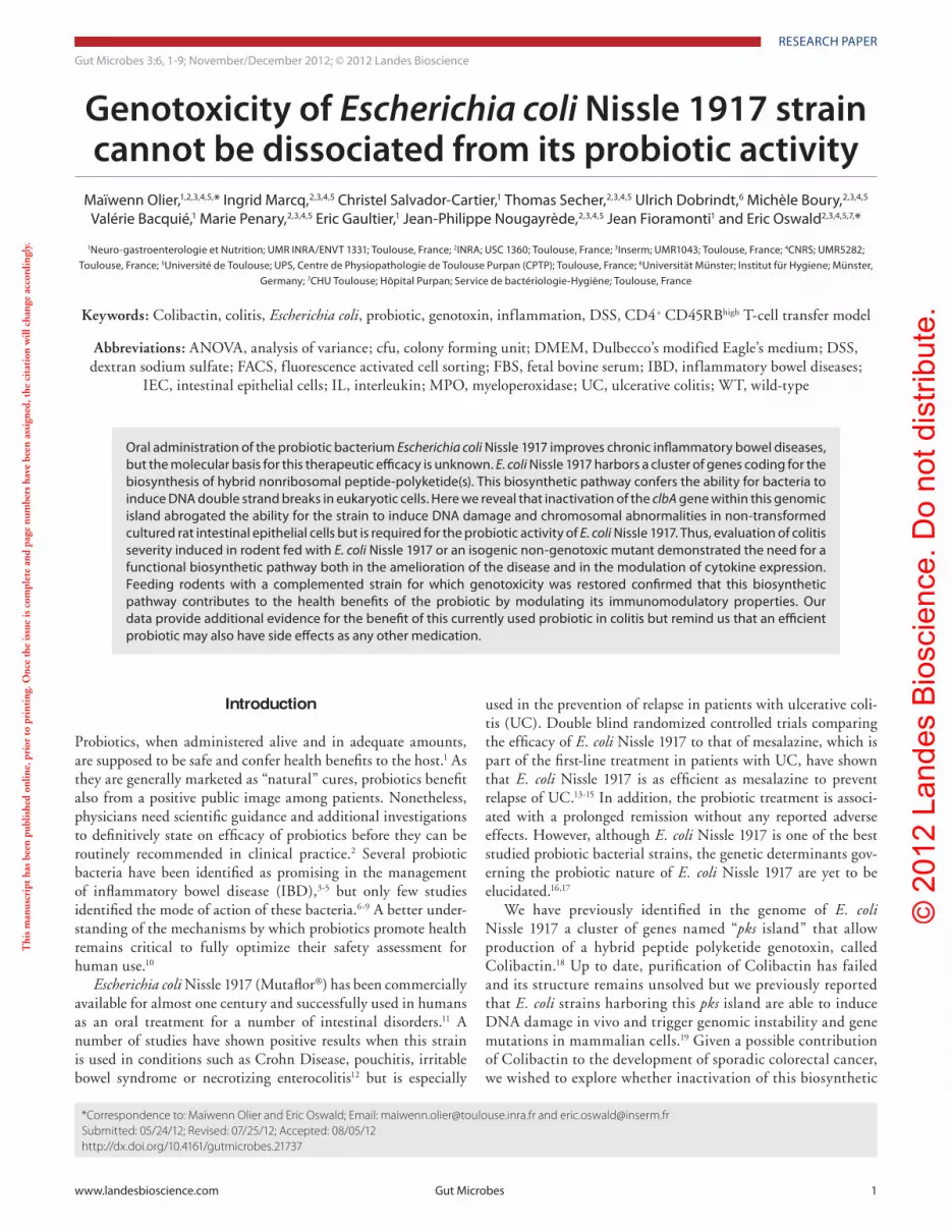
previously observed with other pks+ E. coli strains,19 the isogenic Nissle ΔclbA mutant was unable to induce such damage (Fig. 1B and C).
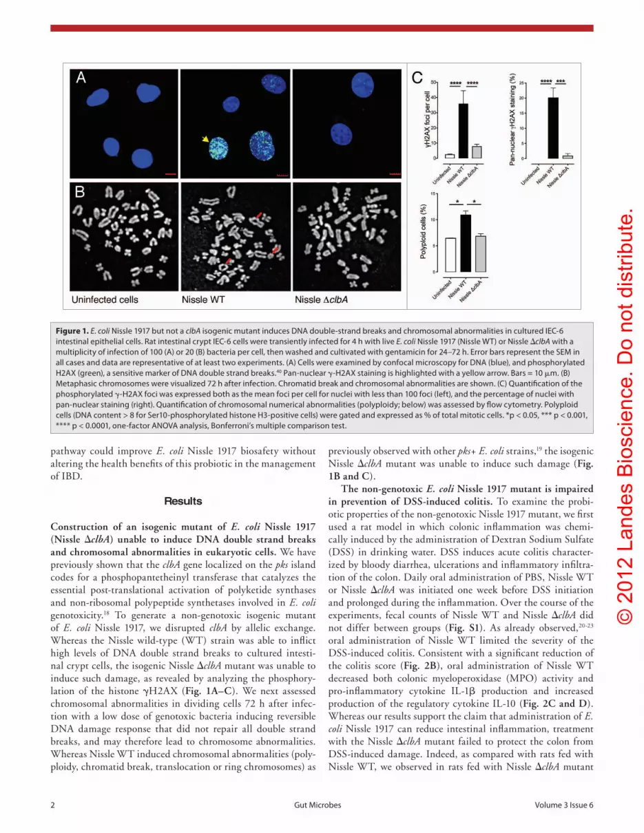
The non-genotoxic E. coli Nissle 1917 mutant is impaired in prevention of DSS-induced colitis. To examine the probi-otic properties of the non-genotoxic Nissle 1917 mutant, we first used a rat model in which colonic inflammation was chemi-cally induced by the administration of Dextran Sodium Sulfate (DSS) in drinking water. DSS induces acute colitis character-ized by bloody diarrhea, ulcerations and inflammatory infiltra-tion of the colon. Daily oral administration of PBS, Nissle WT or Nissle ΔclbA was initiated one week before DSS initiation and prolonged during the inflammation. Over the course of the experiments, fecal counts of Nissle WT and Nissle ΔclbA did not differ between groups (Fig. S1). As already observed,20-23 oral administration of Nissle WT limited the severity of the DSS-induced colitis. Consistent with a significant reduction of the colitis score (Fig. 2B), oral administration of Nissle WT decreased both colonic myeloperoxidase (MPO) activity and pro-inflammatory cytokine IL-1β production and increased production of the regulatory cytokine IL-10 (Fig. 2C and D). Whereas our results support the claim that administration of E. coli Nissle 1917 can reduce intestinal inflammation, treatment with the Nissle ΔclbA mutant failed to protect the colon from DSS-induced damage. Indeed, as compared with rats fed with Nissle WT, we observed in rats fed with Nissle ΔclbA mutant
pathway could improve E. coli Nissle 1917 biosafety without altering the health benefits of this probiotic in the management of IBD.
Results
Construction of an isogenic mutant of E. coli Nissle 1917 (Nissle ΔclbA) unable to induce DNA double strand breaks and chromosomal abnormalities in eukaryotic cells. We have previously shown that the clbA gene localized on the pks island codes for a phosphopantetheinyl transferase that catalyzes the essential post-translational activation of polyketide synthases and non-ribosomal polypeptide synthetases involved in E. coli genotoxicity.18 To generate a non-genotoxic isogenic mutant of E. coli Nissle 1917, we disrupted clbA by allelic exchange. Whereas the Nissle wild-type (WT) strain was able to inflict high levels of DNA double strand breaks to cultured intesti-nal crypt cells, the isogenic Nissle ΔclbA mutant was unable to induce such damage, as revealed by analyzing the phosphory-lation of the histone γH2AX (Fig. 1A–C). We next assessed chromosomal abnormalities in dividing cells 72 h after infec-tion with a low dose of genotoxic bacteria inducing reversible DNA damage response that did not repair all double strand breaks, and may therefore lead to chromosome abnormalities. Whereas Nissle WT induced chromosomal abnormalities (poly-ploidy, chromatid break, translocation or ring chromosomes) as
Figure 1. E. coli Nissle 1917 but not a clbA isogenic mutant induces DNa double-strand breaks and chromosomal abnormalities in cultured Iec-6 intestinal epithelial cells. Rat intestinal crypt Iec-6 cells were transiently infected for 4 h with live E. coli Nissle 1917 (Nissle WT) or Nissle ΔclbA with a multiplicity of infection of 100 (a) or 20 (B) bacteria per cell, then washed and cultivated with gentamicin for 24–72 h. error bars represent the seM in all cases and data are representative of at least two experiments. (a) cells were examined by confocal microscopy for DNa (blue), and phosphorylated h2aX (green), a sensitive marker of DNa double strand breaks.40 pan-nuclear γ-h2aX staining is highlighted with a yellow arrow. Bars = 10 μm. (B) Metaphasic chromosomes were visualized 72 h after infection. chromatid break and chromosomal abnormalities are shown. (c) Quantification of the phosphorylated γ-h2aX foci was expressed both as the mean foci per cell for nuclei with less than 100 foci (left), and the percentage of nuclei with pan-nuclear staining (right). Quantification of chromosomal numerical abnormalities (polyploidy; below) was assessed by flow cytometry. polyploid cells (DNa content > 8 for ser10-phosphorylated histone h3-positive cells) were gated and expressed as % of total mitotic cells. *p < 0.05, *** p < 0.001, **** p < 0.0001, one-factor aNOVa analysis, Bonferroni’s multiple comparison test.
© 2
012
Land
es B
iosc
ienc
e. D
o no
t dis
tribu
te.
www.landesbioscience.com Gut Microbes 3
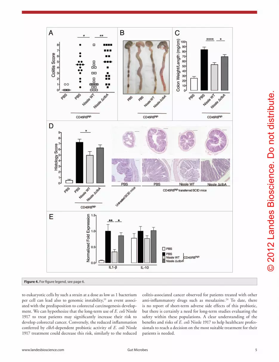
and decreased colonic damage (Fig. 4A and B). Furthermore, Nissle WT treatment improved the shortening and thickening of
a significant worsened body weight gain concomitant with enhanced colitis score and MPO activity. The colonic cytokine profiles of rat fed with Nissle ΔclbA mutant were also similar to that seen in DSS-treated rats fed with PBS (Fig. 2).
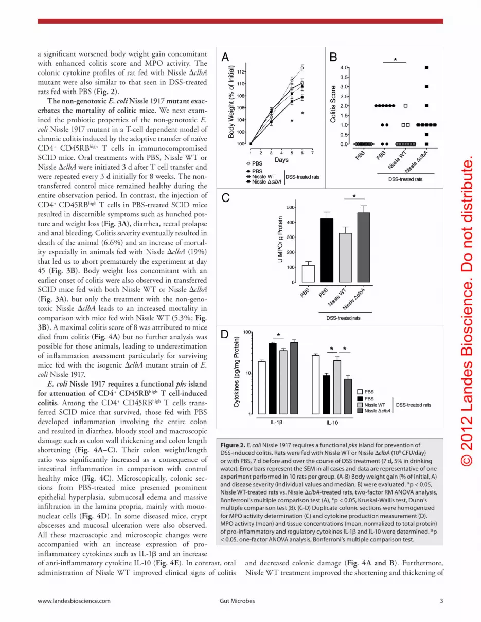
The non-genotoxic E. coli Nissle 1917 mutant exac-erbates the mortality of colitic mice. We next exam-ined the probiotic properties of the non-genotoxic E. coli Nissle 1917 mutant in a T-cell dependent model of chronic colitis induced by the adoptive transfer of naïve CD4+ CD45RBhigh T cells in immunocompromised SCID mice. Oral treatments with PBS, Nissle WT or Nissle ΔclbA were initiated 3 d after T cell transfer and were repeated every 3 d initially for 8 weeks. The non-transferred control mice remained healthy during the entire observation period. In contrast, the injection of CD4+ CD45RBhigh T cells in PBS-treated SCID mice resulted in discernible symptoms such as hunched pos-ture and weight loss (Fig. 3A), diarrhea, rectal prolapse and anal bleeding. Colitis severity eventually resulted in death of the animal (6.6%) and an increase of mortal-ity especially in animals fed with Nissle ΔclbA (19%) that led us to abort prematurely the experiment at day 45 (Fig. 3B). Body weight loss concomitant with an earlier onset of colitis were also observed in transferred SCID mice fed with both Nissle WT or Nissle ΔclbA (Fig. 3A), but only the treatment with the non-geno-toxic Nissle ΔclbA leads to an increased mortality in comparison with mice fed with Nissle WT (5.3%; Fig. 3B). A maximal colitis score of 8 was attributed to mice died from colitis (Fig. 4A) but no further analysis was possible for those animals, leading to underestimation of inflammation assessment particularly for surviving mice fed with the isogenic ΔclbA mutant strain of E. coli Nissle 1917.
E. coli Nissle 1917 requires a functional pks island for attenuation of CD4+ CD45RBhigh T cell-induced colitis. Among the CD4+ CD45RBhigh T cells trans-ferred SCID mice that survived, those fed with PBS developed inflammation involving the entire colon and resulted in diarrhea, bloody stool and macroscopic damage such as colon wall thickening and colon length shortening (Fig. 4A–C). Their colon weight/length ratio was significantly increased as a consequence of intestinal inflammation in comparison with control healthy mice (Fig. 4C). Microscopically, colonic sec-tions from PBS-treated mice presented prominent epithelial hyperplasia, submucosal edema and massive infiltration in the lamina propria, mainly with mono-nuclear cells (Fig. 4D). In some diseased mice, crypt abscesses and mucosal ulceration were also observed. All these macroscopic and microscopic changes were accompanied with an increase expression of pro-inflammatory cytokines such as IL-1β and an increase of anti-inflammatory cytokine IL-10 (Fig. 4E). In contrast, oral administration of Nissle WT improved clinical signs of colitis
Figure 2. E. coli Nissle 1917 requires a functional pks island for prevention of Dss-induced colitis. Rats were fed with Nissle WT or Nissle ΔclbA (109 cFU/day) or with pBs, 7 d before and over the course of Dss treatment (7 d, 5% in drinking water). error bars represent the seM in all cases and data are representative of one experiment performed in 10 rats per group. (a-B) Body weight gain (% of initial, a) and disease severity (individual values and median, B) were evaluated. *p < 0.05, Nissle WT-treated rats vs. Nissle ΔclbA-treated rats, two-factor RM aNOVa analysis, Bonferroni’s multiple comparison test (a), *p < 0.05, Kruskal-Wallis test, Dunn’s multiple comparison test (B). (c-D) Duplicate colonic sections were homogenized for MpO activity determination (c) and cytokine production measurement (D). MpO activity (mean) and tissue concentrations (mean, normalized to total protein) of pro-inflammatory and regulatory cytokines IL-1β and IL-10 were determined. *p < 0.05, one-factor aNOVa analysis, Bonferroni’s multiple comparison test.
© 2
012
Land
es B
iosc
ienc
e. D
o no
t dis
tribu
te.
4 Gut Microbes Volume 3 Issue 6
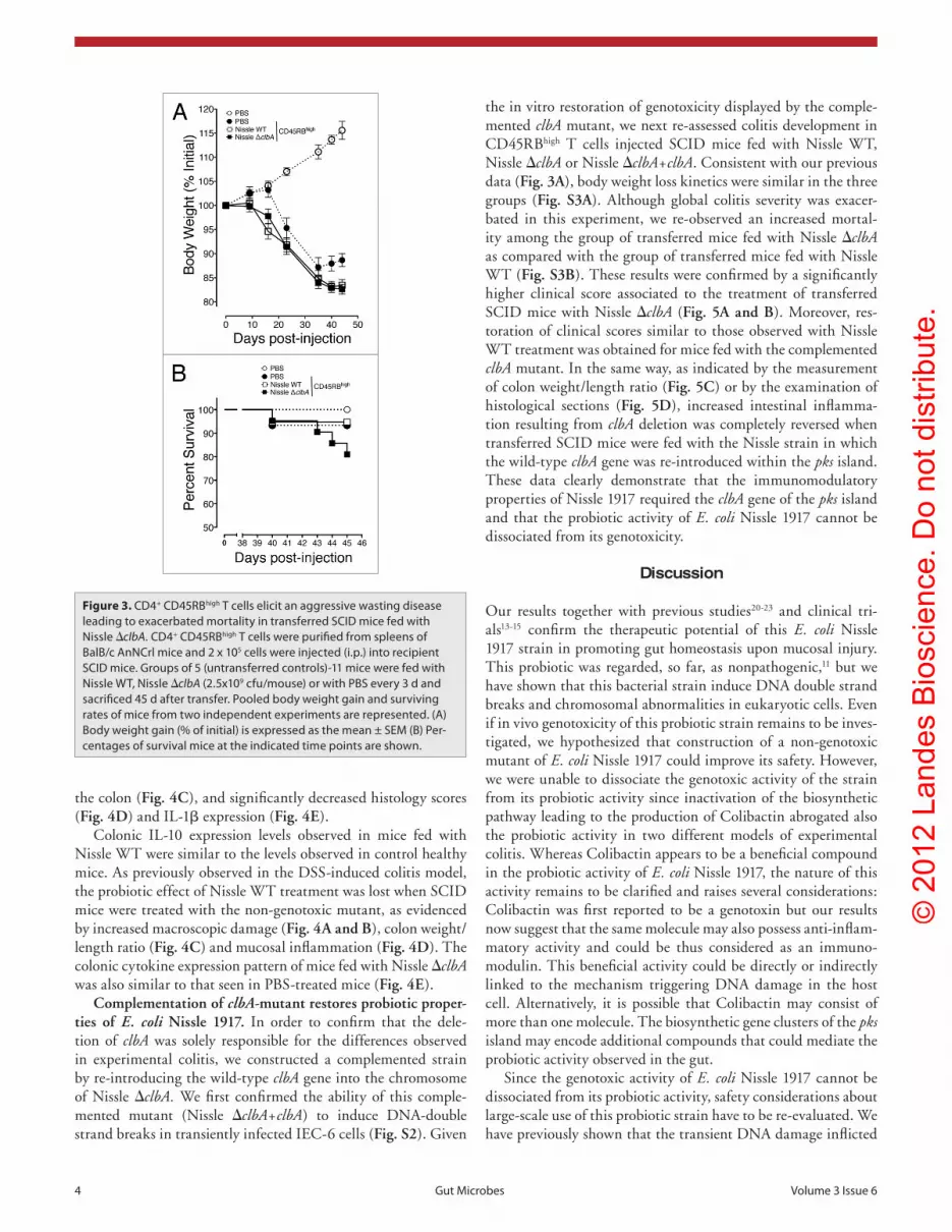
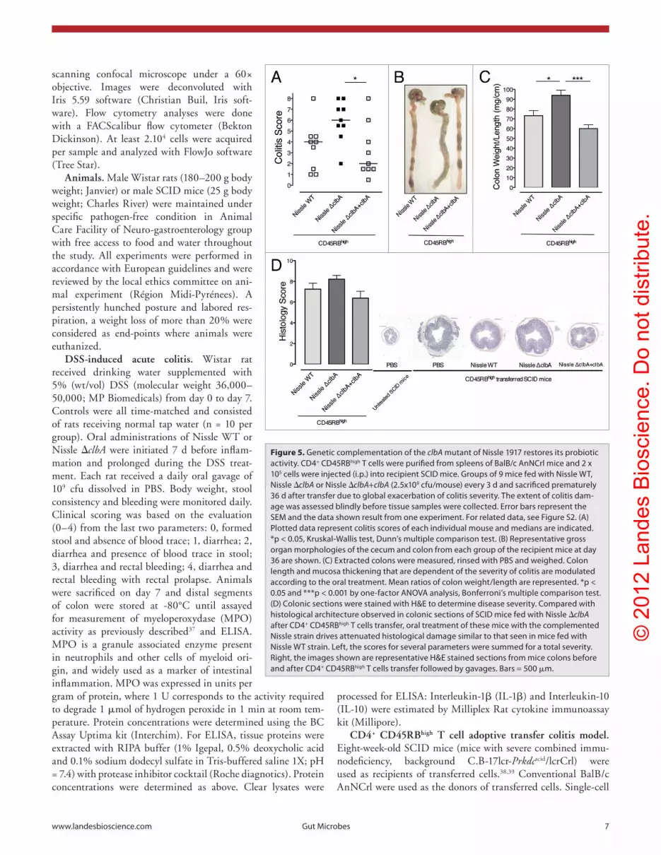
the in vitro restoration of genotoxicity displayed by the comple-mented clbA mutant, we next re-assessed colitis development in CD45RBhigh T cells injected SCID mice fed with Nissle WT, Nissle ΔclbA or Nissle ΔclbA+clbA. Consistent with our previous data (Fig. 3A), body weight loss kinetics were similar in the three groups (Fig. S3A). Although global colitis severity was exacer-bated in this experiment, we re-observed an increased mortal-ity among the group of transferred mice fed with Nissle ΔclbA as compared with the group of transferred mice fed with Nissle WT (Fig. S3B). These results were confirmed by a significantly higher clinical score associated to the treatment of transferred SCID mice with Nissle ΔclbA (Fig. 5A and B). Moreover, res-toration of clinical scores similar to those observed with Nissle WT treatment was obtained for mice fed with the complemented clbA mutant. In the same way, as indicated by the measurement of colon weight/length ratio (Fig. 5C) or by the examination of histological sections (Fig. 5D), increased intestinal inflamma-tion resulting from clbA deletion was completely reversed when transferred SCID mice were fed with the Nissle strain in which the wild-type clbA gene was re-introduced within the pks island. These data clearly demonstrate that the immunomodulatory properties of Nissle 1917 required the clbA gene of the pks island and that the probiotic activity of E. coli Nissle 1917 cannot be dissociated from its genotoxicity.
Discussion
Our results together with previous studies20-23 and clinical tri-als13-15 confirm the therapeutic potential of this E. coli Nissle 1917 strain in promoting gut homeostasis upon mucosal injury. This probiotic was regarded, so far, as nonpathogenic,11 but we have shown that this bacterial strain induce DNA double strand breaks and chromosomal abnormalities in eukaryotic cells. Even if in vivo genotoxicity of this probiotic strain remains to be inves-tigated, we hypothesized that construction of a non-genotoxic mutant of E. coli Nissle 1917 could improve its safety. However, we were unable to dissociate the genotoxic activity of the strain from its probiotic activity since inactivation of the biosynthetic pathway leading to the production of Colibactin abrogated also the probiotic activity in two different models of experimental colitis. Whereas Colibactin appears to be a beneficial compound in the probiotic activity of E. coli Nissle 1917, the nature of this activity remains to be clarified and raises several considerations: Colibactin was first reported to be a genotoxin but our results now suggest that the same molecule may also possess anti-inflam-matory activity and could be thus considered as an immuno-modulin. This beneficial activity could be directly or indirectly linked to the mechanism triggering DNA damage in the host cell. Alternatively, it is possible that Colibactin may consist of more than one molecule. The biosynthetic gene clusters of the pks island may encode additional compounds that could mediate the probiotic activity observed in the gut.
Since the genotoxic activity of E. coli Nissle 1917 cannot be dissociated from its probiotic activity, safety considerations about large-scale use of this probiotic strain have to be re-evaluated. We have previously shown that the transient DNA damage inflicted
the colon (Fig. 4C), and significantly decreased histology scores (Fig. 4D) and IL-1β expression (Fig. 4E).
Colonic IL-10 expression levels observed in mice fed with Nissle WT were similar to the levels observed in control healthy mice. As previously observed in the DSS-induced colitis model, the probiotic effect of Nissle WT treatment was lost when SCID mice were treated with the non-genotoxic mutant, as evidenced by increased macroscopic damage (Fig. 4A and B), colon weight/length ratio (Fig. 4C) and mucosal inflammation (Fig. 4D). The colonic cytokine expression pattern of mice fed with Nissle ΔclbA was also similar to that seen in PBS-treated mice (Fig. 4E).
Complementation of clbA-mutant restores probiotic proper-ties of E. coli Nissle 1917. In order to confirm that the dele-tion of clbA was solely responsible for the differences observed in experimental colitis, we constructed a complemented strain by re-introducing the wild-type clbA gene into the chromosome of Nissle ΔclbA. We first confirmed the ability of this comple-mented mutant (Nissle ΔclbA+clbA) to induce DNA-double strand breaks in transiently infected IEC-6 cells (Fig. S2). Given
Figure 3. cD4+ cD45RBhigh T cells elicit an aggressive wasting disease leading to exacerbated mortality in transferred scID mice fed with Nissle ΔclbA. cD4+ cD45RBhigh T cells were purified from spleens of BalB/c anNcrl mice and 2 x 10⁵ cells were injected (i.p.) into recipient scID mice. Groups of 5 (untransferred controls)-11 mice were fed with Nissle WT, Nissle ΔclbA (2.5x10⁹ cfu/mouse) or with pBs every 3 d and sacrificed 45 d after transfer. pooled body weight gain and surviving rates of mice from two independent experiments are represented. (a) Body weight gain (% of initial) is expressed as the mean ± seM (B) per-centages of survival mice at the indicated time points are shown.
© 2
012
Land
es B
iosc
ienc
e. D
o no
t dis
tribu
te.
www.landesbioscience.com Gut Microbes 5
colitis-associated cancer observed for patients treated with other anti-inflammatory drugs such as mesalazine.24 To date, there is no report of short-term adverse side effects of this probiotic, but there is certainly a need for long-term studies evaluating the safety within these populations. A clear understanding of the benefits and risks of E. coli Nissle 1917 to help healthcare profes-sionals to reach a decision on the most suitable treatment for their patients is needed.
to eukaryotic cells by such a strain at a dose as low as 1 bacterium per cell can lead also to genomic instability,19 an event associ-ated with the predisposition to colorectal carcinogenesis develop-ment. We can hypothesize that the long-term use of E. coli Nissle 1917 to treat patients may significantly increase their risk to develop colorectal cancer. Conversely, the reduced inflammation conferred by clbA-dependent probiotic activity of E. coli Nissle 1917 treatment could decrease this risk, similarly to the reduced
Figure 4. For figure legend, see page 6.
© 2
012
Land
es B
iosc
ienc
e. D
o no
t dis
tribu
te.
6 Gut Microbes Volume 3 Issue 6
Materials and Methods
Bacterial strains and mutagenesis procedures. In order to inves-tigate the potential role of Colibactin production in protection from colitis, we generated chromosomal isogenic mutants of E. coli Nissle 1917 (Mutaflor, DSM 6601, serotype O6:K5:H1) by using the lambda Red recombinase method as described previ-ously.36 Procedures, primers and templates used for directed-mutagenesis and resultant mutants are given in Supplemental Material and Table S1. Successful mutagenesis was confirmed by PCR using flanking primers and sequencing.
Cell culture, in vitro infection and genotoxicity assay. Non transformed rat intestinal epithelial IEC-6 cells (ATCC CRL-1592) were maintained in Dulbecco’s modified Eagle’s medium (DMEM GlutaMax; Invitrogen) supplemented with 10% fetal bovine serum (FBS, Invitrogen), 80 μg ml–1 gentamicin, and bovine insulin (0.1 unit ml–1; Sigma) at 37°C in a 5% CO
2
atmosphere. IEC-6 (~75% confluent) were washed four times and incubated in infection medium based on DMEM supple-mented with 25 mM Hepes and 10% FBS (Invitrogen). Bacteria were pre-grown in infection medium to the midlogarithmic phase, and then the infection dose was calculated to a multiplic-ity of infection of 20 or 100 bacteria per cell. After a 4h infection at 37°C and 5% CO
2, the cells were washed and grown until
analysis in cell culture medium supplemented with 200 μg/ml gentamicin.
DNA double strand breaks were demonstrated by γ-H2AX immunofluorescence analysis. Mouse anti-γ-H2AX (Millipore) was diluted 1:500 in blocking solution and incubated overnight at 4°C. FITC-conjugated goat anti mouse secondary antibody (Invitrogen) was diluted 1:100 in blocking solution and incu-bated for 1h. DNA was conterstained for 5min with TO-PRO-3 (Invitrogen). Slides were mounted in Vectashield containing DAPI (Vectorlabs). Images were acquired with an Olympus IX70 laser scanning confocal microscope, in sequential mode, with the Fluoview FV500 software. At least 15 random fields have been evaluated blindly for γ-H2AX foci counting under 60× apochro-mic objective for each experiment. Pan-nuclear γ-H2AX staining consisted of nuclei with more than 100 γ-H2AX foci.
Metaphasic chromosome spreading, visualization of chro-mosomal abnormalities and polyploid cells were done as previ-ously described.19 Chromosomes were stained with TO-PRO-3 (Invitrogen) and examined with an Olympus IX70 laser
The endogenous gastrointestinal microbiota is known to play a pivotal role in the development and maintenance of gut physi-ology and immune homeostasis.25 Alterations in its composition referred as dysbiosis play an important part in the pathogenesis of IBD.26,27 E. coli is an Enterobacteriaceae and bacteria of this taxonomic family are increasingly being recognized as microbial “inflammatory allies” contributing to the dysbiosis in IBD.28,29 As previously observed in other experimental models of IBD,28,30 the onset of colitis was followed by a spontaneously increase of endogenous Enterobacteriaceae feces levels in comparison with healthy control untransferred mice (Fig. S4). In addition, we have shown that the loss of the biosynthetic pathway encoded by the pks island not only abolishes probiotic properties of E. coli Nissle 1917 but also leads to the exacerbation of colitis severity (Figs. 2 and 3). Similar observations have been noticed in DSS-treated mice orally challenged with the reference adherent-invasive E. coli strain LF82.31 These pathogenic adherent-invasive strains of E. coli (AIEC) are abnormally predominant in ileal lesions of Crohn disease and seem to contribute to perpetuate colitis. Interestingly, LF82 stain belongs to the same B2 phylogenetic group than E. coli Nissle 1917. Similarly to E. coli Nissle 1917, E. coli strain LF82 does not harbor any known virulence factors expressed by pathogenic E. coli,32 but in contrast to E. coli Nissle 1917, LF82 strain does not harbor the pks island. Other studies have also suggested that certain commensal E. coli strains could potentiate the aggressiveness of intestinal inflammation by using E. coli monoassociated murine models of experimental colitis such as germ-free IL-10 deficient mice33 or IL-2 deficient mice.34 These opposite effects encountered among closely bacteria from the same species highlights that the crosstalk between probiotics, intestinal microbiota and host is probably more complex than previously supposed.
In conclusion, although intervention studies with probiotics have yielded promising results,3,12,34 it remains difficult to draw general conclusions. This is probably due to the highly variable nature of the probiotic used since health benefit of one probiotic strain cannot be extrapolated to others35 and bacteria from the same species may have opposite effects. Deciphering the precise molecular mechanisms supporting probiotic action, and poten-tial side effects, are required to delineate the optimal use of pro-biotics as supplementary therapy. From this perspective, it is time to connect probiotic researches with approaches used for patho-gens in the field of infectious disease.
Figure 4. E. coli Nissle 1917 requires a functional pks island for attenuation of cD4+ cD45RBhigh T cell-induced colitis. The extent of colitis and colonic damage were assessed blindly before tissue samples were collected from scID mice followed as in Figure 3. error bars represent the seM and the data shown are the pooled results from duplicate experiments performed in 5–11 mice per group. (a) plotted data represent pooled colitis scores of each individual mouse and medians are indicated. *p < 0.05 and ** p < 0.01, Kruskal-Wallis test, Dunn’s multiple comparison test. (B) Representative gross organ morphologies of the cecum and colons at day 45 are shown. (c) sampled colons were measured, rinsed with pBs and weighed. colon length and mucosa thickening that are dependent of the severity of colitis are modulated according to the oral treatment. Means of colon weight/length ratios are represented. *p < 0.05 and **** p < 0.0001 by one-factor aNOVa analysis, Bonferroni’s multiple comparison test. (D) colonic sections were stained with h&e to determine disease severity. Left, the scores for several parameters were summed for a total severity score. *p < 0.05, Kruskal-Wallis test, Dunn’s multiple comparison test. Right, representative h&e stained sections from mice colons are shown. Images in each row are the same magnification. Bars = 500 μm (above) or 100μm (below). The higher magnification below shows massive epithelial hyperplasia and inflammatory cell infiltration into colonic tissues in mice fed with pBs and Nissle ΔclbA compared with those fed with Nissle WT. (e) colonic samples were homogenized for cytokine expression measurements by q-pcR. Transcriptional expression of IL-1β and IL-10 was normalized to hpRT expression. Represented fold expressions were normalized to pBs-treated colitic mice. *p < 0.05 and ** p < 0.01 by one-factor aNOVa analysis, Bonferroni’s multiple comparison test.
© 2
012
Land
es B
iosc
ienc
e. D
o no
t dis
tribu
te.
www.landesbioscience.com Gut Microbes 7
processed for ELISA: Interleukin-1β (IL-1β) and Interleukin-10 (IL-10) were estimated by Milliplex Rat cytokine immunoassay kit (Millipore).
CD4+ CD45RBhigh T cell adoptive transfer colitis model. Eight-week-old SCID mice (mice with severe combined immu-nodeficiency, background C.B-17lcr-Prkdcscid/lcrCrl) were used as recipients of transferred cells.38,39 Conventional BalB/c AnNCrl were used as the donors of transferred cells. Single-cell
scanning confocal microscope under a 60× objective. Images were deconvoluted with Iris 5.59 software (Christian Buil, Iris soft-ware). Flow cytometry analyses were done with a FACScalibur flow cytometer (Bekton Dickinson). At least 2.104 cells were acquired per sample and analyzed with FlowJo software (Tree Star).
Animals. Male Wistar rats (180–200 g body weight; Janvier) or male SCID mice (25 g body weight; Charles River) were maintained under specific pathogen-free condition in Animal Care Facility of Neuro-gastroenterology group with free access to food and water throughout the study. All experiments were performed in accordance with European guidelines and were reviewed by the local ethics committee on ani-mal experiment (Région Midi-Pyrénees). A persistently hunched posture and labored res-piration, a weight loss of more than 20% were considered as end-points where animals were euthanized.
DSS-induced acute colitis. Wistar rat received drinking water supplemented with 5% (wt/vol) DSS (molecular weight 36,000–50,000; MP Biomedicals) from day 0 to day 7. Controls were all time-matched and consisted of rats receiving normal tap water (n = 10 per group). Oral administrations of Nissle WT or Nissle ΔclbA were initiated 7 d before inflam-mation and prolonged during the DSS treat-ment. Each rat received a daily oral gavage of 109 cfu dissolved in PBS. Body weight, stool consistency and bleeding were monitored daily. Clinical scoring was based on the evaluation (0–4) from the last two parameters: 0, formed stool and absence of blood trace; 1, diarrhea; 2, diarrhea and presence of blood trace in stool; 3, diarrhea and rectal bleeding; 4, diarrhea and rectal bleeding with rectal prolapse. Animals were sacrificed on day 7 and distal segments of colon were stored at -80°C until assayed for measurement of myeloperoxydase (MPO) activity as previously described37 and ELISA. MPO is a granule associated enzyme present in neutrophils and other cells of myeloid ori-gin, and widely used as a marker of intestinal inflammation. MPO was expressed in units per gram of protein, where 1 U corresponds to the activity required to degrade 1 μmol of hydrogen peroxide in 1 min at room tem-perature. Protein concentrations were determined using the BC Assay Uptima kit (Interchim). For ELISA, tissue proteins were extracted with RIPA buffer (1% Igepal, 0.5% deoxycholic acid and 0.1% sodium dodecyl sulfate in Tris-buffered saline 1X; pH = 7.4) with protease inhibitor cocktail (Roche diagnotics). Protein concentrations were determined as above. Clear lysates were
Figure 5. Genetic complementation of the clbA mutant of Nissle 1917 restores its probiotic activity. cD4+ cD45RBhigh T cells were purified from spleens of BalB/c anNcrl mice and 2 x 10⁵ cells were injected (i.p.) into recipient scID mice. Groups of 9 mice fed with Nissle WT, Nissle ΔclbA or Nissle ΔclbA+clbA (2.5x10⁹ cfu/mouse) every 3 d and sacrificed prematurely 36 d after transfer due to global exacerbation of colitis severity. The extent of colitis dam-age was assessed blindly before tissue samples were collected. error bars represent the seM and the data shown result from one experiment. For related data, see Figure s2. (a) plotted data represent colitis scores of each individual mouse and medians are indicated. *p < 0.05, Kruskal-Wallis test, Dunn’s multiple comparison test. (B) Representative gross organ morphologies of the cecum and colon from each group of the recipient mice at day 36 are shown. (c) extracted colons were measured, rinsed with pBs and weighed. colon length and mucosa thickening that are dependent of the severity of colitis are modulated according to the oral treatment. Mean ratios of colon weight/length are represented. *p < 0.05 and ***p < 0.001 by one-factor aNOVa analysis, Bonferroni’s multiple comparison test. (D) colonic sections were stained with h&e to determine disease severity. compared with histological architecture observed in colonic sections of scID mice fed with Nissle ΔclbA after cD4+ cD45RBhigh T cells transfer, oral treatment of these mice with the complemented Nissle strain drives attenuated histological damage similar to that seen in mice fed with Nissle WT strain. Left, the scores for several parameters were summed for a total severity. Right, the images shown are representative h&e stained sections from mice colons before and after cD4+ cD45RBhigh T cells transfer followed by gavages. Bars = 500 μm.
© 2
012
Land
es B
iosc
ienc
e. D
o no
t dis
tribu
te.
8 Gut Microbes Volume 3 Issue 6
Histology. Rat or mouse colonic tissues fixed in Duboscq-Brazil were embedded in paraffin, sectioned (7-μm slices) and stained with Hematoxylin and Eosin (H&E). Colonic damage were graded semi quantitatively in blinded fashion by one or two of the authors (I.M.; C.C.). C.C. was blinded to the experimen-tal protocol used. Each of 5 histological parameters was scored as absent (0), mild (1), moderate (2), or severe (3): density of inflammatory cells, epithelial injury, edema, crypt abscess and epithelial hyperplasia. Histological score was the sum of these parameters.
Statistical analysis. Results are expressed as mean ± SEM or median unless otherwise stated. For in vivo experiments, N refers to the number of animals per group used for each experi-ment. Statistical significance of differences between experimental groups was performed using one-way or two-way analysis of vari-ance (ANOVA) with Bonferroni multiple-comparison post-test, or Kruskal-Wallis non-parametric ANOVA with Dunn’s multi-ple-comparison post-test as apppropriate (Prism 5, Graph Pad Software). Two-side analyses were used throughout and p-values < 0.05 were considered statistically significant.
Disclosure of Potential Conflicts of Interest
No potential conflicts of interest were disclosed.
Acknowledgments
We thank the staff of the Cytometry platform of the Centre de Physiopathologie de Toulouse-Purpan for excellent technical help. This research was supported by grants from the non-profit Association F. Aupetit (AFA) and the French National Research Agency (ANR-09-MIEN-005-01). The funders had no role in study design, data collection and analysis, decision to publish or preparation of the manuscript.
Supplemental Material
Supplemental Materials may be found here:http://www.landesbioscience.com/journals/gutmicrobes/
article/21737/
suspensions of spleens from sex-matched mice were treated with red blood cell lysing buffer. CD4+ T cells were isolated using a negative selection CD4+ isolation kit (Miltenyi Biotec). Cells were stained with 5μg/ml anti-CD4-FITC and 2 μg/ml anti-CD45Rb-PE (eBioscience). CD4+ CD45RBhigh cells were iso-lated by fluorescence activated cell sorting (FACS). Cells (2 x 10⁵) were injected intraperitoneally into SCID mice (n = 9–11 per group, 2 independent experiments). Injected SCID mice were orally administered Nissle WT or Nissle ΔclbA (2.5 109 cfu/mouse) or PBS every 3 d for 45 d. Controls consisted of non-injected SCID mice (1 to 5 mice per experiment) treated with PBS. In a second set of experiments, injected SCID mice were treated with Nissle WT, Nissle ΔclbA or Nissle ΔclbA+clbA every 3 d for 35 d (n = 9 per group, one experiment). Body weight and survival rate were measured daily after the induction of coli-tis. Immediately after sacrifice, the extent of colitis damage was assessed before tissue sampling. The macroscopic parameters analyzed were (ranging from 0 to 7): stool consistency (0 to 2 = formed to severe diarrhea), tissue adhesions (0 to 2 = absence to severe adhesions) and colonic damage (0 to 3 = normal to severe). A total score of 8 was assigned when animals died from colitis. Samples of distal colon were stored at -80°C until assayed for cytokine expression measurement. For RNA extraction, fro-zen colonic samples were homogenized and disrupted in buffer RLT (Qiagen) using Precellys beads kit (CK-MIX beads; Bertin Technologies). Tubes were shaken in a Precellys® 24 bead apparatus (Bertin Technologies) at 6000 rpm for 2 cycles of 10 sec with interval of 10 sec. RNA was then extracted by using Qiagen kit, as recommended by the manufacturer. RNA (1μg) was treated with DNase (Sigma) prior to cDNA conversion using iSript cDNA synthesis kit (Bio-rad). qRT-PCR assays (see Table S2) was performed using IQ SYBR Green Supermix (Bio-rad). Reactions were performed on a Bio-Rad CFX96TM. For cyto-kine analysis, samples were normalized to the housekeeping gene hypoxanthine guanine phosphoribosyl transferase 1 (HPRT) for intersample variation. Normalized fold expressions analyzed using CFX-manager software (Bio-rad) were done to PBS-treated colitis mice.
References1. FAO/WHO. Food and Agricultural Organization
/ World Health Organization. Report -Health and Nutritional Properties of Probiotics in Food Including PowderMilk with Live Lactic Acid Bacteria. Cordoba, Argentina 2001.
2. Sanders ME, Levy DD. The science and regulations of probiotic food and supplement product label-ing. Ann N Y Acad Sci 2011; 1219(Suppl 1):E1-23; PMID:21332758; http://dx.doi.org/10.1111/j.1749-6632.2010.05956.x.
3. Gareau MG, Sherman PM, Walker WA. Probiotics and the gut microbiota in intestinal health and dis-ease. Nat Rev Gastroenterol Hepatol 2010; 7:503-14; PMID:20664519; http://dx.doi.org/10.1038/nrgas-tro.2010.117.
4. Seksik P, Dray X, Sokol H, Marteau P. Is there any place for alimentary probiotics, prebiotics or synbiotics, for patients with inflammatory bowel disease? Mol Nutr Food Res 2008; 52:906-12; PMID:18384087; http://dx.doi.org/10.1002/mnfr.200700147.
5. Wallace TC, Guarner F, Madsen K, Cabana MD, Gibson G, Hentges E, et al. Human gut microbiota and its relationship to health and disease. Nutr Rev 2011; 69:392-403; PMID:21729093; http://dx.doi.org/10.1111/j.1753-4887.2011.00402.x.
6. Mazmanian SK, Round JL, Kasper DL. A micro-bial symbiosis factor prevents intestinal inflammatory disease. Nature 2008; 453:620-5; PMID:18509436; http://dx.doi.org/10.1038/nature07008.
7. Mohamadzadeh M, Pfeiler EA, Brown JB, Zadeh M, Gramarossa M, Managlia E, et al. Regulation of induced colonic inflammation by Lactobacillus acidophilus deficient in lipoteichoic acid. Proc Natl Acad Sci U S A 2011; 108(Suppl 1):4623-30; PMID:21282652; http://dx.doi.org/10.1073/pnas.1005066107.
8. Yan F, Cao H, Cover TL, Washington MK, Shi Y, Liu L, et al. Colon-specific delivery of a probiotic-derived soluble protein ameliorates intestinal inflammation in mice through an EGFR-dependent mechanism. J Clin Invest 2011; 121:2242-53; PMID:21606592; http://dx.doi.org/10.1172/JCI44031.
9. von Schillde MA, Hörmannsperger G, Weiher M, Alpert CA, Hahne H, Bäuerl C, et al. Lactocepin secreted by Lactobacillus exerts anti-inflammatory effects by selectively degrading proinflammatory chemokines. Cell Host Microbe 2012; 11:387-96; PMID:22520466; http://dx.doi.org/10.1016/j.chom.2012.02.006.
10. Sanders ME, Akkermans LM, Haller D, Hammerman C, Heimbach J, Hörmannsperger G, et al. Safety assessment of probiotics for human use. Gut Microbes 2010; 1:164-85; PMID:21327023; http://dx.doi.org/10.4161/gmic.1.3.12127.
11. Schultz M. Clinical use of E. coli Nissle 1917 in inflammatory bowel disease. Inflamm Bowel Dis 2008; 14:1012-8; PMID:18240278; http://dx.doi.org/10.1002/ibd.20377.
12. Yan F, Polk DB. Probiotics: progress toward novel ther-apies for intestinal diseases. Curr Opin Gastroenterol 2010; 26:95-101; PMID:19952741; http://dx.doi.org/10.1097/MOG.0b013e328335239a.
© 2
012
Land
es B
iosc
ienc
e. D
o no
t dis
tribu
te.
www.landesbioscience.com Gut Microbes 9
32. Darfeuille-Michaud A, Neut C, Barnich N, Lederman E, Di Martino P, Desreumaux P, et al. Presence of adherent Escherichia coli strains in ileal mucosa of patients with Crohn’s disease. Gastroenterology 1998; 115:1405-13; PMID:9834268; http://dx.doi.org/10.1016/S0016-5085(98)70019-8.
33. Kim SC, Tonkonogy SL, Karrasch T, Jobin C, Sartor RB. Dual-association of gnotobiotic IL-10-/- mice with 2 nonpathogenic commensal bacteria induces aggres-sive pancolitis. Inflamm Bowel Dis 2007; 13:1457-66; PMID:17763473; http://dx.doi.org/10.1002/ibd.20246.
34. Waidmann M, Bechtold O, Frick JS, Lehr HA, Schubert S, Dobrindt U, et al. Bacteroides vulgatus protects against Escherichia coli-induced colitis in gno-tobiotic interleukin-2-deficient mice. Gastroenterology 2003; 125:162-77; PMID:12851881; http://dx.doi.org/10.1016/S0016-5085(03)00672-3.
35. Azaïs-Braesco V, Bresson JL, Guarner F, Corthier G. Not all lactic acid bacteria are probiotics, ...but some are. Br J Nutr 2010; 103:1079-81; PMID:20230653; http://dx.doi.org/10.1017/S0007114510000723.
36. Datsenko KA, Wanner BL. One-step inactivation of chromosomal genes in Escherichia coli K-12 using PCR products. Proc Natl Acad Sci U S A 2000; 97:6640-5; PMID:10829079; http://dx.doi.org/10.1073/pnas.120163297.
37. Houdeau E, Moriez R, Leveque M, Salvador-Cartier C, Waget A, Leng L, et al. Sex steroid regulation of macrophage migration inhibitory factor in normal and inflamed colon in the female rat. Gastroenterology 2007; 132:982-93; PMID:17324399; http://dx.doi.org/10.1053/j.gastro.2006.12.028.
38. Morrissey PJ, Charrier K, Braddy S, Liggitt D, Watson JD. CD4+ T cells that express high levels of CD45RB induce wasting disease when transferred into congenic severe combined immunodeficient mice. Disease devel-opment is prevented by cotransfer of purified CD4+ T cells. J Exp Med 1993; 178:237-44; PMID:8100269; http://dx.doi.org/10.1084/jem.178.1.237.
39. Powrie F, Leach MW, Mauze S, Caddle LB, Coffman RL. Phenotypically distinct subsets of CD4+ T cells induce or protect from chronic intestinal inflammation in C. B-17 scid mice. Int Immunol 1993; 5:1461-71; PMID:7903159; http://dx.doi.org/10.1093/intimm/5.11.1461.
40. Rogakou EP, Pilch DR, Orr AH, Ivanova VS, Bonner WM. DNA double-stranded breaks induce histone H2AX phosphorylation on serine 139. J Biol Chem 1998; 273:5858-68; PMID:9488723; http://dx.doi.org/10.1074/jbc.273.10.5858.
22. Schultz M, Strauch UG, Linde HJ, Watzl S, Obermeier F, Göttl C, et al. Preventive effects of Escherichia coli strain Nissle 1917 on acute and chronic intestinal inflammation in two different murine models of colitis. Clin Diagn Lab Immunol 2004; 11:372-8; PMID:15013990.
23. Ukena SN, Singh A, Dringenberg U, Engelhardt R, Seidler U, Hansen W, et al. Probiotic Escherichia coli Nissle 1917 inhibits leaky gut by enhancing mucosal integrity. PLoS One 2007; 2:e1308; PMID:18074031; http://dx.doi.org/10.1371/journal.pone.0001308.
24. Velayos FS, Terdiman JP, Walsh JM. Effect of 5-ami-nosalicylate use on colorectal cancer and dysplasia risk: a systematic review and metaanalysis of observa-tional studies. Am J Gastroenterol 2005; 100:1345-53; PMID:15929768; http://dx.doi.org/10.1111/j.1572-0241.2005.41442.x.
25. Cerf-Bensussan N, Gaboriau-Routhiau V. The immune system and the gut microbiota: friends or foes? Nat Rev Immunol 2010; 10:735-44; PMID:20865020; http://dx.doi.org/10.1038/nri2850.
26. Nell S, Suerbaum S, Josenhans C. The impact of the microbiota on the pathogenesis of IBD: lessons from mouse infection models. Nat Rev Microbiol 2010; 8:564-77; PMID:20622892; http://dx.doi.org/10.1038/nrmicro2403.
27. Sartor RB. Microbial influences in inflammatory bowel diseases. Gastroenterology 2008; 134:577-94; PMID:18242222; http://dx.doi.org/10.1053/j.gas-tro.2007.11.059.
28. Garrett WS, Gallini CA, Yatsunenko T, Michaud M, DuBois A, Delaney ML, et al. Enterobacteriaceae act in concert with the gut microbiota to induce sponta-neous and maternally transmitted colitis. Cell Host Microbe 2010; 8:292-300; PMID:20833380; http://dx.doi.org/10.1016/j.chom.2010.08.004.
29. Lupp C, Robertson ML, Wickham ME, Sekirov I, Champion OL, Gaynor EC, et al. Host-mediated inflammation disrupts the intestinal microbiota and promotes the overgrowth of Enterobacteriaceae. Cell Host Microbe 2007; 2:204; PMID:18030708; http://dx.doi.org/10.1016/j.chom.2007.08.002.
30. Lee IA, Bae EA, Hyun YJ, Kim DH. Dextran sul-fate sodium and 2,4,6-trinitrobenzene sulfonic acid induce lipid peroxidation by the proliferation of intestinal gram-negative bacteria in mice. J Inflamm (Lond) 2010; 7:7; PMID:20181058; http://dx.doi.org/10.1186/1476-9255-7-7.
31. Carvalho FA, Barnich N, Sauvanet P, Darcha C, Gelot A, Darfeuille-Michaud A. Crohn’s disease-associated Escherichia coli LF82 aggravates colitis in injured mouse colon via signaling by flagellin. Inflamm Bowel Dis 2008; 14:1051-60; PMID:18338780; http://dx.doi.org/10.1002/ibd.20423.
13. Kruis W, Fric P, Pokrotnieks J, Lukás M, Fixa B, Kascák M, et al. Maintaining remission of ulcer-ative colitis with the probiotic Escherichia coli Nissle 1917 is as effective as with standard mesalazine. Gut 2004; 53:1617-23; PMID:15479682; http://dx.doi.org/10.1136/gut.2003.037747.
14. Kruis W, Schütz E, Fric P, Fixa B, Judmaier G, Stolte M. Double-blind comparison of an oral Escherichia coli preparation and mesalazine in maintaining remis-sion of ulcerative colitis. Aliment Pharmacol Ther 1997; 11:853-8; PMID:9354192; http://dx.doi.org/10.1046/j.1365-2036.1997.00225.x.
15. Rembacken BJ, Snelling AM, Hawkey PM, Chalmers DM, Axon AT. Non-pathogenic Escherichia coli ver-sus mesalazine for the treatment of ulcerative coli-tis: a randomised trial. Lancet 1999; 354:635-9; PMID:10466665; http://dx.doi.org/10.1016/S0140-6736(98)06343-0.
16. Grozdanov L, Raasch C, Schulze J, Sonnenborn U, Gottschalk G, Hacker J, et al. Analysis of the genome structure of the nonpathogenic probiotic Escherichia coli strain Nissle 1917. J Bacteriol 2004; 186:5432-41; PMID:15292145; http://dx.doi.org/10.1128/JB.186.16.5432-5441.2004.
17. Grozdanov L, Zähringer U, Blum-Oehler G, Brade L, Henne A, Knirel YA, et al. A single nucleotide exchange in the wzy gene is responsible for the semirough O6 lipopolysaccharide phenotype and serum sensitiv-ity of Escherichia coli strain Nissle 1917. J Bacteriol 2002; 184:5912-25; PMID:12374825; http://dx.doi.org/10.1128/JB.184.21.5912-5925.2002.
18. Nougayrède JP, Homburg S, Taieb F, Boury M, Brzuszkiewicz E, Gottschalk G, et al. Escherichia coli induces DNA double-strand breaks in eukaryotic cells. Science 2006; 313:848-51; PMID:16902142; http://dx.doi.org/10.1126/science.1127059.
19. Cuevas-Ramos G, Petit CR, Marcq I, Boury M, Oswald E, Nougayrède JP. Escherichia coli induces DNA damage in vivo and triggers genomic instabil-ity in mammalian cells. Proc Natl Acad Sci U S A 2010; 107:11537-42; PMID:20534522; http://dx.doi.org/10.1073/pnas.1001261107.
20. Grabig A, Paclik D, Guzy C, Dankof A, Baumgart DC, Erckenbrecht J, et al. Escherichia coli strain Nissle 1917 ameliorates experimental colitis via toll-like receptor 2- and toll-like receptor 4-dependent pathways. Infect Immun 2006; 74:4075-82; PMID:16790781; http://dx.doi.org/10.1128/IAI.01449-05.
21. Kamada N, Inoue N, Hisamatsu T, Okamoto S, Matsuoka K, Sato T, et al. Nonpathogenic Escherichia coli strain Nissle1917 prevents murine acute and chronic colitis. Inflamm Bowel Dis 2005; 11:455-63; PMID:15867585; http://dx.doi.org/10.1097/01.MIB.0000158158.55955.de.
© 2
012
Land
es B
iosc
ienc
e. D
o no
t dis
tribu
te.
Related Documents


