ARTICLE Received 16 Apr 2013 | Accepted 8 Aug 2013 | Published 16 Sep 2013 Genome signature-based dissection of human gut metagenomes to extract subliminal viral sequences Lesley A. Ogilvie 1 , Lucas D. Bowler 1 , Jonathan Caplin 2 , Cinzia Dedi 1 , David Diston 2,w , Elizabeth Cheek 3 , Huw Taylor 2 , James E. Ebdon 2 & Brian V. Jones 1 Bacterial viruses (bacteriophages) have a key role in shaping the development and functional outputs of host microbiomes. Although metagenomic approaches have greatly expanded our understanding of the prokaryotic virosphere, additional tools are required for the phage- oriented dissection of metagenomic data sets, and host-range affiliation of recovered sequences. Here we demonstrate the application of a genome signature-based approach to interrogate conventional whole-community metagenomes and access subliminal, phylogen- etically targeted, phage sequences present within. We describe a portion of the biological dark matter extant in the human gut virome, and bring to light a population of potentially gut- specific Bacteroidales-like phage, poorly represented in existing virus like particle-derived viral metagenomes. These predominantly temperate phage were shown to encode functions of direct relevance to human health in the form of antibiotic resistance genes, and provided evidence for the existence of putative ‘viral-enterotypes’ among this fraction of the human gut virome. DOI: 10.1038/ncomms3420 OPEN 1 Centre for Biomedical and Health Science Research, School of Pharmacy and Biomolecular Sciences, University of Brighton, Brighton BN2 4GJ, UK. 2 School of Environment and Technology, University of Brighton, Brighton BN2 4GJ, UK. 3 School of Computing, Engineering and Mathematics, University of Brighton, Brighton BN2 4GJ, UK. w Present address: Mikrobiologische and Biotechnologische Risiken Bundesamt fu ¨r Gesundheit BAG, 3003 Bern, Switzerland. Correspondence and requests for materials should be addressed to B.V.J. (email: [email protected]). NATURE COMMUNICATIONS | 4:2420 | DOI: 10.1038/ncomms3420 | www.nature.com/naturecommunications 1 & 2013 Macmillan Publishers Limited. All rights reserved.

Welcome message from author
This document is posted to help you gain knowledge. Please leave a comment to let me know what you think about it! Share it to your friends and learn new things together.
Transcript
ARTICLE
Received 16 Apr 2013 | Accepted 8 Aug 2013 | Published 16 Sep 2013
Genome signature-based dissection of human gutmetagenomes to extract subliminal viral sequencesLesley A. Ogilvie1, Lucas D. Bowler1, Jonathan Caplin2, Cinzia Dedi1, David Diston2,w, Elizabeth Cheek3,
Huw Taylor2, James E. Ebdon2 & Brian V. Jones1
Bacterial viruses (bacteriophages) have a key role in shaping the development and functional
outputs of host microbiomes. Although metagenomic approaches have greatly expanded our
understanding of the prokaryotic virosphere, additional tools are required for the phage-
oriented dissection of metagenomic data sets, and host-range affiliation of recovered
sequences. Here we demonstrate the application of a genome signature-based approach to
interrogate conventional whole-community metagenomes and access subliminal, phylogen-
etically targeted, phage sequences present within. We describe a portion of the biological
dark matter extant in the human gut virome, and bring to light a population of potentially gut-
specific Bacteroidales-like phage, poorly represented in existing virus like particle-derived viral
metagenomes. These predominantly temperate phage were shown to encode functions of
direct relevance to human health in the form of antibiotic resistance genes, and provided
evidence for the existence of putative ‘viral-enterotypes’ among this fraction of the human gut
virome.
DOI: 10.1038/ncomms3420 OPEN
1 Centre for Biomedical and Health Science Research, School of Pharmacy and Biomolecular Sciences, University of Brighton, Brighton BN2 4GJ, UK. 2 Schoolof Environment and Technology, University of Brighton, Brighton BN2 4GJ, UK. 3 School of Computing, Engineering and Mathematics, University of Brighton,Brighton BN2 4GJ, UK. w Present address: Mikrobiologische and Biotechnologische Risiken Bundesamt fur Gesundheit BAG, 3003 Bern, Switzerland.Correspondence and requests for materials should be addressed to B.V.J. (email: [email protected]).
NATURE COMMUNICATIONS | 4:2420 | DOI: 10.1038/ncomms3420 |www.nature.com/naturecommunications 1
& 2013 Macmillan Publishers Limited. All rights reserved.
Viruses are the most abundant infectious agents on theplanet, and collectively constitute a highly diverse andlargely unexplored gene-space, which accounts for much
of the ‘biological dark matter’ in Earth’s biosphere1–3. Bacterialviruses (bacteriophage or phage) are considered the mostnumerous viral entities, and through their effects on hostbacteria, phage can influence processes ranging from globalgeochemical cycles to bacterial virulence and pathogenesis1–5.The study of this expansive family of viruses continues tounderpin many fundamental insights into microbial physiologyand evolution, with the interplay of bacteria and phage nowstudied at scales ranging from the individual componentsof single-phage species, to community-level surveys of viralassemblages and their impacts on host microbial ecosystems.
The development of metagenomic tools for analysis of phagepopulations constitutes a major advance in this regard, which ispoised to deliver unprecedented insight into the prokaryoticvirosphere. This powerful culture-independent approach over-comes many limitations of traditional methods for phageisolation and characterization, ultimately promising almostunrestricted access to the genetic content of host microbiomesand their attendant viral collectives3,6–11. Application of thesetechniques to the study of microbial viromes has already providedmajor insights into a number of phage communities, includingthose associated with microbial ecosystems that develop in or onthe human body7,11,12.
In particular, the retinue of phage associated with the humangut microbiome is now increasingly recognized as an importantfacet of this ecosystem, which may significantly influence itsimpact on human health3,5,13–16. Gut-associated phage havealready been shown to encode genes that confer production oftoxins, virulence factors or antibiotic resistance upon hostbacteria5,17,18, and have the potential to modulate communitystructure and metabolic output through elimination of hostspecies or introduction of new traits1,16,19. Furthermore, viromecomposition also appears to be altered in disease states, which hasgiven rise to the hypothesis that the human gut virome mayhave a role in the pathogenesis of disorders associated withperturbation of the gut ecosystem14. Phage also hold considerablebiotechnological and pharmaceutical potential, with the gutvirome now a viable target for bio-prospecting and thedevelopment of novel therapeutic or diagnostic tools3,13.
However, current strategies for generating viral metagenomesare not without limitations, and are typically based on analysisof nucleic acids derived from purified virus like particles(VLPs)3,7,11,20. As such, these approaches are targeted towardsanalysis of free-phage particles present at the time of sampling,which restricts access to the quiescent virome fraction andobscures host-range information8. VLP-based approaches willalso poorly represent phage not efficiently recovered duringvirion purification stages, and typically rely on subsequentamplification of extracted viral DNA before sequencing, whichcan also exclude some phage types3,7,11,20. Although these caveatsdo not undermine the overall utility of the VLP approach (whichretains a clear advantage in accessing actively replicating phage),much scope remains to develop complementary strategies toaccess and analyse microbial viromes.
In this context, it is notable that conventional metagenomicdata sets, derived from total community DNA, have been foundto contain significant fractions of phage sequence data, and in thecase of the gut microbiome, this has been estimated to be up to17% of microbial DNA recovered from stool samples7,11,21.Owing to the focus on acquisition of chromosomal sequences andan independence from VLP extracts, these data sets are likely tocapture prophage not readily accessed by VLP-based surveys8,and will by default also contain much genetic material from
phage–host species or closely related organisms. The latter shouldfacilitate inference of host-range and permit a more in-depthanalysis of the local ecological landscape populated by recoveredphage, and together with the former stands to provide analternative and novel perspective on the gut virome. Therefore,whole-community metagenomes may constitute valuableresources for the analysis of phage communities, and inconjunction with VLP-derived data sets, provide a more completeunderstanding of phage concurrent with the human gut and otherecosystems8.
Nevertheless, the resolution and host-range affiliation of phagefragments present in conventional metagenomes remains challen-ging, with particular problems arising from the paucity of well-characterized phage reference genomes with established hostranges, a lack of universally conserved and robust phylogeneticanchors in phage genomes (akin to bacterial 16S rRNA genes), aswell as the mosaic nature of phage genomes, and the fragmentarynature of metagenomic data sets8,13. These factors, in conjunctionwith the potential value of standard metagenomes for viromeanalysis, highlight the need to develop robust approaches forphage-oriented dissection of these repositories, and host-rangeaffiliation of recovered phage sequences.
Here we demonstrate the application of a genome signature-based approach for retrieval of subliminal, phylogeneticallytargeted phage sequences present within conventional gutmicrobial metagenomes. Application of this strategy permittedthe identification of a subset of gut-specific Bacteroidales-likephage sequences poorly represented in existing VLP-derived viralmetagenomes. These phage sequences were shown to encodefunctions of direct relevance to human health, and provided newinsights into the structure and composition of the human gutvirome.
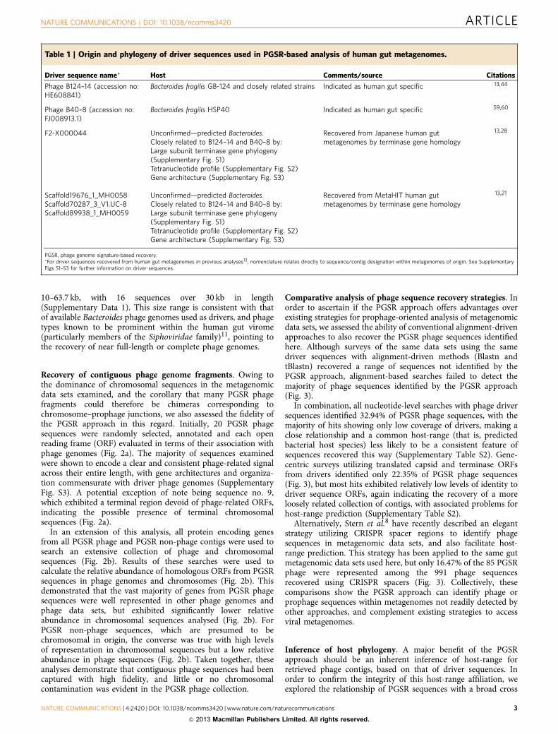
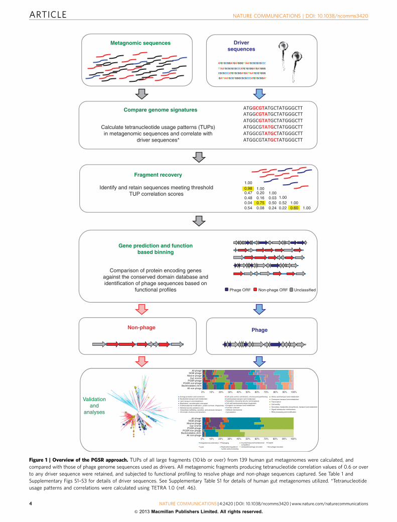
ResultsGenome signature-based recovery of ‘Bacteroidales-like’ phage.Members of the Bacteroidales, and in particular the genus Bac-teroides, are abundant and important constituents of the humangut microbiome for which few complete phage genomes areavailable, with this region of the gut virome believed to remainlargely uncharted13. To more fully explore this novel phage gene-space, we utilized Bacteroidales phage sequences as ‘drivers’ tointerrogate 139 human gut metagenomes based on tetranucleotideusage profiles (TUPs) and functional profiles of contigs (Table 1,Supplementary Figs S1–S3, Supplementary Table S1).
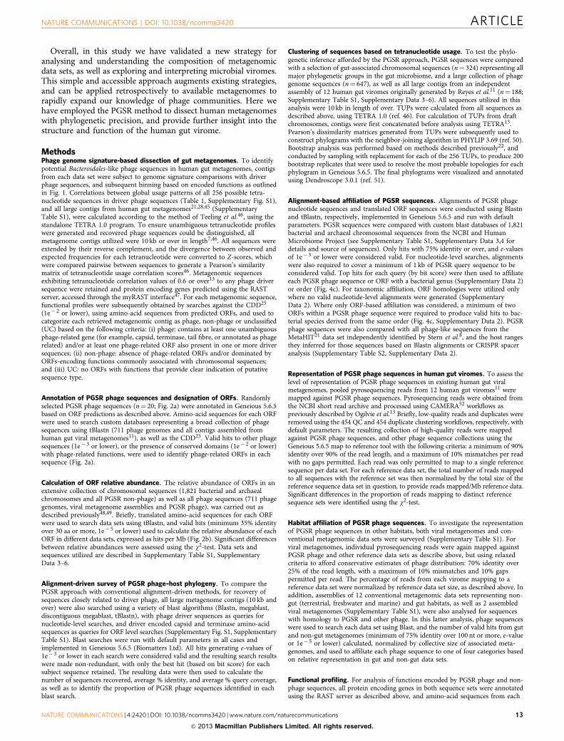
This strategy takes advantage of similarities in global nucleotideusage patterns, or the genome signature, arising between phageinfecting the same or related host bacterial species22–24. We exploitthis phenomenon to identify contigs related to Bacteroidales phagedriver sequences in assembled gut metagenomes, and subsequentfunction-based binning to resolve phage fragments recovered inthis process (Fig. 1). We refer to this strategy as phage genomesignature-based recovery (PGSR), and denote sequences obtainedin this way with the PGSR prefix.
Interrogation of all large contigs (10 kb and over) from humangut metagenomes (Supplementary Table S1) recovered 408metagenomic fragments with TUPs similar to Bacteroidalesphage drivers. Eighty five fragments were categorized as phagebased on functional profiling, and the remainder classified asnon-phage (presumed chromosomal, n¼ 320), or could not becategorized (n¼ 3) (Supplementary Data 1). The proportion ofsequences categorized as phage within the total pool of 408sequences recovered by PGSR (20.83%; 85/408) is congruent withrecent studies estimating that up to 17% of total metagenomicDNA derived from stool samples may be viral in origin7,11,21.Of the PGSR sequences classified as phage, sizes ranged from
ARTICLE NATURE COMMUNICATIONS | DOI: 10.1038/ncomms3420
2 NATURE COMMUNICATIONS | 4:2420 | DOI: 10.1038/ncomms3420 |www.nature.com/naturecommunications
& 2013 Macmillan Publishers Limited. All rights reserved.
10–63.7 kb, with 16 sequences over 30 kb in length(Supplementary Data 1). This size range is consistent with thatof available Bacteroides phage genomes used as drivers, and phagetypes known to be prominent within the human gut virome(particularly members of the Siphoviridae family)11, pointing tothe recovery of near full-length or complete phage genomes.
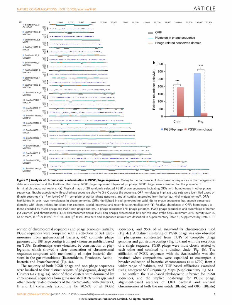
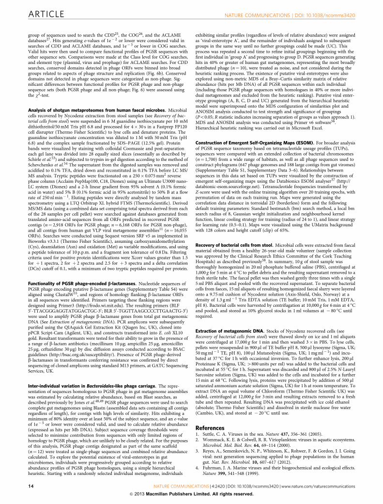
Recovery of contiguous phage genome fragments. Owing tothe dominance of chromosomal sequences in the metagenomicdata sets examined, and the corollary that many PGSR phagefragments could therefore be chimeras corresponding tochromosome–prophage junctions, we also assessed the fidelity ofthe PGSR approach in this regard. Initially, 20 PGSR phagesequences were randomly selected, annotated and each openreading frame (ORF) evaluated in terms of their association withphage genomes (Fig. 2a). The majority of sequences examinedwere shown to encode a clear and consistent phage-related signalacross their entire length, with gene architectures and organiza-tion commensurate with driver phage genomes (SupplementaryFig. S3). A potential exception of note being sequence no. 9,which exhibited a terminal region devoid of phage-related ORFs,indicating the possible presence of terminal chromosomalsequences (Fig. 2a).
In an extension of this analysis, all protein encoding genesfrom all PGSR phage and PGSR non-phage contigs were used tosearch an extensive collection of phage and chromosomalsequences (Fig. 2b). Results of these searches were used tocalculate the relative abundance of homologous ORFs from PGSRsequences in phage genomes and chromosomes (Fig. 2b). Thisdemonstrated that the vast majority of genes from PGSR phagesequences were well represented in other phage genomes andphage data sets, but exhibited significantly lower relativeabundance in chromosomal sequences analysed (Fig. 2b). ForPGSR non-phage sequences, which are presumed to bechromosomal in origin, the converse was true with high levelsof representation in chromosomal sequences but a low relativeabundance in phage sequences (Fig. 2b). Taken together, theseanalyses demonstrate that contiguous phage sequences had beencaptured with high fidelity, and little or no chromosomalcontamination was evident in the PGSR phage collection.
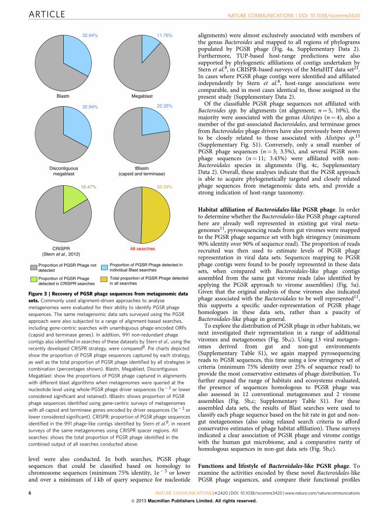
Comparative analysis of phage sequence recovery strategies. Inorder to ascertain if the PGSR approach offers advantages overexisting strategies for prophage-oriented analysis of metagenomicdata sets, we assessed the ability of conventional alignment-drivenapproaches to also recover the PGSR phage sequences identifiedhere. Although surveys of the same data sets using the samedriver sequences with alignment-driven methods (Blastn andtBlastn) recovered a range of sequences not identified by thePGSR approach, alignment-based searches failed to detect themajority of phage sequences identified by the PGSR approach(Fig. 3).
In combination, all nucleotide-level searches with phage driversequences identified 32.94% of PGSR phage sequences, with themajority of hits showing only low coverage of drivers, making aclose relationship and a common host-range (that is, predictedbacterial host species) less likely to be a consistent feature ofsequences recovered this way (Supplementary Table S2). Gene-centric surveys utilizing translated capsid and terminase ORFsfrom drivers identified only 22.35% of PGSR phage sequences(Fig. 3), but most hits exhibited relatively low levels of identity todriver sequence ORFs, again indicating the recovery of a moreloosely related collection of contigs, with associated problems forhost-range prediction (Supplementary Table S2).
Alternatively, Stern et al.8 have recently described an elegantstrategy utilizing CRISPR spacer regions to identify phagesequences in metagenomic data sets, and also facilitate host-range prediction. This strategy has been applied to the same gutmetagenomic data sets used here, but only 16.47% of the 85 PGSRphage were represented among the 991 phage sequencesrecovered using CRISPR spacers (Fig. 3). Collectively, thesecomparisons show the PGSR approach can identify phage orprophage sequences within metagenomes not readily detected byother approaches, and complement existing strategies to accessviral metagenomes.
Inference of host phylogeny. A major benefit of the PGSRapproach should be an inherent inference of host-range forretrieved phage contigs, based on that of driver sequences. Inorder to confirm the integrity of this host-range affiliation, weexplored the relationship of PGSR sequences with a broad cross
Table 1 | Origin and phylogeny of driver sequences used in PGSR-based analysis of human gut metagenomes.
Driver sequence name* Host Comments/source Citations
Phage B124–14 (accession no:HE608841)
Bacteroides fragilis GB-124 and closely related strains Indicated as human gut specific 13,44
Phage B40–8 (accession no:FJ008913.1)
Bacteroides fragilis HSP40 Indicated as human gut specific 59,60
F2-X000044 Unconfirmed—predicted Bacteroides.Closely related to B124–14 and B40–8 by:Large subunit terminase gene phylogeny(Supplementary Fig. S1)Tetranucleotide profile (Supplementary Fig. S2)Gene architecture (Supplementary Fig. S3)
Recovered from Japanese human gutmetagenomes by terminase gene homology
13,28
Scaffold19676_1_MH0058Scaffold70287_3_V1.UC-8Scaffold89938_1_MH0059
Unconfirmed—predicted Bacteroides.Closely related to B124–14 and B40–8 by:Large subunit terminase gene phylogeny(Supplementary Fig. S1)Tetranucleotide profile (Supplementary Fig. S2)Gene architecture (Supplementary Fig. S3)
Recovered from MetaHIT human gutmetagenomes by terminase gene homology
13,21
PGSR, phage genome signature-based recovery.*For driver sequences recovered from human gut metagenomes in previous analyses13, nomenclature relates directly to sequence/contig designation within metagenomes of origin. See SupplementaryFigs S1–S3 for further information on driver sequences.
NATURE COMMUNICATIONS | DOI: 10.1038/ncomms3420 ARTICLE
NATURE COMMUNICATIONS | 4:2420 | DOI: 10.1038/ncomms3420 |www.nature.com/naturecommunications 3
& 2013 Macmillan Publishers Limited. All rights reserved.
Metagnomic sequences
Compare genome signatures
Calculate tetranucleotide usage patterns (TUPs)in metagenomic sequences and correlate with
driver sequences*
Identify and retain sequences meeting thresholdTUP correlation scores
1.00
1.000.980.470.480.040.54 0.08 0.24 0.22
0.520.03
0.200.160.75 0.50
0.60 1.001.00
1.001.00
Comparison of protein encoding genesagainst the conserved domain database andidentification of phage sequences based on
functional profiles Phage ORF Non-phage ORF Unclassified
Fragment recovery
Gene prediction and functionbased binning
Non-phage Phage
Driversequences
Validationand
analyses
Figure 1 | Overview of the PGSR approach. TUPs of all large fragments (10 kb or over) from 139 human gut metagenomes were calculated, and
compared with those of phage genome sequences used as drivers. All metagenomic fragments producing tetranucleotide correlation values of 0.6 or over
to any driver sequence were retained, and subjected to functional profiling to resolve phage and non-phage sequences captured. See Table 1 and
Supplementary Figs S1–S3 for details of driver sequences. See Supplementary Table S1 for details of human gut metagenomes utilized. *Tetranucleotide
usage patterns and correlations were calculated using TETRA 1.0 (ref. 46).
ARTICLE NATURE COMMUNICATIONS | DOI: 10.1038/ncomms3420
4 NATURE COMMUNICATIONS | 4:2420 | DOI: 10.1038/ncomms3420 |www.nature.com/naturecommunications
& 2013 Macmillan Publishers Limited. All rights reserved.
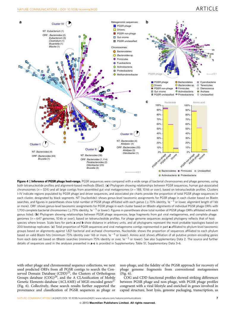
section of chromosomal sequences and phage genomes. Initially,PGSR sequences were compared with a collection of 324 chro-mosomes from gut-associated bacteria, 647 complete phagegenomes and 188 large contigs from gut virome assemblies, basedon TUPs. Relationships were visualized by construction of phy-lograms, which showed a clear association of chromosomalsequences congruent with membership of major bacterial divi-sions in the gut microbiome (Bacteroidetes, Firmicutes, Actino-bacteria and Proteobacteria) (Fig. 4a).
The majority of both PGSR phage and non-phage sequenceswere localized to four distinct regions of phylograms, designatedClusters I–IV (Fig. 4a). Most of these clusters were dominated bychromosomal sequences from gut-associated Bacteroides spp., andother closely related members of the Bacteroidales, with clusters I,II and III collectively accounting for 90.69% of all PGSR
sequences, and 95% of all Bacteroidales chromosomes used(Fig. 4a). A distinct clustering of PGSR phage was also observedin phylograms constructed from TUPs of complete phagegenomes and gut virome contigs (Fig. 4b), and with the exceptionof a single sequence, PGSR phage were most closely related toeach other and confined to a distinct clade (Fig. 4b). Theaffiliation of PGSR sequences with the Bacteroidales was alsoretained when comparisons, were expanded to encompass abroader collection of bacterial chromosomes (n¼ 1,700) from awider range of habitats, and TUP-based affiliations examinedusing Emergent Self Organizing Maps (Supplementary Fig. S4).
To confirm the TUP-based phylogenetic inference for PGSRsequences, and the implied host-range for PGSR phage,alignment-based searches of 1,821 bacterial and archaealchromosomes at both the nucleotide (Blastn) and ORF (tBlastn)
1. Scaffold4730_3O2.UC-18
1a
b
2,500 5,000 7,500 10,000 12,500 15,000 17,500 20,000 22,500 25,000
ORF
Homolog in phage sequence
Phage-related conserved domain
350
***
***
300
250
200
150
100
50
0
Rel
ativ
e ab
unda
nce
(hits
/Mb)
Chrm
PGSR-phage PGSR non-phage
Phg
27,500 30,000 32,500 35,000 37,138
100%50%0%
100%50%0%
100%50%0%
100%50%0%
100%50%0%
100%50%0%
100%50%0%
100%50%0%
100%50%0%
100%50%0%
100%50%0%
100%50%0%
100%50%0%
100%50%0%
100%50%0%
100%50%0%
100%50%0%
100%50%0%
100%50%0%
100%50%0%
2. Scaffold10385_2MH0084
3. Scaffold93605_5MH0011
4. Scaffold19851_6MH0051
5. Scaffold8153_2MH0069
6. Scaffold6082_3MH0068
7. Scaffold33311_3MH0083
8. Scaffold23106_1MH0070
9. Scaffold10269_2MH0076
10. Scaffold16062_5MH0063
11. Scaffold71140_1MH0070
12. Scaffold2660_7O2.UC-18
13. Scaffold158359_1MH0012
14. Scaffold90163_2MH0025
15. Scaffold33361_1MH0067
16. Scaffold82832_1MH0038
17. Scaffold29958_1MH0083
18. Scaffold8035_2V1.CD-12
19. Scaffold21092_1MH0080
20. Scaffold11813_5MH0082
Figure 2 | Analysis of chromosomal contamination in PGSR phage sequences. Owing to the dominance of chromosomal sequences in the metagenomic
data sets analysed and the likelihood that many PGSR phage represent integrated prophage, PGSR phage were examined for the presence of
terminal chromosomal regions. (a) Physical maps of 20 randomly selected PGSR phage sequences indicating ORFs with homologues in other phage
sequences. Graphs associated with each phage sequence show % GþC across the sequence. ORF homologues in phage data sets were identified based on
tBlastn searches (1e� 3 or lower) of 711 complete or partial phage genomes, and all contigs assembled from human gut viral metagenomes11. ORFs
highlighted in cyan have homologues in phage genomes. ORFs highlighted in red generated no valid hits to phage sequences but encode conserved
domains with phage-related functions (for example, capsid, integrase and recombination/replication). (b) Relative abundance of ORFs homologous to
those encoded by PGSR phage and PGSR non-phage contigs, in phage sequences (711 phage genomes, PGSR phage sequences and assemblies of human
gut viromes) and chromosomes (1,821 chromosomes and all PGSR non-phage) expressed as hits per Mb DNA (valid hits¼minimum 35% identity over 30
aa or more, 1e� 5 or lower). ***Pr0.001 (w2-test). Data sets and sequences utilized are described in Supplementary Table S1, Supplementary Data 3–6).
NATURE COMMUNICATIONS | DOI: 10.1038/ncomms3420 ARTICLE
NATURE COMMUNICATIONS | 4:2420 | DOI: 10.1038/ncomms3420 |www.nature.com/naturecommunications 5
& 2013 Macmillan Publishers Limited. All rights reserved.
level were also conducted. In both searches, PGSR phagesequences that could be classified based on homology tochromosome sequences (minimum 75% identity, 1e� 5 or lowerand over a minimum of 1 kb of query sequence for nucleotide
alignments) were almost exclusively associated with members ofthe genus Bacteroides and mapped to all regions of phylogramspopulated by PGSR phage (Fig. 4a, Supplementary Data 2).Furthermore, TUP-based host-range predictions were alsosupported by phylogenetic affiliations of contigs undertaken byStern et al.8, in CRISPR-based surveys of the MetaHIT data set21.In cases where PGSR phage contigs were identified and affiliatedindependently by Stern et al.8, host-range associations werecomparable, and in most cases identical to, those assigned in thepresent study (Supplementary Data 2).
Of the classifiable PGSR phage sequences not affiliated withBacteroides spp. by alignments (nt alignment; n¼ 5, 10%), themajority were associated with the genus Alistipes (n¼ 4), also amember of the gut-associated Bacteroidales, and terminase genesfrom Bacteroidales phage drivers have also previously been shownto be closely related to those associated with Alistipes sp.13
(Supplementary Fig. S1). Conversely, only a small number ofPGSR phage sequences (n¼ 3; 3.5%), and several PGSR non-phage sequences (n¼ 11; 3.43%) were affiliated with non-Bacteroidales species in alignments (Fig. 4c, SupplementaryData 2). Overall, these analyses indicate that the PGSR approachis able to acquire phylogenetically targeted and closely relatedphage sequences from metagenomic data sets, and provide astrong indication of host-range taxonomy.
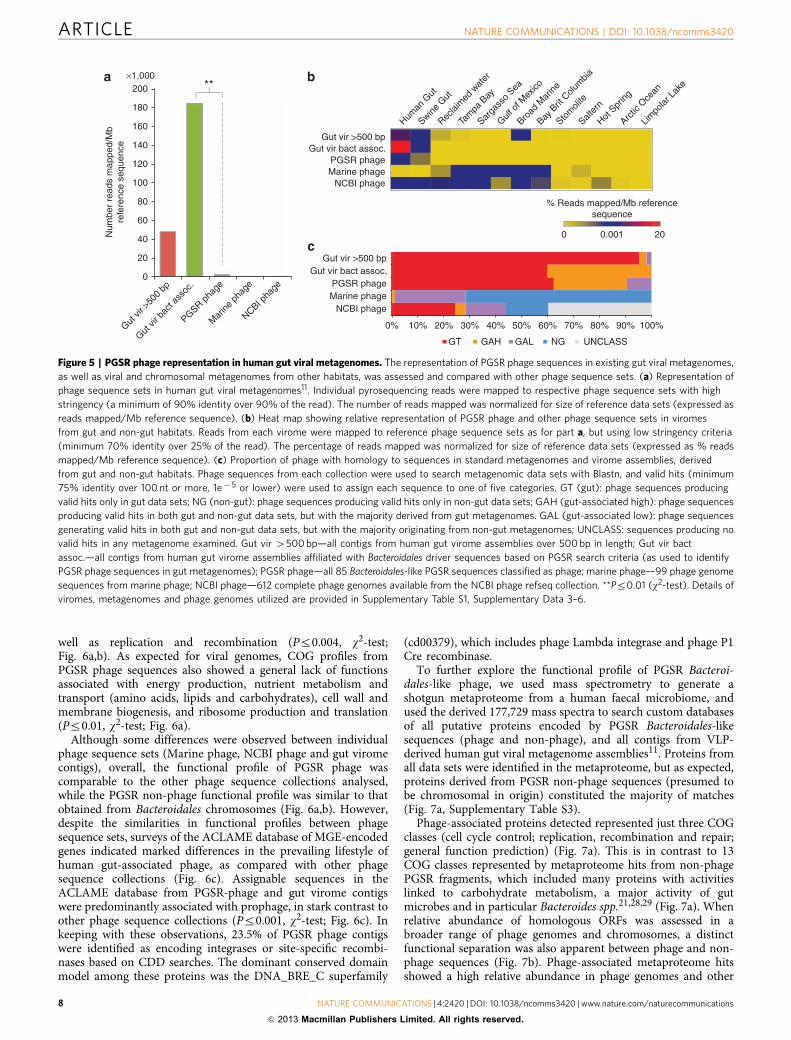
Habitat affiliation of Bacteroidales-like PGSR phage. In orderto determine whether the Bacteroidales-like PGSR phage capturedhere are already well represented in existing gut viral meta-genomes11, pyrosequencing reads from gut viromes were mappedto the PGSR phage sequence set with high stringency (minimum90% identity over 90% of sequence read). The proportion of readsrecruited was then used to estimate levels of PGSR phagerepresentation in viral data sets. Sequences mapping to PGSRphage contigs were found to be poorly represented in these datasets, when compared with Bacteroidales-like phage contigsassembled from the same gut virome reads (also identified byapplying the PGSR approach to virome assemblies) (Fig. 5a).Given that the original analysis of these viromes also indicatedphage associated with the Bacteroidales to be well represented11,this supports a specific under-representation of PGSR phagehomologues in these data sets, rather than a paucity ofBacteroidales-like phage in general.
To explore the distribution of PGSR phage in other habitats, wenext investigated their representation in a range of additionalviromes and metagenomes (Fig. 5b,c). Using 13 viral metagen-omes derived from gut and non-gut environments(Supplementary Table S1), we again mapped pyrosequencingreads to PGSR sequences, this time using a low stringency set ofcriteria (minimum 75% identity over 25% of sequence read) toprovide the most conservative estimates of phage distribution. Tofurther expand the range of habitats and ecosystems evaluated,the presence of sequences homologous to PGSR phage wasalso assessed in 12 conventional metagenomes and 2 viromeassemblies (Fig. 5b,c; Supplementary Table S1). For theseassembled data sets, the results of Blast searches were used toclassify each phage sequence based on the hit rate in gut and non-gut metagenomes (also using relaxed search criteria to affordconservative estimates of phage habitat affiliation). These surveysindicated a clear association of PGSR phage and virome contigswith the human gut microbiome, and a comparative rarity ofhomologous sequences in non-gut data sets (Fig. 5b,c).
Functions and lifestyle of Bacteroidales-like PGSR phage. Toexamine the activities encoded by these novel Bacteroidales-likePGSR phage sequences, and compare their functional profiles
Blastn Megablast
Discontiguousmegablast
tBlastn(capsid and terminase)
CRISPR(Stern et al., 2012)
All searches
32.94% 11.76%
32.94%
16.47%
22.35%
50.59%
Proportion of PGSR Phage notdetected
Proportion of PGSR Phage detected inindividual Blast searches
Proportion of PGSR Phagedetected in CRISPR searches
Total proportion of PGSR Phage detectedin all searches
Figure 3 | Recovery of PGSR phage sequences from metagenomic data
sets. Commonly used alignment-driven approaches to analyse
metagenomes were evaluated for their ability to identify PGSR phage
sequences. The same metagenomic data sets surveyed using the PGSR
approach were also subjected to a range of alignment-based searches,
including gene-centric searches with unambiguous phage-encoded ORFs
(capsid and terminase genes). In addition, 991 non-redundant phage
contigs also identified in searches of these datasets by Stern et al., using the
recently developed CRISPR strategy, were compared8. Pie charts depicted
show the proportion of PGSR phage sequences captured by each strategy,
as well as the total proportion of PGSR phage identified by all strategies in
combination (percentages shown). Blastn, Megablast, Discontiguous
Megablast: show the proportions of PGSR phage captured in alignments
with different blast algorithms when metagenomes were queried at the
nucleotide level using whole-PGSR phage driver sequences (1e� 3 or lower
considered significant and retained). tBlastn: shows proportion of PGSR
phage sequences identified using gene-centric surveys of metagenomes
with all capsid and terminase genes encoded by driver sequences (1e� 3 or
lower considered significant). CRISPR: proportion of PGSR phage sequences
identified in the 991 phage-like contigs identified by Stern et al.8, in recent
surveys of the same metagenomes using CRISPR spacer regions. All
searches: shows the total proportion of PGSR phage identified in the
combined output of all searches conducted above.
ARTICLE NATURE COMMUNICATIONS | DOI: 10.1038/ncomms3420
6 NATURE COMMUNICATIONS | 4:2420 | DOI: 10.1038/ncomms3420 |www.nature.com/naturecommunications
& 2013 Macmillan Publishers Limited. All rights reserved.
with other phage and chromosomal sequence collections, we nextused predicted ORFs from all PGSR contigs to search the Con-served Domain Database (CDD)25, the Clusters of OrthologousGroups database (COG)26, and the A CLAssification of MobileGenetic Elements database (ACLAME) of MGE-encoded genes27
(Fig. 6). Collectively, these search results further supported theprovenance and classification of PGSR sequences as phage or
non-phage, and the fidelity of the PGSR approach for recovery ofphage genome fragments from conventional metagenomes(Fig. 6).
COG and CDD functional profiles showed striking differencesbetween PGSR phage and non-phage, with PGSR phage profilescongruent with a viral lifestyle and enriched in genes involved incapsid structure, host lysis, genome packaging, transcription, as
Cluster IV:
Cluster III:
Cluster II:
Cluster I:
NT: Eubacterium (1)
NT: Bacteroides (4)
NT: Bacteroides (38)
NT: Bacteroides (4) Alistipes (4)
ORF: Bacteroides (55) Alistipes (2) Odoribacter (1)
ORF: Bacteroides (63) Brucella (1) ORF: Bacteroides (1,114)
Parabacteroides (2) Odoribacter (23) Brucella (3)
Metagenomic sequences:
Chromosomes:
PGSR phage
Drivers
PGSR non-phage
Gut virome
PGSR unclassified
ORF: Bacteroides (2)Eubacterium (3)Clostridium (1)Bryantella (1)Blautia (1)
Bacteroidetes
Bacteroides sp.
Firmicutes
Fusobacteria
PGSR phage 0.1
0.1
PGSR phage
100%Nucleotide Amino acid
90%
80%
70%
60%
50%
40%
30%
20%
10%
0%
Bacteroidetes
Actinobacteria
Firmicutes
Proteobacteria
Unclassified
PGSR non
-pha
ge
PGSR pha
ge
Virom
e
PGSR non
-pha
ge
PGSR pha
ge
Virom
e
100%
90%
80%
70%
60%
50%
40%
30%
20%
10%
0%
Bacteroidetes Cyanobacteria
DienococcusTenericutes
ArchaeaUnclassified
FirmicutesActinobacteriaProteobacteria
Bacteroides sp.DriversPGSR non-phageGut viromePGSR unclassified
Actinobacteria
Proteobacteria
Methanobrevibacter
a b
c
Figure 4 | Inference of PGSR phage host-range. PGSR sequences were compared with a wide range of bacterial chromosomes and phage genomes, using
both tetranucleotide profiles and alignment-based methods (Blast). (a) Phylogram showing relationships between PGSR sequences, human gut-associated
chromosomes (n¼ 324) and all large contigs from assembled gut viral metagenomes (n¼ 188, 10 kb or over), based on tetranucleotide profiles. Clusters
I–IV indicate regions populated by PGSR phage and driver sequences, and associated pie charts provide the proportion of total PGSR phage sequences in
each cluster, designated by black segments. NT (nucleotide): shows genus-level taxonomic assignments for PGSR phage in each cluster based on Blastn
searches, and figures in parentheses show total number of PGSR phage affiliated with each genus (Z75% identity, 1e� 5 or lower, alignment length of 1 kb
or more). ORF: shows genus-level taxonomic assignments for PGSR phage in each cluster based on tBlastn alignments of individual PGSR phage ORFs with
1,700 complete bacterial chromsomes (Z75% identity, 1e� 5 or lower). Figures in parentheses show total number of PGSR phage ORFs affiliated with each
genus listed. (b) Phylogram showing relationships between PGSR phage sequences, large fragments from gut viral metagenomes, and complete phage
genomes (n¼647 genomes, 10 kb or over), based on tetranucleotide profiles. For phage genome sequences assigned phylogeny reflects that of host
species where known. Scale bars for parts a and b show distance in arbitrary units, and all phylograms represent the most probable topologies based on
200 bootstrap replicates. (c) Total proportion of PGSR sequences and viral metagenome contigs represented in part a affiliated to phylum-level taxonomic
groups based on alignments against 1,821 bacterial and archaeal chromsomes. Nucleotide: shows the proportion of sequences affiliated to each phylum
based on valid Blastn hits (minimum 75% identity over 1 kb or more, 1e� 5 or lower). Amino acid: shows affiliation of all putative protein encoding genes
from each data set based on tBlastn searches (minimum 75% identity or over, 1e� 5 or lower). See also Supplementary Data 2. The source and further
details of sequences used in the analyses presented in a–c is provided in Supplementary Table S1, Supplementary Data 3–6.
NATURE COMMUNICATIONS | DOI: 10.1038/ncomms3420 ARTICLE
NATURE COMMUNICATIONS | 4:2420 | DOI: 10.1038/ncomms3420 |www.nature.com/naturecommunications 7
& 2013 Macmillan Publishers Limited. All rights reserved.
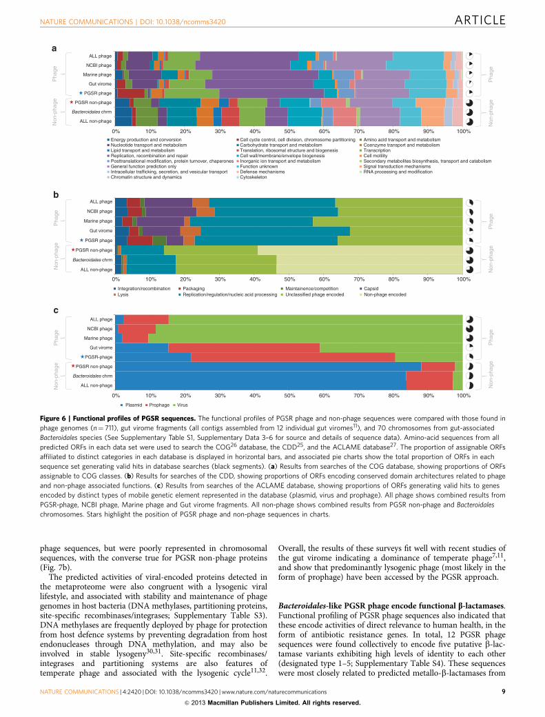
well as replication and recombination (Pr0.004, w2-test;Fig. 6a,b). As expected for viral genomes, COG profiles fromPGSR phage sequences also showed a general lack of functionsassociated with energy production, nutrient metabolism andtransport (amino acids, lipids and carbohydrates), cell wall andmembrane biogenesis, and ribosome production and translation(Pr0.01, w2-test; Fig. 6a).
Although some differences were observed between individualphage sequence sets (Marine phage, NCBI phage and gut viromecontigs), overall, the functional profile of PGSR phage wascomparable to the other phage sequence collections analysed,while the PGSR non-phage functional profile was similar to thatobtained from Bacteroidales chromosomes (Fig. 6a,b). However,despite the similarities in functional profiles between phagesequence sets, surveys of the ACLAME database of MGE-encodedgenes indicated marked differences in the prevailing lifestyle ofhuman gut-associated phage, as compared with other phagesequence collections (Fig. 6c). Assignable sequences in theACLAME database from PGSR-phage and gut virome contigswere predominantly associated with prophage, in stark contrast toother phage sequence collections (Pr0.001, w2-test; Fig. 6c). Inkeeping with these observations, 23.5% of PGSR phage contigswere identified as encoding integrases or site-specific recombi-nases based on CDD searches. The dominant conserved domainmodel among these proteins was the DNA_BRE_C superfamily
(cd00379), which includes phage Lambda integrase and phage P1Cre recombinase.
To further explore the functional profile of PGSR Bacteroi-dales-like phage, we used mass spectrometry to generate ashotgun metaproteome from a human faecal microbiome, andused the derived 177,729 mass spectra to search custom databasesof all putative proteins encoded by PGSR Bacteroidales-likesequences (phage and non-phage), and all contigs from VLP-derived human gut viral metagenome assemblies11. Proteins fromall data sets were identified in the metaproteome, but as expected,proteins derived from PGSR non-phage sequences (presumed tobe chromosomal in origin) constituted the majority of matches(Fig. 7a, Supplementary Table S3).
Phage-associated proteins detected represented just three COGclasses (cell cycle control; replication, recombination and repair;general function prediction) (Fig. 7a). This is in contrast to 13COG classes represented by metaproteome hits from non-phagePGSR fragments, which included many proteins with activitieslinked to carbohydrate metabolism, a major activity of gutmicrobes and in particular Bacteroides spp.21,28,29 (Fig. 7a). Whenrelative abundance of homologous ORFs was assessed in abroader range of phage genomes and chromosomes, a distinctfunctional separation was also apparent between phage and non-phage sequences (Fig. 7b). Phage-associated metaproteome hitsshowed a high relative abundance in phage genomes and other
200×1,000
Gut vir >500 bp
Human
Gut
Swine G
ut
Reclai
med
wat
er
Tam
pa B
ay
Sarga
sso
Sea
Gulf o
f Mex
ico
Broad
Mar
ine
Bay B
rit C
olum
bia
Stom
olite
Salter
n
Hot S
pring
Arctic
Oce
an
Limpo
lar L
ake
Gut vir bact assoc.PGSR phageMarine phage
NCBI phage
Gut vir >500 bpGut vir bact assoc.
PGSR phageMarine phage
NCBI phage
% Reads mapped/Mb referencesequence
0
0% 10% 20%
GT GAH GAL NG UNCLASS
30% 40% 50% 60% 70% 80% 90% 100%
0.001 20
**
180
160
140
120
100
80
60
40
20
0
Gut vi
r >50
0 bp
Gut vi
r bac
t ass
oc.
PGSR pha
ge
Mar
ine p
hage
NCBI pha
ge
Num
ber
read
s m
appe
d/M
bre
fere
nce
sequ
ence
Figure 5 | PGSR phage representation in human gut viral metagenomes. The representation of PGSR phage sequences in existing gut viral metagenomes,
as well as viral and chromosomal metagenomes from other habitats, was assessed and compared with other phage sequence sets. (a) Representation of
phage sequence sets in human gut viral metagenomes11. Individual pyrosequencing reads were mapped to respective phage sequence sets with high
stringency (a minimum of 90% identity over 90% of the read). The number of reads mapped was normalized for size of reference data sets (expressed as
reads mapped/Mb reference sequence). (b) Heat map showing relative representation of PGSR phage and other phage sequence sets in viromes
from gut and non-gut habitats. Reads from each virome were mapped to reference phage sequence sets as for part a, but using low stringency criteria
(minimum 70% identity over 25% of the read). The percentage of reads mapped was normalized for size of reference data sets (expressed as % reads
mapped/Mb reference sequence). (c) Proportion of phage with homology to sequences in standard metagenomes and virome assemblies, derived
from gut and non-gut habitats. Phage sequences from each collection were used to search metagenomic data sets with Blastn, and valid hits (minimum
75% identity over 100nt or more, 1e� 5 or lower) were used to assign each sequence to one of five categories. GT (gut): phage sequences producing
valid hits only in gut data sets; NG (non-gut): phage sequences producing valid hits only in non-gut data sets; GAH (gut-associated high): phage sequences
producing valid hits in both gut and non-gut data sets, but with the majority derived from gut metagenomes. GAL (gut-associated low): phage sequences
generating valid hits in both gut and non-gut data sets, but with the majority originating from non-gut metagenomes; UNCLASS: sequences producing no
valid hits in any metagenome examined. Gut vir 4500bp—all contigs from human gut virome assemblies over 500bp in length; Gut vir bact
assoc.—all contigs from human gut virome assemblies affiliated with Bacteroidales driver sequences based on PGSR search criteria (as used to identify
PGSR phage sequences in gut metagenomes); PGSR phage—all 85 Bacteroidales-like PGSR sequences classified as phage; marine phage—99 phage genome
sequences from marine phage; NCBI phage—612 complete phage genomes available from the NCBI phage refseq collection. **Pr0.01 (w2-test). Details ofviromes, metagenomes and phage genomes utilized are provided in Supplementary Table S1, Supplementary Data 3–6.
ARTICLE NATURE COMMUNICATIONS | DOI: 10.1038/ncomms3420
8 NATURE COMMUNICATIONS | 4:2420 | DOI: 10.1038/ncomms3420 |www.nature.com/naturecommunications
& 2013 Macmillan Publishers Limited. All rights reserved.
phage sequences, but were poorly represented in chromosomalsequences, with the converse true for PGSR non-phage proteins(Fig. 7b).
The predicted activities of viral-encoded proteins detected inthe metaproteome were also congruent with a lysogenic virallifestyle, and associated with stability and maintenance of phagegenomes in host bacteria (DNA methylases, partitioning proteins,site-specific recombinases/integrases; Supplementary Table S3).DNA methylases are frequently deployed by phage for protectionfrom host defence systems by preventing degradation from hostendonucleases through DNA methylation, and may also beinvolved in stable lysogeny30,31. Site-specific recombinases/integrases and partitioning systems are also features oftemperate phage and associated with the lysogenic cycle11,32.
Overall, the results of these surveys fit well with recent studies ofthe gut virome indicating a dominance of temperate phage7,11,and show that predominantly lysogenic phage (most likely in theform of prophage) have been accessed by the PGSR approach.
Bacteroidales-like PGSR phage encode functional b-lactamases.Functional profiling of PGSR phage sequences also indicated thatthese encode activities of direct relevance to human health, in theform of antibiotic resistance genes. In total, 12 PGSR phagesequences were found collectively to encode five putative b-lac-tamase variants exhibiting high levels of identity to each other(designated type 1–5; Supplementary Table S4). These sequenceswere most closely related to predicted metallo-b-lactamases from
ALL phage
NCBI phage
Marine phage
Gut virome
PGSR phage
PGSR non-phage
Bacteroidales chrm
ALL non-phage
0% 10% 20% 30% 40% 50% 60% 70% 80% 90% 100%
Non
-pha
geP
hage
Non
-pha
geP
hage
ALL phage
NCBI phage
Marine phage
Gut virome
PGSR phage
PGSR non-phage
Bacteroidales chrm
ALL non-phage
0% 10%
Integration/recombination PackagingReplication/regulation/nucleic acid processing
Maintainence/competitionUnclassified phage encoded
CapsidNon-phage encodedLysis
20% 30% 40% 50% 60% 70% 80% 90% 100%
Non
-pha
geP
hage
Non
-pha
geP
hage
ALL phage
NCBI phage
Marine phage
Gut virome
PGSR-phage
PGSR non-phage
Bacteroidales chrm
ALL non-phage
0% 10%
Plasmid Prophage Virus
20% 30% 40% 50% 60% 70% 80% 90% 100%
Non
-pha
geP
hage
Non
-pha
geP
hage
Energy production and conversionNucleotide transport and metabolismLipid transport and metabolismReplication, recombination and repairPosttranslational modification, protein turnover, chaperonesGeneral function prediction onlyIntracellular trafficking, secretion, and vesicular transportChromatin structure and dynamics Cytoskeleton
Cell cycle control, cell division, chromosome partitioning
RNA processing and modification
Carbohydrate transport and metabolismTranslation, ribosomal structure and biogenesisCell wall/membrane/envelope biogenesisInorganic ion transport and metabolismFunction unknownDefense mechanisms
Amino acid transport and metabolismCoenzyme transport and metabolismTranscriptionCell motilitySecondary metabolites biosynthesis, transport and catabolismSignal transduction mechanisms
Figure 6 | Functional profiles of PGSR sequences. The functional profiles of PGSR phage and non-phage sequences were compared with those found in
phage genomes (n¼ 711), gut virome fragments (all contigs assembled from 12 individual gut viromes11), and 70 chromosomes from gut-associated
Bacteroidales species (See Supplementary Table S1, Supplementary Data 3–6 for source and details of sequence data). Amino-acid sequences from all
predicted ORFs in each data set were used to search the COG26 database, the CDD25, and the ACLAME database27. The proportion of assignable ORFs
affiliated to distinct categories in each database is displayed in horizontal bars, and associated pie charts show the total proportion of ORFs in each
sequence set generating valid hits in database searches (black segments). (a) Results from searches of the COG database, showing proportions of ORFs
assignable to COG classes. (b) Results for searches of the CDD, showing proportions of ORFs encoding conserved domain architectures related to phage
and non-phage associated functions. (c) Results from searches of the ACLAME database, showing proportions of ORFs generating valid hits to genes
encoded by distinct types of mobile genetic element represented in the database (plasmid, virus and prophage). All phage shows combined results from
PGSR-phage, NCBI phage, Marine phage and Gut virome fragments. All non-phage shows combined results from PGSR non-phage and Bacteroidales
chromosomes. Stars highlight the position of PGSR phage and non-phage sequences in charts.
NATURE COMMUNICATIONS | DOI: 10.1038/ncomms3420 ARTICLE
NATURE COMMUNICATIONS | 4:2420 | DOI: 10.1038/ncomms3420 |www.nature.com/naturecommunications 9
& 2013 Macmillan Publishers Limited. All rights reserved.
Bacteroides sp. D22, Bacteroides sp. 1_1_30 and Bacteroides ster-coris, but showed no significant homology to entries in theAntibiotic Resistance Genes Database33 (minimum 20% identity,1e� 2 or lower).
To confirm the functionality of these putative resistancedeterminants, corresponding regions of PGSR phage wereamplified from total gut metagenomic DNA, cloned andexpressed in E. coli. Transformants were then tested for theirsusceptibility to a range of b-lactam antibiotics. Only Type-2PGSR phage-encoded b-lactamases were successfully amplifiedand cloned, but were capable of conferring resistance againstmecillinam (Supplementary Fig. S5), a member of the amidino-penicillin family with high affinity for Gram-negative penicillin-binding protein 2, but little activity against Gram-positivebacteria34. This antibiotic is not widely used in many Europeancountries or the USA, but has been identified as potentially usefulin the treatment of multi-drug resistant infections caused byGram-negative species35. As such, identification of viable mecillinamresistance genes circulating among lysogenic Bacteroides phage inthe gut mobile metagenome is of particular significance, and
highlights the potential for dissemination and spread of theseresistance determinants via horizontal gene transfer.
Inter-individual variation in Bacteroidales-like phage carriage.To assess inter-individual variation in carriage of PGSR phageand related sequences, we calculated the relative abundance ofsequences homologous to PGSR phage in individual gut meta-genomes (minimum 80% identity over 50% of the subjectsequence, 1e� 5 or lower). This indicated that such sequences arebroadly distributed among the gut microbiomes examined(Fig. 8a), with the incidence of PGSR homologues ranging from51.8–82.73% of metagenomes for the five most broadly repre-sented PGSR phage (encompassing both Japanese and Europeanindividuals) (Fig. 8a). Notably, these apparently broadly dis-tributed virotypes included sequences with homology to PGSRphage harbouring type-2 b-lactamases with proven function.
Heat maps of relative abundance data also suggested theexistence of several distinct patterns of Bacteroidales-like phagecarriage shared by multiple individuals (Fig. 8a). To investigate
0.007PGSR non-phage
PGSR phage
Gut virome
*****
***
*****
0.006
0.005
0.004
0.003Hit
rate
(hits
/ num
ber
of p
redi
cted
pro
tein
s)0.002
0.001
0C
C – Energy production and conversion; L – Replication, recombination and repair;M – Cell wall/membrane/envelope biogenesis;O – Posttranslational modification, protein turnover, chaperones;Q – Secondary metabolites biosynthesis, transport and catabolism;R – General function prediction only;S – Function unknown;T – Signal transduction mechanism;
PGSR non-phage
ChromosomesPGSR non-phage
PGSR phageGut virome
Phage genomesAll chromosomes
All phage
PGSR phage
Relative abundance (hits per Mb)
0 1 5
Gut virome
D – Cell cycle control, cell division, chromosome partitioning;E – Amino acid transport and metabolism;G – Carbohydrate transport and metabolism;I – Lipid transport and metabolism;J – Translation, ribosomal structure and biogenesis;K – Transcription;
D E G I J KCOG class
L M O Q R S T Total
Figure 7 | Representation of PGSR phage sequences in the human gut metaproteome. To further explore the functional profile of PGSR Bacteroidales-like
phage, and their contribution to the human gut metaproteome, a shotgun metaproteome was generated from a human faecal microbiome and the resulting
177,729 mass spectra used to search custom databases of all putative proteins encoded PGSR phage, PGSR non-phage and VLP-derived contigs from
human gut viral metagenomes11. (a) Shows relative hit rates in the gut metaproteome, for amino-acid sequences originating in each data set used to query
mass spectra (PGSR phage, PGSR non-phage, VLP-derived gut virome). Relative hit rates were calculated by normalizing the number of proteins from each
data set detected in the gut metaproteome by the total number of ORFs in parental data sets (expressed as hits per total number of predicted proteins in
each data set). Symbols above bars indicate statistically significant differences in relative hit rate with the data set of corresponding symbol colour
(**P¼0.01 or lower; ***P¼0.001 or lower; w2-test). Putative functions of identified proteins were based on COG searches (1e� 2 or lower; Supplementary
Table S3). (b) Heat map shows relative abundance of sequences homologous to those detected in the gut metaproteome, within a broad cross section of
bacterial and archaeal chromosomal sequences (n¼ 1,821, PGSR non-phage), and phage sequences (711 phage genomes, PGSR phage sequences and
assemblies of human gut viromes), expressed as hits per Mb DNA48,49 (valid hits¼minimum 35% identity over 30 aa or more, 1e� 5 or lower). See
Supplementary Table S1, Supplementary Data 3–6 for sources and details of sequences used.
ARTICLE NATURE COMMUNICATIONS | DOI: 10.1038/ncomms3420
10 NATURE COMMUNICATIONS | 4:2420 | DOI: 10.1038/ncomms3420 |www.nature.com/naturecommunications
& 2013 Macmillan Publishers Limited. All rights reserved.
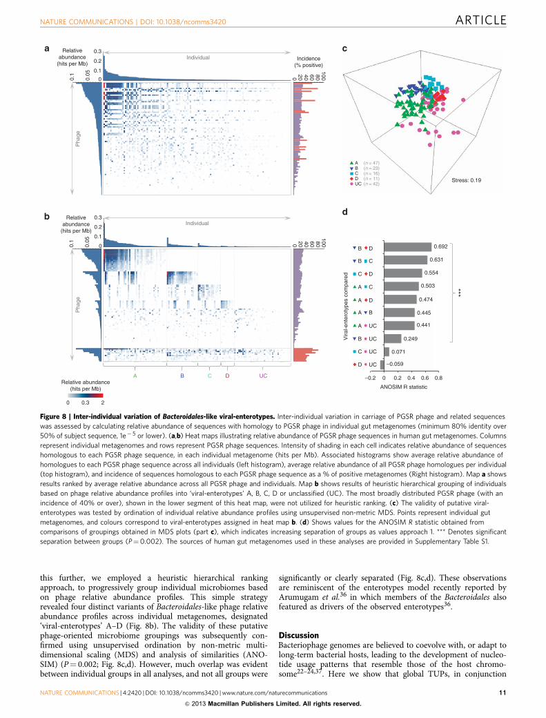
this further, we employed a heuristic hierarchical rankingapproach, to progressively group individual microbiomes basedon phage relative abundance profiles. This simple strategyrevealed four distinct variants of Bacteroidales-like phage relativeabundance profiles across individual metagenomes, designated‘viral-enterotypes’ A–D (Fig. 8b). The validity of these putativephage-oriented microbiome groupings was subsequently con-firmed using unsupervised ordination by non-metric multi-dimensional scaling (MDS) and analysis of similarities (ANO-SIM) (P¼ 0.002; Fig. 8c,d). However, much overlap was evidentbetween individual groups in all analyses, and not all groups were
significantly or clearly separated (Fig. 8c,d). These observationsare reminiscent of the enterotypes model recently reported byArumugam et al.36 in which members of the Bacteroidales alsofeatured as drivers of the observed enterotypes36.
Discussion.Bacteriophage genomes are believed to coevolve with, or adapt tolong-term bacterial hosts, leading to the development of nucleo-tide usage patterns that resemble those of the host chromo-some22–24,37. Here we show that global TUPs, in conjunction
Stress: 0.19
20 40 60 800
0.1
0.3
0
0.2
0.1
0.05
100
Relativeabundance
(hits per Mb)Incidence
(% positive)Individual
Pha
ge
0 20.3
Relative abundance(hits per Mb)
A B C D UC
Individual
Pha
ge
ANOSIM R statistic
0.071
0.249
0.441
0.445
0.474
0.503
0.554
0.631
0.692
–0.2 0 0.2 0.4 0.6 0.8
Vira
l-ent
erot
ypes
com
pare
d
***
–0.059
(n = 47)
(n = 42)
(n = 23)(n = 16)(n = 11)
0.1
0.3
0
0.2
0.1
0.05
Relativeabundance
(hits per Mb)
B D
B C
C D
A C
A D
A B
A UC
UC
UC
UC
C
D
B
ABCDUC
20 40 60 800
100
Figure 8 | Inter-individual variation of Bacteroidales-like viral-enterotypes. Inter-individual variation in carriage of PGSR phage and related sequences
was assessed by calculating relative abundance of sequences with homology to PGSR phage in individual gut metagenomes (minimum 80% identity over
50% of subject sequence, 1e� 5 or lower). (a,b) Heat maps illustrating relative abundance of PGSR phage sequences in human gut metagenomes. Columns
represent individual metagenomes and rows represent PGSR phage sequences. Intensity of shading in each cell indicates relative abundance of sequences
homologous to each PGSR phage sequence, in each individual metagenome (hits per Mb). Associated histograms show average relative abundance of
homologues to each PGSR phage sequence across all individuals (left histogram), average relative abundance of all PGSR phage homologues per individual
(top histogram), and incidence of sequences homologous to each PGSR phage sequence as a % of positive metagenomes (Right histogram). Map a shows
results ranked by average relative abundance across all PGSR phage and individuals. Map b shows results of heuristic hierarchical grouping of individuals
based on phage relative abundance profiles into ‘viral-enterotypes’ A, B, C, D or unclassified (UC). The most broadly distributed PGSR phage (with an
incidence of 40% or over), shown in the lower segment of this heat map, were not utilized for heuristic ranking. (c) The validity of putative viral-
enterotypes was tested by ordination of individual relative abundance profiles using unsupervised non-metric MDS. Points represent individual gut
metagenomes, and colours correspond to viral-enterotypes assigned in heat map b. (d) Shows values for the ANOSIM R statistic obtained from
comparisons of groupings obtained in MDS plots (part c), which indicates increasing separation of groups as values approach 1. *** Denotes significant
separation between groups (P¼0.002). The sources of human gut metagenomes used in these analyses are provided in Supplementary Table S1.
NATURE COMMUNICATIONS | DOI: 10.1038/ncomms3420 ARTICLE
NATURE COMMUNICATIONS | 4:2420 | DOI: 10.1038/ncomms3420 |www.nature.com/naturecommunications 11
& 2013 Macmillan Publishers Limited. All rights reserved.
with functional profiling, can be employed for the direct phage-oriented dissection of conventional metagenomes, permitting theresolution and host-range affiliation of subliminal viromefractions contained within. A major advantage of the use ofgenome signatures in this application is the gene-independent,alignment-free nature of this approach. As nucleotide signaturesare generally pervasive across genomes23,37, the requirement forthe presence of conserved genes or motifs typically used foridentification and classification of sequences is circumvented.
As such, genome signatures are well suited to analysis ofsequence types lacking robust and universally conservedphylogenetic anchors, and fragmentary data sets where conven-tional gene-centric alignment-driven methods often performpoorly37–42. Metagenomes, and phage (or other MGE sequences)captured within, constitute prime examples of such data sets andsequence types, with the PGSR approach shown to resolve phagesequences not readily detected by conventional alignment-drivenapproaches, even when used in conjunction with phage-relatedsequence motifs or genes.
However, this method does not overcome all disadvantages ofmetagenomic approaches for viral discovery. For example, thefocus on acquisition and analysis of chromosomal DNA inconventional metagenomic data sets will exclude RNA phage, andthere remains a need for continued culture-based isolation ofphage to provide well-characterized driver sequences. Despitethese caveats, the PGSR approach can recover many additionalphage sequences from few initial driver sequences, access phagenot well represented in VLP-based censuses, and potentially beused to mine metagenomes for other MGE and semi-conservedsequences.
Furthermore, the use of well characterised phage sequenceswith known host-ranges, as drivers in the PGSR approach,permits recovery of contigs with a common taxonomic imprint,automatically providing an indication of host phylogeny. A highlevel of congruence between TUP inferred phage–host associa-tions, and established host ranges for cultivable bacteria and theirphage has previously been demonstrated23, and also indicated tohold true for viral sequences represented in metagenomic datasets37. Importantly, previous genome signature-based analyses ofwhole-community shotgun metagenomes have shown that theshared selective pressures placed upon microbes occupying agiven habitat do not obscure the taxonomic imprint rooted inTUPs, even when the community is subject to strong andconstant environmental stress, the genus-level resolution ofmetagenomic fragments remains feasible37. These observationsare exemplified by the clear and consistent association of PGSRacquired contigs with Bacteroides spp. and members of the widerBacteroidales in the present study.
Conversely, a small number of PGSR phage sequences (n¼ 3)were affiliated with non-Bacteroidales species in alignment-drivensurveys, and mapped to regions of phylograms closely related tomembers of the Clostridiales, but also populated by a mixture ofBacteroidales-affiliated and unaffiliated sequences. This variegatedphylogenetic signal could be the result of convergent evolutionaryprocesses that generate similar TUPs in unrelated organisms orphage genomes, obscuring the taxonomic imprint and leading tospurious host-range affiliations22,23. There is also the possibilitythat these sequences represent examples of viruses with verybroad host-ranges43, or those in the process of adapting to newhost species. Alternatively, the acquisition of new genetic materialby horizontal gene transfer in phage is also well documented, andcould account for the discordant alignment-based affiliations ofthe PGSR sequences in question. These issues are not unique togenome signature-based approaches and are also importantconsiderations in gene-centric taxonomy22,23, constituting apotential limitation in both strategies.
The utilization of standard metagenomes in the PGSRapproach should also provide access to fractions of bacteriophagecommunities that may be poorly represented by other methods.In light of the reported dominance of temperate phage in thehuman gut ecosystem7,11, it would be expected that greater accessto quiescent phage will be important in further exploration of thisviral community and will yield much insight into its structure andfunction. As such it is notable that the PGSR phage captured herewere indicated to be predominantly prophage, and not wellrepresented in existing VLP-derived gut viral data sets,supporting the identification and analysis of phage sequencesnot readily accessed by other approaches. However, variation inthe geographic origins of the metagenomes and viromes utilizedfor these analyses cannot be excluded as a possible factor in thelow level of PGSR phage representation in VLP-based data sets,with gut metagenomes from which PGSR phage were retrievedEuropean in origin, but viral data sets generated from Americanindividuals11,13,21. Alternatively, phage sequences recovered heremay mostly represent inactivated prophage, which no longercontribute to the active, extrinsic VLP pool sampled in otherstudies.
Subsequent analyses showed PGSR phage not only encodefunctions directly relevant to human health (reinforcing the roleof phage in spread of antibiotic resistance determinants) but alsothe potential specificity of PGSR phage to the human gut habitat,which is relevant to biotechnological applications of phage suchas microbial source tracking13,44. In addition, the possibleexistence of ‘viral-enterotypes’ in this region of the gut viromewas also revealed when individual gut metagenomes werecompared. The phage-oriented grouping of microbiomes isreminiscent of the enterotypes model recently reported byArumugam et al.36, where individuals were grouped based onsimilarities in microbiome composition. Notably, two of the threemicrobial enterotypes presented by Arumugam et al.36 weredriven by members of the Bacteroidales (Bacteroides andPrevotella), and it seems logical that examination of gut-specifictemperate phage associated with these genera should generateconcordant findings.
However, the Bacteroidales-like phage-oriented microbiomegroupings observed here appear less well-defined and may beindicative of inter-individual gradients in phage populationstructure rather than entirely discrete groupings (as has alsobeen posited for microbial enterotypes). Moreover, the groupingof individuals based on virome structure is inconsistent withother recent studies of the gut virome, where no such associationswere observed7,8,11. These discrepancies may be due to thephylogenetically targeted analysis afforded by the PGSR approachcoupled with the nature of the data sets from which PGSR phageare derived. In conjunction, these attributes should provide accessto a closely related population of predominantly lysogenic phage(as prophage), expected to represent a more stable region of thephage ecological landscape in the gut microbiome.
Collectively, these factors could permit resolution of inter-individual similarities in gut virome structure obscured in studiesfocused on the virome as a whole, or the free, replicating viromefraction accessed through VLP libraries. Nevertheless, the datasets utilized here present only a ‘snapshot’ of the gut microbiomeand do not capture the temporal dynamics of phage–hostinteractions. Much scope also remains to refine criteria andstrategies used to identify and explore these putative viral-enterotypes. Although our observations provide the first indica-tion that such groupings may exist in the gut virome, it is clearthat further work will be required to confirm or refute thepotential existence of viral-enterotypes within the Bacteroidalesphage gene-space, and their significance, if any, for ecosystemfunction and development.
ARTICLE NATURE COMMUNICATIONS | DOI: 10.1038/ncomms3420
12 NATURE COMMUNICATIONS | 4:2420 | DOI: 10.1038/ncomms3420 |www.nature.com/naturecommunications
& 2013 Macmillan Publishers Limited. All rights reserved.
Overall, in this study we have validated a new strategy foranalysing and understanding the composition of metagenomicdata sets, as well as exploring and interpreting microbial viromes.This simple and accessible approach augments existing strategies,and can be applied retrospectively to available metagenomes torapidly expand our knowledge of phage communities. Here wehave employed the PGSR method to dissect human metagenomeswith phylogenetic precision, and provide further insight into thestructure and function of the human gut virome.
MethodsPhage genome signature-based dissection of gut metagenomes. To identifypotential Bacteroidales-like phage sequences in human gut metagenomes, contigsfrom each data set were subject to genome signature comparisons with driverphage sequences, and subsequent binning based on encoded functions as outlinedin Fig. 1. Correlations between global usage patterns of all 256 possible tetra-nucleotide sequences in driver phage sequences (Table 1, Supplementary Fig. S1),and all large contigs from human gut metagenomes21,28,45 (SupplementaryTable S1), were calculated according to the method of Teeling et al.46, using thestandalone TETRA 1.0 program. To ensure unambiguous tetranucleotide profileswere generated and recovered phage sequences could be distinguished, allmetagenome contigs utilized were 10 kb or over in length7,46. All sequences wereextended by their reverse complement, and the divergence between observed andexpected frequencies for each tetranucleotide were converted to Z-scores, whichwere compared pairwise between sequences to generate a Pearson’s similaritymatrix of tetranucleotide usage correlation scores46. Metagenomic sequencesexhibiting tetranucleotide correlation values of 0.6 or over13 to any phage driversequence were retained and protein encoding genes predicted using the RASTserver, accessed through the myRAST interface47. For each metagenomic sequence,functional profiles were subsequently obtained by searches against the CDD25
(1e� 2 or lower), using amino-acid sequences from predicted ORFs, and used tocategorize each retrieved metagenomic contig as phage, non-phage or unclassified(UC) based on the following criteria: (i) phage: contains at least one unambiguousphage-related gene (for example, capsid, terminase, tail fibre, or annotated as phagerelated) and/or at least one phage-related ORF also present in one or more driversequences; (ii) non-phage: absence of phage-related ORFs and/or dominated byORFs-encoding functions commonly associated with chromosomal sequences;and (iii) UC: no ORFs with functions that provide clear indication of putativesequence type.
Annotation of PGSR phage sequences and designation of ORFs. Randomlyselected PGSR phage sequences (n¼ 20; Fig. 2a) were annotated in Geneious 5.6.5based on ORF predictions as described above. Amino-acid sequences for each ORFwere used to search custom databases representing a broad collection of phagesequences using tBlastn (711 phage genomes and all contigs assembled fromhuman gut viral metagenomes11), as well as the CDD25. Valid hits to other phagesequences (1e� 3 or lower), or the presence of conserved domains (1e� 2 or lower)with phage-related functions, were used to identify phage-related ORFs in eachsequence (Fig. 2a).
Calculation of ORF relative abundance. The relative abundance of ORFs in anextensive collection of chromosomal sequences (1,821 bacterial and archaealchromosomes and all PGSR non-phage) as well as all phage sequences (711 phagegenomes, viral metagenome assemblies and PGSR phage), was carried out asdescribed previously48,49. Briefly, translated amino-acid sequences for each ORFwere used to search data sets using tBlastn, and valid hits (minimum 35% identityover 30 aa or more, 1e� 5 or lower) used to calculate the relative abundance of eachORF in different data sets, expressed as hits per Mb (Fig. 2b). Significant differencesbetween relative abundances were assessed using the w2-test. Data sets andsequences utilized are described in Supplementary Table S1, SupplementaryData 3–6.
Alignment-driven survey of PGSR phage–host phylogeny. To compare thePGSR approach with conventional alignment-driven methods, for recovery ofsequences closely related to driver phage, all large metagenome contigs (10 kb andover) were also searched using a variety of blast algorithms (Blastn, megablast,discontiguous megablast, tBlastn), with phage driver sequences as queries fornucleotide-level searches, and driver encoded capsid and terminase amino-acidsequences as queries for ORF level searches (Supplementary Fig. S1, SupplementaryTable S1). Blast searches were run with default parameters in all cases andimplemented in Geneious 5.6.5 (Biomatters Ltd). All hits generating e-values of1e� 3 or lower in each search were considered valid and the resulting search resultswere made non-redundant, with only the best hit (based on bit score) for eachsubject sequence retained. The resulting data were then used to calculate thenumber of sequences recovered, average % identity, and average % query coverage,as well as to identify the proportion of PGSR phage sequences identified in eachblast search.
Clustering of sequences based on tetranucleotide usage. To test the phylo-genetic inference afforded by the PGSR approach, PGSR sequences were comparedwith a selection of gut-associated chromosomal sequences (n¼ 324) representing allmajor phylogenetic groups in the gut microbiome, and a large collection of phagegenome sequences (n¼ 647), as well as all large contigs from an independentassembly of 12 human gut viromes originally generated by Reyes et al.11 (n¼ 188;Supplementary Table S1, Supplementary Data 3–6). All sequences utilized in thisanalysis were 10 kb in length of over. TUPs were calculated from all sequences asdescribed above, using TETRA 1.0 (ref. 46). For calculation of TUPs from draftchromosomes, contigs were first concatenated before analysis using TETRA13.Pearson’s dissimilarity matrices generated from TUPs were subsequently used toconstruct phylograms with the neighbor-joining algorithm in PHYLIP 3.69 (ref. 50).Bootstrap analysis was performed based on methods described previously22, andconducted by sampling with replacement for each of the 256 TUPs, to produce 200bootstrap replicates that were used to resolve the most probable topologies for eachphylogram in Geneious 5.6.5. The final phylograms were visualized and annotatedusing Dendroscope 3.0.1 (ref. 51).
Alignment-based affiliation of PGSR sequences. Alignments of PGSR phagenucleotide sequences and translated ORF sequences were conducted using Blastnand tBlastn, respectively, implemented in Geneious 5.6.5 and run with defaultparameters. PGSR sequences were compared with custom blast databases of 1,821bacterial and archaeal chromosomal sequences from the NCBI and HumanMicrobiome Project (see Supplementary Table S1, Supplementary Data 3,4 fordetails and source of sequences). Only hits with 75% identity or over, and e-valuesof 1e� 5 or lower were considered valid. For nucleotide-level searches, alignmentswere also required to cover a minimum of 1 kb of PGSR query sequence to beconsidered valid. Top hits for each query (by bit score) were then used to affiliateeach PGSR phage sequence or ORF with a bacterial genus (Supplementary Data 2)or order (Fig. 4c). For taxonomic affiliation, ORF homologies were utilized onlywhere no valid nucleotide-level alignments were generated (SupplementaryData 2). Where only ORF-based affiliation was considered, a minimum of twoORFs within a PGSR phage sequence were required to produce valid hits to bac-terial species derived from the same order (Fig. 4c, Supplementary Data 2). PGSRphage sequences were also compared with all phage-like sequences from theMetaHIT21 data set independently identified by Stern et al.8, and the host rangesthey inferred for those sequences based on Blastn alignments or CRISPR spaceranalysis (Supplementary Table S2, Supplementary Data 2).
Representation of PGSR phage sequences in human gut viromes. To assess thelevel of representation of PGSR phage sequences in existing human gut viralmetagenomes, pooled pyrosequencing reads from 12 human gut viromes11 weremapped against PGSR phage sequences. Pyrosequencing reads were obtained fromthe NCBI short read archive and processed using CAMERA52 workflows aspreviously described by Ogilvie et al.13 Briefly, low-quality reads and duplicates wereremoved using the 454 QC and 454 duplicate clustering workflows, respectively, withdefault parameters. The resulting collection of high-quality reads were mappedagainst PGSR phage sequences, and other phage sequence collections using theGeneious 5.6.5 map to reference tool with the following criteria: a minimum of 90%identity over 90% of the read length, and a maximum of 10% mismatches per readwith no gaps permitted. Each read was only permitted to map to a single referencesequence per data set. For each reference data set, the total number of reads mappedto all sequences with the reference set was then normalized by the total size of thereference sequence data set in question, to provide reads mapped/Mb reference data.Significant differences in the proportion of reads mapping to distinct referencesequence sets were identified using the w2-test.
Habitat affiliation of PGSR phage sequences. To investigate the representationof PGSR phage sequences in other habitats, both viral metagenomes and con-ventional metagenomic data sets were surveyed (Supplementary Table S1). Forviral metagenomes, individual pyrosequencing reads were again mapped againstPGSR phage and other reference data sets as describe above, but using relaxedcriteria to afford conservative estimates of phage distribution: 70% identity over25% of the read length, with a maximum of 10% mismatches and 10% gapspermitted per read. The percentage of reads from each virome mapping to areference data set were normalized by reference data set size, as described above. Inaddition, assemblies of 12 conventional metagenomic data sets representing non-gut (terrestrial, freshwater and marine) and gut habitats, as well as 2 assembledviral metagenomes (Supplementary Table S1), were also analysed for sequenceswith homology to PGSR and other phage. In this latter analysis, phage sequenceswere used to search each data set using Blast, and the number of valid hits from gutand non-gut metagenomes (minimum of 75% identity over 100 nt or more, e-valueor 1e� 5 or lower) calculated, normalized by collective size of associated meta-genomes, and used to affiliate each phage sequence to one of four categories basedon relative representation in gut and non-gut data sets.
Functional profiling. For analysis of functions encoded by PGSR phage and non-phage sequences, all protein encoding genes in both sequence sets were annotatedusing the RAST server as described above, and amino-acid sequences from each
NATURE COMMUNICATIONS | DOI: 10.1038/ncomms3420 ARTICLE
NATURE COMMUNICATIONS | 4:2420 | DOI: 10.1038/ncomms3420 |www.nature.com/naturecommunications 13
& 2013 Macmillan Publishers Limited. All rights reserved.
group of sequences used to search the CDD25, the COG26, and the ACLAMEdatabases27. Hits generating e-values of 1e� 2 or lower were considered valid insearches of CDD and ACLAME databases, and 1e� 3 or lower in COG searches.Valid hits were then used to compare functional profiles of PGSR sequences withother sequence sets. Comparisons were made at the Class level for COG searches,and element type (plasmid, virus and prophage) for ACLAME searches. For CDDsearches, conserved domains detected in phage ORFs were binned into broadgroups related to aspects of phage structure and replication (Fig. 6b). Conserveddomains not detected in phage sequences were categorized as non-phage. Sig-nificant differences between functional profiles for PGSR phage and non-phagesequence sets (both PGSR phage and all non-phage; Fig. 6) were assessed usingthe w2-test.
Analysis of shotgun metaproteomes from human faecal microbes. Microbialcells recovered by Nycodenz extraction from stool samples (see Recovery of bac-terial cells from stool) were suspended in 6 M guanidine isothiocyanate per 10 mMdithiothreitol/50mM Tris pH 6.8 and processed for 4� 30 s in a Fastprep FP120cell disrupter (Thermo Fisher Scientific) to lyse cells and denature proteins. Theguanidine isothiocyanate concentration was diluted to 1M with 50mM Tris (pH6.8) and the complex sample fractionated by SDS–PAGE (12.5% gel). Proteinbands were visualized by staining with colloidal Coomassie and post-separationeach gel lane was divided into 28 equally sized slices (essentially as described bySchirle et al.53) and subjected to trypsin in-gel digestion according to the method ofSchevchenko et al.54 The supernatant from the digested samples was removed andacidified to 0.1% TFA, dried down and reconstituted in 0.1% TFA before LC MS/MS analysis. Tryptic peptides were fractionated on a 250� 0.075mm2 reversephase column (Acclaim PepMap100, C18, Dionex) using an Ultimate U3000 nano-LC system (Dionex) and a 2-h linear gradient from 95% solvent A (0.1% formicacid in water) and 5% B (0.1% formic acid in 95% acetonitrile) to 50% B at a flowrate of 250 nlmin� 1. Eluting peptides were directly analysed by tandem massspectrometry using a LTQ Orbitrap XL hybrid FTMS (ThermoScientific). DerivedMS/MS data (using a combined data set comprising total spectra derived from eachof the 28 samples per cell pellet) were searched against databases generated fromtranslated amino-acid sequences from all ORFs predicted in recovered PGSRcontigs (n¼ 2,918 ORFs for PGSR phage; n¼ 6,168 ORFs for PGSR non-phage),and all contigs from human gut VLP viral metagenome assemblies11 (n¼ 16,055ORFs). Searches were conducted using Sequest version SRF v5 as implemented inBioworks v3.3.1 (Thermo Fisher Scientific), assuming carboxyamidomethylation(Cys), deamidation (Asn) and oxidation (Met) as variable modifications, and usinga peptide tolerance of 10 p.p.m. and a fragment ion tolerance of 0.8Da. Filteringcriteria used for positive protein identifications were Xcorr values greater than 1.5for þ 1 spectra, 2 for þ 2 spectra and 2.5 for þ 3 spectra and a delta correlation(DCn) cutoff of 0.1, with a minimum of two tryptic peptides required per protein.
Functionality of PGSR phage-encoded b-lactamases. Nucleotide sequences ofPGSR phage encoding putative b-lactamase genes (Supplementary Table S4) werealigned using ClustalW55, and regions of homology flanking b-lactamase ORFsin all sequences were identified. Primers targeting these flanking regions weredesigned using Primer3 (http://frodo.wi.mit.edu). The resulting primers (BLF50-TTACGGGAGGTATGGACTGC-30; BLR 50-TGGTTAAGCCCCTTGAACTG-30)were used to amplify PGSR phage b-lactamase genes from total gut metagenomicDNA (See Extraction of metagenomic DNA). PCR amplicons were subsequentlypurified using the QIAquick Gel Extraction Kit (Qiagen Inc, UK), cloned intopPCR Script-Cam (Agilent, UK), and constructs transformed into E. coli XL10gold. Resultant transformants were tested for their ability to grow in the presence ofa range of b-lactam antibiotics (mecillinam 10 mg; ampicillin 25 mg, amoxicillin25mg, ceftazidime 30mg) by disc diffusion assays conducted according to BSACguidelines (http://bsac.org.uk/susceptibility/). Presence of PGSR phage-derivedb-lactamases in transformants conferring resistance was confirmed by directsequencing of cloned amplicons using standard M13 primers, at GATC SequencingServices, UK.
Inter-individual variation in Bacteroidales-like phage carriage. The repre-sentation of sequences homologous to PGSR phage in gut metagenome assemblieswas estimated by calculating relative abundance, based on Blast searches, asdescribed previously by Jones et al.48,49 PGSR phage sequences were used to searchcomplete gut metagenomes using Blastn (assembled data sets containing all contigsregardless of length), for contigs with high levels of similarity. Hits exhibiting aminimum of 80% identity over at least 50% of the subject sequence, and an e-valueof 1e� 5 or lower were considered valid, and used to calculate relative abundance(expressed as hits per Mb DNA). Subject sequence coverage thresholds wereselected to minimize contribution from sequences with only limited regions ofhomology to PGSR phage, which are unlikely to be closely related. For the purposesof this analysis, PGSR phage contigs designated as part of the same scaffold(n¼ 12) were treated as single-phage sequences and combined relative abundancecalculated. To explore the potential existence of viral-enterotypes in gutmicrobiomes, individuals were progressively grouped according to relativeabundance profiles of PGSR phage homologues, using a simple hierarchicalheuristic. Starting with a randomly selected individual metagenome, individuals
exhibiting similar profiles (regardless of levels of relative abundance) were assignedas ‘viral-enterotype A’, and the remainder of individuals assigned to subsequentgroups in the same way until no further groupings could be made (UC). Thisprocess was repeated a second time to refine initial groupings beginning with thefirst individual in ‘group A’ and progressing to group D. PGSR sequences generatinghits in 40% or greater of human gut metagenomes, representing the most broadlydistributed phage (n¼ 10), were treated as noise, and not considered during theheuristic ranking process. The existence of putative viral-enterotypes were alsoexplored using non-metric MDS of a Bray–Curtis similarity matrix of relativeabundance (hits per Mb DNA) of all PGSR sequences within each individual(including those PGSR phage sequences with homologues in 40% or more indivi-dual metagenomes and excluded from the heuristic ranking). Putative viral enter-otype groupings (A, B, C, D and UC) generated from the hierarchical heuristicmodel were superimposed onto the MDS configuration of similarities plot andANOSIM analysis conducted to test strength and significance of groupings(Po0.05; R statistic indicates increasing separation of groups as values approach 1).MDS and ANOSIM analysis was conducted using Primer v6 software56.Hierarchical heuristic ranking was carried out in Microsoft Excel.
Construction of Emergent Self-Organizing Maps (ESOM). For broader analysisof PGSR sequence taxonomy based on tetranucleotide useage profiles (TUPs),sequences were compared with an extended collection of bacterial chromosomes(n¼ 1,700) from a wide range of habitats, as well as all phage sequences used toconstruct phylograms (647 phage genomes and 188 large contigs from gut viromes)(Supplementary Table S1, Supplementary Data 3–6). Relationships betweensequences in this data set based on TUPs were visualized by the construction ofemergent self-organizing maps using the Databionics ESOM analyser57 (http://databionic-esom.sourceforge.net). Tetranucleotide frequencies transformed byZ-score were used with the online training algorithm over 20 training epochs, withpermutation of data on each training run. Maps were generated using thecorrelation data distance in torroidal 2D (borderless) form and the followingdefault training parameters: Standard bestmatch (bm) search method, a local bmsearch radius of 8, Gaussian weight initialization and neighbourhood kernelfunction, linear cooling strategy for training (radius of 24 to 1), and linear strategyfor learning rate (0.5–0.1). Maps were visualized using the UMatrix backgroundwith 128 colors and height cutoff (clip) of 65%.
Recovery of bacterial cells from stool. Microbial cells were extracted from faecalmaterial obtained from a healthy 26-year-old male volunteer (sample collectionwas approved by the Clinical Research Ethics Committee of the Cork TeachingHospitals) as described previously58. In summary, 10 g of stool sample wasthoroughly homogenized in 20ml phosphate buffered saline (PBS), centrifuged at1,000 g for 5min at 4 �C to pellet debris and the resulting supernatant removed to afresh sterile tube. The faecal pellet was then washed gently three times with a single5ml PBS aliquot and pooled with the recovered supernatant. To separate bacterialcells from faeces, 15ml aliquots of resulting homogenized faecal slurry were layeredonto a 9.75ml cushion of Nycodenz solution (Axis-Shield, Oslo, Norway) at adensity of 1.3 gml� 1 Tris EDTA solution (TE buffer; 10mM Tris, 1mM EDTA,pH 8). Bacterial cells were harvested by centrifugation at 10,000 g for 6min at 4 �Cand pooled, and stored as 10% glycerol stocks in 1ml volumes at � 80 �C untilrequired.
Extraction of metagenomic DNA. Stocks of Nycodenz recovered cells (seeRecovery of bacterial cells from stool) were thawed slowly on ice and 1ml aliquotswere centrifuged at 17,000 g for 1min and then washed 3� in PBS. To lyse cells,pellets were resuspended in 900 ml of TE buffer pH 8, 500ml lysosyme (Sigma, UK;50mgml� 1 TE, pH 8), 100ml Mutanolysin (Sigma, UK; 1mgml� 1) and incu-bated at 37 �C for 1 h with occasional inversion. To further enhance lysis, 200 mlProteinase K (Sigma, UK; 4800 units per ml) was added to the bacterial cells andincubated at 55 �C for 1 h. Supernatant was discarded and 800 ml of 2.5% N-LaurylSarcosine solution (Sigma, UK) was added to the cells and incubated for a further15min at 68 �C. Following lysis, proteins were precipitated by addition of 500 mlsaturated ammonium acetate solution (Sigma, UK) for 1 h at room temperature. Toextract DNA an equal volume of Chloroform (Thermo Fisher Scientific UK) wasadded, centrifuged at 12,000 g for 3min and resulting extracts removed to a freshtube and then repeated. Resulting DNA was precipitated with ice cold ethanol(absolute; Thermo Fisher Scientific) and dissolved in sterile nuclease free water(Cambio, UK), and stored at � 20 �C until use.
References1. Suttle, C. A. Viruses in the sea. Nature 437, 356–361 (2005).2. Wommack, K. E. & Colwell, R. R. Virioplankton: viruses in aquatic ecosystems.
Microbiol. Mol. Biol. Rev. 64, 69–114 (2000).3. Reyes, A., Semenkovich, N. P., Whiteson, K., Rohwer, F. & Gordon, J. I. Going
viral: next generation sequencing applied to phage populations in the humangut. Nat. Rev. Microbiol. 10, 607–617 (2012).
4. Fuhrman, J. A. Marine viruses and their biogeochemical and ecological effects.Nature 399, 541–548 (1999).
ARTICLE NATURE COMMUNICATIONS | DOI: 10.1038/ncomms3420
14 NATURE COMMUNICATIONS | 4:2420 | DOI: 10.1038/ncomms3420 |www.nature.com/naturecommunications
& 2013 Macmillan Publishers Limited. All rights reserved.
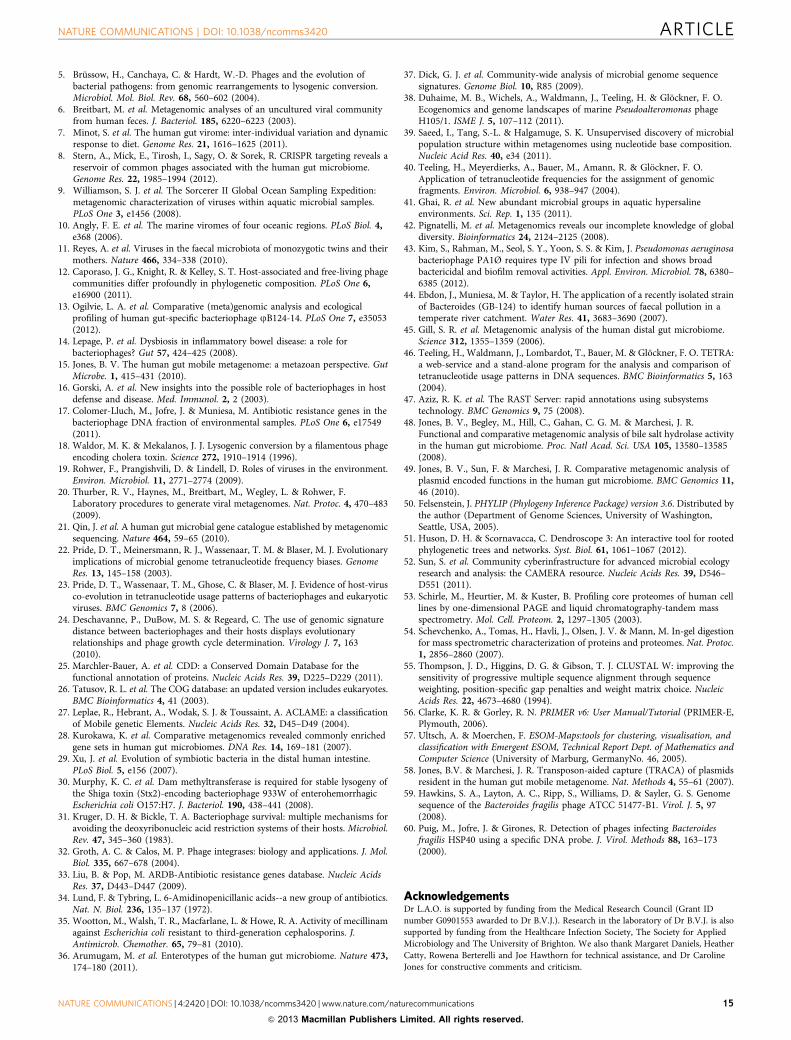
5. Brussow, H., Canchaya, C. & Hardt, W.-D. Phages and the evolution ofbacterial pathogens: from genomic rearrangements to lysogenic conversion.Microbiol. Mol. Biol. Rev. 68, 560–602 (2004).
6. Breitbart, M. et al. Metagenomic analyses of an uncultured viral communityfrom human feces. J. Bacteriol. 185, 6220–6223 (2003).
7. Minot, S. et al. The human gut virome: inter-individual variation and dynamicresponse to diet. Genome Res. 21, 1616–1625 (2011).
8. Stern, A., Mick, E., Tirosh, I., Sagy, O. & Sorek, R. CRISPR targeting reveals areservoir of common phages associated with the human gut microbiome.Genome Res. 22, 1985–1994 (2012).
9. Williamson, S. J. et al. The Sorcerer II Global Ocean Sampling Expedition:metagenomic characterization of viruses within aquatic microbial samples.PLoS One 3, e1456 (2008).
10. Angly, F. E. et al. The marine viromes of four oceanic regions. PLoS Biol. 4,e368 (2006).
11. Reyes, A. et al. Viruses in the faecal microbiota of monozygotic twins and theirmothers. Nature 466, 334–338 (2010).
12. Caporaso, J. G., Knight, R. & Kelley, S. T. Host-associated and free-living phagecommunities differ profoundly in phylogenetic composition. PLoS One 6,e16900 (2011).
13. Ogilvie, L. A. et al. Comparative (meta)genomic analysis and ecologicalprofiling of human gut-specific bacteriophage jB124-14. PLoS One 7, e35053(2012).
14. Lepage, P. et al. Dysbiosis in inflammatory bowel disease: a role forbacteriophages? Gut 57, 424–425 (2008).
15. Jones, B. V. The human gut mobile metagenome: a metazoan perspective. GutMicrobe. 1, 415–431 (2010).
16. Gorski, A. et al. New insights into the possible role of bacteriophages in hostdefense and disease. Med. Immunol. 2, 2 (2003).
17. Colomer-Lluch, M., Jofre, J. & Muniesa, M. Antibiotic resistance genes in thebacteriophage DNA fraction of environmental samples. PLoS One 6, e17549(2011).
18. Waldor, M. K. & Mekalanos, J. J. Lysogenic conversion by a filamentous phageencoding cholera toxin. Science 272, 1910–1914 (1996).
19. Rohwer, F., Prangishvili, D. & Lindell, D. Roles of viruses in the environment.Environ. Microbiol. 11, 2771–2774 (2009).
20. Thurber, R. V., Haynes, M., Breitbart, M., Wegley, L. & Rohwer, F.Laboratory procedures to generate viral metagenomes. Nat. Protoc. 4, 470–483(2009).
21. Qin, J. et al. A human gut microbial gene catalogue established by metagenomicsequencing. Nature 464, 59–65 (2010).
22. Pride, D. T., Meinersmann, R. J., Wassenaar, T. M. & Blaser, M. J. Evolutionaryimplications of microbial genome tetranucleotide frequency biases. GenomeRes. 13, 145–158 (2003).
23. Pride, D. T., Wassenaar, T. M., Ghose, C. & Blaser, M. J. Evidence of host-virusco-evolution in tetranucleotide usage patterns of bacteriophages and eukaryoticviruses. BMC Genomics 7, 8 (2006).
24. Deschavanne, P., DuBow, M. S. & Regeard, C. The use of genomic signaturedistance between bacteriophages and their hosts displays evolutionaryrelationships and phage growth cycle determination. Virology J. 7, 163(2010).
25. Marchler-Bauer, A. et al. CDD: a Conserved Domain Database for thefunctional annotation of proteins. Nucleic Acids Res. 39, D225–D229 (2011).
26. Tatusov, R. L. et al. The COG database: an updated version includes eukaryotes.BMC Bioinformatics 4, 41 (2003).
27. Leplae, R., Hebrant, A., Wodak, S. J. & Toussaint, A. ACLAME: a classificationof Mobile genetic Elements. Nucleic Acids Res. 32, D45–D49 (2004).
28. Kurokawa, K. et al. Comparative metagenomics revealed commonly enrichedgene sets in human gut microbiomes. DNA Res. 14, 169–181 (2007).
29. Xu, J. et al. Evolution of symbiotic bacteria in the distal human intestine.PLoS Biol. 5, e156 (2007).
30. Murphy, K. C. et al. Dam methyltransferase is required for stable lysogeny ofthe Shiga toxin (Stx2)-encoding bacteriophage 933W of enterohemorrhagicEscherichia coli O157:H7. J. Bacteriol. 190, 438–441 (2008).
31. Kruger, D. H. & Bickle, T. A. Bacteriophage survival: multiple mechanisms foravoiding the deoxyribonucleic acid restriction systems of their hosts. Microbiol.Rev. 47, 345–360 (1983).
32. Groth, A. C. & Calos, M. P. Phage integrases: biology and applications. J. Mol.Biol. 335, 667–678 (2004).
33. Liu, B. & Pop, M. ARDB-Antibiotic resistance genes database. Nucleic AcidsRes. 37, D443–D447 (2009).
34. Lund, F. & Tybring, L. 6-Amidinopenicillanic acids--a new group of antibiotics.Nat. N. Biol. 236, 135–137 (1972).
35. Wootton, M., Walsh, T. R., Macfarlane, L. & Howe, R. A. Activity of mecillinamagainst Escherichia coli resistant to third-generation cephalosporins. J.Antimicrob. Chemother. 65, 79–81 (2010).
36. Arumugam, M. et al. Enterotypes of the human gut microbiome. Nature 473,174–180 (2011).
37. Dick, G. J. et al. Community-wide analysis of microbial genome sequencesignatures. Genome Biol. 10, R85 (2009).
38. Duhaime, M. B., Wichels, A., Waldmann, J., Teeling, H. & Glockner, F. O.Ecogenomics and genome landscapes of marine Pseudoalteromonas phageH105/1. ISME J. 5, 107–112 (2011).
39. Saeed, I., Tang, S.-L. & Halgamuge, S. K. Unsupervised discovery of microbialpopulation structure within metagenomes using nucleotide base composition.Nucleic Acid Res. 40, e34 (2011).
40. Teeling, H., Meyerdierks, A., Bauer, M., Amann, R. & Glockner, F. O.Application of tetranucleotide frequencies for the assignment of genomicfragments. Environ. Microbiol. 6, 938–947 (2004).
41. Ghai, R. et al. New abundant microbial groups in aquatic hypersalineenvironments. Sci. Rep. 1, 135 (2011).
42. Pignatelli, M. et al. Metagenomics reveals our incomplete knowledge of globaldiversity. Bioinformatics 24, 2124–2125 (2008).
43. Kim, S., Rahman, M., Seol, S. Y., Yoon, S. S. & Kim, J. Pseudomonas aeruginosabacteriophage PA1Ø requires type IV pili for infection and shows broadbactericidal and biofilm removal activities. Appl. Environ. Microbiol. 78, 6380–6385 (2012).
44. Ebdon, J., Muniesa, M. & Taylor, H. The application of a recently isolated strainof Bacteroides (GB-124) to identify human sources of faecal pollution in atemperate river catchment. Water Res. 41, 3683–3690 (2007).
45. Gill, S. R. et al. Metagenomic analysis of the human distal gut microbiome.Science 312, 1355–1359 (2006).
46. Teeling, H., Waldmann, J., Lombardot, T., Bauer, M. & Glockner, F. O. TETRA:a web-service and a stand-alone program for the analysis and comparison oftetranucleotide usage patterns in DNA sequences. BMC Bioinformatics 5, 163(2004).
47. Aziz, R. K. et al. The RAST Server: rapid annotations using subsystemstechnology. BMC Genomics 9, 75 (2008).
48. Jones, B. V., Begley, M., Hill, C., Gahan, C. G. M. & Marchesi, J. R.Functional and comparative metagenomic analysis of bile salt hydrolase activityin the human gut microbiome. Proc. Natl Acad. Sci. USA 105, 13580–13585(2008).
49. Jones, B. V., Sun, F. & Marchesi, J. R. Comparative metagenomic analysis ofplasmid encoded functions in the human gut microbiome. BMC Genomics 11,46 (2010).
50. Felsenstein, J. PHYLIP (Phylogeny Inference Package) version 3.6. Distributed bythe author (Department of Genome Sciences, University of Washington,Seattle, USA, 2005).
51. Huson, D. H. & Scornavacca, C. Dendroscope 3: An interactive tool for rootedphylogenetic trees and networks. Syst. Biol. 61, 1061–1067 (2012).
52. Sun, S. et al. Community cyberinfrastructure for advanced microbial ecologyresearch and analysis: the CAMERA resource. Nucleic Acids Res. 39, D546–D551 (2011).
53. Schirle, M., Heurtier, M. & Kuster, B. Profiling core proteomes of human celllines by one-dimensional PAGE and liquid chromatography-tandem massspectrometry. Mol. Cell. Proteom. 2, 1297–1305 (2003).
54. Schevchenko, A., Tomas, H., Havli, J., Olsen, J. V. & Mann, M. In-gel digestionfor mass spectrometric characterization of proteins and proteomes. Nat. Protoc.1, 2856–2860 (2007).
55. Thompson, J. D., Higgins, D. G. & Gibson, T. J. CLUSTAL W: improving thesensitivity of progressive multiple sequence alignment through sequenceweighting, position-specific gap penalties and weight matrix choice. NucleicAcids Res. 22, 4673–4680 (1994).
56. Clarke, K. R. & Gorley, R. N. PRIMER v6: User Manual/Tutorial (PRIMER-E,Plymouth, 2006).
57. Ultsch, A. & Moerchen, F. ESOM-Maps:tools for clustering, visualisation, andclassification with Emergent ESOM, Technical Report Dept. of Mathematics andComputer Science (University of Marburg, GermanyNo. 46, 2005).
58. Jones, B.V. & Marchesi, J. R. Transposon-aided capture (TRACA) of plasmidsresident in the human gut mobile metagenome. Nat. Methods 4, 55–61 (2007).
59. Hawkins, S. A., Layton, A. C., Ripp, S., Williams, D. & Sayler, G. S. Genomesequence of the Bacteroides fragilis phage ATCC 51477-B1. Virol. J. 5, 97(2008).
60. Puig, M., Jofre, J. & Girones, R. Detection of phages infecting Bacteroidesfragilis HSP40 using a specific DNA probe. J. Virol. Methods 88, 163–173(2000).
AcknowledgementsDr L.A.O. is supported by funding from the Medical Research Council (Grant IDnumber G0901553 awarded to Dr B.V.J.). Research in the laboratory of Dr B.V.J. is alsosupported by funding from the Healthcare Infection Society, The Society for AppliedMicrobiology and The University of Brighton. We also thank Margaret Daniels, HeatherCatty, Rowena Berterelli and Joe Hawthorn for technical assistance, and Dr CarolineJones for constructive comments and criticism.
NATURE COMMUNICATIONS | DOI: 10.1038/ncomms3420 ARTICLE
NATURE COMMUNICATIONS | 4:2420 | DOI: 10.1038/ncomms3420 |www.nature.com/naturecommunications 15
& 2013 Macmillan Publishers Limited. All rights reserved.
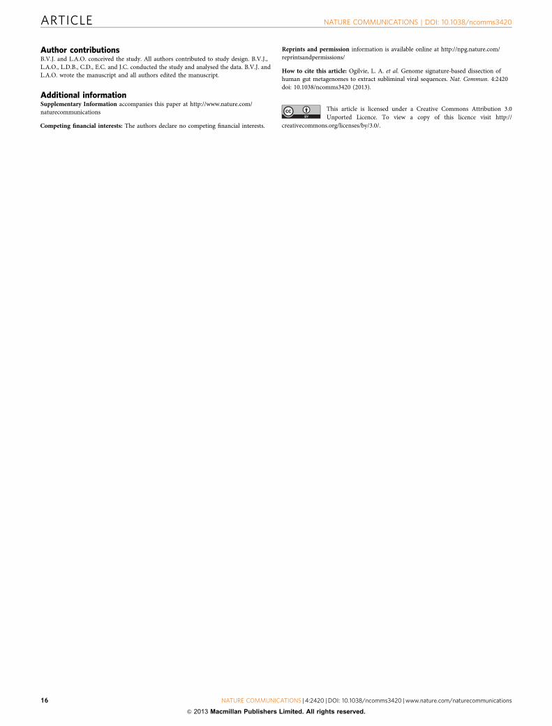
Author contributionsB.V.J. and L.A.O. conceived the study. All authors contributed to study design. B.V.J.,L.A.O., L.D.B., C.D., E.C. and J.C. conducted the study and analysed the data. B.V.J. andL.A.O. wrote the manuscript and all authors edited the manuscript.
Additional informationSupplementary Information accompanies this paper at http://www.nature.com/naturecommunications
Competing financial interests: The authors declare no competing financial interests.
Reprints and permission information is available online at http://npg.nature.com/reprintsandpermissions/
How to cite this article: Ogilvie, L. A. et al. Genome signature-based dissection ofhuman gut metagenomes to extract subliminal viral sequences. Nat. Commun. 4:2420doi: 10.1038/ncomms3420 (2013).
This article is licensed under a Creative Commons Attribution 3.0Unported Licence. To view a copy of this licence visit http://
creativecommons.org/licenses/by/3.0/.
ARTICLE NATURE COMMUNICATIONS | DOI: 10.1038/ncomms3420
16 NATURE COMMUNICATIONS | 4:2420 | DOI: 10.1038/ncomms3420 |www.nature.com/naturecommunications
& 2013 Macmillan Publishers Limited. All rights reserved.
Related Documents