ARTICLE OPEN Exploring human-genome gut-microbiome interaction in Parkinson’s disease Zachary D. Wallen 1 , William J. Stone 1 , Stewart A. Factor 2 , Eric Molho 3 , Cyrus P. Zabetian 4 , David G. Standaert 1 and Haydeh Payami 1 ✉ The causes of complex diseases remain an enigma despite decades of epidemiologic research on environmental risks and genome- wide studies that have uncovered tens or hundreds of susceptibility loci for each disease. We hypothesize that the microbiome is the missing link. Genetic studies have shown that overexpression of alpha-synuclein, a key pathological protein in Parkinson’s disease (PD), can cause familial PD and variants at alpha-synuclein locus confer risk of idiopathic PD. Recently, dysbiosis of gut microbiome in PD was identified: altered abundances of three microbial clusters were found, one of which was composed of opportunistic pathogens. Using two large datasets, we found evidence that the overabundance of opportunistic pathogens in PD gut is influenced by the host genotype at the alpha-synuclein locus, and that the variants responsible modulate alpha-synuclein expression. Results put forth testable hypotheses on the role of gut microbiome in the pathogenesis of PD, the incomplete penetrance of PD susceptibility genes, and potential triggers of pathology in the gut. npj Parkinson’s Disease (2021)7:74 ; https://doi.org/10.1038/s41531-021-00218-2 INTRODUCTION Parkinson’s disease (PD) affects over 6 million people worldwide, having doubled in one decade, and continues to rapidly increase in prevalence with the aging of the world population 1 . PD is a progressive degenerative disease which affects the brain, the peripheral nervous system, and the gastrointestinal tract, causing progressive, debilitating movement disorders, gastrointestinal and autonomic dysfunction, sleep disorders, and cognitive impair- ment. Currently, there is no prevention, cure, or treatment known to slow the progression of the disease. Like other common late-onset disorders, PD has Mendelian forms caused by rare mutations, but the vast majority of cases remain idiopathic. Both genetic and environmental risk factors have been identified 2–4 , but none have large enough effect sizes individually or in combination to fully encapsulate disease risk 5–8 . The triggers that initiate the onset of PD pathology are unknown. There is a connection between PD and the gastrointestinal tract 9,10 and the gut microbiome 11 . The gut microbiome is a relatively new and increasingly active area of research in human disease 12–14 . Studies on PD have consistently found altered gut microbiome, with depletion of short-chain fatty acid (SCFA) producing bacteria, and enrichment of Lactobacillus and Bifidobacterium 11,15,16 . Most studies to date have been modest in size, and therefore have examined mostly common microorganisms. We recently reported a microbiome-wide association study in PD, using two large datasets and internal replication, which enabled investigation of less common taxa not reported before 11 . In these datasets, reduced SCFA- producing bacteria and elevated Lactobacillus and Bifidobacterium were robustly confirmed. In addition, a significant increase was detected in the relative abundance of a poly-microbial cluster of opportunistic pathogens, including Corynebacterium_1 (C. amycola- tum, C. lactis), Porphyromonas (P. asaccharolytica, P. bennonis, P. somerae, P. uenonis), and Prevotella (P. bivia, P. buccalis, P. disiens, P. timonensis). These are commensal bacteria with normally low abundance in the gut; in fact, Corynebacterium is commensal to skin microbiome not the gut. They are referred to as opportunistic pathogens in the literature (as opposed to pathobionts) because they are not prevalent native members of the gut, rather they are known to be able to cause infections in any part of the body if they gain access to a sterile site through wounds, surgery or permeable membranes and are allowed to grow due to a compromised immune system (literature reviewed in ref. 11 ). Overabundance of opportunistic pathogens in PD gut was of interest because it harks back to the hypothesis advanced by Professor Heiko Braak which proposes that in non-familial forms of PD, the disease is triggered by an unknown pathogen in the gut and spreads to the brain 17,18 . Braak’s hypothesis was based on pathological studies of postmortem human brain, stained using antibodies to alpha-synuclein. Misfolded alpha-synuclein, the pathologic hallmark of PD, has been seen to form in enteric neurons early in disease 19–21 , and has been shown to propagate in a prion-like manner from the gut to the brain in animal models 22 . The gene that encodes alpha-synuclein is SNCA. SNCA gene multiplication results in drastic overexpression of alpha-synuclein and causes Mendelian autosomal dominant PD. Variants in the SNCA region are associated with risk of idiopathic PD 23 , and are expression quantitative trait loci (eQTL) associated with expression levels of SNCA 24–26 . Increased alpha-synuclein expression has been noted with infections unrelated to PD 27,28 . We hypothesized that if opportunistic pathogens are involved in disease pathogenesis, there might be an interaction between genetic variants in the SNCA region and dysbiosis of the gut in PD. RESULTS Overview of analyses The two case-control cohorts used here are those previously employed by Wallen et al. to characterize the PD gut microbiome 11 . 1 Department of Neurology, University of Alabama at Birmingham, Birmingham, AL, USA. 2 Department of Neurology, Emory University School of Medicine, Atlanta, GA, USA. 3 Department of Neurology, Albany Medical College, Albany, NY, USA. 4 VA Puget Sound Health Care System and Department of Neurology, University of Washington, Seattle, WA, USA. ✉ email: [email protected] www.nature.com/npjparkd Published in partnership with the Parkinson’s Foundation 1234567890():,;

Welcome message from author
This document is posted to help you gain knowledge. Please leave a comment to let me know what you think about it! Share it to your friends and learn new things together.
Transcript
ARTICLE OPEN
Exploring human-genome gut-microbiome interaction inParkinson’s diseaseZachary D. Wallen 1, William J. Stone 1, Stewart A. Factor2, Eric Molho3, Cyrus P. Zabetian 4, David G. Standaert 1 andHaydeh Payami 1✉
The causes of complex diseases remain an enigma despite decades of epidemiologic research on environmental risks and genome-wide studies that have uncovered tens or hundreds of susceptibility loci for each disease. We hypothesize that the microbiome isthe missing link. Genetic studies have shown that overexpression of alpha-synuclein, a key pathological protein in Parkinson’sdisease (PD), can cause familial PD and variants at alpha-synuclein locus confer risk of idiopathic PD. Recently, dysbiosis of gutmicrobiome in PD was identified: altered abundances of three microbial clusters were found, one of which was composed ofopportunistic pathogens. Using two large datasets, we found evidence that the overabundance of opportunistic pathogens in PDgut is influenced by the host genotype at the alpha-synuclein locus, and that the variants responsible modulate alpha-synucleinexpression. Results put forth testable hypotheses on the role of gut microbiome in the pathogenesis of PD, the incompletepenetrance of PD susceptibility genes, and potential triggers of pathology in the gut.
npj Parkinson’s Disease (2021) 7:74 ; https://doi.org/10.1038/s41531-021-00218-2
INTRODUCTIONParkinson’s disease (PD) affects over 6 million people worldwide,having doubled in one decade, and continues to rapidly increasein prevalence with the aging of the world population1. PD is aprogressive degenerative disease which affects the brain, theperipheral nervous system, and the gastrointestinal tract, causingprogressive, debilitating movement disorders, gastrointestinal andautonomic dysfunction, sleep disorders, and cognitive impair-ment. Currently, there is no prevention, cure, or treatment knownto slow the progression of the disease.Like other common late-onset disorders, PD has Mendelian
forms caused by rare mutations, but the vast majority of casesremain idiopathic. Both genetic and environmental risk factorshave been identified2–4, but none have large enough effect sizesindividually or in combination to fully encapsulate disease risk5–8.The triggers that initiate the onset of PD pathology are unknown.There is a connection between PD and the gastrointestinal tract9,10
and the gut microbiome11. The gut microbiome is a relatively newand increasingly active area of research in human disease12–14.Studies on PD have consistently found altered gut microbiome, withdepletion of short-chain fatty acid (SCFA) producing bacteria, andenrichment of Lactobacillus and Bifidobacterium11,15,16. Most studiesto date have been modest in size, and therefore have examinedmostly common microorganisms. We recently reported amicrobiome-wide association study in PD, using two large datasetsand internal replication, which enabled investigation of less commontaxa not reported before11. In these datasets, reduced SCFA-producing bacteria and elevated Lactobacillus and Bifidobacteriumwere robustly confirmed. In addition, a significant increase wasdetected in the relative abundance of a poly-microbial cluster ofopportunistic pathogens, including Corynebacterium_1 (C. amycola-tum, C. lactis), Porphyromonas (P. asaccharolytica, P. bennonis,P. somerae, P. uenonis), and Prevotella (P. bivia, P. buccalis, P. disiens,P. timonensis). These are commensal bacteria with normally low
abundance in the gut; in fact, Corynebacterium is commensal to skinmicrobiome not the gut. They are referred to as opportunisticpathogens in the literature (as opposed to pathobionts) because theyare not prevalent native members of the gut, rather they are knownto be able to cause infections in any part of the body if they gainaccess to a sterile site through wounds, surgery or permeablemembranes and are allowed to grow due to a compromisedimmune system (literature reviewed in ref. 11).Overabundance of opportunistic pathogens in PD gut was of
interest because it harks back to the hypothesis advanced byProfessor Heiko Braak which proposes that in non-familial forms ofPD, the disease is triggered by an unknown pathogen in the gutand spreads to the brain17,18. Braak’s hypothesis was based onpathological studies of postmortem human brain, stained usingantibodies to alpha-synuclein. Misfolded alpha-synuclein, thepathologic hallmark of PD, has been seen to form in entericneurons early in disease19–21, and has been shown to propagate ina prion-like manner from the gut to the brain in animal models22.The gene that encodes alpha-synuclein is SNCA. SNCA genemultiplication results in drastic overexpression of alpha-synucleinand causes Mendelian autosomal dominant PD. Variants in theSNCA region are associated with risk of idiopathic PD23, and areexpression quantitative trait loci (eQTL) associated with expressionlevels of SNCA24–26. Increased alpha-synuclein expression has beennoted with infections unrelated to PD27,28. We hypothesized that ifopportunistic pathogens are involved in disease pathogenesis,there might be an interaction between genetic variants in theSNCA region and dysbiosis of the gut in PD.
RESULTSOverview of analysesThe two case-control cohorts used here are those previouslyemployed by Wallen et al. to characterize the PD gut microbiome11.
1Department of Neurology, University of Alabama at Birmingham, Birmingham, AL, USA. 2Department of Neurology, Emory University School of Medicine, Atlanta, GA, USA.3Department of Neurology, Albany Medical College, Albany, NY, USA. 4VA Puget Sound Health Care System and Department of Neurology, University of Washington, Seattle, WA,USA. ✉email: [email protected]
www.nature.com/npjparkd
Published in partnership with the Parkinson’s Foundation
1234567890():,;
Here, we generated and added genotype data to investigateinteractions. The sample size for the present analysis was 199 PDand 117 controls in dataset 1, and 312 PD and 174 controls indataset 2. All samples had complete genotypes, 16S microbiomedata, and metadata (Supplementary Table 1).We defined the boundaries of the SNCA region such that it
would encompass known cis-eQTLs for SNCA. Using GTEx eQTLdatabase, we defined the boundaries as ch4:88.9 Mb, down-stream of 3′ SNCA, and ch4:90.6 Mb, upstream of 5′ SNCA. In ourgenome-wide genotype data (see Methods), we had 2,627 singlenucleotide polymorphisms (SNPs) that mapped to this region,had minor allele frequency (MAF) >0.1, imputation quality scorer2 >0.8, and were in common between the two datasets beingstudied here.The taxa examined were grouped and analyzed at genus/
subgenus/clade level as Corynebacterium_1 (C. amycolatum, C.Lactis), Porphyromonas (P. asaccharolytica, P. bennonis, P. somerae,P. uenonis), and Prevotella (P. bivia, P. buccalis, P. disiens, P.timonensis). For simplicity, we will refer to the three microbialgroups as taxa. As we have previously shown, the abundances ofthese taxa are elevated in PD vs. control. These findings werereplicated in the two datasets (Table 1), verified by two statisticalmethods, robust to covariate adjustment (over 40 variablesinvestigated), and yielded no evidence of being the result of PDmedications or disease duration11.The analysis of interaction was structured as follows. First, we
screened for statistical interaction between 2,627 SNP genotypesin the SNCA region, case-control status, and centered log-ratio
(clr) transformed abundance of each taxon and selected the SNPwith the highest statistical significance as the candidateinteracting SNP (Fig. 1a–c). We then tested the association ofeach taxon with case-control status after stratifying the subjectsby the interacting SNP genotype. The effect of SNP on the PD-taxa association was tested statistically (Table 1) and illustratedgraphically (Fig. 2, Supplementary Fig. 1). We tested theassociation of interacting SNPs with PD in the present datasetand in prior GWAS (Table 2). This test was conducted becauseinteraction can exist with or without a main effect of SNP ondisease risk. We then conducted in silico functional analysis ofthe interacting SNPs (Table 2, Fig. 1d, e). All analyses wereperformed in two datasets, followed by meta-analysis.
Corynebacterium_1. The candidate interacting SNP for Coryne-bacterium_1 was rs356229 (interaction P= 2E−3; Fig. 1a). This SNPis located 3′ of SNCA (Fig. 1a, d). The two alleles are rs356229_T(allele frequency= 0.6) and rs356229_C (frequency= 0.4), andwas imputed with imputation quality score of 0.96 in dataset 1and 0.99 in dataset 2.If we do not consider genotype, Corynebacterium_1 abundance
is significantly elevated in PD (OR= 1.6, P= 3E−3). However,when data are stratified by genotype, there is no associationbetween Corynebacterium_1 and PD among individuals withrs356229_TT genotype, who comprised 36% of the study (OR=1.0, P= 0.92). The association of Corynebactreium_1 with PD wasdependent on the presence of the rs356229_C allele. Theabundance of Corynebactreium_1 was nearly 2-fold higher in PDthan controls in heterozygous rs356229_CT (OR= 1.9, P= 1E−3),
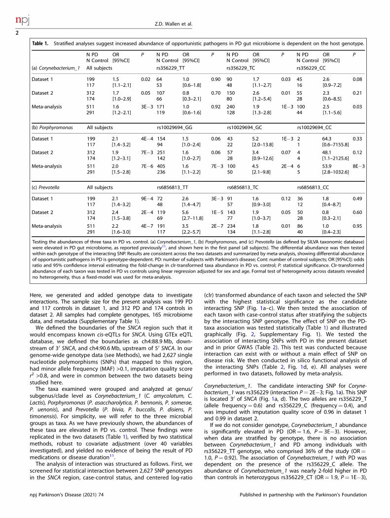
Table 1. Stratified analyses suggest increased abundance of opportunistic pathogens in PD gut microbiome is dependent on the host genotype.
N PDN Control
OR[95%CI]
P N PDN Control
OR[95%CI]
P N PDN Control
OR[95%CI]
P N PDN Control
OR[95%CI]
P
(a) Corynebacterium_1 All subjects rs356229_TT rs356229_TC rs356229_CC
Dataset 1 199117
1.5[1.1–2.1]
0.02 6453
1.0[0.6–1.8]
0.90 9048
1.7[1.1–2.7]
0.03 4516
2.6[0.9–7.2]
0.08
Dataset 2 312174
1.7[1.0–2.9]
0.05 10766
0.8[0.3–2.1]
0.70 15080
2.6[1.2–5.4]
0.01 5528
2.3[0.6–8.5]
0.21
Meta-analysis 511291
1.6[1.2–2.1]
3E−3 171119
1.0[0.6–1.6]
0.92 240128
1.9[1.3–2.8]
1E−3 10044
2.5[1.1–5.6]
0.03
(b) Porphyromonas All subjects rs10029694_GG rs10029694_GC rs10029694_CC
Dataset 1 199117
2.1[1.4–3.2]
4E−4 15494
1.5[1.0–2.4]
0.06 4322
5.2[2.0–13.8]
1E−3 21
64.3[0.6–7155.8]
0.33
Dataset 2 312174
1.9[1.2–3.1]
7E−3 251142
1.6[1.0–2.7]
0.06 5728
3.4[0.9–12.6]
0.07 44
48.1[1.1–2125.6]
0.12
Meta-analysis 511291
2.0[1.5–2.8]
7E−6 405236
1.6[1.1–2.2]
7E−3 10050
4.5[2.1–9.8]
2E−4 65
53.9[2.8–1032.6]
8E−3
(c) Prevotella All subjects rs6856813_TT rs6856813_TC rs6856813_CC
Dataset 1 199117
2.1[1.4–3.2]
9E−4 7248
2.6[1.4–4.7]
3E−3 9157
1.6[0.9–3.0]
0.12 3612
1.8[0.4–8.7]
0.49
Dataset 2 312174
2.4[1.5–3.8]
2E−4 11969
5.6[2.7–11.8]
1E−5 14377
1.9[1.0–3.7]
0.05 5028
0.8[0.3–2.1]
0.60
Meta-analysis 511291
2.2[1.6–3.0]
4E−7 191117
3.5[2.2–5.7]
2E−7 234134
1.8[1.1–2.8]
0.01 8640
1.0[0.4–2.3]
0.95
Testing the abundances of three taxa in PD vs. control. (a) Corynebacterium_1, (b) Porphyromonas, and (c) Prevotella (as defined by SILVA taxonomic database)were elevated in PD gut microbiome, as reported previously11, and shown here in the first panel (all subjects). The differential abundance was then testedwithin each genotype of the interacting SNP. Results are consistent across the two datasets and summarized by meta-analysis, showing differential abundanceof opportunistic pathogens in PD is genotype-dependent. PD: number of subjects with Parkinson’s disease; Cont: number of control subjects; OR [95%CI]: oddsratio and 95% confidence interval estimating the fold-change in clr-transformed taxa abundance in PD vs. control; P: statistical significance. Clr-transformedabundance of each taxon was tested in PD vs controls using linear regression adjusted for sex and age. Formal test of heterogeneity across datasets revealedno heterogeneity, thus a fixed-model was used for meta-analysis.
Z.D. Wallen et al.
2
npj Parkinson’s Disease (2021) 74 Published in partnership with the Parkinson’s Foundation
1234567890():,;
and 2.5-fold higher in the homozygous rs356229_CC individuals(OR= 2.5, P= 0.03). These results can be found in Table 1a, anddata underlying these results are visualized in Fig. 2.The Corynebacterium_1 interacting SNP has been previously
identified in PD GWAS meta-analysis, with the rs356229_C allele
associated with increased PD risk (OR= 1.3, P= 3E−42 withN>100,000 samples23). We also detected an association betweenrs356229_C and PD in the present dataset (OR= 1.3, P= 0.04with N= 802 samples; Table 2). That we estimated an effectsize identical to GWAS, despite the enormous disparity in the
Z.D. Wallen et al.
3
Published in partnership with the Parkinson’s Foundation npj Parkinson’s Disease (2021) 74
sample size and power, speaks to the robustness of the data. Theevidence for interaction does not stem from the association ofSNP with PD (see “Discussion”). Interestingly, the association ofrs356229_C with risk of PD varied by the increasing abundance ofCorynebacterium_1 from no association in the 1st or 2nd quartile(OR= 0.9, P= 0.5; OR= 1.1, P= 0.8) to an emerging and thenstrong association in the 3rd and 4th quartiles (OR= 1.5, P= 0.3and OR= 2.2, P= 5E−3).The rs356229 SNP maps to a distal regulatory element at 3′ of
SNCA (Fig. 1d, e) and is an eQTL for SNCA (Table 2). Data wereobtained by eQTL GWAS conducted in whole blood (eQTLGen.org) and in esophagus mucosa (GTExportal.org). The rs356229_Callele is associated with increased expression of SNCA in blood(eQTL P= 1E−13) and in esophagus mucosa (eQTL P= 9E−5).According to GTEx, rs356229 is also an eQTL for SNCA-AS1 (eQTLP= 2E−7) and RP11-115D19.1 (eQTL P= 3E−14). SNCA-AS1 andRP11-115D19.1 overlap with SNCA and encode long non-codingRNA (lncRNA) that are antisense to SNCA (Fig. 1d) and have beenimplicated in the regulation of SNCA expression29–31.
Porphyromonas. The candidate interacting SNP for Porphyromo-nas was rs10029694 (interaction P= 6E−3; Fig. 1b). The SNP mapsto 3′ of SNCA (Fig. 1b, d). The two alleles are rs10029694_G(frequency= 0.9) and rs10029694_C (frequency= 0.1), and wasimputed with imputation quality score 0.99 in dataset 1 and 0.92in dataset 2.The interacting SNPs for Porphyromonas (rs10029694) and
Corynebacterium_1 (rs356229) map very close to each other, only480 base pairs apart, but they are not in linkage disequilibrium(LD): D′<0.01, R2= 0.Porphyromonas was elevated in PD irrespective of
rs10029694_G/C genotype (OR= 2.0, P= 7E−6), and in everygenotype, but the statistical interaction implied differenceacross genotypes. Shown in stratified analysis (Table 1b, Fig. 2),the rs10029694_GG genotype had a nearly two-fold higherabundance of Porphyromonas in PD vs. controls (OR= 1.6, P=7E−3), rs10029694_GC had nearly five-fold difference (OR=4.5, P= 2E−4) and rs10029694_CC had approximately 54-timeshigher abundance of Porphyromonas in PD than in controls(OR= 53.9, P= 8E−3). Note however that there were only 11individuals with rs10029694_CC genotype. Although thestatistical methods were carefully chosen to be robust to smallsample size, and the P value is quite significant despite thesample size, the fact remains that the OR= 54 was generatedon only 11 people. If we collapse the rare rs10029694_CCgenotype with rs10029694_CG, we have 161 individuals (20%of subjects) with at least one copy of rs10029694_C allele, andwe get a more conservative estimate of OR= 5.1 (P= 2E−5) forassociation of Porphyromonas with PD in people with one ortwo copies of rs10029694_C.The Porphyromonas interacting SNP is also associated with
PD risk. The association was detected in the latest GWAS whichhad 37,688 PD cases and 1.4 million controls (OR= 1.1, P= 2E−14)3. We detected the same effect size (OR= 1.1) but it didnot reach significance (P= 0.6) (Table 2). As would be expected
from the interaction, the frequency of the effect allelers10029694_C in PD vs. control rose with increasing abundanceof Porphyromonas, yielding OR= 0.6 (P= 0.3) for 1st quartileand increasing up to OR= 2.2 (P= 0.08) for the 4th quartile.The rs10029694 SNP maps to a distal regulatory element at 3′
of SNCA, adjacent to another regulatory element wherers356229, the interacting SNP for Corynebacterium_1 resides(Fig. 1d, e). The rs10029694 SNP is an eQTL for two lncRNA thatare antisense to SNCA: RP11-115D19.1 (eQTL P= 1E−5) andRP11-115D19.2 (eQTL P= 7E−6) (Table 2). RP11-115D19.1 over-laps with 3′ of SNCA; RP11-115D19.2 is within SNCA. We did notfind direct evidence for rs10029694 being an eQTL for SNCA.However, RP11-115D19.1 and RP11-115D19.2 are antisense toSNCA which based on current knowledge on function ofantisense lncRNA would be presumed to be regulatory forSNCA30,31, and RP11-115D19.1 has been directly shown toregulate SNCA expression29.
Prevotella. The candidate interacting SNP for Prevotella wasrs6856813 (interaction P= 0.01; Fig. 1c). The SNP is ~100 kbupstream at 5′ of SNCA (Fig. 1c, d). The two alleles are rs6856813_T(frequency= 0.6) and rs6856813_C (frequency= 0.4), and wasimputed with imputation quality score 0.98 in dataset 1 and 0.84in dataset 2. The Prevotella interacting SNP is 300Kb away fromand not in LD with the interacting SNPs of Corynebacterium_1(rs356229, D′= 0.2, R2= 0.04) or Porphyromonas (rs10029694,D′= 0.36, R2= 0.01).Prevotella was elevated two-fold in PD vs. controls (OR= 2.2,
P= 4E−7). Genotype-specific results suggest rs6856813_TT hadthe greatest differential abundance in PD vs. control (OR= 3.5,P= 2E−7), followed by rs6856813_TC (OR= 1.8, P= 0.01), andno difference in rs6856813_CC genotype (OR= 1.0, P= 0.95)(Table 1c, Fig. 2).There is no documented evidence for a direct association
between rs6856813_C/T and PD in this study (OR= 0.9, P= 0.4) orin PD GWAS to date (Table 2). There is a statistically non-significanttrend of increasing frequency of rs6856813_T allele with increas-ing abundance of Prevotella in PD, yielding OR= 0.8 in 1st quartileand increasing to OR= 1.5 in 4th quartile, consistent with thepresence of interaction.Although rs6856813 is ~100 kb upstream of SNCA and does not
map to a known regulatory sequence (Fig. 1d, e), it is a strongeQTL for SNCA: the rs6856813_T allele, which is the effect allele forinteraction with Prevotella, is associated with increased SNCAexpression in blood (eQTL P= 3E−49) and in arteries (eQTL P= 2E−5) (Table 2).
DISCUSSIONNumerous studies have been performed on the association ofgenetic variants with PD and separately of gut microbiome andPD, but none to our knowledge has explored the interactionbetween the two. Here we have used a candidate taxa, candidategene strategy: we used prior knowledge of the association of PDwith elevated abundances of certain opportunistic pathogens in
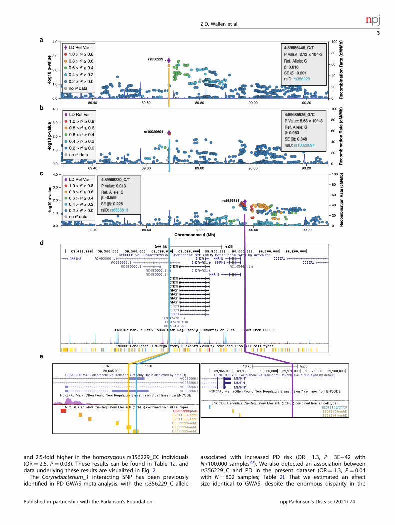
Fig. 1 Genetic map of candidate interacting SNPs. SNPs in SNCA region (chromosome 4: 88.9–90.6 Mb) were tested for interaction on theassociation of three taxa with PD. Results are shown in LocusZoom, where each SNP is plotted according to its base pair position and meta-analysis −log10(P value) for interaction for the three taxa: (a) Corynebacterium_1, (b) Porphyromonas, and (c) Prevotella. The SNP with thehighest significance is shown as a purple diamond, and was chosen as candidate interacting SNP for stratified analysis (Table 1). (d) UCSCGenome Browser shows the interacting SNPs for Corynebacterium_1 and Porphyromonas map to 3′ SNCA in a lncRNA that overlaps with and areantisense to SNCA. The interacting SNP for Prevotella is distal at 5′ of SNCA and MMRN1. (e) The interacting SNPs for Corynebacterium_1 andPorphyromonas, while only 450 base pair apart, are not in LD (R2= 0) and map to adjacent regulatory sequences shown in yellow bars. Theinteracting SNP for Prevotella does not map to any known functional sequence. All three SNPs are eQTLs for SNCA and lncRNA genes SNCA-AS1,RP11-115D19.1 (AC093866.1), and RP11-115D19.2 (AC097478.2) which are associated with expression of SNCA (Table 2). LD: linkagedisequilibrium; Mb: Megabase; P value: P value from meta-analysis; β: beta coefficient of interaction from meta-analysis; SE: standard error;rsID: reference SNP ID for the marked SNP.
Z.D. Wallen et al.
4
npj Parkinson’s Disease (2021) 74 Published in partnership with the Parkinson’s Foundation
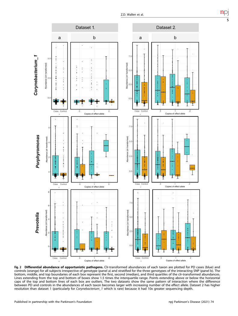
Fig. 2 Differential abundance of opportunistic pathogens. Clr-transformed abundances of each taxon are plotted for PD cases (blue) andcontrols (orange) for all subjects irrespective of genotype (panel a) and stratified for the three genotypes of the interacting SNP (panel b). Thebottom, middle, and top boundaries of each box represent the first, second (median), and third quartiles of the clr-transformed abundances.Lines extending from the top and bottom of boxes show 1.5 times the interquartile range. Points extending above or below the horizontalcaps of the top and bottom lines of each box are outliers. The two datasets show the same pattern of interaction where the differencebetween PD and controls in the abundances of each taxon becomes larger with increasing number of the effect allele. Dataset 2 has higherresolution than dataset 1 (particularly for Corynebacterium_1 which is rare) because it had 10x greater sequencing depth.
Z.D. Wallen et al.
5
Published in partnership with the Parkinson’s Foundation npj Parkinson’s Disease (2021) 74
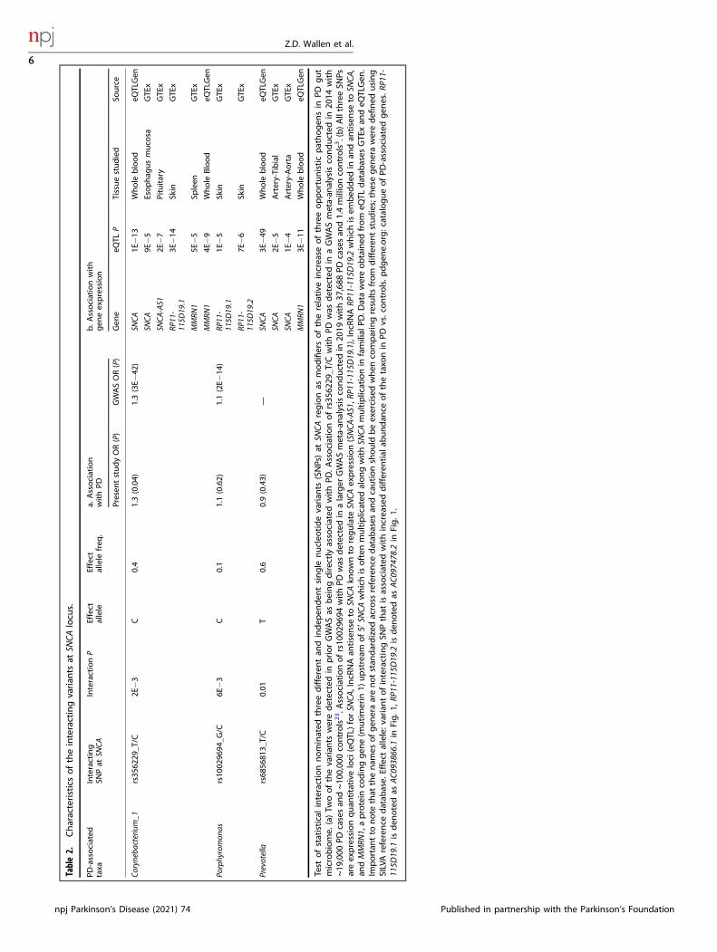
Table2.
Characteristicsoftheinteractingvarian
tsat
SNCA
locu
s.
PD-associated
taxa
Interacting
SNPat
SNCA
InteractionP
Effect
allele
Effect
allele
freq
.a.
Association
withPD
b.A
ssociationwith
gen
eexpression
Presen
tstudyOR(P)
GWASOR(P)
Gen
eeQ
TLP
Tissuestudied
Source
Coryneba
cterium_1
rs35
6229
_T/C
2E−3
C0.4
1.3(0.04)
1.3(3E−
42)
SNCA
1E−13
Whole
blood
eQTLGen
SNCA
9E−5
Esophag
usmuco
saGTE
x
SNCA
-AS1
2E−7
Pituitary
GTE
x
RP11-
115D
19.1
3E−14
Skin
GTE
x
MMRN
15E
−5
Spleen
GTE
x
MMRN
14E
−9
Whole
Blood
eQTLGen
Porphyromon
asrs10
0296
94_G
/C6E
−3
C0.1
1.1(0.62)
1.1(2E−
14)
RP11-
115D
19.1
1E−5
Skin
GTE
x
RP11-
115D
19.2
7E−6
Skin
GTE
x
Prevotella
rs68
5681
3_T/C
0.01
T0.6
0.9(0.43)
—SN
CA3E
−49
Whole
blood
eQTLGen
SNCA
2E−5
Artery-Tibial
GTE
x
SNCA
1E−4
Artery-Aorta
GTE
x
MMRN
13E
−11
Whole
blood
eQTLGen
Test
ofstatisticalinteractionnominated
threedifferen
tan
dindep
enden
tsingle
nucleo
tidevarian
ts(SNPs)at
SNCA
regionas
modifiersoftherelative
increase
ofthreeopportunisticpathogen
sin
PDgut
microbiome.
(a)Tw
oofthevarian
tsweredetectedin
priorGWASas
beingdirectlyassociated
withPD
.Associationofrs35
6229
_T/C
withPD
was
detectedin
aGWASmeta-an
alysisco
nducted
in20
14with
~19
,000
PDcasesan
d~10
0,00
0co
ntrols23.A
ssociationofrs10
0296
94withPD
was
detectedin
alarger
GWASmeta-an
alysisco
nducted
in20
19with37
,688
PDcasesan
d1.4millionco
ntrols3.(b)AllthreeSN
Psareexpressionquan
titative
loci(eQTL)forSN
CA,lncR
NAan
tisense
toSN
CAkn
ownto
regulate
SNCA
expression(SNCA
-AS1,R
P11-115D
19.1),lncR
NARP
11-115D19.2
whichisem
bed
ded
inan
dan
tisense
toSN
CA,
andMMRN
1,aprotein
codinggen
e(m
utimerin
1)upstream
of5′
SNCA
whichisoften
multiplicated
alongwithSN
CAmultiplicationin
familial
PD.D
atawereobtained
from
eQTL
datab
ases
GTE
xan
deQ
TLGen
.Im
portan
tto
note
that
thenam
esofgen
eraarenotstan
dardized
across
reference
datab
ases
andcautionshould
beexercisedwhen
comparingresultsfrom
differen
tstudies;thesegen
eraweredefi
ned
using
SILVAreference
datab
ase.
Effect
allele:v
ariantofinteractingSN
Pthat
isassociated
withincreaseddifferen
tial
abundan
ceofthetaxo
nin
PDvs.controls.p
dgen
e.org:catalogueofPD
-associated
gen
es.R
P11-
115D
19.1
isden
otedas
AC0
93866.1in
Fig.1
,RP11-115D
19.2
isden
otedas
AC0
97478.2in
Fig.1
.
Z.D. Wallen et al.
6
npj Parkinson’s Disease (2021) 74 Published in partnership with the Parkinson’s Foundation
the gut11 and searched for genetic modifiers of these associationsin the SNCA gene region23. Through statistical interaction tests weidentified specific variants in the SNCA region as candidateinteracting variants and through genotype-stratified analyseswe found evidence suggesting that the increases in the relativeabundance of opportunistic pathogens in PD gut are modulatedby host genotype.Statistical interaction tests provide a means to investigate if the
association of one factor with the trait is influenced by a secondfactor. Here, we tested if the association of three opportunisticpathogens with PD (organisms with higher relative abundance inPD cases than similarly aged controls) is dependent on geneticvariations in or around SNCA. Interaction studies require muchlarger sample sizes and power than association studies; theP values for interaction seldom achieve significance, and whenthey do, they are far less significant than the P values for asimilarly sized one-factor association study. To that end, a majorlimitation of this study was the sample size. The raw P values frominteraction tests were significant but did not pass multiple testingcorrection. While the test of interaction in itself is not a powerfulstatistical means to detect modifiers, it is an unbiased screen tonarrow a large region down to a few potential candidates that canbe further interrogated individually in stratified analysis. Wenominate rs356229_T/C and rs10029694_G/C as potential modi-fiers for the association of Coynebacterium_1 and Porphyromonaswith PD based on the following evidence: (a) stratified analysis, ontwo datasets, showed similar and statistically significant differ-ences in taxa abundance by genotype, (b) the SNPs are eQTLaffecting SNCA expression, and (c) both SNPs have been shown inGWAS to be independently associated with PD. The evidence forinteraction did not arise from and is independent of the directassociation of the SNPs with PD. This can be seen in Table 1, wherethe test is between taxa and PD; SNP is not in the test, it is onlyused to divide the samples by genotype, which showed varyingassociation between the taxon and PD as a function of genotypesin both datasets. In fact, the present dataset had marginalevidence for direct association of PD with rs356229_T/C (OR= 1.3,P= 0.04), and was not significant for rs10029694_G/C (OR= 1.1,P= 0.6). The evidence for direct associations with PD come fromthe 2014 GWAS meta-analyses which detected association ofrs356229_T/C with PD at OR= 1.3, P= 3E−42 with N > 100,000cases and controls23, and the 2019 GWAS met-analysis whichdetected association of rs10029694_G/C with PD at OR= 1.1 andP= 2E−14 with N > 1.4 million cases and controls3. Unfortunately,collecting large datasets with microbiome and genotype data ischallenging. Currently, the largest PD datasets that have bothgenotype and microbiome data are the two datasets used here,one has 199 PD and 117 controls and the other 312 PD and 174controls. A major challenge is to secure well-coordinated studieswith large sample sizes that can be pooled or meta-analyzed.Unlike genetic studies which can be combined thanks to thestability of DNA, combining microbiome studies is challenging dueto the effects of collection and storage parameters on outcomes.Standardization of methods can alleviate some of the cross-studyvariations. It is also more difficult to collect stool samples thanblood or even saliva. People are averse to donating stool samples;30% of our research participants who donated blood refused todonate stool. Microbiome researchers are cognizant of the need tojoin resources, create standardized protocols, and coordinate datacollection across laboratories. Within a few years, we will be ableto amass the sample sizes needed to address the interaction ofgenes, environment, and microbiome on a comprehensive scale.Here, limited by sample size, we chose to explore one PD-associated locus (SNCA) and three PD-associated opportunisticpathogens, hoping that the resulting data will help formulatetestable hypotheses.Our rationale for choosing SNCA and opportunistic pathogen as
our candidate gene and candidate taxa stemmed from the
collective literature. SNCA is a key player in PD. Alpha-synucleinaggregates are a pathologic hallmark of PD. Mutations in SNCAcause autosomal dominant PD and variants that affect SNCA geneexpression are the most significant genetic risk factors foridiopathic PD23,32. While the functions of alpha-synuclein are yetto be fully understood, it has been shown to play a key role inactivating the immune system, acting as antigen presented by PD-associated major histocompatibility molecules and recognized byT cells which infiltrate the brain33–35. SNCA expression has alsobeen shown to be critical for inducing immune response againstinfections unrelated to PD27,28. Alpha-synuclein aggregates, whichhave historically been considered as a marker of PD pathology inthe brain, can actually form in the enteric neurons19 and in animalmodels have been shown to propagate from the gut to the brain22
possibly via the vagus nerve36,37. The trigger that induces alpha-synuclein pathology in the gut is unknown. Braak hypothesizedthe trigger is a pathogen17,18. Our choice of opportunisticpathogens as the candidate taxa for interaction testing wasdriven by our recent finding of an overabundance of opportunisticpathogens in PD gut and Braak’s hypothesis. Moreover, a studyconducted in mice has corroborated that intestinal infectiontriggers dopaminergic cell loss and motor impairment in a Pink1knockout model of PD38. Whether the opportunistic pathogensfound in human PD microbiome are triggers of PD is beinginvestigated. In the meantime, we thought that if theseopportunistic pathogens are involved in PD pathogenesis, thereis likely a connection to SNCA genotype worth exploring.Interestingly, three different SNCA-linked genetic variants
emerged as potential modifiers for the association of the threeopportunistic pathogens with PD. They are independent of eachother with no LD among them. All three interacting variants areeQTLs for SNCA and lncRNAs that affect the expression of SNCA.lncRNA are emerging as important regulators of gene expres-sion39. Aberrant expression of lncRNA has been widely reported inPD, often in relation to the expression and aggregation ofSNCA40,41. More specific to the present findings, the lncRNA’s nearSNCA, including SNCA-AS1 identified here, were shown to beunder-expressed while SNCA mRNA was over-expressed insubstantia nigra of autopsied PD brains compared to controls30.Another lncRNA identified here, RP11-115D19.1, was shown torepress SNCA expression in SH-SY5Y human neuroblastoma celllines29. This suggests a link between SNCA expression and thepresence of opportunistic pathogens, and that regulation of thislink may involve different regulatory elements depending on thepathogen. lncRNA is expressed in a cell-specific manner42. It is notknown which cells in the gut are responsible for the expressionand corruption of alpha-synuclein into pathologic species. If theopportunistic pathogens induce SNCA expression or corruption,they may do so by signaling different cell types, hence theinvolvement of different regulatory elements. Prevotella andPorphyromonas are commensal to gastrointestinal and urinarytrack, Corynebacterium is common in skin microbiome. All threecan be found at low abundance in the gut. All three have beenimplicated in causing infections in nearly every type of tissue(reviewed by Wallen et al.11).These data provide new leads and hypotheses that with follow-
up in experimental models may yield a better understanding ofdisease pathogenesis. These data alone cannot resolve cause andeffect. We cannot tell if the SNCA genotype leads to alteredcolonization of the gut, which in turn leads to PD, or is it the otherway around, SNCA genotype causes PD, which leads to gutdysfunction and accumulation of pathogens. Or, maybe thepathogen induces alpha-synuclein expression which elicitsimmune response to infection as seen in other infectionsunrelated to PD, but in individuals with certain regulatorygenotypes at SNCA, the alpha-synuclein expression goes intooverdrive and PD is a downstream consequence. An alternativehypothesis for the interaction of SNCA eQTL and an opportunistic
Z.D. Wallen et al.
7
Published in partnership with the Parkinson’s Foundation npj Parkinson’s Disease (2021) 74
pathogen is that eQTL controls alpha-synuclein concentration inthe cell, bacteria triggers misfolding and aggregation of alpha-synuclein, and since misfolding and aggregation is directlydependent on the concentration of alpha-synuclein in the cell43,individuals with certain SNCA eQTL genotypes are at higher risk ofdeveloping PD pathology from gut-derived insults. One canfurther speculate that these bacteria might promote alpha-synuclein misfolding and aggregation by invading the host cells(all three can invade host cells) or via producing toxic orproinflammatory substances. Prevotella and Porphyromonas pro-duce lipopolysaccharides, gut-derived proinflammatory endotox-ins that when administered to mice, cause intestinal permeabilityand progressive increase in alpha-synuclein expression in the gut,and neuroinflammation and nigral neurodegeneration in thebrain44,45. Further studies in humans conducted over time and inexperimental models will be needed to tease out the underlyingbiology of these interactions.In conclusion, this study was exploratory and hypothesis
generating. Within this cautionary framework, this study suggeststhat genetic susceptibility to disease and the dysbiosis in the gutmicrobiome are not operating independently. Rather, it suggeststhat alterations in gut microbiome should be integrated in thegene–environment interaction paradigm, which has long beensuspected to be the cause of idiopathic disease but is yet toproduce a causative combination. The results also put forth thehypothesis that the PD-associated genetic variants may confersusceptibility via interaction with microbiome; opening a new areato search for the incomplete penetrance of PD susceptibility genes.In addition, while it is yet to be seen if the opportunistic pathogensare part of the cause or consequence of disease (experiments areunderway), the finding that their abundance correlated with PD-associated genotypes adds credence to the hypotheses that theirpresence signifies a role in disease pathogenesis, possibly asthe triggers that Braak originally proposed. With the identity of thecandidate microorganisms in hand, these hypotheses can betested in model systems. Thus, the significance of this work lies noton achieving conclusive discoveries, rather on generating novelhypotheses with tangible leads that can be put to testexperimentally.
METHODSSubjectsThe study was approved by the institutional review boards at allparticipating institutions, namely New York State Department of Health,University of Alabama at Birmingham, VA Puget Sound Health CareSystem, Emory University, and Albany Medical Center. All subjectsprovided written informed consent for their participation. This studyincluded two datasets each composed of persons with PD (case) andneurologically healthy individuals (control). Subject enrollment and datacollection for both datasets were conducted by the NeuroGeneticsResearch Consortium (NGRC) team using uniform protocols. The twodatasets used here were the same datasets used by Wallen et al forcharacterizing the microbiome11; except here we have generated andadded genetic data, and subjects without genotype were excluded(Supplementary Table 1). Methods of subject selection and data collectionhave been described in detail before11. Briefly, PD was diagnosed byNGRC-affiliated movement disorder specialists46. Controls were self-reported free of neurological disease. Metadata were collected on over40 variables including age, sex, race, geography, diet, medication, health,gastrointestinal issues, weight fluctuation, and body mass index. Weenrolled 212 persons with PD and 136 controls in 2014 (dataset 1)47, and323 PD and 184 controls during 2015–2017 (dataset 2)11. Subsequently, weexcluded 11 PD and 4 control samples for failing 16S sequencing, 2 PD forunreliable metadata, and 15 controls for lacking genotypes from dataset 1;and 11 PD and 10 controls were excluded from dataset 2 for lackinggenotype data. The sample size used in current analyses was 199 PD and117 controls in dataset 1, and 312 PD and 174 controls in dataset 2(Supplementary Table 1).
Microbiome dataMethods for collection, processing, and analysis of microbiome data havebeen reported in detail11, and raw sequences are publicly available at NCBISRA BioProject ID PRJNA601994. Each subject provided a single stoolsample at a single time point, and each sample was measured once. Briefly,for both datasets uniformly, DNA/RNA-free sterile cotton swabs were usedto collect stool, DNA was extracted using MoBio extraction kits, and 16SrRNA gene hypervariable region 4 was sequenced using the same primers,but in two laboratories, resulting in 10x greater sequencing depth indataset 2 than dataset 1. Sequences were demultiplexed using QIIME2(core distribution 2018.6)48 for dataset 1 and BCL2FASTQ (Illumina, SanDiego, CA) for dataset 2. Bioinformatics processing of sequences wasperformed separately for each dataset, but using an identical pipeline (seeWallen et al.11 for step-by-step protocol). Unique amplicon sequencevariants (ASVs) were identified using DADA2 v 1.849 and given taxonomicassignment using DADA2 and SILVA (v 132) reference database. Analyseswere performed at genus/subgenus/clade level (here, referred to as taxa).Taxa that were associated with PD were then investigated at species level.This was important because not all species of Corynebacterium_1,Porphyromonas, and Prevotella are opportunistic pathogens. Species thatmade up each taxon were identified by SILVA when an ASV matched aspecies at 100% homology. To augment SILVA, we blasted ASVs that madeup Corynebacterium_1, Porphyromonas, and Prevotella against the NCBI 16SrRNA database for matches that were >99–100% identical with highstatistical confidence.
Defining SNCA regionSince the expression of SNCA has been implicated in PD and the mostsignificant genetic markers of PD map outside SNCA and are eQTL forSNCA, we set out to explore the entire region that includes known cis-eQTLs for SNCA. We used GTEx (V8 release) database and searched foreQTLs for SNCA (https://gtexportal.org/home/gene/SNCA). The searchreturned 1,749 entries which included 601 unique eQTLs. They span fromch4:90.6 Mb at 5′ upstream SNCA to ch4:88.9 Mb at 3′ downstream SNCA(GRCh38/hg38). We had genotypes for 2,627 SNPs in this region (excludingSNPs with MAF < 0.1 and imputation quality score <0.8), and among them,we had captured 413 of the 601 eQTLs for SNCA. Interaction test wasconducted for all 2,627 SNPs and the SNP with the highest interactionP value was chosen for genotype-stratified analysis.
Genotype dataGenotype data for the SNCA region were extracted from GWAS data. Sinceonly some of the GWAS data have been published and most weregenerated recently and unpublished, we will provide the methods indetail. Dataset 1 is composed of a subset of the NGRC subjects who weregenotyped in 2009 using Illumina HumanOmni1-Quad array (GWASpublished in 2010)35 and were subsequently enrolled for microbiomestudy, and additional NGRC samples that were collected for microbiomestudies in 2014 who were genotyped in 2018 using Illumina Infinium Multi-Ethnic array (unpublished data). Dataset 2 was enrolled into NGRC in2015–2017 and genotyped in 2020 using Infinium Global Diversity Array(unpublished data). Genotyping and quality control (QC) of SNP genotypesare described below. Unless otherwise specified, QC was performed usingPLINK 1.9 (v1.90b6.16)50.Approximately 70% of subjects in dataset 1 (N= 244) were genotyped in
2009 using the HumanOmni1-Quad_v1-0_B BeadChip for a GWAS of PD35,resulting in genotypes for 1,012,895 SNPs. Subjects were also genotypedusing the Illumina Immunochip resulting in genotypes for 202,798 SNPs.QC of genotype data had been previously performed using PLINK v1.0735,therefore, this process was redone for current study using an updatedversion of PLINK v1.9. The mean non-Y chromosome call rate for samplesin both arrays was 99.9%. Calculation of identity-by-descent in PLINK usingHumanOmni genotypes revealed no cryptic relatedness between samples(PI_HAT >0.15). A subset of SNP mappings were in NCBI36/hg18 build, andwere converted to GRCh37/hg19 using the liftOver executable andhg18ToHg19.over.chain.gz chain file from UCSC genome browser (down-loaded from https://hgdownload.soe.ucsc.edu/downloads.html). SNP filter-ing for both HumanOmni and Immunochip genotypes included removal ofSNPs with call rate <99%, Hardy-Weinberg equilibrium (HWE) P value < 1E−6, MAF <0.01, and MAF difference between sexes >0.15. HumanOmniand Immunochip data were then merged, and SNPs with significantdifferences in PD patient and control missing rates (P < 1E−5) andduplicate SNPs were removed. To remove duplicate SNPs, we first checked
Z.D. Wallen et al.
8
npj Parkinson’s Disease (2021) 74 Published in partnership with the Parkinson’s Foundation
the genotype concordance between duplicated SNPs. If duplicate SNPswere concordant, we took the SNP with the lowest missing rate, or the firstlisted SNP if missing rates were the same. If duplicate SNPs werediscordant, we removed both SNPs as we do not know which SNP iscorrect. After QC, the remaining number of genotyped SNPs was 910,083with a mean call rate of 99.8%.Approximately 30% of subjects in dataset 1 (N= 89) were enrolled after
the 2010 PD GWAS. These samples were genotyped in 2018 using theInfinium Multi-Ethnic EUR/EAS/SAS-8 array. Raw genotyping intensity fileswere uploaded to GenomeStudio v 2.0.4 where genotype clusterdefinitions and calls were determined for each SNP using intensity datafrom all samples. The GenCall (genotype quality score) threshold for callingSNP genotypes was set at 0.15, and SNPs that resulted in a genotypecluster separation <0.2 were zeroed out for their genotype. Genotypes for1,649,668 SNPs were then exported from GenomeStudio using the PLINKplugin v 2.1.4, and converted to PLINK binary files for further QC. The meannon-Y chromosome call rate for samples was 99.8%. Calculation of identity-by-descent revealed no cryptic relatedness among samples (PI_HAT <0.15).A subset of SNP mappings were in GRCh38/hg38 build, and wereconverted to GRCh37/hg19 using the liftOver executable and hg38ToHg19.over.chain.gz chain file. The same SNP filtering criteria were implementedhere as described above for the first group in dataset 1: call rate <99%,HWE P value < 1E−6, MAF <0.01, MAF difference between sexes >0.15,significant differences in PD patient and control missing rates (P < 1E−5),and removal of duplicate SNPs. After QC, the remaining number ofgenotyped SNPs was 749,362 with a mean call rate of 100%.All subjects in dataset 2 (N= 486) were genotyped at once in 2020 using
the Infinium Global Diversity Array. Genotype clusters were defined usingGenomeStudio v 2011.1 and 99% of the genotyped samples. Genotypeswere not called for SNPs with GenCall score <0.15, and failure criteria forautosomal and X chromosome SNPs included the following: call rate <85%,MAF ≤ 1% and call rate <95%, heterozygote rate ≥80%, cluster separation<0.2, any positive control replicate errors, absolute difference in call ratebetween genders >10% (autosomal only), absolute difference in hetero-zygote rate between genders >30% (autosomal only), and male hetero-zygote rate greater than 1% (X only). All Y chromosome, XY pseudo-autosomal region (PAR), and mitochondrial SNPs were manually reviewed.Genotypes for 1,827,062 SNPs were released in the form of PLINK binaryfiles. The mean non-Y chromosome call rate for samples was 99.2%.Calculation of identity-by-descent showed two subjects were geneticallyrelated as a parent and offspring (PI_HAT= 0.5), which we were alreadyaware of. The same SNP filtering criteria was implemented here as it wasfor dataset 1: call rate <99%, HWE P value < 1E−6, MAF <0.01, MAFdifference between sexes >0.15, significant differences in PD patient andcontrol missing rates (P < 1E−5), and removal of duplicate SNPs. After QC,the remaining number of SNPs for dataset 2 was 783,263 with a mean callrate of 99.9%.
Principal component analysis (PCA)We performed PCA for each genotyping array using 1000 Genomes Phase3 reference genotypes. Study genotypes were first merged with 1000Genomes Phase 3 genotypes (previously filtered for non-triallelic SNPs andSNPs with MAF >5%) using GenotypeHarmonizer v 1.4.2351 and PLINK.Merged genotypes were then LD-pruned as previously described35,resulting in a mean LD-pruned subset of ~148,000 SNPs. Principalcomponents were calculated using pruned SNPs and the top two PCswere plotted using ggplot2 (Supplementary Fig. 1).
ImputationTo increase SNP density, we imputed genotypes using Minimac452 onTrans-Omics for Precision Medicine (TOPMed) Imputation Server(https://imputation.biodatacatalyst.nhlbi.nih.gov)53. To be compatible withTOPMed, we converted SNP coordinates to GRCh38/hg38 using theliftOver executable and hg19ToHg38.over.chain.gz chain file. SNP map-pings were then checked and corrected for use with TOPMed referencepanels using the utility scripts HRC-1000G-check-bim.pl (v4.3.0) andCreateTOPMed.pl (downloaded from https://www.well.ox.ac.uk/~wrayner/tools/), and a TOPMed reference file ALL.TOPMed_freeze5_hg38_dbSNP.vcf.gz (downloaded from https://bravo.sph.umich.edu/freeze5/hg38/download). Running of these utility scripts resulted in a series of PLINKcommands to correct genotypes files for concordance with TOPMed byexcluding SNPs that did not have a match in TOPMed, mitochondrial SNPs,palindromic SNPs with frequency >0.4, SNPs with non-matching alleles to
TOPMed, indels, and duplicates. Once running of PLINK commands wascomplete, genotype files were converted to variant call format (VCF) andsubmitted to the TOPMed Imputation Server using the followingparameters: reference panel TOPMed version r2 2020, array buildGRCh38/hg38, r2 filter threshold 0.3 (although we excluded from down-stream analyses SNPs with r2 <0.8), Eagle v2.4 for phasing, skip QCfrequency check, and run in QC & imputation mode. VCF files withgenotypes and imputed dosage data were then outputted by theimputation server and used in statistical analyses. Directly genotypedand imputed genotypes from HumanOmni1-Quad_v1-0_B BeadChip andInfinium Multi-Ethnic EUR/EAS/SAS-8 Kit arrays were merged to createdataset 1. To merge genotypes, one duplicate subject was first removedfrom the Infinium Multi-Ethnic array VCF files. Then, per chromosome VCFfiles were merged by first indexing the files using tabix, then merging thefiles using bcftools’ merge function (tabix and bcftools v 1.10.2). Thegenome-wide data included 20,263,129 SNPs (1,282,026 genotyped and18,981,103 imputed) for dataset 1 and 21,389,007 SNPs (719,329genotyped and 20,669,678 imputed) for dataset 2.For the present study, the SNCA region was defined as ch4:88.9Mb-
90.6 Mb (as described above). SNPs within SNCA region with MAF<0.1 wereexcluded as there would be too few homozygotes for stratified analysis.Imputed SNPs with imputation quality score r2 <0.8 were also excluded.Analysis included 2,627 SNPs that were directly genotyped or imputed inboth datasets.
Statistical analysis overviewFor all analyses, raw taxa abundances were transformed using the centeredlog-ratio (clr) transformation before including in tests. The clr transforma-tion was performed using Eq. (1) in R:
clr Xtaxað Þ ¼ log Xtaxað Þ �mean log X1;X2 ¼ Xnð Þð Þ½ � (1)
where Xtaxa is the raw abundance of either Corynebacterium_1, Porphyr-omonas, or Prevotella in a single sample with a pseudocount of 1 added,and X1,X2…Xn are the raw abundances of every taxon detected in the samesample with a pseudocount of 1 added.Throughout, tests were conducted in two datasets separately, and
results were meta-analyzed using fixed- and random-effect models, andtested for heterogeneity. If heterogeneity was detected across twodatasets (Cochran’s Q P < 0.1), random-effect meta-analysis results werereported. If no heterogeneity was detected (Cochran’s Q P ≥ 0.1), fixed-effect results were reported. P values were all two-tailed.
Screening for interactionWe tested interaction to identify candidate SNPs that may modify theassociation of Corynebacterium_1, Porphyromonas, or Prevotella with PD.For each dataset separately, linear regression was performed using PLINK 2(v2.3 alpha) --glm function to test the interaction between case/controlstatus and SNP on the abundance of each taxon. Equation (2) shows themodel that was specified for the analyses:
Taxon � SNP x case=controlð Þ þ SNPþ case=controlþ sexþ age½ � (2)
where taxon is the clr-transformed abundance of Corynebacterium_1,Porphyromonas, or Prevotella, and SNP is genotype defined as dosages ofthe minor allele ranging from 0 to 2 in the additive model. The interactiontest was adjusted for sex, age, and main effects of case/control status andSNP. Interaction β and standard errors generated for each taxon were thenused as input for meta-analysis in METASOFT v2.0.154. Summary statisticsare in Supplementary Tables 2–4. For each taxon, the SNP that reached thehighest statistical significance in meta-analysis was tagged as candidateinteracting SNP.
Linkage disequilibriumTo visualize the results across the SNCA region, results from meta-analyseswere uploaded to LocusZoom55. LD between SNPs was calculated inLocusZoom based on the “EUR” LD population. The resulting plots showthe location of the SNPs tested in the region and their LD with candidateinteracting SNP (Fig. 1a–c).To determine if the three candidate interacting SNPs were correlated,
possibly tagging the same variant, or independent, pairwise LD estimateswere calculated using the LDpair tool with 1000 Genome phase 3European data from LDlink v4.156.
Z.D. Wallen et al.
9
Published in partnership with the Parkinson’s Foundation npj Parkinson’s Disease (2021) 74
Association of taxa with PD as a function of genotypeSubjects were grouped by their genotype at the interacting SNP. We usedthe best guessed genotype for the imputed SNPs and directly genotypedSNPs. Association of each taxon with PD (case/control status) was testedwithin each genotype, while adjusting for age and sex, using linearregression via the R function glm from the stats v 3.5.0 package. Oddsratios (OR) and corresponding P values were calculated using linearregression. Each dataset was analyzed separately. Meta-analysis wasperformed using the metagen function of the meta R package v4.9.7,specifying the summary measure to be “OR”. Results are shown in Table 1.Boxplots were created using ggplot2 v 3.1.0 (Fig. 2). Of the two variants ofeach SNP, the one that was associated with enhanced differentialabundance in PD vs. controls was tagged as the effect allele.
Association of interacting SNP with PDTo test whether the interacting SNP had a main effect on PD risk, we usedFirth’s penalized logistic regression (logistf R package v 1.23) testing SNPgenotype (dosages of the effect allele ranging from 0 to 2) in an additivemodel against case-control status adjusting for age and sex. OR, SE andP values were calculated. Results were meta-analyzed using a fixed-effectsmodel as implemented in the metagen function, of the meta R packagev4.9.7, specifying the summary measure to be “OR”.
Functional analysis in silicoWhile we had defined the SNCA region such that it encompassed knowneQTLs, only 413 of 2,676 SNPs tested were eQTL. Thus, if left to chance, theodds that a candidate SNP would be an eQTL was ~15%. We used UCSCGenome Browser (hg38 build) to map the candidate SNPs and visuallyinspect if they were in a regulatory sequence. To determine, for each SNP,if they were found in genome-wide studies to be significantly associatedwith gene expression, we used two eQTL databases, GTEx (https://gtexportal.org) and eQTLGen (https://www.eqtlgen.org).
Reporting summaryFurther information on research design is available in the Nature ResearchReporting Summary linked to this article.
DATA AVAILABILITYAll data that are necessary to generate, verify and extend the research in the articleare publicly available. Individual-level raw 16S sequences and basic metadata arepublicly available at NCBI Sequence Read Archive (SRA) BioProject ID PRJNA601994.Summary statistics of interaction of 2,627 SNPs in SNCA region with PD on clr-transformed abundances of taxa are provided in Supplementary Table 2 forCoryenbacterium_1, Supplementary Table 3 for Porphyromonas, and SupplementaryTable 4 for Prevotella. Individual-level SNP (2,627 SNPs) and phenotype data (sex, age,case/control) that were used in this paper (the SNCA region) are provided inSupplementary Table 5. The full genome-wide genotypes and phenotype (not usedin this article) will be available on dbGaP (accession code phs000196) one year frompublication of this article to allow authors time to analyze the data.
CODE AVAILABILITYNo custom codes were used. All software and packages, their versions, relevantspecification and parameters are stated in the Methods section.
Received: 3 February 2021; Accepted: 23 July 2021;
REFERENCES1. Collaborators, G. B. D. Ps. D. Global, regional, and national burden of Parkinson’s
disease, 1990–2016: a systematic analysis for the Global Burden of Disease Study2016. Lancet Neurol. 17, 939–953 (2018).
2. Chang, D. et al. A meta-analysis of genome-wide association studies identifies 17new Parkinson’s disease risk loci. Nat. Genet. https://doi.org/10.1038/ng.3955(2017).
3. Nalls, M. A. et al. Identification of novel risk loci, causal insights, and heritable riskfor Parkinson’s disease: a meta-analysis of genome-wide association studies.Lancet Neurol. 18, 1091–1102 (2019).
4. Tanner, C. M. Advances in environmental epidemiology. Mov. Disord. 25(Suppl 1),S58–62 (2010).
5. Hamza, T. H. et al. Genome-wide gene-environment study identifies glutamatereceptor gene GRIN2A as a Parkinson’s disease modifier gene via interaction withcoffee. PLoS Genet. 7, e1002237 (2011).
6. Cannon, J. R. & Greenamyre, J. T. Gene-environment interactions in Parkinson’sdisease: specific evidence in humans and mammalian models. Neurobiol. Dis. 57,38–46 (2013).
7. Hill-Burns, E. M. et al. A genetic basis for the variable effect of smoking/nicotineon Parkinson’s disease. Pharmacogenomics J. 13, 530–537 (2013).
8. Biernacka, J. M. et al. Genome-wide gene-environment interaction analysis ofpesticide exposure and risk of Parkinson’s disease. Parkinsonism Relat. Disord. 32,25–30 (2016).
9. Travagli, R. A., Browning, K. N. & Camilleri, M. Parkinson disease and the gut: newinsights into pathogenesis and clinical relevance. Nat. Rev. Gastroenterol. Hepatol.17, 673–685 (2020).
10. Horsager, J. et al. Brain-first versus body-first Parkinson’s disease: a multimodalimaging case-control study. Brain 143, 3077–3088 (2020).
11. Wallen, Z. D. et al. Characterizing dysbiosis of gut microbiome in PD: evidence foroverabundance of opportunistic pathogens. NPJ Parkinsons Dis. 6, 11 (2020).
12. Schmidt, T. S. B., Raes, J. & Bork, P. The human gut microbiome: from associationto modulation. Cell 172, 1198–1215 (2018).
13. Morais, L. H., Schreiber, H. L. & Mazmanian, S. K. The gut microbiota-brain axis inbehaviour and brain disorders. Nat. Rev. Microbiol 19, 241–255 (2020).
14. Fan, Y. & Pedersen, O. Gut microbiota in human metabolic health and disease.Nat. Rev. Microbiol 19, 55–71 (2021).
15. Gerhardt, S. & Mohajeri, M. H. Changes of colonic bacterial composition in Par-kinson’s disease and other neurodegenerative diseases. Nutrients 10, https://doi.org/10.3390/nu10060708 (2018).
16. Boertien, J. M., Pereira, P. A. B., Aho, V. T. E. & Scheperjans, F. Increasing com-parability and utility of gut microbiome studies in Parkinson’s disease: a sys-tematic review. J. Parkinsons Dis. 9, S297–S312 (2019).
17. Braak, H. et al. Staging of brain pathology related to sporadic Parkinson’s disease.Neurobiol. Aging 24, 197–211 (2003).
18. Braak, H., Rub, U., Gai, W. P. & Del Tredici, K. Idiopathic Parkinson’s disease:possible routes by which vulnerable neuronal types may be subject to neu-roinvasion by an unknown pathogen. J. Neural Transm. (Vienna) 110, 517–536(2003).
19. Shannon, K. M. et al. Alpha-synuclein in colonic submucosa in early untreatedParkinson’s disease. Mov. Disord. 27, 709–715 (2012).
20. Breen, D. P., Halliday, G. M. & Lang, A. E. Gut-brain axis and the spread of alpha-synuclein pathology: Vagal highway or dead end? Mov. Disord. 34, 307–316(2019).
21. Knudsen, K. et al. In-vivo staging of pathology in REM sleep behaviour disorder: amultimodality imaging case-control study. Lancet Neurol. 17, 618–628 (2018).
22. Kim, S. et al. Transneuronal propagation of pathologic alpha-synuclein fromthe gut to the brain models Parkinson’s disease. Neuron 103, 627–641 e627(2019).
23. Nalls, M. A. et al. Large-scale meta-analysis of genome-wide association dataidentifies six new risk loci for Parkinson’s disease. Nat. Genet. 46, 989–993 (2014).
24. Mata, I. F. et al. SNCA variant associated with Parkinson disease and plasmaalpha-synuclein level. Arch. Neurol. 67, 1350–1356 (2010).
25. Emelyanov, A. et al. SNCA variants and alpha-synuclein level in CD45+ blood cellsin Parkinson’s disease. J. Neurol. Sci. 395, 135–140 (2018).
26. Consortium, G. Human genomics. The genotype-tissue expression (GTEx) pilotanalysis: multitissue gene regulation in humans. Science 348, 648–660 (2015).
27. Tomlinson, J. J. et al. Holocranohistochemistry enables the visualization of alpha-synuclein expression in the murine olfactory system and discovery of its systemicanti-microbial effects. J. Neural Transm. (Vienna) 124, 721–738 (2017).
28. Stolzenberg, E. et al. A role for neuronal alpha-synuclein in gastrointestinalimmunity. J. Innate Immun. https://doi.org/10.1159/000477990 (2017).
29. Mizuta, I. et al. YY1 binds to alpha-synuclein 3′-flanking region SNP and stimu-lates antisense noncoding RNA expression. J. Hum. Genet. 58, 711–719 (2013).
30. Elkouris, M. et al. Long non-coding RNAs associated with neurodegeneration-linked genes are reduced in Parkinson’s disease patients. Front Cell Neurosci. 13,58 (2019).
31. Villegas, V. E. & Zaphiropoulos, P. G. Neighboring gene regulation by antisenselong non-coding RNAs. Int J. Mol. Sci. 16, 3251–3266 (2015).
32. Chartier-Harlin, M. C. et al. Alpha-synuclein locus duplication as a cause of familialParkinson’s disease. Lancet 364, 1167–1169 (2004).
33. Sulzer, D. et al. T cells from patients with Parkinson’s disease recognize alpha-synuclein peptides. Nature 546, 656–661 (2017).
34. Schonhoff, A. M., Williams, G. P., Wallen, Z. D., Standaert, D. G. & Harms, A. S.Innate and adaptive immune responses in Parkinson’s disease. Prog. Brain Res.252, 169–216 (2020).
Z.D. Wallen et al.
10
npj Parkinson’s Disease (2021) 74 Published in partnership with the Parkinson’s Foundation
35. Hamza, T. H. et al. Common genetic variation in the HLA region is associated withlate-onset sporadic Parkinson’s disease. Nat. Genet. 42, 781–785 (2010).
36. Svensson, E. et al. Vagotomy and subsequent risk of Parkinson’s disease. Ann.Neurol. 78, 522–529 (2015).
37. Liu, B. et al. Vagotomy and Parkinson disease: a Swedish register-based matched-cohort study. Neurology 88, 1996–2002 (2017).
38. Matheoud, D. et al. Intestinal infection triggers Parkinson’s disease-like symptomsin Pink1(−/−) mice. Nature 571, 565–569 (2019).
39. Statello, L., Guo, C. J., Chen, L. L. & Huarte, M. Gene regulation by long non-coding RNAs and its biological functions. Nat. Rev. Mol. Cell Biol. 22, 96–118(2021).
40. Lyu, Y., Bai, L. & Qin, C. Long noncoding RNAs in neurodevelopment and Par-kinson’s disease. Anim. Model Exp. Med 2, 239–251 (2019).
41. Chen, Y. et al. LncRNA SNHG1 promotes alpha-synuclein aggregation and toxicityby targeting miR-15b-5p to activate SIAH1 in human neuroblastoma SH-SY5Ycells. Neurotoxicology 68, 212–221 (2018).
42. Djebali, S. et al. Landscape of transcription in human cells. Nature 489, 101–108(2012).
43. Luna, E. & Luk, K. C. Bent out of shape: alpha-Synuclein misfolding and theconvergence of pathogenic pathways in Parkinson’s disease. FEBS Lett. 589,3749–3759 (2015).
44. Kelly, L. P. et al. Progression of intestinal permeability changes and alpha-synuclein expression in a mouse model of Parkinson’s disease. Mov. Disord. 29,999–1009 (2014).
45. Qin, L. et al. Systemic LPS causes chronic neuroinflammation and progressiveneurodegeneration. Glia 55, 453–462 (2007).
46. Gibb, W. R. & Lees, A. J. A comparison of clinical and pathological features ofyoung- and old-onset Parkinson’s disease. Neurology 38, 1402–1406 (1988).
47. Hill-Burns, E. M. et al. Parkinson’s disease and Parkinson’s disease medicationshave distinct signatures of the gut microbiome. Mov. Disord. 32, 739–749(2017).
48. Bolyen, E. et al. Reproducible, interactive, scalable and extensible microbiomedata science using QIIME 2. Nat. Biotechnol. 37, 852–857 (2019).
49. Callahan, B. J. et al. DADA2: High-resolution sample inference from Illuminaamplicon data. Nat. Methods 13, 581–583 (2016).
50. Chang, C. C. et al. Second-generation PLINK: rising to the challenge of larger andricher datasets. Gigascience 4, 7 (2015).
51. Deelen, P. et al. Genotype harmonizer: automatic strand alignment and formatconversion for genotype data integration. BMC Res. Notes 7, 901 (2014).
52. Das, S. et al. Next-generation genotype imputation service and methods. Nat.Genet 48, 1284–1287 (2016).
53. Taliun, D. et al. Sequencing of 53,831 diverse genomes from the NHLBI TOPMedProgram. Nature 590, 290–299 (2021).
54. Han, B. & Eskin, E. Random-effects model aimed at discovering associations inmeta-analysis of genome-wide association studies. Am. J. Hum. Genet. 88,586–598 (2011).
55. Pruim, R. J. et al. LocusZoom: regional visualization of genome-wide associationscan results. Bioinformatics 26, 2336–2337 (2010).
56. Machiela, M. J. & Chanock, S. J. LDlink: a web-based application for exploringpopulation-specific haplotype structure and linking correlated alleles of possiblefunctional variants. Bioinformatics 31, 3555–3557 (2015).
ACKNOWLEDGEMENTSThis work was supported by the US Army Medical Research Materiel Commandendorsed by the US Army through the Parkinson’s Research Program Investigator-Initiated Research Award under Award number W81XWH1810508 (to H.P.), NationalInstitute of Neurological Disorders and Stroke grant R01 NS036960 (to H.P.), NIH Udallgrants P50 NS062684 (to C.P.Z.) and P50 NS108675 (to D.G.S.), NIH Training Grant T32NS095775 (to Z.D.W.) and NIH T32 GM008361 Medical Scientist Training Program (toW.J.S). Opinions, interpretations, conclusions, and recommendations are those of theauthors and are not necessarily endorsed by the US Army or the NIH.
AUTHOR CONTRIBUTIONSConception: H.P., design: H.P., Z.D.W., S.A.F., E.M., C.P.Z., D.G.S., data acquisition: H.P.,S.A.F., E.M., C.P.Z., D.G.S., data analysis: H.P., Z.D.W., W.J.S., interpretation: H.P., Z.D.W.,drafting the manuscript: Z.D.W., H.P. and revising it critically for important intellectualcontent (all authors).
COMPETING INTERESTSThe authors declare no competing interests.
ADDITIONAL INFORMATIONSupplementary information The online version contains supplementary materialavailable at https://doi.org/10.1038/s41531-021-00218-2.
Correspondence and requests for materials should be addressed to H.P.
Reprints and permission information is available at http://www.nature.com/reprints
Publisher’s note Springer Nature remains neutral with regard to jurisdictional claimsin published maps and institutional affiliations.
Open Access This article is licensed under a Creative CommonsAttribution 4.0 International License, which permits use, sharing,
adaptation, distribution and reproduction in anymedium or format, as long as you giveappropriate credit to the original author(s) and the source, provide a link to the CreativeCommons license, and indicate if changes were made. The images or other third partymaterial in this article are included in the article’s Creative Commons license, unlessindicated otherwise in a credit line to the material. If material is not included in thearticle’s Creative Commons license and your intended use is not permitted by statutoryregulation or exceeds the permitted use, you will need to obtain permission directlyfrom the copyright holder. To view a copy of this license, visit http://creativecommons.org/licenses/by/4.0/.
© The Author(s) 2021
Z.D. Wallen et al.
11
Published in partnership with the Parkinson’s Foundation npj Parkinson’s Disease (2021) 74
Related Documents