Lehrstuhl für Biotechnologie der Nutztiere, Prof. Angelika Schnieke, Ph.D. Genetic engineering of the porcine embryo Bernhard Johannes Klinger Vollständiger Abdruck der von der Fakultät TUM School of Life Sciences der Technischen Universität München zur Erlangung des akademischen Grades eines Doktors der Naturwissenschaften genehmigten Dissertation. Vorsitzende/-r: Prof. Dr. Harald Luksch Prüfende-/r der Dissertation: 1. Prof. Angelika Schnieke, Ph.D. 2. Prof. Dr. Eckhard Wolf Die Dissertation wurde am 22.09.2020 bei der Technischen Universität München eingereicht und durch die Fakultät TUM School of Life Sciences am 01.12.2020 angenommen.

Welcome message from author
This document is posted to help you gain knowledge. Please leave a comment to let me know what you think about it! Share it to your friends and learn new things together.
Transcript
Lehrstuhl für Biotechnologie der Nutztiere, Prof. Angelika Schnieke, Ph.D.
Genetic engineering of the porcine embryo
Bernhard Johannes Klinger
Vollständiger Abdruck der von der Fakultät TUM School of Life Sciences der Technischen
Universität München zur Erlangung des akademischen Grades eines
Doktors der Naturwissenschaften
genehmigten Dissertation.
Vorsitzende/-r: Prof. Dr. Harald Luksch
Prüfende-/r der Dissertation:
1. Prof. Angelika Schnieke, Ph.D.
2. Prof. Dr. Eckhard Wolf
Die Dissertation wurde am 22.09.2020 bei der Technischen Universität München eingereicht
und durch die Fakultät TUM School of Life Sciences am 01.12.2020 angenommen.

Table of contents II
II
TABLE OF CONTENTS
ABSTRACT .................................................................................................................. 1
ZUSAMMENFASSUNG ............................................................................................. 3
1. INTRODUCTION ..................................................................................... 5
1.1. The toolbox for genome engineering of livestock ................................... 7
1.1.1. Traditional methods for genome engineering of pigs ................................. 7
1.1.1.1. Pronuclear DNA microinjection .................................................................. 7
1.1.1.2. Sperm-mediated gene transfer ..................................................................... 8
1.1.1.3. Viral vectors ................................................................................................ 9
1.1.1.4. Transposon-mediated transgenesis ............................................................ 10
1.1.1.5. Somatic cell nuclear transfer ..................................................................... 12
1.1.1.6. Handmade cloning ..................................................................................... 13
1.1.2. CRISPRS/Cas9 mediated genome engineering directly in embryos ......... 14
1.1.2.1. Microinjection of site-specific endonucleases .......................................... 14
1.1.2.2. Electroporation .......................................................................................... 15
1.2. In vitro production of porcine embryos ................................................. 17
1.2.1. In vitro maturation ..................................................................................... 18
1.2.1.1. Cytoplasmic and nuclear maturation ......................................................... 19
1.2.1.2. Conditions for in vitro maturation ............................................................. 20
1.2.2. In vitro fertilisation .................................................................................... 21
1.2.3. In vitro culture ........................................................................................... 23
1.3. Precise genetic modification ................................................................... 25
1.3.1. Zinc finger nucleases ................................................................................. 26
1.3.2. TALENs .................................................................................................... 27
1.3.3. CRISPR/CAS ............................................................................................ 28
1.4. Goals of the project ................................................................................. 30
2. MATERIALS AND METHODS ............................................................ 31
2.1. Materials ................................................................................................... 31
2.1.1. Chemicals, buffers and solutions ............................................................... 31
2.1.2. Enzymes and enzyme buffers .................................................................... 33
2.1.3. Kits ............................................................................................................ 33
Table of contents III
III
2.1.4. Cells ........................................................................................................... 34
2.1.4.1. Bacteria ...................................................................................................... 34
2.1.4.2. Eukaryotic cells ......................................................................................... 34
2.1.5. Oligonucleotides ........................................................................................ 34
2.1.5.1. Primers ....................................................................................................... 34
2.1.5.2. gRNA oligonucleotides ............................................................................. 35
2.1.6. Nucleic acid ladders .................................................................................. 36
2.1.7. Molecular cloning vectors and DNA constructs ....................................... 36
2.1.8. Embryo culture media, supplements and reagents .................................... 36
2.1.9. Bacterial culture media and supplements .................................................. 37
2.1.10. Tissue culture media and supplements ...................................................... 38
2.1.11. Laboratory equipment ............................................................................... 38
2.1.12. Buffers and solutions ................................................................................. 41
2.1.13. Handmade cloning stocks .......................................................................... 42
2.1.14. Consumables ............................................................................................. 42
2.1.15. Software and online tools .......................................................................... 43
2.1.16. Veterinarian medicinal products and equipment ....................................... 44
2.2. Methods .................................................................................................... 46
2.2.1. Embryology ............................................................................................... 46
2.2.1.1. Collection and transport of ovaries ........................................................... 46
2.2.1.2. Oocyte collection and classification .......................................................... 46
2.2.1.3. In vitro maturation ..................................................................................... 47
2.2.1.4. In vitro fertilisation .................................................................................... 48
2.2.1.5. In vitro embryo culture .............................................................................. 49
2.2.1.6. Aceto-Orcein staining ................................................................................ 50
2.2.1.7. Microinjection ........................................................................................... 51
2.2.1.8. Injection needle fabrication ....................................................................... 52
2.2.1.9. DNA/RNA extraction from blastocysts .................................................... 52
2.2.1.10. Parthenogenesis ......................................................................................... 53
2.2.1.10.1. Chemical activation ................................................................................... 53
2.2.1.10.2. Electrical activation ................................................................................... 53
2.2.1.11. Synchronisation of recipients .................................................................... 54
2.2.1.12. Embryo transfer ......................................................................................... 54
2.2.1.13. Flushing of in vivo zygotes ........................................................................ 55

Table of contents IV
IV
2.2.1.14. Freezing of porcine semen ........................................................................ 55
2.2.1.15. Handmade cloning ..................................................................................... 56
2.2.2. Microbiology ............................................................................................. 60
2.2.2.1. Cultivation of bacteria ............................................................................... 60
2.2.2.2. Transformation of bacteria ........................................................................ 60
2.2.2.3. Cryopreservation of bacteria ..................................................................... 60
2.2.2.4. Isolation of plasmid DNA ......................................................................... 60
2.2.3. Molecular biology ..................................................................................... 61
2.2.3.1. Measurement of DNA and RNA concentration ........................................ 61
2.2.3.2. Polymerase chain reaction (PCR) .............................................................. 61
2.2.3.3. Colony PCR ............................................................................................... 62
2.2.3.4. Agarose gel electrophoresis ....................................................................... 62
2.2.3.5. Restriction digest ....................................................................................... 62
2.2.3.6. Ligation ..................................................................................................... 63
2.2.3.7. Blunting ..................................................................................................... 63
2.2.3.8. Isolation of DNA from agarose gels .......................................................... 64
2.2.3.9. Annealing of oligonucleotides ................................................................... 64
2.2.3.10. Production of CRISPR/Cas9 vectors ......................................................... 64
2.2.3.11. Generation of sgRNAs .............................................................................. 65
2.2.3.12. Phenol-chloroform extraction .................................................................... 65
2.2.3.13. Sanger sequencing ..................................................................................... 66
2.2.3.14. Evaluation of editing efficiency ................................................................ 66
2.2.4. Tissue culture ............................................................................................ 67
2.2.4.1. Cell isolation .............................................................................................. 67
2.2.4.2. Cell cultivation .......................................................................................... 67
2.2.4.3. Freezing and thawing of cells .................................................................... 67
2.2.4.4. Counting of cells ....................................................................................... 68
2.2.4.5. Transfection of cells .................................................................................. 68
2.2.4.5.1. Lipofection ................................................................................................ 68
2.2.4.5.2. Electroporation .......................................................................................... 68
2.2.4.6. Selection and isolation of single cell clones .............................................. 69
2.2.4.7. Isolation of genomic DNA ........................................................................ 69
2.2.4.8. Preparation of cells for handmade cloning ................................................ 69
3. RESULTS ................................................................................................. 70

Table of contents V
V
3.1. In vitro embryo production ..................................................................... 71
3.1.1. In vitro maturation ..................................................................................... 71
3.1.2. Cryopreservation of porcine sperm ........................................................... 75
3.1.3. In vitro fertilisation .................................................................................... 77
3.1.3.1. Establishment of a working IVF system ................................................... 77
3.1.3.2. Identification of suitable sperm isolates for IVF ....................................... 78
3.1.3.3. Optimisation of sperm to oocyte ratio ....................................................... 79
3.1.4. Flushing of in vivo zygotes ........................................................................ 81
3.2. Genome engineering directly in porcine embryos ................................ 82
3.2.1. Viability of IVP embryos after microinjection .......................................... 82
3.2.2. Genome engineering in IVP embryos ....................................................... 83
3.2.2.1. NANOS2 ................................................................................................... 83
3.2.2.1.1. Comparison of NANOS2 guide RNAs ..................................................... 84
3.2.2.1.2. Detection of NANOS2 GE in porcine blastocysts .................................... 85
3.2.2.2. Reactivation of the porcine UCP1 gene .................................................... 86
3.2.2.3. Precise excision of the ΔARE element from the TNFα gene .................... 87
3.2.3. Simultaneous genome editing of CMAH and B4GALNT2 ...................... 88
3.2.3.1. Transposon mediated transgenesis via cytoplasmic injection of embryos 89
3.3. Generation of porcine models for biomedicine ..................................... 92
3.3.1. Embryo transfer ......................................................................................... 92
3.3.2. Porcine model for Crohn’s Disease ........................................................... 93
3.3.3. Preliminary work towards a porcine Hepatitis model ............................... 95
3.3.4. Simultaneous GE of multiple genes relevant for xenotransplantation ...... 96
3.3.5. Porcine model for pancreatic cancer ......................................................... 97
3.4. Handmade cloning ................................................................................... 99
4. DISCUSSION ......................................................................................... 101
4.1. In vitro embryo production .................................................................. 101
4.1.1. In vitro maturation ................................................................................... 101
4.1.2. In vitro fertilisation .................................................................................. 102
4.1.3. Cryopreservation of boar sperm .............................................................. 105
4.2. Genome engineering in IVP embryos .................................................. 107
4.3. Generation of porcine models for biomedicine ................................... 111
Table of contents VI
VI
4.3.1. Porcine model for Crohn’s Disease ......................................................... 112
4.3.2. Porcine Hepatitis model .......................................................................... 113
4.3.3. Simultaneous GE of multiple genes relevant for xenotransplantation .... 113
4.3.4. Porcine model for pancreatic cancer ....................................................... 114
4.4. Handmade cloning ................................................................................. 116
5. CONCLUSION AND OUTLOOK ....................................................... 118
6. ABBREVIATIONS ............................................................................... 119
7. LIST OF TABLES ................................................................................. 122
8. LIST OF FIGURES ............................................................................... 124
9. LITERATURE ....................................................................................... 126
10. DANKSAGUNG .................................................................................... 151

1
ABSTRACT
The pig has become an increasingly important model organism in biomedical research. Due to
similarities in size and physiology to humans, porcine disease models can bridge the gap
between fundamental research and clinical studies. The development of CRISPR/CAS9
revolutionised genome engineering by enabling efficient targeting of specific sequences.
However, compared to other species, in vitro production of viable embryos is difficult and
remains a bottleneck for the creation of disease models in pigs.
This work describes the development of a robust in vitro system to generate and culture porcine
embryos. First an efficient protocol for in vitro maturation of porcine oocytes was established.
Sources of sperm suitable for in vitro fertilisation were identified and protocols for
cryopreservation of porcine semen put in place. Conditions for in vitro fertilisation were refined
and optimal sperm to oocyte ratios were determined for each boar individually. A high
proportion of embryos developed to the blastocyst stage, and this was accompanied by a low
level of polyspermic fertilisation, previously a major problem for porcine in vitro fertilisation.
CRISPR/Cas9 vectors targeting multiple different genes were designed and delivered into
zygotes by intracytoplasmic microinjection. This approach resulted in efficient genome editing,
as confirmed by a high ratio of modified blastocysts. In vitro derived embryos were surgically
transferred into synchronised surrogate sows and viable genetically modified founder animals
born. During the course of this project animal models for inflammatory bowel disease,
thermogenesis, hepatitis research and xenotransplantation were generated. For a porcine model
of pancreatic cancer, a Cre-driver-line was produced by intracytoplasmic microinjection of
zygotes using transposon vectors. These models proved that a robust protocol for in vitro
embryo production has been established, eliminating the need for in vivo embryo isolation,
reducing the number of animals required, which is in accordance with the 3R principle. The
quality of the embryos was sufficient to support development of viable offspring even after
inactivation of single or multiple genes or addition of transgenes.
If even more complex genetic manipulations are required it might be advantageous to carry
these out in vitro in cultured cells in order to verify the accuracy of genetic modification prior
to the generation of the animal. The final part of this work therefore describes the establishment

2
of a handmade cloning system as a reliable alternative for traditional somatic cell nuclear
transfer.
3
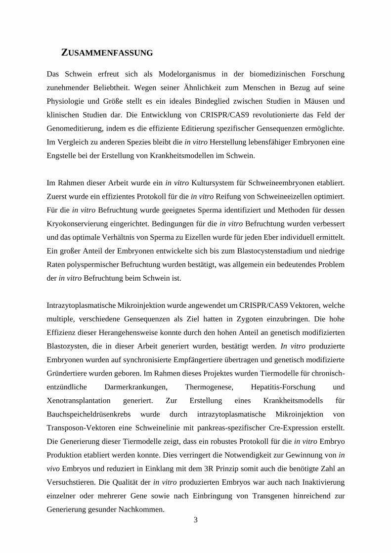
ZUSAMMENFASSUNG
Das Schwein erfreut sich als Modelorganismus in der biomedizinischen Forschung
zunehmender Beliebtheit. Wegen seiner Ähnlichkeit zum Menschen in Bezug auf seine
Physiologie und Größe stellt es ein ideales Bindeglied zwischen Studien in Mäusen und
klinischen Studien dar. Die Entwicklung von CRISPR/CAS9 revolutionierte das Feld der
Genomeditierung, indem es die effiziente Editierung spezifischer Gensequenzen ermöglichte.
Im Vergleich zu anderen Spezies bleibt die in vitro Herstellung lebensfähiger Embryonen eine
Engstelle bei der Erstellung von Krankheitsmodellen im Schwein.
Im Rahmen dieser Arbeit wurde ein in vitro Kultursystem für Schweineembryonen etabliert.
Zuerst wurde ein effizientes Protokoll für die in vitro Reifung von Schweineeizellen optimiert.
Für die in vitro Befruchtung wurde geeignetes Sperma identifiziert und Methoden für dessen
Kryokonservierung eingerichtet. Bedingungen für die in vitro Befruchtung wurden verbessert
und das optimale Verhältnis von Sperma zu Eizellen wurde für jeden Eber individuell ermittelt.
Ein großer Anteil der Embryonen entwickelte sich bis zum Blastocystenstadium und niedrige
Raten polyspermischer Befruchtung wurden bestätigt, was allgemein ein bedeutendes Problem
der in vitro Befruchtung beim Schwein ist.
Intrazytoplasmatische Mikroinjektion wurde angewendet um CRISPR/CAS9 Vektoren, welche
multiple, verschiedene Gensequenzen als Ziel hatten in Zygoten einzubringen. Die hohe
Effizienz dieser Herangehensweise konnte durch den hohen Anteil an genetisch modifizierten
Blastozysten, die in dieser Arbeit generiert wurden, bestätigt werden. In vitro produzierte
Embryonen wurden auf synchronisierte Empfängertiere übertragen und genetisch modifizierte
Gründertiere wurden geboren. Im Rahmen dieses Projektes wurden Tiermodelle für chronisch-
entzündliche Darmerkrankungen, Thermogenese, Hepatitis-Forschung und
Xenotransplantation generiert. Zur Erstellung eines Krankheitsmodells für
Bauchspeicheldrüsenkrebs wurde durch intrazytoplasmatische Mikroinjektion von
Transposon-Vektoren eine Schweinelinie mit pankreas-spezifischer Cre-Expression erstellt.
Die Generierung dieser Tiermodelle zeigt, dass ein robustes Protokoll für die in vitro Embryo
Produktion etabliert werden konnte. Dies verringert die Notwendigkeit zur Gewinnung von in
vivo Embryos und reduziert in Einklang mit dem 3R Prinzip somit auch die benötigte Zahl an
Versuchstieren. Die Qualität der in vitro produzierten Embryos war auch nach Inaktivierung
einzelner oder mehrerer Gene sowie nach Einbringung von Transgenen hinreichend zur
Generierung gesunder Nachkommen.

4
Für Anwendungen, die komplexere genetische Manipulationen erfordern ist es vorteilhaft,
diese zuerst in vitro in der Zellkultur durchzuführen, um vor der Herstellung von Tieren die
Genauigkeit der genetischen Modifikation sicherzustellen. Der letzte Teil dieser Arbeit
beschreibt hierzu die Etablierung eines Handmade Cloning Systems, das eine zuverlässige
Alternative zum herkömmlichen Kerntransfer darstellt.
5
1. INTRODUCTION
Mice are the most commonly used species in biomedical research, mostly because they are
relatively inexpensive to house and techniques for their genetic modification are well
established [1, 2]. Mouse studies have provided extensive insights into the underlying
mechanisms of many human diseases but often they do not mimic human disease pathology
or phenotypes accurately [3].
The “3R” principle demands replacement, reduction and refinement of animal experiments
whenever possible [4] which means that the predictive value of data generated in animal
experiments has to be maximised [5]. Regulatory agencies require preclinical studies in
nonrodent species which makes large animal models of human diseases indispensable [6].
Similarities in organ anatomy, physiology, body size, diet and pathophysiology make pigs
a useful model organism to gain insights into human diseases [7]. Surgical interventions
and diagnostic procedures like imaging of vessels and organs can be carried out using
standard equipment [8]. Public acceptance for the use of livestock in animal experiments
is less controversial than for primate species or companion animals. Pigs are highly fertile
and housing conditions including specific-pathogen-free (SPF) are well established [9, 10].
Genome engineering (GE) combined with sequencing of the whole porcine genome [11]
has promised the generation of tailored porcine disease models for a variety of human
conditions but the practical implementation remains challenging [12]. GE pigs that
replicate human phenotypes and disease mechanisms functionally and on the molecular
level have potential in translational medicine by “bridging the gap between bench and
bedside” [13]. Porcine disease models have been generated for cancer research [14, 15],
xenotransplantation [16, 17], diabetes [18], cystic fibrosis [19] and Duchene muscular
dystrophy [20] but in the past the efficiency in generating these models has been low and
restricted to a few groups worldwide.
Genome engineering also holds great promise for agriculture. It has the potential to
revolutionise animal breeding [21], improve productivity, animal welfare, reduce use of

6
antibiotics in livestock production and protect the environment [22]. GE pigs resistant to
porcine reproductive and respiratory syndrome (PRRSV) virus are exemplary [23].
7
1.1. The toolbox for genome engineering of livestock
1.1.1. Traditional methods for genome engineering of pigs
The first genetically modified pigs were created in 1985 by pronuclear DNA microinjection
[24, 25]. Other methods for genome engineering of livestock include sperm-mediated gene
transfer [26], viral vectors [27], somatic cell nuclear transfer (SCNT) [28, 29] and its close
variation handmade cloning (HMC) [30].
1.1.1.1. Pronuclear DNA microinjection
Pronuclear DNA microinjection was the first, and for a while the most common method of
generating genetically modified large animals [31]. Mice were the first species in which this
method was successfully applied [32, 33] with pigs and other livestock animals following
shortly after [24, 25]. Microinjected DNA can be integrated at the pronuclear stage but also in
subsequent cell divisions [34] which leads to the generation of mosaic animals [35]. Other
downsides are the need for expensive micromanipulation equipment and highly trained
operators. In livestock species it is necessary to centrifuge the oocytes to visualise the pronuclei
because of their pigmentation [36]. Perhaps the greatest drawback is however the low
proportion of transgenic animals produced, about 3% in mice and lower in livestock [37] due
to interspecies variation in DNA integration [38], and the lack of control over transgene
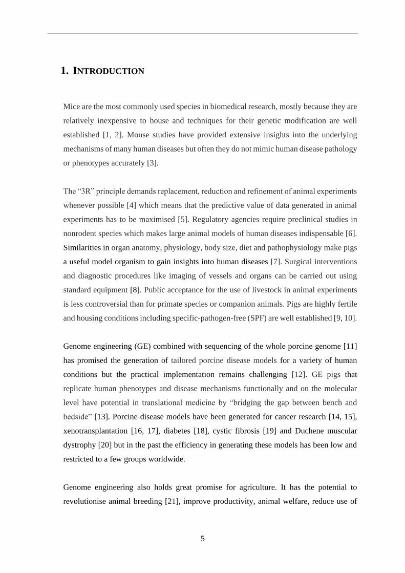
integration. In its basic form pronuclear DNA microinjection (see Figure 1) results in the
addition of transgenes at random locations in the host genome [39]. This leads to the 'position
effect' in which the expression levels of integrated transgenes can differ widely under the
influence of adjacent DNA sequences [40].
Figure 1: DNA microinjection into the male pronucleus of a one-cell mouse embryo A) The injection needle is
inserted into the pronucleus (indicated by the arrow). B) Approximately 2pl of DNA is injected and the diameter
of the pronucleus increases by about 50% under hydrostatic pressure. (adapted from DeMayo et al. [41]).

8
1.1.1.2. Sperm-mediated gene transfer
Sperm-mediated gene transfer (SMGT) was developed as a means of avoiding the need for
embryo micromanipulation, embryo culture and embryo transfer (ET). The natural ability of
sperm to transfer DNA into oocytes is employed to co-transfer exogenous DNA [42]. The
procedure comprises of sperm collection, coincubation with DNA constructs and artificial
insemination (AI) (illustrated in Figure 2). Following its first implementation in mice [43] there
are several reports of transgenic livestock generated using this approach [26, 44]. However
despite its simplicity, the successful implementation of SMGT has been limited to certain
laboratories [45] rendering its validity questionable [46]. SMGT seems to work only with sperm
samples from some donors for inexplicable reasons [47] which is a possible explanation for
those mixed results. Another drawback is that that transgenes introduced this way are frequently
fragmented [48].
Figure 2: Sperm-mediated gene transfer. Sperm is collected from suitable donors and co-incubated with
exogeneous DNA followed by artificial insemination. (adapted from Lavitrano et al.[47]).
Linker-based SMGT is a more recent approach in which uptake of DNA by sperm is facilitated
through endocytosis of DNA-antibody complexes [49]. Another modification is
intracytoplasmic sperm injection (ICSI) mediated gene transfer [50] which is claimed to be
useful for transferring large transgenes [51] and eliminates problems associated with
polyspermy in IVF. While ease of use would seem to make SMGT a superior method for the

9
generation of genetically modified livestock, issues of efficiency and reproducibility have
prevented more widespread adoption [52].
1.1.1.3. Viral vectors
Lentiviruses are part of the family Retroviridae and possess the ability to infect cells and reverse
transcribe their RNA to DNA. Viral DNA is randomly integrated into the host’s genome and
passed on to offspring through germline transmission. This ability can be utilised to transfer
DNA sequences from one organism to another, a process termed transgenesis [53]. Following
its initial use in mice [54] lentiviral gene transfer was successfully applied in porcine
transgenesis [27, 55].
The advantages of lentiviral vectors include reportedly highly efficient transgenesis in livestock
[56] and the ability to transduce non-dividing cells, which allows transduction of very early
embryos thus reducing the likelihood of mosaicism [57]. Their disadvantages include the
limited insert size (~ 5.2 kb) [58], multiple independent integration events resulting in transgene
segregation in subsequent generations [59] and the possibility of vector recombination with a
wild-type virus, leading to the production of infectious virions [60]. Another drawback of all
viral vector systems is the time and labour-intensive preparation and concentration of viral
particles.
Adenoviral vectors can infect a variety of different cell types, have a high infection efficiency
and do not have to be integrated into the host genome [61]. This makes them especially suitable
to deliver site-specific nucleases for genome engineering [62]. Delivery of targeting constructs
via adenoviral vectors has been reported as an efficient means of producing gene targeted
animals [63]. Drawbacks to adenoviral vectors are their relatively high immunogenicity and
cytotoxicity [64].
Adeno associated viruses (AAVs) are a safe alternative for the delivery of RNA-guided
endonucleases. They are not associated with any diseases in humans, rely on helper virus for
replication and in vectors sequences for nearly all viral structural genes are removed [65]. The
biggest drawback to AAVs is their small capacity of about 4.7 kb [58]. This is problematic
when AAVs are to be used in combination with the clustered regularly interspaced short
palindromic repeats (CRISPR) / CRISPR-associated protein (Cas) system. The coding sequence

10
for the components of the CRISPR/Cas system plus the required promotor sequence exceeds
5kb [66]. This packaging limit can be bypassed with an innovative split-Cas9 system in which
the N and C-terminal parts of Cas9 are fused to split-intein units that reconstitute the complete
Cas9 protein upon co-expression [67].
1.1.1.4. Transposon-mediated transgenesis
Transposons or “jumping genes” are mobile genetic elements that are able to relocate within
the genome [68]. Transposable elements (TE) make up a significant proportion of many
species’ genomes [69]. They can be categorised into class I or retrotransposons and class II or
DNA transposons [70]. Class I transposons use a “copy and paste” mechanism based on reverse
transcription to generate a copy of themself [71]. Class II transposons encode the protein
transposase which recognises the inverted terminal repeat (ITR) sequences that flank a
transposon, excises it from its current position in the genome and re-integrates the transposable
element. This is termed a “cut and paste” mechanism (see Figure 3) [72]. Depending on the
transposon type local hopping or a more random re-integration at “TTAA” sites occur.
Figure 3: Mechanisms of transposition A) Class I Transposons rely on an RNA intermediate and reverse
transcription. B) Class II transposons are excised by transposase and relocated by creating DSBs (adapted from
Saha et al. [72]).
11
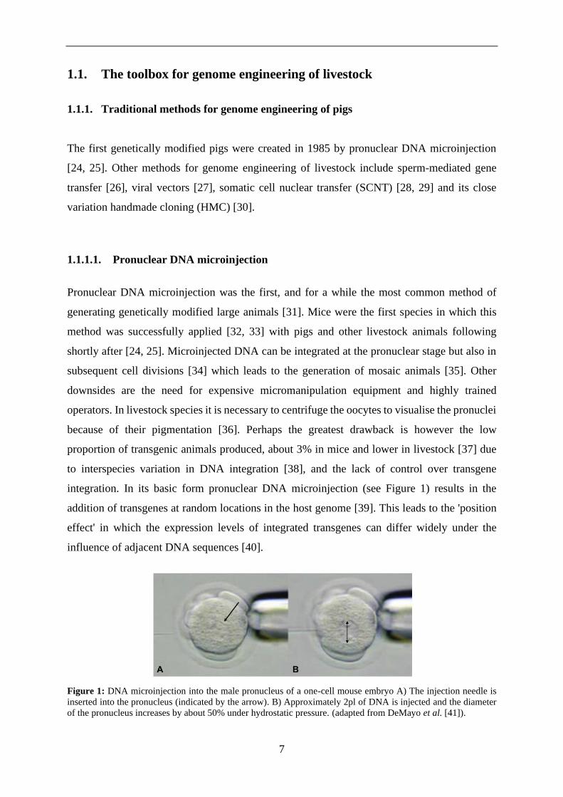
The class II transposon system has been adapted as a tool for the generation of transgenic
animals by replacing the transposon gene with the gene of interest and providing the transposon
activity via a second expression vector or as mRNA [73]. A number of transposon vectors have
been developed. The most commonly used are PiggyBac (see Figure 4) and Sleeping Beauty
transposon systems as these have the highest transposition activity in mammalian cells [74].
Transgenic pigs have been generated using both systems. [75, 76].
Figure 4: The mechanism of PiggyBac transposition. Transposase binds the PiggyBac ITRs. It nicks and attacks
the TTAA ends leading to hairpin formation and transposon excision. The transposon is integrated into genomic
DNA at TTAA sites (adapted from Woodard et al. [77]).
Transposon-mediated transgenesis facilitates efficient transgene insertion and stable expression
compared to DNA microinjection [77]. Advantages over viral vectors are larger cargo size, ease
of implementation and biosafety [78]. This approach only allows for random integration of
transgenes and depends on microinjection as delivery method [78].
12
1.1.1.5. Somatic cell nuclear transfer
Traditionally genetically modified mice have been generated by pronuclear DNA
microinjection [32], or by modification of embryonic stem cells (ESCs) - notably using HR to
effect gene targeting [79]. The ability of ESCs to maintain pluripotency and a normal karyotype
during long term culture [80] combined with the high frequency with which they support HR
makes them a powerful tool [81]. Typically genetically modified ESCs are injected into a
recipient blastocyst or aggregated with a precompaction stage embryo then transferred to a
pseudo-pregnant female to gestate chimeric offspring [82]. Appropriate breeding results in mice
carrying the desired genotype [83]. Totipotent porcine ESCs, capable of populating the germ
line, are still unavailable but there are promising reports about pluripotent stem cells with
enhanced differentiation potential [84].
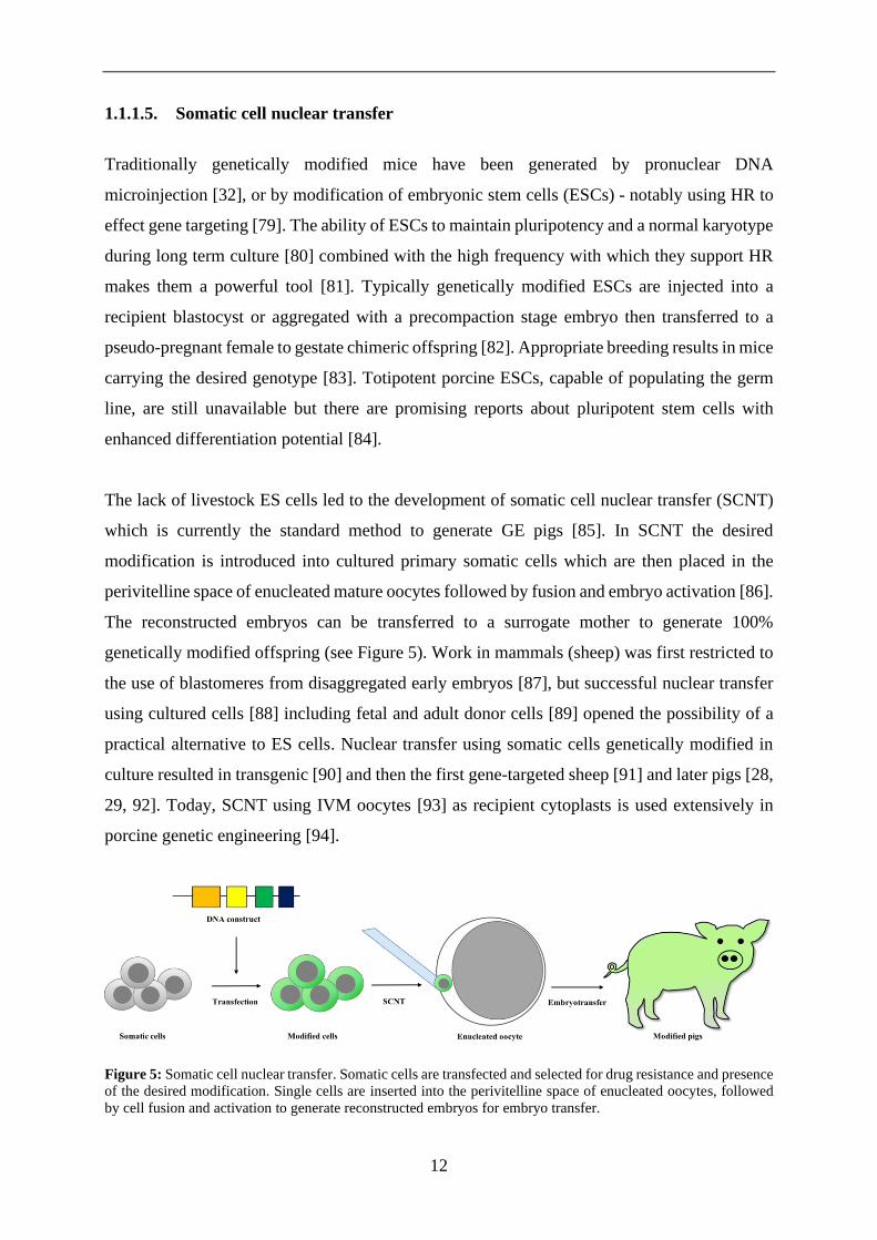
The lack of livestock ES cells led to the development of somatic cell nuclear transfer (SCNT)
which is currently the standard method to generate GE pigs [85]. In SCNT the desired
modification is introduced into cultured primary somatic cells which are then placed in the
perivitelline space of enucleated mature oocytes followed by fusion and embryo activation [86].
The reconstructed embryos can be transferred to a surrogate mother to generate 100%
genetically modified offspring (see Figure 5). Work in mammals (sheep) was first restricted to
the use of blastomeres from disaggregated early embryos [87], but successful nuclear transfer
using cultured cells [88] including fetal and adult donor cells [89] opened the possibility of a
practical alternative to ES cells. Nuclear transfer using somatic cells genetically modified in
culture resulted in transgenic [90] and then the first gene-targeted sheep [91] and later pigs [28,
29, 92]. Today, SCNT using IVM oocytes [93] as recipient cytoplasts is used extensively in
porcine genetic engineering [94].
Figure 5: Somatic cell nuclear transfer. Somatic cells are transfected and selected for drug resistance and presence
of the desired modification. Single cells are inserted into the perivitelline space of enucleated oocytes, followed
by cell fusion and activation to generate reconstructed embryos for embryo transfer.

13
The advantages of SCNT include the ability to engineer DNA sequence replacement, deletion
or addition via HR or genome editing. One also has the possibility to choose the sex of the
resultant offspring [93] and mosaicism is unlikely to occur so all resulting offspring should
carry the desired genetic modification [95]. This can be ensured by extensive selection and
screening of the donor cells before producing embryos. Downsides to the method include the
high level of skill required by the operator, the relatively low numbers of embryos that can be
processed, the low efficiency in terms of the number of animals born per reconstructed embryo
transferred [96], and the occurrence of health problems and high mortality in the resulting
offspring due to deficient epigenetic reprogramming [97].
While the core SCNT procedure has undergone very few changes since it was first developed
for mammals, progress in the enabling technologies such as IVM, IVC and oocyte activation
have improved cloning efficiencies over time [86, 98].
1.1.1.6. Handmade cloning
Handmade cloning (HMC) is a micromanipulator-free alternative to traditional
micromanipulator-based cloning (TC) [99]. The eponymous feature of this method is
enucleation of oocytes with a handheld blade after partial zona pellucida (ZP) digestion. Two
of the resulting cytoplasts are fused with a donor cell and activated. Culture to the blastocyst
stage (see Figure 6) in a well of the well (WOW) system [100] is followed by transfer to a
synchronised recipient [101]. This procedure was first performed in cattle [102, 103] and
quickly adapted to porcine embryos [30] and used to produce GE pigs [104]. While porcine
oocytes are more sensitive to manipulation than other livestock species [105] reported
efficiencies of cloned pigs resulting from HMC are equal or higher than TC [106].
14
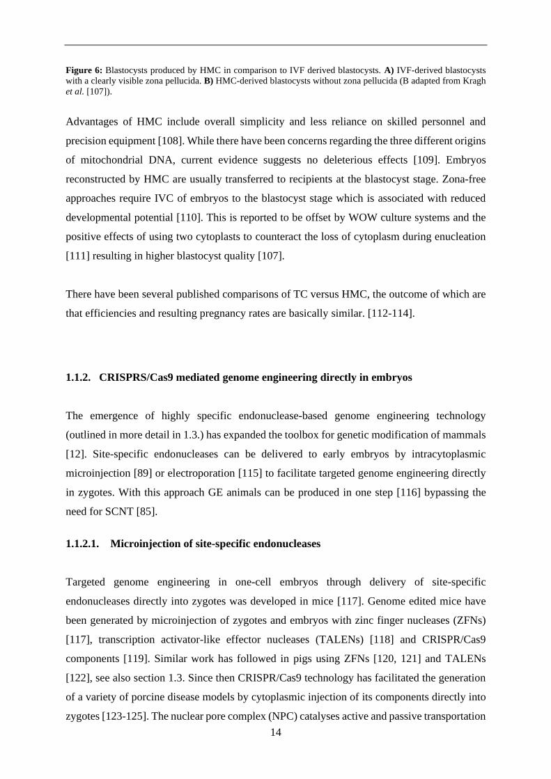
Figure 6: Blastocysts produced by HMC in comparison to IVF derived blastocysts. A) IVF-derived blastocysts
with a clearly visible zona pellucida. B) HMC-derived blastocysts without zona pellucida (B adapted from Kragh
et al. [107]).
Advantages of HMC include overall simplicity and less reliance on skilled personnel and
precision equipment [108]. While there have been concerns regarding the three different origins
of mitochondrial DNA, current evidence suggests no deleterious effects [109]. Embryos
reconstructed by HMC are usually transferred to recipients at the blastocyst stage. Zona-free
approaches require IVC of embryos to the blastocyst stage which is associated with reduced
developmental potential [110]. This is reported to be offset by WOW culture systems and the
positive effects of using two cytoplasts to counteract the loss of cytoplasm during enucleation
[111] resulting in higher blastocyst quality [107].
There have been several published comparisons of TC versus HMC, the outcome of which are
that efficiencies and resulting pregnancy rates are basically similar. [112-114].
1.1.2. CRISPRS/Cas9 mediated genome engineering directly in embryos
The emergence of highly specific endonuclease-based genome engineering technology
(outlined in more detail in 1.3.) has expanded the toolbox for genetic modification of mammals
[12]. Site-specific endonucleases can be delivered to early embryos by intracytoplasmic
microinjection [89] or electroporation [115] to facilitate targeted genome engineering directly
in zygotes. With this approach GE animals can be produced in one step [116] bypassing the
need for SCNT [85].
1.1.2.1. Microinjection of site-specific endonucleases
Targeted genome engineering in one-cell embryos through delivery of site-specific
endonucleases directly into zygotes was developed in mice [117]. Genome edited mice have
been generated by microinjection of zygotes and embryos with zinc finger nucleases (ZFNs)
[117], transcription activator-like effector nucleases (TALENs) [118] and CRISPR/Cas9
components [119]. Similar work has followed in pigs using ZFNs [120, 121] and TALENs
[122], see also section 1.3. Since then CRISPR/Cas9 technology has facilitated the generation
of a variety of porcine disease models by cytoplasmic injection of its components directly into
zygotes [123-125]. The nuclear pore complex (NPC) catalyses active and passive transportation

15
of DNA, RNA, proteins and small molecules through the nuclear membrane [126]. Therefore,
cytoplasmic microinjection of CRISPR/Cas9 components as DNA, RNA or protein molecules,
are all considered practical options [119, 127]. This approach can be used to introduce indels
[128], or together with single strand DNA templates to effect homologous sequence
replacement in the genome of individual embryos [129]. Targeted multiplex genome
engineering in one step is also possible by using multiple sgRNAs [119]. Another advantage of
microinjection is the ability to introduce multiple different reagents like DNA vectors, guide
RNAs and polypeptides at once [130] without constraints regarding type or size of the construct.
Importantly, animals generated by microinjection of site-specific nucleases have not so far
displayed any of the developmental defects [85] associated with SCNT [97].
This approach frequently causes mosaicism which arises from the ability of the CRISPR/Cas9
system to continuously edit cells at various stages of embryonic development [131, 132].
Mosaicism complicates genotype analysis and requires outcrossing of the mosaic founders to
generate new genetically modified lines which is especially problematic in pigs due to the long
generation interval [85].
Another limitation to this method is the difficulty of introducing modifications via homology
directed repair (HDR) [129]. While pigs with targeted knock-ins have been created using this
approach [133] large insertions remain challenging [134].
Genome engineering directly in porcine embryos is further limited by the need for large
numbers of high-quality porcine zygotes, which is a problem due to the inefficiency of current
porcine IVM and IVF systems [135], see also section 1.2. Researchers have therefore mainly
used genome engineering in in vivo derived oocytes or zygotes with few exceptions [124, 136,
137].
1.1.2.2. Electroporation
Electroporation of zygotes is a high-throughput method of introducing CRISPR/Cas9
components into early embryos to produce genetically modified animals [115]. Electroporation
was demonstrated to be a viable method for nucleic acid delivery to oocytes and zygotes in
mice [138]. Initially this approach was limited by the need to remove the ZP, resulting in low
16
development and pregnancy rates [139]. Advances in electroporator technology facilitated ZP
penetration which resulted in the generation of genetically modified rats by delivery of RNA
guided endonucleases into early embryos via electroporation [140]. Application of poring
pulses to create micro-holes in the ZP and oolemma followed by transfer pulses to deliver
mRNA into the ooplasm [140] promotes high transfection efficiencies and embryo viability
[141].
A recent publication reported the generation of a GE pig via electroporation of in vitro derived
zygotes [142]. While the reported efficiencies are still very low, successful electroporation in
embryos could offer a simpler alternative to microinjection [31].

17
1.2. In vitro production of porcine embryos
Production of GE pigs by direct manipulation of porcine zygotes requires a vast number of
porcine embryos. The anatomy of the porcine genital tract makes it very difficult to carry out
non-surgical ovum pick up from living animals, so in vivo matured oocytes or zygotes can only
be collected by flushing the oviducts of slaughtered donor sows, which is expensive and
requires a large number of experimental animals [143]. Thus in vitro production (IVP) of
porcine embryos using slaughterhouse-derived immature oocytes is the only practical means of
providing a sufficient supply of porcine embryos while reducing the number of experimental
animals in accordance with the “3R” principle. [144]
Assisted reproductive techniques (ART) that facilitate efficient IVP of embryos are highly
developed in humans and many livestock species. In pigs however, in vitro embryo culture
conditions are still considered suboptimal [145]. The high costs involved, and the relatively low
financial value of individual pigs make such methods commercially unappealing for routine
agricultural applications. However, pigs are playing an ever more important role in translational
medicine [8, 12, 13], and ARTs are valuable tools for their use in biomedicine [146]. IVP of
embryos comprises three steps: In vitro maturation (IVM) of oocytes, in vitro fertilisation (IVF)
and in vitro culture (IVC). Spurred by the prospect of creating clinically relevant animal models
for a wide spectrum of human conditions IVP systems for porcine embryos have been
developed (see Table 1) but they are still considered inefficient compared to other species [147].
Problems like polyspermic fertilisation, insufficient cytoplasmic maturation of IVM oocytes
and suboptimal culture conditions remain largely unsolved and result in reduced viability of
IVP embryos [148]. Further optimisation is necessary to realise the full potential of this
technology [94, 149].
Table 1: Milestones of porcine in vitro production. Modified from Grupen et al. [94].
Year Details of manipulation / IVP procedure Reference
1985 In vivo zygotes / transgene insertion by microinjection Brem et al. [24]
Hammer et al. [25]
1986 In vivo oocytes / IVF with fresh ejaculated sperm Cheng et al. [150]
1988 In vivo oocytes / IVF with FT epidydimal sperm Nagai et al. [151]

18
1989 IVM oocytes / IVF with extended ejaculated sperm
In vivo oocytes / NT using 4-cell stage blastomeres
In vivo embryos / FT at the peri-hatching blastocyst stage
Mattioli et al. [152]
Prather et al. [153]
Hayashi et al. [154]
1993 IVM oocytes / IVF with fresh ejaculated sperm Yoshida et al. [155]
1995 In vivo embryos / frozen-thawed at the 4-cell stage Nagashima et al. [156]
1997 In vivo oocytes / IVF with sex-sorted sperm Rath et al. [157]
1998 IVM oocytes / IVF with sex-sorted sperm Abeydeera et al. [158]
2000 In vivo oocytes / SCNT
IVM oocytes / SCNT embryos
In vivo oocytes / NT using 4-cell stage blastomeres
Onishi et al. [29]
Polejaeva et al. [92]
Betthauser et al. [28]
Li et al. [159]
2001 IVP embryos / IVC to the 2- to 4-cell and blastocyst stages
Somatic cell nuclear transfer (SCNT) embryos / GM donor cells
Marchal et al. [160]
Park et al. [161]
2002 IVP embryos / IVC to the blastocyst stage
In vivo zygotes / IVC medium chemically defined
SCNT embryos / targeted GM donor cells
Kikuchi et al. [162]
Yoshioka et al. [163]
Dai et al. [164]
2003 IVM oocytes / IVF and IVC media chemically defined Yoshioka et al. [165]
2006 SCNT embryos / IVC to the blastocyst stage
SCNT embryos / FT at the blastocyst stage
Lagutina et al. [166]
Li et al. [167]
2007 IVP embryos / FT at the 4- to 8-cell stage
SCNT embryos / handmade cloning / GM donor cells
Nagashima et al. [168]
Du et al. [30]
2009 IVP embryos/IVM, IVF and IVC media chemically defined
IVP zygotes / FT at the pronuclear stage
SCNT embryos / handmade cloning / GM donor cells
Akaki et al. [169]
Somfai et al. [170]
Kragh et al. [171]
2011 SCNT embryos / FT at the morula stage
SCNT embryos / handmade cloning / targeted GM donor cells
Nakano et al. [172]
Luo et al. [173]
2012 IVP embryos / non-surgical embryo transfer
IVP embryos / FT at the morula stage
Yoshioka et al. [174]
Maehara et al. [175]
2013 In vivo oocytes / intrafallopian insemination / GM donor sperm Umeyama et al. [176]
2017 IVP embryos / triple cytokine supplemented (FLI)medium
Parthenogenesis / iPSC injection / human-pig chimeric embryo
Yuan et al. [177]
Wu et al. [178]
2019 In vivo zygotes / non-surgical ovum pickup Yoshioka et al. [179]
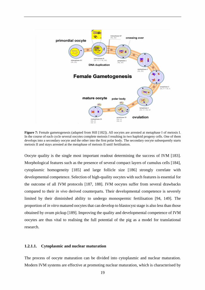
1.2.1. In vitro maturation
In female mammals, all oocytes ever produced (200.000 – 400.000) are arrested at the diplotene
stage (prophase I) of meiosis I until sexual maturity [180]. Maturation describes a complex
process during which oocytes undergo various cellular changes in which they gain the ability
to be fertilised and proceed through embryogenesis (see Figure 7) [181]. As meiosis I resumes,
one set of chromosomes is extruded forming the first polar body. The haploid secondary oocyte
then advances to the metaphase of meiosis II where it is arrested once again until fertilisation.
19
Figure 7: Female gametogenesis (adapted from Hill [182]). All oocytes are arrested at metaphase I of meiosis I.
In the course of each cycle several oocytes complete meiosis I resulting in two haploid progeny cells. One of them
develops into a secondary oocyte and the other into the first polar body. The secondary oocyte subsequently starts
meiosis II and stays arrested at the metaphase of meiosis II until fertilisation.
Oocyte quality is the single most important readout determining the success of IVM [183].
Morphological features such as the presence of several compact layers of cumulus cells [184],
cytoplasmic homogeneity [185] and large follicle size [186] strongly correlate with
developmental competence. Selection of high-quality oocytes with such features is essential for
the outcome of all IVM protocols [187, 188]. IVM oocytes suffer from several drawbacks
compared to their in vivo derived counterparts. Their developmental competence is severely
limited by their diminished ability to undergo monospermic fertilisation [94, 149]. The
proportion of in vitro matured oocytes that can develop to blastocyst stage is also less than those
obtained by ovum pickup [189]. Improving the quality and developmental competence of IVM
oocytes are thus vital to realising the full potential of the pig as a model for translational
research.
1.2.1.1. Cytoplasmic and nuclear maturation
The process of oocyte maturation can be divided into cytoplasmic and nuclear maturation.
Modern IVM systems are effective at promoting nuclear maturation, which is characterised by

20
the resumption of meiosis and extrusion of the first polar body. However, cytoplasmic
maturation distinguished by relocation of mitochondria, cortical granules and other cell
organelles is still defective [190]. The poor developmental competence and high rate of
polyspermy from IVF observed using IVM oocytes is commonly attributed to deficiencies in
those cytoplasmic processes [191, 192].
1.2.1.2. Conditions for in vitro maturation
Modern IVM systems are typically based on the culture media formulations TCM-199 or
NCSU-23 supplemented with hormones [94] that are designed to mimic in vivo conditions as
closely as possible [183]. The reduced developmental potential of IVM oocytes has been
identified as largely due to defective interactions between oocytes and cumulus cells due to
suboptimal culture conditions [193]. Several media additives such as epidermal growth factor
(EGF), cysteine, glutamine, sodium pyruvate and β mercaptoethanol promote better
cytoplasmic maturation [194]. Supplementation with porcine follicular fluid (PFF) to protect
oocytes from oxidative stress and enhance formation of male pronuclei [195, 196] has been
standard practise for decades [197, 198]. However, PFF contains maturation inhibitors [199]
and the mechanism how PFF affects maturation is unclear [200]. Furthermore, variation
between batches of PFF make it difficult to standardise culture conditions. There have thus been
efforts to replace PFF with chemically defined alternatives [177, 201]. Better understanding of
the proteins and peptides contained in PFF and the mechanisms involved in oocyte maturation
[202] has led to the development of chemically-defined maturation media [169]. This has
improved reliability and also eliminated the risk of introducing contaminating pathogens from
PFF and other biological fluids. Cytokine supplementation with fibroblast growth factor 2
(FGF2), leukaemia inhibitory factor (LIF) and insulin-like growth factor (IGF) facilitates more
synchronised nuclear and cytoplasmic maturation [202]. This results in more efficient
blastocyst production after IVF and higher mean litter size after ET [177]. Another approach is
the addition of dibutyryl cyclic adenosine monophosphate (dbcAMP) during the first half of the
maturation process. This reversibly inhibits meiosis, enhancing synchrony of cytoplasmic and
nuclear maturation [203]. Further efforts to optimise IVM conditions include co-culture of
oocytes with porcine oviduct epithelial cells during maturation [204, 205] and the use of
medium conditioned by such co-culture [206].
21
1.2.2. In vitro fertilisation
IVF is a procedure whereby an egg is fertilized by sperm in a test tube or elsewhere outside the
body to form a zygote [207]. In pigs, polyspermy and insufficient male pronucleus (MPN)
formation have been the biggest hurdles in establishing efficient IVF protocols [149]. MPN
formation could be greatly increased by supplementation of IVF media with cysteamine [208],
cysteine and glutathione (GSH) [209]. Other attempts at improving MPN formation have
included exposure of gametes to oviduct fluid [210], oviduct epithelial cells [211] or oviduct-
specific glycoprotein [212].
Polyspermy is a multifactorial problem and therefore difficult to address directly [149, 213].
Rates of polyspermy in porcine IVF systems can reach up to 90% [214, 215]. The ratio of sperm
to oocytes during fertilisation is closely related to the degree of polyspermy in IVF [216]. A
high number of porcine spermatozoa is necessary to attain acceptable fertilisation rates in vitro
compared to the amount that reaches the oviduct in vivo [217]. Experimenters are thus forced
to compromise between optimal fertilisation and acceptable rates of polyspermy, because
reducing the number of sperm cells also reduces the fertilisation rate [218]. Oocyte quality is
another critical factor affecting polyspermy [219, 220]. Oocytes used for IVF are commonly
recovered from prepubertal gilts because they are readily available from the slaughterhouse.
However these have a poor ability to block polyspermy compared to oocytes from adult sows
[160]. Other variables affecting oocyte quality are follicle size [186, 221], high temperatures
resulting from processing of pig carcasses after slaughter [222] and seasonal infertility of pigs
in summer [223]. Selection and preparation of sperm plays an important role in IVF success.
Seminal plasma contains decapacitating factors that must be removed for fresh sperm IVF. This
is usually conducted by simple centrifugation [151], but Percoll gradient centrifugation [224]
[225] provides better rates of fertilisation [226] and blastocyst formation [227].
Frozen-thawed sperm drawn from the epididymis of “good freezer” boars [228] is reported as
the most suitable choice for current IVF systems. It yields reproducible results [176, 229] while
eliminating variability between batches of ejaculates [176]. The availability of good quality
frozen sperm for IVF is however severely limited due to difficulties associated with
cryopreservation. Pig sperm is more sensitive to oxidative stress, temperature fluctuation,
osmolarity and pH-value than most other mammalian species [230, 231]. The membrane of
porcine spermatozoa contains a high ratio of polyunsaturated to saturated fatty acids [232, 233].
This makes them more susceptible to cellular damage caused by the freeze-thaw process

22
compared to other species [234]. Moreover, differences between boars in maintaining fertility
after cryopreservation [235] and even between ejaculates from the same boar [236] make the
procedure unreliable and therefore commercially unappealing.
Efforts to reduce the incidence of polyspermy such as microchannel IVF [237], straw IVF [238],
rolling culture systems [239] and modified swim-up method [240] all attempt to mimic in vivo
selection of the most motile spermatozoa. Selection of sperm that quickly bind to zona pellucida
(ZP) by shorter co-incubation has a similar effect [241] while minimizing detrimental effects
caused by dying spermatozoa in IVF medium [242]. For optimal results, IVF parameters have
to be optimised individually for each boar [235] and for fresh, frozen, ejaculated and epididymal
sperm [243]. The latest innovations combine sperm selection methods with short co-incubation
to reduce polyspermy (see Figure 8) [244].
Figure 8: Methods to reduce polyspermy A) Microchannel IVF, B) straw IVF and C) rolling systems try to mimic
in vivo conditions. They work by selecting the most motile spermatozoa (modified from Clark, Li, Kitaji et al
[237-239]).
Detection of polyspermy is another persistent problem, because it does not reduce the embryos
ability to develop to blastocyst, making this an unsuitable measure of monospermic fertilisation
[245]. To do so, pronuclei can be visualized by aceto-orcein staining [246] or through
polarization of lipid droplets by centrifugation (shown in Figure 9) [247].

23
Figure 9: Visualization of pronuclei to detect polyspermic fertilisation. In illustrations A-D visualization of
pronuclei is facilitated through aceto-orcein staining. A) porcine zygote with two pronuclei B) metaphase spindle
C) porcine zygote with three pronuclei indicating polyspermy D) abnormal porcine oocyte. In illustration E-F
pronuclei are made visible through centrifugation. This is necessary due to the high lipid content of porcine oocytes
otherwise obstructing the view. Polarized lipid droplets form a dark matter visible to the left. E) porcine zygote
with two pronuclei F) porcine zygote with three pronuclei (adapted and modified from Kurome et al. [50] and Gil
et al. [247]).
1.2.3. In vitro culture
Current in vitro culture (IVC) systems for porcine zygotes are able to surpass the historically
critical four-cell stage [248] and support embryonic development up to the blastocyst stage
[135]. However, any period of IVC results in delayed embryo development [249] and lower
cell counts in blastocysts [110].
To support optimal embryonic development, culture media are designed to mimic oviduct fluid
composition [250]. Supplementation of media with oviduct fluid and co-culture with oviduct
epithelial cells [251] has now been replaced by defined media to improve reproducibility and
biosafety [252]. Comparative studies between Whitten’s medium [253], NCSU-23-medium
[251] and Beltsville embryo culture medium [254] proved that NCSU-23 medium is superior
in facilitating blastocyst development [250, 251]. The inclusion of the amino acids glycine,

24
hypotaurine and taurine in NCSU-23 medium was found to especially benefit early embryo
development [251, 255].
Porcine zygote medium 5 (PZM5) [163] was developed based on information regarding the
concentrations of energy substrates [256, 257] and inorganic elements [258] in porcine
oviducts. PZM5 has repeatedly been confirmed as the current medium of choice [165] for
parthenogenetic [259], SCNT [260] and IVP embryos [94]. Recently, porcine blastocyst
medium was shown to facilitate reliable hatching of blastocysts in vitro [261].
Results regarding other culture variables such as oxygen tension with reported optimal values
between 5% [262] and 20% [263] or the physical culture environment, such as drop culture,
IVF plates or well of the well (WOW) systems [101] have been inconsistent, making them
difficult to optimise.

25
1.3. Precise genetic modification
Non-homologous end joining (NHEJ) in which double strand DNA breaks are re-ligated
without the assistance of repair templates is the most common DNA repair pathway in
mammalian cells [264]. This mechanism frequently results in insertions or deletions (indels)
that can disrupt regulatory elements, or cause frameshift errors in coding regions, and so affect
gene function [265].
HR is another natural DNA repair mechanism induced by DNA double strand breaks (DSB) in
which homologous sequences are consulted to make accurate repair [266]. While HR is rarer
than NHEJ [267] it can be utilized for targeted genome engineering by enabling recombination
between the target site and exogenous DNA fragments (see Figure 10) [79]. This facilitates
targeted transgene insertion [268] but results in low targeting efficiencies [269] of around one
targeting event per 106 to 107 cells [270]. Precise transgene placement through site specific
recombination [271] and HR [91] is preferable to random integration of transgenes which leads
to varying expression levels [272], transgene segregation during breeding, and can impede
functions of endogenous genes causing health problems [273].
Figure 10: Repair of nuclease induced DSBs through HDR or NHEJ (adapted from Kim et al. [274]).
Exogenous repair templates facilitate repair of nuclease induced DSBs via HDR thereby allowing for targeted
modifications and transgene insertion. The NHEJ pathway frequently causes indel mutations.

26
Traditional gene targeting vectors comprising of selection cassette and transgene flanked by
homologous arms can be utilized in genome engineering [164]. Strategies to improve targeting
efficiency involve optimising length of homologous arms [275], gene trapping [276] and
positive/negative selection [267].
The development of tailor-made highly specific endonucleases has facilitated the introduction
of DSBs into unique sites within the host genome [277, 278] to trigger DNA repair mechanisms
[279] thus enabling efficient genome engineering [280]. Categories of site-specific
endonucleases include ZFNs [121], TALENs [122] and the CRISPR/Cas9 system [123].
1.3.1. Zinc finger nucleases
ZFNs are dimeric fusion proteins consisting of two DNA binding domains each connected to
an unspecific DNA cleavage domain derived from the restriction enzyme FokI (see Figure 11)
[281]. An active nuclease is formed through FokI dimerization when two monomers bind to
their target sequence [282]. The resulting DSB induces endogenous DNA repair mechanisms,
NHEJ and HR, therefore facilitating genome engineering [283]. Three to six zinc finger motifs
each binding to a three base pair sequence provide specific recognition of 18 to 36 base pair
(bp) target DNA sequences [284].
Successful genome engineering using ZFNs has been reported in a variety of species [120, 285,
286] but the application of ZFNs is limited by narrow design requirements that allow only one
ZFN pair per 100bp. High targeting specificity can be achieved by employing multiple zinc
fingers [287] and delivery to zygotes via microinjection is possible. However, ZFN design and
production is labour intense [288] and unspecific interactions can cause high cytotoxicity [289].

27
Figure 11: Zinc finger nuclease dimer binding to its target site. A dimer of ZFNs with three zinc fingers binding
to a target site. The DNA recognition site is connected by a peptide link to the FokI derived DNA cleavage domain
(adapted from Porteus et al. [290]).
1.3.2. TALENs
TALENs are artificial dimeric structures made of a TAL effector DNA binding domain derived
from the bacterium Xanthomonas [291] fused to a FokI nuclease (see Figure 12) [288]. Tandem
amino acid sequences each recognizing a single nucleotide facilitate sequence specific DNA
binding. Base specificity is mediated by two amino acids termed the “repeat variable
diresidues” [292]. Attachment of two TALEN monomers to their target sequence results in
dimerisation of the FokI nuclease causing DSBs that can be repaired by HR or NHEJ, similar
to ZFNs [280].
Figure 12: TALEN structure. Dimerization of two TALENs is necessary to enable FokI-mediated DNA cleavage.
The target sequence is recognized by the TAL-effector DNA binding domain (adapted from Cermak et al. [292]).
Unlike ZFNs, TAL DNA binding domains can be artificially engineered to target any DNA
sequence [40]. Due to their high targeting efficiency [293] TALENs have been used for genome
28
engineering in various livestock species [122]. The main disadvantage of TALENs lies in the
complexity of designing DNA binding sequences for new targets [40].
1.3.3. CRISPR/CAS
The CRISPR/Cas system is part of the adaptive immune system in bacteria and archaea [294]
that has been adapted for genome engineering [295]. CRISPR are repeating sequences
intermediated by short protospacer segments containing genetic information originating from
viruses or plasmids [296]. These sequences function as an immunological memory system in
prokaryotes to combat viral infection [297]. Cas9 protein is an endonuclease guided by CRISPR
RNA (crRNA) to induce DSBs in sequences complementary to spacer segments [298]. There
are three types of CRISPR systems in prokaryotes [299]. CRISPR type II systems require only
Cas9, crRNA and transactivating crRNA (tracrRNA) necessary for maturation of crRNA [300]
to induce DSBs [301] whereas type I and III systems are more complex. Further simplification
can be achieved by connecting the 3’ end of crRNA to the 5’ end of tracrRNA with a loop
structure to form a synthetic single guide RNA (sgRNA) [302].
In contrast to ZFNs and TALENs, the CRISPR/Cas9 system can be adapted to recognize nearly
any target sequence without protein engineering by using different sgRNAs [280]. Application
of multiple sgRNAs enables targeting of multiple genetic loci [119]. Constraints are only
imposed by the need for a “NGG” protospacer adjacent motif (PAM) sequence located 3’ of
the target sequence [298]. Plasmids coding for sgRNA, Cas9 protein and resistance markers
facilitating selection of transfected cells [303] make this a very simple and versatile system
(see Figure 13) [304].
Due to its high adaptability, usability, simple production and high efficiency CRISPR/Cas has
become the preferred method for genome engineering [304, 305]. The CRISPR system has been
used to generate GE plants [306], livestock [123] and humans [307]. The biggest downside to
Cas9 and other site-specific nucleases are off-target effects, that is the induction of DSBs at
unwanted locations [308].

29
Figure 13: The CRISPR/Cas9 system as a tool for genome engineering. Natural CRISPR/Cas systems are guided
to their target sequence by an RNA complex composed of crRNA and tracrRNA. For genome engineering purposes
this complex has been replaced by an artificially engineered single guide RNA (sgRNA). This sgRNA has been
generated by connecting the 3’ end of crRNA with the 5’ end of tracrRNA with a loop structure. Upon target size
recognition two separate Cas9 domains cleave each DNA strand to make a DSB (adapted from Jinek et al. [302]).
CRISPR/Cas9 technology is rapidly advancing and many variants are being developed.
Modifications to the CRISPR system include 'nickases' that cleave single strand breaks leading
to HDR while reducing mutations in comparison to the original version [309]. So-called 'double
nicking' approaches can create DSBs with high precision [310].
Another approach termed “base editing” converts specific bases into another without causing
DSBs [311]. This is carried out by a deaminase enzyme fused to an inactive Cas9 protein used
for DNA binding. However, this approach has been shown to cause frequent off-target
mutations [312, 313].
'Prime editing' is a new approach that uses a catalytically inactive Cas9 connected to a reverse
transcriptase enzyme. The target site plus the intended edit are both specified by a prime editing
guide RNA at the same time. First reports claim higher efficiencies and reduced off-target
effects compared to traditional Cas9 approaches [314].
30
1.4. Goals of the project
The pig is an important animal species in agriculture and biomedicine. Genome engineering
provides new possibilities in both areas. It can be used to assess the function of genes, improve
animal health and generate disease models or pigs for organ xenotransplantation. A reliable in
vitro embryo production system based on slaughterhouse-derived ovaries is essential to
minimise the required number of experimental animals. Protocols for the in vitro production of
porcine embryos are however still suboptimal compared to other species.
The main goal of this project was to optimise the in vitro production of porcine embryos to
facilitate the generation of genetically engineered pigs. This entails the identification of suitable
sperm isolates, refinement of semen cryopreservation, establishment of a sperm bank and
improvement of embryo culture conditions. Next the suitability of IVP embryos for the
generation of transgenic, single or multiple genome edited and for a simplified cloning method
was to be assessed.
A further objective of this work was the optimisation of genome engineering directly in porcine
zygotes using CRISPR/Cas9 technology and subsequently proof that the resulting embryos are
developmentally competent.
Direct manipulation of porcine zygotes is a powerful method for the inactivation of genes but
its efficiency for more complex genome alterations is much lower. Somatic cell nuclear transfer
is a more suitable tool for such applications because it allows pre-screening for the desired
modification in cell culture. An additional goal was the implementation of handmade cloning
as an efficient alternative to traditional cloning for this purpose.

31
2. MATERIALS AND METHODS
2.1. Materials
2.1.1. Chemicals, buffers and solutions
Table 2: Chemicals, buffers and solutions
Name Source
Acetic acid (C2H4O2) AppliChem, Darmstadt, GER
Agarose Sigma-Aldrich, Steinheim, GER
Ammonium acetate (C2H7NO2) Sigma-Aldrich, Steinheim, GER
Ammonium chloride (NH4Cl) Sigma-Aldrich, Steinheim, GER
Amphotericin B Sigma-Aldrich, Steinheim, GER
Biocoll Biochrom, Berlin, GER
Bisbenzimide (Hoechst staining) Sigma-Aldrich, Steinheim, GER
Bromphenol blue Sigma-Aldrich, Steinheim, GER
BSA (fraction V) Biomol, Hamburg, GER
Caffeine Sigma-Aldrich, Steinheim, GER
Calcium chloride (CaCl2) Sigma-Aldrich, Steinheim, GER
Cetyltrimethylammonium ammonium
bromide
Sigma-Aldrich, Steinheim, GER
Chloroform (99%) Sigma-Aldrich, Steinheim, GER
CutSmart Buffer New England Biolabs, Ipswich, USA
Cycloheximide Sigma-Aldrich, Steinheim, GER
Cysteine (C3H7NO2S) Sigma-Aldrich, Steinheim, GER
Cytochalasin B Sigma-Aldrich, Steinheim, GER
Cycloheximide Sigma-Aldrich, Steinheim, GER
Demecolcine Sigma-Aldrich, Steinheim, GER
Deoxynucleotide (dNTP) solution mix New England Biolabs, Frankfurt, GER
DEPC-treated water (H20) Thermo Fisher, Waltham, MA, USA
Dimethyl-sulfoxide (DMSO) Sigma-Aldrich, Steinheim, GER
Dulbecco’s phosphate buffered saline
(dPBS)
Sigma-Aldrich, Steinheim, GER
Ethanol (EtOH) absolute Fisher Scientific, Loughborough, GBR

32
Ethanol (EtOH) denatured CLN GmbH, Niederhummel, GER
Ethylene diamine tetra-acetic acid
(EDTA)
AppliChem, Darmstadt, GER
Foetal calf serum (FCS) PAA laboratories, Pasching, Austria
Gel loading dye, purple (6x) New England Biolabs, Frankfurt, GER
Glucose (C6H12O6) Sigma-Aldrich, Steinheim, GER
Glutamine Invitrogen GmbH, Darmstadt, GER
Glycerol (C3H8O3) AppliChem, Darmstadt, GER
Glycine (C2H5NO2) Carl Roth, Karlsruhe, GER
Heparin sodium salt Sigma-Aldrich, Steinheim, GER
HEPES buffer Sigma-Aldrich, Steinheim, GER
KCl Sigma-Aldrich, Steinheim, GER
Magnesium chloride (MgCl2) Carl Roth, Karlsruhe, GER
Methanol (CH3OH) Sigma-Aldrich, Steinheim, GER
MgSO4 Sigma-Aldrich, Steinheim, GER
Mineral oil Sigma-Aldrich, Steinheim, GER
Penicillin-Streptomycin Sigma-Aldrich, Steinheim, GER
peqGREEN VWR International, Ismaning, GER
Phenol red Sigma-Aldrich, Steinheim, GER
Phenol-chloroform-alcohol AppliChem, Darmstadt, GER
Polyvinyl alcohol (C2H4O) Sigma-Aldrich, Steinheim, GER
Potassium chloride (KCL) Carl Roth, Karlsruhe, GER
Potassium-bicarbonate (KHCO3) Sigma-Aldrich, Steinheim, GER
Propanolol (C3H8O) Fisher Scientific, Loughborough, GBR
Silicon grease Obermeier, Bad Berleburg, GER
Sodium acetate (C2H3NaO2) AppliChem, Darmstadt, GER
Sodium bicarbonate (NaHCO3) Sigma-Aldrich, Steinheim, GER
Sodium chloride (NaCl) Sigma-Aldrich, Steinheim, GER
Sodium hydroxide (NaOH) Sigma-Aldrich, Steinheim, GER
Sodium pyruvate Sigma-Aldrich, Steinheim, GER
Sorbitol Sigma-Aldrich, Steinheim, GER
Sucrose (C12H22O11) Carl Roth, Karlsruhe, GER
Tris AppliChem, Darmstadt, GER
Tris-HCL Sigma-Aldrich, Steinheim, GER
33
Triton X100 Omnilab-Laborzentrum, Bremen, GER
Trypan blue Thermo Fisher, Waltham, MA, USA
Trypsin Sigma-Aldrich, Steinheim, GER
Tween 20 Sigma-Aldrich, Steinheim, GER
2.1.2. Enzymes and enzyme buffers
Table 3: Enzymes and enzyme buffers
Name Source
5x Green GoTaq reaction buffer Promega, Mannheim, GER
Bam-HF restriction enzyme New England Biolabs, Ipswich, USA
DNA Polymerase I, Large Klenow
Fragment
New England Biolabs, Ipswich, USA
GoTaq G2 DNA polymerase Promega, Mannheim, GER
HindIII-HF restriction enzyme New England Biolabs, Ipswich, USA
Hyaluronidase Sigma-Aldrich, Steinheim, GER
Pronase Sigma-Aldrich, Steinheim, GER
Proteinase K (20mg/ml) Sigma-Aldrich, Steinheim, GER
Q5 high fidelity DNA polymerase New England Biolabs, Ipswich, USA
Restriction endonucleases New England Biolabs, Ipswich, USA
T4 DNA Ligase New England Biolabs, Ipswich, USA
2.1.3. Kits
Table 4: Kits
Name Source
DNeasy Blood and tissue kit Quiagen GmbH, Hilden, GER
innuSPEED RNA kit Analytik Jena AG, Jena, GER
Lipofectamine 2000 Jena Analytic, Jena, GER
MEGAclear kit Ambion, Austin, TX, USA
MEGAshortscript T7 kit Ambion, Austin, TX, USA
Mix2Seq kit Eurofins, Ebersberg, GER
NucleoBond Xtra Midi kit Macherey-Nagel, Düren, GER
PlateSeq DNA kit Eurofins, Ebersberg, GER
Poly-A tailing kit Ambion, Austin, TX, USA

34
SurePrep RNA/DNA/protein
purification kit
Fisher Scientific, Hampton, NH, USA
Wizard SV gel and PCR clean-up
system
Promega, Mannheim, GER
2.1.4. Cells
2.1.4.1. Bacteria
Table 5: Bacteria
Name Genotype: Source:
E. coli
ElectroMAX
DH10B
F-mcrA Δ(mrr-hsdRMS-mcrBC)
Φ80lacZΔM15 ΔlacX74 recA1 endA1
araD139Δ(ara, leu)7697 galU galK λ-
rpsL nupG
Thermo Fisher
Scientific,
Waltham, MA,
USA
2.1.4.2. Eukaryotic cells
Table 6: Eukaryotic cells
Cell type Genotype Source
Porcine sperm Wild type, TP53,
KRAS, CD46,
CD55, CD59, HO-
1, GAL, CMAH,
B4G, R26M
Bayerngenetik GmbH,
Altenbach, GER;
Chair of Livestock
Biotechnology, TUM,
Freising, GER;
Porcine foetal fibroblasts
(several preparations)
Wild type Chair of Livestock
Biotechnology, TUM,
Freising, GER
Porcine kidney fibroblasts
(several preparations)
Wild type Chair of Livestock
Biotechnology, TUM,
Freising, GER
Porcine oocytes Wild type
TP 53
Vion food GmbH,
Landshut, GER
2.1.5. Oligonucleotides
2.1.5.1. Primers
Table 7: Primers and probes. All oligonucleotides were purchased from MWG Eurofins, Ebersberg, GER
Name Sequence
B4G I1 F1
ACCAGACATCGTTCCCAGTG
B4G I2 R1 AACTGGCTGTAAAGTGGGCA
B4G Scr I2 F2 GAACCTTGCGGCCCTAAAAA

35
B4G Scr I3 R1 AGCTTCCGCTCCATCTCAGG
CMAH Scr E10 F2
TGCCGTAAACAAAGAGGGGATT
CMAH Scr E10 R2 TTGTCTGCTGGGTGGGATTC
F.GAPDH s.scrofa TTCCACGGCACAGTCAAGGC
Gal Scr E7T56F GCCAGTCACCACAAGCCATG
Gal Scr E7T56R TGGCCCTGTGACACCATTCT
Gal Scr E8 T3 F
AAGACCATCGGGGAGCACAT
Gal Scr E8 T3 R GGCTTTCATCATGCCACTCG
MHCI F1 CCAGTGGTCACATGAGGCTGC
MHCI R1 GCGCCCTCCTTACCCCATCT
pNCTP scr E2 F1
TGACCACCTGCTCCACCTTC
pNTCP scr E2 R1 CGCACATATTGTGGCCGTTT
R. GAPDH s.scrofa GCAGGTCAGGTCCACAAC
TNF check_F2: GGGTTTGGATTCCTGGATGC
TNF check_R2: GCGGTTACAGACACAACTCC
TNF α F2 GGGTTTGGATTCCTGGATGC
TNF α R2 GCGGTTACAGACACAACTCC
UCP1_hs_5F GGACTACTCCCAATCTGATGAGAAG
UCP1_sus_12R GTTGTGAAGACCACTGCCCT
2.1.5.2. gRNA oligonucleotides
Table 8: gRNA oligonucleotides. All oligonucleotides were purchased from MWG Eurofins, Ebersberg, GER
Name Sequence
B4GALNT2_E3T1 F CACCGTGACGCCTTCGGGCATC
B4GALNT2_E3T1 R AAACGATGCCCGAAGGCGTCAC
CMAH-E6-T3 F GTCCTGCTTTTGCGCGAGGA
CMAH-E6-T3 R TCCTCGCGCAAAAGCAGGAC
Gal-E8-T3-F GACGAGTTCACCTACGAG
Gal-E8-T3-R CTCGTAGGTGAACTCGTC
Nanos g7 F1 GACTACTTCAACCTGAGCC
Nanos g7 R1 GGCTCAGGTTGAAGTAGTC
Px-B4GNT2-E2-T1 F CGATCCTCAAGATATCGA
Px-B4GNT2-E2-T1 R TCGATATCTTGAGGATCGC

36
2.1.6. Nucleic acid ladders
Table 9: Nucleic acid ladders
Name Source
1 kb DNA ladder New England Biolabs, Frankfurt, GER
100 bp DNA ladder New England Biolabs, Frankfurt, GER
2-log DNA ladder (0.1-10.0 kb) New England Biolabs, Frankfurt, GER
Ribo Ruler high range RNA ladder Thermo Scientific, Waltham, MA,
USA
2.1.7. Molecular cloning vectors and DNA constructs
Table 10: Molecular cloning vectors and DNA constructs
Name Specificity
pmaxGFP Kan, maxGFP
PX330 3xKO GGTA1, CMAH, B4GNT2
PX330 4xKO GGTA1, CMAH, B4GNT2, B2M
PX330 hNTCP – guide 15 hNTCP – guide 15, Cas9
PX330 hNTCP– guide 16 hNTCP – guide 16, Cas9
PX330 hNTCP plasmid Bla, hNTCP
PX330 NANOS – guide 1 NANOS2 – guide1, Cas9
PX330 NANOS – guide 2 NANOS2 – guide2, Cas9
PX330 NANOS – guide 3 NANOS2 – guide3, Cas9
PX330 NANOS – guide 4 NANOS2 – guide4, Cas9
PX330 TNF ΔARE TNF ΔARE, Cas9
PX330 UCP1 UCP1, Cas9
2.1.8. Embryo culture media, supplements and reagents
Table 11: Embryo culture media, supplements and reagents
Name Source
Amphotericin B Sigma-Aldrich, Steinheim, GER
Androstar cryo plus sperm freezing
medium
Minitube, Tiefenbach, GER
Androstar plus sperm dilution medium Minitube, Tiefenbach, GER
BSA (fraction V) Sigma-Aldrich, St. Louis, MO, USA
37
Ca-ionophore Sigma-Aldrich, St. Louis, MO, USA
Cysteine Sigma-Aldrich, St. Louis, MO, USA
D-glucose Sigma-Aldrich, St. Louis, MO, USA
EDTA Sigma-Aldrich, St. Louis, MO, USA
Egg yolk pasteurized Minitube, Tiefenbach, GER
Epidermal growth factor (EGF) Sigma-Aldrich, St. Louis, MO, USA
FBS Superior Biochrom GmbH, Berlin, GER
Fibroblast growth factor (FGF) Sigma-Aldrich, St. Louis, MO, USA
Glacial acetic acid Sigma-Aldrich, Steinheim, GER
Hyaluronidase Sigma-Aldrich, St. Louis, MO, USA
Insulin like growth factor (IGF) Sigma-Aldrich, St. Louis, MO, USA
Intergonan (PMSG/ECG) MSD-Tiergesundheit,
Unterschleißheim, GER
Leukaemia inhibitory factor (LIF) Sigma-Aldrich, St. Louis, MO, USA
Mannitol Sigma-Aldrich, St. Louis, MO, USA
MgCl2 Sigma-Aldrich
, St. Louis, MO, USA
MgSO4 Sigma-Aldrich, St. Louis, MO, USA
Mineral oil Sigma-Aldrich, St. Louis, MO, USA
Ovogest (HCG)
MSD-Tiergesundheit,
Unterschleißheim, GER
Penicillin/Streptomycin Sigma-Aldrich, St. Louis, MO, USA
Phosphate-buffered saline (PBS) Sigma-Aldrich, St. Louis, MO, USA
Phytohaemagglutinin Sigma-Aldrich, St. Louis, MO, USA
Polyvinyl alcohol Sigma-Aldrich, St. Louis, MO, USA
Porcine fertilisation medium (PFM) Fujihira Industry, Tokyo, JAP
Porcine zygote medium 5 (PZM5) Fujihira Industry, Tokyo, JAP
Sodium bicarbonate Sigma-Aldrich, St. Louis, MO, USA
Sodium pyruvate Sigma-Aldrich, St. Louis, MO, USA
Sodium pyruvate Sigma-Aldrich, St. Louis, MO, USA
Tissue culture medium 199 Sigma-Aldrich, Steinheim, GER
Tissue culture medium 199 hepes-
modification
Sigma-Aldrich, Steinheim, GER
2.1.9. Bacterial culture media and supplements
Table 12: Bacterial culture media and supplements
Name Source

38
Ampicillin (C16H19N3O4S) Carl Roth, Karlsruhe, GER
Chloramphenicol (C11H12Cl2N2O5) Sigma-Aldrich, Steinheim, GER
LB agar, Miller (Luria-Bertani) Difco BD, Sparks, MD, USA
Luria Broth, Base, Miller Difco BD, Sparks, MD, USA
2.1.10. Tissue culture media and supplements
Table 13: Tissue culture media and supplements
Name Source
Accutase Sigma-Aldrich, Steinheim, GER
Ala-Gln, 200mM Sigma-Aldrich, Steinheim, GER
Amphotericin B Sigma-Aldrich, Steinheim, GER
Blasticidin S InvivoGen, San Diego, CA, USA
Cell culture water Sigma-Aldrich, Steinheim, GER
DMSO Sigma-Aldrich, Steinheim, GER
Dulbecco’s Modified Eagle’s Medium
(DMEM)
Sigma-Aldrich, Steinheim, GER
Foetal calf serum PAA Laboratories, Pasching, Austria
G418 Genaxxon Bioscience, Ulm, GER
GlutaMAX Gibco, BRL, Paisley, UK
Hygromycin AppliChem, Darmstadt, GER
Hypo-osmolar buffer Eppendorf, Hamburg, GER
Lipofectamine 2000 Thermo Fisher Scientific, Waltham,
MA, USA
MEM non-essential amino acids 100x Sigma-Aldrich, Steinheim, GER
Opti-MEM Gibco Life Technologies, Carlsbad,
CA, USA
Penicillin/Streptomycin Sigma-Aldrich, Steinheim, GER
Phosphate-buffered saline (PBS) Sigma-Aldrich, Steinheim, GER
Puromycin InvivoGen, San Diego, CA, USA
Sodium pyruvate solution, 100mM Sigma-Aldrich, Steinheim, GER
Trypan blue Gibco Life Technologies, Paisley,
GBR
Trypsin-EDTA PAA Laboratories, Pasching, Austria
2.1.11. Laboratory equipment
Table 14: Laboratory equipment
39
Name Source
20G needle: Neolus Becton and Dickinson Company, NJ,
USA
7500 fast real-time PCR cycler Life Technologies, Carlsbad, CA,
USA
Aggregation needle DN-09/B BLS Ltd. Budapest, Hungary
Accu-jet pro Brand, Dietenhofen, GER
Blue light table Serva, Heidelberg, GER
Bunsen burner “Gasprofi 2“akku WLD-TEC GmbH, Arenshausen,
GER
Camera AxioCam MR (Axiovision) Carl Zeiss Jena GmbH, Jena, GER
Cell counting chamber: Neubauer
improved
Brand GmbH, Wertheim, GER
Centrifuges „Sigma 3-16KL “„Sigma 1-
15“
Sigma „1-15K “, „Sigma 4K15“
Sigma, Osterode, GER
Countess automatic cell counter Invitrogen, Carlsbad, CA, USA
Digital microscope “M8” PreciPoint, Freising, GER
Dry block heater PCH2 Grant Instruments, Royston, GBR
Dry block heater/cooler “PCH-2” Grant instruments, Royston, GBR
Electrophoresis system (buffer,
chamber, gel trays, combs)
Peqlab Biotechnologie, Erlangen,
GER
Electroporation cuvettes PEQLAB Biotechnologie GmbH,
Erlangen, GER
Electroporator: BTX ECM 830
Electroporation generator
BTX, Holliston, MA, USA
Electroporator: Multiporator Eppendorf, Hamburg, GER
FemtoJet Express Eppendorf, Hamburg, GER
Freezer -20°C “GS 2481“ Liebherr, Bulle, SUI
Freezer -80°C “Forma 900 Series “ Thermo Fisher Scientific, Waltham,
MA, USA
Fusion machine “BLS CF-150/C BLS Ltd. Budapest, Hungary
Fusion chamber BTX microslide 0.5
mm, model 450
Thermo Fisher Scientific, Waltham,
MA, USA
Gel documentation imaging system
“Quantum ST5”
Vilber Lourmat, Eberhardzell, GER
Gel electrophoresis chamber + power
adapter
Bio-Rad Laboratories GmbH, Munich,
GER
Glasware Marienfeld GmbH, Landa, GER
Heating plate HT 200 Minitube, Tiefenbach, GER
Heating plate HT50 Minitube, Tiefenbach, GER
Heating plate SC 300 Minitube, Tiefenbach, GER
40
Hera Safe clean bench Heraeus Instruments, Hanau, GER
Ice machine Manitowoc Ice, Manitowoc, WI, USA
Incubator (Heracell VioS 160i) Thermo Fisher Scientific, Waltham,
MA, USA
Incubator Steri-cycle CO2 Thermo Fisher Scientific, Waltham,
MA, USA
Magnetic stirrer “AREC_X”, “AGE” VELP Scientific, Usmate, ITA
Microblade “ESE 020” Bioniche Animal Health, Clonee,
Ireland
Microinjector: CellTram vario/air/pro Eppendorf, Hamburg, GER
Microscope “Axiovert 40CLF”,
“Axiovert 200M”, “Primo Star”
Carl Zeiss GmbH, Jena, GER
Microwave “MW17M70G-AU” MDA Haushaltswaren, Barsbüttel,
GER
Mini centrifuge “perfect spin mini” Peqlab Biotechnologie, Erlangen,
GER
Mr. Frosty freezing container Thermo Fisher Scientific, Waltham,
MA, USA
Nunc 4-well IVF plate Thermo Fisher Scientific, Waltham,
MA, USA
Orbital shaker Thermo Fisher Scientific, Waltham,
MA, USA
P97-micropipette puller Sutter Instrument, CA, USA
PCR cycler “DNA Engine DYAD, PTC
0220”
Biorad Laboratories, Munich, GER
PCR cycler “peqStar” Peqlab Biotechnologie, Erlangen,
GER
Pipettes “Pipetman “2ul, 20ul, 1000ul” Gilson, Middleton, WI, USA
Power supply “EPS 301” Amersham Bioscience, Little
Chalfont, UK
Power supply “peqPOWER” Peqlab Biotechnologie, Erlangen,
GER
Refrigerator “TSE1283” Beko, Neu-Isenburg, GER
Rocker shaker “Unitwist 3-D” Uniequip, Munich, GER
Safety cabinets HERA safe HSP Heraeus Instruments, München, GER
Safety Workbench Hera safe class 2H Heraeus Instruments, Munich, GER
Spectrophotometer: Bio photometer Eppendorf, Hamburg, GER
Sperm filling machine: SFS Minitube, Tiefenbach, GER
Sperm freezer: Ice Cube 14 S-A Minitube, Tiefenbach, GER
Stereomicroscope Stemi 508 Carl Zeiss, Göttingen, Germany
Stereomicroscope Stemi 508 Carl Zeiss, Göttingen, Germany
Syringe filter (0.22 µm) Berrytec, Grünwald, GER
Table centrifuge Sigma-Aldrich GmbH, Steinheim,
GER

41
Thermos container Alfi GmbH, Wertheim, GER
Transfer man NK2 micromanipulator Eppendorf, Hamburg, GER
Transportable incubator Minitube, Tiefenbach, GER
Vacuum pump: Jun Air Jun-Air, Redditch, UK
Vortex mixer “Vortex Genie 2” Scientific industries, Bohemia, NY,
USA
Water bath Memmert, Schwabach, GER
2.1.12. Buffers and solutions
Table 15: Buffers and solutions
Type Components Amount
DNA miniprep solution I C12H22O11
EDTA
Tris
H2O
1.7 g
2.9 g
3.0 g
Fill up to 1 l
DNA miniprep solution
II
NaOH
SDS
H2O
8.0 g
10.0 g
Fill up to 1 l
DNA miniprep solution
III
C2H3NaO2
H2O
246.1 g
Fill up to 1 l
Electrophoresis buffer
10x
Tris
C2H5NO2
SDS
H2O
30 g
144 g
10 g
Fill up to 1 l
SDS 10% SDS
H2O
10 g
Fill up to 100 ml
Sodium citrate buffer C6H5NaO7 x 2 H2O
H2O
2.9 g Fill up to 1 l
TAE 10x Tris
0.5 M EDTA
C2H4O2
H2O
242 g 100 ml 57.1 ml Fill up to 5 l
TBE 10X Tris
H3BO3
EDTA
H2O
545 g
275 g
39.2 g
Fill up to 5 l
TE buffer Tris-HCl
EDTA
H2O
158 mg 29 mg Fill up to 100 ml
TTE buffer Tris
Triton X 100
EDTA
H2O
242 mg
1 ml
584 mg
Fill up to 100 ml
Oocyte transportation
solution
PBS
Penicillin/Streptomycin
Amphotericin B
500 ml
5 ml
5 ml

42
2.1.13. Handmade cloning stocks
Table 16: Handmade cloning stocks
Type Component Amount
Bovine fusion medium
(cFM)
Stock A
Stock B
Mannitol-PVA-solution
2 ml
2 ml
196 ml
Cycloheximide stock Cycloheximide 1 mg/ml
25 µl aliquots
Cytochalasin B stock Cytochalasin B
5 mg/ml
5 µl aliquots
Hoechst stock Hoechst stain
H2O
3 mg
3 ml
Hyaluronidase stock Hyaluronidase
TCM 199
1 mg/ml
500 µl aliquots
Mannitol-PVA-solution Mannitol
PVA
H2O
10.93 g
0.2 g
196 ml
Phytohaemagglutinin
(PHA) stock
Phytohemagglutinin
TCM 199
5 mg/ml
20 µl aliquots
Porcine fusion medium
(pFM)
Same as cFM, but no
stock A and B are added
Pronase stock Pronase
T10
10 mg/ml
200 µl aliquots
Stock A MgSO4
H2O
25 mg (9.96 mM)
20 ml
Stock B CaCl2
H2O
14.7 mg (5.0 mM)
20 ml
T10 FCS
TCM 199
10 %
2 ml aliquots
T2 FCS
TCM 199
2 %
10 ml aliquots
T20 FCS
TCM 199
20 %
2 ml aliquots
2.1.14. Consumables
Table 17: Consumables
Name Source
Borosilicate glass with filament Sutter Instruments, CA, USA
Carbon dioxide gas cylinders, 200 bar
(CO2)
Westfalen AG, Münster, GER
Cell culture flasks Corning Inc., Corning, NY, USA
Cell culture plates Corning Inc., Corning, NY, USA
CellStar tubes (15ml and 50 ml) Greiner Bio-One, Frickenhausen,
GER

43
Cloning rings Brand, Wertheim, GER
Cover slips (24x60mm) Menzel, Braunschweig, GER
Cryo tube vials Nunc, Wiesbaden, GER
Cryo-vials Corning Inc., Corning, NY, USA
Electroporation cuvettes (2mm/4mm) Peqlab Biotechnology, Erlangen,
GER
Filter pipette tips „Fisher brand Sure
One “
Fisher Scientific, Hampton, NH,
USA
Glass Pasteur pipettes Brand, Wertheim, GER
IVF 4-well plates (nonunclon treated
surface)
Fisher Scientific, Waltham, MA,
USA
Micro loader Tip Eppendorf, Hamburg, GER
Nitrogen gas cylinders, 200 bar (N2) Westfalen AG, Münster, GER
Oxygen gas cylinders, 200 bar (O2) Westfalen AG, Münster, GER
PCR tubes 0.2 ml 8-strip PCR tubes Starlab, Hamburg, GER
Petri dishes Greiner Bio-One, Frickenhausen,
GER
Pipette tips Brand, Wertheim, GER
Plastic pipettes „Costar Stripette“(1-
50ml)
Corning Inc., Corning, NY, USA
Reaction tubes (5ml) Starlab, Hamburg, GER
Reaction tubes, (1.5ml and 2ml) Zefa Laborservice, Harthausen, GER
Sperm straws Minitube, Tiefenbach, GER
Sterile filter 0.22μm Berrytec, Grünwald, GER
Syringes BD Bioscience, Le Pont De Claix,
FRA
Tissue culture flasks (T25,75,150) Corning Inc., Corning, NY, USA
Tissue culture plates (10cm, 6-, 12-, 24-
well)
Corning Inc., Corning, NY, USA
Tubes (15ml) Corning Inc., Corning, NY, USA
Tubes (50ml) Corning Inc., Corning, NY, USA
Vacutip Eppendorf, Hamburg, GER
White cap falcon Greiner Bio-One, Frickenhausen,
GER
2.1.15. Software and online tools
Table 18: Software and online tools
Name Source
Benchling https://www.benchling.com/

44
Chromatogram viewer” Finch TV” Digital world biology LLC, CA,
USA
Crispr design tool http://crispor.tefor.net/crispor.py
Gel documentation software “Quantum
ST5”
Vilber Lourmat, Eberhandzell, GER
Genome database “Ensembl”
https://www.ensembl.org/index.html
Microscope software “Axio Vision” Carl Zeiss, Göttingen, Germany
Primer design tool “Primer3”
http://primer3.ut.ee/
Reverse Complement web tool https://www.bioinformatics.org/sms/
rev_comp.html
Sequence alignment tool “Clustal
Omega”
https://www.ebi.ac.uk/Tools/msa/clu
stalo/
TIDE: Tracking of Indels by
DEcomposition
https://tide.deskgen.com/
Uniprot https://www.uniprot.org/
Vector design software “Everyvector”
http://www.everyvector.com/
2.1.16. Veterinarian medicinal products and equipment
Table 19: Veterinarian medicinal products and equipment
Name Sequence
Altrenogest (Regumate®) MSD-Tiergesundheit,
Unterschleißheim, GER
Azaperone Elanco GmbH, Bad Homburg, GER
Cauter HBH Medizintechnik, Tuttlingen, GER
Cellulose swabs B. Braun AG, Melsungen, GER
Disposable razor B. Braun AG, Melsungen, GER
Disposable scalpels Braun, Melsungen, GER
Intergonan (ECG / PMSG) MSD-Tiergesundheit,
Unterschleißheim, GER
Katheter Careflow 5F, 300mm Merit Medical, Jordan, UT, USA
Ketanest Elanco GmbH, Bad Homburg, GER
Needle holder, Matthieu, 20cm Omega Medical, Winnenden, GER
Ovogest (HCG)
MSD-Tiergesundheit,
Unterschleißheim, GER
Surgical drape B. Braun AG, Melsungen, GER
Surgical gloves, Peha-taft latex Omega Medical, Winnenden, GER
Surgical instruments HBH Medizintechnik, Tuttlingen, GER

45
Surgicryl 910 HS 48, 5 (2), 90cm
Omega Medical, Winnenden, GER
Surgicryl Monofilament DS 24, 3.0
(2/0) 75cm
Omega Medical, Winnenden, GER
Syringes, (1ml,5ml,10ml,20ml) B. Braun AG, Melsungen, GER

46
2.2. Methods
2.2.1. Embryology
2.2.1.1. Collection and transport of ovaries
Ovaries from prepubertal gilts were collected and transported to the laboratory at 38°C in
phosphate buffered saline (PBS) supplemented with antibiotics and antimycotics.
Transportation and handling times were kept as short as possible.
2.2.1.2. Oocyte collection and classification
Ovaries were rinsed several times with warm PBS supplemented with 1%
Hexadecyltrimethylammonium bromide (CTAB). A second washing step was conducted
utilizing only warm PBS solution to get rid of the detergent. The clean ovaries were placed in
warm PBS and kept at 38°C during the puncturing process.
Follicles with a diameter of 3 to 6mm were punctured using a 10 ml syringe and a 18G needle
(see Figure 14). Porcine follicular fluid (PFF) was extracted and stored at 38°C until further
processing. PFF was removed from the falcon while cumulus oocyte complexes (COCs)
sedimented at the bottom of the tube. Then 6-8ml of working medium (WM) supplemented
with 1% Amphotericin B (Ampho B) and 1% Penicillin-Streptomycin were mixed with the
sediment. Oocytes and working medium were transferred to a petri dish for collection. High
quality oocytes with dark, evenly granulated cytoplasm and several compact layers of cumulus
cells were identified under a stereomicroscope equipped with a heating plate.
Figure 14: Porcine ovaries; Antral follicles with a diameter of 3-6mm are best suited for oocyte aspiration.
47
Oocytes were rinsed twice in WM to get rid of cell detritus. For all transfer steps a mouth pipette
and self-made glass capillaries of adequate diameter (approximately 300µm) were used to make
the washing steps as efficient as possible. Glass capillaries were pulled by hand from sterilized
Pasteur pipettes over the flame of a Bunsen burner.
2.2.1.3. In vitro maturation
For in vitro maturation oocytes were transferred to a triple gas incubator (5% O2, 5% CO2, 90%
N2, set up at 38,5°C humidified atmosphere). IVF was conducted in IVF four-well dishes
containing 500 µl of maturation medium. Groups of 50 COCs were rinsed in maturation
medium and transferred to a separate maturation well. After 45 hours successful maturation
was confirmed by visual assessment of polar body extrusion from a sample group of ovaries.
During the first half of the project maturation was carried out in NCSU-23 medium
supplemented with hormones and PFF. Due to better reproducibility and maturation results this
approach was later replaced by a chemically defined maturation medium.
Table 20: Composition of maturation media
NCSU-23 maturation medium
Component Concentration
CaCl2 1.70 mM
Cysteine 0.6 mM
ECG 10 IU/ml
EGF 10 ng/ml
Glucose 5.55 Mm
HCG 10 IU/ml
Hypotaurine 5 mM
KCl 4.78 mM
KH2PO4 1.19mM
L-glutamine 1 mM
MgSO4 1,19 mM
NaCl 108.73 mM

48
NaHCO3 25.07 mM
Penicillin-G 65 mg/L
Porcine follicular fluid 10% v/v
Streptomycin sulphate 50 mg/L
Taurine 7.0 mM
Chemically defined TCM 199 based maturation medium (FLI-medium)
Component Concentration
Cysteine 0.57mM
ECG 1 IU/ml
EGF 10ng/ml
FGF2 40ng/ml
Glucose 3.05mM
HCG 1 IU /ml
IGF1 20ng/ml
LIF 20ng/ml
PVA 0.1% w/v
Sodium pyruvate 0.91mM
TCM 199 -
2.2.1.4. In vitro fertilisation
After maturation all COCs were rinsed twice in working medium and once in equilibrated
porcine fertilisation medium (PFM) then placed in 500 µl of PFM for 30 minutes. Frozen sperm
was thawed, washed with prewarmed sperm diluent and centrifuged at 800G. Supernatant was
discarded, and an identical washing step was repeated one more time. The resulting sperm pellet
was dissolved in 500 µl of PFM and stored in the incubator until fertilisation. Post-thaw sperm
quality was analysed regarding motility, morphology and sperm count. Sperm concentration
was determined with a Neubauer improved counting chamber at a 1:50 dilution.
During IVF on average 7500 motile spermatozoa per oocyte were co-incubated with 50
cumulus oocyte complexes (COCs) for seven hours. The optimal sperm to oocyte ratio was
determined for each boar individually. On average 375.000 live spermatozoa were added to
49
each IVF well with optimal individual numbers varying from 250.000 to 1.000.000 motile
sperm cells per well. After IVF zygotes were denuded by gently pipetting them up and down in
WM supplemented with hyaluronidase (1 mg/ml).
2.2.1.5. In vitro embryo culture
In vitro embryo culture (IVC) was carried out in a triple gas incubator (5% O2, 5% CO2, 90%
N2) set up at 38,5°C humidified atmosphere. Prior to culture all embryos were rinsed in working
medium then in equilibrated culture medium to avoid contamination and transfer of media used
in previous steps to the culture dish.
Zygotes and parthenogenetically activated embryos were cultured in 500 µl of PZM5 covered
by mineral oil. Zona-free reconstructed embryos generated by handmade cloning were cultured
in individual WOWs that were created with an aggregation needle in the culture dish. During
the first half of this thesis all embryos were cultured in commercially available PZM 5 medium.
To minimise variability between batches of IVC medium PZM3 was prepared in bulk, tested,
aliquoted and frozen at -80°C. All further experiments were conducted using the same batch of
culture medium previously proven to support embryonic development.
Table 21: Porcine zygote medium 3 (PZM3)
Component Concentration
NaCl 108.00 mM
NaHCO3 25.07 mM
KCl 10.00 mM
KH2PO4 0.35 mM
MgSO4
0.40 mM
Ca-Lactate-5H2O 2.00 mM
Na-pyruvate 0.2 mM
Myo-Inositol 2.78 mM
Phenol Red 0.27 mM
L-Glutamine 1.00 mM
Hypotaurine 5.00 mM
Gentamicin 0.04 g/L
BSA 3 g/L
NEAA100X 1% (v/v)

50
EAA50X 1% (v/v)
Adjust pH value to 7.2-7.4; osmolarity to 280 +/-8 mOsm, filter through 0.22 µm filter, freeze
at -80°C.
2.2.1.6. Aceto-Orcein staining
Aceto-Orcein staining was conducted to determine optimal sperm to oocyte ratios during IVF
experiments for each individual boar. Groups of five zygotes were denuded 10-18 hours after
IVF (outlined in 5.1.2.4) and fixated on an object slide. The cover slip was glued to the object
slide using fine stripes consisting of Vaseline, hair grease and hair wax (see Figure 15). The
whole slide mounted with zygotes was submerged in methanol glacial acetic acid solution (3:1)
for a minimum of 7 days at room temperature (RT).
Aceto-orcein staining solution was prepared by boiling 1g of orcein in 45ml of acetic acid
followed by dilution with an equal amount of water. Zygotes were stained with this solution for
10 minutes then washed in a solution of glacial acetic acid, glycerol and MQ water (1:1:3).
Analysis of cells was conducted under a phase contrast microscope. Zygotes were categorized
as correctly fertilized if they showed two visible pronuclei. Oocytes without a visible
pronucleus were classified as not fertilized while those with more than two pronuclei were
graded as polyspermic. According to the results of this analysis sperm concentrations were
adjusted for each individual boar to obtain the highest blastocyst development rate possible
while minimising the rate of polyspermic fertilisation.
Figure 15: Fixation set-up for aceto-orcein staining of zygotes. Zygotes are positioned below a cover slip. Glue
strips function as spacers to avoid squashing of the cells.

51
2.2.1.7. Microinjection
After IVF zygotes were visually examined and those extruding the first polar body were
selected for microinjection. Approximately 20-30 zygotes at a time were transferred to a 4 µl
droplet of working medium covered by mineral oil. Injection needles with a side filament were
backfilled with gene targeting vectors dissolved in low tris EDTA buffer (10 mmol/L Tris-
HCL, pH 7.6 and 0.25 mmol/L EDTA, pH 8.0) at a concentration of 5 ng/µl. Alternatively
sgRNAs (prepared as outlined in 5.2.3.11) and Cas9 protein were delivered as RNA-protein-
complexes (50ng/µl Cas9 protein, 100 ng/µl sgRNA). The components of the transposon
system were delivered as PiggyBac transposon DNA vector (5ng/µl) plus PB Transposase
mRNA (10ng/µl).
Zygotes, holding pipette and injection pipette were brought onto the same horizontal plane.
Zygotes were fixed with the holding pipette positioning the polar body at the twelve or six o’
clock orientation by carefully applying suction with a pulsed flow microinjector. The tip of the
injection needle was opened by gently tapping it against the holding pipette (Vacutip). The
injection pipette was gently inserted into the cytoplasm of each oocyte and approximately 10
pl of injection solution were delivered. Successful microinjection was visually confirmed by
observing movement of the intracellular lipid droplets caused by the influx of injection solution.
Injected zygotes were placed in the lower part of the droplet to separate them from non-injected
ones (see Figure 16). Temperature was maintained at 38.5 °C during the whole microinjection
procedure with a heating plate integrated into the microscope table.
Figure 16: Micromanipulation drop. Non-injected zygotes were placed at the top of the drop. The injected ones
were moved to the lower part of the drop to prevent them from mingling.

52
Injected zygotes were washed in equilibrated PZM then transferred to the triple gas incubator
(5% O2, 5% CO2, 90% N2, set up at 38,5°C humidified atmosphere). Groups of 50 zygotes were
cultured in 500 µl of PZM covered by mineral oil. Zygotes destined for embryo transfer were
cultured for 12-36 hours. Those that were subsequently used for DNA extraction to analyse of
targeting efficiency were cultured to the blastocyst stage (six days).
2.2.1.8. Injection needle fabrication
Borosilicate glass needles suitable for microinjection were produced using a P-97 Flaming
Brown micropipette puller with a through filament according to the manufacturers’ instructions.
A ramp test was performed to determine optimal melting temperatures for different strengths
of borosilicate glass tubing. The diameter of holding pipettes was adjusted to 150 µm, an angle
of 35° was given by hand with a blowtorch.
Table 22: Parameters for Flaming Brown micropipette puller
Parameter Value
Heat 750
Pressure 500
Pull 72
Time 210
Velocity 42
2.2.1.9. DNA/RNA extraction from blastocysts
To analyse the efficiency of different targeting approaches DNA was extracted from blastocyst
stage embryos using a protocol first described in [315]. Each individual embryo was washed
twice in PBS then transferred to a PCR tube containing 10 µl of lysis buffer.
Table 23: Lysis buffer for DNA extraction
Component Concentration
KCL 50 mM
MgCl2 1.5 mM
Nonidet P-40 0.5% (w/v)
Proteinase K 100 µg/ml
Tris-Cl (pH 8.0) 10 mM
Tween-20 0.5% (v/v)
53
Incubation took place at 65°C for one hour followed by 95°C for ten minutes for inactivation
of proteinase K. The lysate was then used as a template for PCR, agarose gel electrophoresis or
DNA sequencing.
2.2.1.10. Parthenogenesis
For each experiment of in vitro embryo production, approximately 50 oocytes were
parthenogenetically activated to provide a control group for IVF. Parthenogenesis followed by
microinjection was also carried out to analyse targeting efficiency for new plasmids prior to
using them for IVF. During the better part of this work parthenogenesis was carried out using
chemical activation. Once the necessary equipment could be obtained it was replaced with
electrical activation due to the higher efficiency of this method.
2.2.1.10.1. Chemical activation
Forty-five hours after starting maturation oocytes were denuded in WM supplemented with
1mg/ml hyaluronidase and rinsed twice in working medium. They were examined for evenly
granulated cytoplasm and extrusion of the first polar body under a stereo microscope. Chemical
activation was conducted by placing them in WM supplemented with 25 µm Ionomycin
(calcium ionophore) for ten minutes. The oocytes were washed twice in WM and once in PZM5
then placed in the incubator in 500 µl of PZM5 supplemented with 5µg/ml of Cytochalasin for
3 hours. Afterwards they were rinsed twice in working medium and once in PZM 5.
Subsequently they were cultured in vitro for 6 days to the blastocyst stage.
2.2.1.10.2. Electrical activation
For electrical activation oocytes were prepared as shown in 5.2.1.9.1. Then they were rinsed
twice in activation medium and transferred to a fusion chamber (electrode distance 1.0mm)
connected to a BTX electroporator. A single activating pulse (150V, 100µs) was applied then
the identical procedure outlined for chemical activation was carried out.
Table 24: Activation medium
Component Concentration
CaCl2 0.05 mM
H2O as necessary

54
Mannitol 280 mM
MgSO4 0.1 mM
PVA 0.01 % w/v
Medium was sterile filtrated (22µm). PH was adjusted to 7.2-7.4 with NaOH. Osmolarity was
adjusted to 300 Ω.
2.2.1.11. Synchronisation of recipients
Gilts aged six to seven months with a weight of 110-130kg were selected as recipients. Their
oestrus cycle was synchronised by administering Altrenogest (Regumate ®) orally for 15 days
followed by an injection of 750 IU ECG intramuscularly (i.m.) 24 hours after the last
Altrenogest dispensation. Eighty hours after the administration of ECG, 750 IU of HCG was
injected intramuscularly. Embry transfer was carried out one to two days after HCG
administration.
2.2.1.12. Embryo transfer
Before surgery pigs were fasted for twelve hours. They were anaesthetized by intramuscular
(i.m.) application of 5mg/kg bodyweight (BW) azaperone and 25mg/kg BW ketamine.
Furthermore 0.4mg/kg BW of meloxicam and 15mg/kg BW were applied i.m. peri-operative.
Recipients were elevated on a surgery table and fixed in a 30 ° head down position. The
operating area was cleaned with warm water and soap, then disinfected with alcohol and iodine
solution. A self-adhesive surgery drape was placed at the abdominal area and the skin incision
was put at height of the second to last pair of teats at the Linea Alba. Fat and muscle tissue were
separated bluntly, and one oviduct was placed on the surgical drape. Prior to the transfer of
embryos, the correct ovulation state was controlled by visual assessment of preovulation
follicles or fresh ovulation sites.
A sterile catheter was inserted as far as possible into the oviduct and 150-200 embryos were
injected. The abdomen was closed in three layers using single stitches for peritoneum and
muscle layers and running stitches for skin.
55
2.2.1.13. Flushing of in vivo zygotes
Flushing of in vivo zygotes was conducted for experiments requiring embryos with a specific
genotype. Three to five prepubertal gilts were synchronized (outlined in 2.2.1.10) and
artificially inseminated with sperm from GE boars 39 and 46 hours after HCG application.
Seventeen hours after the second insemination donor gilts were euthanized. Their oviducts were
flushed with warm PBS supplemented with 10% FCS and 1%Penicillin-Streptomycin.
2.2.1.14. Freezing of porcine semen
Sperm was kindly provided by Bayerngenetik GmbH and the Chair of molecular animal
breeding and biotechnology (LMU). The sperm rich fraction of ejaculates from breeding boars
was obtained using the gloved hand method. Sperm was diluted with Androstar® Plus sperm
dilution medium and stored at 17°C overnight in a cooling centrifuge.
Sperm concentration was determined using a Neubauer counting chamber. Boar semen was
centrifuged at 17°C, 800g for 20 minutes in 50ml centrifuge tubes. Supernatant was discarded
and the sperm pellet was resuspended with Androstar® CryoPlus cooling extender prepared
according to the manufacturer’s instructions until 50% of the final intended volume was
reached. Semen was placed in a cold room and slowly cooled for 1.5 hours. Temperature was
monitored and upon reaching 5°C sperm concentration was adjusted to 1x10⁹ sperm cells/ml
by adding the respective amount of Androstar® CryoPlus freezing extender necessary to reach
the intended concentration.
Table 25: Androstar® CryoPlus cooling extender
Component Amount necessary for 970 ml
Androstar® CryoPlus powder 84.9 g
H20 bidistilled 770 ml
Pasteurized egg yolk 200 ml

56
Table 26: Composition of Androstar® CryoPlus freezing extender
Component Amount necessary for 500 ml
Androstar® CryoPlus cooling extender 470 ml
Equex paste 5g
Glycerine 30 ml
A semiautomatic filling and sealing machine installed in a cold room at 4°C was used to fill the
sperm into 0.5ml straws. All straws were sealed with small metal balls and handled with cold
protection gloves to minimize temperature changes. The straws were transferred to a
programmable freezing chamber (IceCube) and frozen by decreasing temperature at a rate of
30°C per minute. After completion of the freezing program all straws were transferred to a
liquid nitrogen container for long term preservation.
Table 27: Boar semen freezing curve (adapted from IceCube user manual).
Step # Temperature
°C
Time
elapsed
(min)
Temperature
change
(°C)
Time
required
(min)
Temp
decrease
(°C/min)
1 4 0 - - -
2 1 1.5 -3 1.5 -2
3 -25 2.4 -26 0.9 -30
4 -140 6.2 -115 3.8 -30
5 -140 21.2 0 15 0
2.2.1.15. Handmade cloning
After maturation oocytes were denuded, examined for extrusion of the first polar body and
moved to T2 (hepes-buffered M199, 2% FCS) drops in a bisection dish (60mm petri dish) while
discarding dead and damaged oocytes. ZP digest was conducted by placing oocytes in pronase
drops (3.3mg/ml) until deformation of the ZP could be observed. Then they were rinsed in a
drop of T20 (hepes-buffered M199, 20% FCS) to deactivate the enzyme. Twenty oocytes at a
time were transferred to a cytochalasin B drop (25µg/ml) aligning their polar bodies at the 12
o’ clock position. Enucleation was performed by removing about one third of the ooplasm
located around the polar body with a handheld microblade (see Figure 17). The resulting
enucleated cytoplasts were washed in T2 and stored in fresh T2 drops until further processing.

57
Successful enucleation was confirmed by placing a sample of cytoplasts in Hoechst staining
solution (10 µg/ml) for 10 minutes. All karyoplasts and incompletely enucleated cytoplasts
were discarded.
Figure 17: Enucleation of oocytes during handmade cloning. A) Bisection dish prepared for the enucleation of
oocytes during HMC (20µl droplets in 60mm petri dish). CB = cytochalasin B in T2; Pro = pronase. B) The red
line indicates where the bisection cut should be set thereby removing about one third of the ooplasm located around
the polar body (adapted from Li et al. [316]).
Genetically modified donor cells were harvested with accutase and resuspended with 500µl
T10 (hepes-buffered M199, 10% FCS). Half of the cytoplasts were transferred to a cell fusion
dish and placed in T10 droplets. Five of them at a time were placed in PHA drops
(phytohaemagglutinin 0.4mg/ml) to make their surface adhesive and one donor cell was
attached to each cytoplast.
Five cytoplast-cell-complexes (CCCs) at a time were washed in porcine fusion medium, moved
to the fusion chamber and aligned. Fusion was conducted by applying a single DC (direct
current) pulse (100V, 9µs) to each CCC individually. CCCs were examined for successful
fusion then covered and left on a warm heating plate for one hour to allow for reprogramming.
Porcine fusion medium was then replaced with cow fusion medium (cFM) and the remaining
half of complemental cytoplasts was moved to a T10 drop in the fusion dish (see Figure 18).
Ten CCCs and their corresponding cytoplasts were moved to the fusion chamber, aligned and
fused with a single DC pulse (85V, 80µs). The calcium ions present in cFM facilitate electrical
activation in the same step.
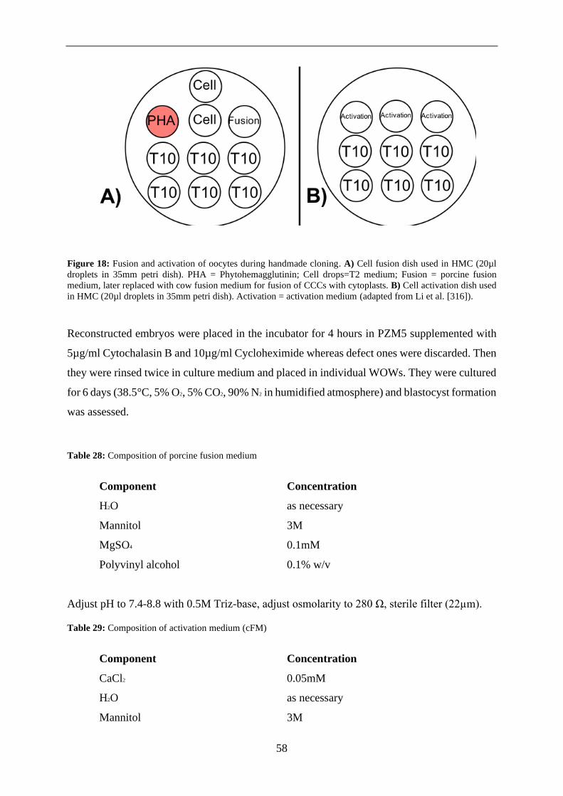
58
Figure 18: Fusion and activation of oocytes during handmade cloning. A) Cell fusion dish used in HMC (20µl
droplets in 35mm petri dish). PHA = Phytohemagglutinin; Cell drops=T2 medium; Fusion = porcine fusion
medium, later replaced with cow fusion medium for fusion of CCCs with cytoplasts. B) Cell activation dish used
in HMC (20µl droplets in 35mm petri dish). Activation = activation medium (adapted from Li et al. [316]).
Reconstructed embryos were placed in the incubator for 4 hours in PZM5 supplemented with
5µg/ml Cytochalasin B and 10µg/ml Cycloheximide whereas defect ones were discarded. Then
they were rinsed twice in culture medium and placed in individual WOWs. They were cultured
for 6 days (38.5°C, 5% O2, 5% CO2, 90% N2 in humidified atmosphere) and blastocyst formation
was assessed.
Table 28: Composition of porcine fusion medium
Component Concentration
H2O as necessary
Mannitol 3M
MgSO4 0.1mM
Polyvinyl alcohol 0.1% w/v
Adjust pH to 7.4-8.8 with 0.5M Triz-base, adjust osmolarity to 280 Ω, sterile filter (22µm).
Table 29: Composition of activation medium (cFM)
Component Concentration
CaCl2 0.05mM
H2O as necessary
Mannitol 3M

59
MgSO4 99.6nM
Polyvinyl alcohol 0.1% w/v
Adjust pH to 7.4-8.8 with 0.5M Triz-base, adjust osmolarity to 280 Ω, sterile filter (22µm).

60
2.2.2. Microbiology
2.2.2.1. Cultivation of bacteria
Bacteria were cultivated overnight at 37°C on agar plates or in LB-medium supplemented with
100 μg/ml ampicillin on an orbital shaker at 220 rpm.
2.2.2.2. Transformation of bacteria
Plasmid DNA was introduced into E. coli ElectroMAX DH10B bacteria by electroporation. 50
μl of bacteria were thawed on ice, mixed with 1-5 μl of ligation reaction and moved to a 2 mm
electroporation cuvette. A single pulse (2500V, 5ms) was applied then bacteria were cultivated
for 30 minutes in LB medium. Afterwards they were plated on LB plates supplemented with
antibiotics selecting for the corresponding plasmid and incubated at 37°C overnight.
2.2.2.3. Cryopreservation of bacteria
To conserve plasmid bearing bacteria 0.5ml of overnight culture was mixed with an equal
amount of 99% glycerol and stored at -80°C.
2.2.2.4. Isolation of plasmid DNA
Plasmid bearing bacteria from a glycerol stock were cultured overnight in 100ml of LB-medium
as outlined in 5.2.2.1. Then the NucleoBond Xtra Midi Kit was carried out according to the
manufacturer`s instructions. The resulting pellet was dissolved in low Tris EDTA buffer and
used for microinjection.

61
2.2.3. Molecular biology
2.2.3.1. Measurement of DNA and RNA concentration
DNA and RNA concentrations were measured using the NanoDrop® Lite spectrophotometer
according to the manufacturer‘s instructions.
2.2.3.2. Polymerase chain reaction (PCR)
Polymerase chain reaction (PCR) was used to amplify specific sequences from genomic DNA
and plasmid DNA. Screening PCRs and the amplification of shorter sequences was carried out
using GoTaq® Polymerase. When proofreading was required, or DNA was extracted from
blastocysts yielding only low concentrations Q5® polymerase was used.
Table 30: PCR conditions for GoTaq® G2 and Q5 polymerase
GoTaq® G2 Polymerase
PCR mixture Cycling conditions
Component Concentration Step Temper
ature Duration Cycles
DNA 10-250 ng
Initial
Denaturatio
n
98°C 2 min 1
5x buffer 1x Denaturatio
n 98°C 30 sec
35-40
dNTPs 200 μm each Annealing 58-62°C 30 sec
Primer F 0.2 μM Extension 72°C 1 min/kb 1
Primer R 0.2 μM Final
extension 72°C 5 min
Polymerase 0.03 U/μl Storage 8°C ∞
H20 Up to 25 μl
Q5® Polymerase
PCR mix Cycling conditions
Componen
t Concentration Step
Temper
ature Duration Cycles
DNA 1 pg-1 μg
Initial
Denaturatio
n
98°C 30 sec 1

62
5x buffer 1x Denaturatio
n 98°C 10 sec
35-40
dNTPs 200 μm each Annealing 58-62°C 30 sec
Primer F 0.5 μM Extension 72°C 30 sec/kb 1
Primer R 0.5 μM Final
extension 72°C 2 min
Polymerase 0.02 U/μl Storage 8°C ∞
H20 Up to 25 μl
2.2.3.3. Colony PCR
After transformation E. coli colonies were screened for the intended plasmid via colony PCR.
DNA templates for this PCR were generated by placing single bacterial clones from a LB plate
in 30 μl TTE buffer and incubating this mix at 95°C for 5 min. Colony PCR was conducted
using 2 μl of DNA solution with one primer designed to bind to the plasmid backbone and the
second one binding to the plasmid insert.
2.2.3.4. Agarose gel electrophoresis
DNA fragments were analysed by agarose gel electrophoresis. DNA samples were loaded on
gels prepared from 1xTBE or 1xTAE buffer and 1-2% agarose supplemented with 4 μl
PeqGreen. DNA fragments were separated by size by applying 80-120V until adequate
separation could be achieved (usually 1-5 hours). Subsequent analysis of DNA fragments was
conducted under UV light (254-366nm) with the Quantum ST5 gel documentation system.
2.2.3.5. Restriction digest
Restriction digests were conducted to generate linearized plasmids for cloning and transfection
or to confirm the correct length of plasmids.
Table 31: Conditions for restriction digest
Component Amount
DNA Linearization digest: 10-15 μg
Analytical digest: 2-3 μg

63
10x NEB Buffer 5 μl
Digestive enzyme 3 U/ μg
H20 up to 50 μl
Restriction digests were carried out at the optimal temperature for each respective enzyme
according to the manufacturer’s instructions. The solution was co-incubated with 2 μl of calf
intestinal alkaline phosphatase at 37°C for at 30 minutes. This prevents re-ligation by stripping
the vector backbone of its 5’ phosphates.
2.2.3.6. Ligation
Ligation of vector backbones with DNA fragments was performed using T4 Ligase according
to the manufacturer’s protocol. Components of ligation mix were co-incubated for 2 hours at
RT then left at 4°C overnight.
2.2.3.7. Blunting
When blunt ends were required for cloning, they were generated using DNA Polymerase I
Large (Klenow) Fragment to remove 3’ overhangs and fill in 5’ overhangs. The reaction was
carried out according to the manufacturer’s protocol and dNTPs were supplemented to inhibit
the polymerase’s 3’-5’ exonuclease activity.
Table 32: Conditions for blunting of DNA-fragments
Component Amount
DNA 5 μg
10x NEBuffer 5 μl
dNTPs (2mM) 1.5 μl
Klenow enzyme 1U/ μg DNA
H2O up to 50 μl
All components were co-incubated for 15 minutes at 25 °C followed by the addition of 10 mM
EDTA and an increase in temperature to 75 °C for 20 minutes to stop the reaction.
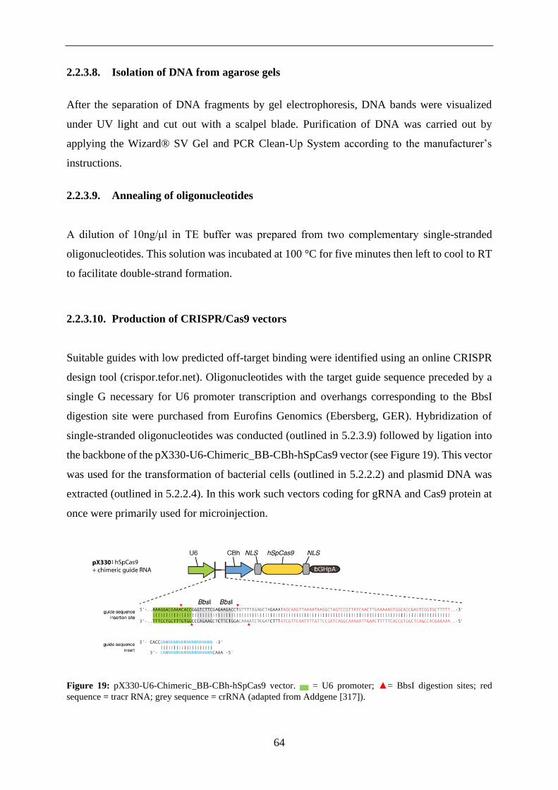
64
2.2.3.8. Isolation of DNA from agarose gels
After the separation of DNA fragments by gel electrophoresis, DNA bands were visualized
under UV light and cut out with a scalpel blade. Purification of DNA was carried out by
applying the Wizard® SV Gel and PCR Clean-Up System according to the manufacturer’s
instructions.
2.2.3.9. Annealing of oligonucleotides
A dilution of 10ng/μl in TE buffer was prepared from two complementary single-stranded
oligonucleotides. This solution was incubated at 100 °C for five minutes then left to cool to RT
to facilitate double-strand formation.
2.2.3.10. Production of CRISPR/Cas9 vectors
Suitable guides with low predicted off-target binding were identified using an online CRISPR
design tool (crispor.tefor.net). Oligonucleotides with the target guide sequence preceded by a
single G necessary for U6 promoter transcription and overhangs corresponding to the BbsI
digestion site were purchased from Eurofins Genomics (Ebersberg, GER). Hybridization of
single-stranded oligonucleotides was conducted (outlined in 5.2.3.9) followed by ligation into
the backbone of the pX330-U6-Chimeric_BB-CBh-hSpCas9 vector (see Figure 19). This vector
was used for the transformation of bacterial cells (outlined in 5.2.2.2) and plasmid DNA was
extracted (outlined in 5.2.2.4). In this work such vectors coding for gRNA and Cas9 protein at
once were primarily used for microinjection.
Figure 19: pX330-U6-Chimeric_BB-CBh-hSpCas9 vector. ▅ = U6 promoter; ▲= BbsI digestion sites; red
sequence = tracr RNA; grey sequence = crRNA (adapted from Addgene [317]).

65
2.2.3.11. Generation of sgRNAs
Generation of sgRNAs by in vitro transcription of DNA templates was performed using the
MEGAshortscript T7 kit according to the manufacturer’s instructions. Polyadenylation and
subsequent purification of the RNA transcript was conducted using the poly-A tailing kit and
the MEGAclear kit as instructed by the manufacturer. In this work such sgRNAs were primarily
used to generate RNA-protein (RNP) -complexes with Cas9 protein for microinjection.
2.2.3.12. Phenol-chloroform extraction
DNA extraction from mammalian tissue and from sperm was carried out via phenol-chloroform
extraction. About 1g of tissue or sperm pellet was incubated in 1ml of lysis buffer overnight at
55° C.
Table 33: Lysis buffer for phenol-chloroform extraction
Component Concentration
Tris-HCL 83 mM
SDS (sodium dodecyl sulphate) 0.8%
EDTA 0.2 M
NaCL 0.2 M
Proteinase K 100 μg/ml
H20 -
Then an equal amount of phenol-chloroform-isoamyl alcohol (25:24:1) was incubated with the
solution (10 minutes at RT) followed by centrifugation (10 minutes at 13.000g). The resulting
supernatant was mixed with an equal volume of chloroform (99%) and centrifuged (10min,
17.000g). Supplementation of 10% v/v sodium acetate (5M) and 0.7% v/v isopropanol followed
by thorough shaking resulted in DNA precipitation. Centrifugation (5 min, 13.000g) and rinsing
of the pellet with 70% ethanol was followed by an identical centrifugation step. Finally, the
resulting DNA pellet was air-dried and dissolved in TE buffer.
Purification of DNA fragments for in vitro transcription was conducted using a modified
variation of phenol-chloroform extraction. Hereby the DNA containing supernatant was
incubated (2 hours, -20°C) with 1/10 volume 5 M sodium acetate and two volumes of ethanol

66
(100%) after the first centrifugation step. The rest of the procedure was carried out as described
above.
2.2.3.13. Sanger sequencing
DNA fragments were prepared for sequencing using the Mix2Seq kit according to the
manufacturer’s instructions. DNA sequencing was carried out by MWG Eurofins (Ebersberg,
GER).
2.2.3.14. Evaluation of editing efficiency
The efficiency of genome engineering was assessed by quantifying the frequency of insertions
and deletions (indels) in embryos, single cell clones or cell pools. After transfection or
microinjection with CRISPR/Cas9 vectors DNA was extracted and used for PCR followed by
sequencing. Monoallelic and biallelic frequency of indels was calculated by determining the
ratio of edited cells in proportion to the total number of cells.
When analysing blastocysts or single cell clones sequencing data was analysed individually
whereas the frequency of mutations in cell pools was assessed by “Tracking of Indels by
Decomposition” (TIDE) analysis. This online tool (available at https://tide.deskgen.com/)
facilitates determination of indel frequency and spectrum within a cell pool from sequencing
data. Reliability of this data was confirmed by R2 values above 0.9 indicating low negative
interference caused by large deletions and sequencing noise.
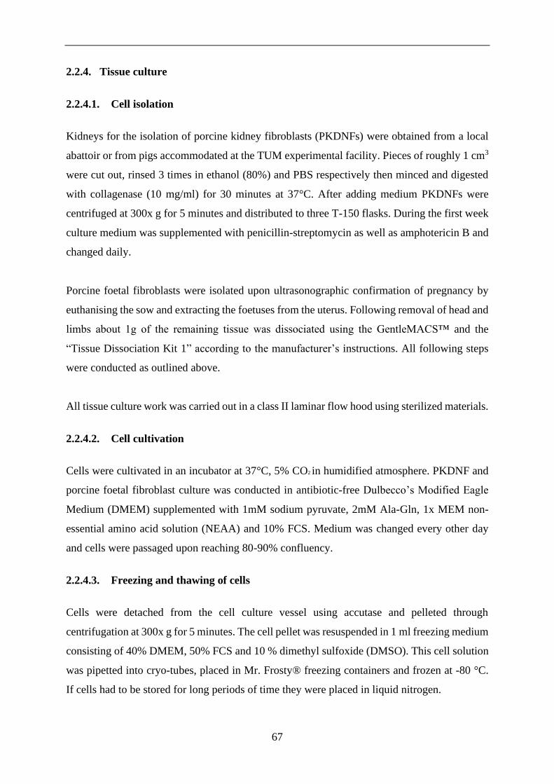
67
2.2.4. Tissue culture
2.2.4.1. Cell isolation
Kidneys for the isolation of porcine kidney fibroblasts (PKDNFs) were obtained from a local
abattoir or from pigs accommodated at the TUM experimental facility. Pieces of roughly 1 cm3
were cut out, rinsed 3 times in ethanol (80%) and PBS respectively then minced and digested
with collagenase (10 mg/ml) for 30 minutes at 37°C. After adding medium PKDNFs were
centrifuged at 300x g for 5 minutes and distributed to three T-150 flasks. During the first week
culture medium was supplemented with penicillin-streptomycin as well as amphotericin B and
changed daily.
Porcine foetal fibroblasts were isolated upon ultrasonographic confirmation of pregnancy by
euthanising the sow and extracting the foetuses from the uterus. Following removal of head and
limbs about 1g of the remaining tissue was dissociated using the GentleMACS™ and the
“Tissue Dissociation Kit 1” according to the manufacturer’s instructions. All following steps
were conducted as outlined above.
All tissue culture work was carried out in a class II laminar flow hood using sterilized materials.
2.2.4.2. Cell cultivation
Cells were cultivated in an incubator at 37°C, 5% CO2 in humidified atmosphere. PKDNF and
porcine foetal fibroblast culture was conducted in antibiotic-free Dulbecco’s Modified Eagle
Medium (DMEM) supplemented with 1mM sodium pyruvate, 2mM Ala-Gln, 1x MEM non-
essential amino acid solution (NEAA) and 10% FCS. Medium was changed every other day
and cells were passaged upon reaching 80-90% confluency.
2.2.4.3. Freezing and thawing of cells
Cells were detached from the cell culture vessel using accutase and pelleted through
centrifugation at 300x g for 5 minutes. The cell pellet was resuspended in 1 ml freezing medium
consisting of 40% DMEM, 50% FCS and 10 % dimethyl sulfoxide (DMSO). This cell solution
was pipetted into cryo-tubes, placed in Mr. Frosty® freezing containers and frozen at -80 °C.
If cells had to be stored for long periods of time they were placed in liquid nitrogen.

68
Cells were thawed in a water bath at 37 °C until the medium became liquid again. The cell
suspension was immediately diluted with 5 ml prewarmed medium and pelleted through
centrifugation at 300x g for 5 minutes. The pellet was resuspended in prewarmed culture
medium and transferred to the incubator for cultivation.
2.2.4.4. Counting of cells
Counting of cells was carried out using the Countess™ automated cell counter according to the
manufacturer’s instructions.
2.2.4.5. Transfection of cells
Transfection of PKDNF cells with DNA was carried out by lipofection or electroporation.
2.2.4.5.1. Lipofection
Prior to lipofection cells were cultivated to 50-70% confluency. The following day cells were
rinsed with PBS and cultivated in 4 ml of Opti-MEM® in 10 cm cell culture dishes. Five μg
DNA was dissolved in Opti-MEM® to a total volume of 300 μl and 6 μl Lipofectamine 2000
mixed with 294 μl Opti-MEM®. After 5 minutes of incubation at room temperature the
Lipofectamine mix was gently added to the DNA solution and co-incubated for 25 minutes at
room temperature. This compound solution was then trickled on the cells. After 6 hours of
cultivation 8 ml of medium was added to the cell culture dish followed by overnight cultivation
and a medium change the next day.
2.2.4.5.2. Electroporation
For electroporation cells were detached with accutase, counted and pelleted through
centrifugation at 300x g for 5 minutes. Then 1x10⁶ cells were transferred to 400 μl hypo-
osmolar buffer containing 5 μg of linearized plasmid DNA and transferred to an electroporation
cuvette with a diameter of 4 mm. After five minutes of incubation at room temperature one
pulse of 1200V was applied for 85μs. Following another five minutes of incubation at room
temperature the cell suspension was transferred to a 10 cm dish with fresh medium. Medium
was changed the following day.
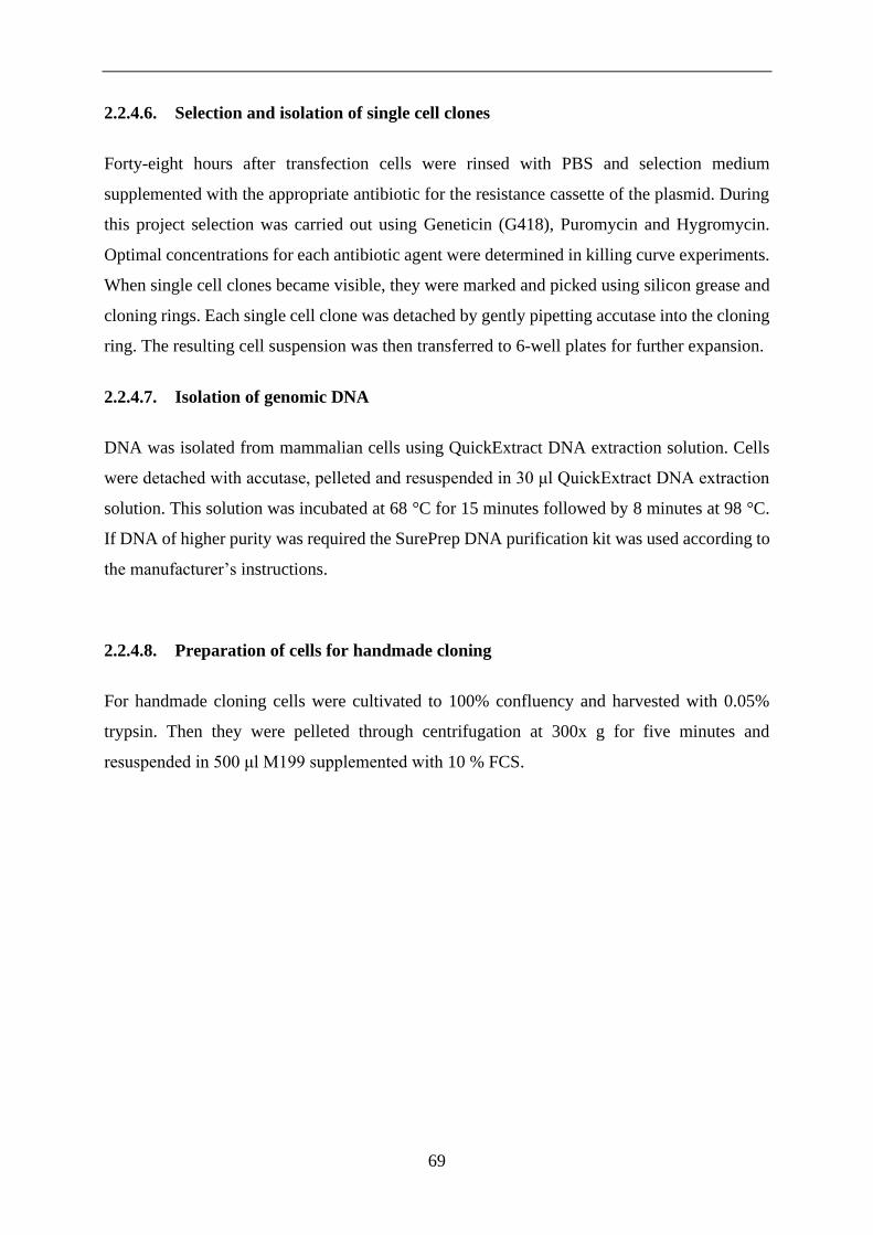
69
2.2.4.6. Selection and isolation of single cell clones
Forty-eight hours after transfection cells were rinsed with PBS and selection medium
supplemented with the appropriate antibiotic for the resistance cassette of the plasmid. During
this project selection was carried out using Geneticin (G418), Puromycin and Hygromycin.
Optimal concentrations for each antibiotic agent were determined in killing curve experiments.
When single cell clones became visible, they were marked and picked using silicon grease and
cloning rings. Each single cell clone was detached by gently pipetting accutase into the cloning
ring. The resulting cell suspension was then transferred to 6-well plates for further expansion.
2.2.4.7. Isolation of genomic DNA
DNA was isolated from mammalian cells using QuickExtract DNA extraction solution. Cells
were detached with accutase, pelleted and resuspended in 30 μl QuickExtract DNA extraction
solution. This solution was incubated at 68 °C for 15 minutes followed by 8 minutes at 98 °C.
If DNA of higher purity was required the SurePrep DNA purification kit was used according to
the manufacturer’s instructions.
2.2.4.8. Preparation of cells for handmade cloning
For handmade cloning cells were cultivated to 100% confluency and harvested with 0.05%
trypsin. Then they were pelleted through centrifugation at 300x g for five minutes and
resuspended in 500 μl M199 supplemented with 10 % FCS.

70
3. RESULTS
The goal of this thesis was to establish and improve methods for genome engineering in porcine
embryos.
For this purpose, systems for in vitro production (IVP) of porcine embryos comprising in vitro
maturation (IVM), in vitro fertilisation (IVF) and in vitro culture (IVC) were established.
Techniques for the cryopreservation of boar sperm were optimised, used to freeze semen
isolates suitable for IVF and build a sperm bank for genetically modified pig lines. Flushing of
in vivo zygotes was standardised (addressed in 3.1).
CRISPR/Cas9 mediated genome engineering was performed directly in early stage embryos.
Targeting vectors for the inactivation of the porcine NANOS2 gene were created. A variety of
gRNAs with minimal predicted off-target effects were evaluated for their genome engineering
efficiency. Those vectors and several others provided by colleagues were delivered to porcine
zygotes by microinjection and assessed for embryotoxicity and editing efficiency. A transposon
system was employed and the efficiency of this approach for transgenesis was evaluated.
(outlined in 3.2).
Promising constructs were used to generate genetically modified embryos. Surgical embryo
transfer was established and fourteen genetically modified pigs with four distinct genetic
modifications were obtained (explained in 3.3).
Finally, handmade cloning was implemented to facilitate more complex genome alterations that
require homology directed repair. These are inefficient and somatic cell nuclear transfer allows
pre-screening for the desired modification in cell culture, which is not possible when
performing the direct manipulation of porcine zygotes. Reconstructed embryos with the desired
genotype were generated and cultured to the blastocyst stage (described in 3.4).

71
3.1. In vitro embryo production
The Chair of Livestock Biotechnology specialises in creating porcine disease models for a
variety of human conditions and diseases. Reliable supply of in vivo derived porcine embryos
in sufficient quantity for this purpose would entail immense financial costs and sacrifice of
many donor animals. State of the art systems for in vitro production (IVP) of porcine embryos
comprising in vitro maturation (IVM), in vitro fertilisation (IVF) and in vitro culture (IVC)
were established during this work (see Figure 20). The optimised procedures for the IVP of
porcine embryos described in detail in section 2.2.1. are the most important result of this thesis.
Figure 20: In vitro embryo production. Oocytes were isolated from porcine ovaries sourced from a local
slaughterhouse. These were matured in vitro and served as cytoplast donors for handmade cloning or were in vitro
fertilised followed by microinjection, in vitro culture and embryo transfer.
3.1.1. In vitro maturation
An efficient in vitro maturation system is the foundation of in vitro embryo production. IVF
and handmade cloning require large quantities of mature oocytes, therefore both methods
benefit from improved IVM outcomes. Porcine oocytes are highly sensitive regarding
temperature, osmolarity, oxygen tension and medium composition. The objective was to
optimise IVM conditions and thereby improve the quantity and quality of IVM oocytes
measured by polar body extrusion, cleavage rate, blastocyst development and embryonic cell
count.

72
NCSU23 based maturation medium was compared to chemically defined cytokine-enhanced
maturation medium based on TCM199 (FLI-medium). FLI-medium attempts to approximately
mimic the composition of porcine oviductal fluid through supplementation with FGF, LIF and
IGF to promote more synchronous cytoplasmic and nuclear maturation. The composition of
both media is outlined in table 20. An equal number (100-150) of high-quality cumulus oocyte
complexes (COCs) with homogenous, dark, evenly granulated cytoplasm covered by multiple
compact layers of cumulus cells (see Figure 21) was matured in each medium for 45 hours
under otherwise identical conditions (outlined in 2.2.1.3).
Figure 21: Cumulus oocyte complexes selected for IVM. A) High-quality COCs with homogenous, dark, evenly
granulated cytoplasm covered by multiple compact layers of cumulus cells. B) Low-quality COCs sparsely
covered by cumulus cells.
Maturation rate was analysed by visually determining the percentage of oocytes showing
extrusion of the first polar body and cleavage rate (see Figure 22).

73
Figure 22: Extrusion of the first polar body. A) Porcine oocyte with polar body located at the 9 o’ clock position.
B) Hoechst staining of porcine oocyte with the polar body located at the 11 o’ clock position and nucleus slightly
below.
Oocytes matured in both media were parthenogenetically activated to induce embryonic
development without fertilisation. This approach was chosen to exclude variability caused by
sperm quality and IVF parameters. All embryos were cultured under identical conditions to
compare their developmental competence. Embryonic cleavage and blastocyst development
rates were evaluated. Five rounds of IVM were carried out with each maturation medium (see
Table 35).
Table 34: Comparison of NCSU23 medium and chemically defined FLI-medium
Maturation
medium Total (n) Maturation (%) Dead (%)
NCSU 23 1473 1042 (70.7%) 172 (11.68%)
TCM 199 (FLI) 1296 1063 (82.02%) 130 (10.03%)
Parthenogenesis Total (n) Cleavage (%) Blastocyst (%)
NCSU 23 1042 719 (69.00%) 217 (20.83%)
TCM 199 (FLI) 1063 753 (70.84%) 378 (35.56%)
Hoechst-staining was performed to determine the embryonic cell number on day 6 which is a
widely used indicator of blastocyst quality (shown in Figure 23). In vivo derived blastocysts
reach a cell number of approximately 100 cells at this point of embryonic development [318].
The average number of cells in blastocysts matured in NCSU 23 medium was 59.5±8.2 and
76.2±7.9 in FLI medium. Due to this data cytokine enhanced FLI maturation medium was used
in all further IVM experiments consistently resulting in average maturation rates of 80-85%.

74
Figure 23: Parthenogenetically generated blastocyst stained with Hoechst.
Parthenogenesis was further used to analyse the editing efficiency and cytotoxicity for a variety
of target genes. The efficiency of chemical and electrical parthenogenetic activation of porcine
embryos (outlined in 2.2.1.9.2) was compared over ten experiments. The average blastocyst
rate resulting from electrical activation was 50% (see Figure 24) compared to 36% after
chemical activation. Thus, electrical activation was used for all further parthenogenesis
experiments.
Figure 24: Blastocyst development rate of 50% after electrical parthenogenesis.

75
3.1.2. Cryopreservation of porcine sperm
High-quality frozen sperm facilitates consistent IVF outcomes over multiple experiments by
eliminating inter-ejaculate variability. The high sensitivity of porcine spermatozoa to oxidative
stress, temperature fluctuations, osmolarity and pH-value however makes their
cryopreservation challenging [231]. The objective was to optimise the freezing of porcine
sperm and compare the post-thaw survival rate of in-house sperm from GE boars and
commercial wildtype sperm.
During this project a system for the cryopreservation and storage of boar sperm was established.
Utilising the protocol outlined in 2.2.1.13 ejaculates from five breeding boars and ten GE pig
lines were frozen and a sperm bank was established (see Table 39).
Table 35: Sperm bank. Semen from GE pig lines is numerically labelled, samples from GE boars with the
respective names.
Boar: Genotype: Motility:
#123, #710 KRAS G12D/WT 30%, 40%
#227 APC 1311/WT, KRAS G12D/WT 50 %
#278, #3 TP 53 R167H/R167H 60%, 40%
#598 APC 1311/WT 60%
#662, #750, #908 humanised IgH and IgK 40%, 40%, 50%
#1530 TNF ΔARE/WT 30%
#760 R26-mT, reporter 10%
#869 humanised CD46, CD55,
CD59, HO1, A20; GGTA1 +/-
50%
#87 GGTA1-/-, CMAH-/-, B4GNT2-/- 40%
#10261 (2x) GGTA1-/-, CMAH-/-, B4GNT2-/-
, B2M-/-
40%, 50%
Fadros wildtype 40%
Fossil wildtype 30%
Uteus wildtype 40%

76
Wadtbandt wildtype 40%
Wal wildtype 30%
Cryopreservation decreased past-thaw sperm motility on average by 30% ranging from 25-
50%. The average post-thaw motility rate between all samples was 42%. Similar post-thaw
survival rates of at least 30% could be obtained for semen from wildtype boars and semen from
GE boars except boar #760. Only ejaculates of subpar quality could be obtained from this
subfertile boar expressing red fluorescent protein. Three sows were artificially inseminated with
cryopreserved sperm from boar #10261 resulting in three pregnancies.

77
3.1.3. In vitro fertilisation
The inefficiency of IVF due to polyspermy and insufficient male pronucleus formation is the
biggest hurdle for the IVP of porcine embryos [149]. The objective was to reduce the rate of
polyspermic fertilisation in porcine IVF and make the generation of GE pigs more efficient.
First, a working IVF system was established using proven sperm. Then semen isolates from
breeding boars were assessed for IVF suitability and the sperm to oocyte ratio was individually
optimised.
3.1.3.1. Establishment of a working IVF system
To supply the necessary number of embryos required to produce GE pigs by microinjection an
efficient IVF system needed to be established as part of this project. For this purpose, boar
semen repeatedly proven to generate blastocysts in IVF was kindly provided by Dr. Mayuko
Kurome (Chair for Molecular Animal Breeding and Biotechnology, LMU). Five IVF
experiments were conducted (as outlined in 2.2.1.4) adding 1x10⁶ spermatozoa to each IVF
well. Average blastocyst formation rates of 21.3% could be obtained (see Table 36 and Figure
25).
Table 36: Blastocyst development rates using sperm provided by Dr. Mayuko Kurome.
Oocytes (n) Blastocysts (%)
195 52 (26.7%)
243 66 (27.1%)
198 48 (24.2%)
207 24 (11.6%)
189 30 (15.9%)
Total: 1032 Total: 220 (21.3%)
Figure 25: Porcine Blastocysts produced by IVF; hatching blastocysts are marked with an asterisk.

78
3.1.3.2. Identification of suitable sperm isolates for IVF
Under 3.1.3.1. the basic protocol for IVF was established. The next step was to identify eligible
sperm donors because only semen from a minority of boars is suitable for IVF after
cryopreservation [151]. The goal was to identify such sperm isolates and assess in vitro
blastocyst development. High quality ejaculates from eighteen breeding boars and three GE
boars present at the animal facility were collected. Eight samples were frozen in house, thirteen
could be obtained frozen. At least three rounds of IVF were carried out for sperm from each
individual boar. In total 21 different sperm isolates were analysed for their IVF suitability
(shown in Table 37).
Motility rates after thawing were vastly different for all sperm samples. The initial sperm to
oocyte ratio was set at 7500 motile spermatozoa per oocyte to make results more comparable.
This was known to be a reasonable baseline from previous experiments.
Table 37: IVF suitability of 21 different boars. The first three numerically labelled sperm isolates are from boars
present at the TUM facility. The 18 animals identified by their names originate from a breeding company.
Boar Total oocytes (n) Total Blastocysts (%)
260 390 42 (11%)
420 300 14 (5%)
869 395 3 (1%)
Cadura 250 41 (16%)
Cor 320 36 (11%)
Fadros 700 167 (24%)
Fossil 300 6 (2%)
Icerico 250 1 (0%)
Igelspitz 250 7 (3%)
Madura 250 2 (1%)
Maswald 200 1 (0%)
Mozzi 250 8 (3%)
Orlaki 500 40 (8%)
Pablura 300 26 (9%)
Ryder 250 1 (0%)
Uteus 300 27 (9%)

79
Wadtbandt 300 22 (7%)
Wadtlise 300 6 (2%)
Wadtpill 300 12 (4%)
Wadttext 300 9 (3%)
Wal 200 4 (2%)
With a total number of 167 blastocysts produced from 700 oocytes (24%) Fadros showed the
highest performance in IVF. Consequently, semen from this boar was used for all further
experiments. The quality of Fadros’ sperm could later be confirmed by the generation of
twenty-nine healthy piglets through IVF (outlined in 3.3).
3.1.3.3. Optimisation of sperm to oocyte ratio
A high sperm to oocyte ratio increases the fertilisation rate in IVF but also raises the degree of
polyspermy [216]. The objective was to optimise the sperm to oocyte ratio to avoid polyspermy
while maintaining high fertilisation rates and blastocyst development.
Several IVF experiments using three different sperm donors and different sperm concentrations
were conducted for this purpose (see Table 38). Blastocyst development occurs at a normal rate
in polyspermic embryos which makes it an unsuitable parameter for measuring monospermic
fertilisation [245]. The rate of polyspermic fertilisation was therefore evaluated by performing
aceto-orcein staining (outlined in 2.2.1.5). An example is shown in Figure 26.
Figure 26: Zygotes in different fertilisation states. A) Unfertilized oocyte characterised by metaphase plate
(indicated by the arrow) B) Polyspermic fertilisation indicated by more than two pronuclei C) Monospermic
fertilisation indicated by exactly two visible pronuclei. Arrows indicate the positions of pronuclei.

80
Again, the most promising results were obtained for Fadros sperm for which monospermic
fertilisation rates of 55-60% could be confirmed repeatedly when using a concentration of
20.000 spermatozoa per oocyte (see Table 38). The overall fertilisation rate was 80% and
polyspermy could be limited to 23% of all oocytes. Higher sperm to oocyte ratios led to better
fertilisation rates but also caused a disproportionate increase in polyspermy. Considering the
average motility rate of 30% after thawing for this sperm this corresponds to a ratio of 6.000
motile spermatozoa per oocyte.
Table 38: Comparison of monospermic fertilisation rates from three different boars by aceto-orcein staining.
Optimal sperm concentration is indicated by a high percentage of oocytes with two pronuclei.
Boar Sperm/oocyte Monospermy % Polyspermy % Fertilised %
869 25.000 5.2 % 0 % 5.2 %
869 38.000 0 % 0 % 0 %
869 51.000 0 % 6.67 % 6.67 %
Uteus 25.000 0 % 0 % 0 %
Uteus 50.000 8 % 4 % 12 %
Uteus 100.000 0 % 30 % 30 %
Fadros 10.000 45 % 18 % 63 %
Fadros 20.000 57 % 23 % 80 %
Fadros 50.000 38 % 47 % 85 %
In summary, an efficient IVF system was established, suitable sperm isolates were identified
and the sperm to oocyte ratio was optimised. Monospermic fertilisation rates of 57% were
achieved while polyspermy could be reduced to 23%. All further IVF experiments were
conducted following this protocol.

81
3.1.4. Flushing of in vivo zygotes
The quality of in vitro produced porcine embryos is inferior to that of their in vivo derived
counterparts [149]. Furthermore, only wild type oocytes can be extracted from slaughterhouse-
derived ovaries. Flushing of in vivo zygotes is an effective method to obtain high-quality
porcine zygotes from GE pig lines if the necessary number of experimental animals is available.
Further genome alterations can then be performed on this genetic background.
Two TP53R167H/R167H gilts were super ovulated, artificially inseminated twice with sperm from
a TP53R167H/WT boar and euthanised 17 hours after the second AI (protocol outlined in 2.2.1.12).
Flushing of in vivo zygotes was performed yielding 35 one-cell-stage zygotes. To confirm their
developmental competence all zygotes were cultured in vitro for six days resulting in 16
blastocysts (see Figure 27). As expected, the rate of blastocyst development for in vivo derived
zygotes (45.7%) was much higher compared to the best result from in vitro generated zygotes
(24%).
Figure 27: Morphological comparison of in vivo and in vitro generated zygotes. A) In vitro generated zygotes;
two polar bodies are visible (top right). B) In vivo zygotes; Their greater size, bigger perivitelline space and dark,
even cytoplasm indicates their high quality. C) Blastocyst development after IVC of in vivo zygotes.
This experiment shows that in vivo zygotes could be extracted and successfully cultured to the
blastocyst stage in vitro with high efficiency. This method facilitates future projects that require
high quality embryos from GE pig lines to introduce additional modifications.

82
3.2. Genome engineering directly in porcine embryos
The objective of this part of the project was to use IVP porcine embryos for genome engineering
to generate new animal models. This method facilitates introduction of indels, or together with
single strand DNA templates to effect homologous sequence replacement through direct
manipulation of individual embryos. In combination with an efficient IVP system for porcine
embryos this approach can “fast-track” the generation of GE pigs for biomedical applications.
3.2.1. Viability of IVP embryos after microinjection
Microinjection facilitates delivery of transgenes, donor DNA and genome engineering
components but also causes cellular damage which adversely impacts embryo development
[319]. The average survival and blastocyst development rate after microinjection for in vitro
derived porcine zygotes is reported at 40-60% and 5-25% respectively [320]. The goal was to
optimise different technical parameters to improve the viability of IVP embryos after
microinjection. A variety of different needle types, shapes and diameters, injection volumes
and pressures were tested resulting in the protocol described in 2.2.1.6.
To visualize successful delivery of the injection solution eGFP mRNA was injected into the
cytoplasm of parthenogenetically activated porcine embryos. Twenty-four hours after injection
72% of all zygotes showed green fluorescence (see Figure 28), 15% were dead.
Figure 28: Green fluorescent porcine zygotes generated through microinjection of eGFP mRNA. A) One-cell-
stage; B) Cleaved oocyte 24 hours after eGFP injection.
Cleavage occurred in 55% of the oocytes showing eGFP expression and 22% developed to the
blastocyst stage. The control group that had undergone parthenogenetic activation without

83
microinjection showed cleavage rates of 70.84% and 35.56% blastocyst development. These
numbers indicate a 15.84% decrease in cleavage and 13.56% reduction in blastocyst
development after microinjection. An identical experiment was conducted injecting porcine
zygotes created by IVF resulting in blastocyst development rates of 14%. In the non-injected
control group 24% of all embryos developed to the blastocyst stage.
Overall, the optimised protocol for microinjection still had a negative impact on embryo
development but the outcomes compare favourably to the literature and facilitate the use of IVP
embryos for genome engineering.
3.2.2. Genome engineering in IVP embryos
The efficiency of genome engineering and embryotoxicity was assessed for a variety of target
genes and applications. DNA expression vectors are normally injected into one of the pronuclei
but this is difficult in pigs due to the pigmentation of porcine oocytes. Here we explored if the
cytoplasmic injection of DNA expression vectors is a suitable method for GE.
First, a DNA GE vector containing an expression cassette for both Cas9 and sgRNA was
microinjected into the cytoplasm of in vitro derived porcine zygotes. The efficiency of this
approach for the insertion of DNA fragments via homologous recombination was explored.
Then the precise excision of a DNA fragment using two gRNAs and simultaneous GE of
multiple target genes were tested. A transposon system was employed and the efficiency of this
approach for transgenesis was evaluated.
3.2.2.1. NANOS2
NANOS2 plays a key role in the sexual differentiation of germ cells. Male animals with a
homozygous knockout of this gene have intact testis completely lacking germ cells while
females carrying the same modification have a normal phenotype. Such males could therefore
be ideal recipients for the transfer of spermatogonial stem cells to enhance the reproductive
potential of GE boars or valuable breeding animals [321].
84
GE of the NANOS2 gene was used extensively to establish and improve technical aspects of
micromanipulation, DNA isolation protocols from porcine blastocysts and evaluation of
genome editing events.
3.2.2.1.1. Comparison of NANOS2 guide RNAs
Four different gRNAs for the NANOS2 gene (NANOS2 G1-4) with minimal predicted off-
target activity were identified and cloned into the pX330-U6-Chimeric_BB-CBh-hSpCas9
vector (see figure 29).
Figure 29: Structure of pX330-U6-Chimeric_BB-CBh-hSpCas9-NANOS2. It contains the CBh promoter and
hSpCas9 gene flanked by two nuclear localization signals (NLS), followed by a bGH-poly-A terminator sequence.
The sgRNA sequence includes an 18 bp gRNA homologous to the target sequence in exon 1 of the NANOS2 gene
followed by a gRNA scaffold driven by the U6 promotor.
Porcine kidney fibroblasts (PKDNFs) (isolate 250515) were transfected with each of the four
vectors (as outlined in 2.2.4.5). DNA was isolated from the pool of transfected cells and PCR
amplification was performed across the target sites. DNA sequencing was conducted and the
frequency of indel mutations determined by TIDE analysis (see figure 30).
85
Figure 30: Comparison of frequency and spectrum of indels at the NANOS2 target site by TIDE analysis.
A) NANOS2 G1 (49,1%), B) NANOS2 G2 (33%), C) NANOS2 G3 (26,4%), D) NANOS2 G4 (41%).
Out of the tested gRNAs NANOS2 G1 reached the highest on-target cleavage efficiency of
49,1% and was therefore used for GE in porcine embryos.
3.2.2.1.2. Detection of NANOS2 GE in porcine blastocysts
Most protocols for the isolation of DNA from blastocysts are optimised for mice. Porcine IVP
blastocysts contain only 50-75 cells and store large amounts of RNAs, proteins and especially
lipids. This reduces the quality of extracted DNA and makes downstream assays such as PCR
less efficient [322]. The objective was to identify suitable protocols for the isolation of DNA
from porcine blastocysts and assess if the injection of DNA GE vectors into the cytoplasm of
in vitro derived porcine zygotes results in efficient genome editing.
Two-hundred porcine oocytes were microinjected with the NANOS2G1 targeting vector
(5gn/µl) followed by parthenogenesis. Fifty-two embryos (26%) developed to the blastocyst
stage. Methods for the extraction of DNA from porcine blastocysts based on freeze-drying
[323], proteinase K digest [324], chemical lysis [315] and commercial DNA isolation kits were
compared. DNA of sufficient quality and quantity could only be obtained from 10 blastocysts
using the DNA isolation protocol described by Li et al which was used for all further
experiments [315].
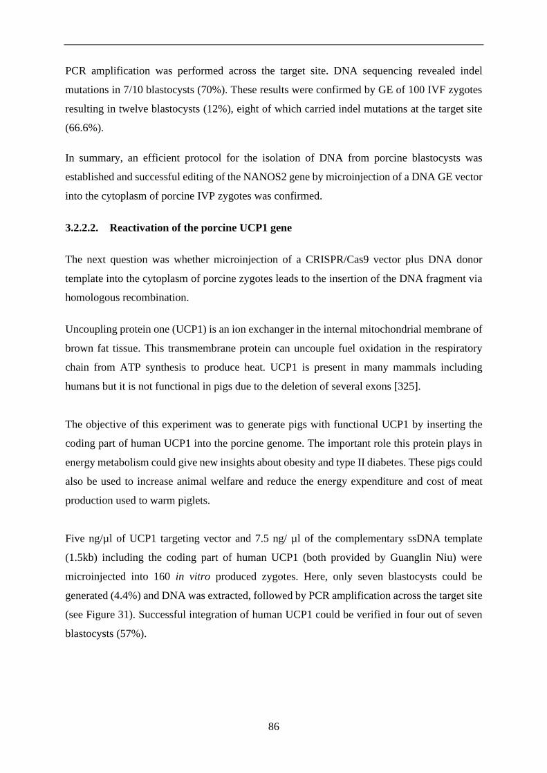
86
PCR amplification was performed across the target site. DNA sequencing revealed indel
mutations in 7/10 blastocysts (70%). These results were confirmed by GE of 100 IVF zygotes
resulting in twelve blastocysts (12%), eight of which carried indel mutations at the target site
(66.6%).
In summary, an efficient protocol for the isolation of DNA from porcine blastocysts was
established and successful editing of the NANOS2 gene by microinjection of a DNA GE vector
into the cytoplasm of porcine IVP zygotes was confirmed.
3.2.2.2. Reactivation of the porcine UCP1 gene
The next question was whether microinjection of a CRISPR/Cas9 vector plus DNA donor
template into the cytoplasm of porcine zygotes leads to the insertion of the DNA fragment via
homologous recombination.
Uncoupling protein one (UCP1) is an ion exchanger in the internal mitochondrial membrane of
brown fat tissue. This transmembrane protein can uncouple fuel oxidation in the respiratory
chain from ATP synthesis to produce heat. UCP1 is present in many mammals including
humans but it is not functional in pigs due to the deletion of several exons [325].
The objective of this experiment was to generate pigs with functional UCP1 by inserting the
coding part of human UCP1 into the porcine genome. The important role this protein plays in
energy metabolism could give new insights about obesity and type II diabetes. These pigs could
also be used to increase animal welfare and reduce the energy expenditure and cost of meat
production used to warm piglets.
Five ng/µl of UCP1 targeting vector and 7.5 ng/ µl of the complementary ssDNA template
(1.5kb) including the coding part of human UCP1 (both provided by Guanglin Niu) were
microinjected into 160 in vitro produced zygotes. Here, only seven blastocysts could be
generated (4.4%) and DNA was extracted, followed by PCR amplification across the target site
(see Figure 31). Successful integration of human UCP1 could be verified in four out of seven
blastocysts (57%).
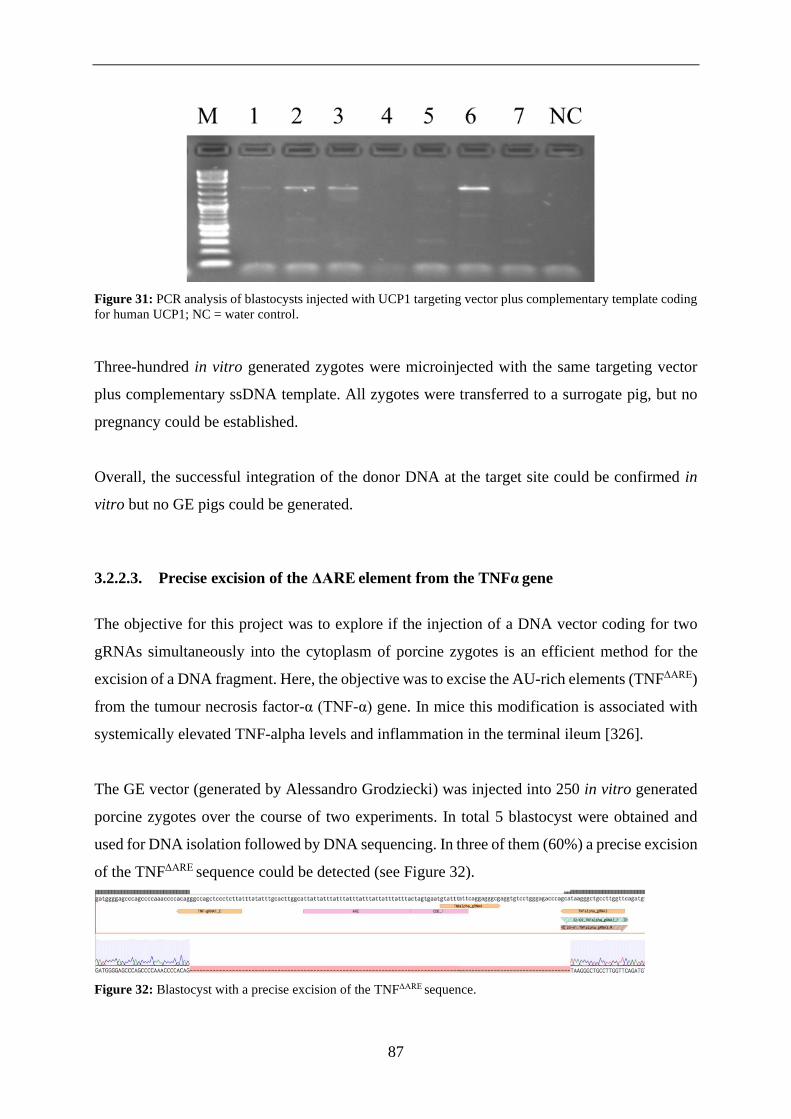
87
Figure 31: PCR analysis of blastocysts injected with UCP1 targeting vector plus complementary template coding
for human UCP1; NC = water control.
Three-hundred in vitro generated zygotes were microinjected with the same targeting vector
plus complementary ssDNA template. All zygotes were transferred to a surrogate pig, but no
pregnancy could be established.
Overall, the successful integration of the donor DNA at the target site could be confirmed in
vitro but no GE pigs could be generated.
3.2.2.3. Precise excision of the ΔARE element from the TNFα gene
The objective for this project was to explore if the injection of a DNA vector coding for two
gRNAs simultaneously into the cytoplasm of porcine zygotes is an efficient method for the
excision of a DNA fragment. Here, the objective was to excise the AU-rich elements (TNFΔARE)
from the tumour necrosis factor-α (TNF-α) gene. In mice this modification is associated with
systemically elevated TNF-alpha levels and inflammation in the terminal ileum [326].
The GE vector (generated by Alessandro Grodziecki) was injected into 250 in vitro generated
porcine zygotes over the course of two experiments. In total 5 blastocyst were obtained and
used for DNA isolation followed by DNA sequencing. In three of them (60%) a precise excision
of the TNFΔARE sequence could be detected (see Figure 32).
Figure 32: Blastocyst with a precise excision of the TNFΔARE sequence.

88
In summary, the TNFΔARE sequence was successfully excised using a GE vector that codes for
two gRNAs simultaneously.
3.2.3. Simultaneous genome editing of CMAH and B4GALNT2
The goal was to explore whether the injection of a DNA vector into the cytoplasm of porcine
zygotes is a suitable method to simultaneously edit multiple target genes. The removal of
xenoreactive antigens through inactivation of porcine genes can minimise the rejection of pig
organs by human recipients in xenotransplantation. CMAH and B4GALNT2 are genes coding
for major xenogeneic antigens. During this thesis a targeting vector aimed at the inactivation of
these two genes (provided by Beate Rieblinger) was tested in porcine embryos.
The plasmid was microinjected into 373 in vitro produced zygotes. Seventy-three zygotes were
cultured in vitro for six days and five blastocysts (6.8%) could be obtained compared to twelve
out of 88 (13.6%) blastocysts in the non-injected control group. The remaining 300 injected
zygotes were transferred into the oviduct of a synchronised recipient, but no pregnancy could
be established.
DNA isolation from the injected blastocysts and analysis by PCR amplification followed by
DNA sequencing of the target sequences of the CMAH and B4GALNT2 genes was conducted
by Thomas Winogrodzki (master student). Homozygous knockouts of CMAH and B4GALNT2
could be verified in one (20%) blastocyst whereas heterozygous knockouts of CMAH and
B4GALNT2 could be observed in three (60%) blastocysts (see Table 40).

89
Table 39: DNA sequencing results from blastocysts microinjected with CMAH- B4GALNT2-double knockout
vector.
Blastocyst CMAH B4GNT2
1 +1 Heterozygous
T-insert
Wildtype
2 +1 Heterozygous
T-insert
Multiple
mutations
3 +1 Homozygous
T-insert
Multiple
mutations
4 +1 Heterozygous
T-insert
Heterozygous T-
deletion
5 Homozygous T-
insert
Homozygous T-
insertion
In brief, two target genes could simultaneously be inactivated by microinjection of a GE vector
into the cytoplasm of in vitro derived porcine zygotes.
3.2.3.1. Transposon mediated transgenesis via cytoplasmic injection of embryos
The objective for this project was to explore if the injection of a PiggyBac (PB) transposon
DNA vector plus PB Transposase mRNA into the cytoplasm of porcine zygotes is an efficient
method for transgenesis. PB Transposase recognises the inverted terminal repeats (ITRs) of the
transposon vector excises it from the plasmid backbone and randomly integrates the vector
containing the gene of interest into the genome at “TTAA” sites via a cut and paste mechanism
[78].
Here the gene of interest was the codon-improved Cre recombinase (iCre) driven by the
pancreas-specific mouse pancreas duodenum homeobox-1 (mPdx1) promoter for the activation
of conditional oncogenic mutations (see 3.3.5).
The transposon plasmid and transposase-mCherry mRNA (both generated by Daniela Kalla,
see Figure 33) were injected into 100 in vitro generated porcine zygotes followed by
parthenogenetic activation.

90
Figure 33: Structure of the components of the transposon system A) MPdx1-iCre Transposon DNA vector. It
contains the mPdx1 promoter, rabbit beta globin intron, iCre and bGH-poly-A flanked by two ITRs. B) Structure
of PB transposase expression vector. It contains the CMV promoter, T7 promoter, PB Transposase, a T2A, the
mCherry sequence and bGH-poly-A.
Upon translation of the injected mRNA the polypeptide is broken apart at the T2A site via
ribosome skipping into PB Transposase and mCherry [327]. Twenty-four hours after
microinjection the oocytes were assessed for expression of mCherry and PB Transposase
indicated by red fluorescence (see Figure 34).
Figure 34: Oocytes 24 hours after microinjection with transposon plasmid and transposase mRNA. Red
fluorescence indicates the expression of mCherry and PB Transposase. A) Bright field: The oocytes have not
undergone cleavage but their membranes are intact. B) Dark field, fluorescence imaging: MCherry fluorescence
is clearly visible. C) Overlay.
After six days of in vitro culture 42/100 (42%) oocytes developed to the blastocyst stage
compared to 25/50 (50%) in the non-injected control group. DNA isolation followed by PCR
amplification across the target site revealed the mPdx1-iCre sequence in 18/42 (43%)
blastocysts (see Figure 35).

91
Figure 35: PCR analysis of blastocysts injected with mPdx1-iCre transposon plasmid plus PB Transposase
mRNA. Black arrows indicate the positive samples; CTRL = non-injected blastocysts; H2O = water control.
In Summary, the mPdx1-iCre sequence was successfully ascertained after cytoplasmic injection
of a DNA transposon plasmid plus Transposase mRNA.

92
3.3. Generation of porcine models for biomedicine
The generation of porcine disease models was the main objective of this project. All previously
described methods, such as in vitro production of porcine embryos, cryopreservation of sperm,
genome engineering of porcine zygotes by microinjection and embryo transfer were optimised
to facilitate this goal.
3.3.1. Embryo transfer
During this thesis endoscopic and surgical embryo transfer (ET) were carried out. Training for
endoscopic ET was obtained from Dr. Barbara Kessler. Surgical ET was established as an
alternative for endoscopic ET (see Figure 36). Both techniques facilitate transfer of one to two-
cell stage embryos to the oviduct. Surgical ET also allows for bicornual transfer of zona-free
blastocysts directly to the uterus. This is necessary for HMC because zona-free embryos must
be cultured to the blastocyst stage in vitro to be able to survive in vivo.
Figure 36: Surgical embryo transfer. The recipient pig was fixated on the surgery table. The abdomen was opened,
and embryos were transferred directly into the oviduct with a sterile catheter. The surgery wound was stitched in
three layers and aluminium spray was applied.
Vectors that were previously tested in in vitro generated and cultured embryos (outlined in 3.3)
were used to generate genetically modified embryos. In total twenty-two ETs were carried out
and eleven pregnancies were established. Twelve of them were performed endoscopically by
Dr. Barbara Kessler (Chair for Molecular Animal Breeding and Biotechnology) and ten
surgically by me (as outlined in 2.2.1.11). Five out of ten surgical ETs and six out of twelve
endoscopic ETs resulted in pregnancies yielding an equal pregnancy rate of 50% for both
methods. One surgical ET experiment was discontinued due to postoperative complications and
the pig was euthanised. Three pregnancies were confirmed by sonography on day 21 but due to
resorption of the embryos they were not carried to full term. Five pregnancies resulted in the
birth of 29 piglets with fourteen of them carrying four different genetic modifications. One
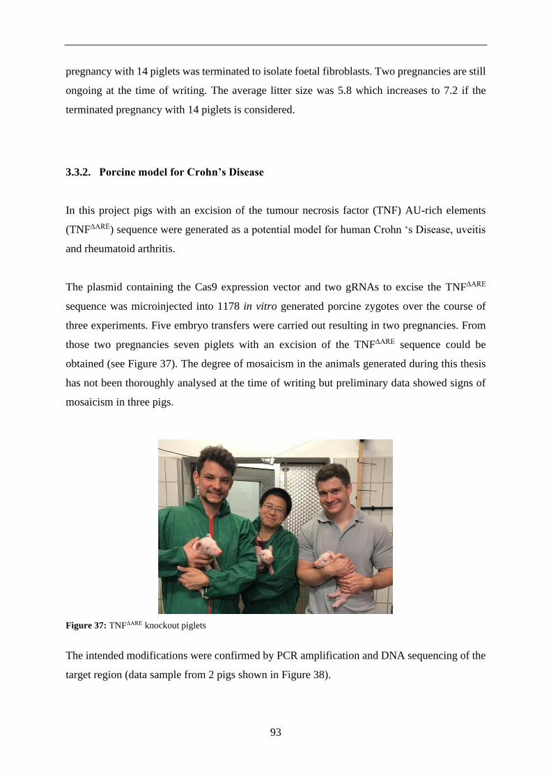
93
pregnancy with 14 piglets was terminated to isolate foetal fibroblasts. Two pregnancies are still
ongoing at the time of writing. The average litter size was 5.8 which increases to 7.2 if the
terminated pregnancy with 14 piglets is considered.
3.3.2. Porcine model for Crohn’s Disease
In this project pigs with an excision of the tumour necrosis factor (TNF) AU-rich elements
(TNFΔARE) sequence were generated as a potential model for human Crohn ‘s Disease, uveitis
and rheumatoid arthritis.
The plasmid containing the Cas9 expression vector and two gRNAs to excise the TNFΔARE
sequence was microinjected into 1178 in vitro generated porcine zygotes over the course of
three experiments. Five embryo transfers were carried out resulting in two pregnancies. From
those two pregnancies seven piglets with an excision of the TNFΔARE sequence could be
obtained (see Figure 37). The degree of mosaicism in the animals generated during this thesis
has not been thoroughly analysed at the time of writing but preliminary data showed signs of
mosaicism in three pigs.
Figure 37: TNFΔARE knockout piglets
The intended modifications were confirmed by PCR amplification and DNA sequencing of the
target region (data sample from 2 pigs shown in Figure 38).

94
Figure 38: Data sample from two TNFΔARE pigs. A) PCR amplification of the target site reveals mutant bands at
440bp for pigs #1530 and #1532. B) DNA sequencing of the target region confirms the excision of the TNFΔARE
sequence.
Overall, seven pigs with an excision of the TNFΔARE sequence were generated in this project. It
remains to be seen if these animals develop a disease phenotype with age.

95
3.3.3. Preliminary work towards a porcine Hepatitis model
The objective for this project was to generate GE pigs susceptible to hepatitis B virus (HBV)
infection. Similar to the editing experiment of the UCP1 gene this project requires the targeted
insertion of a DNA sequence via homologous recombination but here the sequence is much
shorter (20bp compared to 1.5 kb).
Viral hepatitis is a major global health problem causing approximately 880.000 deaths per year
worldwide [328]. HBV binds to the natrium-taurocholate co-transporting polypeptide (NTCP)
which is a bile acid transporter encoded by the SLC10A1 gene [329]. HBV infections are
limited to great apes (Hominidae) and humans due to interspecies variations in the amino acid
sequence of the NTCP receptor. The NTCP amino acid sequence has been modified in mice to
match the human equivalent but these animals were not susceptible to HBV infection. Porcine
hepatocytes expressing the human NTCP receptor however, have been shown to enable
productive HBV infections which could make NTCP humanized pigs a suitable animal model
for HBV research [330].
The targeting vector and sgRNA required for the generation of a possible model for human
Hepatitis were produced by Dr. Konrad Fischer. In five experiments this targeting vector plus
ssDNA donor template was injected into 1672 in vitro generated porcine zygotes. Five embryo
transfers were carried out resulting in two pregnancies from which in total five piglets were
obtained. In two piglets (40%) indel mutations in the porcine NTCP gene could be identified
by PCR amplification and DNA sequencing (see Figure 39). The human NTCP sequence could
not be ascertained in any of those pigs.

96
Figure 39: A) Two pigs with indel mutations in the porcine NTCP gene. B) DNA Sequencing across the target
site reveals indel mutations in the porcine NTCP gene but the intended humanisation of the NTCP gene could not
be achieved.
In summary, two pigs with indel mutations in the NTCP gene could be generated but the
integration of the human NTCP sequence was not successful.
3.3.4. Simultaneous GE of multiple genes relevant for xenotransplantation
The objective for this project was to evaluated if quadruple knockout pigs can be generated for
xenotransplantation, and if so with which efficiency. The gene editing vector tested in 3.3.2.4
aimed to knockout two genes that synthesize xenogeneic glycosylation patterns (CMAH and
B4GALNT2). The fourfold knockout vector used for this experiment (created by Beate
Rieblinger) targets the GGTA1 and SLA class I genes in addition the other two genes
(GGTA1/CMAH/B4GALNT2/SLA class I).
The vector was microinjected into 200 in vitro produced zygotes. Those zygotes were
transferred to a synchronized recipient alongside 200 injected with the TNFΔARE vector resulting
in a pregnancy. Seven piglets were born, two of them with an excision of the TNFΔARE sequence

97
(see 3.3.2.) and one of them with homozygous knockouts of the GGTA1 and B4GALNT2 genes
(see Figure 40). No indel mutations could be found in the CMAH and SLA class I genes.
Figure 40: Sequencing of the GGTA1 B4GALNT2 knockout pig #1529.
In summary, a pig with homozygous knockouts of the GGTA1 and B4GALNT2 genes could
be generated but the other two target genes remained unmodified.
3.3.5. Porcine model for pancreatic cancer
The overarching goal of this project is the generation of pigs predisposed to pancreatic cancer
by conditional, tissue-specific activation of oncogenic mutations. For this purpose, a pig line
carrying oncogenic KRASG12D and TP53R167H mutations silenced by a “stop” cassette had been
previously generated [331, 332]. Here the objective was to generate a pig line expressing Cre-
recombinase specifically in the pancreas to activate these conditional mutations by
crossbreeding. For this purpose, the mPdx1 promoter was used to direct Cre expression to the
developing pancreas.
The mPdx1-iCre transposon vector and PB transposase/mCherry mRNA (see 3.2.2.5.) was
injected into 1227 in vitro generated porcine zygotes over the course of three experiments. Five
embryo transfers were carried out resulting in two pregnancies. One pregnancy was confirmed
sonographically on day 21 but was not carried to full term due to resorption of the embryos.
From the other pregnancy ten piglets were obtained, three of them carrying the desired mPdx1-
iCre sequence (see Figure 41).

98
Figure 41: A) PCR amplification reveals mutant bands at 2226bp for pigs #2017, 2022 and 2024 (indicated by the
pink arrows). B) Three transgenic pigs carrying the mPdx1-iCre sequence. C) DNA sequencing of the target region
confirms the integration of the mPdx1-iCre sequence (data sample from pig #2017).
In summary, three transgenic mPdx1-iCre pigs were generated. It remains to be seen if these
animals develop a disease phenotype after crossbreeding with the KRASG12D and TP53R167H
line.

99
3.4. Handmade cloning
Previous experiments have shown that the integration of DNA fragments using GE-vectors and
DNA donor templates is inefficient. These more complex genome alterations are better carried
out in somatic cells because this approach enables pre-selection for the desired modification.
Modified donor cells can then be used for somatic cell nuclear transfer to generate GE pigs.
Therefore, the next goal of this thesis was to establish handmade cloning to facilitate projects
that require homologous recombination or editing of multiple genes simultaneously. Initial
instructions for HMC were given by Prof. Lin (Department of biomedicine, Aarhus University).
The first objective was to generate cytoplasts and test their viability after enucleation. Mature
oocytes were enucleated with a handheld blade after zona pellucida removal (as outlined in
2.2.1.14). Successful enucleation and generation of the resulting zona free cytoplasts was
confirmed by Hoechst staining (see Figure 42).
Figure 42: Hoechst-staining of zona-free cytoplasts generated through enucleation of mature oocytes. A) Bright
field, B) Dark field; Insufficient enucleation is indicated by blue fluorescence (oocyte to the right).
The next objective was to generate reconstructed embryos and assess their developmental
potential. Cytoplasts were fused with porcine kidney fibroblasts in two steps to generate eight
reconstructed embryos. They were cultured in vitro for six days in a well-of-the-well system.
Two blastocysts were generated (25%) and four embryos developed to the morula stage (50%)
(see Figure 43).

100
Figure 43: Fusion steps during handmade cloning procedure resulting in blastocysts after six days of in vitro
culture A) Fusion chamber used for both fusion steps in HMC. B) Somatic cell fused to cytoplast (indicated by
red arrow). C) Reconstructed embryos after fusion with second cytoplast (indicated by red arrows) D) Blastocysts
developed from reconstructed HMC embryos after six days of in vitro culture.
Preliminary, somatic cell nuclear transfer was successfully conducted using the handmade
cloning technique. Twenty-five percent of reconstructed embryos developed to the blastocyst
stage but this number of is insufficient for embryo transfer.

101
4. DISCUSSION
The objective of this work was to optimise the in vitro production of porcine zygotes and
establish a system for direct manipulation of embryos to “fast-track” the generation of porcine
models for biomedicine.
Section 4.1 addresses the optimisation of the IVP system for porcine embryos including in vitro
maturation, in vitro fertilisation, in vitro culture and the cryopreservation of boar sperm.
CRISPR/Cas9 mediated genome engineering directly in early embryos and transgenesis using
a transposon system is examined in segment 4.2. The generation of thirteen GE pigs with four
distinct genotypes is debated in passage 4.3. Finally, the establishment of HMC as an alternative
to TC is discussed is section 4.4
4.1. In vitro embryo production
In vitro embryo production is a multistage process that requires proper interaction of a variety
of techniques including IVM, sperm preparation, IVF and IVC of embryos [218] which are
discussed in this section. The quality of embryos measured by embryonic cell count, maturation
rates and blastocyst development generated with the IVP system described here could be
improved markedly over the course of the project.
4.1.1. In vitro maturation
An efficient in vitro maturation system is essential to supply an adequate number of mature
oocytes for SCNT and IVF [85]. The maturation protocol that was established and optimised in
this thesis reliably promotes maturation rates above 80% which matches or slightly exceeds
rates described in most publications [144, 149]. The average blastocyst development rate of
50% after electrical activation further exemplifies the high developmental potential of oocytes
generated with this IVM system. The close proximity of a slaughterhouse decreased
transportation times for ovaries to less than 30 minutes which is known to positively influence
oocyte quality [333].
102
Maturation media are usually supplemented with EGF to support maturation [334] but analysis
of follicular fluid provided evidence that several other growth factors are required to adequately
support oocyte maturation [335]. Meiotic arrest of oocytes in the follicle is mediated by high
levels of cAMP [336]. A sudden decrease in cAMP levels induced by removal of oocytes from
their follicular environment causes asynchronous cytoplasmic and nuclear maturation and
impairs embryonic development [202]. FGF, IGF and LIF are all cytokines that are known to
be present in porcine follicular fluid (PFF) [335, 337]. Together they effectively regulate cAMP
levels leading to more synchronous cytoplasmic and nuclear maturation [202].
This effect can be replicated by including PFF in maturation media for its growth factor content
but PFF also contains potent maturation inhibitors such as hypoxanthine [199]. Chemically
defined media as used here eliminate the potential for transmission of pathogens that might be
present in biological fluids [338]. They also lead to higher reproducibility by eliminating
biological variables, such as the quality of PFF and FCS [169, 339].
Nuclear maturation rates consistently exceeding 80% and a 15% higher blastocyst rate after
parthenogenesis compared to the PFF supplemented medium show the effectiveness of the
chemically defined, cytokine enhanced approach. The increased embryonic cell count,
comparatively high rate of monospermic fertilisation and high consistency support the validity
of this IVM system. Downsides are its slightly higher complexity and costs.
In summary, the IVM system established during this thesis consistently yielded mature oocytes
of high quality. Similar efficiencies are described for other cytokine-supplemented IVM media
[177]. Performance metrics regarding embryo quality compare favourably to most other IVM
outcomes described in the literature [135, 169, 181, 201].
4.1.2. In vitro fertilisation
IVF remains a limiting factor for the IVP of porcine embryos due to the unsolved issue of
polyspermy [149]. Polyspermy is a complex problem to solve because it is influenced by many
different variables, such as gamete coincubation times [242], supplementation of media with
different molecules [212], sperm quality [340] and sperm to oocyte ratio [341].

103
All IVF parameters were optimised exclusively for frozen-thawed sperm to standardize for
differences between boars and ejaculates from the same boar [236]. The starting point for all
experiments was a proven IVF protocol utilising sperm provided by Dr. Mayuko Kurome (Chair
for Molecular Animal Breeding and Biotechnology, LMU). The next step was the identification
of suitable sperm donors to further improve IVF outcomes.
Ejaculates from 21 different boars were analysed for their IVF suitability. Blastocysts could be
produced with most sperm samples but IVF performance was vastly different among boars
which is also described in the literature [235, 342]. Many publications suggest that only sperm
from a minority of boars is suitable for IVF after cryopreservation [151, 343]. The data
generated during this thesis supports this assumption even though exclusively high-quality
ejaculates from highly fertile breeding boars were used for IVF.
Blastocyst formation rates of on average 24% using sperm from the highest performing boar
(Fadros) are in line with reports from other IVF laboratories using in vitro derived oocytes [149,
165]. While there are publications reporting much higher blastocyst development rates these
numbers are usually achieved using in vivo derived oocytes [125] or associated with rates of
polyspermy reaching up to 90% [214, 215]. Reports of high blastocyst development rates
calculated based on pre-selected subgroups of mature or fertilised oocytes should be seen in
perspective [344].
Blastocyst development rates are only relevant in the context of monospermic fertilisation
which is a more suitable parameter to measure the validity of an IVF system [245]. The degree
of polyspermy in IVF is closely tied to the ratio of sperm to oocytes during fertilisation [216].
Individual optimisation of IVF parameters resulted in monospermic fertilisation rates of 57%
accompanied by 23% of polyspermy which is consistent with the 50-60% efficiency reported
for modern IVF systems [218]. The slightly higher fertilisation rates that were observed when
adding more spermatozoa during IVF came with a strong increase in polyspermy (results are
shown in table 24). This confirms the prevailing assumption that a compromise has to be made
between optimal fertilisation and acceptable rates of polyspermy [218].
Oocyte quality is another critical factor affecting polyspermy [219, 220]. Here, a chemically
defined, cytokine supplemented maturation medium was used to improve the quality of IVM
oocytes (discussed in 4.1.1). Higher rates of monospermic fertilisation can be achieved using

104
oocytes from adult sows [160] or in vivo derived zygotes [247] but the ability of in vitro matured
oocytes to block polyspermy remains low [345]. The IVF protocol used here foregoes removal
of cumulus cell. This reduces polyspermy by posing an additional barrier mimicking in vivo
selection of the most motile spermatozoa [346].
In vivo fertilisation takes place in oviductal fluid (OF) which makes it a logical supplement for
IVM media [347]. Its chemical composition and beneficial effects on monospermic fertilisation
are dependent on the phase of the oestrous cycle upon collection [348, 349]. This variability in
composition is further increased because the presence of spermatozoa [350], oocytes, or a
combination of both [351] which leads to large alterations in the oviduct proteome. While OF
supplementation has positive effects on IVF outcomes when using fresh sperm [352]
detrimental effects have been reported for frozen-thawed sperm [348]. These findings
demonstrate OFs potential as a supplement for IVF media, but they also highlight its high
complexity and variability. Sanitary certified OF, classified for oestrous cycle and biological
activity is an auspicious additive that could reduce the incidence of polyspermy [218].
A 3-dimensional IVF system within an organ-on-a-chip system [353] is another promising
concept to improve the quality of in vitro derived porcine embryos. Until such options become
commercially available a combination of sperm selection methods with short co-incubation
times [244] is a practical approach to further optimise the IVF system discussed here.
In summary, an IVF system was established and optimised reaching 57% efficiency measured
by the rate of monospermic fertilisation. While polyspermy remains an unsolved problem, it
was minimised by optimising the sperm to oocyte for each boar and improving the quality of
in vitro matured oocytes. The generation of 29 pigs using zygotes derived from this IVF system
(discussed in 4.3) during the first year of implementation further supports its validity.

105
4.1.3. Cryopreservation of boar sperm
Standardised, efficient in vitro production of porcine embryos requires high-quality frozen
sperm suitable for IVF [176, 229] to eliminate inter-ejaculate variability and improve IVF
consistency [176]. However, only sperm from a minority of “good freezer” boars [228] is
suitable for IVF after cryopreservation [151, 343]. The high sensitivity of pig sperm to oxidative
stress, temperature fluctuation, osmolarity and pH-value [230, 231] make its cryopreservation
challenging and commercially unappealing [236]. Therefore, good quality frozen sperm for IVF
is scarce. A growing demand for such sperm due to the increasing popularity of porcine disease
models in combination with improved cryopreservation methods might help to alleviate this
shortage in the future.
During this project cryopreservation of sperm was performed using a controlled-rate freezer.
This method minimises ice crystal formation which improves sperm survival rates [354]. The
resulting average post-thaw motility rate of 42% lies right in the middle of the broad 20-60%
range described by literature [228, 355]. Some ejaculates were already characterised by low
motility prior to cryopreservation which has a negative impact on post-thaw survival rates
[356]. The large variability in sperm function after freezing can be explained by male-to-male
variability [235] and has even been described between ejaculates from the same boar [236].
Artificial insemination of three sows with cryopreserved sperm from boar #10261 resulted in
three pregnancies. This suggests that the quality of this batch of frozen semen is sufficient for
the re-derivation of GE pig lines.
Post-thaw motility is a suitable parameter to predict boar sperm fertilisation competence during
artificial insemination (AI) [357] but there is only a weak correlation to the rate of monospermic
fertilisation in IVF [236, 356]. Therefore, it is an inadequate indicator for the suitability of cryo-
conserved boar sperm for IVF which can only be assessed by measuring the rate of
monospermic fertilisation [358].
The same cryopreservation protocol was used to build a sperm bank (shown in table 39) for GE
pig lines to prevent the loss of genetic information due to infection or injury-related death of
valuable boars. A plausible threat is African Swine Fever Virus which is present in both
neighbour countries France and Poland at the time of writing [359, 360]. Frozen sperm proven
suitable for IVF can be shipped to other laboratories together with the optimised protocols to

106
facilitate consistent IVF experiments there. Moreover, frozen sperm from GE boars can be
delivered to other research institutes and used for crossbreeding via AI.
In summary, the sperm freezing system established during this thesis promotes post thaw
motility rates similar to other publications [228, 355]. It was successfully used to freeze sperm
from breeding boars for IVF and establish a sperm bank for GE pig lines.

107
4.2. Genome engineering in IVP embryos
Microinjection has been a foundational technology in manipulating mammalian genomes since
the creation of one of the first genetically modified animals in 1974 but it only allowed for
random transgene addition [33]. The emergence of nuclease-based genome engineering
technology has expanded the potential of microinjection by contributing the ability to
efficiently introduce targeted modifications during embryogenesis [361]. Here, genome
engineering was performed directly in early embryos by delivering several GE vectors and the
constituents of a transposon system to porcine zygotes via microinjection.
Microinjection facilitates efficient delivery of genome engineering components but it also has
a negative impact on embryo development [319]. A decrease of 15.84% in cleavage rate and
10% in blastocyst development rate was observed in microinjected IVF embryos compared to
the non-injected control group. Similar adverse effects were seen in parthenogenetic embryos
which can be explained through the cellular damage caused by microinjection itself [362].
EGFP mRNA was used to visualize successful delivery of the injection solution. Therefore, the
cytotoxicity and immunogenicity of GFP is another possible explanation [363].
Besides its adverse effect on embryo development microinjection of site specific nucleases also
frequently leads to mosaicism which is especially problematic in pigs due to the long generation
interval [85]. In rodents the incidence of mosaicism lies between 20-70% [364, 365] whereas
the rate of mosaicism in pigs is reported at approximately 10-20% [124, 136]. Differences in
embryo development, timing of microinjection and varying efficiencies of the genome
engineering components used in each study are plausible explanations for this disparity [366].
The degree of mosaicism in pigs created during this project has not been thoroughly analysed
at the time of writing but preliminary data revealed mosaicism in at least four pigs. Increasing
the concentrations of CRISPR components reduces mosaicism but it diminishes embryonic
viability at the same time [367]. Another approach is to make Cas9 protein less persistent by
tagging it with an ubiquitin-proteasome degradation signal [368] or to use multiple sgRNAs
targeting the same gene [369]. Injection of Cas9 protein instead of plasmids or Cas9 RNA was
shown to reduce mosaicism [370]. Timing of the microinjection is another critical factor. Here,
microinjection was performed right after the IVF protocol which takes 7 hours to complete.

108
This is before the time frame of pronucleus formation at 12-15h after fertilisation where genome
replication reportedly takes place [371].
In this thesis GE was performed using DNA GE vectors coding for the specific sgRNA and
Cas9 protein. Other possible options include delivery of the CRISPR/Cas9 components as Cas9
protein or mRNA together with the sgRNA. These approaches don’t require transcription and
translation to facilitate genome engineering which can reduce the rate of mosaicism [127, 372].
They also eliminate the risk for unwanted integration of the DNA vector into the host genome
but are more labour intense to prepare [303].
A concern regarding CRISPR/Cas9 technology in general is the potential for off-target cleavage
[373]. Such unintended DSBs occur at sites that differ by up to 5 bases from the target sequence
and can result in adverse phenotypic consequences [374]. Detection of off-target mutations is
hampered by the limited usefulness of in silico tools that predict possible off-target sites based
on their similarity to the target sequence [375]. Whole genome sequencing and another
approach termed genome-wide off-target analysis by two-cell embryo injection report the
frequency of off-target effects caused by the CRISPR/Cas9 system to be relatively low [312,
376]. However, there are strategies to further improve the specificity of genome engineering
such as pairing two Nickases [310], “Base Editing” [311] and “Prime Editing” which uses a
catalytically inactive Cas9 connected to a reverse transcriptase enzyme [314].
During in vitro testing of NANOS2 guide RNAs indel mutations in 70% of all parthenogenetic
and 66.6% of IVF embryos and a blastocyst rate of 26% and 12% respectively was obtained.
This data shows the potential of this approach to efficiently knock out specific genes but it also
highlights the detrimental effects microinjection and potentially cytotoxic GE vectors have on
embryo development. Most publications describe knockout efficiencies in the 60 - 70% range
[123, 125], similar to what we observed here while some report efficiencies up to 100% [137].
Variations in the effectiveness of CRISPR/Cas9 mediated genome engineering when targeting
different genomic loci caused by chromatin state and secondary structure of gRNAs are
plausible explanations for this variance [377].
In vitro testing of the porcine UCP1 guide RNAs plus donor DNA for the human UCP1 gene
resulted in correct integration of the human sequence in 57% of the blastocysts. This shows that
targeted knock-ins are possible with this approach which is also supported by literature [133].

109
GE knock-in pigs have been generated using CRISPR/Cas9 plus DNA donor templates but not
many groups have been able to replicate this feat [378]. Targeting of embryos through HDR
[129], especially the introduction of large insertions remains challenging [134]. This is
exasperated by the high cytotoxicity of double-stranded DNA donor templates [379] which
could explain the low blastocyst rate of 4.4% and futile attempts at establish a pregnancy.
Single-stranded donor templates can be a solution for this problem because they are less
cytotoxic [379]. HDR efficiency could further be increased by inhibiting the more frequent non-
homologous end joining (NHEJ) pathway [380] or by taking advantage of the open chromatin
structure during G2 phase by performing microinjection at the two-cell stage [381].
Several groups have reported targeted multiplex genome engineering directly in early embryos
by using multiple sgRNAs [119, 125]. Here, this strategy was tested in vitro and then applied
to generate a pig carrying multiple knockouts from in vitro derived oocytes (discussed in 4.3).
However, the intended homozygous modifications could only be verified in two (20%)
embryos. The rest carried no modifications which highlights the limitations of this approach.
Pronuclear microinjection, SCNT using transgenic donor cells, ESC mediated gene transfer or
viral-based based approaches are the predominant methods for the generation of transgenic
animals [382]. However, pronuclear microinjection and SCNT show low efficiencies in
livestock and viral transgenesis is hampered by biosafety considerations and limited transgene
size [13, 22, 57]. True porcine ESCs that meet the strict array of criteria for pluripotency have
not yet been established [383]. However, the recent derivation of porcine expanded potential
stem cells (EPSCs) that express key pluripotency genes, differentiate to all three germ layers in
chimeras and produce germ cell-like cells in vitro is promising [384].
Transposon systems have the ability to efficiently integrate large transgenes into a host genome
but unlike lentiviruses they are not capable of traversing the cell membrane [78]. Here,
cytoplasmic injection of a PiggyBac (PB) transposon DNA vector plus PB Transposase mRNA
into porcine zygotes was evaluated as a method for transgenesis. A similar experiment was
conducted by Li et al. using a single DNA vector containing all the transpositional elements
necessary for transgenesis [75]. We observed blastocyst development of 42% in the injected
group compared to 50% in the non-injected control group which compares favourably to the
12-27% range reported by Li et al. [75]. However, the proportion of transgenic embryos was
lower here (43%) compared to the other study (53%) which might be explained by differences

110
in plasmid concentration. Both experiments lead to the conclusion that microinjecting the
components of a transposon system into the cytoplasm of porcine zygotes is an efficient method
for the generation of transgenic porcine embryos.
In summary, microinjection of DNA GE vectors into the cytoplasm of in vitro derived porcine
zygotes is a suitable and effective method for the generation of GE embryos. By combining this
method with a transposon system even large transgenes can be efficiently introduced into the
host genome. The biggest limitations to this approach are the difficulty of introducing large,
targeted insertions via HDR [134], mosaicism [85], the potential for off-target mutations [373]
and the requirement for large numbers of high-quality zygotes [135]. Due to its simplicity and
efficiency genome engineering directly in porcine embryos is a potent addition to the toolbox
for the generation of GE pigs despite these drawbacks.

111
4.3. Generation of porcine models for biomedicine
In this thesis in vitro embryo production was combined with cytoplasmic microinjection of GE
vectors and the components of a transposon system to “fast-track” the generation of porcine
models for biomedical research.
Twenty-two embryo transfers were carried out with eleven of them resulting in pregnancies
(50.0%). This is comparable to the 50-80% range reported by literature for in vivo derived
zygotes [214, 385]. However, reports of genetically modified pigs generated from entirely in
vitro derived pig embryos are a more suitable reference group. To date, there have been very
few such publications but they report similar efficiencies as observed here [124, 162, 165].
Embryo quality is important but the selection of recipients also has a major impact on pregnancy
rates [386]. Prepubertal gilts were used as recipients during this thesis but use of sows is
preferable due to better endocrine and uterine development [386]. Fourteen out of twenty-nine
pigs (45%) generated in this project carried genetic modifications. This proportion is similar to
other reports using microinjection [123, 125].
The average number of piglets obtained from each pregnancy (5.8) is higher than commonly
reported for similar IVF procedures (3.8) but the litter size previously reported for cytokine
supplemented maturation media (8.6) could not be replicated [177]. This number is highly
influenced by the genetic modification, number of embryos transferred, breed and age of
surrogate pigs [97]. Porkers were used for most embryo transfers which might have negatively
affected pregnancy rates [387] due to their comparably lower fertility compared to German
Landrace or other breeds selected for fertility [388]. Other factors that adversely influence
fertility such as high temperature, infectious diseases [389] and low quality of feed [390] were
controlled and can therefore be ruled out. Here foetal resorption was observed in three out of
eleven pregnancies but this is a common physiological occurrence in pigs, especially during the
early stages of pregnancy [391].
The pregnancy rates for surgical and endoscopic embryo transfer were very similar. This was
expected because both methods are reportedly equally efficient [392, 393]. The endoscopic
procedure is less invasive [86, 394] but is less suitable for bicornual transfer of zona-free
embryos generated by HMC [105].

112
Animals generated by microinjection of site-specific nucleases don’t have the characteristic
developmental defects [85] caused by deficient epigenetic reprogramming frequently observed
in animals generated by SCNT [395]. None of the 29 pigs that were generated showed obvious
developmental abnormalities at birth other than the phenotype caused by the intended genetic
modification.
Recently a new method termed genome editing via oviductal nucleic acids delivery (GONAD)
has been shown to facilitate in vivo genome editing of preimplantation embryos in mice [396].
This approach still has low efficiencies and high rates of mosaicism [397]. However, if those
problems can be overcome GONAD has the potential to be the simplest method of gene delivery
to embryos, eliminating isolation, handling, culture, manipulation of embryos and embryo
transfer. This would be especially beneficial in species such as pigs where those steps are
difficult [31].
4.3.1. Porcine model for Crohn’s Disease
In this thesis seven pigs with an excision of the TNFΔARE sequence were generated. Due to their
systemically elevated TNF-alpha levels these pigs are a potential model for Crohn‘s Disease,
uveitis or rheumatoid arthritis [326].
This project required the excision of a single DNA sequence using two gRNAs which can be
efficiently performed using CRISPR/Cas9 technology [398]. Here this was achieved by
microinjection of a GE vector coding for two gRNAs simultaneously into the cytoplasm of
porcine zygotes. Similar excisions directly in porcine zygotes have been reported by other
groups but all of then used RNA-protein complexes for this task [123, 137]. Furthermore, with
few exceptions all of them were conducted using in vivo derived oocytes [85, 124].
The observations made here are in accordance with the consensus in the literature that genome
engineering by injection of DNA into the cytoplasm of porcine IVP embryos is an effective
method for the excision of DNA fragments in pigs that avoids many of the drawbacks of nuclear
transfer [12, 85, 218, 399].

113
4.3.2. Porcine Hepatitis model
Two pigs with indel mutations in the NTCP gene were generated in this project but the original
goal of replacing the porcine with the human NTCP sequence could not be met. The pigs
generated here are of little utility to study HBF infection but they could be useful to study the
function of the NTCP receptor.
CRISPR-based strategies have been used to create targeted insertions via one-step delivery
directly to zygotes but overall this strategy is inefficient [129]. Especially the targeted
introduction of large insertions is difficult [134]. A limited number of knock-in mice has been
created with this approach [1, 400] but publications in pigs are scarce [133]. The concentration
of CRISPR/Cas9 components influences insertion efficiency but other parameters are largely
unknown [401]. A targeting strategy that combines CRISPR RNP complexes with long (~1600
bp) ssDNA donor templates was shown to increase the efficiency of targeted DNA cassette
insertions in mouse zygotes [402].
The cytotoxicity of single-stranded DNA templates as used here is lower than that of double-
stranded DNA donor templates [379] but it could still have affected litter size as only three
piglets were born. This is consistent with the comparably low number of blastocysts that were
obtained during in vitro testing of the NTCP and UCP1 targeting vectors plus DNA donor
templates.
In summary, the targeted insertion of DNA fragments by homologous recombination directly
in zygotes is possible but inefficient. Therefore, introducing the desired insertion into somatic
cells followed by SCNT remains a more efficient approach to produce transgenic pigs.
4.3.3. Simultaneous GE of multiple genes relevant for xenotransplantation
One GGTA1/B4GALNT2-double knockout pig was generated during this thesis but no indel
mutations in the CMAH and SLA class I genes could be observed. Editing of multiple genes
was successful but the goal of producing pigs in which all four xenoreactive antigen genes had
been inactivated could not be met.
CRISPR/Cas9 technology can be used to edit multiple genes simultaneously by encoding
multiple guide sequences into a single CRISPR array [403]. This approach has been
114
implemented directly in zygotes to generate multi-knockout mice [119] and rabbits [404] but
not in pigs. These results show that pigs with multiple different modifications can be generated
in one step but not all target genes could be inactivated here. Parallel experiments in which
porcine somatic cells were edited, selected for the inactivation of all four genes and then used
for SCNT led to the production of viable pigs [405].
Targeting of multiple genes directly in zygotes is challenging because every single incidence
of genome editing is a separate stochastic event. Donor cells for SCNT can be submitted to
antibiotic selection and screened to make sure they carry all intended modifications
simultaneously which is difficult in early embryos [303]. Pre-implantation embryo biopsies
could be used to detect CRISPR/Cas9 induced mutations and select only correctly edited
embryos [406]. However, the procedure is labour and time intensive which makes screening
large numbers of embryos impractical.
Selection of efficient gRNAs is especially important when targeting multiple genes to maximise
the probability of all modifications occurring in the same cell. Targeting efficiency and the
frequency of off-target cleavage is influenced by the length of the gRNA sequence [407, 408].
The gRNAs used here had previously been tested and applied in cell culture followed by SCNT
to generate pigs carrying all four desired knockouts simultaneously [405].
In summary, these observations indicate that knocking out multiple genes simultaneously
directly in porcine zygotes is possible but challenging. Production of donor cells carrying
intended modification followed by SCNT remains the method of choice to generate pigs with
multiple genetic modifications.
4.3.4. Porcine model for pancreatic cancer
Three transgenic mPdx1-iCre pigs were generated in this project. The generation of transgenic
pigs by cytoplasmic microinjection of transposons has been reported by several groups but all
of them used in vivo derived porcine zygotes [75, 76, 409]. There have been previous attempts
to apply this approach to in vitro derived porcine embryos but both publications conclude that
the quality of in vitro derived porcine embryos is insufficient [75, 409]. Here, the components
of the transposon system were injected into in vitro derived zygotes and ten piglets could be
obtained from one pregnancy, three of them transgenic. This compares favourably with

115
pronuclear DNA microinjection where the proportion of transgenic animals remains below 1%
for livestock species [37]. A higher concentration of transposon vector or transposase mRNA
might increase the proportion of transgenic animals but this also reduces embryo viability [75].
It remains to be seen if the mPdx1-iCre pigs actually express Cre-recombinase specifically in
the pancreas and if they develop a disease phenotype after crossbreeding with the KRASG12D
and TP53R167H lines.
In summary, cytoplasmic injection of transposons is an efficient method for the generation of
transgenic pigs from in vitro derived porcine zygotes.

116
4.4. Handmade cloning
The final objective of this thesis was to establish handmade cloning as an alternative to
traditional cloning (TC). The only change in mammalian SCNT technology since it was first
published in 1984 [87] is the use of somatic cells instead of blastomeres as donor cells [89].
HMC technology is a radical simplification of SCNT that requires only minimal equipment in
form of a stereomicroscope and a fusion machine. This greatly reduces the required investment
to transform an IVF laboratory into a cloning facility.
HMC is in theory a simple, easy-to-learn and time efficient technique which is crucial as time
spent outside the incubator adversely affects embryo quality [108]. An experienced operator
can produce 30-50 transferrable blastocysts per workday [410]. This is enough for one embryo
transfer into pigs but reaching this level of productivity requires several months of intensive
practice [107]. Besides experience in handling porcine embryos the first reconstructed embryos
could be produced within three to four hours of HMC but speed should improve with practise.
The relevant performance variables of HMC match or exceed those of TC. Pregnancy rates of
~ 50% have been reported using cloned, zona-free embryos in pigs [166], cattle [411], horses
[412] and mice [413]. Zona-free embryos overcome problems related to hatching which
favourably impacts litter size [410]. The greatest litter (ten piglets) and highest number of pigs
per transferred embryo (22%) from SCNT have been generated by HMC. Sample size is too
low to draw definitive conclusions but pregnancy and farrowing rates are at least identical with
those reported after TC [30]. HMC has potential for automation using microchannel technology
which could enable large-scale standardised production of cloned embryos [108].
One disadvantage of HMC is the tendency of zona-free embryos to attach to each other. Their
separation is time intensive and can result in losses but with proper handling this problem can
be minimised [410]. Removal of the zona pellucida increases the potential for disease
transmission but the zona is not intact in TC either which equalises this theoretical risk for both
approaches. HMC introduces mitochondria from three different animals into one individual but
no experimental or practical disadvantages of this heteroplasmy have been reported so far [109,
414].

117
HMC requires more oocytes than TC as two cytoplasts are required for every single
reconstructed embryo. This inefficiency is more than compensated for by the positive effect of
the bigger volume of cytoplasm on the efficiency of all further steps like enucleation, fusion
and blastocyst development [415]. In fact, several reports suggest that the quality of cloned
embryos and cloning efficiency is better in HMC compared to TC [98, 416]. A reliable IVM
system such as the one optimised during this thesis makes the higher requirement for oocytes
even less of a practical consideration.
In summary, HMC is a simple and efficient alternative to TC that decreases costs while possibly
increasing productivity. Reconstructed embryos were successfully generated during this thesis.
Their number is still insufficient for embryo transfer but with additional practice the generation
of adequate numbers of embryos is highly feasible.
118
5. CONCLUSION AND OUTLOOK
As part of this thesis systems for porcine embryo IVP and direct manipulation of porcine
zygotes were optimised. The focus of this work was to overcome the inefficiency of porcine
IVF to facilitate the generation of porcine models for biomedicine.
Genome engineering is essential to realise the full potential of pigs, both as livestock and as
animal models for biomedicine. SCNT and direct manipulation of zygotes are the prevalent
methods for the generation of GE pigs. CRISPR/Cas9 mediated genome engineering directly
in early embryos is a convenient and efficient method that excels at introducing indels via
NHEJ. Cytoplasmic injection of transposons is an efficient method for transgenesis. SCNT and
its simpler version HMC facilitate pre-screening of donor cells for the intended modification
which makes them a more suitable alternative for targeting several genes simultaneously or
introducing DNA fragments into the genome via HDR. The individual strengths and
weaknesses of these approaches complement each other well and together they provide an
efficient toolbox for the generation of GE pigs. The TNFΔARE pigs generated during this thesis
will find application as a potential disease model for Crohn‘s Disease. This line will be bred
with the mutant APC1311 line available at our chair to investigate the interaction between
inflammation and colorectal cancer. The transgenic mPdx1-iCre pigs will be crossbred with the
KRASG12D and TP53R167H line to generate a potential porcine model for pancreatic cancer.
Polyspermy remains an unsolved problem but optimised IVM protocols, sperm selection and
optimisation of sperm to oocyte ratios can greatly reduce its incidence. In vitro production of
porcine embryos and cryopreservation of sperm will continue to be improved. This increases
the efficiency of both SCNT and genome engineering in zygotes thereby benefiting agriculture
and biomedical research. Despite all progress the problem of polyspermy in IVF could remain
the limiting factor for the generation of GE pigs for the foreseeable future. Establishment of
porcine pluripotent stem cells would be a big step to make the production of GE pigs more
efficient. Electroporation of porcine zygotes could render microinjection obsolete and make
high-throughput genome engineering in livestock a reality. Ultimately, the IVP of embryos
could be replaced altogether by in vivo electroporation of porcine zygotes directly in the
maternal oviduct.
119
6. ABBREVIATIONS
∞ infinitely
AAV Adeno associated viruses
AI Artificial insemination
Ala Alanine
Ampho B Amphotericin B
ART Assisted reproductive techniques
Bp Base pair
BSA Bovine serum albumin
BW Body weight
CCCs Cytoplasm-cell-complexes
cFM Bovine fusion medium
COCs Cumulus oocyte complexes
CRISPR Clustered regularly interspaced short
palindromic repeats
CRISPR/CAS 9 Clustered regularly interspaced short
palindromic repeats / Cas9
crRNA CRISPR-RNA
D-PBS Dulbecco’s phosphate buffered saline
dbcAMP Dibutyryl cyclic adenosine monophosphate
DMEM Dulbecco’s Modified Eagle Medium
DMSO Dimethyl sulfoxide
DNA Deoxyribonucleic acid
DSB Double strand break
DSB Double-strand break
EDTA Ethylenediaminetetraacetic acid
EGF Epidermal growth factor
EGFP Enhanced green fluorescent protein
EPSC Expanded potential stem cells
ESC Embryonic stem cell
ET Embryo transfer
EtOH Ethanol
FCS Foetal calf serum
FGF Fibroblast growth factor
FLI FGF2 LIF IGF
GE Genome engineered

120
Gln Glutamine
GSH Glutathione
HBV Hepatitis B virus
HDR Homology-directed repair
HMC Handmade cloning
HR Homologous recombination
HR Homologous recombination
iCre Codon-improved Cre recombinase
ICSI Intracytoplasmic sperm injection
IGF Insulin-like growth factor
iPSC Induced pluripotent stem cell
ITR Inverted terminal repeat
IVC In vitro culture
IVF In vitro fertilisation
IVP In vitro production
LIF Leukaemia inhibitory factor
MII-phase Metaphase II
MPdx1 mouse pancreas duodenum homeobox-1
MPN Male pronucleus formation
mRNA Messenger ribonucleic acid
NEAA Non-essential amino acid
NHEJ Non-homologous end joining
NPC Nuclear pore complex
NTCP Natrium-taurocholate co-transporting
polypeptide
OF Oviductal fluid
PB PiggyBac
PBS Phosphate buffered saline
PCR Polymerase chain reaction
PFF Porcine follicular fluid
pFM Porcine fusion medium
PFM Porcine fertilisation medium
PHA Phytohemagglutinin
PKDNF Porcine kidney fibroblasts
Pro Pronase
PVA Polyvinyl alcohol
PZM5 Porcine zygote medium 5
RNP RNA-protein complexes

121
RT Room temperature
SCNT Somatic cell nuclear transfer
SDS Sodium dodecyl sulphate
SMGT Sperm mediated gene transfer
SPF Specific-pathogen-free
SSC Spermatogonial stem cell
ssODN Single stranded oligonucleotide
T10 TCM 199, hepes modification supplemented
with 10% FCS
T2 TCM 199, hepes modification supplemented
with 2% FCS
T20 TCM 199, hepes modification supplemented
with 20% FCS
TAE Tris-acetate-EDTA-buffer
TALEN Transcription activator-like effector nuclease
TBE Tris-borate-EDTA-buffer
TC Traditional micromanipulator-based cloning
TCM 199 Tissue culture medium 199
TE Transposable element
tracrRNA Trans-activating CRISPR RNA
WM Working medium
ZFN Zinc finger nuclease
ZP Zona pellucida
122
7. LIST OF TABLES
Table 1: Milestones of porcine in vitro production. ................................................................ 17
Table 2: Chemicals, buffers and solutions ............................................................................... 31
Table 3: Enzymes and enzyme buffers ..................................................................................... 33
Table 4: Kits ............................................................................................................................. 33
Table 5: Bacteria ..................................................................................................................... 34
Table 6: Eukaryotic cells ......................................................................................................... 34
Table 7: Primers and probes. .................................................................................................. 34
Table 8: gRNA oligonucleotides. ............................................................................................. 35
Table 9: Nucleic acid ladders .................................................................................................. 36
Table 10: Molecular cloning vectors and DNA constructs ...................................................... 36
Table 11: Embryo culture media, supplements and reagents .................................................. 36
Table 12: Bacterial culture media and supplements ............................................................... 37
Table 13: Tissue culture media and supplements .................................................................... 38
Table 14: Laboratory equipment ............................................................................................. 38
Table 15: Buffers and solutions ............................................................................................... 41
Table 16: Handmade cloning stocks ........................................................................................ 42
Table 17: Consumables ............................................................................................................ 42
Table 18: Software and online tools ........................................................................................ 43
Table 19: Veterinarian medicinal products and equipment .................................................... 44
Table 20: Composition of maturation media ........................................................................... 47
Table 22: Porcine zygote medium 3 (PZM3) ........................................................................... 49
Table 23: Parameters for Flaming Brown micropipette puller ............................................... 52
Table 24: Lysis buffer for DNA extraction ............................................................................... 52
Table 25: Activation medium ................................................................................................... 53
Table 26: Androstar® CryoPlus cooling extender .................................................................. 55
Table 27: Composition of Androstar® CryoPlus freezing extender ........................................ 56
Table 28: Boar semen freezing curve. ..................................................................................... 56
Table 29: Composition of porcine fusion medium ................................................................... 58
Table 30: Composition of activation medium (cFM) ............................................................... 58
Table 31: PCR conditions for GoTaq® G2 and Q5 polymerase ............................................. 61
Table 32: Conditions for restriction digest .............................................................................. 62
Table 33: Conditions for blunting of DNA-fragments ............................................................. 63

123
Table 34: Lysis buffer for phenol-chloroform extraction ........................................................ 65
Table 35: Comparison of NCSU23 medium and chemically defined FLI-medium ................. 73
Table 39: Sperm bank. ............................................................................................................. 75
Table 36: Blastocyst development rates ................................................................................... 77
Table 37: IVF suitability of 21 different boars. ....................................................................... 78
Table 38: Comparison of monospermic fertilisation rates ...................................................... 80
Table 40: DNA sequencing results double knockout vector. ................................................... 89
124
8. LIST OF FIGURES
Figure 1: DNA microinjection into the male pronucleus of a one-cell mouse embryo ............. 7
Figure 2: Sperm-mediated gene transfer. .................................................................................. 8
Figure 3: Mechanisms of transposition ................................................................................... 10
Figure 4: The mechanism of PiggyBac transposition. ............................................................ 11
Figure 5: Somatic cell nuclear transfer. .................................................................................. 12
Figure 6: Blastocysts produced by HMC in comparison to IVF derived blastocysts.............. 14
Figure 7: Female gametogenesis ............................................................................................. 19
Figure 8: Methods to reduce polyspermy ................................................................................ 22
Figure 9: Visualization of pronuclei to detect polyspermic fertilisation. ................................ 23
Figure 10: Repair of nuclease induced DSBs through HDR or NHEJ ................................... 25
Figure 11: Zinc finger nuclease ............................................................................................... 27
Figure 12: TALEN structure.. .................................................................................................. 27
Figure 13: The CRISPR/Cas9 system as a tool for genome engineering. ............................... 29
Figure 14: Porcine ovaries; .................................................................................................... 46
Figure 15: Fixation set-up for aceto-orcein staining of zygotes. ............................................ 50
Figure 16: Micromanipulation drop ........................................................................................ 57
Figure 18: Fusion and activation of oocytes during handmade cloning. ................................ 58
Figure 19: pX330-U6-Chimeric_BB-CBh-hSpCas9 vector. ................................................... 64
Figure 20: In vitro embryo production. ................................................................................... 71
Figure 21: Cumulus oocyte complexes .................................................................................... 72
Figure 22: Extrusion of the first polar body.. .......................................................................... 73
Figure 23: Parthenogenetically generated blastocyst stained with Hoechst. ......................... 74
Figure 24: Blastocyst development rate after electrical parthenogenesis. ............................. 74
Figure 25: Porcine Blastocysts produced by IVF; .................................................................. 77
Figure 26: Zygotes in different fertilisation states. ................................................................. 79
Figure 27: Morphological comparison of in vivo and in vitro generated zygotes. ................. 81
Figure 28: Green fluorescent porcine zygotes ........................................................................ 82
Figure 29: Structure of pX330-U6-Chimeric_BB-CBh-hSpCas9-NANOS2. .......................... 84
Figure 30: Comparison of frequency and spectrum of indels at the NANOS2 target site ...... 85
Figure 31: PCR analysis of blastocysts injected with UCP1 targeting vector ........................ 87
Figure 32: Blastocyst with a precise excision of the TNFΔARE sequence. ................................ 87
Figure 33: Structure of the components of the transposon system. ......................................... 90

125
Figure 34: Oocytes after microinjection with transposon plasmid and transposase mRNA. .. 90
Figure 35: PCR analysis of blastocysts injected with mPdx1-iCre transposon plasmid plus PB
Transposase mRNA. ................................................................................................................. 91
Figure 36: Surgical embryo transfer.. ..................................................................................... 92
Figure 37: TNFΔARE knockout piglets ...................................................................................... 93
Figure 38: Data sample from two TNFΔARE pigs. .................................................................... 94
Figure 39: Pigs with indel mutations in the porcine NTCP gene. ........................................... 96
Figure 40: Sequencing of the GGTA1 B4GALNT2 knockout pig ............................................ 97
Figure 41: Transgenic pigs carrying the mPdx1-iCre sequence. ........................................... 98
Figure 42: Hoechst-staining of zona-free cytoplasts ............................................................... 99
Figure 43: Fusion steps during handmade cloning procedure resulting in blastocysts ....... 100

126
9. LITERATURE
[1] V. T. Chu et al., "Efficient generation of Rosa26 knock-in mice using CRISPR/Cas9
in C57BL/6 zygotes," BMC Biotechnol, vol. 16, p. 4, Jan 16 2016, doi: 10.1186/s12896-016-
0234-4.
[2] W. C. Skarnes et al., "A conditional knockout resource for the genome-wide study of
mouse gene function," Nature, vol. 474, no. 7351, pp. 337-42, Jun 15 2011, doi:
10.1038/nature10163.
[3] I. W. Mak, N. Evaniew, and M. Ghert, "Lost in translation: animal models and clinical
trials in cancer treatment," Am J Transl Res, vol. 6, no. 2, pp. 114-8, 2014. [Online].
Available: https://www.ncbi.nlm.nih.gov/pubmed/24489990.
[4] (2010). Directive 2010/63/EU - On the protection of animals used for scientific
purposes.
[5] M. J. Justice and P. Dhillon, "Using the mouse to model human disease: increasing
validity and reproducibility," Dis Model Mech, vol. 9, no. 2, pp. 101-3, Feb 2016, doi:
10.1242/dmm.024547.
[6] C. Perleberg, A. Kind, and A. Schnieke, "Genetically engineered pigs as models for
human disease," Dis Model Mech, vol. 11, no. 1, Jan 22 2018, doi: 10.1242/dmm.030783.
[7] S. N. Heinritz, R. Mosenthin, and E. Weiss, "Use of pigs as a potential model for
research into dietary modulation of the human gut microbiota," Nutr Res Rev, vol. 26, no. 2,
pp. 191-209, Dec 2013, doi: 10.1017/S0954422413000152.
[8] J. K. Lunney, "Advances in swine biomedical model genomics," Int J Biol Sci, vol. 3,
no. 3, pp. 179-84, Feb 10 2007, doi: 10.7150/ijbs.3.179.
[9] D. H. Sachs, "The pig as a potential xenograft donor," Vet Immunol Immunopathol,
vol. 43, no. 1-3, pp. 185-91, Oct 1994, doi: 10.1016/0165-2427(94)90135-x.
[10] P. D. Constable, D. C. Blood, and O. M. Radostits, Veterinary medicine : a textbook of
the diseases of cattle, horses, sheep, pigs, and goats, 11th edition. ed. St. Louis, Missouri:
Elsevier, 2017, p. 2 volumes.
[11] M. A. Groenen et al., "Analyses of pig genomes provide insight into porcine
demography and evolution," Nature, vol. 491, no. 7424, pp. 393-8, Nov 15 2012, doi:
10.1038/nature11622.
[12] M. Dmochewitz and E. Wolf, "Genetic engineering of pigs for the creation of
translational models of human pathologies," Animal Frontiers, vol. 5, pp. 50-56, 2015.
[13] B. Aigner et al., "Transgenic pigs as models for translational biomedical research," J
Mol Med (Berl), vol. 88, no. 7, pp. 653-64, Jul 2010, doi: 10.1007/s00109-010-0610-9.
[14] T. Flisikowska, A. Kind, and A. Schnieke, "Pigs as models of human cancers,"
Theriogenology, vol. 86, no. 1, pp. 433-7, Jul 1 2016, doi:
10.1016/j.theriogenology.2016.04.058.
[15] T. Flisikowska, A. Kind, and A. Schnieke, "The new pig on the block: modelling
cancer in pigs," Transgenic Res, vol. 22, no. 4, pp. 673-80, Aug 2013, doi: 10.1007/s11248-
013-9720-9.
[16] K. Fischer et al., "Efficient production of multi-modified pigs for xenotransplantation
by 'combineering', gene stacking and gene editing," Sci Rep, vol. 6, p. 29081, Jun 29 2016,
doi: 10.1038/srep29081.
[17] B. Rieblinger et al., "Strong xenoprotective function by single-copy transgenes placed
sequentially at a permissive locus," Xenotransplantation, vol. 25, no. 2, p. e12382, Mar 2018,
doi: 10.1111/xen.12382.
[18] E. Wolf, C. Braun-Reichhart, E. Streckel, and S. Renner, "Genetically engineered pig
models for diabetes research," Transgenic Res, vol. 23, no. 1, pp. 27-38, Feb 2014, doi:
10.1007/s11248-013-9755-y.

127
[19] C. S. Rogers et al., "The porcine lung as a potential model for cystic fibrosis," Am J
Physiol Lung Cell Mol Physiol, vol. 295, no. 2, pp. L240-63, Aug 2008, doi:
10.1152/ajplung.90203.2008.
[20] N. Klymiuk et al., "Dystrophin-deficient pigs provide new insights into the hierarchy
of physiological derangements of dystrophic muscle," Hum Mol Genet, vol. 22, no. 21, pp.
4368-82, Nov 1 2013, doi: 10.1093/hmg/ddt287.
[21] J. Ruan, J. Xu, R. Y. Chen-Tsai, and K. Li, "Genome editing in livestock: Are we
ready for a revolution in animal breeding industry?," Transgenic Res, vol. 26, no. 6, pp. 715-
726, Dec 2017, doi: 10.1007/s11248-017-0049-7.
[22] G. Laible, J. Wei, and S. Wagner, "Improving livestock for agriculture - technological
progress from random transgenesis to precision genome editing heralds a new era,"
Biotechnol J, vol. 10, no. 1, pp. 109-20, Jan 2015, doi: 10.1002/biot.201400193.
[23] K. M. Whitworth et al., "Gene-edited pigs are protected from porcine reproductive and
respiratory syndrome virus," Nat Biotechnol, vol. 34, no. 1, pp. 20-2, Jan 2016, doi:
10.1038/nbt.3434.
[24] G. Brem, B. Brenig, H. Goodman, R. Selden, F. Graf, and B. Kruff, "Production of
transgenic rabbits, sheep and pigs by microinjection," Zuchthygiene, vol. 20, pp. 251-2, Jun
20-26 1985. [Online]. Available: https://www.ncbi.nlm.nih.gov/pubmed/3892305.
[25] R. E. Hammer et al., "Production of transgenic rabbits, sheep and pigs by
microinjection," Nature, vol. 315, no. 6021, pp. 680-3, Jun 20-26 1985, doi:
10.1038/315680a0.
[26] M. Lavitrano et al., "Sperm-mediated gene transfer: production of pigs transgenic for a
human regulator of complement activation," Transplant Proc, vol. 29, no. 8, pp. 3508-9, Dec
1997, doi: 10.1016/s0041-1345(97)00998-6.
[27] A. Hofmann et al., "Efficient transgenesis in farm animals by lentiviral vectors,"
EMBO Rep, vol. 4, no. 11, pp. 1054-60, Nov 2003, doi: 10.1038/sj.embor.embor7400007.
[28] J. Betthauser et al., "Production of cloned pigs from in vitro systems," Nat Biotechnol,
vol. 18, no. 10, pp. 1055-9, Oct 2000, doi: 10.1038/80242.
[29] A. Onishi et al., "Pig cloning by microinjection of fetal fibroblast nuclei," Science, vol.
289, no. 5482, pp. 1188-90, Aug 18 2000, doi: 10.1126/science.289.5482.1188.
[30] Y. Du et al., "Piglets born from handmade cloning, an innovative cloning method
without micromanipulation," Theriogenology, vol. 68, no. 8, pp. 1104-10, Nov 2007, doi:
10.1016/j.theriogenology.2007.07.021.
[31] M. Sato, M. Ohtsuka, S. Watanabe, and C. B. Gurumurthy, "Nucleic acids delivery
methods for genome editing in zygotes and embryos: the old, the new, and the old-new," Biol
Direct, vol. 11, no. 1, p. 16, Mar 31 2016, doi: 10.1186/s13062-016-0115-8.
[32] J. W. Gordon, G. A. Scangos, D. J. Plotkin, J. A. Barbosa, and F. H. Ruddle, "Genetic
transformation of mouse embryos by microinjection of purified DNA," Proc Natl Acad Sci U
S A, vol. 77, no. 12, pp. 7380-4, Dec 1980, doi: 10.1073/pnas.77.12.7380.
[33] R. Jaenisch and B. Mintz, "Simian virus 40 DNA sequences in DNA of healthy adult
mice derived from preimplantation blastocysts injected with viral DNA," Proc Natl Acad Sci
U S A, vol. 71, no. 4, pp. 1250-4, Apr 1974, doi: 10.1073/pnas.71.4.1250.
[34] A. W. Chan, G. Kukolj, A. M. Skalka, and R. D. Bremel, "Timing of DNA integration,
transgenic mosaicism, and pronuclear microinjection," Mol Reprod Dev, vol. 52, no. 4, pp.
406-13, Apr 1999, doi: 10.1002/(SICI)1098-2795(199904)52:4<406::AID-MRD9>3.0.CO;2-
P.
[35] M. Sato et al., "Direct Injection of CRISPR/Cas9-Related mRNA into Cytoplasm of
Parthenogenetically Activated Porcine Oocytes Causes Frequent Mosaicism for Indel
Mutations," Int J Mol Sci, vol. 16, no. 8, pp. 17838-56, Aug 3 2015, doi:
10.3390/ijms160817838.
128
[36] B. G. Tatham, A. H. Sathananthan, V. Dharmawardena, D. Y. Munesinghe, I. Lewis,
and A. O. Trounson, "Centrifugation of bovine oocytes for nuclear micromanipulation and
sperm microinjection," Hum Reprod, vol. 11, no. 7, pp. 1499-503, Jul 1996, doi:
10.1093/oxfordjournals.humrep.a019425.
[37] M. Uchida et al., "Production of transgenic miniature pigs by pronuclear
microinjection," Transgenic Res, vol. 10, no. 6, pp. 577-82, Dec 2001. [Online]. Available:
https://www.ncbi.nlm.nih.gov/pubmed/11817545.
[38] L. M. Houdebine, "Transgenic animal models in biomedical research," Methods Mol
Biol, vol. 360, pp. 163-202, 2007, doi: 10.1385/1-59745-165-7:163.
[39] D. A. Dunn, C. A. Pinkert, and D. L. Kooyman, "Foundation Review: Transgenic
animals and their impact on the drug discovery industry," Drug Discov Today, vol. 10, no. 11,
pp. 757-67, Jun 1 2005, doi: 10.1016/S1359-6446(05)03452-5.
[40] Y. Luo, L. Lin, L. Bolund, T. G. Jensen, and C. B. Sorensen, "Genetically modified
pigs for biomedical research," J Inherit Metab Dis, vol. 35, no. 4, pp. 695-713, Jul 2012, doi:
10.1007/s10545-012-9475-0.
[41] J. L. Demayo, J. Wang, D. Liang, R. Zhang, and F. J. Demayo, "Genetically
Engineered Mice by Pronuclear DNA microinjection," Curr Protoc Mouse Biol, vol. 2, pp.
245-262, Sep 1 2012, doi: 10.1002/9780470942390.mo110168.
[42] M. Lavitrano, M. Busnelli, M. G. Cerrito, R. Giovannoni, S. Manzini, and A.
Vargiolu, "Sperm-mediated gene transfer," Reprod Fertil Dev, vol. 18, no. 1-2, pp. 19-23,
2006, doi: 10.1071/rd05124.
[43] M. Lavitrano, A. Camaioni, V. M. Fazio, S. Dolci, M. G. Farace, and C. Spadafora,
"Sperm cells as vectors for introducing foreign DNA into eggs: genetic transformation of
mice," Cell, vol. 57, no. 5, pp. 717-23, Jun 2 1989, doi: 10.1016/0092-8674(89)90787-3.
[44] M. Lavitrano et al., "Human decay accelerating factor transgenic pigs for
xenotransplantation obtained by sperm-mediated gene transfer," Transplant Proc, vol. 31, no.
1-2, pp. 972-4, Feb-Mar 1999, doi: 10.1016/s0041-1345(98)01863-6.
[45] N. L. Webster et al., "Multi-transgenic pigs expressing three fluorescent proteins
produced with high efficiency by sperm mediated gene transfer," Mol Reprod Dev, vol. 72,
no. 1, pp. 68-76, Sep 2005, doi: 10.1002/mrd.20316.
[46] R. L. Brinster, E. P. Sandgren, R. R. Behringer, and R. D. Palmiter, "No simple
solution for making transgenic mice," Cell, vol. 59, no. 2, pp. 239-41, Oct 20 1989, doi:
10.1016/0092-8674(89)90282-1.
[47] M. Lavitrano et al., "Sperm mediated gene transfer in pig: Selection of donor boars
and optimization of DNA uptake," Mol Reprod Dev, vol. 64, no. 3, pp. 284-91, Mar 2003,
doi: 10.1002/mrd.10230.
[48] M. Lavitrano, R. Giovannoni, and M. G. Cerrito, "Methods for sperm-mediated gene
transfer," Methods Mol Biol, vol. 927, pp. 519-29, 2013, doi: 10.1007/978-1-62703-038-0_44.
[49] K. Chang et al., "Effective generation of transgenic pigs and mice by linker based
sperm-mediated gene transfer," BMC Biotechnol, vol. 2, p. 5, Apr 19 2002. [Online].
Available: https://www.ncbi.nlm.nih.gov/pubmed/11964188.
[50] M. Kurome, H. Ueda, R. Tomii, K. Naruse, and H. Nagashima, "Production of
transgenic-clone pigs by the combination of ICSI-mediated gene transfer with somatic cell
nuclear transfer," Transgenic Res, vol. 15, no. 2, pp. 229-40, Apr 2006, doi: 10.1007/s11248-
006-0004-5.
[51] T. Osada et al., "Production of inbred and hybrid transgenic mice carrying large (>
200 kb) foreign DNA fragments by intracytoplasmic sperm injection," Mol Reprod Dev, vol.
72, no. 3, pp. 329-35, Nov 2005, doi: 10.1002/mrd.20319.
[52] J. Parrington, K. Coward, and J. Gadea, "Sperm and testis mediated DNA transfer as a
means of gene therapy," Syst Biol Reprod Med, vol. 57, no. 1-2, pp. 35-42, Feb 2011, doi:
10.3109/19396368.2010.514022.
129
[53] D. Murphy and D. A. Carter, "Introduction to transgenesis," Methods Mol Biol, vol.
18, pp. 3-5, 1993, doi: 10.1385/0-89603-245-0:3.
[54] R. Jaenisch, "Infection of mouse blastocysts with SV40 DNA: normal development of
the infected embryos and persistence of SV40-specific DNA sequences in the adult animals,"
Cold Spring Harb Symp Quant Biol, vol. 39 Pt 1, pp. 375-80, 1975, doi:
10.1101/sqb.1974.039.01.049.
[55] C. B. Whitelaw et al., "Efficient generation of transgenic pigs using equine infectious
anaemia virus (EIAV) derived vector," FEBS Lett, vol. 571, no. 1-3, pp. 233-6, Jul 30 2004,
doi: 10.1016/j.febslet.2004.06.076.
[56] M. Reichenbach et al., "Germ-line transmission of lentiviral PGK-EGFP integrants in
transgenic cattle: new perspectives for experimental embryology," Transgenic Res, vol. 19,
no. 4, pp. 549-56, Aug 2010, doi: 10.1007/s11248-009-9333-5.
[57] A. Pfeifer and A. Hofmann, "Lentiviral transgenesis," Methods Mol Biol, vol. 530, pp.
391-405, 2009, doi: 10.1007/978-1-59745-471-1_21.
[58] Z. Wu, H. Yang, and P. Colosi, "Effect of genome size on AAV vector packaging,"
Mol Ther, vol. 18, no. 1, pp. 80-6, Jan 2010, doi: 10.1038/mt.2009.255.
[59] A. Hofmann et al., "Epigenetic regulation of lentiviral transgene vectors in a large
animal model," Mol Ther, vol. 13, no. 1, pp. 59-66, Jan 2006, doi:
10.1016/j.ymthe.2005.07.685.
[60] H. Sid and B. Schusser, "Applications of Gene Editing in Chickens: A New Era Is on
the Horizon," Front Genet, vol. 9, p. 456, 2018, doi: 10.3389/fgene.2018.00456.
[61] A. Harui, S. Suzuki, S. Kochanek, and K. Mitani, "Frequency and stability of
chromosomal integration of adenovirus vectors," J Virol, vol. 73, no. 7, pp. 6141-6, Jul 1999.
[Online]. Available: https://www.ncbi.nlm.nih.gov/pubmed/10364373.
[62] M. Holkers, I. Maggio, S. F. Henriques, J. M. Janssen, T. Cathomen, and M. A.
Goncalves, "Adenoviral vector DNA for accurate genome editing with engineered nucleases,"
Nat Methods, vol. 11, no. 10, pp. 1051-7, Oct 2014, doi: 10.1038/nmeth.3075.
[63] T. Tsukui, S. Miyake, S. Azuma, H. Ichise, I. Saito, and Y. Toyoda, "Gene transfer
and expression in mouse preimplantation embryos by recombinant adenovirus vector," Mol
Reprod Dev, vol. 42, no. 3, pp. 291-7, Nov 1995, doi: 10.1002/mrd.1080420305.
[64] M. K. Chuah, D. Collen, and T. VandenDriessche, "Biosafety of adenoviral vectors,"
Curr Gene Ther, vol. 3, no. 6, pp. 527-43, Dec 2003, doi: 10.2174/1566523034578140.
[65] M. A. Kay and H. Nakai, "Looking into the safety of AAV vectors," Nature, vol. 424,
no. 6946, p. 251, Jul 17 2003, doi: 10.1038/424251b.
[66] H. Nishimasu et al., "Crystal structure of Cas9 in complex with guide RNA and target
DNA," Cell, vol. 156, no. 5, pp. 935-49, Feb 27 2014, doi: 10.1016/j.cell.2014.02.001.
[67] D. J. Truong et al., "Development of an intein-mediated split-Cas9 system for gene
therapy," Nucleic Acids Res, vol. 43, no. 13, pp. 6450-8, Jul 27 2015, doi:
10.1093/nar/gkv601.
[68] C. B. Mc, "The origin and behavior of mutable loci in maize," Proc Natl Acad Sci U S
A, vol. 36, no. 6, pp. 344-55, Jun 1950, doi: 10.1073/pnas.36.6.344.
[69] P. SanMiguel et al., "Nested retrotransposons in the intergenic regions of the maize
genome," Science, vol. 274, no. 5288, pp. 765-8, Nov 1 1996, doi:
10.1126/science.274.5288.765.
[70] H. H. Kazazian, Jr., "Mobile elements: drivers of genome evolution," Science, vol.
303, no. 5664, pp. 1626-32, Mar 12 2004, doi: 10.1126/science.1089670.
[71] B. Pierce, Genetics : a conceptual approach, Seventh edition. ed. New York:
Macmillan Learning, 2019, p. pages cm.
[72] S. W. Saha, M.H., "Transposons for Non-Viral Gene Transfer," in Gene Therapy -
Tools and Potential Applications, 2013.
130
[73] K. Katter et al., "Transposon-mediated transgenesis, transgenic rescue, and tissue-
specific gene expression in rodents and rabbits," FASEB J, vol. 27, no. 3, pp. 930-41, Mar
2013, doi: 10.1096/fj.12-205526.
[74] A. Kim and I. Pyykko, "Size matters: versatile use of PiggyBac transposons as a
genetic manipulation tool," Mol Cell Biochem, vol. 354, no. 1-2, pp. 301-9, Aug 2011, doi:
10.1007/s11010-011-0832-3.
[75] Z. Li et al., "Generation of transgenic pigs by cytoplasmic injection of piggyBac
transposase-based pmGENIE-3 plasmids," Biol Reprod, vol. 90, no. 5, p. 93, May 2014, doi:
10.1095/biolreprod.113.116905.
[76] W. Garrels et al., "Germline transgenic pigs by Sleeping Beauty transposition in
porcine zygotes and targeted integration in the pig genome," PLoS One, vol. 6, no. 8, p.
e23573, 2011, doi: 10.1371/journal.pone.0023573.
[77] L. E. Woodard and M. H. Wilson, "piggyBac-ing models and new therapeutic
strategies," Trends Biotechnol, vol. 33, no. 9, pp. 525-33, Sep 2015, doi:
10.1016/j.tibtech.2015.06.009.
[78] S. Zhao et al., "PiggyBac transposon vectors: the tools of the human gene encoding,"
Transl Lung Cancer Res, vol. 5, no. 1, pp. 120-5, Feb 2016, doi: 10.3978/j.issn.2218-
6751.2016.01.05.
[79] M. R. Capecchi, "Altering the genome by homologous recombination," Science, vol.
244, no. 4910, pp. 1288-92, Jun 16 1989, doi: 10.1126/science.2660260.
[80] M. J. Evans and M. H. Kaufman, "Establishment in culture of pluripotential cells from
mouse embryos," Nature, vol. 292, no. 5819, pp. 154-6, Jul 9 1981, doi: 10.1038/292154a0.
[81] A. Bradley, M. Evans, M. H. Kaufman, and E. Robertson, "Formation of germ-line
chimaeras from embryo-derived teratocarcinoma cell lines," Nature, vol. 309, no. 5965, pp.
255-6, May 17-23 1984, doi: 10.1038/309255a0.
[82] M. R. Capecchi, "The new mouse genetics: altering the genome by gene targeting,"
Trends Genet, vol. 5, no. 3, pp. 70-6, Mar 1989, doi: 10.1016/0168-9525(89)90029-2.
[83] B. H. Koller and O. Smithies, "Altering genes in animals by gene targeting," Annu Rev
Immunol, vol. 10, pp. 705-30, 1992, doi: 10.1146/annurev.iy.10.040192.003421.
[84] F. Etoc and A. Brivanlou, "A boost towards totipotency for stem cells," Nat Cell Biol,
vol. 21, no. 6, pp. 671-673, Jun 2019, doi: 10.1038/s41556-019-0340-3.
[85] J. Ryu, R. S. Prather, and K. Lee, "Use of gene-editing technology to introduce
targeted modifications in pigs," J Anim Sci Biotechnol, vol. 9, p. 5, 2018, doi:
10.1186/s40104-017-0228-7.
[86] M. Kurome, B. Kessler, A. Wuensch, H. Nagashima, and E. Wolf, "Nuclear transfer
and transgenesis in the pig," Methods Mol Biol, vol. 1222, pp. 37-59, 2015, doi: 10.1007/978-
1-4939-1594-1_4.
[87] S. M. Willadsen, "Nuclear transplantation in sheep embryos," Nature, vol. 320, no.
6057, pp. 63-5, Mar 6-12 1986, doi: 10.1038/320063a0.
[88] K. H. Campbell, J. McWhir, W. A. Ritchie, and I. Wilmut, "Sheep cloned by nuclear
transfer from a cultured cell line," Nature, vol. 380, no. 6569, pp. 64-6, Mar 7 1996, doi:
10.1038/380064a0.
[89] I. Wilmut, A. E. Schnieke, J. McWhir, A. J. Kind, and K. H. Campbell, "Viable
offspring derived from fetal and adult mammalian cells," Nature, vol. 385, no. 6619, pp. 810-
3, Feb 27 1997, doi: 10.1038/385810a0.
[90] A. E. Schnieke et al., "Human factor IX transgenic sheep produced by transfer of
nuclei from transfected fetal fibroblasts," Science, vol. 278, no. 5346, pp. 2130-3, Dec 19
1997, doi: 10.1126/science.278.5346.2130.
[91] K. J. McCreath, J. Howcroft, K. H. Campbell, A. Colman, A. E. Schnieke, and A. J.
Kind, "Production of gene-targeted sheep by nuclear transfer from cultured somatic cells,"
Nature, vol. 405, no. 6790, pp. 1066-9, Jun 29 2000, doi: 10.1038/35016604.
131
[92] I. A. Polejaeva et al., "Cloned pigs produced by nuclear transfer from adult somatic
cells," Nature, vol. 407, no. 6800, pp. 86-90, Sep 7 2000, doi: 10.1038/35024082.
[93] S. L. Stice et al., "Cloning: new breakthroughs leading to commercial opportunities,"
Theriogenology, vol. 49, no. 1, pp. 129-38, Jan 1 1998, doi: 10.1016/s0093-691x(97)00407-x.
[94] C. G. Grupen, "The evolution of porcine embryo in vitro production," Theriogenology,
vol. 81, no. 1, pp. 24-37, Jan 1 2014, doi: 10.1016/j.theriogenology.2013.09.022.
[95] E. Wolf, W. Schernthaner, V. Zakhartchenko, K. Prelle, M. Stojkovic, and G. Brem,
"Transgenic technology in farm animals--progress and perspectives," Exp Physiol, vol. 85, no.
6, pp. 615-25, Nov 2000. [Online]. Available:
https://www.ncbi.nlm.nih.gov/pubmed/11187957.
[96] D. Solter, "Mammalian cloning: advances and limitations," Nat Rev Genet, vol. 1, no.
3, pp. 199-207, Dec 2000, doi: 10.1038/35042066.
[97] M. Kurome et al., "Factors influencing the efficiency of generating genetically
engineered pigs by nuclear transfer: multi-factorial analysis of a large data set," BMC
Biotechnol, vol. 13, p. 43, May 20 2013, doi: 10.1186/1472-6750-13-43.
[98] H. Callesen, Y. Liu, H. S. Pedersen, R. Li, and M. Schmidt, "Increasing efficiency in
production of cloned piglets," Cell Reprogram, vol. 16, no. 6, pp. 407-10, Dec 2014, doi:
10.1089/cell.2014.0053.
[99] G. Vajta et al., "Handmade somatic cell cloning in cattle: analysis of factors
contributing to high efficiency in vitro," Biol Reprod, vol. 68, no. 2, pp. 571-8, Feb 2003, doi:
10.1095/biolreprod.102.008771.
[100] G. Vajta et al., "New method for culture of zona-included or zona-free embryos: the
Well of the Well (WOW) system," Mol Reprod Dev, vol. 55, no. 3, pp. 256-64, Mar 2000,
doi: 10.1002/(SICI)1098-2795(200003)55:3<256::AID-MRD3>3.0.CO;2-7.
[101] G. Vajta, Y. Zhang, and Z. Machaty, "Somatic cell nuclear transfer in pigs: recent
achievements and future possibilities," Reprod Fertil Dev, vol. 19, no. 2, pp. 403-23, 2007,
doi: 10.1071/rd06089.
[102] B. G. Tatham, A. T. Dowsing, and A. O. Trounson, "Enucleation by centrifugation of
in vitro-matured bovine oocytes for use in nuclear transfer," Biol Reprod, vol. 53, no. 5, pp.
1088-94, Nov 1995, doi: 10.1095/biolreprod53.5.1088.
[103] G. Vajta, P. Bartels, J. Joubert, M. de la Rey, R. Treadwell, and H. Callesen,
"Production of a healthy calf by somatic cell nuclear transfer without micromanipulators and
carbon dioxide incubators using the Handmade Cloning (HMC) and the Submarine
Incubation System (SIS)," Theriogenology, vol. 62, no. 8, pp. 1465-72, Nov 2004, doi:
10.1016/j.theriogenology.2004.02.010.
[104] D. M. Floss, J. Kumlehn, U. Conrad, and I. Saalbach, "Haploid technology allows for
the efficient and rapid generation of homozygous antibody-accumulating transgenic tobacco
plants," Plant Biotechnol J, vol. 7, no. 7, pp. 593-601, Sep 2009, doi: 10.1111/j.1467-
7652.2009.00426.x.
[105] G. Vajta and H. Callesen, "Establishment of an efficient somatic cell nuclear transfer
system for production of transgenic pigs," Theriogenology, vol. 77, no. 7, pp. 1263-74, Apr
15 2012, doi: 10.1016/j.theriogenology.2011.10.040.
[106] P. Zhang et al., "Handmade cloned transgenic piglets expressing the nematode fat-1
gene," Cell Reprogram, vol. 14, no. 3, pp. 258-66, Jun 2012, doi: 10.1089/cell.2011.0073.
[107] P. M. Kragh, G. Vajta, T. J. Corydon, S. Purup, L. Bolund, and H. Callesen,
"Production of transgenic porcine blastocysts by hand-made cloning," Reprod Fertil Dev, vol.
16, no. 3, pp. 315-8, 2004, doi: 10.10371/RD04007.
[108] G. Vajta, P. M. Kragh, N. R. Mtango, and H. Callesen, "Hand-made cloning approach:
potentials and limitations," Reprod Fertil Dev, vol. 17, no. 1-2, pp. 97-112, 2005, doi:
10.1071/rd04116.

132
[109] S. Hiendleder and E. Wolf, "The mitochondrial genome in embryo technologies,"
Reprod Domest Anim, vol. 38, no. 4, pp. 290-304, Aug 2003. [Online]. Available:
https://www.ncbi.nlm.nih.gov/pubmed/12887568.
[110] L. F. Beebe, S. M. McIlfatrick, I. M. Vassiliev, and M. B. Nottle, "Development of an
Improved Porcine Embryo Culture Medium for Cloning, Transgenesis and Embryonic Stem
Cell Isolation," Clon. Transgen, vol. 2, no. 2, 2013, doi: 10.4172/2168-9849.1000107.
[111] T. T. Peura, I. M. Lewis, and A. O. Trounson, "The effect of recipient oocyte volume
on nuclear transfer in cattle," Mol Reprod Dev, vol. 50, no. 2, pp. 185-91, Jun 1998, doi:
10.1002/(SICI)1098-2795(199806)50:2<185::AID-MRD9>3.0.CO;2-G.
[112] R. T. Tecirlioglu, J. Guo, and A. O. Trounson, "Interspecies somatic cell nuclear
transfer and preliminary data for horse-cow/mouse iSCNT," Stem Cell Rev, vol. 2, no. 4, pp.
277-87, 2006, doi: 10.1007/BF02698054.
[113] R. T. Tecirlioglu et al., "Comparison of two approaches to nuclear transfer in the
bovine: hand-made cloning with modifications and the conventional nuclear transfer
technique," Reprod Fertil Dev, vol. 17, no. 5, pp. 573-85, 2005, doi: 10.1071/rd04122.
[114] B. Oback and D. N. Wells, "Cloning cattle: the methods in the madness," Adv Exp
Med Biol, vol. 591, pp. 30-57, 2007, doi: 10.1007/978-0-387-37754-4_3.
[115] M. Hashimoto and T. Takemoto, "Electroporation enables the efficient mRNA
delivery into the mouse zygotes and facilitates CRISPR/Cas9-based genome editing," Sci Rep,
vol. 5, p. 11315, Jun 11 2015, doi: 10.1038/srep11315.
[116] I. J. Huijbers, "Generating Genetically Modified Mice: A Decision Guide," Methods
Mol Biol, vol. 1642, pp. 1-19, 2017, doi: 10.1007/978-1-4939-7169-5_1.
[117] I. D. Carbery et al., "Targeted genome modification in mice using zinc-finger
nucleases," Genetics, vol. 186, no. 2, pp. 451-9, Oct 2010, doi: 10.1534/genetics.110.117002.
[118] Y. H. Sung et al., "Knockout mice created by TALEN-mediated gene targeting," Nat
Biotechnol, vol. 31, no. 1, pp. 23-4, Jan 2013, doi: 10.1038/nbt.2477.
[119] H. Wang et al., "One-step generation of mice carrying mutations in multiple genes by
CRISPR/Cas-mediated genome engineering," Cell, vol. 153, no. 4, pp. 910-8, May 9 2013,
doi: 10.1016/j.cell.2013.04.025.
[120] J. Hauschild et al., "Efficient generation of a biallelic knockout in pigs using zinc-
finger nucleases," Proc Natl Acad Sci U S A, vol. 108, no. 29, pp. 12013-7, Jul 19 2011, doi:
10.1073/pnas.1106422108.
[121] J. J. Whyte et al., "Gene targeting with zinc finger nucleases to produce cloned eGFP
knockout pigs," Mol Reprod Dev, vol. 78, no. 1, p. 2, Jan 2011, doi: 10.1002/mrd.21271.
[122] D. F. Carlson et al., "Efficient TALEN-mediated gene knockout in livestock," Proc
Natl Acad Sci U S A, vol. 109, no. 43, pp. 17382-7, Oct 23 2012, doi:
10.1073/pnas.1211446109.
[123] T. Hai, F. Teng, R. Guo, W. Li, and Q. Zhou, "One-step generation of knockout pigs
by zygote injection of CRISPR/Cas system," Cell Res, vol. 24, no. 3, pp. 372-5, Mar 2014,
doi: 10.1038/cr.2014.11.
[124] K. M. Whitworth et al., "Use of the CRISPR/Cas9 system to produce genetically
engineered pigs from in vitro-derived oocytes and embryos," Biol Reprod, vol. 91, no. 3, p.
78, Sep 2014, doi: 10.1095/biolreprod.114.121723.
[125] B. Petersen et al., "Efficient production of biallelic GGTA1 knockout pigs by
cytoplasmic microinjection of CRISPR/Cas9 into zygotes," Xenotransplantation, vol. 23, no.
5, pp. 338-46, Sep 2016, doi: 10.1111/xen.12258.
[126] X. Wei, V. G. Henke, C. Strubing, E. B. Brown, and D. E. Clapham, "Real-time
imaging of nuclear permeation by EGFP in single intact cells," Biophys J, vol. 84, no. 2 Pt 1,
pp. 1317-27, Feb 2003, doi: 10.1016/S0006-3495(03)74947-9.

133
[127] C. Liu, L. Zhang, H. Liu, and K. Cheng, "Delivery strategies of the CRISPR-Cas9
gene-editing system for therapeutic applications," J Control Release, vol. 266, pp. 17-26, Nov
28 2017, doi: 10.1016/j.jconrel.2017.09.012.
[128] H. H. Yu et al., "Porcine Zygote Injection with Cas9/sgRNA Results in DMD-
Modified Pig with Muscle Dystrophy," Int J Mol Sci, vol. 17, no. 10, Oct 9 2016, doi:
10.3390/ijms17101668.
[129] K. E. Park et al., "Targeted gene knock-in by CRISPR/Cas ribonucleoproteins in
porcine zygotes," Sci Rep, vol. 7, p. 42458, Feb 14 2017, doi: 10.1038/srep42458.
[130] D. W. Rose, "Genetic manipulation of Mammalian cells by microinjection," CSH
Protoc, vol. 2007, p. pdb prot4754, May 1 2007, doi: 10.1101/pdb.prot4754.
[131] M. Mehravar, A. Shirazi, M. Nazari, and M. Banan, "Mosaicism in CRISPR/Cas9-
mediated genome editing," Dev Biol, vol. 445, no. 2, pp. 156-162, Jan 15 2019, doi:
10.1016/j.ydbio.2018.10.008.
[132] D. Goodrich et al., "A randomized and blinded comparison of qPCR and NGS-based
detection of aneuploidy in a cell line mixture model of blastocyst biopsy mosaicism," J Assist
Reprod Genet, vol. 33, no. 11, pp. 1473-1480, Nov 2016, doi: 10.1007/s10815-016-0784-3.
[133] J. Peng et al., "Production of Human Albumin in Pigs Through CRISPR/Cas9-
Mediated Knockin of Human cDNA into Swine Albumin Locus in the Zygotes," Sci Rep, vol.
5, p. 16705, Nov 12 2015, doi: 10.1038/srep16705.
[134] X. Wang et al., "Efficient CRISPR/Cas9-mediated biallelic gene disruption and site-
specific knockin after rapid selection of highly active sgRNAs in pigs," Sci Rep, vol. 5, p.
13348, Aug 21 2015, doi: 10.1038/srep13348.
[135] B. K. Redel, L. D. Spate, and R. S. Prather, "In Vitro Maturation, Fertilization, and
Culture of Pig Oocytes and Embryos," Methods Mol Biol, vol. 2006, pp. 93-103, 2019, doi:
10.1007/978-1-4939-9566-0_6.
[136] S. Lei et al., "Increased and prolonged human norovirus infection in RAG2/IL2RG
deficient gnotobiotic pigs with severe combined immunodeficiency," Sci Rep, vol. 6, p.
25222, Apr 27 2016, doi: 10.1038/srep25222.
[137] K. M. Whitworth et al., "Zygote injection of CRISPR/Cas9 RNA successfully
modifies the target gene without delaying blastocyst development or altering the sex ratio in
pigs," Transgenic Res, vol. 26, no. 1, pp. 97-107, Feb 2017, doi: 10.1007/s11248-016-9989-6.
[138] J. B. Grabarek, B. Plusa, D. M. Glover, and M. Zernicka-Goetz, "Efficient delivery of
dsRNA into zona-enclosed mouse oocytes and preimplantation embryos by electroporation,"
Genesis, vol. 32, no. 4, pp. 269-76, Apr 2002. [Online]. Available:
https://www.ncbi.nlm.nih.gov/pubmed/11948914.
[139] R. A. Bronson and A. McLaren, "Transfer to the mouse oviduct of eggs with and
without the zona pellucida," J Reprod Fertil, vol. 22, no. 1, pp. 129-37, Jun 1970, doi:
10.1530/jrf.0.0220129.
[140] T. Kaneko, T. Sakuma, T. Yamamoto, and T. Mashimo, "Simple knockout by
electroporation of engineered endonucleases into intact rat embryos," Sci Rep, vol. 4, p. 6382,
Oct 1 2014, doi: 10.1038/srep06382.
[141] Y. Nakagawa and T. Kaneko, "Rapid and efficient production of genome-edited
animals by electroporation into oocytes injected with frozen or freeze-dried sperm,"
Cryobiology, vol. 90, pp. 71-74, Oct 2019, doi: 10.1016/j.cryobiol.2019.08.004.
[142] F. Tanihara et al., "Generation of CD163-edited pig via electroporation of the
CRISPR/Cas9 system into porcine in vitro-fertilized zygotes," Anim Biotechnol, pp. 1-8, Sep
26 2019, doi: 10.1080/10495398.2019.1668801.
[143] M. Nakai et al., "Viable piglets generated from porcine oocytes matured in vitro and
fertilized by intracytoplasmic sperm head injection," Biol Reprod, vol. 68, no. 3, pp. 1003-8,
Mar 2003, doi: 10.1095/biolreprod.102.009506.

134
[144] Y. Yuan and R. L. Krisher, "In vitro maturation (IVM) of porcine oocytes," Methods
Mol Biol, vol. 825, pp. 183-98, 2012, doi: 10.1007/978-1-61779-436-0_14.
[145] T. Nagai, H. Funahashi, K. Yoshioka, and K. Kikuchi, "Up date of in vitro production
of porcine embryos," Front Biosci, vol. 11, pp. 2565-73, Sep 1 2006, doi: 10.2741/1991.
[146] W. A. Kues and H. Niemann, "The contribution of farm animals to human health,"
Trends Biotechnol, vol. 22, no. 6, pp. 286-94, Jun 2004, doi: 10.1016/j.tibtech.2004.04.003.
[147] M. A. Gil, C. Cuello, I. Parrilla, J. M. Vazquez, J. Roca, and E. A. Martinez,
"Advances in swine in vitro embryo production technologies," Reprod Domest Anim, vol. 45
Suppl 2, pp. 40-8, Jun 2010, doi: 10.1111/j.1439-0531.2010.01623.x.
[148] T. Q. Dang-Nguyen et al., "In vitro production of porcine embryos: current status,
future perspectives and alternative applications," Anim Sci J, vol. 82, no. 3, pp. 374-82, Jun
2011, doi: 10.1111/j.1740-0929.2011.00883.x.
[149] L. R. Abeydeera, "In vitro production of embryos in swine," Theriogenology, vol. 57,
no. 1, pp. 256-73, Jan 1 2002, doi: 10.1016/s0093-691x(01)00670-7.
[150] W. T. K. Cheng, C. Polge, and R. M. Moor, "In vitro fertilization of pig and sheep
oocytes. 25:146," Theriogenology, vol. 25:146, 1986.
[151] T. Nagai et al., "In-vitro fertilization of pig oocytes by frozen boar spermatozoa," J
Reprod Fertil, vol. 84, no. 2, pp. 585-91, Nov 1988, doi: 10.1530/jrf.0.0840585.
[152] M. Mattioli, M. L. Bacci, G. Galeati, and E. Seren, "Developmental competence of pig
oocytes matured and fertilized in vitro," Theriogenology, vol. 31, no. 6, pp. 1201-7, Jun 1989,
doi: 10.1016/0093-691x(89)90089-7.
[153] R. S. Prather, M. M. Sims, and N. L. First, "Nuclear transplantation in early pig
embryos," Biol Reprod, vol. 41, no. 3, pp. 414-8, Sep 1989, doi: 10.1095/biolreprod41.3.414.
[154] S. Hayashi, K. Kobayashi, J. Mizuno, K. Saitoh, and S. Hirano, "Birth of piglets from
frozen embryos," Vet Rec, vol. 125, no. 2, pp. 43-4, Jul 8 1989, doi: 10.1136/vr.125.2.43.
[155] M. Yoshida, Y. Mizoguchi, K. Ishigaki, T. Kojima, and T. Nagai, "Birth of piglets
derived from in vitro fertilization of pig oocytes matured in vitro.," Theriogenology, vol. 39,
pp. 1303-1311, 1993.
[156] H. Nagashima, N. Kashiwazaki, R. J. Ashman, C. G. Grupen, and M. B. Nottle,
"Cryopreservation of porcine embryos," Nature, vol. 374, no. 6521, p. 416, Mar 30 1995, doi:
10.1038/374416a0.
[157] D. Rath, L. A. Johnson, J. R. Dobrinsky, G. R. Welch, and H. Niemann, "Production
of piglets preselected for sex following in vitro fertilization with X and Y chromosome-
bearing spermatozoa sorted by flow cytometry," Theriogenology, vol. 47, no. 4, pp. 795-800,
Mar 1997, doi: 10.1016/s0093-691x(97)00035-6.
[158] L. R. Abeydeera et al., "Birth of piglets preselected for gender following in vitro
fertilization of in vitro matured pig oocytes by X and Y chromosome bearing spermatozoa
sorted by high speed flow cytometry," Theriogenology, vol. 50, no. 7, pp. 981-8, Nov 1998,
doi: 10.1016/s0093-691x(98)00201-5.
[159] G. P. Li et al., "Cloned piglets born after nuclear transplantation of embryonic
blastomeres into porcine oocytes matured in vivo," Cloning, vol. 2, no. 1, pp. 45-52, 2000,
doi: 10.1089/15204550050145120.
[160] R. Marchal, J. M. Feugang, C. Perreau, E. Venturi, M. Terqui, and P. Mermillod,
"Meiotic and developmental competence of prepubertal and adult swine oocytes,"
Theriogenology, vol. 56, no. 1, pp. 17-29, Jul 1 2001, doi: 10.1016/s0093-691x(01)00539-8.
[161] K. W. Park et al., "Production of nuclear transfer-derived swine that express the
enhanced green fluorescent protein," Anim Biotechnol, vol. 12, no. 2, pp. 173-81, Nov 2001.
[Online]. Available: https://www.ncbi.nlm.nih.gov/pubmed/11808633.
[162] K. Kikuchi et al., "Successful piglet production after transfer of blastocysts produced
by a modified in vitro system," Biol Reprod, vol. 66, no. 4, pp. 1033-41, Apr 2002, doi:
10.1095/biolreprod66.4.1033.

135
[163] K. Yoshioka, C. Suzuki, A. Tanaka, I. M. Anas, and S. Iwamura, "Birth of piglets
derived from porcine zygotes cultured in a chemically defined medium," Biol Reprod, vol. 66,
no. 1, pp. 112-9, Jan 2002, doi: 10.1095/biolreprod66.1.112.
[164] Y. Dai et al., "Targeted disruption of the alpha1,3-galactosyltransferase gene in cloned
pigs," Nat Biotechnol, vol. 20, no. 3, pp. 251-5, Mar 2002, doi: 10.1038/nbt0302-251.
[165] K. Yoshioka, C. Suzuki, S. Itoh, K. Kikuchi, S. Iwamura, and H. Rodriguez-Martinez,
"Production of piglets derived from in vitro-produced blastocysts fertilized and cultured in
chemically defined media: effects of theophylline, adenosine, and cysteine during in vitro
fertilization," Biol Reprod, vol. 69, no. 6, pp. 2092-9, Dec 2003, doi:
10.1095/biolreprod.103.020081.
[166] I. Lagutina, G. Lazzari, and C. Galli, "Birth of cloned pigs from zona-free nuclear
transfer blastocysts developed in vitro before transfer," Cloning Stem Cells, vol. 8, no. 4, pp.
283-93, Winter 2006, doi: 10.1089/clo.2006.8.283.
[167] R. Li et al., "Cloned transgenic swine via in vitro production and cryopreservation,"
Biol Reprod, vol. 75, no. 2, pp. 226-30, Aug 2006, doi: 10.1095/biolreprod.106.052514.
[168] H. Nagashima et al., "Production of live piglets following cryopreservation of
embryos derived from in vitro-matured oocytes," Biol Reprod, vol. 76, no. 5, pp. 900-5, May
2007, doi: 10.1095/biolreprod.106.052779.
[169] Y. Akaki, K. Yoshioka, M. Noguchi, H. Hoshi, and H. Funahashi, "Successful piglet
production in a chemically defined system for in-vitro production of porcine embryos:
dibutyryl cyclic amp and epidermal growth factor-family peptides support in-vitro maturation
of oocytes in the absence of gonadotropins," J Reprod Dev, vol. 55, no. 4, pp. 446-53, Aug
2009, doi: 10.1262/jrd.20219.
[170] T. Somfai et al., "Live piglets derived from in vitro-produced zygotes vitrified at the
pronuclear stage," Biol Reprod, vol. 80, no. 1, pp. 42-9, Jan 2009, doi:
10.1095/biolreprod.108.070235.
[171] P. M. Kragh et al., "Hemizygous minipigs produced by random gene insertion and
handmade cloning express the Alzheimer's disease-causing dominant mutation APPsw,"
Transgenic Res, vol. 18, no. 4, pp. 545-58, Aug 2009, doi: 10.1007/s11248-009-9245-4.
[172] K. Nakano et al., "Cloned porcine embryos can maintain developmental ability after
cryopreservation at the morula stage," J Reprod Dev, vol. 57, no. 2, pp. 312-6, Apr 2011, doi:
10.1262/jrd.10-142a.
[173] Y. Luo et al., "High efficiency of BRCA1 knockout using rAAV-mediated gene
targeting: developing a pig model for breast cancer," Transgenic Res, vol. 20, no. 5, pp. 975-
88, Oct 2011, doi: 10.1007/s11248-010-9472-8.
[174] K. Yoshioka, M. Noguchi, and C. Suzuki, "Production of piglets from in vitro-
produced embryos following non-surgical transfer," Anim Reprod Sci, vol. 131, no. 1-2, pp.
23-9, Mar 2012, doi: 10.1016/j.anireprosci.2012.01.018.
[175] M. Maehara et al., "Hollow fiber vitrification provides a novel method for
cryopreserving in vitro maturation/fertilization-derived porcine embryos," Biol Reprod, vol.
87, no. 6, p. 133, Jun 2012, doi: 10.1095/biolreprod.112.100339.
[176] K. Umeyama et al., "Production of diabetic offspring using cryopreserved epididymal
sperm by in vitro fertilization and intrafallopian insemination techniques in transgenic pigs," J
Reprod Dev, vol. 59, no. 6, pp. 599-603, Dec 17 2013, doi: 10.1262/jrd.2013-069.
[177] Y. Yuan et al., "Quadrupling efficiency in production of genetically modified pigs
through improved oocyte maturation," Proc Natl Acad Sci U S A, vol. 114, no. 29, pp. E5796-
E5804, Jul 18 2017, doi: 10.1073/pnas.1703998114.
[178] J. Wu et al., "Interspecies Chimerism with Mammalian Pluripotent Stem Cells," Cell,
vol. 168, no. 3, pp. 473-486 e15, Jan 26 2017, doi: 10.1016/j.cell.2016.12.036.
[179] K. Yoshioka, K. Uchikura, T. Suda, and S. Matoba, "Production of piglets from in
vitro-produced blastocysts by ultrasound-guided ovum pick-up from live donors,"

136
Theriogenology, vol. 141, pp. 113-119, Sep 12 2019, doi:
10.1016/j.theriogenology.2019.09.019.
[180] K. J. Grive and R. N. Freiman, "The developmental origins of the mammalian ovarian
reserve," Development, vol. 142, no. 15, pp. 2554-63, Aug 1 2015, doi: 10.1242/dev.125211.
[181] K. Hardy, C. S. Wright, S. Franks, and R. M. Winston, "In vitro maturation of
oocytes," Br Med Bull, vol. 56, no. 3, pp. 588-602, 2000, doi: 10.1258/0007142001903391.
[182] D. M. Hill, UNSW Embryology. 2019.
[183] P. Lonergan and T. Fair, "Maturation of Oocytes in Vitro," Annu Rev Anim Biosci, vol.
4, pp. 255-68, 2016, doi: 10.1146/annurev-animal-022114-110822.
[184] E. H. Cheng et al., "Evaluation of telomere length in cumulus cells as a potential
biomarker of oocyte and embryo quality," Hum Reprod, vol. 28, no. 4, pp. 929-36, Apr 2013,
doi: 10.1093/humrep/det004.
[185] M. Jackowska et al., "The morphology of porcine oocytes is associated with zona
pellucida glycoprotein transcript contents," Reprod Biol, vol. 9, no. 1, pp. 79-85, Mar 2009.
[Online]. Available: https://www.ncbi.nlm.nih.gov/pubmed/19352420.
[186] R. Marchal, C. Vigneron, C. Perreau, A. Bali-Papp, and P. Mermillod, "Effect of
follicular size on meiotic and developmental competence of porcine oocytes,"
Theriogenology, vol. 57, no. 5, pp. 1523-32, Mar 15 2002, doi: 10.1016/s0093-
691x(02)00655-6.
[187] T. Miyano and N. Manabe, "Oocyte growth and acquisition of meiotic competence,"
Soc Reprod Fertil Suppl, vol. 63, pp. 531-8, 2007. [Online]. Available:
https://www.ncbi.nlm.nih.gov/pubmed/17566297.
[188] W. H. Wang, Q. Y. Sun, M. Hosoe, Y. Shioya, and B. N. Day, "Quantified analysis of
cortical granule distribution and exocytosis of porcine oocytes during meiotic maturation and
activation," Biol Reprod, vol. 56, no. 6, pp. 1376-82, Jun 1997, doi:
10.1095/biolreprod56.6.1376.
[189] P. J. Stokes, L. R. Abeydeera, and H. J. Leese, "Development of porcine embryos in
vivo and in vitro; evidence for embryo 'cross talk' in vitro," Dev Biol, vol. 284, no. 1, pp. 62-
71, Aug 1 2005, doi: 10.1016/j.ydbio.2005.05.001.
[190] R. M. Moor, M. Mattioli, J. Ding, and T. Nagai, "Maturation of pig oocytes in vivo
and in vitro," J Reprod Fertil Suppl, vol. 40, pp. 197-210, 1990. [Online]. Available:
https://www.ncbi.nlm.nih.gov/pubmed/2192038.
[191] Q. Y. Sun et al., "Translocation of active mitochondria during pig oocyte maturation,
fertilization and early embryo development in vitro," Reproduction, vol. 122, no. 1, pp. 155-
63, Jul 2001. [Online]. Available: https://www.ncbi.nlm.nih.gov/pubmed/11425340.
[192] W. H. Wang, L. R. Abeydeera, R. S. Prather, and B. N. Day, "Polymerization of
nonfilamentous actin into microfilaments is an important process for porcine oocyte
maturation and early embryo development," Biol Reprod, vol. 62, no. 5, pp. 1177-83, May
2000, doi: 10.1095/biolreprod62.5.1177.
[193] M. Mattioli, G. Galeati, and E. Seren, "Effect of follicle somatic cells during pig
oocyte maturation on egg penetrability and male pronucleus formation," Gamete Res, vol. 20,
no. 2, pp. 177-83, Jun 1988, doi: 10.1002/mrd.1120200208.
[194] L. R. Abeydeera, W. H. Wang, T. C. Cantley, A. Rieke, and B. N. Day, "Coculture
with follicular shell pieces can enhance the developmental competence of pig oocytes after in
vitro fertilization: relevance to intracellular glutathione," Biol Reprod, vol. 58, no. 1, pp. 213-
8, Jan 1998, doi: 10.1095/biolreprod58.1.213.
[195] M. Yoshida, Y. Ishizaki, H. Kawagishi, K. Bamba, and Y. Kojima, "Effects of pig
follicular fluid on maturation of pig oocytes in vitro and on their subsequent fertilizing and
developmental capacity in vitro," J Reprod Fertil, vol. 95, no. 2, pp. 481-8, Jul 1992, doi:
10.1530/jrf.0.0950481.

137
[196] K. Naito, Y. Fukuda, and Y. Toyoda, "Effects of porcine follicular fluid on male
pronucleus formation in porcine oocytes matured in vitro," Gamete Res, vol. 21, no. 3, pp.
289-95, Nov 1988, doi: 10.1002/mrd.1120210310.
[197] G. Vatzias and D. R. Hagen, "Effects of porcine follicular fluid and oviduct-
conditioned media on maturation and fertilization of porcine oocytes in vitro," Biol Reprod,
vol. 60, no. 1, pp. 42-8, Jan 1999, doi: 10.1095/biolreprod60.1.42.
[198] O. Algriany, M. Bevers, E. Schoevers, B. Colenbrander, and S. Dieleman, "Follicle
size-dependent effects of sow follicular fluid on in vitro cumulus expansion, nuclear
maturation and blastocyst formation of sow cumulus oocytes complexes," Theriogenology,
vol. 62, no. 8, pp. 1483-97, Nov 2004, doi: 10.1016/j.theriogenology.2004.02.008.
[199] K. Wigglesworth, K. B. Lee, M. J. O'Brien, J. Peng, M. M. Matzuk, and J. J. Eppig,
"Bidirectional communication between oocytes and ovarian follicular somatic cells is required
for meiotic arrest of mammalian oocytes," Proc Natl Acad Sci U S A, vol. 110, no. 39, pp.
E3723-9, Sep 24 2013, doi: 10.1073/pnas.1314829110.
[200] J. F. Cruz, D. Rondina, and V. J. Freitas, "Ovarian follicular dynamics during
anoestrus in Anglo-Nubian and Saanen goats raised in tropical climate," Trop Anim Health
Prod, vol. 37, no. 5, pp. 395-402, Jul 2005, doi: 10.1007/s11250-005-4166-6.
[201] J. X. Jin et al., "A potential role of knockout serum replacement as a porcine follicular
fluid substitute for in vitro maturation: Lipid metabolism approach," J Cell Physiol, vol. 233,
no. 9, pp. 6984-6995, Sep 2018, doi: 10.1002/jcp.26489.
[202] S. Sela-Abramovich, I. Edry, D. Galiani, N. Nevo, and N. Dekel, "Disruption of gap
junctional communication within the ovarian follicle induces oocyte maturation,"
Endocrinology, vol. 147, no. 5, pp. 2280-6, May 2006, doi: 10.1210/en.2005-1011.
[203] H. Funahashi, T. C. Cantley, and B. N. Day, "Synchronization of meiosis in porcine
oocytes by exposure to dibutyryl cyclic adenosine monophosphate improves developmental
competence following in vitro fertilization," Biol Reprod, vol. 57, no. 1, pp. 49-53, Jul 1997,
doi: 10.1095/biolreprod57.1.49.
[204] S. H. Lee et al., "Effect of co-culture canine cumulus and oviduct cells with porcine
oocytes during maturation and subsequent embryo development of parthenotes in vitro,"
Theriogenology, vol. 106, pp. 108-116, Jan 15 2018, doi:
10.1016/j.theriogenology.2017.09.015.
[205] S. H. Lee, H. J. Oh, M. J. Kim, E. M. N. Setyawan, Y. B. Choi, and B. C. Lee, "Effect
of co-culture human endothelial progenitor cells with porcine oocytes during maturation and
subsequent embryo development of parthenotes in vitro," Mol Reprod Dev, vol. 85, no. 4, pp.
336-347, Apr 2018, doi: 10.1002/mrd.22969.
[206] W. H. Eyestone and N. L. First, "Co-culture of early cattle embryos to the blastocyst
stage with oviducal tissue or in conditioned medium," J Reprod Fertil, vol. 85, no. 2, pp. 715-
20, Mar 1989, doi: 10.1530/jrf.0.0850715.
[207] A. Stevenson and M. Waite, Concise Oxford English dictionary, 12th ed. Oxford ;
New York: Oxford University Press, 2011, pp. xxix, 1696 p.
[208] C. G. Grupen, H. Nagashima, and M. B. Nottle, "Cysteamine enhances in vitro
development of porcine oocytes matured and fertilized in vitro," Biol Reprod, vol. 53, no. 1,
pp. 173-8, Jul 1995, doi: 10.1095/biolreprod53.1.173.
[209] M. Yoshida, K. Ishigaki, and V. G. Pursel, "Effect of maturation media on male
pronucleus formation in pig oocytes matured in vitro," Mol Reprod Dev, vol. 31, no. 1, pp.
68-71, Jan 1992, doi: 10.1002/mrd.1080310112.
[210] N. H. Kim, B. N. Day, J. G. Lim, H. T. Lee, and K. S. Chung, "Effects of oviductal
fluid and heparin on fertility and characteristics of porcine spermatozoa," Zygote, vol. 5, no. 1,
pp. 61-5, Feb 1997. [Online]. Available: https://www.ncbi.nlm.nih.gov/pubmed/9223246.

138
[211] T. Nagai and R. M. Moor, "Effect of oviduct cells on the incidence of polyspermy in
pig eggs fertilized in vitro," Mol Reprod Dev, vol. 26, no. 4, pp. 377-82, Aug 1990, doi:
10.1002/mrd.1080260413.
[212] A. J. Kouba, L. R. Abeydeera, I. M. Alvarez, B. N. Day, and W. C. Buhi, "Effects of
the porcine oviduct-specific glycoprotein on fertilization, polyspermy, and embryonic
development in vitro," Biol Reprod, vol. 63, no. 1, pp. 242-50, Jul 2000, doi:
10.1095/biolreprod63.1.242.
[213] H. Funahashi, "Polyspermic penetration in porcine IVM-IVF systems," Reprod Fertil
Dev, vol. 15, no. 3, pp. 167-77, 2003, doi: 10.1071/rd02076.
[214] K. P. Brussow, H. Torner, W. Kanitz, and J. Ratky, "In vitro technologies related to
pig embryo transfer," Reprod Nutr Dev, vol. 40, no. 5, pp. 469-80, Sep-Oct 2000. [Online].
Available: https://www.ncbi.nlm.nih.gov/pubmed/11140817.
[215] K. Niwa, "Effectiveness of in vitro maturation and in vitro fertilization techniques in
pigs," J Reprod Fertil Suppl, vol. 48, pp. 49-59, 1993. [Online]. Available:
https://www.ncbi.nlm.nih.gov/pubmed/8145214.
[216] D. Rath, "Experiments to improve in vitro fertilization techniques for in vivo-matured
porcine oocytes," Theriogenology, vol. 37, no. 4, pp. 885-96, Apr 1992, doi: 10.1016/0093-
691x(92)90050-2.
[217] C. Soriano-Úbeda, F. A. García-Vázquez, J. Romero-Aguirregomezcorta, and C.
Matás, "Improving porcine in vitro fertilization output by simulating the oviductal
environment," Scientific Reports, vol. 7, no. 1, p. 43616, 2017/03/06 2017, doi:
10.1038/srep43616.
[218] R. Romar, H. Funahashi, and P. Coy, "In vitro fertilization in pigs: New molecules and
protocols to consider in the forthcoming years," Theriogenology, vol. 85, no. 1, pp. 125-34,
Jan 1 2016, doi: 10.1016/j.theriogenology.2015.07.017.
[219] R. Webb, P. C. Garnsworthy, B. K. Campbell, and M. G. Hunter, "Intra-ovarian
regulation of follicular development and oocyte competence in farm animals,"
Theriogenology, vol. 68 Suppl 1, pp. S22-9, Sep 1 2007, doi:
10.1016/j.theriogenology.2007.04.036.
[220] M. G. Hunter, R. S. Robinson, G. E. Mann, and R. Webb, "Endocrine and paracrine
control of follicular development and ovulation rate in farm species," Anim Reprod Sci, vol.
82-83, pp. 461-77, Jul 2004, doi: 10.1016/j.anireprosci.2004.05.013.
[221] M. A. Bagg, M. B. Nottle, D. T. Armstrong, and C. G. Grupen, "Relationship between
follicle size and oocyte developmental competence in prepubertal and adult pigs," Reprod
Fertil Dev, vol. 19, no. 7, pp. 797-803, 2007, doi: 10.1071/rd07018.
[222] G. Q. Tong, B. C. Heng, N. Q. Chen, W. Y. Yip, and S. C. Ng, "Effects of elevated
temperature in vivo on the maturational and developmental competence of porcine germinal
vesicle stage oocytes," J Anim Sci, vol. 82, no. 11, pp. 3175-80, Nov 2004, doi:
10.2527/2004.82113175x.
[223] M. Bertoldo, P. K. Holyoake, G. Evans, and C. G. Grupen, "Oocyte developmental
competence is reduced in sows during the seasonal infertility period," Reprod Fertil Dev, vol.
22, no. 8, pp. 1222-9, 2010, doi: 10.1071/RD10093.
[224] M. Yeste, R. E. Lloyd, E. Badia, M. Briz, S. Bonet, and W. V. Holt, "Direct contact
between boar spermatozoa and porcine oviductal epithelial cell (OEC) cultures is needed for
optimal sperm survival in vitro," Anim Reprod Sci, vol. 113, no. 1-4, pp. 263-78, Jul 2009,
doi: 10.1016/j.anireprosci.2008.08.018.
[225] H. D. Guthrie and G. R. Welch, "Use of fluorescence-activated flow cytometry to
determine membrane lipid peroxidation during hypothermic liquid storage and freeze-thawing
of viable boar sperm loaded with 4, 4-difluoro-5-(4-phenyl-1,3-butadienyl)-4-bora-3a,4a-
diaza-s-indacene-3-undecanoic acid," J Anim Sci, vol. 85, no. 6, pp. 1402-11, Jun 2007, doi:
10.2527/jas.2006-787.

139
[226] C. Matas et al., "Effects of centrifugation through three different discontinuous Percoll
gradients on boar sperm function," Anim Reprod Sci, vol. 127, no. 1-2, pp. 62-72, Aug 2011,
doi: 10.1016/j.anireprosci.2011.06.009.
[227] B. S. Jeong and X. Yang, "Cysteine, glutathione, and Percoll treatments improve
porcine oocyte maturation and fertilization in vitro," Mol Reprod Dev, vol. 59, no. 3, pp. 330-
5, Jul 2001, doi: 10.1002/mrd.1038.
[228] M. Hernandez et al., "Cryosurvival and in vitro fertilizing capacity postthaw is
improved when boar spermatozoa are frozen in the presence of seminal plasma from good
freezer boars," J Androl, vol. 28, no. 5, pp. 689-97, Sep-Oct 2007, doi:
10.2164/jandrol.107.002725.
[229] H. Ikeda et al., "Effect of preincubation of cryopreserved porcine epididymal sperm,"
Theriogenology, vol. 57, no. 4, pp. 1309-18, Mar 1 2002, doi: 10.1016/s0093-691x(02)00630-
1.
[230] D. Kong, H. Shang, K. Guo, Y. Liu, J. Zhang, and H. Wei, "A study on optimizing the
cryopreservation methods for Bama miniature pig semen," Exp Anim, vol. 61, no. 5, pp. 533-
42, 2012, doi: 10.1538/expanim.61.533.
[231] E. De Mercado, A. Rodriguez, E. Gomez, and E. Sanz, "Cryopreservation of Iberian
pig spermatozoa. Comparison of different freezing extenders based on post-thaw sperm
quality," Anim Reprod Sci, vol. 118, no. 1, pp. 54-61, Mar 2010, doi:
10.1016/j.anireprosci.2009.06.006.
[232] R. Bathgate, "Antioxidant mechanisms and their benefit on post-thaw boar sperm
quality," Reprod Domest Anim, vol. 46 Suppl 2, pp. 23-5, Sep 2011, doi: 10.1111/j.1439-
0531.2011.01826.x.
[233] J. L. Bailey, J. F. Bilodeau, and N. Cormier, "Semen cryopreservation in domestic
animals: a damaging and capacitating phenomenon," J Androl, vol. 21, no. 1, pp. 1-7, Jan-Feb
2000. [Online]. Available: https://www.ncbi.nlm.nih.gov/pubmed/10670514.
[234] R. V. Knox, "The Fertility of Frozen Boar Sperm When used for Artificial
Insemination," Reprod Domest Anim, vol. 50 Suppl 2, pp. 90-7, Jul 2015, doi:
10.1111/rda.12552.
[235] M. A. Gil, C. Alminana, J. Roca, J. M. Vazquez, and E. A. Martinez, "Boar semen
variability and its effects on IVF efficiency," Theriogenology, vol. 70, no. 8, pp. 1260-8, Nov
2008, doi: 10.1016/j.theriogenology.2008.06.004.
[236] B. W. Daigneault, K. A. McNamara, P. H. Purdy, R. L. Krisher, R. V. Knox, and D. J.
Miller, "Novel and traditional traits of frozen-thawed porcine sperm related to in vitro
fertilization success," Theriogenology, vol. 82, no. 2, pp. 266-73, Jul 15 2014, doi:
10.1016/j.theriogenology.2014.04.006.
[237] S. G. Clark, K. Haubert, D. J. Beebe, C. E. Ferguson, and M. B. Wheeler, "Reduction
of polyspermic penetration using biomimetic microfluidic technology during in vitro
fertilization," Lab Chip, vol. 5, no. 11, pp. 1229-32, Nov 2005, doi: 10.1039/b504397m.
[238] Y. H. Li, W. Ma, M. Li, Y. Hou, L. H. Jiao, and W. H. Wang, "Reduced polyspermic
penetration in porcine oocytes inseminated in a new in vitro fertilization (IVF) system: straw
IVF," Biol Reprod, vol. 69, no. 5, pp. 1580-5, Nov 2003, doi: 10.1095/biolreprod.103.018937.
[239] H. Kitaji, S. Ookutsu, M. Sato, and K. Miyoshi, "A new rolling culture-based in vitro
fertilization system capable of reducing polyspermy in porcine oocytes," Anim Sci J, vol. 86,
no. 5, pp. 494-8, May 2015, doi: 10.1111/asj.12327.
[240] C. H. Park, S. G. Lee, D. H. Choi, and C. K. Lee, "A modified swim-up method
reduces polyspermy during in vitro fertilization of porcine oocytes," Anim Reprod Sci, vol.
115, no. 1-4, pp. 169-81, Oct 2009, doi: 10.1016/j.anireprosci.2008.12.004.
[241] C. Alminana et al., "Effects of ultrashort gamete co-incubation time on porcine in
vitro fertilization," Anim Reprod Sci, vol. 106, no. 3-4, pp. 393-401, Jul 2008, doi:
10.1016/j.anireprosci.2007.05.017.

140
[242] M. A. Gil, M. Ruiz, J. M. Vazquez, J. Roca, B. N. Day, and E. A. Martinez, "Effect of
short periods of sperm-oocyte coincubation during in vitro fertilization on embryo
development in pigs," Theriogenology, vol. 62, no. 3-4, pp. 544-52, Aug 2004, doi:
10.1016/j.theriogenology.2003.11.001.
[243] C. Alminana et al., "Adjustments in IVF system for individual boars: value of
additives and time of sperm-oocyte co-incubation," Theriogenology, vol. 64, no. 8, pp. 1783-
96, Nov 2005, doi: 10.1016/j.theriogenology.2005.04.008.
[244] C. Alminana et al., "In vitro fertilization (IVF) in straws and a short gamete
coincubation time improves the efficiency of porcine IVF," Reprod Domest Anim, vol. 43, no.
6, pp. 747-52, Dec 2008, doi: 10.1111/j.1439-0531.2007.00995.x.
[245] Y. M. Han et al., "Growth retardation of inner cell mass cells in polyspermic porcine
embryos produced in vitro," Biol Reprod, vol. 60, no. 5, pp. 1110-3, May 1999, doi:
10.1095/biolreprod60.5.1110.
[246] J. R. Prentice-Biensch, J. Singh, B. Alfoteisy, and M. Anzar, "A simple and high-
throughput method to assess maturation status of bovine oocytes: comparison of anti-lamin
A/C-DAPI with an aceto-orcein staining technique," Theriogenology, vol. 78, no. 7, pp. 1633-
8, Oct 15 2012, doi: 10.1016/j.theriogenology.2012.07.013.
[247] M. A. Gil et al., "The in vitro and in vivo developmental capacity of selected porcine
monospermic zygotes," Theriogenology, vol. 79, no. 2, pp. 392-8, Jan 15 2013, doi:
10.1016/j.theriogenology.2012.10.012.
[248] D. L. Davis, "Culture and storage of pig embryos," J Reprod Fertil Suppl, vol. 33, pp.
115-24, 1985. [Online]. Available: https://www.ncbi.nlm.nih.gov/pubmed/3910819.
[249] B. Gajda, "Factors and methods of pig oocyte and embryo quality improvement and
their application in reproductive biotechnology," Reprod Biol, vol. 9, no. 2, pp. 97-112, Jul
2009. [Online]. Available: https://www.ncbi.nlm.nih.gov/pubmed/19734950.
[250] C. R. Long, J. R. Dobrinsky, and L. A. Johnson, "In vitro production of pig embryos:
comparisons of culture media and boars," Theriogenology, vol. 51, no. 7, pp. 1375-90, May
1999, doi: 10.1016/S0093-691X(99)00081-3.
[251] R. M. Petters and K. D. Wells, "Culture of pig embryos," J Reprod Fertil Suppl, vol.
48, pp. 61-73, 1993. [Online]. Available: https://www.ncbi.nlm.nih.gov/pubmed/8145215.
[252] B. D. Bavister, "Culture of preimplantation embryos: facts and artifacts," Hum Reprod
Update, vol. 1, no. 2, pp. 91-148, Mar 1995, doi: 10.1093/humupd/1.2.91.
[253] A. R. Menino, Jr. and R. W. Wright, Jr., "Development of one-cell porcine embryos in
two culture systems," J Anim Sci, vol. 54, no. 3, pp. 583-8, Mar 1982, doi:
10.2527/jas1982.543583x.
[254] J. R. Dobrinsky, L. A. Johnson, and D. Rath, "Development of a culture medium
(BECM-3) for porcine embryos: effects of bovine serum albumin and fetal bovine serum on
embryo development," Biol Reprod, vol. 55, no. 5, pp. 1069-74, Nov 1996, doi:
10.1095/biolreprod55.5.1069.
[255] T. Mito, K. Yoshioka, S. Yamashita, C. Suzuki, M. Noguchi, and H. Hoshi, "Glucose
and glycine synergistically enhance the in vitro development of porcine blastocysts in a
chemically defined medium," Reprod Fertil Dev, vol. 24, no. 3, pp. 443-50, 2012, doi:
10.1071/RD11197.
[256] R. Nichol, R. H. Hunter, D. K. Gardner, H. J. Leese, and G. M. Cooke,
"Concentrations of energy substrates in oviductal fluid and blood plasma of pigs during the
peri-ovulatory period," J Reprod Fertil, vol. 96, no. 2, pp. 699-707, Nov 1992, doi:
10.1530/jrf.0.0960699.
[257] A. Iritani, E. Sato, and Y. Nishikawa, "Secretion rates and chemical composition of
oviduct and uterine fluids in sows," J Anim Sci, vol. 39, no. 3, pp. 582-8, Sep 1974, doi:
10.2527/jas1974.393582x.

141
[258] D. K. Gardner, "Changes in requirements and utilization of nutrients during
mammalian preimplantation embryo development and their significance in embryo culture,"
Theriogenology, vol. 49, no. 1, pp. 83-102, Jan 1 1998, doi: 10.1016/s0093-691x(97)00404-4.
[259] L. Nanassy, K. Lee, A. Javor, and Z. Machaty, "Effects of activation methods and
culture conditions on development of parthenogenetic porcine embryos," Anim Reprod Sci,
vol. 104, no. 2-4, pp. 264-74, Mar 3 2008, doi: 10.1016/j.anireprosci.2007.01.019.
[260] G. S. Im et al., "In vitro development of preimplantation porcine nuclear transfer
embryos cultured in different media and gas atmospheres," Theriogenology, vol. 61, no. 6, pp.
1125-35, Apr 15 2004, doi: 10.1016/j.theriogenology.2003.06.006.
[261] T. Mito and H. Hoshi, "In Vitro Culture of Late Stage Pig Embryos in a Chemically
Defined Medium, Porcine Blastocyst Medium (PBM)," Methods Mol Biol, vol. 2006, pp. 105-
113, 2019, doi: 10.1007/978-1-4939-9566-0_7.
[262] N. W. Karja et al., "Effects of oxygen tension on the development and quality of
porcine in vitro fertilized embryos," Theriogenology, vol. 62, no. 9, pp. 1585-95, Dec 2004,
doi: 10.1016/j.theriogenology.2004.03.012.
[263] Y. Kitagawa, K. Suzuki, A. Yoneda, and T. Watanabe, "Effects of oxygen
concentration and antioxidants on the in vitro developmental ability, production of reactive
oxygen species (ROS), and DNA fragmentation in porcine embryos," Theriogenology, vol.
62, no. 7, pp. 1186-97, Oct 1 2004, doi: 10.1016/j.theriogenology.2004.01.011.
[264] F. Fattah, E. H. Lee, N. Weisensel, Y. Wang, N. Lichter, and E. A. Hendrickson, "Ku
regulates the non-homologous end joining pathway choice of DNA double-strand break repair
in human somatic cells," PLoS Genet, vol. 6, no. 2, p. e1000855, Feb 26 2010, doi:
10.1371/journal.pgen.1000855.
[265] K. M. Kramer, J. A. Brock, K. Bloom, J. K. Moore, and J. E. Haber, "Two different
types of double-strand breaks in Saccharomyces cerevisiae are repaired by similar RAD52-
independent, nonhomologous recombination events," Mol Cell Biol, vol. 14, no. 2, pp. 1293-
301, Feb 1994, doi: 10.1128/mcb.14.2.1293.
[266] J. D. Watson, et al. , Die homologe Rekombination auf molekularer Ebene ( Watson
Molekularbiologie). Pearson, 2010.
[267] D. I. Jin et al., "Targeting efficiency of a-1,3-galactosyl transferase gene in pig fetal
fibroblast cells," Exp Mol Med, vol. 35, no. 6, pp. 572-7, Dec 31 2003, doi:
10.1038/emm.2003.75.
[268] W. a. E. K. Janning, Genetik. Thieme, 2008.
[269] C. Denning, P. Dickinson, S. Burl, D. Wylie, J. Fletcher, and A. J. Clark, "Gene
targeting in primary fetal fibroblasts from sheep and pig," Cloning Stem Cells, vol. 3, no. 4,
pp. 221-31, 2001, doi: 10.1089/15362300152725945.
[270] K. M. Vasquez, K. Marburger, Z. Intody, and J. H. Wilson, "Manipulating the
mammalian genome by homologous recombination," Proc Natl Acad Sci U S A, vol. 98, no.
15, pp. 8403-10, Jul 17 2001, doi: 10.1073/pnas.111009698.
[271] J. P. Russell, D. W. Chang, A. Tretiakova, and M. Padidam, "Phage Bxb1 integrase
mediates highly efficient site-specific recombination in mammalian cells," Biotechniques, vol.
40, no. 4, pp. 460, 462, 464, Apr 2006, doi: 10.2144/000112150.
[272] C. Wilson, H. J. Bellen, and W. J. Gehring, "Position effects on eukaryotic gene
expression," Annu Rev Cell Biol, vol. 6, pp. 679-714, 1990, doi:
10.1146/annurev.cb.06.110190.003335.
[273] A. J. Clark et al., "Chromosomal position effects and the modulation of transgene
expression," Reprod Fertil Dev, vol. 6, no. 5, pp. 589-98, 1994, doi: 10.1071/rd9940589.
[274] H. Kim and J. S. Kim, "A guide to genome engineering with programmable
nucleases," Nat Rev Genet, vol. 15, no. 5, pp. 321-34, May 2014, doi: 10.1038/nrg3686.

142
[275] K. R. Thomas and M. R. Capecchi, "Site-directed mutagenesis by gene targeting in
mouse embryo-derived stem cells," Cell, vol. 51, no. 3, pp. 503-12, Nov 6 1987, doi:
10.1016/0092-8674(87)90646-5.
[276] J. M. Sedivy and P. A. Sharp, "Positive genetic selection for gene disruption in
mammalian cells by homologous recombination," Proc Natl Acad Sci U S A, vol. 86, no. 1,
pp. 227-31, Jan 1989, doi: 10.1073/pnas.86.1.227.
[277] M. Bibikova, K. Beumer, J. K. Trautman, and D. Carroll, "Enhancing gene targeting
with designed zinc finger nucleases," Science, vol. 300, no. 5620, p. 764, May 2 2003, doi:
10.1126/science.1079512.
[278] J. Ruan et al., "Highly efficient CRISPR/Cas9-mediated transgene knockin at the H11
locus in pigs," Sci Rep, vol. 5, p. 14253, Sep 18 2015, doi: 10.1038/srep14253.
[279] D. C. van Gent, J. H. Hoeijmakers, and R. Kanaar, "Chromosomal stability and the
DNA double-stranded break connection," Nat Rev Genet, vol. 2, no. 3, pp. 196-206, Mar
2001, doi: 10.1038/35056049.
[280] T. Gaj, C. A. Gersbach, and C. F. Barbas, 3rd, "ZFN, TALEN, and CRISPR/Cas-
based methods for genome engineering," Trends Biotechnol, vol. 31, no. 7, pp. 397-405, Jul
2013, doi: 10.1016/j.tibtech.2013.04.004.
[281] Y. G. Kim, J. Cha, and S. Chandrasegaran, "Hybrid restriction enzymes: zinc finger
fusions to Fok I cleavage domain," Proc Natl Acad Sci U S A, vol. 93, no. 3, pp. 1156-60, Feb
6 1996, doi: 10.1073/pnas.93.3.1156.
[282] J. Bitinaite, D. A. Wah, A. K. Aggarwal, and I. Schildkraut, "FokI dimerization is
required for DNA cleavage," Proc Natl Acad Sci U S A, vol. 95, no. 18, pp. 10570-5, Sep 1
1998, doi: 10.1073/pnas.95.18.10570.
[283] M. H. Porteus and D. Carroll, "Gene targeting using zinc finger nucleases," Nat
Biotechnol, vol. 23, no. 8, pp. 967-73, Aug 2005, doi: 10.1038/nbt1125.
[284] M. S. Bhakta et al., "Highly active zinc-finger nucleases by extended modular
assembly," Genome Res, vol. 23, no. 3, pp. 530-8, Mar 2013, doi: 10.1101/gr.143693.112.
[285] M. Bibikova, M. Golic, K. G. Golic, and D. Carroll, "Targeted chromosomal cleavage
and mutagenesis in Drosophila using zinc-finger nucleases," Genetics, vol. 161, no. 3, pp.
1169-75, Jul 2002. [Online]. Available: https://www.ncbi.nlm.nih.gov/pubmed/12136019.
[286] V. Zakhartchenko et al., "Cell-mediated transgenesis in rabbits: chimeric and nuclear
transfer animals," Biol Reprod, vol. 84, no. 2, pp. 229-37, Feb 2011, doi:
10.1095/biolreprod.110.087098.
[287] C. B. Whitelaw, T. P. Sheets, S. G. Lillico, and B. P. Telugu, "Engineering large
animal models of human disease," J Pathol, vol. 238, no. 2, pp. 247-56, Jan 2016, doi:
10.1002/path.4648.
[288] A. J. Bogdanove and D. F. Voytas, "TAL effectors: customizable proteins for DNA
targeting," Science, vol. 333, no. 6051, pp. 1843-6, Sep 30 2011, doi:
10.1126/science.1204094.
[289] T. I. Cornu et al., "DNA-binding Specificity Is a Major Determinant of the Activity
and Toxicity of Zinc-finger Nucleases," Mol Ther, vol. 16, no. 2, pp. 352-358, Feb 2008, doi:
10.1038/sj.mt.6300357.
[290] M. H. Porteus, "Mammalian gene targeting with designed zinc finger nucleases," Mol
Ther, vol. 13, no. 2, pp. 438-46, Feb 2006, doi: 10.1016/j.ymthe.2005.08.003.
[291] J. C. Miller et al., "A TALE nuclease architecture for efficient genome editing," Nat
Biotechnol, vol. 29, no. 2, pp. 143-8, Feb 2011, doi: 10.1038/nbt.1755.
[292] T. Cermak et al., "Efficient design and assembly of custom TALEN and other TAL
effector-based constructs for DNA targeting," Nucleic Acids Res, vol. 39, no. 12, p. e82, Jul
2011, doi: 10.1093/nar/gkr218.

143
[293] D. Reyon, S. Q. Tsai, C. Khayter, J. A. Foden, J. D. Sander, and J. K. Joung, "FLASH
assembly of TALENs for high-throughput genome editing," Nat Biotechnol, vol. 30, no. 5,
pp. 460-5, May 2012, doi: 10.1038/nbt.2170.
[294] Y. Ishino, H. Shinagawa, K. Makino, M. Amemura, and A. Nakata, "Nucleotide
sequence of the iap gene, responsible for alkaline phosphatase isozyme conversion in
Escherichia coli, and identification of the gene product," J Bacteriol, vol. 169, no. 12, pp.
5429-33, Dec 1987, doi: 10.1128/jb.169.12.5429-5433.1987.
[295] F. J. Mojica, C. Diez-Villasenor, E. Soria, and G. Juez, "Biological significance of a
family of regularly spaced repeats in the genomes of Archaea, Bacteria and mitochondria,"
Mol Microbiol, vol. 36, no. 1, pp. 244-6, Apr 2000, doi: 10.1046/j.1365-2958.2000.01838.x.
[296] F. J. Mojica, C. Diez-Villasenor, J. Garcia-Martinez, and E. Soria, "Intervening
sequences of regularly spaced prokaryotic repeats derive from foreign genetic elements," J
Mol Evol, vol. 60, no. 2, pp. 174-82, Feb 2005, doi: 10.1007/s00239-004-0046-3.
[297] R. Barrangou et al., "CRISPR provides acquired resistance against viruses in
prokaryotes," Science, vol. 315, no. 5819, pp. 1709-12, Mar 23 2007, doi:
10.1126/science.1138140.
[298] J. A. Doudna and E. Charpentier, "Genome editing. The new frontier of genome
engineering with CRISPR-Cas9," Science, vol. 346, no. 6213, p. 1258096, Nov 28 2014, doi:
10.1126/science.1258096.
[299] D. Bhaya, M. Davison, and R. Barrangou, "CRISPR-Cas systems in bacteria and
archaea: versatile small RNAs for adaptive defense and regulation," Annu Rev Genet, vol. 45,
pp. 273-97, 2011, doi: 10.1146/annurev-genet-110410-132430.
[300] E. Deltcheva et al., "CRISPR RNA maturation by trans-encoded small RNA and host
factor RNase III," Nature, vol. 471, no. 7340, pp. 602-7, Mar 31 2011, doi:
10.1038/nature09886.
[301] P. D. Hsu, E. S. Lander, and F. Zhang, "Development and applications of CRISPR-
Cas9 for genome engineering," Cell, vol. 157, no. 6, pp. 1262-78, Jun 5 2014, doi:
10.1016/j.cell.2014.05.010.
[302] M. Jinek, K. Chylinski, I. Fonfara, M. Hauer, J. A. Doudna, and E. Charpentier, "A
programmable dual-RNA-guided DNA endonuclease in adaptive bacterial immunity,"
Science, vol. 337, no. 6096, pp. 816-21, Aug 17 2012, doi: 10.1126/science.1225829.
[303] F. A. Ran, P. D. Hsu, J. Wright, V. Agarwala, D. A. Scott, and F. Zhang, "Genome
engineering using the CRISPR-Cas9 system," Nat Protoc, vol. 8, no. 11, pp. 2281-2308, Nov
2013, doi: 10.1038/nprot.2013.143.
[304] P. Mali et al., "RNA-guided human genome engineering via Cas9," Science, vol. 339,
no. 6121, pp. 823-6, Feb 15 2013, doi: 10.1126/science.1232033.
[305] J. D. Sander and J. K. Joung, "CRISPR-Cas systems for editing, regulating and
targeting genomes," Nat Biotechnol, vol. 32, no. 4, pp. 347-55, Apr 2014, doi:
10.1038/nbt.2842.
[306] W. Jiang, H. Zhou, H. Bi, M. Fromm, B. Yang, and D. P. Weeks, "Demonstration of
CRISPR/Cas9/sgRNA-mediated targeted gene modification in Arabidopsis, tobacco, sorghum
and rice," Nucleic Acids Res, vol. 41, no. 20, p. e188, Nov 2013, doi: 10.1093/nar/gkt780.
[307] H. Jiankui, "Shock greets claim of CRISPR-edited babies," Science, vol. 362, no.
6418, pp. 978-979, 30.11.2018 2018.
[308] K. R. Anderson et al., "CRISPR off-target analysis in genetically engineered rats and
mice," Nat Methods, vol. 15, no. 7, pp. 512-514, Jul 2018, doi: 10.1038/s41592-018-0011-5.
[309] M. Jinek, A. East, A. Cheng, S. Lin, E. Ma, and J. Doudna, "RNA-programmed
genome editing in human cells," Elife, vol. 2, p. e00471, Jan 29 2013, doi:
10.7554/eLife.00471.

144
[310] F. A. Ran et al., "Double nicking by RNA-guided CRISPR Cas9 for enhanced genome
editing specificity," Cell, vol. 154, no. 6, pp. 1380-9, Sep 12 2013, doi:
10.1016/j.cell.2013.08.021.
[311] H. A. Rees and D. R. Liu, "Base editing: precision chemistry on the genome and
transcriptome of living cells," Nat Rev Genet, vol. 19, no. 12, pp. 770-788, Dec 2018, doi:
10.1038/s41576-018-0059-1.
[312] E. Zuo et al., "Cytosine base editor generates substantial off-target single-nucleotide
variants in mouse embryos," Science, vol. 364, no. 6437, pp. 289-292, Apr 19 2019, doi:
10.1126/science.aav9973.
[313] S. Jin et al., "Cytosine, but not adenine, base editors induce genome-wide off-target
mutations in rice," Science, vol. 364, no. 6437, pp. 292-295, Apr 19 2019, doi:
10.1126/science.aaw7166.
[314] J. Cohen, "Prime editing promises to be a cut above CRISPR," Science, vol. 366, no.
6464, p. 406, Oct 25 2019, doi: 10.1126/science.366.6464.406.
[315] Y. Wang et al., "The meganuclease I-SceI containing nuclear localization signal
(NLS-I-SceI) efficiently mediated mammalian germline transgenesis via embryo cytoplasmic
microinjection," PLoS One, vol. 9, no. 9, p. e108347, 2014, doi:
10.1371/journal.pone.0108347.
[316] R. Li, J. Miao, and Z. Wang, "Production of Genetically Engineered Porcine Embryos
by Handmade Cloning," Methods Mol Biol, vol. 1874, pp. 347-360, 2019, doi: 10.1007/978-
1-4939-8831-0_20.
[317] M. addgene, USA. "pX330-U6-Chimeric_BB-CBh-hSpCas9, Plasmid #42230."
(accessed.
[318] S. A. Cheong, E. Kim, S. S. Kwak, Y. Jeon, and S. H. Hyun, "Improvement in the
blastocyst quality and efficiency of putative embryonic stem cell line derivation from porcine
embryos produced in vitro using a novel culturing system," Mol Med Rep, vol. 12, no. 2, pp.
2140-8, Aug 2015, doi: 10.3892/mmr.2015.3634.
[319] P. C. Polites HG, Transgenic animal technology — a laboratory handbook (DNA
microinjection and transgenic animal production). Academic Press, New York, 1994.
[320] M. S. Buddika, R.; Jun-Bo, W.; , "Establishment of Efficient Microinjection System in
the Porcine Embryos," Journal of Embryo Transfer, 2014/03/30, doi:
10.12750/JET.2014.29.1.59.
[321] A. Suzuki, M. Tsuda, and Y. Saga, "Functional redundancy among Nanos proteins and
a distinct role of Nanos2 during male germ cell development," Development, vol. 134, no. 1,
pp. 77-83, Jan 2007, doi: 10.1242/dev.02697.
[322] C. Schrader, A. Schielke, L. Ellerbroek, and R. Johne, "PCR inhibitors - occurrence,
properties and removal," J Appl Microbiol, vol. 113, no. 5, pp. 1014-26, Nov 2012, doi:
10.1111/j.1365-2672.2012.05384.x.
[323] R. Apfelbaum, "Strategie zur Erstellung eines FanconiAnämie Modells im Schwein
mits Clustered Regularly Interspaced Short Palindromic Repeats (CRISPR)," 2018.
[324] N. D. Meeker, S. A. Hutchinson, L. Ho, and N. S. Trede, "Method for isolation of
PCR-ready genomic DNA from zebrafish tissues," Biotechniques, vol. 43, no. 5, pp. 610, 612,
614, Nov 2007, doi: 10.2144/000112619.
[325] L. Hou, J. Shi, L. Cao, G. Xu, C. Hu, and C. Wang, "Pig has no uncoupling protein 1,"
Biochem Biophys Res Commun, vol. 487, no. 4, pp. 795-800, Jun 10 2017, doi:
10.1016/j.bbrc.2017.04.118.
[326] D. Kontoyiannis et al., "Genetic dissection of the cellular pathways and signaling
mechanisms in modeled tumor necrosis factor-induced Crohn's-like inflammatory bowel
disease," J Exp Med, vol. 196, no. 12, pp. 1563-74, Dec 16 2002, doi: 10.1084/jem.20020281.
145
[327] Z. Liu et al., "Systematic comparison of 2A peptides for cloning multi-genes in a
polycistronic vector," Sci Rep, vol. 7, no. 1, p. 2193, May 19 2017, doi: 10.1038/s41598-017-
02460-2.
[328] O. WH., "Global Hepatitis Report 2017," 2017.
[329] Z. Zhang, B. Zehnder, C. Damrau, and S. Urban, "Visualization of hepatitis B virus
entry - novel tools and approaches to directly follow virus entry into hepatocytes," FEBS Lett,
vol. 590, no. 13, pp. 1915-26, Jul 2016, doi: 10.1002/1873-3468.12202.
[330] M. Dandri and J. Petersen, "Animal models of HBV infection," Best Pract Res Clin
Gastroenterol, vol. 31, no. 3, pp. 273-279, Jun 2017, doi: 10.1016/j.bpg.2017.04.014.
[331] S. Li et al., "Viable pigs with a conditionally-activated oncogenic KRAS mutation,"
Transgenic Res, vol. 24, no. 3, pp. 509-17, Jun 2015, doi: 10.1007/s11248-015-9866-8.
[332] S. Leuchs et al., "Inactivation and inducible oncogenic mutation of p53 in gene
targeted pigs," PLoS One, vol. 7, no. 10, p. e43323, 2012, doi: 10.1371/journal.pone.0043323.
[333] R. S. Barberino, J. R. V. Silva, J. R. Figueiredo, and M. H. T. Matos, "Transport of
Domestic and Wild Animal Ovaries: A Review of the Effects of Medium, Temperature, and
Periods of Storage on Follicular Viability," Biopreserv Biobank, vol. 17, no. 1, pp. 84-90,
2019, doi: 10.1089/bio.2018.0057.
[334] J. Y. Park, Y. Q. Su, M. Ariga, E. Law, S. L. Jin, and M. Conti, "EGF-like growth
factors as mediators of LH action in the ovulatory follicle," Science, vol. 303, no. 5658, pp.
682-4, Jan 30 2004, doi: 10.1126/science.1092463.
[335] A. Malamitsi-Puchner, A. Sarandakou, S. Baka, D. Hasiakos, E. Kouskouni, and G.
Creatsas, "In vitro fertilization: angiogenic, proliferative, and apoptotic factors in the
follicular fluid," Ann N Y Acad Sci, vol. 997, pp. 124-8, Nov 2003, doi:
10.1196/annals.1290.043.
[336] L. M. Mehlmann et al., "The Gs-linked receptor GPR3 maintains meiotic arrest in
mammalian oocytes," Science, vol. 306, no. 5703, pp. 1947-50, Dec 10 2004, doi:
10.1126/science.1103974.
[337] E. E. Nilsson and M. K. Skinner, "Kit ligand and basic fibroblast growth factor
interactions in the induction of ovarian primordial to primary follicle transition," Mol Cell
Endocrinol, vol. 214, no. 1-2, pp. 19-25, Feb 12 2004, doi: 10.1016/j.mce.2003.12.001.
[338] D. A. Stringfellow and M. D. Givens, "Epidemiologic concerns relative to in vivo and
in vitro production of livestock embryos," Anim Reprod Sci, vol. 60-61, pp. 629-42, Jul 2
2000, doi: 10.1016/s0378-4320(00)00104-4.
[339] K. Yoshioka, C. Suzuki, and A. Onishi, "Defined system for in vitro production of
porcine embryos using a single basic medium," J Reprod Dev, vol. 54, no. 3, pp. 208-13, Jun
2008, doi: 10.1262/jrd.20001.
[340] P. Coy, E. Martinez, S. Ruiz, J. M. Vazquez, J. Roca, and C. Matas, "Sperm
concentration influences fertilization and male pronuclear formation in vitro in pigs,"
Theriogenology, vol. 40, no. 3, pp. 539-46, Sep 1993, doi: 10.1016/0093-691x(93)90407-v.
[341] W. H. Wang, L. R. Abeydeera, R. S. Prather, and B. N. Day, "Morphologic
comparison of ovulated and in vitro-matured porcine oocytes, with particular reference to
polyspermy after in vitro fertilization," Mol Reprod Dev, vol. 49, no. 3, pp. 308-16, Mar 1998,
doi: 10.1002/(SICI)1098-2795(199803)49:3<308::AID-MRD11>3.0.CO;2-S.
[342] H. Suzuki, Y. Saito, N. Kagawa, and X. Yang, "In vitro fertilization and polyspermy
in the pig: factors affecting fertilization rates and cytoskeletal reorganization of the oocyte,"
Microsc Res Tech, vol. 61, no. 4, pp. 327-34, Jul 1 2003, doi: 10.1002/jemt.10345.
[343] M. A. Sirard, A. Dubuc, D. Bolamba, Y. Zheng, and K. Coenen, "Follicle-oocyte-
sperm interactions in vivo and in vitro in pigs," J Reprod Fertil Suppl, vol. 48, pp. 3-16, 1993.
[Online]. Available: https://www.ncbi.nlm.nih.gov/pubmed/8145212.

146
[344] A. W. Brown, K. A. Kaiser, and D. B. Allison, "Issues with data and analyses: Errors,
underlying themes, and potential solutions," Proc Natl Acad Sci U S A, vol. 115, no. 11, pp.
2563-2570, Mar 13 2018, doi: 10.1073/pnas.1708279115.
[345] G. M. Wu et al., "Birth of piglets by in vitro fertilization of zona-free porcine
oocytes," Theriogenology, vol. 62, no. 8, pp. 1544-56, Nov 2004, doi:
10.1016/j.theriogenology.2004.02.016.
[346] W. E. Maalouf, J. H. Lee, and K. H. Campbell, "Effects of caffeine, cumulus cell
removal and aging on polyspermy and embryo development on in vitro matured and fertilized
ovine oocytes," Theriogenology, vol. 71, no. 7, pp. 1083-92, Apr 15 2009, doi:
10.1016/j.theriogenology.2008.12.001.
[347] N. H. Kim, H. Funahashi, L. R. Abeydeera, S. J. Moon, R. S. Prather, and B. N. Day,
"Effects of oviductal fluid on sperm penetration and cortical granule exocytosis during
fertilization of pig oocytes in vitro," J Reprod Fertil, vol. 107, no. 1, pp. 79-86, May 1996,
doi: 10.1530/jrf.0.1070079.
[348] L. Ballester, J. Romero-Aguirregomezcorta, C. Soriano-Ubeda, C. Matas, R. Romar,
and P. Coy, "Timing of oviductal fluid collection, steroid concentrations, and sperm
preservation method affect porcine in vitro fertilization efficiency," Fertil Steril, vol. 102, no.
6, pp. 1762-8 e1, Dec 2014, doi: 10.1016/j.fertnstert.2014.08.009.
[349] A. Seytanoglu, A. S. Georgiou, E. Sostaric, P. F. Watson, W. V. Holt, and A. Fazeli,
"Oviductal cell proteome alterations during the reproductive cycle in pigs," J Proteome Res,
vol. 7, no. 7, pp. 2825-33, Jul 2008, doi: 10.1021/pr8000095.
[350] A. S. Georgiou et al., "Modulation of the oviductal environment by gametes," J
Proteome Res, vol. 6, no. 12, pp. 4656-66, Dec 2007, doi: 10.1021/pr070349m.
[351] A. S. Georgiou et al., "Gametes alter the oviductal secretory proteome," Mol Cell
Proteomics, vol. 4, no. 11, pp. 1785-96, Nov 2005, doi: 10.1074/mcp.M500119-MCP200.
[352] P. Coy et al., "Oviduct-specific glycoprotein and heparin modulate sperm-zona
pellucida interaction during fertilization and contribute to the control of polyspermy," Proc
Natl Acad Sci U S A, vol. 105, no. 41, pp. 15809-14, Oct 14 2008, doi:
10.1073/pnas.0804422105.
[353] M. Ferraz, H. H. W. Henning, T. A. E. Stout, P. Vos, and B. M. Gadella, "Designing
3-Dimensional In Vitro Oviduct Culture Systems to Study Mammalian Fertilization and
Embryo Production," Ann Biomed Eng, vol. 45, no. 7, pp. 1731-1744, Jul 2017, doi:
10.1007/s10439-016-1760-x.
[354] M. Yeste, "Sperm cryopreservation update: Cryodamage, markers, and factors
affecting the sperm freezability in pigs," Theriogenology, vol. 85, no. 1, pp. 47-64, Jan 1
2016, doi: 10.1016/j.theriogenology.2015.09.047.
[355] D. V. Alkmin et al., "Boar sperm cryosurvival is better after exposure to seminal
plasma from selected fractions than to those from entire ejaculate," Cryobiology, vol. 69, no.
2, pp. 203-10, Oct 2014, doi: 10.1016/j.cryobiol.2014.07.004.
[356] J. Roca, M. Hernandez, G. Carvajal, J. M. Vazquez, and E. A. Martinez, "Factors
influencing boar sperm cryosurvival," J Anim Sci, vol. 84, no. 10, pp. 2692-9, Oct 2006, doi:
10.2527/jas.2006-094.
[357] M. Yeste, J. E. Rodriguez-Gil, and S. Bonet, "Artificial insemination with frozen-
thawed boar sperm," Mol Reprod Dev, vol. 84, no. 9, pp. 802-813, Sep 2017, doi:
10.1002/mrd.22840.
[358] D. Rath and H. Niemann, "In vitro fertilization of porcine oocytes with fresh and
frozen-thawed ejaculated or frozen-thawed epididymal semen obtained from identical boars,"
Theriogenology, vol. 47, no. 4, pp. 785-93, Mar 1997, doi: 10.1016/s0093-691x(97)00034-4.
[359] M. Andraud, T. Halasa, A. Boklund, and N. Rose, "Threat to the French Swine
Industry of African Swine Fever: Surveillance, Spread, and Control Perspectives," Front Vet
Sci, vol. 6, p. 248, 2019, doi: 10.3389/fvets.2019.00248.

147
[360] Z. Pejsak et al., "Four years of African swine fever in Poland. New insights into
epidemiology and prognosis of future disease spread," Pol J Vet Sci, vol. 21, no. 4, pp. 835-
841, Dec 2018, doi: 10.24425/pjvs.2018.125598.
[361] D. Seruggia and L. Montoliu, "The new CRISPR-Cas system: RNA-guided genome
engineering to efficiently produce any desired genetic alteration in animals," Transgenic Res,
vol. 23, no. 5, pp. 707-16, Oct 2014, doi: 10.1007/s11248-014-9823-y.
[362] H. Niemann and B. Reichelt, "Manipulating early pig embryos," J Reprod Fertil
Suppl, vol. 48, pp. 75-94, 1993. [Online]. Available:
https://www.ncbi.nlm.nih.gov/pubmed/8145216.
[363] A. M. Ansari et al., "Cellular GFP Toxicity and Immunogenicity: Potential
Confounders in in Vivo Cell Tracking Experiments," Stem Cell Rev Rep, vol. 12, no. 5, pp.
553-559, Oct 2016, doi: 10.1007/s12015-016-9670-8.
[364] S. T. Yen et al., "Somatic mosaicism and allele complexity induced by CRISPR/Cas9
RNA injections in mouse zygotes," Dev Biol, vol. 393, no. 1, pp. 3-9, Sep 1 2014, doi:
10.1016/j.ydbio.2014.06.017.
[365] D. Oliver, S. Yuan, H. McSwiggin, and W. Yan, "Pervasive Genotypic Mosaicism in
Founder Mice Derived from Genome Editing through Pronuclear Injection," PLoS One, vol.
10, no. 6, p. e0129457, 2015, doi: 10.1371/journal.pone.0129457.
[366] S. C. Isom, R. F. Li, K. M. Whitworth, and R. S. Prather, "Timing of first embryonic
cleavage is a positive indicator of the in vitro developmental potential of porcine embryos
derived from in vitro fertilization, somatic cell nuclear transfer and parthenogenesis," Mol
Reprod Dev, vol. 79, no. 3, pp. 197-207, Mar 2012, doi: 10.1002/mrd.22013.
[367] U. Midic et al., "Quantitative assessment of timing, efficiency, specificity and genetic
mosaicism of CRISPR/Cas9-mediated gene editing of hemoglobin beta gene in rhesus
monkey embryos," Hum Mol Genet, vol. 26, no. 14, pp. 2678-2689, Jul 15 2017, doi:
10.1093/hmg/ddx154.
[368] Z. Tu et al., "Promoting Cas9 degradation reduces mosaic mutations in non-human
primate embryos," Sci Rep, vol. 7, p. 42081, Feb 3 2017, doi: 10.1038/srep42081.
[369] E. Zuo et al., "One-step generation of complete gene knockout mice and monkeys by
CRISPR/Cas9-mediated gene editing with multiple sgRNAs," Cell Res, vol. 27, no. 7, pp.
933-945, Jul 2017, doi: 10.1038/cr.2017.81.
[370] T. Horii et al., "Validation of microinjection methods for generating knockout mice by
CRISPR/Cas-mediated genome engineering," Sci Rep, vol. 4, p. 4513, Mar 28 2014, doi:
10.1038/srep04513.
[371] P. G. Adenot, Y. Mercier, J. P. Renard, and E. M. Thompson, "Differential H4
acetylation of paternal and maternal chromatin precedes DNA replication and differential
transcriptional activity in pronuclei of 1-cell mouse embryos," Development, vol. 124, no. 22,
pp. 4615-25, Nov 1997. [Online]. Available: https://www.ncbi.nlm.nih.gov/pubmed/9409678.
[372] A. Burger et al., "Maximizing mutagenesis with solubilized CRISPR-Cas9
ribonucleoprotein complexes," Development, vol. 143, no. 11, pp. 2025-37, Jun 1 2016, doi:
10.1242/dev.134809.
[373] Y. Fu et al., "High-frequency off-target mutagenesis induced by CRISPR-Cas
nucleases in human cells," Nat Biotechnol, vol. 31, no. 9, pp. 822-6, Sep 2013, doi:
10.1038/nbt.2623.
[374] P. D. Hsu et al., "DNA targeting specificity of RNA-guided Cas9 nucleases," Nat
Biotechnol, vol. 31, no. 9, pp. 827-32, Sep 2013, doi: 10.1038/nbt.2647.
[375] X. H. Zhang, L. Y. Tee, X. G. Wang, Q. S. Huang, and S. H. Yang, "Off-target Effects
in CRISPR/Cas9-mediated Genome Engineering," Mol Ther Nucleic Acids, vol. 4, p. e264,
Nov 17 2015, doi: 10.1038/mtna.2015.37.

148
[376] A. Veres et al., "Low incidence of off-target mutations in individual CRISPR-Cas9
and TALEN targeted human stem cell clones detected by whole-genome sequencing," Cell
Stem Cell, vol. 15, no. 1, pp. 27-30, Jul 3 2014, doi: 10.1016/j.stem.2014.04.020.
[377] K. T. Jensen et al., "Chromatin accessibility and guide sequence secondary structure
affect CRISPR-Cas9 gene editing efficiency," FEBS Lett, vol. 591, no. 13, pp. 1892-1901, Jul
2017, doi: 10.1002/1873-3468.12707.
[378] S. Lai et al., "Generation of Knock-In Pigs Carrying Oct4-tdTomato Reporter through
CRISPR/Cas9-Mediated Genome Engineering," PLoS One, vol. 11, no. 1, p. e0146562, 2016,
doi: 10.1371/journal.pone.0146562.
[379] I. Takara Bio USA. I. Takara Bio USA. (2019). Highly specific gene knockins of long
sequences using CRISPR/Cas9 and a single-stranded DNA donor template.
[380] M. Liu et al., "Methodologies for Improving HDR Efficiency," Front Genet, vol. 9, p.
691, 2018, doi: 10.3389/fgene.2018.00691.
[381] B. Gu, E. Posfai, and J. Rossant, "Efficient generation of targeted large insertions by
microinjection into two-cell-stage mouse embryos," Nat Biotechnol, vol. 36, no. 7, pp. 632-
637, Aug 2018, doi: 10.1038/nbt.4166.
[382] R. G. Wakchaure, S.; Praveen, P.; Para, P., "Transgenic Animals: A Review on its
Various Dimensions and Applications in Animal Biotechnology," International Journal of
Emerging Technology and Advanced Engineering, vol. 5, 2015.
[383] T. Ezashi, Y. Yuan, and R. M. Roberts, "Pluripotent Stem Cells from Domesticated
Mammals," Annu Rev Anim Biosci, vol. 4, pp. 223-53, 2016, doi: 10.1146/annurev-animal-
021815-111202.
[384] X. Gao et al., "Establishment of porcine and human expanded potential stem cells,"
Nat Cell Biol, vol. 21, no. 6, pp. 687-699, Jun 2019, doi: 10.1038/s41556-019-0333-2.
[385] D. L. Davis and J. E. James, "Embryo transfer in swine," Mod Vet Pract, vol. 65, no.
3, pp. 191-5, Mar 1984. [Online]. Available: https://www.ncbi.nlm.nih.gov/pubmed/6727854.
[386] J. R. Klaus-Peter Brüssow, Paweł Antosik, Bartosz Kempisty, Jędrzej Maria
Jaśkowski, "Embryo transfer in swine - an indispensable key for the application of
reproductive techniques," ELECTRONIC JOURNAL OF POLISH AGRICULTURAL
UNIVERSITIES, vol. 21
, no. 3, 2018.
[387] J. Ratky, K. P. Brussow, L. Solti, H. Torner, and P. Sarlos, "Ovarian response, embryo
recovery and results of embryo transfer in a Hungarian native pig breed," Theriogenology,
vol. 56, no. 5, pp. 969-78, Sep 15 2001, doi: 10.1016/s0093-691x(01)00623-9.
[388] W. S. Kwon, M. S. Rahman, D. Y. Ryu, A. Khatun, and M. G. Pang, "Comparison of
markers predicting litter size in different pig breeds," Andrology, vol. 5, no. 3, pp. 568-577,
May 2017, doi: 10.1111/andr.12332.
[389] W. T. Christianson, "Stillbirths, mummies, abortions, and early embryonic death," Vet
Clin North Am Food Anim Pract, vol. 8, no. 3, pp. 623-39, Nov 1992, doi: 10.1016/s0749-
0720(15)30708-8.
[390] O. Wayman, Iwanaga, II, and W. I. Hugh, "Fetal resorption in swine caused by
Leucaena leucocephala (Lam.) de Wit. in the diet," J Anim Sci, vol. 30, no. 4, pp. 583-8, Apr
1970, doi: 10.2527/jas1970.304583x.
[391] C. N. Saunders, "The young pig. Abortions, stillbirths and congenital abnormalities,"
Vet Rec, vol. 80, no. 25, pp. Suppl 9:xv-xvi, Jun 24 1967, doi: 10.1136/vr.80.25.xv.
[392] U. Besenfelder, J. Modl, M. Muller, and G. Brem, "Endoscopic embryo collection and
embryo transfer into the oviduct and the uterus of pigs," Theriogenology, vol. 47, no. 5, pp.
1051-60, Apr 1 1997, doi: 10.1016/s0093-691x(97)00062-9.
[393] D. W. Ducro-Steverink, C. G. Peters, C. C. Maters, W. Hazeleger, and J. W. Merks,
"Reproduction results and offspring performance after non-surgical embryo transfer in pigs,"

149
Theriogenology, vol. 62, no. 3-4, pp. 522-31, Aug 2004, doi:
10.1016/j.theriogenology.2003.11.010.
[394] J. M. Galvin, D. B. Killian, and A. N. Stewart, "A procedure for successful
nonsurgical embryo transfer in swine," Theriogenology, vol. 41, no. 6, pp. 1279-89, 1994,
doi: 10.1016/0093-691x(94)90486-3.
[395] C. Wrenzycki and H. Niemann, "Epigenetic reprogramming in early embryonic
development: effects of in-vitro production and somatic nuclear transfer," Reprod Biomed
Online, vol. 7, no. 6, pp. 649-56, Dec 2003, doi: 10.1016/s1472-6483(10)62087-1.
[396] G. Takahashi, C. B. Gurumurthy, K. Wada, H. Miura, M. Sato, and M. Ohtsuka,
"GONAD: Genome-editing via Oviductal Nucleic Acids Delivery system: a novel
microinjection independent genome engineering method in mice," Sci Rep, vol. 5, p. 11406,
Jun 22 2015, doi: 10.1038/srep11406.
[397] M. Sato, E. Akasaka, I. Saitoh, M. Ohtsuka, and S. Watanabe, "In vivo gene transfer in
mouse preimplantation embryos after intraoviductal injection of plasmid DNA and
subsequent in vivo electroporation," Syst Biol Reprod Med, vol. 58, no. 5, pp. 278-87, Oct
2012, doi: 10.3109/19396368.2012.688088.
[398] C. V. Campenhout et al., "Guidelines for optimized gene knockout using
CRISPR/Cas9," Biotechniques, vol. 66, no. 6, pp. 295-302, Jun 2019, doi: 10.2144/btn-2018-
0187.
[399] J. Ryu and K. Lee, "CRISPR/Cas9-Mediated Gene Targeting during Embryogenesis in
Swine," Methods Mol Biol, vol. 1605, pp. 231-244, 2017, doi: 10.1007/978-1-4939-6988-
3_16.
[400] Y. Wu et al., "Correction of a genetic disease by CRISPR-Cas9-mediated gene editing
in mouse spermatogonial stem cells," Cell Res, vol. 25, no. 1, pp. 67-79, Jan 2015, doi:
10.1038/cr.2014.160.
[401] A. Raveux, S. Vandormael-Pournin, and M. Cohen-Tannoudji, "Optimization of the
production of knock-in alleles by CRISPR/Cas9 microinjection into the mouse zygote," Sci
Rep, vol. 7, p. 42661, Feb 17 2017, doi: 10.1038/srep42661.
[402] R. M. Quadros et al., "Easi-CRISPR: a robust method for one-step generation of mice
carrying conditional and insertion alleles using long ssDNA donors and CRISPR
ribonucleoproteins," Genome Biol, vol. 18, no. 1, p. 92, May 17 2017, doi: 10.1186/s13059-
017-1220-4.
[403] L. Cong et al., "Multiplex genome engineering using CRISPR/Cas systems," Science,
vol. 339, no. 6121, pp. 819-23, Feb 15 2013, doi: 10.1126/science.1231143.
[404] Q. Yan et al., "Generation of multi-gene knockout rabbits using the Cas9/gRNA
system," Cell Regen (Lond), vol. 3, no. 1, p. 12, 2014, doi: 10.1186/2045-9769-3-12.
[405] K. Fischer et al., "Viable pigs after simultaneous inactivation of porcine MHC class I
and three xenoreactive antigen genes GGTA1, CMAH and B4GALNT2,"
Xenotransplantation, p. e12560, Oct 8 2019, doi: 10.1111/xen.12560.
[406] M. Vilarino et al., "Mosaicism diminishes the value of pre-implantation embryo
biopsies for detecting CRISPR/Cas9 induced mutations in sheep," Transgenic Res, vol. 27,
no. 6, pp. 525-537, Dec 2018, doi: 10.1007/s11248-018-0094-x.
[407] S. W. Cho et al., "Analysis of off-target effects of CRISPR/Cas-derived RNA-guided
endonucleases and nickases," Genome Res, vol. 24, no. 1, pp. 132-41, Jan 2014, doi:
10.1101/gr.162339.113.
[408] Y. Fu, J. D. Sander, D. Reyon, V. M. Cascio, and J. K. Joung, "Improving CRISPR-
Cas nuclease specificity using truncated guide RNAs," Nat Biotechnol, vol. 32, no. 3, pp. 279-
284, Mar 2014, doi: 10.1038/nbt.2808.
[409] Z. Ivics et al., "Germline transgenesis in pigs by cytoplasmic microinjection of
Sleeping Beauty transposons," Nat Protoc, vol. 9, no. 4, pp. 810-27, Apr 2014, doi:
10.1038/nprot.2014.010.

150
[410] G. Vajta, "Handmade cloning: the future way of nuclear transfer?," Trends Biotechnol,
vol. 25, no. 6, pp. 250-3, Jun 2007, doi: 10.1016/j.tibtech.2007.04.004.
[411] I. Lagutina et al., "Comparative aspects of somatic cell nuclear transfer with
conventional and zona-free method in cattle, horse, pig and sheep," Theriogenology, vol. 67,
no. 1, pp. 90-8, Jan 1 2007, doi: 10.1016/j.theriogenology.2006.09.011.
[412] I. Lagutina et al., "Somatic cell nuclear transfer in horses: effect of oocyte
morphology, embryo reconstruction method and donor cell type," Reproduction, vol. 130, no.
4, pp. 559-67, Oct 2005, doi: 10.1530/rep.1.00772.
[413] R. Ribas et al., "Modifications to improve the efficiency of zona-free mouse nuclear
transfer," Cloning Stem Cells, vol. 8, no. 1, pp. 10-5, 2006, doi: 10.1089/clo.2006.8.10.
[414] R. Steinborn et al., "Mitochondrial DNA heteroplasmy in cloned cattle produced by
fetal and adult cell cloning," Nat Genet, vol. 25, no. 3, pp. 255-7, Jul 2000, doi:
10.1038/77000.
[415] W. Sayaka et al., "Effect of volume of oocyte cytoplasm on embryo development after
parthenogenetic activation, intracytoplasmic sperm injection, or somatic cell nuclear transfer,"
Zygote, vol. 16, no. 3, pp. 211-22, Aug 2008, doi: 10.1017/S0967199408004620.
[416] Y. Liu et al., "Developmental Competence and Epigenetic Profile of Porcine Embryos
Produced by Two Different Cloning Methods," Cell Reprogram, vol. 19, no. 3, pp. 171-179,
Jun 2017, doi: 10.1089/cell.2016.0055.

151
10. DANKSAGUNG
Ganz herzlich möchte ich mich bei Prof. Angelika Schnieke für die Möglichkeit bedanken,
meine Promotion an ihrem Lehrstuhl durchzuführen. Sie hat mir viel Freiraum gelassen um
meine Projekte selbst zu planen und mich gleichzeitig, wann immer es nötig war unterstützt. Es
hat mir immer viel Spaß gemacht am Lehrstuhl zu arbeiten und ich möchte mich für ihren
Einsatz und das entgegengebrachte Vertrauen bedanken.
Ebenfalls möchte ich mich bei Alex Kind für die zahlreichen guten Ratschläge und
Unterstützung beim Verfassen meiner Dissertation bedanken.
Mein besonderer Dank geht an den Betreuer meiner Doktorarbeit, Dr. Konrad Fischer. Er hat
mir sehr viel im Labor beigebracht und in jeder Hinsicht zum Erfolg dieser Arbeit beigetragen.
Außerdem ist er ein echt guter Kerl, grillt super Spanferkel und ist unsere Geheimwaffe bei den
Highland Games.
Bei meiner Mentorin Dr. Mayuko Kurome möchte ich mich dafür bedanken, dass sie mir alles
in der Embryologie beigebracht hat und mir bei Problemen jeglicher Art immer mit Rat und
Tat zur Seite stand.
Kristof und Tatiana Flisikowski danke ich für die schönen Partys, und treue Begleitung als
Trainingspartner.
Bei allen TAs möchte ich mich für die tatkräftige Unterstützung in allen Bereichen bedanken.
Sulith Christan hat stets dafür gesorgt, dass wir alles haben, was wir für unsere Experimente
brauchen. Alex Carrapeiro, Lea Radomsky, Kristina Mosandl und Johanna Tebbing möchte ich
ganz herzlich für die Hilfe beim punktieren der Eierstöcke, Zubereitung von Medien und
zahlreiche Midi-Präpps danken. Nina Simm möchte ich für die großartige Einarbeitung in der
Zellkultur danken. Auch bei Peggy Müller-Fliedner und Marlene Stummbaum möchte ich mich
für die angenehme Zusammenarbeit bedanken.
Auch Barbara Bauer möchte ich für die super Organisation und unkomplizierte Hilfe beim
Ausfüllen von Papierkram jeglicher Art danken.

152
Steffen und Viola Löbnitz sowie Sascha Plach möchte ich für die gute Zusammenarbeit und
gute Pflege unserer Tiere danken.
Bei all meinen PhD Kollegen Andrea Fischer, Alessandro Grodziecki, Daniela Huber, Carolin
Perleberg, Beate Rieblinger, David Preisinger, Melanie Nusselt, Guanling Niu, Daniela Kalla
und Agnieska Bak, Ying Wang und Yue Zhang möchte ich mich für die schöne gemeinsame
Zeit am Lehrstuhl und die zahlreichen schönen Treffen bedanken. Besonders möchte ich mich
bei meinen beiden Embryologie Kollegen Liang Wei und Thomas Winogrodzki bedanken, dass
sie sich immer abends mit mir getroffen haben um Schweinchen zu machen.
Von ganzem Herzen möchte ich mich bei meiner Familie bedanken. Danke Mama und Papa,
dass ihr mich immer unterstützt und gefördert habt. Ganz besonders möchte ich mich auch bei
meiner Mina bedanken – ich weiß echt, was ich an dir hab.
Related Documents