Research paper Genetic dissection of the formation of the forebrain in Medaka, Oryzias latipes Daiju Kitagawa a , Tomomi Watanabe a , Kota Saito a , Satoshi Asaka a , Takao Sasado b , Chikako Morinaga b , Hiroshi Suwa b , Katsutoshi Niwa b , Akihito Yasuoka c , Tomonori Deguchi d , Hiroki Yoda d , Yukihiro Hirose e , Thorsten Henrich b , Norimasa Iwanami f , Sanae Kunimatsu f , Masakazu Osakada g , Chritoph Winkler h , Harun Elmasri h , Joachim Wittbrodt i , Felix Loosli i , Rebecca Quiring i , Matthias Carl i , Clemens Grabher i , Sylke Winkler i , Filippo Del Bene i , Akihiro Momoi d , Toshiaki Katada a , Hiroshi Nishina a , Hisato Kondoh b,d , Makoto Furutani-Seiki b, * a Department of Physiological Chemistry, Graduate School of Pharmaceutical Sciences, The University of Tokyo, Tokyo 113-0033, Japan b Japan Science and Technology Agency, ERATO, Kondoh Differentiation Signaling Project, Kawaracho14, Yoshida, Sakyoku, Kyoto 606-8305, Japan c Graduate School of Agricultural and Life Sciences, The University of Tokyo, Tokyo 113-0033, Japan d Graduate School of Frontier Biosciences, Osaka University, Osaka, 565-0871, Japan e Graduate School of Biostudies, Kyoto University, Kyoto 606-8502, Japan f Division of Experimental Immunology, Institute for Genome Research, The University of Tokushima, Tokushima 770-8503, Japan g Department of Molecular Medicine and Pathophysiology, Research Institute, Osaka Medical Center for Cancer and Cardiovascular Diseases, Osaka 537-8511, Japan h Department of Physiological Chemistry I, Biocenter, University of Wuerzburg, Wuerzburg, Germany i Developmental Biology Programme, EMBL, D-69117, Heidelberg, Germany Received 1 February 2004; received in revised form 16 March 2004; accepted 18 March 2004 Abstract The forebrain, consisting of the telencephalon and diencephalon, is essential for processing sensory information. To genetically dissect formation of the forebrain in vertebrates, we carried out a systematic screen for mutations affecting morphogenesis of the forebrain in Medaka. Thirty-three mutations defining 25 genes affecting the morphological development of the forebrain were grouped into two classes. Class 1 mutants commonly showing a decrease in forebrain size, were further divided into subclasses 1A to 1D. Class 1A mutation (1 gene) caused an early defect evidenced by the lack of bf1 expression, Class 1B mutations (6 genes) patterning defects revealed by the aberrant expression of regional marker genes, Class 1C mutation (1 gene) a defect in a later stage, and Class 1D (3 genes) a midline defect analogous to the zebrafish one-eyed pinhead mutation. Class 2 mutations caused morphological abnormalities in the forebrain without considerably affecting its size, Class 2A mutations (6 genes) caused abnormalities in the development of the ventricle, Class 2B mutations (2 genes) severely affected the anterior commissure, and Class 2C (6 genes) mutations resulted in a unique forebrain morphology. Many of these mutants showed the compromised sonic hedgehog expression in the zona-limitans-intrathalamica (zli), arguing for the importance of this structure as a secondary signaling center. These mutants should provide important clues to the elucidation of the molecular mechanisms underlying forebrain development, and shed new light on phylogenically conserved and divergent functions in the developmental process. q 2004 Elsevier Ireland Ltd. All rights reserved. Keywords: Forebrain; Telencephalon; Diencephalon; Mutants; Medaka; Mutagenesis screen 1. Introduction The vertebrate forebrain, consisting of the telencephalon and diencephalon, is formed at the most rostral portion of the developing central nervous system (CNS). The tele- ncephalon is the highest-order processor of neural functions, 0925-4773/$ - see front matter q 2004 Elsevier Ireland Ltd. All rights reserved. doi:10.1016/j.mod.2004.03.010 Mechanisms of Development 121 (2004) 673–685 www.elsevier.com/locate/modo * Corresponding author. Tel./fax: þ 81-75-771-9362. E-mail address: [email protected] (M. Furutani-Seiki).

Welcome message from author
This document is posted to help you gain knowledge. Please leave a comment to let me know what you think about it! Share it to your friends and learn new things together.
Transcript

Research paper
Genetic dissection of the formation of the forebrain
in Medaka, Oryzias latipes
Daiju Kitagawaa, Tomomi Watanabea, Kota Saitoa, Satoshi Asakaa, Takao Sasadob,Chikako Morinagab, Hiroshi Suwab, Katsutoshi Niwab, Akihito Yasuokac, Tomonori Deguchid,
Hiroki Yodad, Yukihiro Hirosee, Thorsten Henrichb, Norimasa Iwanamif, Sanae Kunimatsuf,Masakazu Osakadag, Chritoph Winklerh, Harun Elmasrih, Joachim Wittbrodti, Felix Looslii,
Rebecca Quiringi, Matthias Carli, Clemens Grabheri, Sylke Winkleri, Filippo Del Benei,Akihiro Momoid, Toshiaki Katadaa, Hiroshi Nishinaa, Hisato Kondohb,d,
Makoto Furutani-Seikib,*
aDepartment of Physiological Chemistry, Graduate School of Pharmaceutical Sciences, The University of Tokyo, Tokyo 113-0033, JapanbJapan Science and Technology Agency, ERATO, Kondoh Differentiation Signaling Project, Kawaracho14, Yoshida, Sakyoku, Kyoto 606-8305, Japan
cGraduate School of Agricultural and Life Sciences, The University of Tokyo, Tokyo 113-0033, JapandGraduate School of Frontier Biosciences, Osaka University, Osaka, 565-0871, Japan
eGraduate School of Biostudies, Kyoto University, Kyoto 606-8502, JapanfDivision of Experimental Immunology, Institute for Genome Research, The University of Tokushima, Tokushima 770-8503, Japan
gDepartment of Molecular Medicine and Pathophysiology, Research Institute, Osaka Medical Center for Cancer
and Cardiovascular Diseases, Osaka 537-8511, JapanhDepartment of Physiological Chemistry I, Biocenter, University of Wuerzburg, Wuerzburg, Germany
iDevelopmental Biology Programme, EMBL, D-69117, Heidelberg, Germany
Received 1 February 2004; received in revised form 16 March 2004; accepted 18 March 2004
Abstract
The forebrain, consisting of the telencephalon and diencephalon, is essential for processing sensory information. To genetically dissect
formation of the forebrain in vertebrates, we carried out a systematic screen for mutations affecting morphogenesis of the forebrain in
Medaka. Thirty-three mutations defining 25 genes affecting the morphological development of the forebrain were grouped into two classes.
Class 1 mutants commonly showing a decrease in forebrain size, were further divided into subclasses 1A to 1D. Class 1A mutation (1 gene)
caused an early defect evidenced by the lack of bf1 expression, Class 1B mutations (6 genes) patterning defects revealed by the aberrant
expression of regional marker genes, Class 1C mutation (1 gene) a defect in a later stage, and Class 1D (3 genes) a midline defect analogous
to the zebrafish one-eyed pinhead mutation. Class 2 mutations caused morphological abnormalities in the forebrain without considerably
affecting its size, Class 2A mutations (6 genes) caused abnormalities in the development of the ventricle, Class 2B mutations (2 genes)
severely affected the anterior commissure, and Class 2C (6 genes) mutations resulted in a unique forebrain morphology. Many of these
mutants showed the compromised sonic hedgehog expression in the zona-limitans-intrathalamica (zli), arguing for the importance of this
structure as a secondary signaling center. These mutants should provide important clues to the elucidation of the molecular mechanisms
underlying forebrain development, and shed new light on phylogenically conserved and divergent functions in the developmental process.
q 2004 Elsevier Ireland Ltd. All rights reserved.
Keywords: Forebrain; Telencephalon; Diencephalon; Mutants; Medaka; Mutagenesis screen
1. Introduction
The vertebrate forebrain, consisting of the telencephalon
and diencephalon, is formed at the most rostral portion of
the developing central nervous system (CNS). The tele-
ncephalon is the highest-order processor of neural functions,
0925-4773/$ - see front matter q 2004 Elsevier Ireland Ltd. All rights reserved.
doi:10.1016/j.mod.2004.03.010
Mechanisms of Development 121 (2004) 673–685
www.elsevier.com/locate/modo
* Corresponding author. Tel./fax: þ81-75-771-9362.
E-mail address: [email protected] (M. Furutani-Seiki).
and the diencephalon is the conduit for ascending sensory
information. Each territory of the forebrain is further
regionalized along the respective anteroposterior (AP)
and dorsoventral (DV) axes. These structures in the
forebrain and their connections are essential for processing
sensory information, integrating of new sensory information
with established memories, and then formulating and
effecting behavioral responses (Wilson and Rubenstein,
2000; Rallu et al., 2002).
The vertebrate forebrain was proposed to be subdivided
in a segment-like manner into transverse neuromeric
domains (prosomeres) analogous to rhombomeres in the
hindbrain; on the basis of restricted expression patterns of
transcription factors (neuromeric model, Bulfone et al.,
1993; Figdor and Stern, 1993; Puelles and Rubenstein,
1993; Hauptmann and Gerster, 2000).
In all vertebrates, the developing telencephalon is
subdivided into the dorsal (pallial region, expressing
emx1) and ventral (subpallial region, expressing dlx2)
domains. In the mammalian telencephalon, dorsal, pallial
regions give rise to the cortex, while ventral, subpallial
regions give rise to the basal ganglia. Similar pallial and
subpallial subdivisions of the telencephalon exist in all
vertebrates (Fernandez et al., 1998; Puelles et al., 2000),
although the adult derivatives of these subdivisions vary
among species.
The diencephalon is proposed to be divided into four
longitudinal neuronal zones—dorsally, epithalamus, dorsal
thalamus, ventral thalamus; and ventrally, hypothalamus
(Figdor and Stern, 1993; Hauptmann et al., 2002). The
dorsal and ventral thalami are divided by the zona limitans
intrathalamica (zli). Although it is yet to be proven, the zli
has been suggested to be a secondary signaling center, since
the secreted signaling protein sonic hedgehog (shh) is
expressed in the zli.
Theories of the formation of the subdivisions of the
forebrain, however, are established on the bases of restricted
expression patterns, mostly of transcription factors. Most of
these transcription factors were cloned either by the
homology of genes identified in the forward genetic mutant
screening of invertebrates, such as Drosophila melanogaster
and Caenorhabditis elegans, or by expression pattern
screening. Functional studies of these genes have been
carried out by the reverse genetic approach in the mouse or
gain-of-function studies in chick, Xenopus and zebrafish.
These studies, however, were still limited to the genes
initially cloned by homology or expression patterns, but not
their functions.
Genome-wide forward genetic screening based on the
functions of genes, carried out using zebrafish for the first
time in vertebrates, together with gene knock-out mice,
established a genetic basis for the three key signaling
pathways for the patterning of the forebrain. The Nodal
pathway acts upstream of Shh signaling to specify the
ventral telencephalon (Rohr et al., 2001; Varga et al., 2001),
but Nodal and Shh signaling have distinct and cooperative
roles in the development of the ventral diencephalon
(Mathieu et al., 2002). However, the precise roles of Shh
signaling, such as the source and time of action for the
patterning the forebrain, remain to be elucidated. Wnt
signaling is also reported to be important for patterning of
the forebrain along the anteroposterior (A–P) axis (Kim
et al., 2000; Heisenberg et al., 2001). The first row of cells at
the rostral margin of the neural plate were shown to pattern
the anterior forebrain anteroposteriorly (Houart et al., 1998)
and its function has been ascribed to the secretion of the Wnt
antagonist, tlc (Houart et al., 2002), corroborating the
importance of Wnt signaling for A–P patterning in the
forebrain in zebrafish. Nevertheless, insights obtained from
existing mutants are still fragmentary due to the limited
number of mutants.
In vertebrates in which the functions of multiple genes
often overlap, mutagenesis screens in a single species is not
sufficient for uncovering all functioning genes in a genetic
cascade or in the development of an organ, but this
limitation should be largely alleviated by the use of another
related animal species. Thus, to define the genetic
components of the signaling required for patterning of the
forebrain and their interplay, we have undertaken a large-
scale mutagenesis screen in Medaka. Here, we report the
initial characterization of 33 mutations in 25 complementa-
tion groups exhibiting specific defects in the development of
the forebrain. These mutants show a reduction in the size of
the telencephalon, and defects in the formation of the
ventricle or axogenesis. These mutants are often pheno-
typically distinct from those of mutants isolated in zebrafish
(Brand et al., 1996a,b; Furutani-Seiki et al., 1996; Heisen-
berg et al., 1996; Schier et al., 1996), supporting the
importance of this fish species which complements
zebrafish.
2. Results
2.1. Development and regionalization of forebrain
in wild-type Medaka embryos
The forebrain formed at the most rostral portion of the
neural plate is composed of the telencephalon and
diencephalon, occupying its dorsoanterior and ventro-
posterior portions, respectively. In Medaka, the forebrain
becomes distinguishable from the midbrain at the histo-
logical level at stage 19 (st. 19), 27 hours post fertilization
(hpf) at 28 8C (Fig. 1A), (Iwamatsu, 1994), and morpho-
logically at st. 21 (Fig. 1B). During st. 19 and 21, the tissue
initially located at the rostral end of the brain tissue is
displaced to the ventral side of the brain. At st. 23 (41 hpf),
the forebrain ventricle starts to form (Fig. 1C). During st. 23
and st. 27, the initially linear anteroposterior (A–P) axis
through the brain is bent in the diencephalon area forming
the ventral diencephalon (hypothalamus) overlain by both
the dorsal diencephalon and mesensephalon, thereby
D. Kitagawa et al. / Mechanisms of Development 121 (2004) 673–685674
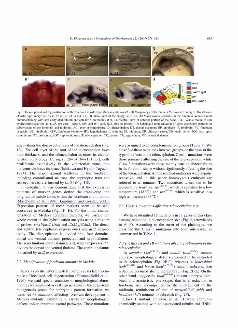
establishing the dorsoventral axis of the diencephalon (Fig.
1D). The cell layer of the roof of the telencephalon loses
their thickness, and the telencephalon assumes its charac-
teristic morphology. During st. 28–34 (64–131 hpf), cells
proliferate extensively in the ventricular zone, and
the ventricle loses its space (Ishikawa and Hyodo-Taguchi,
1994). The major axonal scaffolds in the forebrain,
including commissural neurons, the supraoptic tract and
sensory nerves, are formed by st. 34 (Fig. 1E).
In zebrafish, it was demonstrated that the expression
patterns of marker genes define the transverse and
longitudinal subdivisions within the forebrain and midbrain
(Macdonald et al., 1994; Hauptmann and Gerster, 2000).
Expression patterns of these markers seem to be well
conserved in Medaka (Fig. 1F–H). For the initial charac-
terization of Medaka forebrain mutants, we carried out
whole-mount in situ hybridization analysis using a mixture
of probes, emx1/pax2.1/shh and dlx2/fgf8/slit2. The dorsal
and ventral telencephalon express emx1 and dlx2, respec-
tively. The diencephalon is divided into four domains;
dorsal and ventral thalami, pretectum and hypothalamus.
The zona limitans intrathalamica (zli), which expresses shh,
divides the dorsal and ventral thalami. The ventral thalamus
is marked by dlx2 expression.
2.2. Identification of forebrain mutants in Medaka
Since a specific patterning defect often causes later occur-
rence of localized cell degeneration (Furutani-Seiki et al.,
1996), we paid special attention to morphological abnor-
malities accompanied by cell degeneration. In the large-scale
mutagenesis screen for embryonic pattern formation, we
identified 33 mutations affecting forebrain development in
Medaka mutants, exhibiting a variety of morphological
defects and/or abnormal axonal pathways. These mutations
were assigned to 25 complementation groups (Table 1). We
classified these mutations into two groups, on the basis of the
type of defects in the telencephalon. Class 1 mutations were
those primarily affecting the size of the telencephalon, while
Class 2 mutations were those mainly causing abnormalities
in the forebrain shape without significantly affecting the size
of the telencephalon. All the isolated mutations were zygotic
recessive, and in this paper homozygous embryos are
referred to as mutants. Two mutations turned out to be
temperature sensitive; karj50-4A, which is sensitive to a low
temperature (18 8C); and ikaj94-8A, which is sensitive to a
high temperature (33 8C).
2.3. Class 1 mutations affecting telencephalon size
We have identified 15 mutations in 11 genes of this class
causing reduction in telencephalon size (Fig. 2, arrowheads
in A–F). According to the onset of the phenotype, we
classified the Class 1 mutations into four subclasses, as
summarized in Table 1.
2.3.1. Class 1A and 1B mutations affecting subregions of the
telencephalon
In kentoku (ketj23-3B) and aonibi (aonj9-2F) mutant
embryos, morphological defects appeared to be restricted
to the telencephalon (Fig. 2B,C), whereas in kobesshimi
(kobj35-6D) and bouzu (boujr118-2A) mutant embryos, size
reduction occurred also in the midbrain (Fig. 2D,E). On the
other hand, nopperabo (nopj80-19B) mutant embryos exhi-
bited a characteristic phenotype, that is a reduction in
forebrain size accompanied by the enlargement of the
midbrain, reminiscent of that of masterblind (mbl) and
headless (hdl) mutants in zebrafish (Fig. 2F).
Class 1 mutant embryos at st. 31 were immuno-
chemically stained with anti-acetylated-tubulin and HNK1
Fig. 1. Development and regionalization of the forebrain in wild-type Medaka embryos. (A–D) Morphology of the brain in Medaka live embryos. Dorsal view
of wild-type embryo at (A) st. 19, (B) st. 21, (C) st. 23, (D) lateral view of the embryo at st. 27. (E) Major axonal scaffolds in the forebrain. Whole-mount
immunostaining with anti-acetylated-tubulin and anti-HNK antibodies at st. 31. Ventral view of anterior portion of the head. (F,G) Whole-mount in situ
hybridization analysis at st. 28. (F) emx1, pax2.1, shh; and (G) dlx2, fgf8, slit2 as probes. (H) Schematic representation of gene expression patterns in
subdivisions of the forebrain and midbrain. AC, anterior commissure; D, diencephalon; DT, dorsal thalamus; EP, epiphysis; F, forebrain; FV, forebrain
ventricle; HB, hindbrain; HBV, hindbrain ventricle; HY, hypothalamus; I, isthmus; M, midbrain; OF, olfactory nerve; ON, optic nerve; POC, post-optic
commissure; PT, pretectum; SOT, supraoptic tract; T, telencephalon; TC, tectum; TG, tegmentum; VT, ventral thalamus.
D. Kitagawa et al. / Mechanisms of Development 121 (2004) 673–685 675

antibody to examine the paths and fasciculation of axons
(Fig. 2G–L). In the wild type embryos, the olfactory nerve,
anterior commissure, supraoptic tract and optic nerve were
clearly stained (Fig. 2G). All Class 1 mutants showed
interesting abnormalities in these nerves. In ket mutant
embryos, anterior commissure nerves were not fully
fasciculated (black arrowhead in Fig. 2H), displaced and
lacked association with the olfactory nerve (white arrow-
heads in Fig. 2H), and the supraoptic tract was not clearly
detected. In aon mutant embryos, the olfactory nerve and
supraoptic tract were not detected, the anterior commissure
lacked fasciculation (white arrowhead in Fig. 2I), and
bundle formation of the optic nerve was also affected (black
arrowhead in Fig. 2I). kob mutants were unique in that only
olfactory nerve was affected and lacked fasciculation
(arrowhead in Fig. 2J). In bou mutant embryos, the axons
in the anterior commisure were totally defasciculated,
and the commissure did not form (arrowhead in Fig. 2K).
In the nop mutant embryos, all axonal paths were so
severely affected and in astray that the nerves in the
forebrain were not morphologically distinguishable each
other (arrowhead in Fig. 2L). In addition, the olfactory bulbs
appeared to be missing in nop mutant embryos (Fig. 2L).
Thus, Class 1 mutants share the commonality of a reduced
telencephalon size, but alterations of the nerves and their
paths were affected distinctly.
Class 1 mutants were also examined for the regional
markers of the forebrain by in situ hybridization with two
sets of probes, emx1/pax2.1/shh and dlx2/fgf8/slit2
(Fig. 2M–X). Consistent with a smaller telencephalon,
Class 1 mutant embryos generally showed a reduction in
emx1 or dlx2 expression in the telencephalon.
In ket and nop mutant embryos, the emx1 expression was
strongly reduced (black arrowhead in Fig. 2N,R) and
Table 1
Mutations affecting formation of the forebrain
Gene Symbol Alleles Forebrain phenotype Other phenotypes
Class 1: mutations affecting the size of the telencephalon
Class 1A: mutations affecting early specification of the telencephalon
kentoku ket j23-3B Telencephalon size reduced
Class 1B: mutations affecting regionalization of the telencephalon
aonibi aon j9-2F, j60-3A Telencephalon size reduced Lipid metabolism affected
kobesshimi kob j9-10A, j35-6D, j54-3A Telencephalon size reduced Midbrain slightly reduced
bouzu bou jr118-2A Telencephalon size reduced Midbrain anteroposteriorly reduced
nopperabo nop j80-19B Telencephalon size reduced Eyes missing, midbrain expanded
kumasaka kum j54-20A Telencephalon size reduced
usobuki uso j14-26A Telencephalon size reduced
Class 1C: a mutation affecting maintenance of the telencephalon
hannya han j41-3B Late telencephalon defect –
Class 1D: mutations affecting formation of the midline neural tissue
akatsuki aku j22-15A, jf121-1A Telencephalon size reduced Similar to zebrafish oep
akebono ake j54-7A Telencephalon size reduced Similar to zebrafish oep
mochizuki moc j96-11B Telencephalon size reduced Similar to zebrafish oep
Class 2: mutations affecting morphology of the telencephalon
Class 2A: mutations affecting formation of the forebrain ventricle
sarudahiko sar j106-4A Forebrain ventricle reduced Circulation, midline defect, tectum reduced
tengu ten j2-11A, jr10-4D, j53-4C Forebrain ventricle reduced Circulation, midline defect, tectum reduced
karuna kar j50-4A Forebrain ventricle enlarged Temperature sensitive at 18 8C
oobesshimi oob j103-11A, j58-1A Forebrain ventricle enlarged Tegmentum and hindbrain bumpy
samidare sam j20-26A Forebrain ventricle enlarged Similar to zebrafish parachute mutant
shigure sgu j55-8A Forebrain ventricle enlarged Similar to zebrafish parachute mutant
Class 2B: mutations affecting the formation of the anterior commissure
ikazuchi ika j94-8A Ectopic anterior commissure Temperature sensitive at 33 8C, hindbrain bumpy
shikami shi j92-3A Anterior commissure not formed –
Class 2C: mutations causing forebrain dysmorphology
baltan bal j102-2A Forebrain dysmorphology, edema Eyes small, tectum reduced, circulation defect, somite irregular
fukuwarai fuk j8-33A, j93-4A Forebrain dysmorphology Regions of CNS misplaced
yuzen yuz j107-2D Forebrain dysmorphology Regions of CNS misplaced
kagome kag jr114-2D Forebrain dysmorphology Regions of CNS misplaced
hirame hir j54-20C Flattened, differentiation defect CNS flat, heart beating next to ears
tobi tob jr116-4A Protruding telencephalon Eyes small
D. Kitagawa et al. / Mechanisms of Development 121 (2004) 673–685676
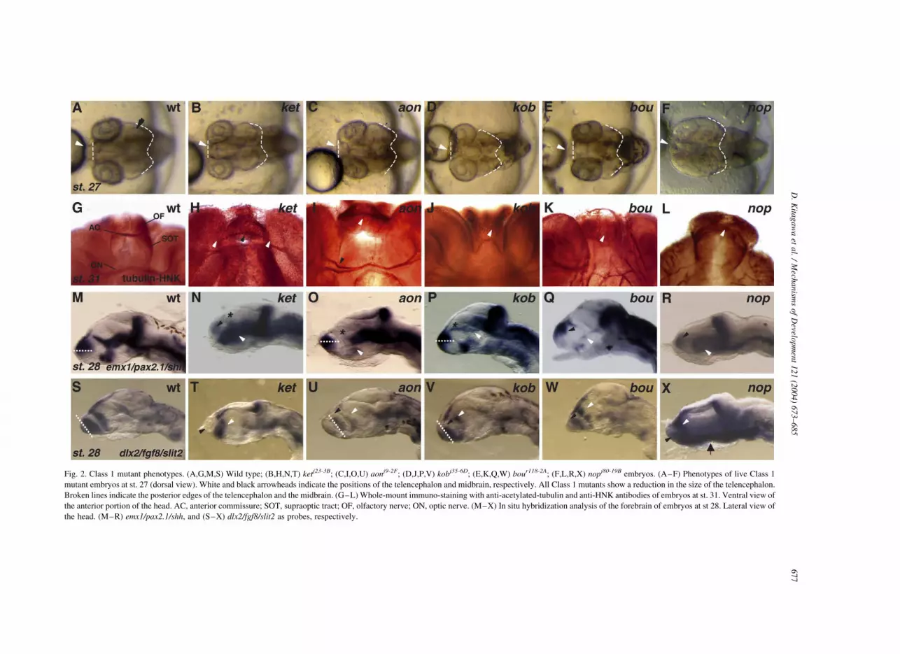
Fig. 2. Class 1 mutant phenotypes. (A,G,M,S) Wild type; (B,H,N,T) ketj23-3B; (C,I,O,U) aonj9-2F; (D,J,P,V) kobj35-6D; (E,K,Q,W) bour118-2A; (F,L,R,X) nopj80-19B embryos. (A–F) Phenotypes of live Class 1
mutant embryos at st. 27 (dorsal view). White and black arrowheads indicate the positions of the telencephalon and midbrain, respectively. All Class 1 mutants show a reduction in the size of the telencephalon.
Broken lines indicate the posterior edges of the telencephalon and the midbrain. (G–L) Whole-mount immuno-staining with anti-acetylated-tubulin and anti-HNK antibodies of embryos at st. 31. Ventral view of
the anterior portion of the head. AC, anterior commissure; SOT, supraoptic tract; OF, olfactory nerve; ON, optic nerve. (M–X) In situ hybridization analysis of the forebrain of embryos at st 28. Lateral view of
the head. (M–R) emx1/pax2.1/shh, and (S–X) dlx2/fgf8/slit2 as probes, respectively.
D.
Kita
ga
wa
eta
l./
Mech
an
isms
of
Develo
pm
ent
12
1(2
00
4)
67
3–
68
56
77

the dlx2 expression in the ventral telencephalon was almost
absent (black arrowheads in Fig. 2T,X). In these mutants,
the shh expression along the floor of the diencephalon
(white arrowheads in Fig. 2N,R) dorsally expanded and, in
ket mutant embryos, the expression in the zona limitans
intrathalamica (zli) was lost (asterisk in Fig. 2N). The dlx2
expression in the ventral thalamus showed an anterior shift
(white arrowhead in Fig. 2T). In nop mutants, the dlx2
expression marking the ventral thalamus anteriorly shifted,
which was accompanied by the anterior expansion of
the dlx2 expression in the pharyngeal arch region (arrow in
Fig. 2X).
In aon mutant embryos, the emx1 expression shifted
ventrally (broken line in Fig. 2M,O). Concomitantly, two
domains of the dlx2 expression in the ventral telence-
phalon and ventral thalamus shifted posteriorly (broken
line in Fig. 2S,U to show the anterior limit of the dlx2
expression; black and white arrowheads in Fig. 2U). The
anteroventral region of the diencephalic shh expression
was reduced (white arrowhead in Fig. 2O) and the shh
expression in the zli was abolished (asterisk in Fig. 2O).
In kob mutants, the emx1 expression shifted ventrally
(broken line in Fig. 2P) and the dlx2 expression in
the ventral telencephalon shifted posteriorly (broken line
and black arrowhead in Fig. 2V), and the dlx2 expression
in the ventral thalamus decreased (white arrowhead in
Fig. 2V). shh expression in the zli and ventral
diencephalon was low (asterisk and white arrowhead in
Fig. 2P, respectively).
In bou mutant embryos, the emx1 expression domain
seems compressed anteroposteriorly (black arrowhead in
Fig. 2Q) and the dlx2 expression in the ventral thalamus
became noncontinuous (white arrow head in Fig. 2W). The
shh expression in the diencephalon was only rudimentary
(white arrowhead in Fig. 2Q).
Thus, different patterning defects in the forebrain are
included in these Class 1 mutants with smaller telencepha-
lon. It is important to note that the majority of the mutants of
this Class had an altered shh expression, particularly a
reduction of shh expression in the zli.
2.3.2. Class 1A mutant kentoku representing an early
fuction in telencephalon development
The expression of an early telencephalic marker bf1 (Tao
and Lai, 1992) was examined in all the Class 1 mutants. In
wild-type Medaka embryos, bf1 expression becomes
detectable in the most anterior region of the brain at st.
19. ket mutant at st. 20 uniquely lacked the bf1 expression
(Fig. 3A,B). This observation indicated that ket is required
in an early step in telencephalon development, possibly in
the induction process. In nop mutant embryos, bf1
expression was reduced at st. 20 probably due to the
expansion of the diencephalon and mesencephalon at the
expense of the telencephalon. In the rest of Class 1 mutants,
bf1 expression appeared normal (data not shown).
2.3.3. Class 1C mutation affecting maintenance of the
telencephalon
In hannya (hanj41-3B) mutant embryos, the distance
between the eyes decreased (arrow in Fig. 4A,E), but the
floor plate was normal (data not shown), ruling out general
midline defects. The expression of dorsal emx1 was reduced
in han mutant embryos (arrowheads in Fig. 4D,H). The
projection pattern of trigeminal nerves was altered such that
they did not extend toward the ventral surface of the
forebrain at st. 31 (arrowheads in Fig. 4B,C,F,G).
By contrast, the anterior commissure appeared normal.
This phenotype was unique to han mutants and distin-
guished them from other Class 1 mutants.
2.3.4. Class 1D mutations exhibiting the phenotype similar
to that of oep in zebrafish
The mutants of akatsuki (akuj22-15A), akebono(akej54-7A)
and mochizuki(mocj96-11B), classified to 1D all displayed a
drastic morphological phenotype similar to that of one-
eyed-pinhead (oep) in zebrafish (Fig. 5A–D), where only
one median eye was formed and the ventral brain tissue was
severely affected. This phenotype was very similar to that of
the zebrafish oep mutant (Schier et al., 1996). These three
Medaka mutations sharing similar forebrain phenotypes
complemented each other.
Fig. 3. Loss of bf1 expression in ketj23-3B mutant embryo at st. 20. (A,B) Whole-mount in situ hybridization analysis of embryos at st. 20 with bf1 probe.
(A) Wild-type embryo. (B) ket mutant embryo.
D. Kitagawa et al. / Mechanisms of Development 121 (2004) 673–685678

2.4. Class 2 mutations affecting the morphology
of the forebrain
Class 2 mutations affecting forebrain shape without
altering telencephalon size was divided into the sub-
classes, 2A to 2C, based on other associated phenotypes
(Tables 1 and 2).
2.4.1. Class 2A mutations affecting the forebrain ventricle
We have identified mutations in 6 genes affecting
the formation of the forebrain ventricle. In sarudahiko
(sarj106-4A) and tengu (tenjr10-4D) mutant embryos, the
ventricle of the forebrain did not inflate (arrowheads in
Fig. 6A–C), the emx1 expression did not extend
ventrally as in the wild type (black arrowheads in
Fig. 6P–R), while the dlx2 expression remained normal
(data not shown). It is remarkable that the shh expression
in either the brain or the floor plate was absent, sug-
gesting a defect in midline signaling (white arrowheads
in Fig. 6Q,R). At the histological level, neuroepithelial
cells in the forebrain and cortical layers of the retina
were round and did not exhibit the characteristic
polarized cell morphology (Fig. 6F–H,K–M). The defect
at the cellular level may account for the defect in the
histogenesis of the forebrain ventricle in these mutants.
sar and ten mutants also shared a common defect in the
cardiovascular system.
In karuna (karj50-4A) and oobesshimi (oobj103-11A) mutant
embryos, in contrast, the forebrain ventricle was abnormally
expanded (arrowheads in Fig. 6A,D–F,I,J). kar mutant
embryos also had small eyes (Fig. 6D,I), and a forebrain
ventricle open on the ventral side (open arrowhead in
Fig. 6I). The shh expression in the diencephalon was altered
in kar mutant embryos (white and black arrowheads in
Fig. 6S), suggesting that the patterning of the ventral
forebrain is affected. In addition, the bilateral retinas were
not completely separated in the midline (black arrowhead in
Fig. 6I). It is interesting to determine whether this is caused
by the altered development of the optic stalk or by a defect
in the morphogenetic movement of diencephalon to split
Fig. 5. Mutants in 3 genes display phenotypes similar to that of oep zebrafish mutants. (A–D) Dorsal view of st. 27 live embryos of (A) wild-type; (B) akuj22-15A
mutant; (C) akej54-7A mutant and (D) mocj96-11B mutant.
Fig. 4. hanj41-3B mutant phenotypes. (A–D) wild type. (E–H) han mutant. (A,E) Live phenotype of embryos at st. 33. Anterior front view. Arrows show the
width of the telencephalon. (B,C,F,G) Whole-mount staining of embryos at st. 31 with anti-acetylated-tubulin and HNK antibodies. (B,F) Ventral view; (C,G)
Anterior front view. Arrowheads show the position of the trigeminal nerve. (D,H) Whole-mount in situ hybridization with emx1/pax2.1/shh probe mixture.
Dorsal view of embryos at st. 28.
D. Kitagawa et al. / Mechanisms of Development 121 (2004) 673–685 679
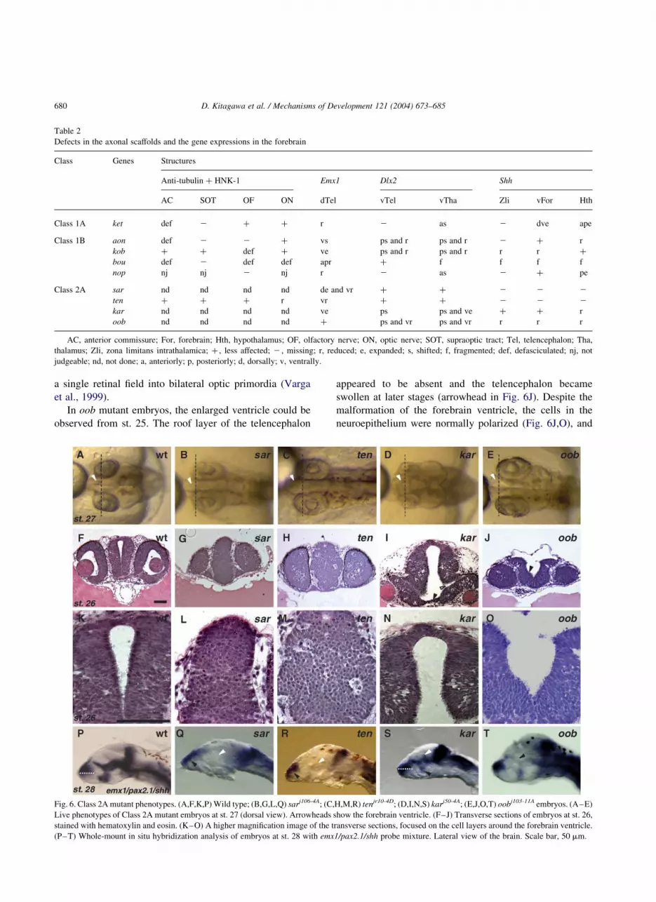
a single retinal field into bilateral optic primordia (Varga
et al., 1999).
In oob mutant embryos, the enlarged ventricle could be
observed from st. 25. The roof layer of the telencephalon
appeared to be absent and the telencephalon became
swollen at later stages (arrowhead in Fig. 6J). Despite the
malformation of the forebrain ventricle, the cells in the
neuroepithelium were normally polarized (Fig. 6J,O), and
Fig. 6. Class 2A mutant phenotypes. (A,F,K,P) Wild type; (B,G,L,Q) sarj106-4A; (C,H,M,R) tenjr10-4D; (D,I,N,S) karj50-4A; (E,J,O,T) oobj103-11A embryos. (A–E)
Live phenotypes of Class 2A mutant embryos at st. 27 (dorsal view). Arrowheads show the forebrain ventricle. (F–J) Transverse sections of embryos at st. 26,
stained with hematoxylin and eosin. (K–O) A higher magnification image of the transverse sections, focused on the cell layers around the forebrain ventricle.
(P–T) Whole-mount in situ hybridization analysis of embryos at st. 28 with emx1/pax2.1/shh probe mixture. Lateral view of the brain. Scale bar, 50 mm.
Table 2
Defects in the axonal scaffolds and the gene expressions in the forebrain
Class Genes Structures
Anti-tubulin þ HNK-1 Emx1 Dlx2 Shh
AC SOT OF ON dTel vTel vTha Zli vFor Hth
Class 1A ket def 2 þ þ r 2 as 2 dve ape
Class 1B aon def 2 2 þ vs ps and r ps and r 2 þ r
kob þ þ def þ ve ps and r ps and r r r þ
bou def 2 def def apr þ f f f f
nop nj nj 2 nj r 2 as 2 þ pe
Class 2A sar nd nd nd nd de and vr þ þ 2 2 2
ten þ þ þ r vr þ þ 2 2 2
kar nd nd nd nd ve ps ps and ve þ þ r
oob nd nd nd nd þ ps and vr ps and vr r r r
AC, anterior commissure; For, forebrain; Hth, hypothalamus; OF, olfactory nerve; ON, optic nerve; SOT, supraoptic tract; Tel, telencephalon; Tha,
thalamus; Zli, zona limitans intrathalamica; þ , less affected; 2 , missing; r, reduced; e, expanded; s, shifted; f, fragmented; def, defasciculated; nj, not
judgeable; nd, not done; a, anteriorly; p, posteriorly; d, dorsally; v, ventrally.
D. Kitagawa et al. / Mechanisms of Development 121 (2004) 673–685680
the general pattern of gene expression was not significantly
affected, except for the reduction in shh expression in the
diencephalon (arrowhead in Fig. 6T).
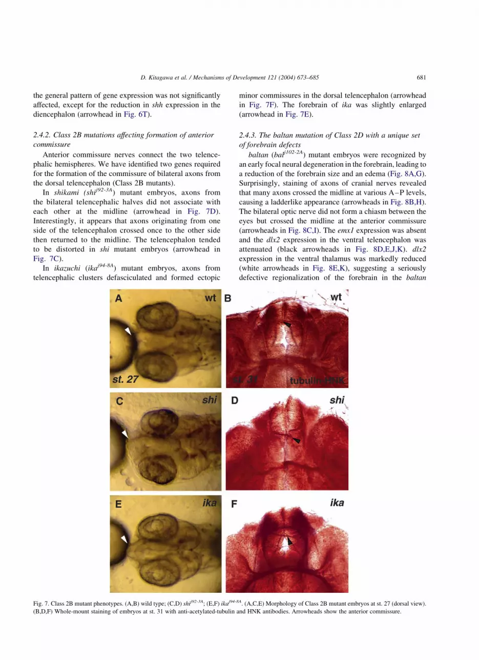
2.4.2. Class 2B mutations affecting formation of anterior
commissure
Anterior commissure nerves connect the two telence-
phalic hemispheres. We have identified two genes required
for the formation of the commissure of bilateral axons from
the dorsal telencephalon (Class 2B mutants).
In shikami (shij92-3A) mutant embryos, axons from
the bilateral telencephalic halves did not associate with
each other at the midline (arrowhead in Fig. 7D).
Interestingly, it appears that axons originating from one
side of the telencephalon crossed once to the other side
then returned to the midline. The telencephalon tended
to be distorted in shi mutant embryos (arrowhead in
Fig. 7C).
In ikazuchi (ikaj94-8A) mutant embryos, axons from
telencephalic clusters defasciculated and formed ectopic
minor commissures in the dorsal telencephalon (arrowhead
in Fig. 7F). The forebrain of ika was slightly enlarged
(arrowhead in Fig. 7E).
2.4.3. The baltan mutation of Class 2D with a unique set
of forebrain defects
baltan (balj102-2A) mutant embryos were recognized by
an early focal neural degeneration in the forebrain, leading to
a reduction of the forebrain size and an edema (Fig. 8A,G).
Surprisingly, staining of axons of cranial nerves revealed
that many axons crossed the midline at various A–P levels,
causing a ladderlike appearance (arrowheads in Fig. 8B,H).
The bilateral optic nerve did not form a chiasm between the
eyes but crossed the midline at the anterior commissure
(arrowheads in Fig. 8C,I). The emx1 expression was absent
and the dlx2 expression in the ventral telencephalon was
attenuated (black arrowheads in Fig. 8D,E,J,K). dlx2
expression in the ventral thalamus was markedly reduced
(white arrowheads in Fig. 8E,K), suggesting a seriously
defective regionalization of the forebrain in the baltan
Fig. 7. Class 2B mutant phenotypes. (A,B) wild type; (C,D) shij92-3A; (E,F) ikaj94-8A. (A,C,E) Morphology of Class 2B mutant embryos at st. 27 (dorsal view).
(B,D,F) Whole-mount staining of embryos at st. 31 with anti-acetylated-tubulin and HNK antibodies. Arrowheads show the anterior commissure.
D. Kitagawa et al. / Mechanisms of Development 121 (2004) 673–685 681

mutant. Moreover, bf1 expression at the induction phase of
telencephalon was remarkably reduced (Fig. 8F,L). These
results suggested that bal is necessary for early specification
of the forebrain as well as proper axonal projection of
cranial nerves.
3. Discussion
3.1. Class 1 mutations causing smaller telencephalon
ket mutants are remarkable for their compromised
expression of the early telencephalon marker bf1(Fig. 3). In
parallel, both emx1 expression in the dorsal telencephalon
and the dlx2 expression in the ventral telencephalon are
strongly reduced, raising the possibility that ket is a key
regulator required for specification of the telencephalon
(Fig. 2N,T). The phenotype of ket mutants is reminiscent of
that of the bf1 knockout mouse with hypoplasia of the
cerebral hemispheres and more severe defects in the basal
region of the telencephalon (Dou et al., 1999). The
expression of both bf1 and emx1 is reduced in tlc- knockdown
embryos after the injection of antisense morpholino
oligonucleotides (Houart et al., 2002). The possible genetic
linkage of ket with bf1 and tlc is currently under
investigation. In mice, Fgf8 induces bf1 expression under
in vitro culture condition of the forebrain tissue (Shimamura
and Rubenstein, 1997). However, telencephalon is somehow
formed in the fgf8 mutants of mice and zebrafish, suggesting
it alone is not responsible for induction of the telencephalon
(Meyers et al., 1998; Reifers et al., 1998; Shanmugalingam
et al., 2000; Shinya et al., 2001).
The major feature of aon and kob mutants is their defect
in dorsoventral (D–V) patterning in the telencephalon. The
tissue area for emx1 expression in the dorsal telencephalon
expands or shifts ventrally, and the dlx2 expression in the
ventral telencephalon is reduced and posteriorly displaced
(Fig. 2O,P,U,V).
shh-null mice lack any signs of the medial ganglionic
eminence (MGE), which is the ventral portion of the basal
ganglia, indicating that Shh is required for the patterning of
the ventral telencephalon (Chiang et al., 1996). However, it
is yet to be determined which part of shh expression in
the forebrain is required for the D–V patterning of the
telencephalon. All the Class 1 mutants have an altered shh
expression, particularly in the zli (Fig. 2N–R). The zli not
only forms a clear histological border between the dorsal
and ventral thalami (Larsen et al., 2001), but, for its shh
expression, is considered to function as a secondary
organizing center. The presence of patterning defects of
the telencephalon in Class 2 mutants corroborates this
notion.
It is to be noted that the phenotype of the Class 1 mutants
associated with the decreased expression of shh in the
forebrain does not resemble those of zebrafish mutants
defective in sonic hedgehog signaling, sonic you (syu),
Fig. 8. balj102-2A mutant phenotypes. (A–F) Wild type; (G–L) balj102-2A embryos. (A,G) Live embryos of wild-type (A) and (G) balj102-2A mutant at st. 30
(lateral view). An arrowhead in G shows an empty space generated by degeneration in the forebrain. (B,C,H,I) Whole-mount immunostaining of st. 31 embryos
using anti-acetylated tubulin and HNK antibodies. (B,H) Ventral view of the head. An arrowhead indicates ladder like axons of unknown origin. (C,I) enlarged
view of (B,H). an arrowhead shows the optic nerve. (D–F,J–L) Whole-mount in situ hybridization analysis of embryos at st.28 (D,E,J,K, dorsal view) and st.21
(F,L, lateral view), using (D,J) emx1/pax2.1/shh; (E,K) dlx2/fgf8/slit2 and (F,L) bf1 as probes. (F,L) Arrowheads indicate the bf1 expression in the
telencephalon.
D. Kitagawa et al. / Mechanisms of Development 121 (2004) 673–685682

detour (dtr), you-too (yot) and slow-muscle-omitted (smu),
in which shh, gli1, gli2, foxh1 and smoothened, respectively,
are mutated and the primary causes are defects in the
midline tissues (Schauerte et al., 1998; Karlstrom et al.,
1999; Barresi et al., 2000; Chen et al., 2001; Varga et al.,
2001; Karlstrom et al., 2003).
The phenotype of the nop mutant, characterized by the
expansion of the diencephalon and mesencephalon at the
expense of the telencephalon tissue (Fig. 2F), is reminiscent
of those of masterblind(mbl) and headless (hdl) mutants of
zebrafish. In these zebrafish mutants, the genes coding for
the negative regulators of Wnt signaling, tcf3 and axin1,
respectively, are mutated, possibly resulting in the over-
activation of Wnt signaling throughout the forebrain (Kim
et al., 2000; Heisenberg et al., 2001). It is also known that a
secreted Wnt antagonist tlc is expressed in the anterior
neural ridge (Houart et al., 2002). Whether the nop gene
function is involved in Wnt signaling is of an immediate
interest.
3.2. Patterning defects resulting in defective formation
of forebrain ventricle
sar and ten mutants lack polarized cell shape in the
neuroepithelium on one hand (Fig. 6L,M), and show an
attenuated shh expression in the forebrain and spinal cord on
the other (Fig. 6Q,R). It is currently under investigation how
these phenotypes correlate with each other.
In oob mutants showing the expansion of the forebrain
ventricle, the roof plate is missing and the basal plate is
thicker (Fig. 6J). This phenotype may reflect an altered
D–V patterning or a defect in the histogenetic process
dependent on cell–cell interactions. It is interesting to note
that the phenotype of oob mutants somewhat resembles that
of the zebrafish parachute mutant of the N-cadherin gene
(Lele et al., 2002; Erdmann et al., 2003).
3.3. Mutations affecting axonal paths in the forebrain
Class 2B mutants have characteristic defects in the
formation of the anterior commissure. Interestingly, the
formation of the anterior commissure is affected differently
in shi and ika mutants, suggesting that shi and ika may be
required for a distinct process in anterior commissure
formation (Fig. 7).
It is suggested in zebrafish that a commissure is formed
through three steps (Bak and Fraser, 2003). The first step is
the extension of the leader axon toward the midline through
the already defined axonal tract. The growth cone senses and
integrates attractive and repulsive midline cues for their
behavior. The second step is the growth and extension of the
follower axon along the leader axon toward the midline and
its fasciculation with the leader axon. The third step is the
interaction of the axons with the leader axon of the opposite
side across the midline and mutual fasciculation. In this
way, the leader axons from two sides cooperate with each
other to establish a commisure across the midline.
In ika mutants, the first step may be disturbed since the
anterior commisure was formed but in an ectopic position
(Fig. 7F). The group of Class 1 mutants, bou, ket and aon,
may have a defect in the second step. The failure of the
axons to properly cross the midline in bou mutants may be
explained by the loss of interaction between the leader
and follower axons (Fig. 2K). In ket and aon mutants, a mild
defasciculation of axons in the anterior commissure was
observed (Fig. 2H,I).
In bal mutants, the anterior commissure and optic nerve
joined together. In the region between the anterior
commissure and the optic nerve, Netrins, the repulsive
cues for axons, are known to be expressed. Indeed, the
netrin expression in this region was altered in noi and ace
zebrafish mutant embryos, accompanied by abnormalities of
the anterior and postoptic commissures (Macdonald et al.,
1997; Shanmugalingam et al., 2000). It would be interesting
to determine whether the netrin expression between the
anterior commissure and the optic nerve is altered in bal
mutants. In shi mutants, the third step may be affected, since
the commissure structure crossing the midline was not
established.
In the Class 1 mutants which have altered gene
expression pattern, the formation of axonal scaffolds in
the forebrain is also affected, as summarized in Table 2. In
zebrafish, the boundaries of regulatory gene expression
domains correspond in many cases to early axonal scaffolds
in the forebrain (Macdonald et al., 1994). It has been
hypothesized that a growth cone extends along the interface
between two domains of cells with different cell surface
properties. Class 1 Medaka mutants should prove useful in
testing this hypothesis.
3.4. Medaka and zebrafish complement to genetically
dissect forebrain formation
We have identified 25 loci required for the formation of
the forebrain in the systematic screen in Medaka. Although
screenings covering the whole genome have already been
carried out in zebrafish, we identified a battery of mutations
resulting in phenotypes that were not observed in
the zebrafish mutant collection. This could reflect (1) the
difference in the functional overlap of genes, (2) the
difference in the regulation of forebrain development, (3)
the difference in susceptibility to mutagens of a genetic
locus between Medaka and zebrafish.
Some other mutant phenotypes corresponded to those of
identified zebrafish mutants. These mutated genes of shared
phenotypes must be involved in the genetic pathways
conserved between the two species. Interestingly, mutations
in 3 genes, aku, ake and moc of Class 1D result in a
phenotype similar to that of one-eyed-pinhead (oep) in
zebrafish (Schier et al., 1997; Strahle et al., 1997). In
zebrafish, mutants of other genes involved in Nodal
D. Kitagawa et al. / Mechanisms of Development 121 (2004) 673–685 683
signaling, cyclops(cyc), squint (sqt), and schmalspur
(sur) displayed slightly different phenotypes, suggesting
a divergence of the molecular components in a signaling
pathway between the two species (Hatta et al., 1994; Brand
et al., 1996b; Malicki et al., 1996; Schier et al., 1996;
Heisenberg and Nusslein-Volhard, 1997; Feldman et al.,
1998; Rebagliati et al., 1998; Sampath et al., 1998; Pogoda
et al., 2000; Sirotkin et al., 2000). Similarly, mutations in 3
genes, oob, sam, and sgu, caused phenotypes that are
reminiscent of those of parachute zebrafish mutant of the
N-cadherin gene. No mutations in other genes are reported
in zebrafish to cause the parachute phenotype (Lele et al.,
2002; Erdmann et al., 2003). Cloning of these mutated genes
in Medaka and the analysis of their function will reveal both
conserved and divergent functions of the genes essential for
forebrain formation. The results of the mutant screen in
Medaka do demonstrate that zebrafish and Medaka
complement each other in uncovering more genes and
more functions involved in the developmental process.
4. Experimental procedures
4.1. Mutant embryos
Fish are mutagenized and screened for mutations as
described in an accompanying paper (Furutani-Seiki et al.,
this issue). Mutant embryos were obtained by mating
heterozygotes, reared at 28 8C, and staged according to the
development of sibling normal embryos. Live embryos were
photographed after removing the chorion and mounting in
2.5% methylcellulose.
4.2. Histological methods
Whole-mount staining of the embryos by in situ
hybridization or with anti-acetylated tubulin/anti-HNK1
antibodies was performed as described by Hammerschmidt
and Nusslein-Volhard (1993). RNA probes for in situ
hybridization were labeled with digoxygenin, and detected
using an anti-digoxygenin antibody conjugated with
alkalinephophatase, and reacted with BM purple (Roche)
for color development. For histological sections, the
embryos were fixed in 4% paraformaldehyde, dehydrated
through ethanol series and embedded in Jung HistoResin
Plus (Leica) and sections 4 mm thickness of were made.
Acknowledgements
We are grateful to Dr Giselbert Hauptmann for input
about regionalization of medaka brain, Drs Takashi Sasaki
and Noboru Nakajima for cloning slit2, dlx2 and bf1 probes.
This work was supported by the ERATO project of the
Japan Science and Technology Agency to H.K.
References
Bak, M., Fraser, S.E., 2003. Axon fasciculation and differences in midline
kinetics between pioneer and follower axons within commissural
fascicles. Development 130, 4999–5008.
Barresi, M.J., Stickney, H.L., Devoto, S.H., 2000. The zebrafish slow-
muscle-omitted gene product is required for Hedgehog signal
transduction and the development of slow muscle identity. Develop-
ment 127, 2189–2199.
Brand, M., Heisenberg, C.P., Jiang, Y.J., Beuchle, D., Lun, K., Furutani-
Seiki, M., et al., 1996a. Mutations in zebrafish genes affecting the
formation of the boundary between midbrain and hindbrain. Develop-
ment 123, 179–190.
Brand, M., Heisenberg, C.P., Warga, R.M., Pelegri, F., Karlstrom, R.O.,
Beuchle, D., et al., 1996b. Mutations affecting development of the
midline and general body shape during zebrafish embryogenesis.
Development 123, 129–142.
Bulfone, A., Puelles, L., Porteus, M.H., Frohman, M.A., Martin, G.R.,
Rubenstein, J.L., 1993. Spatially restricted expression of Dlx-1, Dlx-2
(Tes-1), Gbx-2, and Wnt-3 in the embryonic day 12.5 mouse forebrain
defines potential transverse and longitudinal segmental boundaries.
J. Neurosci. 13, 3155–3172.
Chen, W., Burgess, S., Hopkins, N., 2001. Analysis of the zebrafish
smoothened mutant reveals conserved and divergent functions of
hedgehog activity. Development 128, 2385–2396.
Chiang, C., Litingtung, Y., Lee, E., Young, K.E., Corden, J.L., Westphal,
H., Beachy, P.A., 1996. Cyclopia and defective axial patterning in mice
lacking Sonic hedgehog gene function. Nature 383, 407–413.
Dou, C.L., Li, S., Lai, E., 1999. Dual role of brain factor-1 in regulating
growth and patterning of the cerebral hemispheres. Cereb. Cortex 9,
543–550.
Erdmann, B., Kirsch, F.P., Rathjen, F.G., More, M.I., 2003. N-cadherin is
essential for retinal lamination in the zebrafish. Dev. Dyn. 226,
570–577.
Feldman, B., Gates, M.A., Egan, E.S., Dougan, S.T., Rennebeck, G.,
Sirotkin, H.I., et al., 1998. Zebrafish organizer development and germ-
layer formation require nodal-related signals. Nature 395, 181–185.
Fernandez, A.S., Pieau, C., Reperant, J., Boncinelli, E., Wassef, M., 1998.
Expression of the Emx-1 and Dlx-1 homeobox genes define three
molecularly distinct domains in the telencephalon of mouse, chick,
turtle and frog embryos: implications for the evolution of telencephalic
subdivisions in amniotes. Development 125, 2099–2111.
Figdor, M.C., Stern, C.D., 1993. Segmental organization of embryonic
diencephalon. Nature 363, 630–634.
Furutani-Seiki, M., Jiang, Y.J., Brand, M., Heisenberg, C.P., Houart, C.,
Beuchle, D., et al., 1996. Neural degeneration mutants in the zebrafish,
Danio rerio. Development 123, 229–239.
Hatta, K., Puschel, A.W., Kimmel, C.B., 1994. Midline signaling in the
primordium of the zebrafish anterior central nervous system. Proc. Natl
Acad. Sci. USA 91, 2061–2065.
Hauptmann, G., Gerster, T., 2000. Regulatory gene expression patterns
reveal transverse and longitudinal subdivisions of the embryonic
zebrafish forebrain. Mech. Dev. 91, 105–118.
Hauptmann, G., Soll, I., Gerster, T., 2002. The early embryonic zebrafish
forebrain is subdivided into molecularly distinct transverse and
longitudinal domains. Brain Res. Bull. 57, 371–375.
Heisenberg, C.P., Nusslein-Volhard, C., 1997. The function of silberblick
in the positioning of the eye anlage in the zebrafish embryo. Dev. Biol.
184, 85–94.
Heisenberg, C.P., Brand, M., Jiang, Y.J., Warga, R.M., Beuchle, D., van
Eeden, F.J., et al., 1996. Genes involved in forebrain development in the
zebrafish, Danio rerio. Development 123, 191–203.
Heisenberg, C.P., Houart, C., Take-Uchi, M., Rauch, G.J., Young, N.,
Coutinho, P., et al., 2001. A mutation in the Gsk3-binding domain of
zebrafish Masterblind/Axin1 leads to a fate transformation of
telencephalon and eyes to diencephalon. Genes Dev. 15, 1427–1434.
D. Kitagawa et al. / Mechanisms of Development 121 (2004) 673–685684

Houart, C., Westerfield, M., Wilson, S.W., 1998. A small population of
anterior cells patterns the forebrain during zebrafish gastrulation.
Nature 391, 788–792.
Houart, C., Caneparo, L., Heisenberg, C., Barth, K., Take-Uchi, M.,
Wilson, S., 2002. Establishment of the telencephalon during gastrula-
tion by local antagonism of Wnt signaling. Neuron 35, 255–265.
Ishikawa, Y., Hyodo-Taguchi, Y., 1994. Cranial nerves and brain fiber
systems of the medaka fry as observed by a whole-mount staining
method. Neurosci. Res. 19, 379–386.
Iwamatsu, T., 1994. Stages of normal development in the medaka Oryzias
latipes. Zool. Sci. 11, 825–839.
Karlstrom, R.O., Talbot, W.S., Schier, A.F., 1999. Comparative synteny
cloning of zebrafish you-too: mutations in the Hedgehog target gli2
affect ventral forebrain patterning. Genes Dev. 13, 388–393.
Karlstrom, R.O., Tyurina, O.V., Kawakami, A., Nishioka, N., Talbot, W.S.,
Sasaki, H., Schier, A.F., 2003. Genetic analysis of zebrafish gli1 and
gli2 reveals divergent requirements for gli genes in vertebrate
development. Development 130, 1549–1564.
Kim, C.H., Oda, T., Itoh, M., Jiang, D., Artinger, K.B., Chandrasekharappa,
S.C., et al., 2000. Repressor activity of Headless/Tcf3 is essential for
vertebrate head formation. Nature 407, 913–916.
Larsen, C.W., Zeltser, L.M., Lumsden, A., 2001. Boundary formation and
compartition in the avian diencephalon. J. Neurosci. 21, 4699–4711.
Lele, Z., Folchert, A., Concha, M., Rauch, G.J., Geisler, R., Rosa, F., et al.,
2002. parachute/n-cadherin is required for morphogenesis and main-
tained integrity of the zebrafish neural tube. Development 129,
3281–3294.
Macdonald, R., Xu, Q., Barth, K.A., Mikkola, I., Holder, N., Fjose, A., et al.,
1994. Regulatory gene expression boundaries demarcate sites of
neuronal differentiation in the embryonic zebrafish forebrain. Neuron
13, 1039–1053.
Macdonald, R., Scholes, J., Strahle, U., Brennan, C., Holder, N., Brand, M.,
Wilson, S.W., 1997. The Pax protein Noi is required for commissural
axon pathway formation in the rostral forebrain. Development 124,
2397–2408.
Malicki, J., Neuhauss, S.C., Schier, A.F., Solnica-Krezel, L., Stemple, D.L.,
Stainier, D.Y., et al., 1996. Mutations affecting development of the
zebrafish retina. Development 123, 263–273.
Mathieu, J., Barth, A., Rosa, F.M., Wilson, S.W., Peyrieras, N., 2002.
Distinct and cooperative roles for Nodal and Hedgehog signals during
hypothalamic development. Development 129, 3055–3065.
Meyers, E.N., Lewandoski, M., Martin, G.R., 1998. An Fgf8 mutant allelic
series generated by Cre- and Flp-mediated recombination. Nat. Genet.
18, 136–141.
Pogoda, H.M., Solnica-Krezel, L., Driever, W., Meyer, D., 2000. The
zebrafish forkhead transcription factor FoxH1/Fast1 is a modulator of
nodal signaling required for organizer formation. Curr. Biol. 10,
1041–1049.
Puelles, L., Rubenstein, J.L., 1993. Expression patterns of homeobox and
other putative regulatory genes in the embryonic mouse forebrain
suggest a neuromeric organization. Trends Neurosci. 16, 472–479.
Puelles, L., Kuwana, E., Puelles, E., Bulfone, A., Shimamura, K., Keleher,
J., et al., 2000. Pallial and subpallial derivatives in the embryonic chick
and mouse telencephalon, traced by the expression of the genes Dlx-2,
Emx-1, Nkx-2.1, Pax-6, and Tbr-1. J. Comp. Neurol. 424, 409–438.
Rallu, M., Corbin, J.G., Fishell, G., 2002. Parsing the prosencephalon. Nat.
Rev. Neurosci. 3, 943–951.
Rebagliati, M.R., Toyama, R., Haffter, P., Dawid, I.B., 1998. Cyclops
encodes a nodal-related factor involved in midline signaling. Proc. Natl
Acad. Sci. USA 95, 9932–9937.
Reifers, F., Bohli, H., Walsh, E.C., Crossley, P.H., Stainier, D.Y., Brand,
M., 1998. Fgf8 is mutated in zebrafish acerebellar (ace) mutants and is
required for maintenance of midbrain–hindbrain boundary develop-
ment and somitogenesis. Development 125, 2381–2395.
Rohr, K.B., Barth, K.A., Varga, Z.M., Wilson, S.W., 2001. The nodal
pathway acts upstream of hedgehog signaling to specify ventral
telencephalic identity. Neuron 29, 341–351.
Sampath, K., Rubinstein, A.L., Cheng, A.M., Liang, J.O., Fekany, K.,
Solnica-Krezel, L., et al., 1998. Induction of the zebrafish ventral brain
and floorplate requires cyclops/nodal signalling. Nature 395, 185–189.
Schauerte, H.E., van Eeden, F.J., Fricke, C., Odenthal, J., Strahle, U.,
Haffter, P., 1998. Sonic hedgehog is not required for the induction of
medial floor plate cells in the zebrafish. Development 125, 2983–2993.
Schier, A.F., Neuhauss, S.C., Harvey, M., Malicki, J., Solnica-Krezel, L.,
Stainier, D.Y., et al., 1996. Mutations affecting the development of the
embryonic zebrafish brain. Development 123, 165–178.
Schier, A.F., Neuhauss, S.C., Helde, K.A., Talbot, W.S., Driever, W., 1997.
The one-eyed pinhead gene functions in mesoderm and endoderm
formation in zebrafish and interacts with no tail. Development 124,
327–342.
Shanmugalingam, S., Houart, C., Picker, A., Reifers, F., Macdonald, R.,
Barth, A., et al., 2000. Ace/Fgf8 is required for forebrain commissure
formation and patterning of the telencephalon. Development 127,
2549–2561.
Shimamura, K., Rubenstein, J.L., 1997. Inductive interactions direct early
regionalization of the mouse forebrain. Development 124, 2709–2718.
Shinya, M., Koshida, S., Sawada, A., Kuroiwa, A., Takeda, H., 2001. Fgf
signalling through MAPK cascade is required for development of the
subpallial telencephalon in zebrafish embryos. Development 128,
4153–4164.
Sirotkin, H.I., Gates, M.A., Kelly, P.D., Schier, A.F., Talbot, W.S., 2000.
Fast1 is required for the development of dorsal axial structures in
zebrafish. Curr. Biol. 10, 1051–1054.
Strahle, U., Jesuthasan, S., Blader, P., Garcia-Villalba, P., Hatta, K.,
Ingham, P.W., 1997. One-eyed pinhead is required for development of
the ventral midline of the zebrafish (Danio rerio) neural tube. Genes
Funct. 1, 131–148.
Tao, W., Lai, E., 1992. Telencephalon-restricted expression of BF-1, a new
member of the HNF-3/fork head gene family, in the developing rat
brain. Neuron 8, 957–966.
Varga, Z.M., Wegner, J., Westerfield, M., 1999. Anterior movement of
ventral diencephalic precursors separates the primordial eye field in
the neural plate and requires cyclops. Development 126,
5533–5546.
Varga, Z.M., Amores, A., Lewis, K.E., Yan, Y.L., Postlethwait, J.H., Eisen,
J.S., Westerfield, M., 2001. Zebrafish smoothened functions in ventral
neural tube specification and axon tract formation. Development 128,
3497–3509.
Wilson, S.W., Rubenstein, J.L., 2000. Induction and dorsoventral
patterning of the telencephalon. Neuron 28, 641–651.
D. Kitagawa et al. / Mechanisms of Development 121 (2004) 673–685 685
Related Documents