A novel membrane guanylyl cyclase expressed in medaka (Oryzias latipes ) intestine B Kaori Iio a , Mina Nakauchi a , Sayaka Yamagami a , Makiko Tsutsumi b , Hiroshi Hori b , Kiyoshi Naruse c , Hiroshi Mitani c , Akihiro Shima c , Norio Suzuki a, * a Division of Biological Sciences, Graduate School of Science, Hokkaido University, Sapporo 060-0810, Japan b Division of Biological Science, Graduate School of Science, Nagoya University, Japan c Department of Biological Sciences, Graduate School of Science, University of Tokyo, Japan Received 29 September 2004; received in revised form 29 November 2004; accepted 9 December 2004 Abstract A novel membrane guanylyl cyclase (GC), OlGC9 , was identified in the intestine of the medaka fish Oryzias latipes by the isolation of a full-length cDNA clone (3783 bp). Phylogenetic analysis indicated that OlGC9 belongs in the enterotoxin/guanylin receptor membrane GC subfamily. The nucleotide and deduced amino acid sequences of OlGC9 were highly homologous to those of OlGC6, another enterotoxin/ guanylin receptor membrane GC in medaka fish. Linkage analysis of the medaka fish chromosome demonstrated that the OlGC9 gene was mapped to LG8, which distinguishes it from the OlGC6 gene. Determination of the cGMP concentrations in COS-7 cells expressed with OlGC9 indicated that Escherichia coli heat-stable enterotoxin (STa) stimulated the activity of OlGC9 in a concentration-dependent manner, although it did not activate the OlGC6 expressed in the COS-7 cells. The 5V-flanking region of the OlGC9 gene important for its transcription was partially determined using both CACO-2 cells and COS-1 cells, and was not found to be conserved with respect to either the mammalian GC-C gene or the OlGC6 gene. D 2005 Elsevier Inc. All rights reserved. Keywords: Guanylyl cyclase; Medaka fish; Intestine; Enterotoxin; Transcription; GC-C 1. Introduction Cyclic GMP is a ubiquitous intracellular second messenger produced by an ever-expanding family of receptor guanylyl cyclases (GCs), which are classified into two major forms, those found in the plasma membrane (membrane GC) and those in the cytoplasm (soluble GC) (Drewett and Garbers, 1994; Garbers and Lowe, 1994; Kusakabe and Suzuki, 2000; Loretz and Pollina, 2000; Wedel and Garbers, 2001). The soluble GC is a heme-containing heterodimer and is activated by nitric oxide or carbon monoxide (Drewett and Garbers, 1994). The membrane GC is a protein with a single membrane-spanning region and is activated by various endogenous and exogenous peptides. The membrane GC is further divided into three subfamilies, i.e., the natriuretic peptide (NP) receptors, the sensory organ- specific membrane GCs, and the enterotoxin/guanylin receptors (Seimiya et al., 1997). In mammals, cDNA for two membrane GCs, i.e., GC-A (the receptor for ANP/ BNP) and GC-B (the receptor for CNP), and in medaka fish, cDNAs for three membrane GCs (OlGC1 , OlGC2 , and OlGC7 ) have been identified as members of the NP receptor subfamily (Takeda and Suzuki, 1999; Kusakabe and Suzuki, 2000; Loretz and Pollina, 2000; Wedel and Garbers, 2001; Yamagami et al., 2001). Similarly, cDNAs for three membrane GCs (GC-D, GC-E, and GC-F) and 1096-4959/$ - see front matter D 2005 Elsevier Inc. All rights reserved. doi:10.1016/j.cbpc.2004.12.006 B The nucleotide sequence data reported in this paper have been submitted to the DDBJ, EMBL, and GenBank nucleotide sequence databases, and are available under accession number AB162944 for cDNA of OlGC9. * Corresponding author. Tel.: +81 11 706 4908; fax: +81 11 706 4461. E-mail address: [email protected] (N. Suzuki). Comparative Biochemistry and Physiology, Part B 140 (2005) 569–578 www.elsevier.com/locate/cbpb

Welcome message from author
This document is posted to help you gain knowledge. Please leave a comment to let me know what you think about it! Share it to your friends and learn new things together.
Transcript

A novel membrane guanylyl cyclase expressedin medaka (Oryzias latipes) intestineB
Kaori Iioa, Mina Nakauchia, Sayaka Yamagamia, Makiko Tsutsumib, Hiroshi Horib,Kiyoshi Narusec, Hiroshi Mitanic, Akihiro Shimac, Norio Suzukia,*
aDivision of Biological Sciences, Graduate School of Science, Hokkaido University, Sapporo 060-0810, JapanbDivision of Biological Science, Graduate School of Science, Nagoya University, Japan
cDepartment of Biological Sciences, Graduate School of Science, University of Tokyo, Japan
Received 29 September 2004; received in revised form 29 November 2004; accepted 9 December 2004
Abstract
A novel membrane guanylyl cyclase (GC), OlGC9, was identified in the intestine of the medaka fish Oryzias latipes by the isolation of a
full-length cDNA clone (3783 bp). Phylogenetic analysis indicated that OlGC9 belongs in the enterotoxin/guanylin receptor membrane GCsubfamily. The nucleotide and deduced amino acid sequences of OlGC9 were highly homologous to those of OlGC6, another enterotoxin/guanylin receptor membrane GC in medaka fish. Linkage analysis of the medaka fish chromosome demonstrated that the OlGC9 gene wasmapped to LG8, which distinguishes it from the OlGC6 gene. Determination of the cGMP concentrations in COS-7 cells expressed with
OlGC9 indicated that Escherichia coli heat-stable enterotoxin (STa) stimulated the activity of OlGC9 in a concentration-dependent manner,although it did not activate the OlGC6 expressed in the COS-7 cells. The 5V-flanking region of the OlGC9 gene important for its transcriptionwas partially determined using both CACO-2 cells and COS-1 cells, and was not found to be conserved with respect to either the mammalian
GC-C gene or the OlGC6 gene.D 2005 Elsevier Inc. All rights reserved.
Keywords: Guanylyl cyclase; Medaka fish; Intestine; Enterotoxin; Transcription; GC-C
1. Introduction
Cyclic GMP is a ubiquitous intracellular secondmessenger produced by an ever-expanding family ofreceptor guanylyl cyclases (GCs), which are classifiedinto two major forms, those found in the plasmamembrane (membrane GC) and those in the cytoplasm(soluble GC) (Drewett and Garbers, 1994; Garbers andLowe, 1994; Kusakabe and Suzuki, 2000; Loretz andPollina, 2000; Wedel and Garbers, 2001). The soluble GC
is a heme-containing heterodimer and is activated bynitric oxide or carbon monoxide (Drewett and Garbers,1994). The membrane GC is a protein with a singlemembrane-spanning region and is activated by variousendogenous and exogenous peptides. The membrane GCis further divided into three subfamilies, i.e., thenatriuretic peptide (NP) receptors, the sensory organ-specific membrane GCs, and the enterotoxin/guanylinreceptors (Seimiya et al., 1997). In mammals, cDNA fortwo membrane GCs, i.e., GC-A (the receptor for ANP/BNP) and GC-B (the receptor for CNP), and in medakafish, cDNAs for three membrane GCs (OlGC1, OlGC2,and OlGC7) have been identified as members of the NPreceptor subfamily (Takeda and Suzuki, 1999; Kusakabeand Suzuki, 2000; Loretz and Pollina, 2000; Wedel andGarbers, 2001; Yamagami et al., 2001). Similarly, cDNAsfor three membrane GCs (GC-D, GC-E, and GC-F) and
1096-4959/$ - see front matter D 2005 Elsevier Inc. All rights reserved.
doi:10.1016/j.cbpc.2004.12.006
B The nucleotide sequence data reported in this paper have been
submitted to the DDBJ, EMBL, and GenBank nucleotide sequence
databases, and are available under accession number AB162944 for cDNA
of OlGC9.
* Corresponding author. Tel.: +81 11 706 4908; fax: +81 11 706 4461.
E-mail address: [email protected] (N. Suzuki).
Comparative Biochemistry and Physiology, Part B 140 (2005) 569–578
www.elsevier.com/locate/cbpb

four membrane GCs (OlGC3, OlGC4, OlGC5, andOlGC-R2) have been found in mammalian and medakafish sensory organs, respectively (Seimiya et al., 1997; Yuet al., 1997; Hisatomi et al., 1999; Kusakabe and Suzuki,2000).
In contrast to the two abovementioned subfamilies,only a single membrane GC has been classified in theenterotoxin/guanylin receptor subfamily in both mammals(GC-C) (Schulz et al., 1990) and medaka fish (OlGC6)(Mantoku et al., 1999). Recently, cDNA fragments fortwo membrane GCs (GC-C1 and GC-C2) have beencloned from the intestine of the European eel Anguillaanguilla (Comrie et al., 2001). Furthermore, it has beendemonstrated that the expression of the GC-C2 gene inthe intestine was increased by 100% following thetransfer of fresh water-acclimated eels to sea water;moreover, the developmental maturation of yellow eelsinto pre-migratory silver eels resulted in a significantincrease in the intestinal expression of the GC-C2 gene(Comrie et al., 2001). On the other hand, it has beenreported that the mammalian GC-C gene is expressedexclusively in the intestine, and its translation product isactivated by both endogenous peptides (guanylin anduroguanylin) and exogenous peptides (for example,Escherichia coli heat-stable enterotoxin, STa) (Schulz etal., 1990; Currie et al., 1992; Hamra et al., 1993;Vaandrager and De Jonge, 1994). Upon STa-binding tothe extracellular domain of mammalian GC-C, the intra-cellular cyclase catalytic domain converts GTP to cGMP,causing secretory diarrhea in humans, particularly amongchildren.
In this study, we found that the medaka fish Oryziaslatipes possesses the second intestine-specific membraneGC (OlGC9), although the expression of OlGC9 as well asOlGC6 in adult fish did not change with alterations inenvironmental salinity. Here, we report that using arecombinant COS-7 cell expression system, OlGC9 isactivated by STa, and OlGC6 is activated by endogenousligand(s). In this report, we also describe the 5V-flankingregion of the OlGC9 gene that is required to drive itstranscription; this region was found to be unique andexhibited no similarity to corresponding regions of theOlGC6 gene (Nakauchi and Suzuki, 2003) and the humanGC-C gene (Swenson et al., 1999; Di Guglielmo et al.,2001).
2. Materials and methods
2.1. Animals
Mature adult medaka fish Oryzias latipes of the orange-red variety were purchased from a local dealer. The fishwere kept in indoor tanks under artificial reproductiveconditions (10-h dark, 14-h light cycle; 27 8C) and were fedOtohime B2 (Nisshin Seifun Group Tokyo, Japan).
2.2. Isolation of a cDNA clone for a novel intestine-specificmembrane GC
The first strand of cDNA was synthesized by reverse-transcription using the total RNA (2 Ag) prepared frommedaka fish intestine by the acid guanidinium thiocyanate-phenol-chloroform extraction method (Chomczynski andSacchi, 1987). A cDNA fragment for a novel intestine-specific membrane GC, named OlGC9, was amplified byPCR using the cDNA as the template with a combination ofthree degenerate oligonucleotide primers (P2: 5V-GAYAHT-GNGGNTTYAC-3V; P6: 5V-GTRTTNACNGTRTCNCC-3V;and P7: 5V-ARRCARTANCKNGGCAT-3V) synthesizedbased on the amino acid sequences of three regions(DIVGFT, residues 820–825; GDTVNT, residues 935–940, and MPRYCL, residues 928–933, respectively,) con-served in known membrane GCs. The following conditionswere used for the first PCR amplification with P2 and P6primers: 90 s at 94 8C, 35 cycles of 30 s at 94 8C, 1 min at41 8C and 1 min at 72 8C, and 5 min at 72 8C. The followingconditions were used for the second PCR amplification withP2 and P7 primers: 90 s at 94 8C, 35 cycles of 30 s at 94 8C,1 min at 39 8C and 1 min at 72 8C, and 5 min at 72 8C. The3V- and 5V-portions of the cDNA were amplified by PCRusing each adapter-specific primer (3V-RACE inner primer,3V-RACE outer primer, 5V-RACE inner primer, or 5V-RACEouter primer) and the gene-specific sense or antisenseoligonucleotide primers according to the manufacturer’sprotocol (the FirstChoicek RLM-RACE Kit; Ambion,Austin, TX, USA). To determine the full-length cDNAsequence of OlGC9, the PCR products were cloned intopBluescript II KS(-) (Stratagene, La Jolla, CA, USA) andsequenced using the dideoxy chain termination method(Sanger et al., 1977) and an ABI PRISM 3100 Ge-netic Analyzer (Applied Biosystems, Foster City, CA,USA).
2.3. Molecular phylogenetic analysis
The amino acid sequence (residues 820–932) of OlGC9was compared with those of known GCs using Clustal Wprogram (Thompson et al., 1994) and the sequence editorSeqPub (Gilbert, Indiana University, Bloomington, IN,USA). The rooted phylogenetic tree was constructed usingthe aligned sequence and the neighbor-joining algorithmsin the PROTRAS program of PHYLIP ver.3.572 (Felsen-stein, 1989) and the Clustal W program (Saitou and Nei,1987). For the neighbor-joining analysis, the evolutionarydistance was estimated using Kimura’s empirical method forprotein distances (Kimura, 1983). The following DDBJaccession numbers for the sequences were used: AB004921(OlGC1); AB030274 (OlGC2); AB000899 (OlGC3);AB000900 (OlGC4); AB000901 (OlGC5); AB007192(OlGC6); AB023489 (OlGC7); AB054814 (OlGC8);AB015874 (OlGC-R2 ); AJ291611 (eel GC-C1 );AJ291612 (eel GC-C2); U21101 (sea urchin spermatozoa
K. Iio et al. / Comparative Biochemistry and Physiology, Part B 140 (2005) 569–578570

GC); D49837 (Xenopus GC-C); NM_004963 (human GC-C); D17513 (porcine GC-C); X14773 (rat GC-A); M26896(rat GC-B); M55636 (rat GC-C); L37203 (rat GC-D);L36029 (rat GC-E); L36030 (rat GC-F); and AF024622 (ratGC-G). Complementary DNA sequences of pufferfish(Takifugu rubripes) mGC1 and fugu mGC2 were predictedfrom their genome sequences (Ensemble Gene ID: fugumGC1; SINFRUG00000151673 and fugu mGC2; SINFRU-G00000140444) in the DOE Joint Genome Institute database(http://bahama.jgi-psf.org/fugu/html/fugu_funsheet.html)and were translated into the putative amino acid sequences.
2.4. Expression of the OlGC9 transcripts in medaka fishorgans
Total RNA (1 Ag) prepared from various adult medakafish organs (brain, eye, kidney, intestine, ovary, testis, gill,liver, and spleen) was reverse-transcribed using an oligo(dT) primer according to the manufacturer’s protocol(SuperScriptk III First-Strand Synthesis System for RT-PCR; Invitrogen, Carlsbad, CA, USA). The cDNA fragmentcontaining the 3V untranslated region (UTR) of OlGC9 wasamplified by PCR (5 min at 96 8C, 25 or 28 cycles of 30 s at96 8C, 30 s at 60 8C and 1 min at 72 8C, and 10 min at 728C) using a pair of gene-specific primers: 5V-GGCTGTCA-CAGAGAATCCAAG-3V and 5V-CCTGACCAGCTCACA-CAAGG-3V(291-bp product). As an internal control, OlCA1(the cytoplasmic actin gene of the medaka fish, DDBJaccession number D89627) was also amplified usingfollowing primers: 5V-GGGTCTTTCATGACGGGC-3V and5V-CAAGTCGGAACACATGTGCA-3V (100-bp product).Each cDNA fragment was separated by electrophoresis on1.5% agarose gel in 1!TAE (0.35%(v/v) glacial acetic acid,10 mM EDTA, 40 mM Tris base, pH 8.0) and then eachfragment was stained with ethidium bromide (EtBr).
2.5. Effect of environmental salinity on the expression of theOlGC9 gene
Thirty individual medaka fish were divided into twogroups and transferred into plastic aquaria filled with freshwater (FW) and 50% artificial seawater (SW), respectively.After 24-h acclimation to 50% SW, the fish were transferredinto 100% SW (Inoue and Takei, 2002). In order to expose thefish to similar stress conditions, the fish in the FW group werealso transferred into another plastic aquarium filled with FW.To examine the change in the mRNA levels, at 24 h, 3 days,and 5 days after transfer of the FW fish (n=5 in each case) ineach group, each fish was dissected, and the total RNA wasextracted from the intestine. The total RNA was reverse-transcribed using the SuperScriptk III First-Strand SynthesisSystem (Invitrogen), and the cDNA fragment containing the3V untranslated region (UTR) of OlGC9 or OlGC6 wasamplified by PCR with a pair of gene-specific primers: 5V-GGCTGTCACAGAGAATCCAAG-3V and 5V-CCTGAC-CAGCTCACACAAGG-3V for OlGC9 (291-bp product),
and 5V-ACAACGGAGAACGTCCAGCGT-3V and 5V-TGCAGCCATGTTGCCTCGTTAC-3V for OlGC6 (271-bpproduct) under the following conditions: 5 min at 96 8C, 23cycles of 30 s at 96 8C, 30 s at 60 8C and 1 min at 72 8C,and 10 min at 72 8C. The cDNA fragments separated byelectrophoresis on 1.5% agarose gel in 1!TAE, and thenthe fragments were stained with EtBr.
2.6. Linkage analysis of the OlGC9 gene
To assign the locus of the OlGC9 gene to the linkagegroup, polymerase chain reaction-restriction fragment lengthpolymorphism (PCR-RFLP) was carried out using gene-specific primers (5V-AGATTTACCACAAGCTCGTT-3V and5V-CCTTCCACAGCCTCCTGAAG-3V) according to a pro-cedure described in a previous paper (Naruse et al., 2000).
2.7. Assay of guanylyl cyclase activity
The open reading frame (ORF) of OlGC6 (amino acids1–1075) or OlGC9 (amino acids 1–1076) was amplified byPCR using gene-specific primers (for OlGC6 : 5V-ATGAGCACATTTAACTTGTGGCTG-3 V and 5 V-CTAAAGGAAGGTGCTCAGCGTG-3V; and for OlGC9:5V -ATGTACGGCTTACAGAGTTTACTG-3V and 5V-TCA-CAGGTAAGTACTTGGATTCTC-3V). The following PCRconditions were used: 5 cycles of 10 s at 98 8C, 30 s at 638C and 5 min at 72 8C, 5 cycles of 10 s at 98 8C, 30 s at61 8C and 5 min at 72 8C, 25 cycles of 10 s at 98 8C, 30 sat 59 8C and 5 min at 72 8C, and 10 min at 72 8C. ThePCR products were subcloned into a pCRR 3.1 vector(Invitrogen), and the construct containing OlGC6 orOlGC9 was transfected into COS-7 cells.
The COS-7 cells were cultured in Dulbecco’s modifiedEagle’s medium (DMEM) supplemented with 10% heat-inactivated fetal bovine serum (FBS) (HyCloneR, Logan,UT, USA) and 1!penicillin-streptomycin-glutamine (Invi-trogen) under a humidified 5% CO2 atmosphere. The cellswere plated at 5!105 cells per well in a 24-well plate andwere cultured for 24 h before transfection. The cells were thentransfected by lipofectamin with the plasmid DNA (0.8 Ag ofDNA/2 Al of LIPOFECTAMIN 2000 (LF2000) Reagent perwell) according to the manufacturer’s protocol (Invitrogen).As a control, the pCR 3.1 vector was transfected alone. Forty-eight hours after transfection of the respective constructDNA, the cells were washed with 200 Al DMEM containing10% IBMX, and were treated with various concentrations(10"5 M-10"11 M) of STa or medaka fish intestine extract in200 Al DMEM containing 10% IBMX. After incubation at 378C for 40 min, the medium was removed, and the cGMPconcentration was determined by PROTOCOL 3 of theEnzyme immunoassay (EIA) System (Amersham PharmaciaBiotech., Buckinghamshire, UK) according to the manufac-turer’s protocol.
The intestines obtained from 20 mature adult individualmedaka fish were homogenized in a 300-Al homogenization
K. Iio et al. / Comparative Biochemistry and Physiology, Part B 140 (2005) 569–578 571

buffer containing 10% glycerol, 100 mM NaCl, 1 mMEDTA, and 50 mM HEPES, pH 7.4. The homogenate wascentrifuged at 10,000 !g for 30 min at 48 C, and theresulting supernatant was used as the intestine extract. Theprotein concentrations were determined using a BCAkprotein assay kit (PIERCE, Bonita Springs, FL, USA)according to the manufacturer’s protocol.
2.8. Isolation and characterization of the 5V-flanking regionof the OlGC9 gene
To isolate genomic DNA clones containing the 5V-flankingregion of the OlGC9 gene, the medaka fish BAC library(approximately 4.5!105 plaques) (Matsuda et al., 2001) washybridized with an OlGC9 cDNA fragment (nucleotides3432–3724) as a probe, which was labeled with digoxigenin(DIG)-dNTP using DIG-High Prime (Boehringer Ingelheim,Heidelberg, Germany) according to the manufacturer’sprotocol. The BAC DNAs of positive clones were purifiedusing a HiSpeedk Plasmid Midi Kit (QIAGEN, Hilden,Germany) and were analyzed by Southern hybridizationusing the DIG-labeled probe, after being digested by BamHIor EcoRI. The nucleotide sequence of the longest 5V-flankingregion of theOlGC9 gene was determined as described aboveand was analyzed with GENETYX-MAC/version 7.2.0(Software Development, Tokyo, Japan).
The transcriptional factor binding sites in the 5V flankingregion of the OlGC6 and OlGC9 genes, as well as in humanGC-C, were predicted using Parallel Protein InformationAnalysis system (http://mbs.cbrc.jp/papia/papiaJ.html).
2.9. Promoter analysis of the 5V-flanking region of theOlGC9 gene
The transcription initiation site of the OlGC9 gene,determined by a procedure described in a previous paper(Yamamoto and Suzuki, 2002), was 238 bp upstream of theputative start codon and was designated as +1. Variousgenomic DNA fragments (nucleotides "4016 to +253,"3216 to +253, "2439 to +253, "1485 to +253, "1153to +253, "1153 to +253, "948 to +253, "810 to +253,"745 to +253, "702 to +253, "638 to +253, "583 to +253,"453 to +253,"374 to"253, and"55 to +253) were clonedrespectively into the KpnI–BamHI sites in a pGL3-enhancerluciferase vector (Promega, Madison, WI, USA) and werepurified using the Qiagen Lambda Midi Kit (QIAGEN).
CACO-2 cells (a human intestine-derived cell line) orCOS-1 cells (an African green monkey kidney-derived cellline) were cultured in Dulbecco’s modified Eagle’s medium(DMEM) supplemented with 10% heat-inactivated FBS(HyClone) and 1! penicillin-streptomycin-glutamine (Invi-trogen) under a humidified 5% CO2 atmosphere. For thereporter-enzyme assay, 5!105 CACO-2 cells or 4!105 COS-1 cells were plated per well in a six-well plate, and the cellswere cultured for 24 h before transfection. A plasmidconstruct (20 Ag) consisting of the OlGC9 promoter region
and the luciferase gene was co-transfected with pSV-h-galactosidase (20 Ag) (Promega) into CACO-2 cells or COS-1 cells by the calcium phosphate co-precipitation method(Graham and van der Eb, 1973). After 48-h culture, the cellswere lysed in Reporter Lysis Buffer (Promega), and theluciferase activity was assayed using a Luciferase AssaySystem (Promega) according to the manufacturer’s protocol.h-galactosidase activity was also assayed in order to normal-ize it with respect to variations in transfection efficiency.
3. Results
3.1. Isolation and characterization of a cDNA cloneencoding OlGC9
The isolation and determination of a full-length cDNAclone of OlGC9, obtained by RT-PCR and repeated 3V- and5V-RACE, indicated that the OlGC9 cDNA is 3,783 bp inlength, consisting of a 238-bp 5V-untranslated region(UTR), a 3,225-bp open reading frame (ORF), and a 316-bp 3VUTR with a poly (A)+ tail. Termination codons occurin all three frames upstream of the putative initiation codon(ATG), and nucleotides around the putative initiation codonfit within the preferred sequence for initiation of proteinsynthesis in eukaryotic mRNA (Kozak, 1983). The ORF ofthe OlGC9 cDNA predicts a protein of 1075 amino acids,which contains an amino-terminal signal sequence of 24amino acids (Fig. 1). Cleavage of the signal sequencewould result in a mature protein of 1051 amino acids. Themature protein of OlGC9 is composed of a largeextracellular domain (residues 1–426), a single mem-brane-spanning domain (residues 427–451), a proteinkinase-like domain (residues 471–747), and a cyclasecatalytic domain (residues 779–1006). Membrane GCs aregenerally classified into three major groups; the enter-otoxin/guanylin receptor, the sensory organ-specific recep-tor, and the natriuretic peptide receptor subfamily. GC-G isknown not to be activated by any known ligands formembrane GCs and thus to be classified as an orphanreceptor, although the extracellular domain of GC-G showsstructural similarity with that of natriuretic peptide recep-tors (Schulz et al., 1998). The OlGC8 gene and itstranscript have been identified only in medaka fish(Yamagami et al., 2003). However, its translation productdid not show to be activated by any known ligands formembrane GCs and medaka fish tissue extracts, suggestingthat it is an unique orphan receptor (Yamagami et al.,2003). The phylogenetic analysis demonstrated that OlGC9belongs to the heat-stable enterotoxin/guanylin receptorsubfamily (Fig. 2). Forty-two percent, 65%, and 84%similarity was observed between the following regions ofOlGC9 and OlGC6: the extracellular domain, the proteinkinase-like, and the cyclase catalytic domains, respectively.
As shown in Fig. 3, RT-PCR using total RNA isolatedfrom various adult medaka fish organs (brain, eye, kidney,
K. Iio et al. / Comparative Biochemistry and Physiology, Part B 140 (2005) 569–578572

OlGC9 ---MYGLQSLLFLGTLTMLVSNDMLDDCLESNPRYTMNVVLLEDNTYEWSRPFVQEAVEGAIKKDAEENRKAGLNFTLTANYNWFNTNLYNRQGCGSSTCEGVAILKKLHNTGEVGCVML 117OlGC6 ----MSTFNLWLSVLCAFGVSASCVQDTGQCMDGITVNVILLEDEESPWSLKYVGGQILEAIEKDAAINAEEGMEFNLTVNFEGFNTTLYRQRGCITSACEGAEKLNKLMVTGELGCAVL 116eel ------------------------------------------------------------------------------------------------------------------------human MKTLLLDLALWSLLFQPGWLSFSSQVSQNCHNGSYEISVLMMGNSAFAEPLKNLEDAVNEGLEIVRGRLQNAGLNVTVNATFMYSDGLIHNSGDCRSSTCEGLDLLRKISNAQRMGCVLI 120porcine MKTPLLALALWSLLLQLGLTFWPSSVSQNCHNGSYEISVLMMNNSAFPESLDNLKAVVNEGVNIVRQRLLEAGLTVTVNATFVYSEGVIYKSSDCRSSTCEGLDLLRTISSEKRMGCVLL 120
OlGC9 GPSCTFATFQLVDEEIGLSLSIXVISAGSFGLSCDYKPKLTRILPPARKVSDSLVYFFN-EKNMLKP-IWEKAYVYKKSNNVTEDCFWYNNALEAPSAHFASSK-KREMLRGEEELKKAL 234OlGC6 GPTCTYATFAIADVEKGFNLSTPIISAGSFGSSCDYAMNLQRLLPPARKISDFFINFWK-EKFTIKP-KWRTAYVYKRQPNT-EDCLWYIGALEAD-GRFLVNV-SRTILRHPGDLKDVL 231eel ------------------------------------KAYLTRLLPPARKISNLFIEFLRFEESSLKP-RWEAVYVYKKPENS-EDCFWYINALDAPSAAFNSAI-TRKVLRKPEELHSAF 81human GPSCTYSTFQMY---LDTELSYPMISAGSFGLSCDYKETLTRLMSPARKLMYFLVNFWKTNDLPFKTYSWSTSYVYKNGTET-EDCFWYLNALEASVSYFSHELGFKVVLRQDKEFQDIL 236porcine GPSCTYSTFQMY---LDTDLNYPMISAGSFGLSCDYKETLTRLMSPARKLMYFLVDFWKVNNFPFKPFSWNTAYVFKNSTES-EDCFWYLNALEAGVSYFSQKLSFKEMLRGNEEFQNIL 236
* *::.****: :: * . :. :*. * **:*. : ***:** .**:* . * : :** :::. :
OlGC9 TSK-YRFSNIFILCGSVDDIVSIKGLAKQFH-EDTIFILIDLYNPEYYIN-TTSLAPMRDVLVVTLP-PRNYVN--ESNSTFNNTINDYVAGYHDSALLFGEVLRRKMLS-----QHVPL 343OlGC6 DSQENRTSNLFILCGSPTDLKEVKNISDAADNLDILFILIDLYNDVYYTN-TTSMPEMRNVLVLTMPDTRTYTI--KPDLTGNDTMNDYMAAYHDAVLLVGQVMRDIAIRNPAEMQGMEY 348eel ERK-NRTSNLFIICGTPEDVANLTDNGRRLE-PDIVILLIDLYNHEYHSA-AGSPA-MSNVLVITMPNIRNYTEGWTDNGTLP-EMNDYVAGYHDGVHLFGLVLRQKMLYGEGSVEENAS 196human MDH-NRKSNVIIMCGGPEFLYKLKG--DRAVAEDIVIILVDLFNDQYLEDNVTAPDYMKNVLVLTLSPGNSLLNS-SFSRNLSPTKRDFALAYLNGILLFGHMLKIFLENGE---NITTP 349porcine MDQ-NRKSNVIIMCGAPETVHTLKG--GRAVAEDTVIILVDLFNDHYFMDNVTAPDYMKNVLVLTLPPENSVSNS-SFSKDLSLVKNDFTLAYMNGVLLFGHMLKIFLEKRE---DVTTS 349
: * **::*:** : :.. * :::*:**:* * . : * :***:*:. .. . .*: .* :. *.* ::: :
OlGC9 SDT-PFGNISFEGMAGNYVLDEYGDRDVNFTFIYTSAQTSKYETLSVFDTSQNITIMWHDSPTLPWKDGQLPGDEP-ENTEDLSTQDIIVIVLGVSVVVVTAIALIFYRQNRKERLMQ-M 460OlGC6 VNTNYFRNVSFNGIGGHYKLDSYGDRDVNFSVIYTSTDN-KYKILFSFDTENNRTKQMDPSPTFIWTK-ALPDDKP---GSELETQDIIVVVLGVTVAVVATLAFIFYRQNRKDRRLR-K 462eel VEN-PFKNISFSGIGGQYVLDEHGDRDVNFSVMYMSTTDSQYKVLFEFDTSTNNTAVVDANPTWHWKSSRLPDDRPAQEQVLLATQDIIVIVLGISVVVVTVFALIFYRRNIKDRQAQ-K 314human KFAHAFRNLTFEGYDGPVTLDDWGDVDSTMVLLYTSVDTKKYKVLLTYDTHVNKTYPVDMSPTFTWKNSKLPNDIT---GRGPQILMIAVFTLTGAVVLLLLVALLMLRKYRKDYELRQK 466porcine KFAHAFRNITFEGHMGPVTLDNCGDIDNTMFLLYTSVDTSKYKVLLTYDTRKNYTNPVDKSPTFIWKNHKLPNDIP---GRGPQILMIAVFTLTGTIVLLLLIALLVLRKYKREYALRQK 466
* *::*.* * **. ** * .: .:* *. :*: * :** * * . .** *.. **.* . * *..* ::.:: .*::. *: :: :
OlGC9 KWSHIEPHLIGPLDEKE---VSLKIDEDKRKDSTFFSHRGRYDKKPVVLKELKHPDEDFTEEQKIELNTLLRIDYYNLTKFYGTVKFEYGVFGVFELCQRGSLRYILNDRISYPDETFMD 577OlGC6 RWSHINPDLISLLENNEHNIVSLKIEEERKK--MCQIRRALYDKKIVILKELKKSDGNFNKAQRIELNALLHIDYYSLTKFYGTVKFDQGVFGVFEYGQRGSLRYVLNDKVSYPEGTFMD 580eel QWSHIPPDRITPVDDKDFSLVSLKIDEDQRKDSIDRIRRGRYDQKPIILKELKNTEGYFSEEKRIELNTLLRIDYYNLTKFYGTVKFEYGVFGVYEFCERGSLRYVLNDKISYPEETFMD 434human KWSHIPPENIFPLETNETNHVSLKIDDDKRRDTIQRLRQCKYDKKRVILKDLKHNDGNFTEKQKIELNKLLQIDYYNLTKFYGTVKLDTMIFGVIEYCERGSLREVLNDTISYPDGTFMD 586porcine KWSHIPPENIFPLESNETNHVSLKIDDDRRRDTIQRLRQCKYDKKRVILKDLKHNDGNFTEKQKIELNKLLQIDYYNLTKFYGTVKLDSMIFGVIEYCERGSLREVLNDTISYPDGTFMD 586
:**** *. * :: :: *****:::::: :: **:* ::**:**: : *.: ::**** **:****.*********:: :*** * :***** :*** :***: ****
OlGC9 MEFKISVMYDIAKGMSXLHTSNIPVHGRLKSTNCEVDNRMVVKITDFGCHTILKPAKDLWTAPEHLRKDGVSQKGDVYSYAIIAHEIVTRXPPFYTQYCSDSAEKMYRVQYPRNNNFFRP 697OlGC6 WEFKISVMYDIAKGMSYLHASDIQVHGRLKSTNCVVDNRMVVKITDFGCNAFLSREHDLWTAPEHLRKEGTSQKGDVYSFAIICQEIVLRRSTFYTEASLKRSEKLSRVIT----SYFRP 696eel LEFKISVMHDIVKGMSYLHLSNIAVHGRLKSTNCVVDSRMVVKITDFGCNNILRPSRDLWTAPEHLRNPGTSQKGDVYSFGIISQEILLRKCTFYTAACSDRAEKTYRVQFPDSCSFFRP 554human WEFKISVLYDIAKGMSYLHSSKTEVHGRLKSTNCVVDSRMVVKITDFGCNSILPPKKDLWTAPEHLRQANISQKGDVYSYGIIAQEIILRKETFYTLSCRDRNEKIFRVENSNGMKPFRP 706porcine WEFKISVLYDIAKGMSYLHSSKTEVHGRLKSTNCVVDSRMVVKITDFGCNSILAPKKDLWTAPEHLRRASVSQKGDVYSYGIIAQEIILRRETFYTLSCRDQKEKIFRVENSNGVKPFRP 706
******::**.**** ** *. ********** **.***********: :* :**********. . ********:.**.:**: * .*** . . ** ** . ***
OlGC9 ELNFEGASDRETEVYMLIKNCWDEDPERRPDFKKIELTLGKIFSNLHNQATETYMDNLIRRLQMYSRTLENLVEERTALYKAERDRADHLNFMLLPGPVVRSLKETGRVEPELFEEVTIY 817OlGC6 DLNLETASEKEAEVYMLIKSCWEEDPEKRPDFKKVENLLGKIISKIHNQDNESYMDNMMRRLQMYSKNLEHLVEERTALYKAERDRADRLNFMLLPRPVVKSLKESGAVEPELYDEVTIY 816eel DLNLESAGERERELCGLIKNCWDEDPEKRPDFKKIESCLVKISS-LHSQGNESYMDSMIRRLQMYSRNLEHLVEERTALYKAERDRADQLNFMLLPGPVVQSLKERGYVEPELFDEVSIY 673human DLFLETAEEKELEVYLLVKNCWEEDPEKRPDFKKIETTLAKIFGLFHDQKNESYMDTLIRRLQLYSRNLEHLVEERTQLYKAERDRADRLNFMLLPRLVVKSLKEKGFVEPELYEEVTIY 826porcine DLFLETAEEKELEVYLLVKNCWEEDPEKRPDFKKIENTLAKIFGLFHDQKNESYMDTLIRRLQLYSRNLEHLVEERTQLYKAERDRADRLNFMLLPRLVVKSLKEKGIVEPELYEEVTIY 826
:* :* * ::* *: *:*.**:****:******:* * ** . :*.* .*:***.::****:**:.**:****** **********:******* **:**** * *****::**:**
OlGC9 FSDIVGFTTLCHYSTPMEVVDMLNNIYKNFDSILDHHDVYKVETIGDAYMVASGLPNRNGDRHAVDIAHMALDILSFVGTFELQHLPGIPLWIRIGVHSGPCAAGVVGNKMPRYCLFGDT 937OlGC6 FSDIVGFTTLCQYSTPMEVVDMLNDIYKGFDSIVDHHDVYKVETIGDAYMVASGLPNRNGNMHAVDICRMALDILEFMGTFQLRHLVGIPVWIRIGVHSGPCAAGVVGVKMPRYCLFGDT 936eel FSDIVGFTTLCQYSTPMEVVNMLNEIYKNFDSILDNHDVYKVETIGDAYMVASGLPNRNGNRHAVDISRMALDILSFMGTFRLQHLQELPVWIRIGVHSGPCAAGVVGIKMPRYCLFGDT 793human FSDIVGFTTICKYSTPMEVVDMLNDIYKSFDHIVDHHDVYKVETIGDAYMVASGLPKRNGNRHAIDIAKMALEILSFMGTFELEHLPGLPIWIRIGVHSGPCAAGVVGIKMPRYCLFGDT 946porcine FSDIVGFTTICKYSTPMEVVDMLNDIYKSFDHILDHHDVYKVETIGDAYMVASGLPKRNGNRHAIDIAKMALDILSFMGTFELEHLPGLPIWIRIGIHSGPCAAGVVGIKMPRYCLFGDT 946
*********:*:********:***:***.** *:*:********************:***: **:**.:***:**.*:***.*.** :*:*****:*********** ***********
OlGC9 VNTASRMESTGLPLRIHVSQSTIKILQRTPCKFEFEQRGETYLKGKGKEMTYWLTGVTGGNYNLPTPPTAENFQRLQQDLADMIISSLEKREGGKETIEKRKTLSTRIRWKDRNNSFQKE 1057OlGC6 VNTASRMESTGHPLRIHVSEPTIQILQRTNCKFEYEMRGETYLKGKGTEMTYWLTGESGQDYDLPTPPTTENVQRLQQHLAHMILACLERRSRGSVRRKKQHADQGKNDEDEESGVESES 1056eel VNTASRMESTGLPLRIHVSQPTISILQRTDCQFVFERRGETYLKGRGTETTYWLTGVKGQEYNLPVPPTAENFQRQQQELAHMIESCLEKRSLGGGRKEQRKTLSTRQRRREKEDG-QGD 912human VNTASRMESTGLPLRIHVSGSTIAILKRTECQFLYEVRGETYLKGRGNETTYWLTGMKDQKFNLPTPPTVENQQRLQAEFSDMIANSLQKRQAAGIRSQKPRRVAS-----------YKK 1055porcine VNTASRMESTGLPLRIHVSGSTIAILKRTECQFLYEVRGETYLKGRGTETTYWLTGVKDQEYNLPTPPTAENQQRLQAEFVDMIASSLQKRQASGIKNRKPTRVAS-----------YKK 1055
*********** ******* .** **:** *:* :* ********:*.* ****** ...:: **.***.** ** * .: .** .*::*. . .: .
OlGC9 SPPEYFHLAVTENP-STYL 1075OlGC6 SQPEYLHLATVDNTLSTFL 1075eel QQPAYSALLP--------- 922human GTLEYLQLNTTDKE-STYF 1073porcine GTLEYLQLNTTDNE-STHF 1073
* * **
PDZ protein binding-region
catalytic domain
kinase-like domain
transmembrane
Putative signal sequence
Fig. 1. Alignment of the amino acid sequences of OlGC9, OlGC6, eel GC-C2, human GC-C, and porcine GC-C. Asterisks below the sequences indicate
identical amino acid residues among the three proteins. Gaps in the sequences are indicated by dashes (–). Open boxes indicate conserved Cys residues
(Yu et al., 1997) and Lys residues (Bhandari et al., 2001). N-linked glycosylation sites are underlined, and the glycosylation sites at Asn195 and Asn402 in
porcine GC-C (Hasegawa et al., 1999a) are indicated by open circles. The region in porcine GC-C, which is reported to be involved in STa binding, is
indicated by a shadowed box (Hasegawa et al., 1999b). The predicted oligomerization region in human GC-C is indicated by a double underline. The
consensus phosphorylation sequence containing the target site (Ser1052) of PKC identified in porcine GC-C (Wada et al., 1996) is indicated by a broken
line. The amino acid number indicates the position relative to the first Met, considered as 1.
K. Iio et al. / Comparative Biochemistry and Physiology, Part B 140 (2005) 569–578 573

intestine, ovary, testis, gill, liver, and spleen) demonstratedthat OlGC9 was expressed exclusively in the intestine; thisfinding reflects a similarity to the expression patterns ofOlGC6 (Mantoku et al., 1999). Environmental salinity didnot affect the extent of expression of either OlGC6 orOlGC9 in the intestine (data not shown). The gene mappingstudy revealed that the OlGC9 gene was mapped to thelinkage group (LG) 8 on the medaka fish genome.
3.2. cGMP accumulation in COS-7 cells expressed withOlGC6 or OlGC9 upon treatment of STa and medaka fishintestine extract
The medaka fish intestine extract increased cGMPconcentrations by 24.8-fold in COS-7 cells transfected withthe OlGC6 cDNA, as compared to the values obtainedwithout the extract; however, no obvious increases were
detected with regard to the cGMP concentrations in theCOS-7 cells transfected with the OlGC9 cDNA (Fig. 4A).In contrast, STa at 1!10"6 M led to a 16.8-fold increase inthe cGMP concentration in COS-7 cells transfected withthe OlGC9 cDNA, as compared to the values observedwithout STa, but no significant increases were detected inthe cGMP concentrations in the COS-7 cells transfectedwith the OlGC6 cDNA (Fig. 4B). In both cases, the cellstransfected with the empty vector did not show anyobvious changes in the cGMP concentration in responseto the extract or to STa at 1!10"6 M. The increases in thecGMP concentration in cells transfected with the OlGC9cDNA were concentration-dependent when the STa con-centration ranged from 1!10"11 M to 1!10"6 M, whereasSTa administered within a range of 1!10"11 M to 1!10"6
splee
ntes
tiski
dney
ovar
ygil
lliv
erey
ebr
ainin
testin
e
25 cyclesOlGC9
28 cyclesOlGC9
25 cyclesOlCA1
Fig. 3. Detection of the OlGC9 transcript in various organs of adult medaka
fish by RT-PCR analysis. RT-PCR was carried out for 25 and 28 cycles.
Medaka fish cytoplasmic actin gene (OlCA1) was amplified as an internal
control.
cGM
P fm
ol/w
ell
0
50
100
150
200
250
300
OlGC6
OlGC9
pCR
(A)
cGM
P fm
ol/w
ell
(B)
0
100
200
300
400
500
600
OlGC6
OlGC9
pCR
Fig. 4. GC activity in COS-7 cells transfected with OlGC6 or OlGC9 with
medaka fish intestine extract (A) and STa (B). Black boxes indicate the
cGMP concentrations after treatment of the cells with the intestine extract
(27 Ag protein/Al) or with 1!10"6 M STa. White boxes indicate the cGMP
concentrations in cells not exposed to the extract or to STa. Transfection
and cGMP assay were performed in four independent experiments, and the
values are expressed as the meanFS.D.
Sea urchinRat GC-G
Rat GC-COlGC6Eel GC-C1Eel GC-C2
OlGC9Rat GC-D
OlGC4OlGC3
OlGC5Rat GC-ERat GC-F
OlGC-R2Rat GC-AOlGC7
OlGC2Rat GC-B
OlGC1OlGC8
(I)
(II)
(III)
Fig. 2. Molecular phylogenetic relationship of various membrane GCs in
medaka fish and the rat, and eel GC-C1 and GC-C2. The amino acid
sequences of the catalytic domains of known membrane GCs were
subjected to phylogenetic analysis. (I), (II), and (III) indicate the enter-
otoxin/guanylin receptor, the sensory organ-specific receptor, and the
natriuretic peptide receptor subfamily, respectively.
STa (-logM)
cGM
P fm
ol/w
ell
0
100
200
300
400
500
600
700
11 10 9 8 7 6
Fig. 5. The accumulated cGMP in COS-7 cells transfected with OlGC6 or
OlGC9 after treatment with STa.OlGC6,OlGC9, or empty pCRR 3.1 vector
is indicated by n, x, orE, respectively. Transfection and cGMP assay were
performed in four independent experiments, and the values are expressed as
the meanFS.D.
K. Iio et al. / Comparative Biochemistry and Physiology, Part B 140 (2005) 569–578574
M had no effect on the cGMP concentrations in the COS-7cells transfected with the OlGC6 cDNA or the emptyvector (Fig. 5).
3.3. Characterization of the promoter in the OlGC9 geneusing mammalian cell lines
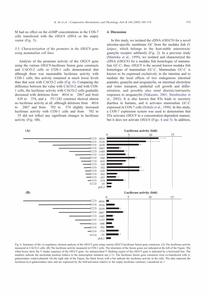
Analysis of the promoter activity of the OlGC9 geneusing the various OlGC9-luciferase fusion gene constructsand CACO-2 cells or COS-1 cells demonstrated thatalthough there was measurable luciferase activity withCOS-1 cells, this activity remained at much lower levelsthan that seen with CACO-2 cells (Fig. 6). Comparing thedifference between the value with CACO-2 and with COS-1 cells, the luciferase activity with CACO-2 cells graduallydecreased with deletions from "4016 to "2067 and from"638 to "374, and a "55/+243 construct showed almostno luciferase activity at all, although deletions from "4016to "2067 and from "702 to "374 slightly increasedluciferase activity with COS-1 cells and from "702 to"55 did not reflect any significant changes in luciferaseactivity (Fig. 6B).
4. Discussion
In this study, we isolated the cDNA (OlGC9) for a novelintestine-specific membrane GC from the medaka fish O.latipes, which belongs to the heat-stable enterotoxin/guanylin receptor subfamily (Fig. 2). In a previous study(Mantoku et al., 1999), we isolated and characterized thecDNA (OlGC6) for a medaka fish homologue of mamma-lian GC-C; thus, OlGC9 is the second known medaka fishhomologue of mammalian GC-C. Mammalian GC-C isknown to be expressed exclusively in the intestine and tomediate the local effects of two endogenous intestinalpeptides, guanylin and uroguanylin, on intestinal electrolyteand water transport, epithelial cell growth and differ-entiation, and possibly also renal diuretic/natriureticresponses to uroguanylin (Nakazato, 2001; Steinbrecher etal., 2002). It is also known that STa leads to secretorydiarrhea in humans, and it activates mammalian GC-Cexpressed in COS-7 cells (Schulz et al., 1990). In this study,a COS-7 expression system was used to demonstrate thatSTa activates OlGC9 in a concentration-dependent manner,but it does not activate OlGC6 (Figs. 4 and 5). In addition,
0 5 10
5 10
15
pGL3-55
-374-702-948
-1153-1485-2067-2439-3216-4016
Luciferase activity (fold)
Luciferase activity (fold)
(A)
(B)
0 15
pGL3-55
-374-453-583-638-702-745-810-948
-1153-1485-2067-2439-3216-4016
Fig. 6. Summary of the cis-regulatory element analysis of the OlGC9 gene using various OlGC9-luciferase fusion gene constructs. (A) The luciferase activity
measured in CACO-2 cells. (B) The luciferase activity measured in COS-1 cells. The structures of the fusion genes are indicated at the left of the Figure. The
white boxes show the 5V-leader sequence of the OlGC9 gene. An untranscribed 5V-flanking region of the OlGC9 gene is indicated by a horizontal line. The
numbers indicate the nucleotide position relative to the transcription initiation site (+1). The luciferase fusion gene constructs were co-transfected with h-galactosidase control plasmid. On the right side of the Figure, the black boxes with a bar indicate the luciferase activity in the cells. The data represent the
luciferase-to-h-galactosidase ratio and are expressed by the fold-activation relative to the empty luciferase construct, considered as 1.
K. Iio et al. / Comparative Biochemistry and Physiology, Part B 140 (2005) 569–578 575

we found that the intestine extract, which was expected tocontain endogenous ligands, activated OlGC6, but notOlGC9 (Fig. 4). These findings suggest that the structuraldifferences in the extracellular domains of OlGC6 andOlGC9 are responsible for differential activation byendogenous ligand(s) and STa, although it has been reportedthat the structure of STa is similar to that of mammalianendogenous ligands, namely, guanylin and uroguanylin(Nakazato, 2001). Recently, three types of guanylins havebeen identified in teleost fish, eel (Yuge et al., 2003).Therefore, in the eel system, it would be interesting toexamine whether two receptor membrane GCs mightdistinguish these three endogenous ligands, or one of themmight be activated by STa alone and other be activated byall of three endogenous ligands as we found in the medakafish.
The mechanism of stimulating the guanylyl cyclaseactivity of mammalian GC-C by STa has been accountedfor from a number of perspectives, such as: (1) the importanceof glycosylation sites (Asn195 and Asn402) in the extracellulardomain for the proper folding of the domain for STa-binding(Nandi et al., 1996; Ghanekar et al., 2004), (2) thephosphorylation of Ser1052 in the intracellular domain byPKC for conformation changes suitable for an active formupon binding of STa to the extracellular domain (Wada et al.,1996), (3) oligomerization (Vaandrager et al., 1994; Vijaya-chandra et al., 2000), (4) the interaction of the carboxyltermini with PDZ protein (Scott et al., 2002; Zhang andWang, 2003), and (5) ATP-binding to the kinase-like domain(Bhandari et al., 2001). Among these features, it was foundthat the following were conserved in both OlGC6 andOlGC9: a region for mediating oligomerization, the PDZprotein-binding region, and a Lys residue in the kinase-like
domain for stabilizing the ATP-binding site (Fig. 1). Inaddition to these conserved features, the residues SPTFIWK,which are thought to be involved in STa binding in porcineGC-C (Hasegawa et al., 1999a,b), were also conserved inbothOlGC6 andOlGC9, suggesting that the STa binding siteidentified in porcine GC-C may not be related to thedifferential activation ofOlGC6 andOlGC9 by STa-binding.On the other hand, as shown in Fig. 1, two glycosylation sites(Asn195 and Asn402) are conserved in OlGC9, but not inOlGC6. In addition, a RNNSFQK sequence (residues1050"1056 in OlGC9), which corresponds to the consensusphosphorylation sequence (RXXS1052XK) found in porcineGC-C, is found inOlGC9, but not inOlGC6. These structuraldifferences betweenOlGC6 andOlGC9 may contribute to anexplanation of the differential binding and subsequentactivation of OlGC6 and OlGC9 by possible endogenousligand(s) and STa.
Several studies have shown that the intestinal transcrip-tional regulator CDX2 mediates intestinal epithelial cell-specific expression of the mammalian GC-C gene (DiGuglielmo et al., 2001), and that hepatocyte nuclear factor-4(HNF-4) is a key regulator of mammalian GC-C expressionin the intestine (Swenson et al., 1999). In a previous study,we demonstrated that the AGACCTTTGC nucleotides inthe 5V-flanking region of the OlGC6 gene play a critical rolein the transcription of the gene (indicated as the star in Fig.7) (Nakauchi and Suzuki, 2003). Since the size of thehuman genome is almost four times that of the medaka fish(Tanaka, 1995), the human genome can be considered tohave undergone more dynamic change in the evolutionaryprocess, which would in turn suggest that transcriptionalregulatory mechanisms on the genome may also have variedsubstantially. Indeed, the regions which have been reported
-2000 -1800 -1600 -1400 -1200 -1000 -800 -600 -400 -200 0
OlGC6
Human GC-C
OlGC9
bp
GATA
HNF-4
C/EBP
TATA box
Cdx2
the region that drives the intestinal transcription of the OlGC6 gene
Fig. 7. Schematic drawing of several consensus sequences for several transcription factors found in the 5V-flanking region of the OlGC6 gene, the OlGC9 gene,
and the human GC-C gene. Closed ellipses, open ellipses, closed boxes, and open boxes represent the consensus sequence for the GATA transcription factor
family, hepatocyte nuclear factor-4 (HNF-4), Cd!2, and the CCAAT/enhancer binding protein family (C/EBP), respectively. The TATA box is indicated as an
open triangle. The star indicates the AGACCTTTGC sequence which plays a critical role in the transcription of the OlGC6 gene (Nakauchi and Suzuki, 2003).
The numbers below the line indicate the length of the 5V-flanking sequence.
K. Iio et al. / Comparative Biochemistry and Physiology, Part B 140 (2005) 569–578576

to regulate the intestinal cell-specific expression of humanGC-C, differ from the regions identified in the OlGC6 gene.In this study, we demonstrated that these potential tran-scriptional regulatory sequences were not found in the 5V-flanking region of the OlGC9 gene (Fig. 7), thus suggestingthat the transcriptional regulatory mechanisms of the OlGC9gene differ from those of the OlGC6 and mammalian GC-Cgenes.
To gain a better understanding of the OlGC9-specificmechanisms of transcription, we analyzed the promoter/enhancer activity of the OlGC9 gene using two cell lines ofdifferent origin, CACO-2 and COS-1 cells. The luciferaseactivity measured with the COS-1 cells remained muchlower than that observed in the case of the CACO-2 cells(Fig. 6), suggesting that the transcriptional activity of theOlGC9 gene is higher in the intestine than in the kidney.This interpretation of the results receives support from thepresent findings showing that the OlGC9 gene is expressedexclusively in the intestine (Fig. 3). With respect to theluciferase activity measured with COS-1 and CACO-2 cells,the regions between "2439 and "2067, "702 and "374,and "374 and "55 are likely to be involved in the type oftranscriptional regulation specific to CACO-2 cells (Fig. 6),suggesting that these sequences are not similar to those ofthe OlGC6 gene, nor to those of the human GC-C gene (Fig.7). Moreover, these sequences might be required forintestine-specific transcription and may contain potentialtranscription factor binding sites.
In this study, we demonstrated that the OlGC9 gene wasmapped to linkage group (LG) 8 on the medaka fish genome,which is different from the linkage group (LG19) mapped forthe OlGC6 gene (Naruse et al., 2000). In addition, as regardsthis difference, it has been suggested that the genes on LG8and LG19 share the same ancestral chromosome (proto-chromosome 2) (Naruse et al., 2004). Therefore, we expectthat the OlGC6 and OlGC9 genes were duplicated from thesame ancestral gene; this interpretation accounts, at least inpart, for the similarity of the two genes at the amino acid level.However, the following two factors suggest that after geneduplication, the nucleotide sequences of both genes, and thesubsequent biological functions of the translation products ofboth genes, were altered independently: 1) the observationregarding the differential activation ofOlGC6 andOlGC9 bypossible endogenous ligand(s) and STa, and 2) the lack ofsimilarity in the 5V-flanking region between the OlGC6 andOlGC9 genes. Here, it should bementioned that the Europeaneel A. anguilla has two GC-Cs (GC-C1 and GC-C2), andincreased expression of the GC-C2 gene in the intestine wasobserved with increases in environmental salinity (Comrie etal., 2001), whereas in this study, no transcriptional changeswere observed in either the OlGC6 gene or the OlGC9 genewith changes in environmental salinity (data not shown). Thisdiscrepancy may reflect the fact that the medaka fish O.latipes has long been accustomed to living in fresh water,although it is also able to survive in seawater for a short periodof time; therefore, the OlGC6 and OlGC9 gene transcription
products are thought to have lost functions related to osmoticcontrol during the process of evolution.
Acknowledgements
We are grateful to the staff members of the Center forAdvanced Science and Technology, Hokkaido University,for the use of their laboratory facilities. We would also liketo thank Dr. Minoru Tanaka, Hokkaido University, for theuse of his laboratory facilities. This study was supported bya Grant-in-Aid for Scientific Research from the Ministry ofEducation, Science, Sports, and Culture of Japan (No.11236202) and by the National Project on Protein Structuraland Functional Analyses.
References
Bhandari, R., Srinivasan, N., Mahaboobi, M., Ghanekar, Y., Suguna, K.,
Visweswariah, S.S., 2001. Functional inactivation of the human
guanylyl cyclase C receptor: modeling and mutation of the protein
kinase-like domain. Biochemistry 40, 9196–9206.
Chomczynski, P., Sacchi, N., 1987. Single-step method of RNA isolation
by acid guanidinium thiocyanate-phenol-chloroform extraction. Anal.
Biochem. 162, 156–159.
Comrie, M.M., Cutler, C.P., Cramb, C.G., 2001. Cloning and expression of
two isoforms of guanylate cyclase C (GC-C) from the European eel
(Anguilla anguilla). Comp. Biochem. Physiol., B 129, 575–586.
Currie, M.G., Fok, K.F., Kato, J., Moore, R.J., Hamra, F.K., Duffin, K.L.,
Smith, C.E., 1992. Guanylin: an endogenous activator of intestinal
guanylate cyclase. Proc. Natl. Acad. Sci. U. S. A. 89, 947–951.
Di Guglielmo, M.D., Park, J., Schulz, S., Waldman, S.A., 2001. Nucleotide
requirements for CDX2 binding to the cis promoter element mediating
intestine-specific expression of guanylyl cyclase C. FEBS Lett. 507,
128–132.
Drewett, J.G., Garbers, D.L., 1994. The family of guanylyl cyclase
receptors and their ligands. Endocr. Rev. 15, 135–162.
Felsenstein, J., 1989. PHYLIP—phylogeny interference package (version
3.2). Cladistics 5, 164–166.
Garbers, D.L., Lowe, D.G., 1994. Guanylyl cyclase receptors. J. Biol.
Chem. 269, 30741–30744.
Ghanekar, Y., Chandrashaker, A., Tatu, U., Visweswariah, S.S., 2004.
Glycosylation of the receptor guanylyl cyclase C: role in ligand binding
and catalytic activity. Biochem. J. 379, 653–663.
Graham, F.L., van der Eb, A.J., 1973. A new technique for the assay of
infectivity of human adenovirus 5 DNA. Virology 52, 456–467.
Hamra, F.K., Forte, L.R., Eber, S.L., Pidhorodeckyj, N.V., Krause, W.J.,
Freeman, R.H., Chin, D.T., Tompkins, J.A., Fok, K.F., Smith, C.E.,
Duffin, K.L., Siegel, N.R., Currie, M.G., 1993. Uroguanylin:
structure and activity of a second endogenous peptide that stimulates
intestinal guanylate cyclase. Proc. Natl. Acad. Sci. U. S. A. 90,
10464–10468.
Hasegawa, M., Hidaka, Y., Matsumoto, Y., Sanni, T., Shimonishi, Y.,
1999a. Determination of the binding site on extracellular domain of
guanylyl cyclase C to heat-stable enterotoxin. J. Biol. Chem. 274,
31713–31718.
Hasegawa, M., Hidaka, Y., Wada, A., Hirayama, T., Shimonishi, Y., 1999b.
The relevance of N-linked glycosylation to the binding of a ligand to
guanylate cyclase C. Eur. J. Biochem. 263, 338–345.
Hisatomi, O., Honkawa, H., Imanishi, Y., Satoh, T., Tokunaga, F., 1999.
Three kinds of guanylyl cyclase expressed in medaka photo-receptor
cells in both retina and pineal organ. Biochem. Biophys. Res. Commun.
255, 216–220.
K. Iio et al. / Comparative Biochemistry and Physiology, Part B 140 (2005) 569–578 577

Inoue, K., Takei, Y., 2002. Diverse adaptability in Oryzias species to high
environmental salinity. Zool. Sci. 19, 727–734.
Kimura, M., 1983. The Neutral Theory of Molecular Evolution. Cambridge
University Press, Cambridge.
Kozak, M., 1983. Comparison of initiation of protein synthesis in
procaryotes, eucaryotes, and organelles. Microbiol. Rev. 47, 1–45.
Kusakabe, T., Suzuki, N., 2000. The guanylyl cyclase family in medaka fish
Oryzias latipes. Zool. Sci. 17, 131–140.
Loretz, C.A., Pollina, C.P., 2000. Natriuretic peptides in fish physiology.
Comp. Biochem. Physiol., B 125, 169–187.
Mantoku, T., Muramatsu, R., Nakauchi, M., Yamagami, S., Kusakabe, T.,
Suzuki, N., 1999. Sequence analysis of cDNA and genomic DNA, and
mRNA expression of the medaka fish homolog of mammalian guanylyl
cyclase C. J. Biochem. 125, 476–486.
Matsuda, M., Kawato, N., Asakawa, S., Shimizu, N., Nagahama, Y.,
Hamaguchi, S., Sakaizumi, M., Hori, H., 2001. Construction of a BAC
library derived from the inbred Hd-rR strain of the teleost fish,Oryzias
latipes. Genes & Genet. Syst. 76, 61–63.
Nakauchi, M., Suzuki, N., 2003. Promoter analysis of a medaka fish
intestinal guanylyl cyclase gene. FEBS Lett. 536, 12–18.
Nakazato, M., 2001. Guanylin family: new intestinal peptides regulating
electrolyte and water homeostasis. J. Gastroenterol. 36, 219–225.
Nandi, A., Mathew, R., Visweswariah, S.S., 1996. Expression of the
extracellular domain of the human heat-stable enterotoxin receptor in
Escherichia coli and generation of neutralization antibodies. Protein
Expr. Purif. 8, 151–159.
Naruse, K., Fukamachi, S., Mitani, H., Kondo, M., Matsuoka, T., Kondo,
S., Hanamura, N., Morita, Y., Hasegawa, K., Nishigaki, R., Shimada,
A., Wada, H., Kusakabe, T., Suzuki, N., Kinoshita, M., Kanamori, A.,
Terada, T., Kimura, H., Nonaka, M., Shima, A., 2000. A detailed
linkage map of medaka, Oryzias latipes: comparative genomics and
genome evolution. Genetics 154, 1773–1784.
Naruse, K., Tanaka, M., Mita, K., Shima, A., Postlethwait, J., Mitani, H.,
2004. A medaka gene map: the trace of ancestral vertebrate proto-
chromosomes revealed by comparative gene mapping. Genome Res. 14,
820–828.
Saitou, N., Nei, M., 1987. The neighbor-joining method: a new method for
reconstructing phylogenetic trees. Mol. Biol. Evol. 4, 406–425.
Sanger, F., Nicklen, S., Coulson, A.R., 1977. DNA sequencing with chain-
terminating inhibitors. Proc. Natl. Acad. Sci. U. S. A. 74, 5463–5467.
Schulz, S., Green, C.K., Yuen, P.S., Garbers, D.L., 1990. Guanylyl cyclase
is a heat-stable enterotoxin receptor. Cell 63, 941–948.
Schulz, S., Wedel, B.J., Matthews, A., Garbers, D.L., 1998. The cloning
and expression of a new guanylyl cyclase orphan receptor. J. Biol.
Chem. 273, 1032–1037.
Scott, R.O., Thelin, W.R., Milgram, S.L., 2002. A novel PDZ protein
regulates the activity of guanylyl cyclase C, the heat-stable enterotoxin
receptor. J. Biol. Chem. 277, 22934–22941.
Seimiya, M., Kusakabe, T., Suzuki, N., 1997. Primary structure and
differential gene expression of three membrane forms of guanylyl
cyclase found in the eye of the teleost Oryzias latipes. J. Biol. Chem.
272, 23407–23417.
Steinbrecher, K.A., Work, S.A., Rudolph, J.A., Witte, D.P., Cohen, M.B.,
2002. Targeted inactivation of the mouse guanylin gene results in
altered dynamics of colonic epithelial proliferation. Am. J. Pathol. 161,
2169–2178.
Swenson, E.S., Mann, E.A., Jump, M.L., Giannella, R.A., 1999.
Hepatocyte nuclear factor-4 regulates intestinal expression of the
guanylin/heat-stable toxin receptor. Am. J. Physiol. 276, G726–G736.
Takeda, K., Suzuki, N., 1999. Genomic structure and expression of the
medaka fish homolog of the mammalian guanylyl cyclase B. J.
Biochem. 126, 104–114.
Tanaka, M., 1995. Characteristics of medaka genes and their promoter
regions. Fish. Biol. J. Medaka 7, 11–14.
Thompson, J.D., Higgins, D.G., Gibson, T.J., 1994. CLUSTAL W:
improving the sensitivity of progressive multiple sequence alignment
through sequence weighting, position-specific gap penalties and weight
matrix choice. Nucleic Acids Res. 22, 4673–4680.
Vaandrager, A.B., De Jonge, H.R., 1994a. Effect of cyclic GMP on
intestinal transport. Adv. Pharmacol. 26, 253–283.
Vaandrager, A.B., van der Wiel, E., Hom, M.L., Luthjens, L.H., de Jonge,
H.R., 1994b. Heat-stable enterotoxin receptor/guanylyl cyclase C is an
oligomer consisting of functionally distinct subunits, which are non-
covalently linked in the intestine. J. Biol. Chem. 269, 16409–16415.
Vijayachandra, K., Guruprasad, M., Bhandari, R., Manjunath, U.H.,
Somesh, B.P., Srinivasan, N., Suguna, K., Visweswariah, S.S., 2000.
Biochemical characterization of the intracellular domain of the human
guanylyl cyclase C receptor provides evidence for a catalytically active
homodimer. Biochemistry 39, 16075–16083.
Wada, A., Hasegawa, M., Matsumoto, K., Niidome, T., Kawano, Y.,
Hidaka, T., Padilla, P.I., Kurazono, H., Shimonishi, Y., Hirayama, T.,
1996. The significance of Ser1029 of the heat-stable enterotoxin receptor
(STaR): relation of STa-mediated guanylyl cyclase activation and
signaling by phorbal myristate acetate. FEBS Lett. 387, 75–77.
Wedel, B., Garbers, D., 2001. The guanylyl cyclase family at Y2K. Annu.
Rev. Physiol. 63, 215–233.
Yamagami, S., Suzuki, K., Suzuki, N., 2001. Expression and exon/intron
organization of two medaka fish homologs of the mammalian guanylyl
cyclase A. J. Biochem. 130, 39–50.
Yamagami, S., Hua, Xu.S., Xu, S., Tsutsumi, M., Hori, H., Suzuki, N., 2003.
Expression and genomic organization of a medaka fish novel membrane
form of guanylyl cyclase/orphan receptor. Zool. Sci. 20, 591–606.
Yamamoto, T., Suzuki, N., 2002. Prompter activity of 5V-flanking regions ofmedaka fish soluble guanylate cyclase alpha 1 and beta 1 subunit genes.
Biochem. J. 361, 337–345.
Yu, S., Avery, L., Baude, E., Garbers, D.L., 1997. Guanylyl cyclase
expression in specific sensory neurons: a new family of chemosensory
receptors. Proc. Natl. Acad. Sci. U. S. A. 94, 3384–3387.
Yuge, S., Inoue, K., Hyodo, S., Takei, Y., 2003. A novel guanylin family
(guanylin, uroguanylin, and renoguanylin) in eels: possible osmor-
egulatory hormones in intestine and kidney. J. Biol. Chem. 278,
22726–22733.
Zhang, M., Wang, W., 2003. Organization of signaling complex by PDZ-
domain scaffold proteins. Acc. Chem. Res. 36, 530–538.
K. Iio et al. / Comparative Biochemistry and Physiology, Part B 140 (2005) 569–578578
Related Documents