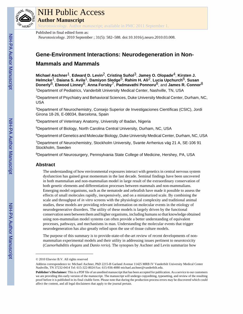
Gene-Environment Interactions: Neurodegeneration in Non- Mammals and Mammals Michael Aschner 1 , Edward D. Levin 2 , Cristina Suñol 3 , James O. Olopade 4 , Kirsten J. Helmcke 1 , Daiana S. Avila 1 , Damiyon Sledge 5 , Rahim H. Ali 2 , Lucia Upchurch 6 , Susan Donerly 6 , Elwood Linney 6 , Anna Forsby 7 , Padmavathi Ponnoru 8 , and James R. Connor 8 1 Department of Pediatrics, Vanderbilt University Medical Center, Nashville, TN, USA 2 Department of Psychiatry and Behavioral Sciences, Duke University Medical Center, Durham, NC, USA 3 Department of Neurochemistry, Consejo Superior de Investigaciones Científicas (CSIC), Jordi Girona 18-26, E-08034, Barcelona, Spain 4 Department of Veterinary Anatomy, University of Ibadan, Nigeria 5 Department of Biology, North Carolina Central University, Durham, NC, USA 6 Department of Genetics and Molecular Biology, Duke University Medical Center, Durham, NC, USA 7 Department of Neurochemistry, Stockholm University, Svante Arrhenius väg 21 A, SE-106 91 Stockholm, Sweden 8 Department of Neurosurgery, Pennsylvania State College of Medicine, Hershey, PA, USA Abstract The understanding of how environmental exposures interact with genetics in central nervous system dysfunction has gained great momentum in the last decade. Seminal findings have been uncovered in both mammalian and non-mammalian model in large result of the extraordinary conservation of both genetic elements and differentiation processes between mammals and non-mammalians. Emerging model organisms, such as the nematode and zebrafish have made it possible to assess the effects of small molecules rapidly, inexpensively, and on a miniaturized scale. By combining the scale and throughput of in vitro screens with the physiological complexity and traditional animal studies, these models are providing relevant information on molecular events in the etiology of neurodegenerative disorders. The utility of these models is largely driven by the functional conservation seen between them and higher organisms, including humans so that knowledge obtained using non-mammalian model systems can often provide a better understanding of equivalent processes, pathways, and mechanisms in man. Understanding the molecular events that trigger neurodegeneration has also greatly relied upon the use of tissue culture models. The purpose of this summary is to provide-state-of-the-art review of recent developments of non- mammalian experimental models and their utility in addressing issues pertinent to neurotoxicity (Caenorhabditis elegans and Danio rerio). The synopses by Aschner and Levin summarize how © 2010 Elsevier B.V. All rights reserved Address correspondence to: Michael Aschner, PhD 2215-B Garland Avenue 11425 MRB IV Vanderbilt University Medical Center Nashville, TN 37232-0414 Tel: 615-322-8024 Fax: 615-936-4080 [email protected]. Publisher's Disclaimer: This is a PDF file of an unedited manuscript that has been accepted for publication. As a service to our customers we are providing this early version of the manuscript. The manuscript will undergo copyediting, typesetting, and review of the resulting proof before it is published in its final citable form. Please note that during the production process errors may be discovered which could affect the content, and all legal disclaimers that apply to the journal pertain. NIH Public Access Author Manuscript Neurotoxicology. Author manuscript; available in PMC 2011 September 1. Published in final edited form as: Neurotoxicology. 2010 September ; 31(5): 582–588. doi:10.1016/j.neuro.2010.03.008. NIH-PA Author Manuscript NIH-PA Author Manuscript NIH-PA Author Manuscript

Welcome message from author
This document is posted to help you gain knowledge. Please leave a comment to let me know what you think about it! Share it to your friends and learn new things together.
Transcript
Gene-Environment Interactions: Neurodegeneration in Non-Mammals and Mammals
Michael Aschner1, Edward D. Levin2, Cristina Suñol3, James O. Olopade4, Kirsten J.Helmcke1, Daiana S. Avila1, Damiyon Sledge5, Rahim H. Ali2, Lucia Upchurch6, SusanDonerly6, Elwood Linney6, Anna Forsby7, Padmavathi Ponnoru8, and James R. Connor81Department of Pediatrics, Vanderbilt University Medical Center, Nashville, TN, USA2Department of Psychiatry and Behavioral Sciences, Duke University Medical Center, Durham, NC,USA3Department of Neurochemistry, Consejo Superior de Investigaciones Científicas (CSIC), JordiGirona 18-26, E-08034, Barcelona, Spain4Department of Veterinary Anatomy, University of Ibadan, Nigeria5Department of Biology, North Carolina Central University, Durham, NC, USA6Department of Genetics and Molecular Biology, Duke University Medical Center, Durham, NC, USA7Department of Neurochemistry, Stockholm University, Svante Arrhenius väg 21 A, SE-106 91Stockholm, Sweden8Department of Neurosurgery, Pennsylvania State College of Medicine, Hershey, PA, USA
AbstractThe understanding of how environmental exposures interact with genetics in central nervous systemdysfunction has gained great momentum in the last decade. Seminal findings have been uncoveredin both mammalian and non-mammalian model in large result of the extraordinary conservation ofboth genetic elements and differentiation processes between mammals and non-mammalians.Emerging model organisms, such as the nematode and zebrafish have made it possible to assess theeffects of small molecules rapidly, inexpensively, and on a miniaturized scale. By combining thescale and throughput of in vitro screens with the physiological complexity and traditional animalstudies, these models are providing relevant information on molecular events in the etiology ofneurodegenerative disorders. The utility of these models is largely driven by the functionalconservation seen between them and higher organisms, including humans so that knowledge obtainedusing non-mammalian model systems can often provide a better understanding of equivalentprocesses, pathways, and mechanisms in man. Understanding the molecular events that triggerneurodegeneration has also greatly relied upon the use of tissue culture models.
The purpose of this summary is to provide-state-of-the-art review of recent developments of non-mammalian experimental models and their utility in addressing issues pertinent to neurotoxicity(Caenorhabditis elegans and Danio rerio). The synopses by Aschner and Levin summarize how
© 2010 Elsevier B.V. All rights reservedAddress correspondence to: Michael Aschner, PhD 2215-B Garland Avenue 11425 MRB IV Vanderbilt University Medical CenterNashville, TN 37232-0414 Tel: 615-322-8024 Fax: 615-936-4080 [email protected]'s Disclaimer: This is a PDF file of an unedited manuscript that has been accepted for publication. As a service to our customerswe are providing this early version of the manuscript. The manuscript will undergo copyediting, typesetting, and review of the resultingproof before it is published in its final citable form. Please note that during the production process errors may be discovered which couldaffect the content, and all legal disclaimers that apply to the journal pertain.
NIH Public AccessAuthor ManuscriptNeurotoxicology. Author manuscript; available in PMC 2011 September 1.
Published in final edited form as:Neurotoxicology. 2010 September ; 31(5): 582–588. doi:10.1016/j.neuro.2010.03.008.
NIH
-PA Author Manuscript
NIH
-PA Author Manuscript
NIH
-PA Author Manuscript

genetic mutants of these species can be used to complement the understanding of molecular andcellular mechanisms associated with neurobehavioral toxicity and neurodegeneration. Next, studiesby Suñol and Olopade detail the predictive value of cultures in assessing neurotoxicity. Suñol andcolleagues summarize present novel information strategies based on in vitro toxicity assays that arepredictive of cellular effects that can be extrapolated to effects on individuals. Olopade and colleaguesdescribe cellular changes caused by sodium metavanadate (SMV) and demonstrate how rat primaryastrocyte cultures can be used as predicitive tools to assess the neuroprotective effects of antidoteson vanadium-induced astrogliosis and demyelination.
KeywordsCaenorhabditis elegans; zebrafish; tissue culture; neurotoxicity; astrocyte; vanadium; in vitro
C. Elegans as an Alternative Model in ToxicologyCaenorhabditis elegans in Neurotoxicology Studies
The search for experimental models that allow live in vivo analysis in the toxicological fieldhas led to the use of several invertebrate organisms. Caenorhabditis elegans (C. elegans) hasdemonstrated great advantages in providing insights into the mechanisms of toxicity andneuronal injury. The codification of the complete genome revealed the extraordinaryconservation of its genome with mammals (60–80% homology) [1] and studies indicating thatworms and mammals share similar biosynthetic and metabolic pathways provide justificationfor its relevance in the toxicological field.
C. elegans has a small size (adults are ~1 mm long), ease of maintenance, ability to be frozenand stored, speedy generation time (3 days), short life span, and large brood size (>300 progenyper hermaphrodite). All these features provide a limitless supply of worms for cellular,molecular, and genetic analyses. The transparency of C. elegans along with the ease ofconstructing reporter gene fusions facilitates the visualization of neuronal morphology andprotein expression patterns within the living system. Furthermore, the intensely studiedgenome, complete cell lineage map, knock-out libraries, and established geneticmethodologies, including mutagenesis, RNAi and transgenesis, provide a variety of optionsfor manipulating and performing molecular analyses in C. elegans.
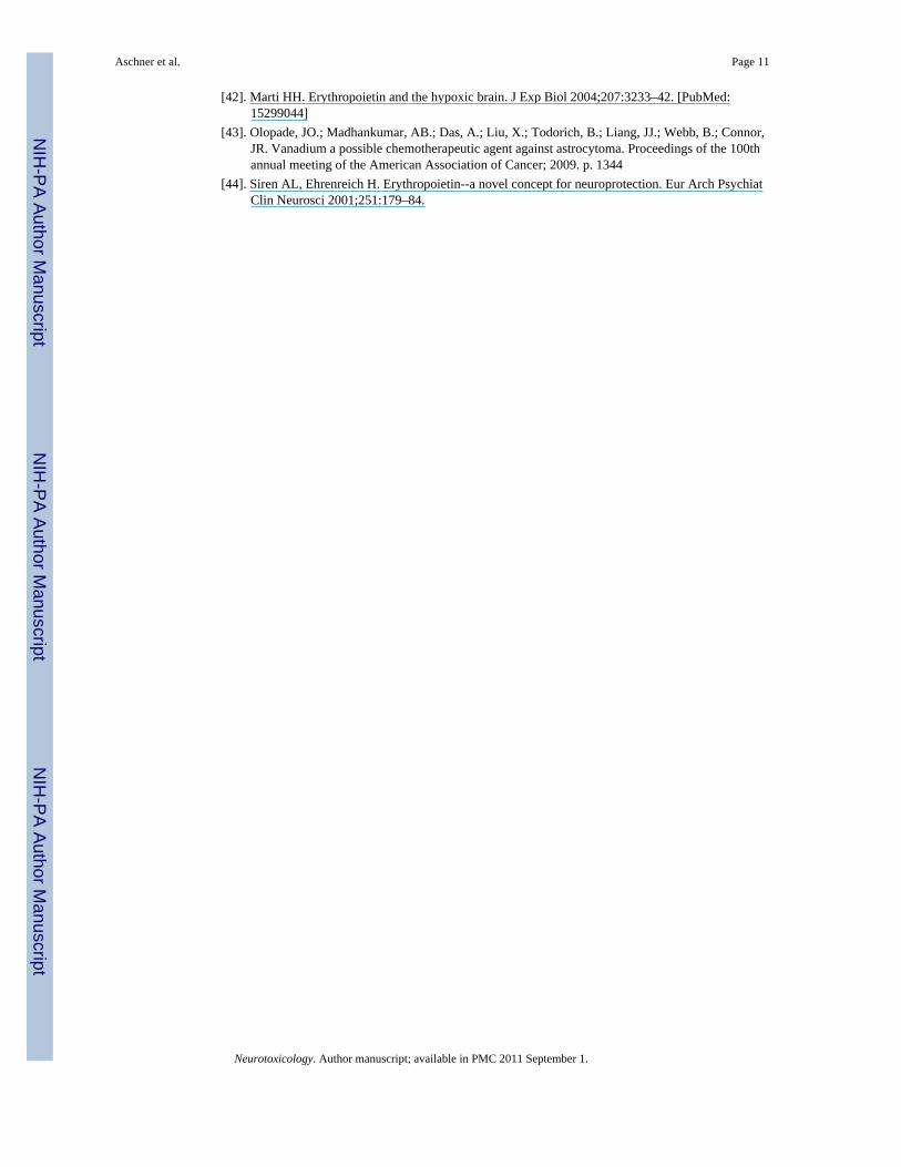
At its first inception, the C. elegans platform was developed as an experimental model to studynervous system development [2]. A complete three-dimensional map of the 302-cell nervoussystem provides the resources for identifying most synapses between neurons [3] and greenfluorescent protein (GFP) reporter strains have been generated to allow for the observation ofdifferent neuronal populations, for example the dat-1∷GFP fusion allows for the observationof all 8 dopaminergic neurons in the worm (Figure 1). The extensive knowledge of the nervoussystem has been exploited in the investigation of C. elegans and has allowed for the study ofvarious neurodegenerative disorders, such as Duchenne muscular dystrophy (DMD),Parkinson's disease, Huntington's disease and Alzheimer's disease. An example is theDYSTROPHIN gene, responsible for DMD, which is conserved in C. elegans and the studyof mutants has revealed progressive muscle degeneration [4]. Exposure of C. elegans to 1-methyl-4-phenylpyridinium (MPP+) [5] or 6-hyrdroxydopamine (6-OHDA) [6] mimicsParkinson's disease as both of these induce damage to the dopaminergic nervous system.Expression of polyQ Huntingtin variants in C. elegans has led to discoveries revealing geneinteractions with Huntingtin, axonal defects and protein aggregation, thereby furthering ourunderstanding of Huntington's disease [7,8]. The investigation into Alzheimer's disease hasbeen advanced with mutant worms expressing human beta-amyloid precursor protein
Aschner et al. Page 2
Neurotoxicology. Author manuscript; available in PMC 2011 September 1.
NIH
-PA Author Manuscript
NIH
-PA Author Manuscript
NIH
-PA Author Manuscript
(betaAPP) or TAU, allowing researchers to develop insights into disease progression anddiscover how other genes are involved [9].
C. elegans has also been used as a model to elucidate the toxicity and toxicological mechanismsof a variety of metals [10]. The toxicity of aluminum, arsenic, barium, cadmium, copper,chromium, lead, mercury, uranium, and zinc has been investigated in the C. elegans modelsystem, in which behavioral, morphological, and developmental alterations were observedupon toxicant exposure [11–13]. For instance, feeding behavior, thrashing, and pharyngealpumping are behavioral endpoints that allow examination of the involvement of specificneuronal networks affected by different metals and studies have indicated a decrease in theseendpoints. Various GFP reporters have been used to demonstrate changes in expression. Forexample, metallothioneins are upregulated upon exposure to cadmium [14].
Paraquat, rotenone, organophosphates, and other pesticides have also been assessed in the C.elegans model, enlightening researchers to the novel neurotoxic mechanisms through whichthese agents exert their effects [11]. Strains of C. elegans have been generated that have eitherincreased [15] or decreased [16] sensitivity to paraquat. Although the identities of many of theproteins encoded by the genes that cause these alterations are yet unknown, researchers havefound that alterations in antioxidants such as superoxide dismutase can cause alterations in theresponse of C. elegans to paraquat. Specifically, it has been found that mutants that lack theseenzymes display increased sensitivity [17] while mutants that overexpress these enzymesdisplay decreased sensitivity [18,19].
C. elegans is an ideal model to use to address the increased interest in the use of high-throughputapproaches to reproducibly and efficiently screen various chemicals. High-throughputscreening tools in C. elegans studies include computer tracking software to assess behaviorand biosorters and microfluidics to manipulate large numbers of worms with great speed,efficiency, and precision [20,21].
In summary, C, elegans is a valuable model for use in biological research, especiallytoxicology. The use of worms in the toxicological field demonstrates great potential forrevealing toxicity mechanisms which elude investigators using other models. The findings ofnumerous C. elegans studies have been reported and reviewed for the scientific community[11,21,22]. Understanding mechanisms of toxicity will help to elucidate ways in which newtherapeutics can be used to mitigate the adverse health effects of various toxicants.
Behavioral Analysis of Xenobiotics in ZebrafishIntroduction
Zebrafish can provide a useful complementary model for developmental neurotoxicology toprovide important mechanistic and screening information in combination with in vitro cell-based models, invertebrate models such as c. Elegans and drosophila as well as classicmammalian rodent models and epidemiological studies. Zebrafish have a rich array of toolswith which determine the molecular biology of neurodevelopment and mal-development.There are a great many zebrafish mutants with which to determine key genomic factors for thedevelopmental process. The use of morpholino techniques to transiently suppress specificmolecular components during early development is particularly powerful in studies ofdevelopmental neurotoxicology. In addition, since zebrafish have a clear chorion reportersystems can be used to highlight individual components of the nervous system nervous systemdevelopment can be visualized continuously and the impact of developmental neurotoxicantson developing neural processes can be determined. These elegant tools to study molecular andcellular processes in a temporally and anatomically intact model can be powerful for
Aschner et al. Page 3
Neurotoxicology. Author manuscript; available in PMC 2011 September 1.
NIH
-PA Author Manuscript
NIH
-PA Author Manuscript
NIH
-PA Author Manuscript

determining critical mechanisms of neurobehavioral toxicity if valid, sensitive, reliable andefficient measures of neurobehavioral function can be developed.
Recently, a variety of laboratories including ours have developed tests of learning and memory,stress response and sensorimotor reactivity for zebrafish, which are sensitive to drug andtoxicant effects, similar to those seen in classic rodent assays and humans. The neurobehavioraltest battery developed to date in our laboratory includes tests of swimming activity in newlyhatched fish, and in adults' sensorimotor response, exploratory behavior and stress response aswell as learning and memory. These tests have proven to be sensitive to toxicant andpharmacological effects and with some the efficiency of the tests are enhanced by automatedcomputerized video analysis.
Behavioral Analyses in ZebrafishSwimming Activity in Newly Hatched Fish—Spontaneous swimming activity can bequantified in zebrafish during the early period after hatching for a rapid read on behavioralconsequences of developmental toxicant exposure. We found that low-dose (0.029 and 0.29μM, 10 and 100 ml/l) exposure during the first five days after fertilization to theorganophosphate pesticide chlorpyrofos caused significant reductions in locomotor activity inyoung zebrafish tested on days 6 and 9 after fertilization [23]. That study used hand scoring ofvideotapes of the fish swimming activity in a three-minute session. More recently we havereproduced the same result using a computerized video tracking system (Noldus EthoVision,Wageningen, The Netherlands).
Sensorimotor Response—A rapid, intense sensory stimulus will in most species producea sudden motor reaction. The same is true in zebrafish. We have developed a test in which asudden tactile stimulus from a tap produced by a solenoid on the bottom of the test environmentproduces a flurry of swimming activity in the fish, an effect, which shows habituation orlessened response with repeated exposure. This sensorimotor response test has proven to besensitive to the persisting effects of developmental exposure to the organophosphate pesticidechlorpyrifos. Since chlorpyrifos inhibits acetylcholinesterase (AChE) and thus acts as anindirect cholinergic agonist we also tested the effects of developmental exposure to directcholinergic agonists for nicotinic and muscarinic acetylcholine receptors nicotine andpilocarpine [24]. All three of these treatments during the first five days after fertilizationproduce significant increase in sensorimotor response when the fish were later tested as adultsbut chlorpyrifos was effective at a much lower dose (0.29 μM) than either nicotine (15 μM) orpilocarpine (1,000 μM) [24].
Exploratory Behavior and Stress Response—When introduced into a novelenvironment zebrafish will dive to the bottom of the tank in what is likely a predation escaperesponse. Gradually, over time the fish swim increasingly to the higher levels of the tank[25]. This effect is specific to the fish being introduced into a novel tank. Placement into afamiliar tank does not produce this effect [26]. The novel tank diving response is reduced byanxiolytic drugs diazepam and buspirone, but not by chlordiazepoxide [26]. Nicotine, whichhas been shown to have anxiolytic effects in mammals including humans also significantlyreduces the novel tank diving response, an effect which is reversed by co-administration ofnicotinic antagonists [25,27]. Recently, we have found that 0.29 μM of chlorpyrifos during thefirst five days after fertilization caused a significant increase in swimming activity in the noveltank diving task as well as significantly less of the normal diving response when the fish weretested as adults after early developmental exposure [26].
Learning and Memory—Zebrafish learn spatial and color discrimination as well as spatialalternation as assessed in a three-chamber test tank we have developed [23,28]. Nicotine
Aschner et al. Page 4
Neurotoxicology. Author manuscript; available in PMC 2011 September 1.
NIH
-PA Author Manuscript
NIH
-PA Author Manuscript
NIH
-PA Author Manuscript
significantly improves both spatial learning and memory in this test paradigm [23,24,29]. Thenicotine-induced learning improvement has been shown to be related to increases in braindopamine activity [24]. The nicotinic antagonist mecamylamine blocks both the nicotine-induced learning improvement and the nicotine-induced increase in dopamine metabolite[24].
With regard to developmental neurotoxicity, we have found that 0.029 or 0.29 μM ofchlorpyrifos in the tank water of zebrafish for the first five days after fertilization causes long-term impairment in memory assessed by the delayed spatial alternation task when the fish weretested as adults [30]. These chlorpyrifos doses had a biphasic effect on swimming speed in thememory test with the lower dose significantly slowing response speed and the higher dosecausing significant hyperactivity. To test the impact of inhibition of the AChE inhibition onthese behavioral functions we produced a zebrafish morpholino, which roughly matched thedegree of AChE inhibition by 0.29μM of CPF in the tank water for the first five days afterfertilization (Figure 2). When these fish and their controls were tested in the three-chamberspatial alternation task when they were adults, the AChE morpholino fish had a significantimpairment in memory relative to controls, quite similar to the chlorpyrifos exposed fish(Figure 3) indicating that it was the inhibition of AChE by chlorpyrifos which was sufficientto produce the memory impairment. Interestingly, the effect of the AChE morpholino onresponse speed did not match the effect of 0.29 μM of chlorpyrifos. The morpholino matchingthe same degree of AChE inhibition as 0.29 μM of chlorpyrifos did not produce hyperactivityas did 0.29 μM chlorpyrifos. If anything there was a slight reduction in speed.
These studies show that zebrafish models can provide useful neurobehavioral informationconcerning the potentially adverse effects of developmental toxicant exposure. Zebrafish canserve as a translational vertebrate model between cell-based and invertebrate and mammalianmodels providing screening of compounds as well as mechanistic information concerningdevelopmental neurobehavioral toxicity. The development of a neurobehavioral test batteryfor zebrafish will bring these advantages to bear for functional neurotoxicity.
A Way Forward for Using In Vitro Neurotoxicity Models in Acute ToxicityTesting Strategy: Update on the FP6 Integrated Project ACuteToxIntroduction
Toxicity risk assessment for chemical-induced human health hazards relies mainly on dataobtained from animal experimentation, human studies and epidemiology. Accepted toxicitytesting since 2001 includes regulations that take into account the concepts of Refining andReducing animal experimentation: the Fixed Dose Procedure (FDP, TG 420), the Acute ToxicClass Method (ATCM, TG 423), and the Up-and-Down Procedure (UDP, TG 425). In 2007,the US National Academies proposed that toxicity testing in the 21st century should takeadvantage of the understanding of “toxicity pathways” (the cellular response pathways thatunderlie adverse health effects when they are sufficiently perturbed). Therefore, theyrecommend using “a predictive strategy based on in vitro toxicity assays which predict cellularlevel effects that can be extrapolated to effects on individuals” (NRC, 2007). In 2005, theEuropean Union supported the ACuteTox project, which has set the very ambitious overallobjective of developing an in-vitro test strategy sufficiently robust and powerful to replace in-vivo testing for predicting acute toxicity of chemicals. The ACuteTox project aims to improvethe previously estimated correlation between in vitro basal cytotoxicity and rodent LD50 values[31], to a level sufficient enough to ensure a valid prediction of human acute toxicity. The coreof the ACuteTox project is composed from the generation of a high quality in vivo data base,the generation of a high quality in vitro data base, and the compilation of cell systems andendpoints. In vitro data include those obtained in cytotoxicity assays and in specific in vitro/
Aschner et al. Page 5
Neurotoxicology. Author manuscript; available in PMC 2011 September 1.
NIH
-PA Author Manuscript
NIH
-PA Author Manuscript
NIH
-PA Author Manuscript
in silico assays for biokinetics, metabolism and organ toxicity (hepatotoxicity, nephrotoxicityand neurotoxicity) [32]. All this information is stored in a data base that will be publiclyavailable on completion of the project. About 60 reference chemicals including pesticides,pharmaceuticals and industrial chemicals, for which data on their acute human toxicity do exist,have been tested. The linear regression analysis between cytotoxicity data (Neutral red uptake(NRU) assay in the mouse fibroblast 3T3 cell line) and human lethal blood concentration(LC50) gave an explained variance R2 = 0.56 for the reference chemicals [33]. It is foreseenthat the integration of biokinetic, metabolism and organ specific toxicity data will provide alerts(against the exclusive use of cytotoxicity data) and correctors (algorithms to be used) for theefficient use of an in vitro-based testing strategy for predicting human acute toxicity.
The ACuteTox Project as a Predictive Tool for Neurotoxicity AssessmentThe nervous system is particularly vulnerable to chemical exposure; its complexity results inmultiple potential target sites with different toxicity features. Acute human toxicity related toadverse neuronal function is mainly a result of over-excitation or depression of the peripheralor central nervous system (CNS). The major molecular and cellular mechanisms involved insuch effects include GABAergic, glutamatergic and cholinergic neurotransmission, regulationof cell and mitochondrial membrane potential, and those critical for maintaining CNSfunctionality, such as controlling cell energy. Up to 57 neural endpoints, including those relatedwith the above mechanisms and others that specifically identify neural types (neurons,astrocytes and microglia) or a cascade of multiple targets (induction of stress and apoptoticgene biomarkers), have been determined in primary cultured rodent neurons, rodent brain cellaggregates and human neuronal cell lines. Unless otherwise cited, this presentation summarizespublished results from the ACuteTox project [34,35]. Cell viability in the neural systems afterexposure to the agents for 24 – 48 hours did not differ from that in non neural cell lines,suggesting that the neuronal cytotoxicity model did not correct the outlier chemicals found inthe NRU-3T3 cytotoxicity assay versus human LC50 correlation [34]. 28 out of 58 testedchemicals were recognized by the GABAA receptor (GABAAR) assay (a radiometric assaydetermining 36Cl− influx in primary cultured neurons) and 10 out of 23 chemicals wererecognized by the GABA transport assay (a radiometric assay measuring [3H]GABA uptakein primary cultured neurons). In contrast, other neurochemical assays (fluorescence assays foracetylcholine and glutamate receptor function, spectrophotometric assay foracetylcholinesterase activity and radiometric/chromatographic assay for glutamate transport)were very specific and recognized few compounds from the reference list. A more generalassay for assessing neuronal excitability (a fluorescence-based assay measuring changes in cellmembrane potential (CMP) in the human neuronal cell line SH-SY5Y) recognized 22 out of58 chemicals. The combination of the CMP and GABAAR assays generated alerts (theneurotoxic endpoints were more sensitive than the cytotoxic endpoint) for outliers (Figure 4)and improved the in vitro predictability of human acute toxicity. The R2 values for thecorrelation of in vitro data vs. human LC50 values were estimated to 0.57 for only the generalcytotoxicity data and 0.64 when considering cytotoxicity data in combination with alertsidentified from the CMP+GABAAR assays. By using appropriate agonists andpharmacological agents, the GABAAR and the CMP assays may cover most of the membrane-based neurotoxicity pathways. As an advantage for the use of these in vitro testing of neuralendpoints, both the GABAAR and the CMP assays were performed in a short period of time(10 and 2 minutes, respectively). However, the drawback of using a highly energeticradionuclide in the GABAAR assay should be overcome by the use of less energetic isotopesor even, non radiometric techniques.
In addition to the GABAAR and CMP assays, genomic and metabolic endpoints were analyzedin a multi-endpoint assay after exposure to rodent brain cell aggregate cultures and theexpression of the stress gene marker caspase 3 was determined in primary cultured neurons.
Aschner et al. Page 6
Neurotoxicology. Author manuscript; available in PMC 2011 September 1.
NIH
-PA Author Manuscript
NIH
-PA Author Manuscript
NIH
-PA Author Manuscript
The multi-endpoint determining changes in the mRNA of NF-H, GFAP, MBP and HSP32, aswell as in the total mRNA level and glucose consumption in the brain cell aggregate culturesthat were exposed for 48 hours to the tested chemicals resulted to be the most sensitiveendpoints producing alerts for severe outliers like digoxin, lindane, malathion and parathion.Furthermore, the caspase-3 mRNA expression gave a good estimate of the human LC50 for aset of the chemicals tested, correcting severe outliers like atropine, digoxin and malathion[34].
ConclusionsIn conclusion, the assays developed in ACuteTox project have provided alerts for all the outliersevolved from the cytotoxicity – human LC50 correlation. The combined genomic and metabolicbiomarkers analyzed in the brain cell aggregate cultures were very sensitive endpoints,generating most alerts. The combination of the GABAAR and CMP assays identified correctoutliers and improved the prediction of human lethal toxicity. These two endpoints cover mostof the membrane-based neurotoxicity. As a whole, it is foreseen that the ACuteTox project willyield “alert” tests to indicate when a cytotoxicity-based test result may not be valid and“corrector” tests to improve the predictive capabilities of a test strategy. A capacity to indicatewhen a cytotoxicity-based in vitro approach may be insufficient and that additional testing isrequired or that a certain compound is beyond its predictive capabilities represents an importantstep forward for in vitro methods.
Erythropoietin, Desferroxamine and Tiron are Protective against Vanadium-Induced Demyelination and Oxidative Stress in the Rat BrainIntroduction
Environmental pollution from fossil fuel burning and the subsequent release of finelyparticulate vanadium compounds in the Arabian gulf [36] and Nigeria's Niger Delta [37]hasbeen on the increase with minimal studies done on both the short and long term effects on thebrain. Hypomyelination and or myelin destruction from lipid peroxidation has been implicatedin part to be responsible for the phenotypic expressions of neuromuscular and behavioraldeficits seen in vanadium toxicosis [38]. The present study assessed novel cellular changescaused by sodium metavanadate (SMV) in astrocyte culture cells and the neuroprotectiveeffects of antidotes desferroxamine (DFO), tiron and erythropoietin (EPO) on vanadium-induced astrogliosis and demyelination.
Studies on the in vitro and in vivo Neurotoxicity of VanadiumThe effects of vanadium treatment (150 μM for 6 hours) on reactive oxygen species (ROS)generation, cellular hypoxia and erythropoietin (EPO) expression were assessed in primary ratastrocytes cultures. ROS generation was determined with the dichlorofluorescein (DCF) assay[measured as relative fluorescence unit (RFU)]. Protein expression was determined by westernblot (WB) analysis, using HIF-1 α (Hypoxia Induction Factor) and EPO antibodies (SantaCruz). We also treated 15-day-old Sprague-Dawley rats (n=5) with 3mg/kg SMV for sevendays; three groups were similarly treated, but were given DFO (300 mg/kg/day), tiron (606mg/kg/day) or EPO (5,000 U/kg/day) as antidotes concurrently for six days (one day aftervanadium treatment) with appropriate controls. Glial fibrillary acidic protein (GFAP)immunohistochemistry was observed in the corpus callosum while myelin basic protein (MBP)was quantified with WB analysis.
There was a significant increase in ROS generation in vanadate treated primary astrocytecultures compared with controls (Figure 5) including up-regulation in HIF-1 α and EPOexpressions as seen by WB analysis (Figure 6). There was also a significant increase in GFAP
Aschner et al. Page 7
Neurotoxicology. Author manuscript; available in PMC 2011 September 1.
NIH
-PA Author Manuscript
NIH
-PA Author Manuscript
NIH
-PA Author Manuscript
immunoreactivity in the corpus callosum (Figure 7) and a decrease in MBP quantification inbrains of vanadium treated rats compared to controls (Figure 8); these observations weresignificantly (P<0.05) attenuated in rats receiving the antidotes DFO and EPO, or tiron andEPO.
In this study, vanadium induced the production of reactive oxygen species (ROS); this causesthe cells to undergo hypoxia, the onset of which leads to vanadate-induced expression ofHIF-1α [39]]. In other cells, such as the kidney cells and prostate cancer; HIF 1 α upregulationleads to increased EPO expression [40]. EPO has been shown to be expressed in the brain[41]; in mammalian tissue, discrepancy between oxygen supply and utilization leads to hypoxiawhich induces a variety of specific adaptation mechanisms including the activation of HIF,which, in turn, has a modulatory role on hypoxically-related genes, such as those encoding forEPO [42]. In ischemia, the de novo up regulation of EPO in the brain creates a protective effectagainst neuronal injury. Though vanadium causes neuronal pathologies and neurobehavioraldeficits [38] in vivo, induces oxidative stress and cellular hypoxia in brain cell cultures [43].The role EPO plays at the cellular, local and systemic level during vanadium neurotoxicosisremains unknown. Our work has shown that vanadium induced HIF-1 α expression can leadto EPO upregulation in primary astrocytes which is indicative of an endogenous protectivemechanism in the neuroglia. This current work also shows that systemic administration ofantidotes can modulate vanadium-induced neuropathologies. The increased expression ofGFAP seen after vanadium treatment was significantly attenuated by the antidotes DFO andEPO, but not by tiron, while the reduced MBP expression caused by vanadium administrationwas significantly reversed by tiron and EPO, indicating that EPO was effective in bothinstances. These actions of EPO are consistent with the fact that EPO has anti-inflammatoryand antioxidative effects, and prevents lipid peroxidation [44]. While in theory, it is expectedthat vanadium entry into the brain will lead to increased generation of ROS, which willconcurrently be responsible for increased GFAP expression, lipid peroxidation and thendemyelination, the fact that DFO in vivo was able to attenuate the increased GFAP expressionafter vanadate treatment, but not the demyelinating process and vice-versa for tiron may beindicative of other independent pathways between lipid peroxidation and demyelination causedby vanadate neurotoxicosis.
ConclusionsIn conclusion, this work demonstrates the utility of primary astrocyte cultures in screening theeffects of vandate on cellular responses, as well as novel information on vandate's ability toinduce increased expression of EPO in astrocyte cell cultures and that antidotes, such asexogenous EPO can attenuate astrogliosis and demyelination caused by vanadateneurotoxicosis.
AcknowledgmentsThis review was supported in part by PHS grant NIEHS R01 ES10563 (MA), the Duke University Superfund BasicResearch Center NIEHS ES010356 (EDL). Sunol and Forsby wish to acknowledge the contribution by Joan AlbertVericat and the group at Neuropharma (Spain), Victor Rimbau and the group at the University of Barcelona (Spain),Paul Honegger and the group at the University of Lausanne (Switzerland), Sandra Coecke and the group at ECVAM(Italy) and the signed authors and the groups at the CSIC (Spain) and Stockholm University (Sweden). Partial supportwas also provided by IBRO Research Fellowship (JOO).
References[1]. Kaletta T, Hengartner MO. Finding function in novel targets: C. elegans as a model organism. Nat
Rev Drug Discov 2006;5:387–98. [PubMed: 16672925][2]. Brenner S. The genetics of Caenorhabditis elegans. Genetics 1974;77:71–94. [PubMed: 4366476][3]. Hobert O. Specification of the nervous system. WormBook 2005:1–19. [PubMed: 18050401]
Aschner et al. Page 8
Neurotoxicology. Author manuscript; available in PMC 2011 September 1.
NIH
-PA Author Manuscript
NIH
-PA Author Manuscript
NIH
-PA Author Manuscript

[4]. Gieseler K, Grisoni K, Segalat L. Genetic suppression of phenotypes arising from mutations indystrophin-related genes in Caenorhabditis elegans. Curr Biol 2000;10:1092–7. [PubMed:10996789]
[5]. Braungart E, Gerlach M, Riederer P, Baumeister R, Hoener MC. Caenorhabditis elegans MPP+ modelof Parkinson's disease for high-throughput drug screenings. Neurodegener Dis 2004;1:175–83.[PubMed: 16908987]
[6]. Nass R, Hall DH, Miller DM 3rd, Blakely RD. Neurotoxin-induced degeneration of dopamineneurons in Caenorhabditis elegans. Proc Natl Acad Sci U S A 2002;99:3264–9. [PubMed:11867711]
[7]. Holbert S, Dedeoglu A, Humbert S, Saudou F, Ferrante RJ, Neri C. Cdc42-interacting protein 4 bindsto huntingtin: neuropathologic and biological evidence for a role in Huntington's disease. Proc NatlAcad Sci U S A 2003;100:2712–7. [PubMed: 12604778]
[8]. Parker JA, Connolly JB, Wellington C, Hayden M, Dausset J, Neri C. Expanded polyglutamines inCaenorhabditis elegans cause axonal abnormalities and severe dysfunction of PLMmechanosensory neurons without cell death. Proc Natl Acad Sci U S A 2001;98:13318–23.[PubMed: 11687635]
[9]. Boyd-Kimball D, Poon HF, Lynn BC, Cai J, Pierce WM Jr. Klein JB, Ferguson J, Link CD, ButterfieldDA. Proteomic identification of proteins specifically oxidized in Caenorhabditis elegans expressinghuman Abeta(1–42): implications for Alzheimer's disease. Neurobiol Aging 2006;27:1239–49.[PubMed: 16099075]
[10]. Berkowitz LA, Hamamichi S, Knight AL, Harrington AJ, Caldwell GA, Caldwell KA. Applicationof a C. elegans dopamine neuron degeneration assay for the validation of potential Parkinson'sdisease genes. J Vis Exp 2008:ii, 835. doi: 10.3791/835.
[11]. Leung MC, Williams PL, Benedetto A, Au C, Helmcke KJ, Aschner M, Meyer JN. Caenorhabditiselegans: an emerging model in biomedical and environmental toxicology. Toxicol Sci 2008;106:5–28. [PubMed: 18566021]
[12]. Helmcke KJ, Syversen T, Miller DM 3rd, Aschner M. Characterization of the effects ofmethylmercury on Caenorhabditis elegans. Toxicol Appl Pharmacol. 2009 ePub ahead of print.
[13]. Guo Y, Yang Y, Wang D. Induction of reproductive deficits in nematode Caenorhabditis elegansexposed to metals at different developmental stages. Reprod Toxicol 2009;28:90–5. [PubMed:19490999]
[14]. Levin ED, Swain HA, Donerly S, Linney E. Developmental chlorpyrifos effects on hatchlingzebrafish swimming behavior. Neurotoxicology & Teratology 2004;26:719–23. [PubMed:15451035]
[15]. Ishii N, Takahashi K, Tomita S, Keino T, Honda S, Yoshino K, Suzuki K. A methyl viologen-sensitive mutant of the nematode Caenorhabditis elegans. Mutat Res 1990;237:165–71. [PubMed:2233820]
[16]. Fujii M, Tanaka N, Miki K, Hossain MN, Endoh M, Ayusawa D. Uncoupling of longevity andparaquat resistance in mutants of the nematode Caenorhabditis elegans. Biosci Biotechnol Biochem2005;69:2015–8. [PubMed: 16244463]
[17]. Anantharam V, Lehrmann E, Kanthasamy A, Yang Y, Banerjee P, Becker KG, Freed WJ,Kanthasamy AG. Microarray analysis of oxidative stress regulated genes in mesencephalicdopaminergic neuronal cells: relevance to oxidative damage in Parkinson's disease. Neurochem Int2007;50:834–47. [PubMed: 17397968]
[18]. Yanase S, Yasuda K, Ishii N. Adaptive responses to oxidative damage in three mutants ofCaenorhabditis elegans (age-1, mev-1 and daf-16) that affect life span. Mech Ageing Dev2002;123:1579–87. [PubMed: 12470895]
[19]. Burmeister C, Luersen K, Heinick A, Hussein A, Domagalski M, Walter RD, Liebau E. Oxidativestress in Caenorhabditis elegans: protective effects of the Omega class glutathione transferase(GSTO-1). FASEB J 2008;22:343–54. [PubMed: 17901115]
[20]. Hulme SE, Shevkoplyas SS, Samuel A. Microfluidics: streamlining discovery in worm biology.Nat Methods 2008;5:589–90. [PubMed: 18587316]
Aschner et al. Page 9
Neurotoxicology. Author manuscript; available in PMC 2011 September 1.
NIH
-PA Author Manuscript
NIH
-PA Author Manuscript
NIH
-PA Author Manuscript

[21]. Peterson RT, Nass R, Boyd WA, Freedman JH, Dong K, Narahashi T. Use of non-mammalianalternative models for neurotoxicological study. Neurotoxicology 2008;29:546–55. [PubMed:18538410]
[22]. Helmcke KJ, Avila DS, Aschner M. Utility of Caenorhabditis elegans in high throughputneurotoxicological research. Neurotoxicol Teratol. 2008 ePub ahead of print.
[23]. Levin ED, Chen E. Nicotinic involvement in memory function in zebrafish. Neurotoxicology andTeratology 2004;26:731–735. [PubMed: 15451037]
[24]. Eddins D, Cerutti D, Williams P, Linney E, Levin ED. Developmental chlorpyrifos causesbehavioral and neurochemical defects in zebrafish. Neurotoxicology and Teratology. 2009 in press.
[25]. Levin ED, Bencan Z, Cerutti DT. Anxiolytic effects of nicotine in zebrafish. Physiology andBehavior 2007;90:54–58. [PubMed: 17049956]
[26]. Bencan Z, Sledge D, Levin ED. Buspirone, chlordiazepoxide and diazepam effects in a zebrafishmodel of anxiety. Pharmacology, Biochemistry and Behavior. 2009 in press.
[27]. Bencan Z, Levin ED. The role of α7 and α4β2 nicotinic receptors in the nicotine-induced anxiolyticeffect in zebrafish. Physiology & Behavior 2008;95:408–412. [PubMed: 18671990]
[28]. Arthur D, Levin ED. Spatial and non-spatial discrimination learning in zebrafish (Danio rerio).Animal Cognition 2001;4:125–131.
[29]. Levin ED, Limpuangthip J, Rachakonda T, Peterson M. Timing of nicotine effects on learning inzebrafish. Psychopharmacology 2006;184:547–552. [PubMed: 16175402]
[30]. Levin ED, Chrysanthis E, Yacisin K, Linney E. Chlorpyrifos exposure of developing zebrafish:effects on survival and long-term effects on response latency and spatial discrimination.Neurotoxicology & Teratology 2003;25:51–7. [PubMed: 12633736]
[31]. Ekwall B, Barile FA, Castano A, Clemedson C, Clothier RH, Dierickx P, Ekwall B, Ferro M,Fiskesjö G, Garza-Ocañas L, et al. MEIC Evaluation of Acute Systemic Toxicity: Part VI. Theprediction of human toxicity by rodent LD50 values and results from 61 in vitro methods. AlterLab Animals 1988;26:617–658.
[32]. Clemedson C. The European ACuteTox project: a modern integrative in vitro approach to betterprediction of acute toxicity. Clin Pharmacol Ther 2008;84:200–2. [PubMed: 18679183]
[33]. Sjostrom M, Kolman A, Clemedson C, Clothier R. Estimation of human blood LC50 values for usein modeling of in vitro-in vivo data of the ACuteTox project. Toxicol In Vitro 2008;22:1405–11.[PubMed: 18514477]
[34]. Forsby A, Bal-Price AK, Camins A, Coecke S, Fabre N, Gustafsson H, Honegger P, Kinsner-Ovaskainen A, Pallas M, Rimbau V, et al. Neuronal in vitro models for the estimation of acutesystemic toxicity. Toxicol In Vitro. 2009 ePub ahead of print.
[35]. Sunol C, Babot Z, Fonfria E, Galofre M, Garcia D, Herrera N, Iraola S, Vendrell I. Studies withneuronal cells: From basic studies of mechanisms of neurotoxicity to the prediction of chemicaltoxicity. Toxicol In Vitro 2008;22:1350–5. [PubMed: 18467072]
[36]. Sasi MM, Haider SS, el-Fakhri M, Ghwarsha KM. Microchromatographic analysis of lipids, protein,and occurrence of lipid peroxidation in various brain areas of vanadium exposed rats: a possiblemechanism of vanadium neurotoxicity. Neurotoxicology 1994;15:413–20. [PubMed: 7991230]
[37]. Igado OO, Olopade JO, Onwuka SK, Chukwudi AC, Daramola OA, Ajufo UE. Evidence ofenvironmental pollution in caprine brains obtained from a relatively unindustrialized area in Nigeria.AJBR 2008;11:305–9.
[38]. Soazo M, Garcia GB. Vanadium exposure through lactation produces behavioral alterations andCNS myelin deficit in neonatal rats. Neurotoxicol Teratol 2007;29:503–10. [PubMed: 17493788]
[39]. Gao HM, Jiang J, Wilson B, Zhang W, Hong JS, Liu B. Microglial activation-mediated delayedand progressive degeneration of rat nigral dopaminergic neurons: relevance to Parkinson's disease.J Neurochem 2002;81:1285–97. [PubMed: 12068076]
[40]. Arcasoy MO. The non-haematopoietic biological effects of erythropoietin. Br J Haematol2008;141:14–31. [PubMed: 18324962]
[41]. Siren AL, Fratelli M, Brines M, Goemans C, Casagrande S, Lewczuk P, Keenan S, Gleiter C,Pasquali C, Capobianco A, Mennini T, Heumann R, A. Ehrenreich H, Ghezzi P. Erythropoietinprevents neuronal apoptosis after cerebral ischemia and metabolic stress. Proc Natl Acad Sci U SA 2001;98:4044–9. [PubMed: 11259643]
Aschner et al. Page 10
Neurotoxicology. Author manuscript; available in PMC 2011 September 1.
NIH
-PA Author Manuscript
NIH
-PA Author Manuscript
NIH
-PA Author Manuscript
[42]. Marti HH. Erythropoietin and the hypoxic brain. J Exp Biol 2004;207:3233–42. [PubMed:15299044]
[43]. Olopade, JO.; Madhankumar, AB.; Das, A.; Liu, X.; Todorich, B.; Liang, JJ.; Webb, B.; Connor,JR. Vanadium a possible chemotherapeutic agent against astrocytoma. Proceedings of the 100thannual meeting of the American Association of Cancer; 2009. p. 1344
[44]. Siren AL, Ehrenreich H. Erythropoietin--a novel concept for neuroprotection. Eur Arch PsychiatClin Neurosci 2001;251:179–84.
Aschner et al. Page 11
Neurotoxicology. Author manuscript; available in PMC 2011 September 1.
NIH
-PA Author Manuscript
NIH
-PA Author Manuscript
NIH
-PA Author Manuscript
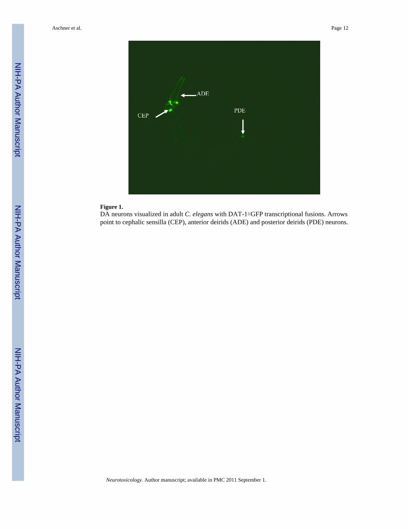
Figure 1.DA neurons visualized in adult C. elegans with DAT-1∷GFP transcriptional fusions. Arrowspoint to cephalic sensilla (CEP), anterior deirids (ADE) and posterior deirids (PDE) neurons.
Aschner et al. Page 12
Neurotoxicology. Author manuscript; available in PMC 2011 September 1.
NIH
-PA Author Manuscript
NIH
-PA Author Manuscript
NIH
-PA Author Manuscript

Figure 2.Effects on acetylcholinesterase activity of 0.29μM (100 ng/ml) of chlorpyrifos for five dayspost-fertilization and AChE morpholino.
Aschner et al. Page 13
Neurotoxicology. Author manuscript; available in PMC 2011 September 1.
NIH
-PA Author Manuscript
NIH
-PA Author Manuscript
NIH
-PA Author Manuscript
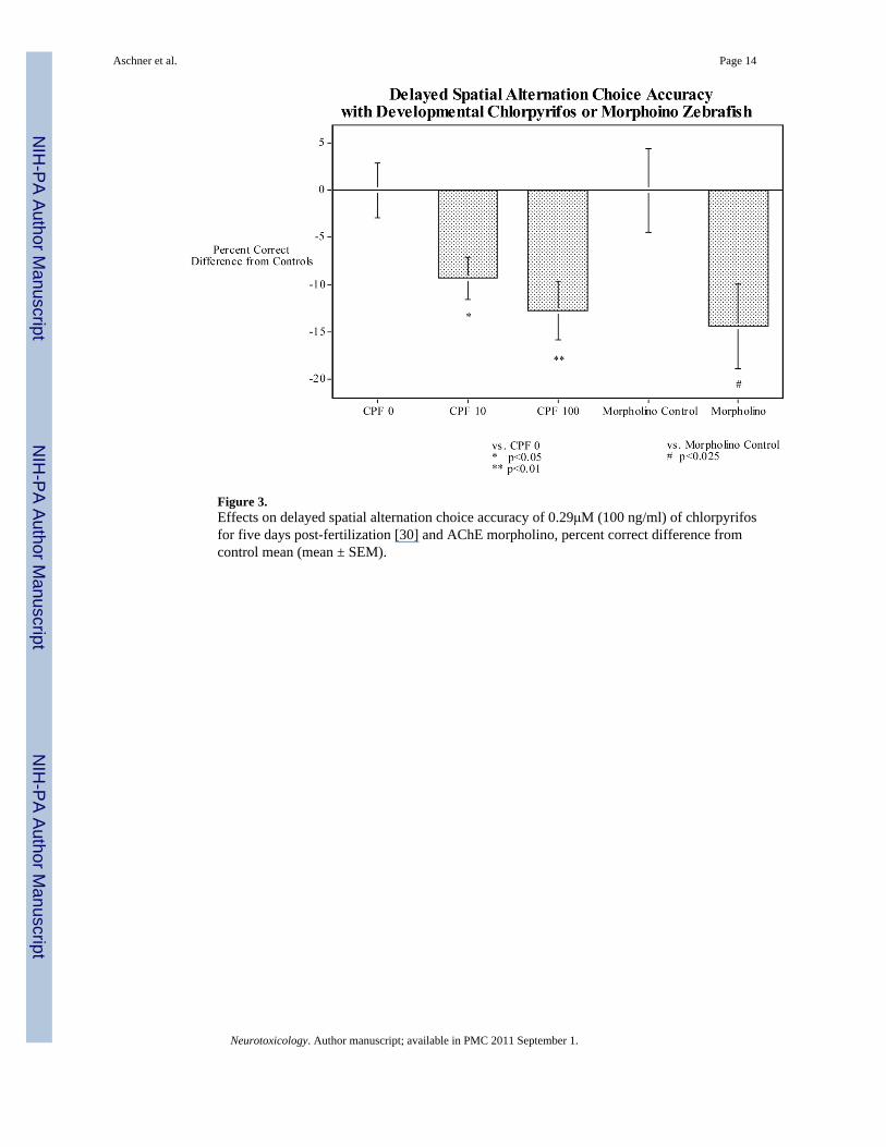
Figure 3.Effects on delayed spatial alternation choice accuracy of 0.29μM (100 ng/ml) of chlorpyrifosfor five days post-fertilization [30] and AChE morpholino, percent correct difference fromcontrol mean (mean ± SEM).
Aschner et al. Page 14
Neurotoxicology. Author manuscript; available in PMC 2011 September 1.
NIH
-PA Author Manuscript
NIH
-PA Author Manuscript
NIH
-PA Author Manuscript
Figure 4.Neurotoxicity alerts determined by the combination of the GABAAR and cell membranepotential assay (CMP). Blue bars represent the lowest EC50/LEC values for the GABAARassay (performed in primary neuronal cultures) or the CMP assay (performed in SH-SY5Ycells) after acutely exposure to the reference chemicals. Red bars represent IC50 values for theNRU cytotoxicity test (performed in 3T3 cells) from Sjöström et al. (2008). Asterisks indicateneurotoxic alerts (significant effects on the GABAAR/CMP assays) were produced atconcentrations at least one order of magnitude lower than those producing cytotoxicity. Arrowsindicate outlier compounds in the NRU-3T3 cytotoxicity assay vs human LC50 valuescorrelation.
Aschner et al. Page 15
Neurotoxicology. Author manuscript; available in PMC 2011 September 1.
NIH
-PA Author Manuscript
NIH
-PA Author Manuscript
NIH
-PA Author Manuscript
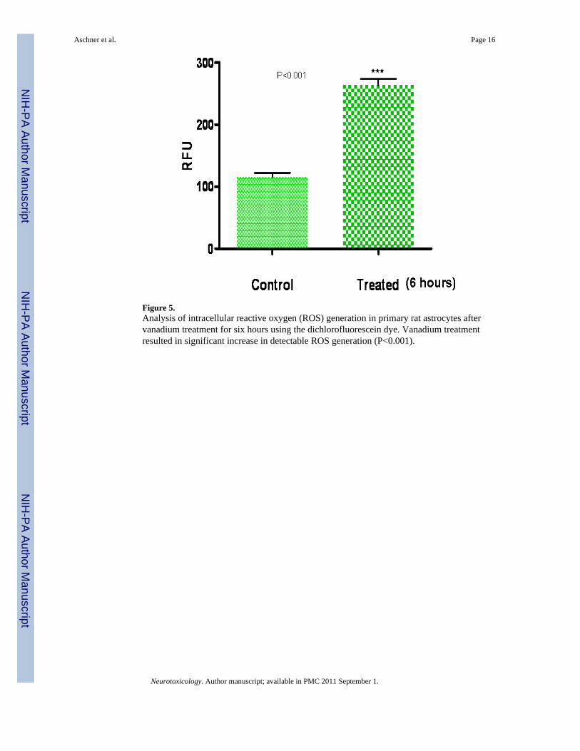
Figure 5.Analysis of intracellular reactive oxygen (ROS) generation in primary rat astrocytes aftervanadium treatment for six hours using the dichlorofluorescein dye. Vanadium treatmentresulted in significant increase in detectable ROS generation (P<0.001).
Aschner et al. Page 16
Neurotoxicology. Author manuscript; available in PMC 2011 September 1.
NIH
-PA Author Manuscript
NIH
-PA Author Manuscript
NIH
-PA Author Manuscript
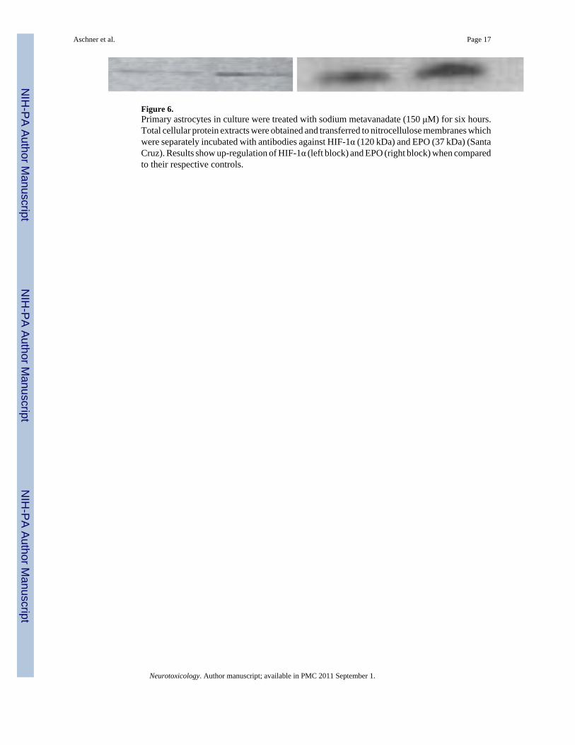
Figure 6.Primary astrocytes in culture were treated with sodium metavanadate (150 μM) for six hours.Total cellular protein extracts were obtained and transferred to nitrocellulose membranes whichwere separately incubated with antibodies against HIF-1α (120 kDa) and EPO (37 kDa) (SantaCruz). Results show up-regulation of HIF-1α (left block) and EPO (right block) when comparedto their respective controls.
Aschner et al. Page 17
Neurotoxicology. Author manuscript; available in PMC 2011 September 1.
NIH
-PA Author Manuscript
NIH
-PA Author Manuscript
NIH
-PA Author Manuscript

Figure 7.Treatment of rat cells 3mg /kg of SMV for seven days caused increased GFAP expression (topright) compared to controls (top left). The staining caused by vanadate in was markedlyattenuated by antidotes DFO and EPO (bottom left and right respectively) but not by tiron(bottom center).
Aschner et al. Page 18
Neurotoxicology. Author manuscript; available in PMC 2011 September 1.
NIH
-PA Author Manuscript
NIH
-PA Author Manuscript
NIH
-PA Author Manuscript

Figure 8.Treatment of rat with3mg /kg of SMV for seven days led to decreased myelin content (G2)compared to controls (G1). Antidotes tiron and EPO (G4and5) when administered withvanadate caused a significant increase in myelin quantity compared to treated group but notthe case with DFO (G3).
Aschner et al. Page 19
Neurotoxicology. Author manuscript; available in PMC 2011 September 1.
NIH
-PA Author Manuscript
NIH
-PA Author Manuscript
NIH
-PA Author Manuscript
Related Documents