RESEARCH Open Access Gene expression profiling of cholangiocarcinoma- derived fibroblast reveals alterations related to tumor progression and indicates periostin as a poor prognostic marker Kusumawadee Utispan 1,7 , Peti Thuwajit 2,7 , Yoshimitsu Abiko 3 , Komgrid Charngkaew 4 , Anucha Paupairoj 5,7 , Siri Chau-in 6,7 , Chanitra Thuwajit 2,7* Abstract Background: Fibroblasts play important roles in several cancers. It was hypothesized that cholangiocarcinoma (CCA)-associated fibroblasts (Cfs) differ from non-tumorigenic liver fibroblasts (Lfs) in their gene expression profiles resulting in the capability to promote cancer. Periostin (PN) is a multi-functional protein and has emerged as a promising marker for tumor progression. The role of PN in CCA, however, has not yet been explored. Results: In this study, the gene expression profile of Cfs in comparison to Lfs was performed using oligonucleotide microarrays. The common- and unique-expressed genes in Cfs and the promising roles in cancer promotion and progression were determined. PN was markedly over-expressed in Cfs confirmed by real time RT-PCR and western blot analysis. Immunohistochemistry examination of a number of patients with intrahepatic CCA showed the expression of PN solely in stromal fibroblasts, but was expressed neither in cancer cells nor immune cells. Low to no expression of PN was observed in tissues of benign liver disease and hepatocellular carcinoma. CCA patients with high levels of PN had significantly shorter survival time than those with low levels (P = 0.026). Multivariate analysis revealed high levels of PN (P = 0.045) and presence of lymph node metastasis (P = 0.002) as independent poor prognostic factors. The in vitro study revealed that recombinant PN induced CCA cell proliferation and invasion. Interestingly, interference RNA against integrin a 5 significantly reduced the cellular response to PN-stimulated proliferation and invasion. Conclusion: The gene expression profile of fibroblasts in CCA is apparently explored for the first time and has determined the genes involving in induction of this cancer progression. High PN can be used to distinguish CCA from other related liver diseases and is proposed as a prognostic factor of poor survival. Regulation of fibroblast- derived PN in CCA proliferation and invasion may be considered as an alternative therapeutic approach. Background Cholangiocarcinoma (CCA) originates from biliary epithelial cells and is a unique cancer in northeastern Thailand where the prevalence of a liver fluke, Opisthorchis viverrini infection is higher than elsewhere in the country. A recent study showed a strong positive correlation of CCA incidence and the prevalence of O. viverrini infection [1]. In other countries, CCA has been shown to correlate with Clonorchis sinesis [2,3], and chronic biliary diseases [4]. Even though CCA is caused from the different etiologies, it is well recognized to contain an abundant fibrous stroma that is mainly com- posed of a-smooth muscle actin (SMA) positive fibro- blasts [5,6]. In addition, the degree of a-SMA expression has been shown to correlate with the survival of patients, in part, via the ability of these cancer-asso- ciated fibroblasts to induce proliferation of bile duct epithelial and cancer cells [6]. The ability of stromal fibroblasts to generate a favor- able microenvironment for cancer cells leading to cancer * Correspondence: [email protected] 2 Department of Immunology, Faculty of Medicine Siriraj Hospital, Mahidol University, 2 Prannok Road, Bangkok Noi, Bangkok 10700, Thailand Utispan et al. Molecular Cancer 2010, 9:13 http://www.molecular-cancer.com/content/9/1/13 © 2010 Utispan et al; licensee BioMed Central Ltd. This is an Open Access article distributed under the terms of the Creative Commons Attribution License (http://creativecommons.org/licenses/by/2.0), which permits unrestricted use, distribution, and reproduction in any medium, provided the original work is properly cited.

Welcome message from author
This document is posted to help you gain knowledge. Please leave a comment to let me know what you think about it! Share it to your friends and learn new things together.
Transcript

RESEARCH Open Access
Gene expression profiling of cholangiocarcinoma-derived fibroblast reveals alterations related totumor progression and indicates periostin as apoor prognostic markerKusumawadee Utispan1,7, Peti Thuwajit2,7, Yoshimitsu Abiko3, Komgrid Charngkaew4, Anucha Paupairoj5,7,Siri Chau-in6,7, Chanitra Thuwajit2,7*
Abstract
Background: Fibroblasts play important roles in several cancers. It was hypothesized that cholangiocarcinoma(CCA)-associated fibroblasts (Cfs) differ from non-tumorigenic liver fibroblasts (Lfs) in their gene expression profilesresulting in the capability to promote cancer. Periostin (PN) is a multi-functional protein and has emerged as apromising marker for tumor progression. The role of PN in CCA, however, has not yet been explored.
Results: In this study, the gene expression profile of Cfs in comparison to Lfs was performed using oligonucleotidemicroarrays. The common- and unique-expressed genes in Cfs and the promising roles in cancer promotion andprogression were determined. PN was markedly over-expressed in Cfs confirmed by real time RT-PCR and westernblot analysis. Immunohistochemistry examination of a number of patients with intrahepatic CCA showed theexpression of PN solely in stromal fibroblasts, but was expressed neither in cancer cells nor immune cells. Low tono expression of PN was observed in tissues of benign liver disease and hepatocellular carcinoma. CCA patientswith high levels of PN had significantly shorter survival time than those with low levels (P = 0.026). Multivariateanalysis revealed high levels of PN (P = 0.045) and presence of lymph node metastasis (P = 0.002) as independentpoor prognostic factors. The in vitro study revealed that recombinant PN induced CCA cell proliferation andinvasion. Interestingly, interference RNA against integrin a5 significantly reduced the cellular response toPN-stimulated proliferation and invasion.
Conclusion: The gene expression profile of fibroblasts in CCA is apparently explored for the first time and hasdetermined the genes involving in induction of this cancer progression. High PN can be used to distinguish CCAfrom other related liver diseases and is proposed as a prognostic factor of poor survival. Regulation of fibroblast-derived PN in CCA proliferation and invasion may be considered as an alternative therapeutic approach.
BackgroundCholangiocarcinoma (CCA) originates from biliaryepithelial cells and is a unique cancer in northeasternThailand where the prevalence of a liver fluke,Opisthorchis viverrini infection is higher than elsewherein the country. A recent study showed a strong positivecorrelation of CCA incidence and the prevalence of O.viverrini infection [1]. In other countries, CCA has been
shown to correlate with Clonorchis sinesis [2,3], andchronic biliary diseases [4]. Even though CCA is causedfrom the different etiologies, it is well recognized tocontain an abundant fibrous stroma that is mainly com-posed of a-smooth muscle actin (SMA) positive fibro-blasts [5,6]. In addition, the degree of a-SMA expressionhas been shown to correlate with the survival ofpatients, in part, via the ability of these cancer-asso-ciated fibroblasts to induce proliferation of bile ductepithelial and cancer cells [6].The ability of stromal fibroblasts to generate a favor-
able microenvironment for cancer cells leading to cancer
* Correspondence: [email protected] of Immunology, Faculty of Medicine Siriraj Hospital, MahidolUniversity, 2 Prannok Road, Bangkok Noi, Bangkok 10700, Thailand
Utispan et al. Molecular Cancer 2010, 9:13http://www.molecular-cancer.com/content/9/1/13
© 2010 Utispan et al; licensee BioMed Central Ltd. This is an Open Access article distributed under the terms of the Creative CommonsAttribution License (http://creativecommons.org/licenses/by/2.0), which permits unrestricted use, distribution, and reproduction inany medium, provided the original work is properly cited.

development, invasion and metastasis has been summar-ized [7,8]. Mitotic substances have been produced fromstromal fibroblasts to promote tumor growth in manycancers [9,10]. In addition, some matrix metalloprotei-nases which are often mentioned as proteolytic sub-stances of the extracellular matrix (ECM) and have beenreported to show increased production from cancer stro-mal fibroblasts. These proteolytic substances appear tohelp to promote cancer cell invasion and metastasis [11].Specifically, in CCA, stromal-derived factor 1 (SDF-1)has been secreted from stromal fibroblasts into themicroenvironment in which it was located at the edge ofcancer masses and was proposed to play important rolein induction of CCA cell invasion and metastasis [12].To understand the roles of fibroblasts in carcinogen-
esis, cancer promotion and progression, gene profilingof cancer fibroblasts have been studied in many cancers[13-15]. Stromal cancer fibroblasts from breast cancerwith invasion were compared with the expression pro-files of fibroblasts in benign breast disorders. HYL(Csk-homologous kinase CHK) involving in regulationof Src kinase, GM CSF-1 (granulocyte monocyte colonystimulating factor-1) and osteopontin were up-regulatedwhich may result in induction of tumor growth andmetastasis [13]. Among genes encoded for secreted pro-teins over-expressed in fibroblasts of human basal cellcarcinoma [14], genes including CTSK (cathepsin K),SFRP2 (secreted frizzled-related protein 2), PDGFRL(platelet-derived growth factor receptor-like protein),and DCN (decorin) were shown to be up-regulated innon-epithelial cells of breast cancer [16]. In contrast,these genes could not be detected in fibroblasts isolatedfrom cancers of pancreas [17], and liver metastases ofcolon cancer [18]. Taken together, it is possible to saythat differential gene expression profile of cancer fibro-blasts is partly similar but actually unique for each can-cer type. This supports the importance of specificrecognition of the concerted performance between fibro-blasts and epithelial cells in carcinogenesis and progres-sion in different organs of origin. So it is of great valueto investigate the specific gene expression profile of theCCA-derived fibroblasts to help us better understandthe molecular mechanisms that fibroblasts use to pro-mote cancer.Periostin (PN) is a secreted protein which was first
identified in bone and implicated in regulating adhesionand differentiation of osteoblasts. The cancer biologyrole of PN has been investigated in a wide range of can-cers including cell proliferation [19,20], migration [21],invasion/metastasis [22,23], and angiogenesis [23,24].When not regarding the specific sources, either fromcancer cells or fibroblasts, secreted PN has beenreported to induce tumorigenic properties of epithelialcells via the activation of integrins (ITGs) receptors [21].
Even though substantial evidence has shown that can-cer-associated fibroblasts are involved in tumor promo-tion and with the evidence that fibroblasts in CCAinduce more aggressive tumorigenic properties of cancercells [6], the role of CCA-derived fibroblasts in this can-cer is yet to be determined. In the present study, fibro-blasts isolated from CCA tissues or CCA-associatedfibroblasts (Cfs) which were already characterized by thepresent group [6], were explored. The genome wideexpressions of these Cfs were determined and comparedto non-tumorigenic liver fibroblasts (Lfs). The alteredexpression of genes focusing on the impact of solubleproducts from Cfs on the promotion and progression ofCCA was investigated. Interestingly, PN, which hasnever been reported in CCA was found at a high levelwhereas no-to-low PN was detected in non-tumor livertissues and cancer of hepatocytes. The overexpression ofPN in CCA tissues was detected solely in fibroblasts andassociated with poor prognosis and short survival of thepatients. The effect of PN to induce cell proliferationand invasion has been examined.
ResultsGene expression analysis of Cf and validation by realtime RT-PCRTo reduce the genetic background of different patients,the gene expression profile of Cfs was compared to thoseof two Lfs namely Lf1 and Lf2. Lf1 was isolated fromnon-tumorigenic liver tissues of hepatectomized liverfrom the CCA patient who Cfs were originated from.The Lf2 was isolated from the other CCA patient. Geneswith differential expressed levels in Cf compared to Lf1were 3,560 for 2-fold or more up-regulation and 2,339for 0.5-fold or less down regulation (Fig 1A and 1B). Thecomparison of the Cf to Lf2 was 4,579 and 3,348 for up-and down-regulation. The common differential geneswhich are genes altered in their expressions in Cfs whencompared to both Lf1 and Lf2 (Cf/Lfs), were 1,466 forup-regulation and 495 for down-regulation. Arylaceta-mide deacetylase (DAC), procollagen C endopeptidaseenhancer 2 (PCPE2), serpin peptidase inhibitor (PAI) andS100 calcium binding protein A4 (S100A4) were predo-minantly over-expressed at high levels in Cfs whereasbone morphogenic protein 2 (BMP2), matrix-remodelingassociated 5 (DKFZp564I1922), bradykinin receptor B1(BRADYB1), response gene to complement 32 (RGC32)and interleukin 24 (IL-24) were down-regulated with ahigh array intensity (Table 1).Most of common differentially expressed genes in Cfs
play roles in controlling cellular metabolism (Table 2 andTable 3). The up-regulated genes encoded for secretedproteins were mostly classified in groups of extracellularregion, proteolysis, and ECM organization/biosynthesiswhich took up to 11% of total genes (Table 2). Among
Utispan et al. Molecular Cancer 2010, 9:13http://www.molecular-cancer.com/content/9/1/13
Page 2 of 20

these genes in addition to the secreted protein encodinggenes that act in cell proliferation and motility, 11 geneshaving several tumorigenic functions were selected forfurther exploration including a disintegrin and matrixmetalloproteinase 12 (ADAM12), amphiregulin (AREG),angiopoietin (AGN2), epiregulin (ER), jagged1 (JAGL1),laminin alpha 5 (LAMA5), nephroblastoma overexpressed (NOV), platelet-derived growth factor-a(PDGF-A), periostin (PN), reelin (RL), and secretogranin2 (SCG2) (Fig 1A).The up-regulated levels of these genes in Cfs were
verified by relative quantification using real timeRT-PCR. In concordance with microarray data, real timeRT-PCR results revealed that ADAM12, AREG, ER,JAGL1, PDGF-A, PN and SCG2 had significant up-regu-lations in Cfs compared to Lfs, but that of NOV was notstatistically significantly increased (Fig 1C). ANG2,LAMA5, and RL, however, showed the opposite direc-tion to the microarray results.
Detection of PN expression in Cf and CCA tissuesUsing different biological preparation lots of Cfs fromthose used in microarray analysis, both real time RT-PCR and western blot analysis confirmed that Cfs hadhigher expressions of PN than Lfs with statistical signifi-cance (Fig 2A and 2B). The expression of PN in KKU-
100, KKU-M055, KKU-M156 and KKU-M213 CCA celllines was detected at a very low level compared to thehigh expression in Cfs (Fig 2C).To check whether the increased expression of PN
mRNA can be found in CCA tissues, real time PCR wasperformed using total RNA extracted from pieces ofCCA mass. Using b-actin and gapdh as the internal con-trols, the results showed the median of PN mRNAexpression was higher in CCA tissues (4.347 and 2.449using b-actin and gapdh respectively) than in benignliver tissues (1.064 and 1.625, respectively) (Fig 2D).This increased up-regulation was not statistically signifi-cant. In addition, to achieve the aim to use a rapidmethod such as real time PCR in place of immunohisto-chemical detection of PN in CCA tissues, the PNmRNA level was related to the intensity of PN immu-noreactivity detected by immunohistochemistry. Theresults indicated the positive correlation of PN mRNAlevel and the encoding protein found in CCA tissueswith statistical significance (P = 0.045) (Fig 2E).
Expression of PN in CCA tissues and clinicopathologicalrelevanceImmunohistochemistry revealed that the expression ofPN was exclusively localized in fibroblasts but not cancercells (Fig 3). Of all 52 cases, 43 cases or 83% were PN
Figure 1 Genome wide expression analysis of Cf and Lfs and gene validation by real time PCR. A Vane diagram showed common up-regulated genes (A) and common down-regulated genes (B) in Cf (Cf/Lfs). In this study, eleven genes encoded secreted proteins involved ininduction of epithelial cell tumorigenesis including proliferation, invasion, metastasis and angiogenesis were selected from 4 main differentgroups of biological functions (A). The numbers in the parentheses represent folds of gene expression level of Cf over those in Lfs. The resultsshow the comparison of gene expression levels measured by real time PCR and oligonucleotide microarray (C). Folds of expression representfold changes of gene expression level in Cfs as compared to that in Lfs.
Utispan et al. Molecular Cancer 2010, 9:13http://www.molecular-cancer.com/content/9/1/13
Page 3 of 20

positive (Table 4). Among these positive cases, 58% ofthem showed high expression levels. High expression ofPN was observed in well- (Fig 3A), moderately- (Fig 3B)and poorly-differentiated malignant tissues (Fig 3C). ForPN-negative CCA tissues, only 17% (9/52) were in thisgroup in which no PN was detected in either fibroblastsor cancer cells (Fig 3D). In contrast, benign liver tissuesshowed no (2/8) to slight (6/8) PN expression. Similar to
benign liver tissues, hepatocellular carcinoma revealedlow PN expression in their stromal cells (Fig 3E and 3F).Moreover, double immunofluorescence staining revealedco-localization of a-SMA and PN in the fibroblastswithin cancerous area (Fig 3G).Cumulative survival of CCA patients with low or high
PN expression in cancer stromal fibroblasts was ana-lyzed using the Kaplan-Meier method. The patients with
Table 1 List of top 20 common up-regulated genes and top 20 common down-regulated genes.
Gene Abbreviation Intensity Mismatch RatioCommon up-regulated genes of Cf binding Cf/Lfs
arylacetamide deacetylase (esterase) DAC 115.92 P 956.45
sparc/osteonectin (testican 3) SPOCK3 16.70 P 669.24
neuropeptide Y receptor Y1 NPYR 20.86 P 416.71
collagen, type XIV, alpha 1 (undulin) COL14A1 47.74 P 245.03
growth associated protein 43 B-50 23.52 P 232.10
procollagen C-endopeptidase enhancer 2 PCPE2 117.81 P 224.83
sorbin and SH3 domain containing 2 SORB2 10.65 P 192.25
myozenin 2 MYOZ2 6.53 P 139.50
serpin peptidase inhibitor, clade B (ovalbumin), member 2 transcript variant 2 PAI2 260.13 P 133.32
doublecortin-like kinase 1 DCLK 19.11 P 112.57
formyl peptide receptor-like 2 FPRL2 3.24 P 111.02
contactin associated protein-like 3 CASPR3 5.09 P 106.75
integrin, beta-like 1 (with EGF-like repeat domains) ITGBL1 13.35 P 82.92
collagen, type IV, alpha 6 COL4A6 76.71 P 77.06
myc target 1 MYCT1 28.15 P 73.88
S100 calcium binding protein A4 S100A4 103.88 P 72.30
phosphodiesterase 1A, calmodulin-dependent HSPDEA1 12.50 P 71.51
neurofilament, light polypeptide 68 kDa NEFL 5.71 P 69.33
ADAMTS-like 1 ADAMTSR1 3.24 P 68.55
early B-cell factor 1 EBF 4.57 P 60.80
ST6 (alpha-N-acetyl-neuraminyl-2,3-beta-galactosyl-1,3)-N-acetylgalactosaminide alpha-2,6-sialyltransferase 5
SIAT7E 0.02 P 3943.41
fibrillin 2 (congenital contractural arachnodactyly) FBN2 0.05 P 1035.17
fibroblast growth factor receptor 2 FGFR2 0.04 P 684.03
pregnancy specific beta-1-glycoprotein 5 PSG 0.07 P 349.77
Sal-like 1 (Drosophila) SALL1 0.03 P 312.29
membrane metallo-endopeptidase MME 0.05 P 274.25
odz, odd Oz/ten-m homolog 2 (Drosophila) TEN-M2 0.93 P 134.10
R-spondin 3 homolog (Xenopus laevis) RSPO3 0.4 P 70.16
bone morphogenetic protein 2 BMP2A 1.19 P 68.96
neuroligin 4, Y-linked NLGN4Y 0.09 P 68.62
matrix-remodelling associated 5 DKFZp564I1922 1.31 P 57.89
collagen, type IV, alpha 4 COL4A4 0.67 P 49.71
bradykinin receptor B1 BRADYB1 1.61 P 48.96
microfibrillar-associated protein 4 MFAP4 0.3 P 32.40
matrix metallopeptidase 3 (stromelysin 1, progelatinase) MMP-3 0.85 P 30.01
chromosome 13 open reading frame 15 RGC32 1.69 P 28.40
fibroblast growth factor 13 FGF13 0.38 P 28.15
ephrin receptor A5 EPHA5 0.40 P 25.68
interleukin 24 IL-24 1.68 P 25.27
Only those having transcripts, not EST or clones in cDNA library are listed.P = presence to detectable intensity
Utispan et al. Molecular Cancer 2010, 9:13http://www.molecular-cancer.com/content/9/1/13
Page 4 of 20

Table 2 Gene ontology of common up-regulated genes. Only genes in the top-five ranking of each group are shown.
Gene ontology (%) Accessionno.
Description Ratio
Cellular metabolism (23.7%) NM_000909 neuropeptide Y receptor Y1 416.71
NM_004734 doublecortin-like kinase 1 112.57
NM_002961 S100 calcium binding protein A4 72.30
AF208502 early B-cell factor 1 60.80
AW004016 ST6 beta-galactosamide alpha-2,6 sialyltransferase 2 59.04
Protein binding (20.8%) BF449063 collagen, type XIV, alpha 1 (undulin) 245.03
NM_002045 growth associated protein 43 232.10
AI659533 sorbin and SH3 domain containing 2 192.25
BF939176 myozenin 2 139.5
AF333769 contactin associated protein-like 3 106.75
Signal transduction (10.9%) AW026543 formyl peptide receptor-like 2 111.02
NM_004791 integrin, beta-like 1 82.92
NM_005019 phosphodiesterase 1A 71.51
AF159570 regulator of G-protein signalling 5 49.64
W67461 angiopoietin-like 1 49.16
Extracellular region (7.8%) NM_001086 arylacetamide deacetylase (esterase) 956.45
AI808090 Sparc/osteonectin, cwcv and kazal-like domains proteoglycan (testican) 3 669.24
NM_013363 procollagen C-endopeptidase enhancer 2 224.83
AI889941 collagen, type IV, alpha 6 77.06
NM_052866 a disintegrin and metalloproteinase with thrombospondin motif-like 1 68.55
Transcription factor (7.5%) AF332197 sine oculis homeobox homolog 2 44.20
AI681917 iroquois homeobox protein 3 35.34
NM_020639 receptor-interacting serine-threonine kinase 4 29.41
AK023792 PBX/knotted 1 homeobox 2 29.40
AF208967 paternally expressed 3 26.74
Protein modification (5.5%) AW975934 Titin 32.01
NM_020639 receptor-interacting serine-threonine kinase 4 29.41
NM_000222 v-kit Hardy-Zuckerman 4 feline sarcoma viral oncogene homolog 25.21
BF446673 hemicentin 1 18.96
NM_002848 protein tyrosine phosphatase, receptor type, O 14.58
Receptor (4.8%) BF941499 G protein-coupled receptor 116 35.62
L35594 ectonucleotide pyrophosphatase/phosphodiesterase 2 32.11
NM_002820 parathyroid hormone-like hormone 30.44
U61276 jagged 1 (Alagille syndrome) 19.60
AK022548 integrin, alpha 7 15.62
Cell differentiation (4.3%) AA343027 Sema domain, immunoglobulin domain (Ig), short basic domain, secreted,(semaphorin) 3D
67.06
NM_000216 Kallmann syndrome 1 sequence 27.91
AL560266 Fc receptor-like A 19.61
AA127691 neuropilin 2 19.35
NM_002506 nerve growth factor, beta polypeptide 19.11
Cell adhesion (3.5%) NM_006727 cadherin 10, type 2 (T2-cadherin) 55.43
NM_000072 CD36 molecule (thrombospondin receptor) 40.71
AL573851 endothelial cell adhesion molecule 22.52
N69091 protocadherin 17 22.32
AA489646 protocadherin beta 13 19.36
Cell cycle (2.4%) NM_003914 cyclin A1 34.8
NM_015714 G0/G1switch 2 26.62
NM_001759 cyclin D2 16.45
NM_001992 coagulation factor II (thrombin) receptor 13.90
Utispan et al. Molecular Cancer 2010, 9:13http://www.molecular-cancer.com/content/9/1/13
Page 5 of 20

survival time under 14 d were identified as peri-opera-tive deaths (n = 1) and excluded from the analysis. Med-ian survival time was 395 ± 157 d for patients with lowand 179 ± 35 d for patients with high PN expression.We found that the patients with high PN positive fibro-blasts had statistically significantly shorter survival timesthan those with low PN positive fibroblasts (P = 0.026)(Fig 4). The prognostic value of PN expression andother clinicopathological factors among CCA patientswas analyzed using multivariate Cox ProportionalHazard Regression model. The results revealed that highPN expression (HR = 2.02, P = 0.045), and the presenceof lymph node metastasis (HR = 3.13, P = 0.002) werethe independent risk factors for the overall survival ofCCA patients after hepatectomy (Table 5). However,lymph node metastasis and other clinical data showedno association with PN expression (Table 6).
PN promotes proliferation and invasion of CCA cellsPN could induce proliferation of KKU-M156, KKU-M213and KKU-M055 CCA cell lines (Fig 5A-C), but not KKU-100 (Fig 5D). In addition, KKU-M156, KKU-M213 andKKU-M055 responded to the proliferative effect of
optimal PN concentration in a time dependent mannerwith statistical significance at the 24 h-treatment for allcell types (Fig 5E). To reinforce the proliferation effect ofPN on CCA cell lines, colony formation assay with andwithout soft agar were performed and the result indicatedthe increased numbers of colonies in the condition of PNtreatment in comparison to the negative control withoutPN stimulation (Fig 5F). In addition, flow cytometric ana-lysis indicated an increased number of KKU-M213 andKKU-M156 cells distributed in S+G2/M when exposedto PN (Fig 6A and 6B).To address the invasion effect of PN on CCA cells, the
invasion assay of cell lines with high ITGa5 expressionwas performed in a Boyden chamber. The resultsshowed that exogenous PN could markedly induce inva-sion of KKU-M156 and KKU-M213 CCA cell lines upto around 210% and 230% of cells without PN treatment(Fig 6C).
Knockdown of ITGa5 attenuates PN-induced proliferationand invasionTreatment of CCA cells with siITGa5 and lipofectamine(mock) for 6 h did not affect cell viability (Fig 7A). The
Table 2: Gene ontology of common up-regulated genes. Only genes in the top-five ranking of each group are shown. (Continued)
AK024082 Tousled-like kinase 2 11.09
Cell motility (2.2%) NM_005045 reelin 17.30
NM_003062 slit homolog 3 (Drosophila) 5.12
M21121 chemokine (C-C motif) ligand 5 5.06
NM_014795 zinc finger E-box binding homeobox 2 4.85
D45864 protein kinase, cGMP-dependent, type I 4.74
Proteolysis (2.0%) NM_001870 carboxypeptidase A3 (mast cell) 20.01
NM_024539 ring finger protein 128 12.68
AL574912 protease, serine, 35 11.59
NM_001873 carboxypeptidase E 9.45
NM_000892 kallikrein B, plasma (Fletcher factor) 1 6.40
Cell proliferation (2.0%) U77914 jagged 1 (Alagille syndrome) 16.76
NM_004624 vasoactive intestinal peptide receptor 1 13.07
BF514079 Kruppel-like factor 4 (gut) 12.89
NM_001432 epiregulin 11.98
BC003355 laminin, alpha 5 10.36
Apoptosis (1.6%) NM_002575 serpin peptidase inhibitor 133.32
NM_000557 growth differentiation factor 5 14.79
NM_003728 unc-5 homolog C (C. elegans) 9.60
BF432648 tumor necrosis factor receptor superfamily 8.73
NM_003551 non-metastatic cells 5, protein expressed in (nucleoside-diphosphate kinase) 6.80
ECM organization and biosynthesis(0.9%)
BC001186 protocadherin beta 5 15.90
M25813 tenascin XB 12.46
NM_002380 matrilin 2 4.89
AY140646 periostin, osteoblast specific factor 2 4.89
NM_004612 transforming growth factor, beta receptor I (activin A receptor type II-like kinase, 53kDa
4.56
Utispan et al. Molecular Cancer 2010, 9:13http://www.molecular-cancer.com/content/9/1/13
Page 6 of 20

Table 3 Gene ontology of common down-regulated genes.
Gene ontology (%) Accessionno.
Description Ratio
Cellular metabolism(31.6%)
NM_030965 ST6 (alpha-N-acetyl-neuraminyl-2,3-beta-galactosyl-1,3)-N-acetylgalactosaminide alpha-2,6-sialyltransferase 5
3943.41
NM_022969 fibroblast growth factor receptor 2 684.03
AU152837 Sal-like 1 (Drosophila) 312.29
NM_007287 membrane metallo-endopeptidase 73.38
BF589322 R-spondin 3 homolog (Xenopus laevis) 70.16
Signal transduction(17.8%)
NM_001200 bone morphogenetic protein 2 68.96
NM_000710 bradykinin receptor B1 48.96
R72286 microfibrillar-associated protein 4 32.40
NM_004114 fibroblast growth factor 13 28.15
BE218107 EPH receptor A5 25.68
Transcription factor(11.5%)
AJ277914 LIM homeobox 9 25.01
NM_001452 forkhead box F2 18.08
AA705845 transducin-like enhancer of split 4 (E(sp1) homolog, Drosophila) 17.16
BG261252 ecotropic viral integration site 1 11.58
NM_020327 activin A receptor, type IB 9.32
Protein modification(7.9%)
AF245505 matrix-remodelling associated 5 57.89
AA725644 ubiquitin specific peptidase 42 18.73
NM_001982 v-erb-b2 erythroblastic leukemia viral oncogene homolog 3 (avian) 11.34
AV727260 protein tyrosine phosphatase, receptor type, D 10.93
NM_002570 proprotein convertase subtilisin/kexin type 6 9.83
Cell differentiation (6.2%) NM_000641 interleukin 11 17.51
BC006454 growth arrest-specific 7 15.20
M69148 midkine (neurite growth-promoting factor 2) 14.03
NM_003991 endothelin receptor type B 10.85
AI758962 EPH receptor A4 7.15
Cell adhesion (5.9%) NM_001999 fibrillin 2 1035.17
NM_014893 neuroligin 4, Y-linked 68.62
AI694562 collagen, type IV, alpha 3 22.21
NM_005864 embryonal Fyn-associated substrate 8.44
AU146651 collagen, type XII, alpha 1 4.89
Cell cycle (5.9%) NM_014059 chromosome 13 open reading frame 15 28.40
M19701 retinoblastoma 1 (including osteosarcoma) 4.93
NM_002009 fibroblast growth factor 7 4.44
NM_014703 Vpr (HIV-1) binding protein 3.92
AI983033 DEAD/H box polypeptide 12 3.91
Cell motility (4.7%) NM_002784 pregnancy specific beta-1-glycoprotein 9 69.22
X99268 twist homolog 1 5.45
NM_015180 spectrin repeat containing, nuclear envelope 2 3.81
AI990816 laminin, alpha 1 3.59
N90777 neuropilin 2 3.57
Cell proliferation (4.5%) NM_016931 NADPH oxidase 4 5.79
AF064826 glypican 4 4.31
NM_004525 low density lipoprotein-related protein 2 3.85
NM_001963 epidermal growth factor (beta-urogastrone) 3.70
AF064103 CDC14 cell division cycle 14 homolog A 3.67
Apoptosis (4.0%) NM_006850 interleukin 24 25.27
NM_002135 nuclear receptor subfamily 4, group A, member 1 5.48
Utispan et al. Molecular Cancer 2010, 9:13http://www.molecular-cancer.com/content/9/1/13
Page 7 of 20

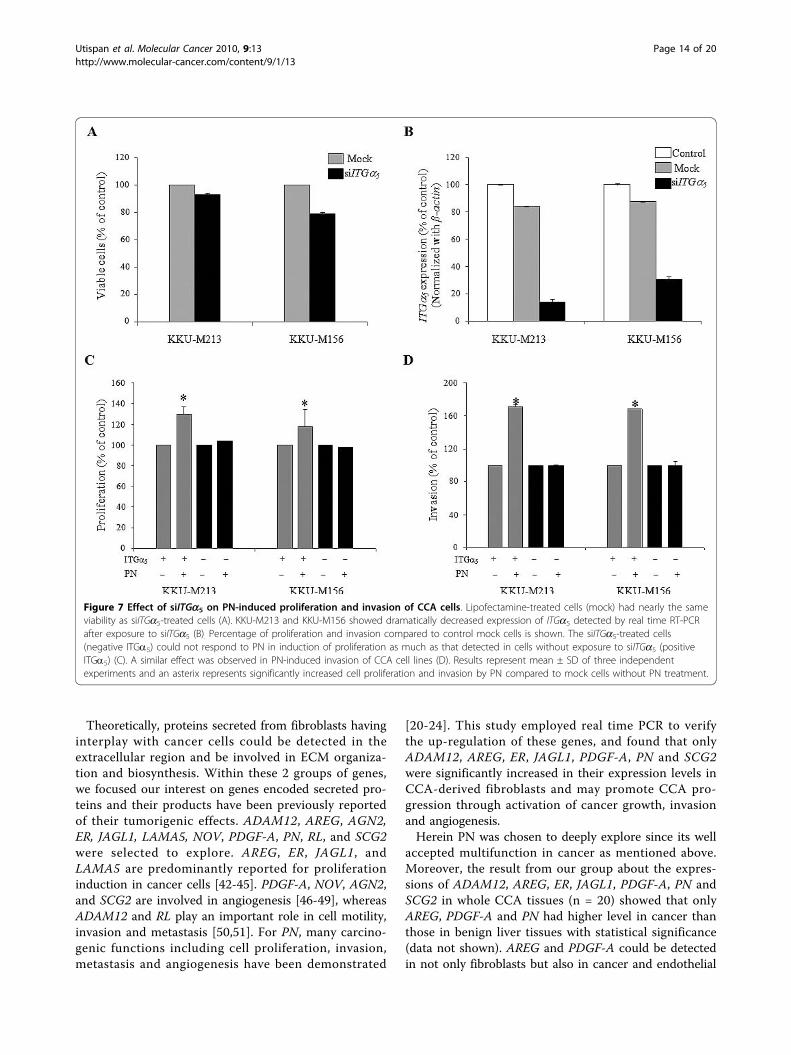
reduction of ITGa5 expression was observed to be 88%of that expressed in both KKU-M213 and KKU-M156CCA cells without transient knockdown of this gene(Fig 7B). The knockdown effect could be detected up to72 h after siITGa5 treatment (data not shown). Thus,the subsequent investigations of cell proliferation andinvasion were done within 72 h after transient knock-down with siITGa5.The reduction of ITGa5 expressions in both KKU-
M213 and KKU-M156 CCA cells resulted in a signifi-cant decreased response of cells to PN-induced cell pro-liferation and invasion (Fig 7C and 7D). A 104% ofKKU-M213 cell proliferation induction was detected insiITGa5-treated cells exposed to PN, whereas cell prolif-eration could increase up to 130% in cells with intrinsicITGa5 expression (Fig 7C). In the same manner, KKU-
M156 showed 118% and 98% of cell proliferation induc-tion observed in cells untreated and treated withsiITGa5. Both ITGa5-knockdown CCA cell lines did notrespond to PN-activated cell invasion whereas PN dra-matically induced invasion of both cell lines having nor-mal intrinsic ITGa5 expression (168% for KKU-M156and 172% for KKU-M213) (Fig 7D). Finally, cells withhigh ITGa5 expressions were more susceptible to PNstimulation to proliferate and invade than cells withoutor minimal ITGa5 expression.
DiscussionCancer-associated fibroblasts have been recognized fortheir impact in the genesis, promotion and progressionof many carcinomas and highlighted in several reviews[8,25]. CCA is notoriously associated with dense
Table 3: Gene ontology of common down-regulated genes. (Continued)
NM_003823 tumor necrosis factor receptor superfamily 4.59
AJ301610 glutamate receptor, ionotropic, kainate 2 3.92
NM_005809 peroxiredoxin 2 3.90
Only genes in the top-five ranking of each group are shown.
Figure 2 PN expressions in Cfs, CCA cell lines and CCA tissues. PN expression in Cfs measured by real time RT-PCR (A) and western blotanalysis (B) using different biological preparations of Cfs and Lfs from those used in microarray. The expression of PN in CCA cell lines andfibroblasts extracted from CCA tissues is graphically depicted (C). Results are expressed as mean ± SD of three independent experiments. Meansof PN mRNA expression levels were measured in 20 CCA cases and compared to 5 cases of benign liver diseases using both b-actin and gapdhas internal controls (D). The positive correlation of mRNA and protein levels of PN in CCA tissues is shown with statistical significance bySpearman correlation analysis (E).
Utispan et al. Molecular Cancer 2010, 9:13http://www.molecular-cancer.com/content/9/1/13
Page 8 of 20

desmoplastic stroma with activated fibroblasts [5,6].Relatively little, however, is known about the contribu-tion of the stromal fibroblasts to CCA. The authors inthe present group have recently shown that CCA stro-mal fibroblasts, with and without direct interaction withcancer cells could induce cancer cell proliferation [6].Herein, this study focused on the gene expression pro-file of CCA-derived fibroblasts in order to investigatethe molecular mechanism of how fibroblasts induce afavorable microenvironment to promote cancer. Eventhough the current study is limited to a single cancerfibroblast line isolated from a single CCA patient, the
validity of array results was strengthened by comparinggene expression levels in cancer fibroblasts to the twolines of normal fibroblasts; one isolated from the sameCCA patient and the other from a second patient. Onlygenes in cancer fibroblasts altered from both normalfibroblast lines were investigated as the common up- ordown-regulated genes. This is to provide evidence thatthe fibroblasts used in our study are valid representa-tives of fibroblasts found in CCA.By comparing gene profiles in fibroblasts from CCA
with those of other tumor types, it is suggested thatCCA fibroblasts display not only common genotypes for
Figure 3 Immunohistochemical staining of PN in CCA tissues. The expression of PN was localized in fibroblasts but not cancer cells. Highexpression of PN was observed in well- (A), moderately- (B) and poorly-differentiated tissues (C), whereas PN negative staining CCA tissue wasdemonstrated (D). Benign liver tissue (E) and hepatocellular carcinoma (F) showed no to slight expression. Magnification, 100×. Doubleimmunofluorescence staining showed co-expression of PN and a-SMA in CCA stromal fibroblasts (G). Magnification, 200×.
Utispan et al. Molecular Cancer 2010, 9:13http://www.molecular-cancer.com/content/9/1/13
Page 9 of 20

activated cells but also unique characteristics. Genesinvolved in metabolism of cells needed to be up-regu-lated in order to support the active function of CCAstromal fibroblasts to produce many supporting proteinsin the cancer environment. Neuropeptide Y receptor Y1has been indicated to receive the activation signal toinduce neuroproliferation [26] and doublecortin-likekinase 1, a microtubule-associated active protein kinaseexpressed in growth cones of postmitotic neurons [27]may help facilitate fibroblast proliferation. In similar tohuman basal cell carcinoma fibroblasts [14], SPARC orosteonectin, was also over-expressed in CCA-derivedfibroblasts. SPARC-null mice were recently demon-strated to resist UV-induced squamous cell carcinoma,suggesting a tumor-promoting role of SPARC [28]. Incontrast to the cancer-associated fibroblasts in meta-static colon cancer to the liver which showed down-reg-ulation of SDF-1 [18], CCA-derived fibroblasts had up-regulated SDF-1 (data not shown).
The highly up-regulated genes in CCA-derived fibro-blasts showed several interesting functions involved incancer progression. Serpin peptidase inhibitor, clade Bmember 2 (SERPINE2) or plasminogen activator inhibitortype 2 (PAI2) is involved in cancer invasion and metasta-sis by controlling serine protease urokinase plasminogenactivator. In a recent review, several studies led to thesuggestion that the significance of PAI2 expression onprognosis of cancers is organ context-dependent [29]. Inbreast cancer, PAI2 was expressed in both stromal andtumor cells and associated with prolonged disease-freesurvival [30]. In contrast, high levels of PAI2 in endome-trial cancer were reported to correlate with the invasionpotential of the cancer [31]. S100 calcium binding proteinA4 (S100A4) has been revealed as the metastasis-inducingprotein [32]. Genes such as procollagen C-endopeptidaseenhancer 2 (PCPE2) were also detected which mayinvolve in collagen synthesis [33]. These results supportthe function of fibroblasts in CCA to promote a desmo-plastic reaction. For down-regulated genes, bone morpho-genetic protein 2 (BMP2A), a multi-functional growthfactor belonging to the transforming growth factor-bsuperfamily was decreased in CCA fibroblasts as reportedin breast cancer-derived fibroblasts [13]. BMP2A-encod-ing protein has been elucidated to induce hypophosphor-ylation of retinoblastoma protein causing cell cycle arrest[34]. Hence, decreased BMP2A in the CCA microenvir-onment may promote cancer cells to enter the cell cycle.Moreover, a decreased level of interleukin 24 (IL-24), anapoptotic inducible cytokine [35], in cancer tissues,attenuates cancer cells from undergoing apoptosis. Theresponse gene to complement 32 (RGC32), a novel p53-inducible gene, and bradykinin receptor B1 (BRADYB1)decreased expression in CCA-derived fibroblasts. Beingintracellular protein and membrane receptor, respectively,RGC32 and BRADYB1 have elucidated the function ofinhibition of fibroblast cell proliferation [36,37]. It seemspossible to conclude that down-regulated genes in fibro-blasts encode proteins, if acting in the intracellularregion, can inhibit the proliferation of fibroblasts them-selves, but if they exist in the extracellular region, theymay involve inhibition of cancer cell proliferation. Thisevidence strengthens the roles of fibroblast-derived pro-
Figure 4 Multivariate analysis using Kaplan-Meier method.Cumulative survival analysis showed significantly shorter survivaltime of the patients with high PN expression in fibroblasts whencompared to those who had low PN expression in fibroblasts (P =0.026).
Table 4 PN expression in CCA tissues compared to benign liver tissues and hepatocellular carcinoma.
Tissues Total cases(n)
PN expression in fibroblasts
Negative PositiveLow High
CCA 52 9 (17%) 13 (25%) 30 (58%)
Benign liver tissue 8 2 (25%) 6 (75%) 0 (0%)
Hepatocellular carcinoma 4 1 (25%) 3 (75%) 0 (0%)
Utispan et al. Molecular Cancer 2010, 9:13http://www.molecular-cancer.com/content/9/1/13
Page 10 of 20

teins released into a tumor environment to induce a highproliferative capability of cancer cells.Fibroblasts have been proposed the bipolar effects in
cancers [38]. In our microarray results, ADAMTS-like 1(ADAMTSR1) was over-expressed in CCA-derived fibro-blasts. The ADAMTS-like proteins have been discussedas the enhancers of ADAMTS proteases [39]. Sincesome ADAMTS have been proven to be anti-angiogenicfactors [40] partly via the trapping of vascular
endothelial growth factor by thrombospondin motifs ofADAMTS [41]. So up-regulation of ADAMTSR1 infibroblasts may inhibit angiogenesis. Moreover, stromely-sin-1 or MMP-3 which can degrade ECM and inducecancer invasion and metastasis, showed the decreasedexpression in fibroblasts. Taken together, the increasedexpression of ADAMTSR1 and the decreased expressionof MMP-3 may highlight fibroblasts in term of suppres-sing CCA progression.
Table 5 Multivariate analysis by Cox proportional hazard regression model for the evaluation of prognostic factors.
Variable(No. of patients)
No. of deadpatients
Hazard ratio 95% confidence interval P
(5-yr survival cut-off) (HR) (CI)
Age in years
≤ 57 (25) 21 1
>57 (26) 22 1.25 0.62-2.48 0.533
PN expression
Low (22) 17 1
High (29) 26 2.02 1.02-4.02 0.045*
Lymph node metastasis
Absence (36) 28 1
Presence (15) 15 3.13 1.54-6.35 0.002*
Histological type
Well-differentiated (20) 16 1
Moderately-differentiated (8) 8 2.77 1.10-6.98 0.031*
Poorly-differentiated (8) 7 1.64 0.63-4.29 0.310
Papillary (15) 12 0.60 0.25-1.44 0.254
Tumor size (cm)
≤ 5 (28) 23 1
>5 (23) 20 1.49 0.76-2.94 0.251
* P value of equal or less than 0.05 means statistical significance
Table 6 Correlation between PN expression level and clinicopathological parameters.
Variable n PN expression (%) Univariate analysis Multivariate analysis
Low High P HR P
Age in years 0.575
≤ 57 26 10 (38.5) 16 (61.5) 1
>57 26 12 (46.2) 14 (53.8) 0.899 0.870
Sex 0.375
Female 20 10 (50.0) 10 (50.0) 1
Male 32 12 (37.5) 20 (62.5) 1.638 0.452
Histological type 0.083
Well-differentiated 21 8 (38.1) 13 (61.9) 1
Moderately-differentiated 8 1 (12.5) 7 (87.5) 3.720 0.271
Poorly-differentiated 8 6 (75.0) 2 (25.0) 0.184 0.86
Papillary 15 7 (46.7) 8 (53.3) 0.611 0.505
Tumor size (cm) 0.123
≤ 5 29 15 (51.7) 14 (48.3) 1
>5 23 7 (30.4) 16 (69.6) 2.493 0.161
Lymph node metastasis 0.830
Absence 37 16 (43.2) 21 (56.8) 1
Presence 15 6 (40.0) 9 (60.0) 1.459 0.590
Utispan et al. Molecular Cancer 2010, 9:13http://www.molecular-cancer.com/content/9/1/13
Page 11 of 20

Figure 5 PN promotes cell proliferation of CCA cell lines. KKU-M156, KKU-M213 and KKU-M055 showed significantly induced proliferationwith different concentrations of PN (A-C) whereas KKU-100 was unresponsive to PN (D). With an optimal dose of PN for each cell line, KKU-M213, KKU-M156, and KKU-M055 increased cell proliferation in a time-dependent manner (E). Triplicate experiments were performed for eachassay. Results are expressed as mean ± SD and an asterix represents a P value less than 0.05 when compared to the negative controls withoutPN treatment. Black and dashed lines represent cells with and without PN treatment, respectively. Colony formation assay with and without agarwas performed (F). Colony of more than 30 cells was counted under inverted microscope. Numbers of colonies/well in 6-well plate of both KKU-M213 and KKU-M156 CCA cell lines were higher in condition of PN treatment than those without PN. Results are expressed as mean ± SD ofduplicate experiments. Pictures of crystal violet-stained cells are of KKU-M213 CCA cells in comparison between with and without PN.
Utispan et al. Molecular Cancer 2010, 9:13http://www.molecular-cancer.com/content/9/1/13
Page 12 of 20

Figure 6 Cell cycle distribution and invasion analysis of cancer cells with and without stimulation by PN. Cell cycle analysis of KKU-M213and KKU-M156 induced by PN (A). PN could drive cells from G1 into S and G2/M phases of the cell cycle when compared to control cellswithout PN treatment (B). Invasion induction by PN on KKU-M213 and KKU-M156 CCA cell lines is shown (C). Numbers of invaded cells when noPN was used served as control and were adjusted to be 100% (white bar). The increase of invaded cells induced by PN is observed and shownby a black bar. Each bar graph represents mean ± SD of three independent experiments. An asterix represents a P value of less than 0.05.
Utispan et al. Molecular Cancer 2010, 9:13http://www.molecular-cancer.com/content/9/1/13
Page 13 of 20
Theoretically, proteins secreted from fibroblasts havinginterplay with cancer cells could be detected in theextracellular region and be involved in ECM organiza-tion and biosynthesis. Within these 2 groups of genes,we focused our interest on genes encoded secreted pro-teins and their products have been previously reportedof their tumorigenic effects. ADAM12, AREG, AGN2,ER, JAGL1, LAMA5, NOV, PDGF-A, PN, RL, and SCG2were selected to explore. AREG, ER, JAGL1, andLAMA5 are predominantly reported for proliferationinduction in cancer cells [42-45]. PDGF-A, NOV, AGN2,and SCG2 are involved in angiogenesis [46-49], whereasADAM12 and RL play an important role in cell motility,invasion and metastasis [50,51]. For PN, many carcino-genic functions including cell proliferation, invasion,metastasis and angiogenesis have been demonstrated
[20-24]. This study employed real time PCR to verifythe up-regulation of these genes, and found that onlyADAM12, AREG, ER, JAGL1, PDGF-A, PN and SCG2were significantly increased in their expression levels inCCA-derived fibroblasts and may promote CCA pro-gression through activation of cancer growth, invasionand angiogenesis.Herein PN was chosen to deeply explore since its well
accepted multifunction in cancer as mentioned above.Moreover, the result from our group about the expres-sions of ADAM12, AREG, ER, JAGL1, PDGF-A, PN andSCG2 in whole CCA tissues (n = 20) showed that onlyAREG, PDGF-A and PN had higher level in cancer thanthose in benign liver tissues with statistical significance(data not shown). AREG and PDGF-A could be detectedin not only fibroblasts but also in cancer and endothelial
Figure 7 Effect of siITGa5 on PN-induced proliferation and invasion of CCA cells. Lipofectamine-treated cells (mock) had nearly the sameviability as siITGa5-treated cells (A). KKU-M213 and KKU-M156 showed dramatically decreased expression of ITGa5 detected by real time RT-PCRafter exposure to siITGa5 (B). Percentage of proliferation and invasion compared to control mock cells is shown. The siITGa5-treated cells(negative ITGa5) could not respond to PN in induction of proliferation as much as that detected in cells without exposure to siITGa5 (positiveITGa5) (C). A similar effect was observed in PN-induced invasion of CCA cell lines (D). Results represent mean ± SD of three independentexperiments and an asterix represents significantly increased cell proliferation and invasion by PN compared to mock cells without PN treatment.
Utispan et al. Molecular Cancer 2010, 9:13http://www.molecular-cancer.com/content/9/1/13
Page 14 of 20

cells, however PN expressed exclusively in CCA fibro-blasts. In order to demonstrate role of fibroblast-derivedproteins in CCA, we determined that PN should be thefirst target to explore.The strong evidence using different biological prepara-
tions of CCA-derived fibroblasts and CCA tissues con-firmed the increased levels of PN at both mRNA andprotein. Most of CCA tissues of all differentiated typeshad high levels of PN and expressed exclusively in a-SMA positive fibroblasts. In the same direction, the find-ings showed no expression of PN in CCA cell lines whencompared to the high level expressed in the fibroblasts.This may strengthen the results of the absence of PN incancer cells in CCA tissues. From these results takentogether, it can be concluded that PN detected in CCAtissues is only of fibroblast origin as reported in somecancers [52-54]. In cancers of head and neck, ovary, andcolon, PN was found in cancer cells and has been pro-posed to induce tumorigenic properties of cancer cellsvia an autocrine mechanism [21,22]. Hence results fromthe present study allow the speculation to propose a phe-nomenon that fibroblast-derived PN in CCA may affectcancer cells by a paracrine mode and has a promisingrole in cancer promotion. These results revealed that ahigh PN level in fibroblasts was an independent risk fac-tor in CCA patients and those having high PN had signif-icantly low cumulative survival time after surgery. PNmight therefore be used as a poor prognostic marker inpatients suffering from CCA. Detections of PN at bothmRNA and encoding protein in CCA tissues are in thesame direction to distinguish CCA from non-cancer syn-dromes of bile ducts. In addition, most benign liver tis-sues and hepatocellular carcinoma showed no to only aminimal expression of PN when compared to the highlevel detected in CCA tissues. Hence, serum PN mayhelp to distinguish CCA from benign conditions and clo-sely-related liver cancer and may use as the prognostic orpredictive marker as previously reported [52,55].To show the tumorigenic impacts of PN on CCA cells,
recombinant PN was employed as extracellular PN tomimic the paracrine effect of PN produced from cancerstromal fibroblasts to induce CCA cell proliferation andinvasion. Though receptors ITGavb3 and ITGavb5 havebeen shown to be the receptors for PN in several cancercells [21], PN promoted invasiveness of pancreatic can-cer cells via the b4 integrin [56]. This suggests the celltype dependent on a specific ITG responded to PN. Thestudy herein reveals that PN-induced cell proliferationand invasion could be inhibited by RNAi against ITGa5.
Hence, ITGa5 is a potentially promising receptor for PNin CCA cells. As the well known receptor for fibronec-tin, the apparent reason for ITGa5 production in CCAcells is to support the abundance of fibronectin found inCCA [57]. In addition, ITGa5 can only be from
dimerization with the b1 subunit and activation ofITGa5b1 has been revealed to support cell survival [58]and induce invasion and angiogenesis [59,60]. Thoughfurther studies need to be performed before such a con-clusion is valid in CCA, this work highlights the PN-induced-ITGa5 pathway as one of the activated path-ways to induce an aggressive CCA.TGF-b has been proposed to induce the expression of
PN [61]. O. viverrini excretory/secretory product hasalso recently been shown to be the stimulator of fibro-blast proliferation via the TGF-b-mediated signal trans-duction pathway [62] and this pathway seems likely tobe the cause of PN expression in CCA-derived fibro-blasts. The authors’ laboratory has checked the effect ofparasitic product-treated fibroblasts and found thatthese fibroblasts increased PN expression compared tothe normal liver fibroblasts without exposure (unpub-lished data). It is interesting to propose that in CCAcells; the expression of fibroblast-derived PN could beinduced by TGF-b produced from infected parasitessince the early stage of carcinogenesis and may be inconcert with TGF-b produced from CCA cells in a latestage of cancer [12]. Though in vivo experiments areneeded to confirm, fibroblast-derived PN may influenceO. viverrini-associated CCA at the early stage of canceras well as to promote cancer progression in the latertime. With this information, targeting the stroma inCCA may not only be effective in treatment of primary,invasive and metastatic tumors, but may also play rolein prevention of tumor development.
ConclusionsTo the authors’ knowledge, this study is the first todescribe the gene expression profile of CCA-derived fibro-blasts. Molecular understanding of fibroblasts in CCA bythe functions of certain up- and down-regulated genes hasbeen revealed and has suggested certain groups of genes incontrolling cancer cell proliferation, invasion, metastasisand angiogenesis (Fig 8). These findings provide evidencethat fibroblasts are important sources of tumorigenic sub-stances, particularly PN, when produced into the microen-vironment of CCA. High levels of PN are found in mostCCA patients and can be used as a poor prognostic mar-ker. In addition, the level of PN can be used to distinguishCCA from other benign liver conditions and hepatocellularcarcinoma. The interaction of fibroblast-derived PN andCCA cells helps to promote cell proliferation and invasionprobably via ITGa5. Though further investigations areneeded, this study suggests promising evidence of the valueof using serum PN as a prognostic marker of poor survivalin CCA patients. Moreover, targeting fibroblasts or fibro-blast-derived-PN-stimulated pathways in cancer cells toattenuate the tumorigenic induction of PN is a furtherchallenge to inhibit CCA progression in the patients.
Utispan et al. Molecular Cancer 2010, 9:13http://www.molecular-cancer.com/content/9/1/13
Page 15 of 20

MethodsCells and culture conditionCfs and Lfs were established and characterized in thislab as previously reported [6]. CCA cell lines includingKKU-M213; KKU-M156; KKU-M055; and KKU-100were received as a kind gift from Associate ProfessorDr. Banchob Sripa (Khon Kaen University). Cfs and Lfswere grown in the complete media which is 10% FBScontaining DMEM with 20% epidermal growth factor(CytoLab Ltd., Rehovot, Israel). CCA cells were culturedin 10% FBS containing Ham-F12 (Invitrogen, Carlsbad,CA) supplemented with antibiotics and an anti-fungalagent at 37°C and in 5% CO2 incubator.
Gene expression profiling study and data analysisTotal RNA was extracted using RNeasy Micro Kit (Qia-gen, Valencia, CA) using the manufacturer’s instructions.The quality of the RNA was assessed by an Agilent RNA6000 Nano Kit (Agilent Technologies, Waldbronn, Ger-many). Affymetrix GeneChip Human Genome U133 plus2.0 Array containing 38,500 human genes (Affymetrix,Santa Clara, CA) was used following Affymetrix’s instruc-tion. The array data were scanned by a GeneChip Scanner3000 (Affymetrix) and analysed by Affymetrix microarraysuite, version 5.0. Raw data from the GeneChips wereused to analyze expression levels and expressed as foldchanges and gene ontology was categorized by GeneSpring G.X.7.3 software (Agilent Technologies). Averagefold change of gene expression was determined by inten-sity comparison between Cf and Lf1 and Lf2.
Semi-quantitative real time PCRComplementary DNA was synthesized from 1 μg oftotal RNA using the first strand cDNA synthesis kit(AMV) (Roche Molecular Biochemicals, Mannheim,Germany) according to the instructions. Relative expres-sion levels in genes of Cfs and Lfs were determined bySYBR Green-based real time PCR using ABI 7500(Applied Biosystem, Foster City, CA) and calculated bythe 2-ΔΔCT equation. In this case, ΔCT = CT (Cf)-CT
(Lf). b-actin served as an internal control to adjust theamount of starting cDNA. The sequences of genestested in this study were retrieved from PubMed http://www.ncbi.nlm.nih.gov and the primers were designed byPrimer 3 (Table 7).
Human CCA tissues and immunohistochemistryFifty-two cases of CCA tissues were obtained frompatients who had undergone hepatectomy using the pro-tocol approved by the Human Research Ethics Commit-tee, Khon Kaen University (HE490143). The age, sex,tumor size, histological type and staging data werederived from the medical charts and pathologicalrecords. Benign liver tissues were characterized aschronic inflammation by other causes rather than CCA.Paraffin-embedded tissues were used and antigens
were retrieved in 10 mM citrate buffer pH 6.0 at 95°Cfor 40 min and endogenous peroxidase was blocked in3% H2O2 for 5 min. After blocking non-specific bindingwith 2% bovine serum albumin for 20 min, 1:10,000 rab-bit anti-human PN (Biovendor, Heidelberg, Germany)
Figure 8 Proposed impacts of CCA-associated fibroblasts revealed in this study. A schematic representation of the main alterations inCCA-associated fibroblasts revealed in this study. The biological functions of protein products from the up-regulated genes in fibroblasts arerepresented. Tumorigenic effects of PN on CCA cancer cells are also proposed.
Utispan et al. Molecular Cancer 2010, 9:13http://www.molecular-cancer.com/content/9/1/13
Page 16 of 20

was applied to the sections at room temperature over-night, followed by anti-rabbit Envision+ System-HRPlabeled polymer (Dako, Carpinteria, CA) for 30 min atroom temperature. The immunoreactive signal wasdeveloped by diaminobenzidine (DAB; Sigma, St Louis,MO) and counterstained with hematoxylin. The signalwas checked under light microscope.PN expression of intratumoral fibroblasts on the histo-
logic sections was semi-quantitatively scored on the basisof PN-positive fibroblasts percentage and the immunos-taining intensity. The number of positive fibroblast cellswere classified as < 10% (negative); 10-25% (+1); 26-50%(+2); and >50% (+3). The intensity of PN expression infibroblasts was scored no staining, 0; weak staining, 1;intermediate or focal weak and focal intense staining, 2;intense staining, 3. The interpretation of PN expressionwas performed by summarization the scores of the per-cent positive cell (0-3) and the scores of staining intensity(1-3) to reach the total final score of 0-6. The resultswere then categorized as follows; low expression, score ≤4; and high expression, score > 4. All samples were anon-ymized and independently scored by one pathologist(KC) and 2 investigators (PT and CT). In case of dis-agreement, the slides were reexamined and a consensuswas reached by at least 2 observers.
Double immunofluorescence staining of a-SMA and PNIn order to localize the expression of PN and a-SMA inCCA tissues, double immunofluorescence staining wasperformed. The 1:200 mouse anti-human a-SMA antibody(Sigma) and 1:500 rabbit anti-human PN antibody (Bio-vendor) were used as primary antibodies. Anti-mouseIgG-Alexa 488 and anti-rabbit IgG-Cy3 (Invitrogen) were
used as the second antibodies. Nucleus was stained withHoechst (Invitrogen). The signal was observed under theLSM 510 Meta laser scanning confocal microscope (CarlZeiss, Jena, Germany) at the Division of Medical Molecu-lar Biology, Office for Research and Development, Facultyof Medicine Siriraj Hospital, Mahidol University.
Protein extraction and western blot analysisTwenty μgs of total protein from the cell lysate were sepa-rated in 10% SDS-PAGE and transferred onto a PVDFmembrane (Millipore, Billerica, MA). For PN detection,1:500 rabbit anti-human PN (Biovendor) and 1:1,000 goatanti-rabbit conjugated HRP (Abcam, Cambridge, MA)were used. The signal was visualized by ECL (Pierce, Rock-ford, IL). The expression of b-actin was used as an internalcontrol to determine an equal amount of loading proteins.
Cell proliferation assayCCA cells with or without treatment with siITGa5 cellswere arrested in HAM-F12 without serum supplementfor 12 h. Different concentrations of recombinant PN(Biovendor) prepared in 1% FBS containing HAM-F12were incubated with cells for 6, 12 and 24 h. The viablecells in each condition were determined using an MTSassay (Promega, Madison, WI) according to the manu-facturer’s instruction.
Cell cycle analysis by flow cytometryCell cycle distribution analysis used cells stained withpropidium iodide (Invitrogen) as previously described[63]. The distribution of cells in each stage of the cellcycle was quantitated in a flow cytometer and CellQuestsoftware (Becton Dickinson, Franklin Lakes, NJ).
Table 7 Primer sequences for real time PCR.
Gene Forward Primer Reverse Primer Size Accession no.5’-3’ 5’-3’ (bp)
ADAM12 tttgggggtcaacagttttc agagctgggttcccttttgt 191 NM_003474
AREG tggggaaaagtccatgaaaa tttcgttcctcagcttctcc 174 NM_001657
AGN2 ccacctgaggaactgtctcg ggtcttgctttggtccgtta 191 NM_001147
ER catatgggagaagggggagt aagtgcaattacagagtgcaaaa 166 NM_001432
JAGL1 gcctgccttaagtgaggaaa gccaagaacaacacatcaaaga 169 U77914
LAMA5 gtgatgaaaagcgggaatgt acctccacagagcgagtcat 221 BC003355
NOV tgcaattccaagaaaatatcactg cttggatttggagcttggaa 167 NM_002514
PDGF-A acacgagcagtgtcaagtgc tctggttggctgctttaggt 250 X03795
PN cactctttgctcccaccaat tcaaagactgctcctcccata 157 AY140646
RL tgctgaatttggggctactt gggagatagggtcttcatcca 198 NM_005045
SCG2 cccgaagaatgatgataccc aaatgttgggatttgcttgg 195 NM_003469
ITGa5 agttgcatttccgagtctgg ccaaacaggatggctaggat 223 NM_002205
b-actin cacactgtgcccatctacga ctccttaatgtcacgcacga 162 X00351
gapdh ctcctcctgttcgacagtca gttaaaagcagccctggtga 140 NM_002046
Note: ADAM12, a disintegrin and matrix metalloproteinase 12; AREG, amphiregulin; AGN2, angiopoietin 2; ER, epiregulin; JAGL1, jagged soluble form; LAMA5,laminin alpha 5; NOV, nephroblastoma over expressed; PDGF-A, platelet-derived growth factor alpha; PN, periostin; RL, reelin; SCG2, secretogranin 2; ITGa5,integrin alpha 5; b-actin, beta-actin; gapdh, glyceraldehyde 3-phosphate dehydrogenase
Utispan et al. Molecular Cancer 2010, 9:13http://www.molecular-cancer.com/content/9/1/13
Page 17 of 20

Numbers of CCA cell lines in S+G2/M phases of thecell cycle were measured and compared between condi-tions with and without recombinant PN treatment.These experiments were repeated two times using repli-cate culture dishes in the same experiment.
Colony formation assayCCA cell lines were cultured in 6-well plate. After 24-hculture, recombinant PN diluted in 1% FBS containingmedium was added and the plate was incubated in CO2
incubator. Soft agar colony formation assay was alsoperformed using 0.5% and 0.35% MetaPhor® agarose(Cambrex Bio Science, Rockland, ME) as lower andupper layers, respectively. After 12 d, cells were fixedwith 5% v/v glutaraldehyde and stained with 0.5% w/vcrystal violet in 40% v/v methanol. Cell growth was esti-mated by counting numbers of colonies with more than30 cells under inverted microscope and comparedbetween those of treat and untreated with PN. Theexperiment was performed in duplicate.
Invasion assayKKU-M213 and KKU-M156 CCA cells were seeded inPN (100 ng/ml) containing medium into the Matrigelinvasion chamber (BD Biosciences, San Jose, CA) andincubated for 24 h. Invaded cells were fixed with 5% v/vglutaraldehyde and stained with 0.5% w/v crystal violetin 40% v/v methanol for 30 min each. The number ofinvaded cells was counted under a microscope by twoindependent investigators using 100× magnificationfields. The assays were done in replicate and three inde-pendent experiments were performed.
Small interfering RNA against receptor integrin a5
Two hundred thousand CCA cells were seeded into a 6-well plate for 24 h before transfection of siITGa5 (SantaCruz Biotechnology, Santa Cruz, CA) by Lipofectamine2000 (Invitrogen). Three siRNA strands (5’-guca-gaauuucgagacaaa-3’, 5’-caccaacaagagagccaaa-3’, and 5’-ccacugaccagaacuagaa-3’) were used to target ITGa5
mRNA. The efficiency of knock down was tested by realtime PCR using b-actin as an internal control.
Statistical analysisStatistical analyses were performed using SPSS version16.0 (SPSS Inc., Chicago, IL). The correlation of PNexpression and pathological parameters of CCApatients was analyzed by the c2-test and binary logisticmultivariate analysis. Patient survival was calculatedfrom the time of surgical resection to death and thesurvival curves were constructed according to Kaplan-Meier, with a Log-Rank test. A multivariate analysiswas performed by the Cox proportional hazard regres-sion model. The significance of the different data was
determined by the Student’s t-test. A P value of equalto or less than 0.05 was defined as statisticallysignificant.
AcknowledgementsWe would like to thank the Commission on Higher Education, Thailand forsupporting by grant fund under the program Strategic Scholarships forFrontier Research Network for the Ph.D. Program Thai Doctoral degree forthis research. This project was co-supported by a Mid-Career Grant(RMU5080069), Thailand Research Fund (TRF) and the ResearchStrengthening Grant 2007 from National Center for Genetic Engineering andBiotechnology (BIOTEC), National Science and Technology DevelopmentAgency (NSTDA). The English editing of this manuscript was kindlyperformed by Professor James A. Will, University of Wisconsin, Madison, WI,USA.
Author details1Department of Biochemistry, Faculty of Medicine, Khon Kaen University, 123Mitraparb Road, Muang, Khon Kaen 40002, Thailand. 2Department ofImmunology, Faculty of Medicine Siriraj Hospital, Mahidol University, 2Prannok Road, Bangkok Noi, Bangkok 10700, Thailand. 3Department ofBiochemistry and Molecular Biology, Nihon University School of Dentistry atMatsudo, Matsudo, Chiba 2718587, Japan. 4Department of Pathology, Facultyof Medicine Siriraj Hospital, Mahidol University, Thailand. 5Department ofPathology, Faculty of Medicine, Khon Kaen University, Thailand. 6Departmentof Surgery, Faculty of Medicine, Khon Kaen University, Thailand. 7Liver Flukeand Cholangiocarcinoma Research Center, Faculty of Medicine, Khon KaenUniversity, Thailand.
Authors’ contributionsKU performed most of the experiments and helped to draft the manuscript.YA contributed to the microarray experiment. PT performed real time PCR ofsome genes and helped KC and AP in immunohistochemical scoring. SCcontributed to the patient clinicopathological data and samples collection.CT contributed to the design of the entire study, data analysis andpreparation of the manuscript.All authors have read and approved the final manuscript.
Competing interestsThe authors declare that they have no competing interests.
Received: 8 September 2009Accepted: 24 January 2010 Published: 24 January 2010
References1. Sriamporn S, Pisani P, Pipitgool V, Suwanrungruang K, Kamsa-ard S,
Parkin DM: Prevalence of Opisthorchis viverrini infection and incidence ofcholangiocarcinoma in Khon Kaen, Northeast Thailand. Trop Med IntHealth 2004, 9:588-594.
2. Lim MK, Ju YH, Franceschi S, Oh JK, Kong HJ, Hwang SS, Park SK, Cho SI,Sohn WM, Kim DI, Yoo KY, Hong ST, Shin HR: Clonorchis sinensis infectionand increasing risk of cholangiocarcinoma in the Republic of Korea. AmJ Trop Med Hyg 2006, 75:93-96.
3. Choi D, Lim JH, Lee KT, Lee JK, Choi SH, Heo JS, Jang KT, Lee NY, Kim S,Hong ST: Cholangiocarcinoma and Clonorchis sinensis infection: a case-control study in Korea. J Hepatol 2006, 44:1066-1073.
4. Lazaridis KN, Gores GJ: Primary sclerosing cholangitis andcholangiocarcinoma. Semin Liver Dis 2006, 26:42-51.
5. Okamura N, Yoshida M, Shibuya A, Sugiura H, Okayasu I, Ohbu M: Cellularand stromal characteristics in the scirrhous hepatocellular carcinoma:comparison with hepatocellular carcinomas and intrahepaticcholangiocarcinomas. Pathol Int 2005, 55:724-731.
6. Chuaysri C, Thuwajit P, Paupairoj A, Chau-In S, Suthiphongchai T,Thuwajit C: Alpha-smooth muscle actin-positive fibroblasts promotebiliary cell proliferation and correlate with poor survival incholangiocarcinoma. Oncol Rep 2009, 21:957-969.
7. Hwang RF, Moore T, Arumugam T, Ramachandran V, Amos KD, Rivera A,Ji B, Evans DB, Logsdon CD: Cancer-associated stromal fibroblastspromote pancreatic tumor progression. Cancer Res 2008, 68:918-926.
Utispan et al. Molecular Cancer 2010, 9:13http://www.molecular-cancer.com/content/9/1/13
Page 18 of 20

8. Micke P, Ostman A: Exploring the tumour environment: cancer-associatedfibroblasts as targets in cancer therapy. Expert Opin Ther Targets 2005,9:1217-1233.
9. Augsten M, Hagglof C, Olsson E, Stolz C, Tsagozis P, Levchenko T,Frederick MJ, Borg A, Micke P, Egevad L, Ostman A: CXCL14 is anautocrine growth factor for fibroblasts and acts as a multi-modalstimulator of prostate tumor growth. Proc Natl Acad Sci USA 2009,106:3414-3419.
10. Tuxhorn JA, Ayala GE, Rowley DR: Reactive stroma in prostate cancerprogression. J Urol 2001, 166:2472-2483.
11. Rosenthal EL, McCrory A, Talbert M, Carroll W, Magnuson JS, Peters GE:Expression of proteolytic enzymes in head and neck cancer-associatedfibroblasts. Arch Otolaryngol Head Neck Surg 2004, 130:943-947.
12. Ohira S, Itatsu K, Sasaki M, Harada K, Sato Y, Zen Y, Ishikawa A, Oda K,Nagasaka T, Nimura Y, Nakanuma Y: Local balance of transforming growthfactor-beta1 secreted from cholangiocarcinoma cells and stromal-derived factor-1 secreted from stromal fibroblasts is a factor involved ininvasion of cholangiocarcinoma. Pathol Int 2006, 56:381-389.
13. Singer CF, Gschwantler-Kaulich D, Fink-Retter A, Haas C, Hudelist G,Czerwenka K, Kubista E: Differential gene expression profile in breast cancer-derived stromal fibroblasts. Breast Cancer Res Treat 2008, 110:273-281.
14. Micke P, Kappert K, Ohshima M, Sundquist C, Scheidl S, Lindahl P,Heldin CH, Botling J, Ponten F, Ostman A: In situ identification of genesregulated specifically in fibroblasts of human basal cell carcinoma.J Invest Dermatol 2007, 127:1516-1523.
15. Fromigue O, Louis K, Dayem M, Milanini J, Pages G, Tartare-Deckert S,Ponzio G, Hofman P, Barbry P, Auberger P, Mari B: Gene expressionprofiling of normal human pulmonary fibroblasts following coculturewith non-small-cell lung cancer cells reveals alterations related to matrixdegradation, angiogenesis, cell growth and survival. Oncogene 2003,22:8487-8497.
16. Allinen M, Beroukhim R, Cai L, Brennan C, Lahti-Domenici J, Huang H,Porter D, Hu M, Chin L, Richardson A, Schnitt S, Sellers WR, Polyak K:Molecular characterization of the tumor microenvironment in breastcancer. Cancer Cell 2004, 6:17-32.
17. Fukushima N, Sato N, Prasad N, Leach SD, Hruban RH, Goggins M:Characterization of gene expression in mucinous cystic neoplasms of thepancreas using oligonucleotide microarrays. Oncogene 2004, 23:9042-9051.
18. Nakagawa H, Liyanarachchi S, Davuluri RV, Auer H, Martin EW Jr, de laChapelle A, Frankel WL: Role of cancer-associated stromal fibroblasts inmetastatic colon cancer to the liver and their expression profiles.Oncogene 2004, 23:7366-7377.
19. Erkan M, Kleeff J, Gorbachevski A, Reiser C, Mitkus T, Esposito I, Giese T,Buchler MW, Giese NA, Friess H: Periostin creates a tumor-supportivemicroenvironment in the pancreas by sustaining fibrogenic stellate cellactivity. Gastroenterology 2007, 132:1447-1464.
20. Tai IT, Dai M, Chen LB: Periostin induction in tumor cell line explants andinhibition of in vitro cell growth by anti-periostin antibodies.Carcinogenesis 2005, 26:908-915.
21. Gillan L, Matei D, Fishman DA, Gerbin CS, Karlan BY, Chang DD: Periostinsecreted by epithelial ovarian carcinoma is a ligand for alpha(V)beta(3)and alpha(V)beta(5) integrins and promotes cell motility. Cancer Res2002, 62:5358-5364.
22. Kudo Y, Ogawa I, Kitajima S, Kitagawa M, Kawai H, Gaffney PM, Miyauchi M,Takata T: Periostin promotes invasion and anchorage-independentgrowth in the metastatic process of head and neck cancer. Cancer Res2006, 66:6928-6935.
23. Siriwardena BS, Kudo Y, Ogawa I, Kitagawa M, Kitajima S, Hatano H,Tilakaratne WM, Miyauchi M, Takata T: Periostin is frequentlyoverexpressed and enhances invasion and angiogenesis in oral cancer.Br J Cancer 2006, 95:1396-1403.
24. Shao R, Bao S, Bai X, Blanchette C, Anderson RM, Dang T, Gishizky ML,Marks JR, Wang XF: Acquired expression of periostin by human breastcancers promotes tumor angiogenesis through up-regulation of vascularendothelial growth factor receptor 2 expression. Mol Cell Biol 2004,24:3992-4003.
25. De Wever O, Demetter P, Mareel M, Bracke M: Stromal myofibroblasts aredrivers of invasive cancer growth. Int J Cancer 2008, 123:2229-2238.
26. Howell OW, Scharfman HE, Herzog H, Sundstrom LE, Beck-Sickinger A,Gray WP: Neuropeptide Y is neuroproliferative for post-natalhippocampal precursor cells. J Neurochem 2003, 86:646-659.
27. Burgess HA, Reiner O: Doublecortin-like kinase is associated withmicrotubules in neuronal growth cones. Mol Cell Neurosci 2000, 16:529-541.
28. Aycock RL, Bradshaw AC, Sage EH, Starcher B: Development of UV-inducedsquamous cell carcinomas is suppressed in the absence of SPARC. JInvest Dermatol 2004, 123:592-599.
29. Croucher DR, Saunders DN, Lobov S, Ranson M: Revisiting the biologicalroles of PAI2 (SERPINB2) in cancer. Nat Rev Cancer 2008, 8:535-545.
30. Duggan C, Kennedy S, Kramer MD, Barnes C, Elvin P, McDermott E,O’Higgins N, Duffy MJ: Plasminogen activator inhibitor type 2 in breastcancer. Br J Cancer 1997, 76:622-627.
31. Osmak M, Babic D, Abramic M, Milicic D, Vrhovec I, Skrk J: Plasminogenactivator inhibitor type 2: potential prognostic factor for endometrialcarcinomas. Neoplasma 2001, 48:462-467.
32. Schmidt-Hansen B, Klingelhofer J, Grum-Schwensen B, Christensen A,Andresen S, Kruse C, Hansen T, Ambartsumian N, Lukanidin E, Grigorian M:Functional significance of metastasis-inducing S100A4(Mts1) in tumor-stroma interplay. J Biol Chem 2004, 279:24498-24504.
33. Steiglitz BM, Keene DR, Greenspan DS: PCOLCE2 encodes a functionalprocollagen C-proteinase enhancer (PCPE2) that is a collagen-bindingprotein differing in distribution of expression and post-translationalmodification from the previously described PCPE1. J Biol Chem 2002,277:49820-49830.
34. Tomari K, Kumagai T, Shimizu T, Takeda K: Bone morphogenetic protein-2induces hypophosphorylation of Rb protein and repression of E2F inandrogen-treated LNCaP human prostate cancer cells. Int J Mol Med2005, 15:253-258.
35. Zheng M, Bocangel D, Ramesh R, Ekmekcioglu S, Poindexter N, Grimm EA,Chada S: Interleukin-24 overcomes temozolomide resistance andenhances cell death by down-regulation of O6-methylguanine-DNAmethyltransferase in human melanoma cells. Mol Cancer Ther 2008,7:3842-3851.
36. Zou J, Ren JH, Feng D, Wang H, Xu J: Compensatory function ofbradykinin B1 receptor in the inhibitory effect of captopril oncardiomyocyte hypertrophy and cardiac fibroblast proliferation inneonatal rats. Chin Med J (Engl) 2008, 121:1220-1225.
37. Saigusa K, Imoto I, Tanikawa C, Aoyagi M, Ohno K, Nakamura Y, Inazawa J:RGC32, a novel p53-inducible gene, is located on centrosomes duringmitosis and results in G2/M arrest. Oncogene 2007, 26:1110-1121.
38. Mueller MM, Fusenig NE: Friends or foes - bipolar effects of the tumourstroma in cancer. Nat Rev Cancer 2004, 4:839-849.
39. Hirohata S, Wang LW, Miyagi M, Yan L, Seldin MF, Keene DR, Crabb JW,Apte SS: Punctin, a novel ADAMTS-like molecule, ADAMTSL-1, inextracellular matrix. J Biol Chem 2002, 277:12182-12189.
40. Vazquez F, Hastings G, Ortega MA, Lane TF, Oikemus S, Lombardo M, Iruela-Arispe ML: METH-1, a human ortholog of ADAMTS-1, and METH-2 aremembers of a new family of proteins with angio-inhibitory activity. J BiolChem 1999, 274:23349-23357.
41. Luque A, Carpizo DR, Iruela-Arispe ML: ADAMTS1/METH1 inhibitsendothelial cell proliferation by direct binding and sequestration ofVEGF165. J Biol Chem 2003, 278:23656-23665.
42. Castillo J, Erroba E, Perugorria MJ, Santamaria M, Lee DC, Prieto J, Avila MA,Berasain C: Amphiregulin contributes to the transformed phenotype ofhuman hepatocellular carcinoma cells. Cancer Res 2006, 66:6129-6138.
43. Morita S, Shirakata Y, Shiraishi A, Kadota Y, Hashimoto K, Higashiyama S,Ohashi Y: Human corneal epithelial cell proliferation by epiregulin andits cross-induction by other EGF family members. Mol Vis 2007,13:2119-2128.
44. Purow BW, Haque RM, Noel MW, Su Q, Burdick MJ, Lee J, Sundaresan T,Pastorino S, Park JK, Mikolaenko I, Maric D, Eberhart CG, Fine HA:Expression of Notch-1 and its ligands, Delta-like-1 and Jagged-1, iscritical for glioma cell survival and proliferation. Cancer Res 2005,65:2353-2363.
45. Gonzales M, Haan K, Baker SE, Fitchmun M, Todorov I, Weitzman S,Jones JC: A cell signal pathway involving laminin-5, alpha3beta1integrin, and mitogen-activated protein kinase can regulate epithelialcell proliferation. Mol Biol Cell 1999, 10:259-270.
46. Shikada Y, Yonemitsu Y, Koga T, Onimaru M, Nakano T, Okano S, Sata S,Nakagawa K, Yoshino I, Maehara Y, Sueishi K: Platelet-derived growthfactor-AA is an essential and autocrine regulator of vascular endothelialgrowth factor expression in non-small cell lung carcinomas. Cancer Res2005, 65:7241-7248.
Utispan et al. Molecular Cancer 2010, 9:13http://www.molecular-cancer.com/content/9/1/13
Page 19 of 20

47. Lin CG, Leu SJ, Chen N, Tebeau CM, Lin SX, Yeung CY, Lau LF: CCN3 (NOV)is a novel angiogenic regulator of the CCN protein family. J Biol Chem2003, 278:24200-24208.
48. Lobov IB, Brooks PC, Lang RA: Angiopoietin-2 displays VEGF-dependentmodulation of capillary structure and endothelial cell survival in vivo.Proc Natl Acad Sci USA 2002, 99:11205-11210.
49. Kirchmair R, Gander R, Egger M, Hanley A, Silver M, Ritsch A, Murayama T,Kaneider N, Sturm W, Kearny M, Fischer-Colbrie R, Kircher B, Gaenzer H,Wiedermann CJ, Ropper AH, Losordo DW, Patsch JR, Schratzberger P: Theneuropeptide secretoneurin acts as a direct angiogenic cytokine in vitroand in vivo. Circulation 2004, 109:777-783.
50. Le Pabic H, Bonnier D, Wewer UM, Coutand A, Musso O, Baffet G,Clement B, Theret N: ADAM12 in human liver cancers: TGF-beta-regulated expression in stellate cells is associated with matrixremodeling. Hepatology 2003, 37:1056-1066.
51. Hashimoto-Torii K, Torii M, Sarkisian MR, Bartley CM, Shen J, Radtke F,Gridley T, Sestan N, Rakic P: Interaction between reelin and notchsignaling regulates neuronal migration in the cerebral cortex. Neuron2008, 60:273-284.
52. Sasaki H, Dai M, Auclair D, Fukai I, Kiriyama M, Yamakawa Y, Fujii Y, Chen LB:Serum level of the periostin, a homologue of an insect cell adhesionmolecule, as a prognostic marker in nonsmall cell lung carcinomas.Cancer 2001, 92:843-848.
53. Fukushima N, Kikuchi Y, Nishiyama T, Kudo A, Fukayama M: Periostindeposition in the stroma of invasive and intraductal neoplasms of thepancreas. Mod Pathol 2008, 21:1044-1053.
54. Kikuchi Y, Kashima TG, Nishiyama T, Shimazu K, Morishita Y, Shimazaki M,Kii I, Horie H, Nagai H, Kudo A, Fukayama M: Periostin is expressed inpericryptal fibroblasts and cancer-associated fibroblasts in the colon. JHistochem Cytochem 2008, 56:753-764.
55. Sasaki H, Yu CY, Dai M, Tam C, Loda M, Auclair D, Chen LB, Elias A:Elevated serum periostin levels in patients with bone metastases frombreast but not lung cancer. Breast Cancer Res Treat 2003, 77:245-252.
56. Baril P, Gangeswaran R, Mahon PC, Caulee K, Kocher HM, Harada T, Zhu M,Kalthoff H, Crnogorac-Jurcevic T, Lemoine NR: Periostin promotesinvasiveness and resistance of pancreatic cancer cells to hypoxia-induced cell death: role of the beta4 integrin and the PI3k pathway.Oncogene 2007, 26:2082-2094.
57. Chen CY, Lin XZ, Tsao HC, Shiesh SC: The value of biliary fibronectin fordiagnosis of cholangiocarcinoma. Hepatogastroenterology 2003,50:924-927.
58. Zhang Z, Vuori K, Reed JC, Ruoslahti E: The alpha 5 beta 1 integrinsupports survival of cells on fibronectin and up-regulates Bcl-2expression. Proc Natl Acad Sci USA 1995, 92:6161-6165.
59. Zeng ZZ, Yao H, Staszewski ED, Rockwood KF, Markwart SM, Fay KS,Spalding AC, Livant DL: Alpha(5)beta(1) integrin ligand PHSRN inducesinvasion and alpha(5) mRNA in endothelial cells to stimulateangiogenesis. Transl Oncol 2009, 2:8-20.
60. Tuomi S, Mai A, Nevo J, Laine JO, Vilkki V, Ohman TJ, Gahmberg CG,Parker PJ, Ivaska J: PKCepsilon regulation of an alpha5 integrin-ZO-1complex controls lamellae formation in migrating cancer cells. Sci Signal2009, 2:ra32.
61. Horiuchi K, Amizuka N, Takeshita S, Takamatsu H, Katsuura M, Ozawa H,Toyama Y, Bonewald LF, Kudo A: Identification and characterization of anovel protein, periostin, with restricted expression to periosteum andperiodontal ligament and increased expression by transforming growthfactor beta. J Bone Miner Res 1999, 14:1239-1249.
62. Thuwajit C, Thuwajit P, Uchida K, Daorueang D, Kaewkes S, Wongkham S,Miwa M: Gene expression profiling defined pathways correlated withfibroblast cell proliferation induced by Opisthorchis viverrini excretory/secretory product. World J Gastroenterol 2006, 12:3585-3592.
63. Thuwajit C, Thuwajit P, Kaewkes S, Sripa B, Uchida K, Miwa M, Wongkham S:Increased cell proliferation of mouse fibroblast NIH-3T3 in vitro inducedby excretory/secretory product(s) from Opisthorchis viverrini. Parasitology2004, 129:455-464.
doi:10.1186/1476-4598-9-13Cite this article as: Utispan et al.: Gene expression profiling ofcholangiocarcinoma-derived fibroblast reveals alterations related totumor progression and indicates periostin as a poor prognostic marker.Molecular Cancer 2010 9:13.
Submit your next manuscript to BioMed Centraland take full advantage of:
• Convenient online submission
• Thorough peer review
• No space constraints or color figure charges
• Immediate publication on acceptance
• Inclusion in PubMed, CAS, Scopus and Google Scholar
• Research which is freely available for redistribution
Submit your manuscript at www.biomedcentral.com/submit
Utispan et al. Molecular Cancer 2010, 9:13http://www.molecular-cancer.com/content/9/1/13
Page 20 of 20
Related Documents