Citation: Ratajczak-Wielgomas, K.; Kmiecik, A.; Dziegiel, P. Role of Periostin Expression in Non-Small Cell Lung Cancer: Periostin Silencing Inhibits the Migration and Invasion of Lung Cancer Cells via Regulation of MMP-2 Expression. Int. J. Mol. Sci. 2022, 23, 1240. https://doi.org/ 10.3390/ijms23031240 Academic Editors: Laura Masuelli and Roberto Bei Received: 14 December 2021 Accepted: 20 January 2022 Published: 22 January 2022 Publisher’s Note: MDPI stays neutral with regard to jurisdictional claims in published maps and institutional affil- iations. Copyright: © 2022 by the authors. Licensee MDPI, Basel, Switzerland. This article is an open access article distributed under the terms and conditions of the Creative Commons Attribution (CC BY) license (https:// creativecommons.org/licenses/by/ 4.0/). International Journal of Molecular Sciences Article Role of Periostin Expression in Non-Small Cell Lung Cancer: Periostin Silencing Inhibits the Migration and Invasion of Lung Cancer Cells via Regulation of MMP-2 Expression Katarzyna Ratajczak-Wielgomas 1, * , Alicja Kmiecik 1 and Piotr Dziegiel 1,2 1 Division of Histology and Embryology, Department of Human Morphology and Embryology, Wroclaw Medical University, 50-368 Wroclaw, Poland; [email protected] (A.K.); [email protected] (P.D.) 2 Department of Human Biology, Faculty of Physiotherapy, University School of Physical Education, 51-612 Wroclaw, Poland * Correspondence: [email protected]; Tel.: +48-7-1784-1365; Fax: +48-7-1784-0082 Abstract: The involvement of periostin (POSTN) in non-small-cell lung cancer (NSCLC) migration, invasion, and its underlying mechanisms has not been well established. The present study aims to determine epithelial POSTN expression in NSCLC and to assess associations with clinicopathological factors and prognosis as well as to explore the effects of POSTN knockdown on tumor microenvi- ronment and the migration and invasion of lung cancer cells. Immunohistochemistry was used to evaluate epithelial POSTN expression in NSCLC. POSTN mRNA expression in the dissected lung cancer cells was confirmed by laser capture microdissection and real-time PCR. A549 cells were used for transfecting shRNA-POSTN lentiviral particles. Wound healing and Transwell invasion assays were used to assess the migratory and invasive abilities of A549 cells transfected with POSTN-specific short hairpin (sh)RNA. The results demonstrated significantly higher cytoplasmic POSTN expression in the whole NSCLC group compared to non-malignant lung tissue (NMLT). POSTN expression in cancer cells may be considered to be an independent prognostic factor for survival in NSCLC. POSTN knockdown significantly inhibited A549 cell migration and invasion capabilities in vitro. The activity and the expression level of matrix metalloproteinase-2 (MMP-2) were significantly de- creased in A549.shRNA compared to control cells. In summary, POSTN may regulate lung cancer cell invasiveness by modulating the expression of MMP-2 and may represent a potential target for novel therapeutic intervention for NSCLC. Keywords: periostin; non-small cell lung carcinoma; invasion; matrix metalloproteinase-2; remodeling; extracellular matrix 1. Introduction Lung cancer, i.e., bronchogenic malignant neoplasms arising from airway epithelioma, is one of the most common malignancies, and 5-year survival rates range from 4% to 17%, depending on the stage of the disease at diagnosis [1,2]. Worldwide, over 1.5 million people annually develop non-small cell lung cancer (NSCLC), which accounts for 80–85% of all lung cancer cases [3,4]. Improvements in the knowledge of molecular alterations and their functional significance have the prospective to influence lung cancer diagnosis, prognostication, and treatment [5]. Tumor invasion and metastasis are one of the major causes of lung cancer-associated mortality. Therefore, to improve lung cancer treatment, the mechanism underlying lung cancer metastasis should be completely understood so that the establishment of methods that suppress tumor metastasis can be facilitated [6]. The molecular mechanisms underlying cancer cell invasion and migration are com- plex. The initial events are related to the proteolytic degradation of the extracellular matrix (ECM), which provides biochemical and mechanical barriers to cancer cell migra- tion [7]. ECM degradation requires the expression and activity of matrix metalloproteinases Int. J. Mol. Sci. 2022, 23, 1240. https://doi.org/10.3390/ijms23031240 https://www.mdpi.com/journal/ijms

Welcome message from author
This document is posted to help you gain knowledge. Please leave a comment to let me know what you think about it! Share it to your friends and learn new things together.
Transcript
�����������������
Citation: Ratajczak-Wielgomas, K.;
Kmiecik, A.; Dziegiel, P. Role of
Periostin Expression in Non-Small
Cell Lung Cancer: Periostin Silencing
Inhibits the Migration and Invasion
of Lung Cancer Cells via Regulation
of MMP-2 Expression. Int. J. Mol. Sci.
2022, 23, 1240. https://doi.org/
10.3390/ijms23031240
Academic Editors: Laura Masuelli
and Roberto Bei
Received: 14 December 2021
Accepted: 20 January 2022
Published: 22 January 2022
Publisher’s Note: MDPI stays neutral
with regard to jurisdictional claims in
published maps and institutional affil-
iations.
Copyright: © 2022 by the authors.
Licensee MDPI, Basel, Switzerland.
This article is an open access article
distributed under the terms and
conditions of the Creative Commons
Attribution (CC BY) license (https://
creativecommons.org/licenses/by/
4.0/).
International Journal of
Molecular Sciences
Article
Role of Periostin Expression in Non-Small Cell Lung Cancer:Periostin Silencing Inhibits the Migration and Invasion of LungCancer Cells via Regulation of MMP-2 ExpressionKatarzyna Ratajczak-Wielgomas 1,* , Alicja Kmiecik 1 and Piotr Dziegiel 1,2
1 Division of Histology and Embryology, Department of Human Morphology and Embryology,Wroclaw Medical University, 50-368 Wroclaw, Poland; [email protected] (A.K.);[email protected] (P.D.)
2 Department of Human Biology, Faculty of Physiotherapy, University School of Physical Education,51-612 Wroclaw, Poland
* Correspondence: [email protected]; Tel.: +48-7-1784-1365; Fax: +48-7-1784-0082
Abstract: The involvement of periostin (POSTN) in non-small-cell lung cancer (NSCLC) migration,invasion, and its underlying mechanisms has not been well established. The present study aims todetermine epithelial POSTN expression in NSCLC and to assess associations with clinicopathologicalfactors and prognosis as well as to explore the effects of POSTN knockdown on tumor microenvi-ronment and the migration and invasion of lung cancer cells. Immunohistochemistry was used toevaluate epithelial POSTN expression in NSCLC. POSTN mRNA expression in the dissected lungcancer cells was confirmed by laser capture microdissection and real-time PCR. A549 cells were usedfor transfecting shRNA-POSTN lentiviral particles. Wound healing and Transwell invasion assayswere used to assess the migratory and invasive abilities of A549 cells transfected with POSTN-specificshort hairpin (sh)RNA. The results demonstrated significantly higher cytoplasmic POSTN expressionin the whole NSCLC group compared to non-malignant lung tissue (NMLT). POSTN expressionin cancer cells may be considered to be an independent prognostic factor for survival in NSCLC.POSTN knockdown significantly inhibited A549 cell migration and invasion capabilities in vitro.The activity and the expression level of matrix metalloproteinase-2 (MMP-2) were significantly de-creased in A549.shRNA compared to control cells. In summary, POSTN may regulate lung cancer cellinvasiveness by modulating the expression of MMP-2 and may represent a potential target for noveltherapeutic intervention for NSCLC.
Keywords: periostin; non-small cell lung carcinoma; invasion; matrix metalloproteinase-2; remodeling;extracellular matrix
1. Introduction
Lung cancer, i.e., bronchogenic malignant neoplasms arising from airway epithelioma,is one of the most common malignancies, and 5-year survival rates range from 4% to17%, depending on the stage of the disease at diagnosis [1,2]. Worldwide, over 1.5 millionpeople annually develop non-small cell lung cancer (NSCLC), which accounts for 80–85%of all lung cancer cases [3,4]. Improvements in the knowledge of molecular alterationsand their functional significance have the prospective to influence lung cancer diagnosis,prognostication, and treatment [5]. Tumor invasion and metastasis are one of the majorcauses of lung cancer-associated mortality. Therefore, to improve lung cancer treatment,the mechanism underlying lung cancer metastasis should be completely understood sothat the establishment of methods that suppress tumor metastasis can be facilitated [6].
The molecular mechanisms underlying cancer cell invasion and migration are com-plex. The initial events are related to the proteolytic degradation of the extracellularmatrix (ECM), which provides biochemical and mechanical barriers to cancer cell migra-tion [7]. ECM degradation requires the expression and activity of matrix metalloproteinases
Int. J. Mol. Sci. 2022, 23, 1240. https://doi.org/10.3390/ijms23031240 https://www.mdpi.com/journal/ijms
Int. J. Mol. Sci. 2022, 23, 1240 2 of 24
(MMPs), which are known to play a major role in lung cancer by favoring the invasion ofcancer cells [7,8]. Among the MMPs, matrix metalloproteinase -2 (MMP-2) activation isrelated to tumor progression and invasion [7,8]. Therefore, the inhibition of the MMP-2expression regulatory pathway is an important therapeutic strategy for preventing lungcancer metastasis.
Periostin (POSTN), a secreted matrix N-glycoprotein that lacks a transmembranedomain, is a multimodular protein with a signal peptide (SP) that is crucial for secretion,a cysteine-rich domain in the EMILIN family (EMI domain) involved in the formationof multimers through cysteine disulfide bonds [9], a tandem of four homologous FAS1domains (FAS1) that interact with integrins (αvβ3, αvβ5, α6β4) [10], and a C-terminalregion (CTRL) regulating the cell–matrix organization and interactions by binding theECM proteins such as collagens, fibronectin, tenascin C, or heparin [9,11]. These propertiesmake POSTN a key player in the regulation of cell behavior and the organization of theECM. POSTN is a protein expressed in various normal human tissues and plays a role inmany normal physiological processes. It has been demonstrated that POSTN is involved inthe physiological process of epithelial–mesenchymal transition (EMT). This glycoproteinplays a significant role in the fibrillogenesis of collagen [12] and the wound healing pro-cess [13]. Additionally, POSTN is also associated with pathological processes, includingthe development of cardiovascular diseases, inflammation, asthma, and tumorigenesis [14].The mechanism by which POSTN interacts with tumors has not been fully understood.Most analyses have indicated that POSTN can be involved in regulating the ECM net-work [14]. It interacts with other ECM proteins to form an extracellular environment wherecells can interact with each other to promote both growth and survival [15]. POSTN has alsobeen confirmed to regulate the development of several types of human cancers by bindingto the integrins to activate the Akt/PKB and FAK-mediated signaling pathways [16,17].POSTN-activated signaling pathways promote cellular survival, motility, and adhesion,which are crucial in tumor growth, angiogenesis, invasion, and metastasis. [17]. Addi-tionally, it induces neovascularization and supports tumor growth by inducing vascularendothelial growth factor receptor expression in vascular endothelial cells, also stimulatingthe survival of vascular endothelial cells through the Akt pathway [18–20]. Currently,many studies have indicated that the overexpression of POSTN is connected with tumorprogression. However, others have reported that POSTN might inhibit the invasion andmetastasis of bladder cancer cells [21]. Furthermore, Kanno et al. [22] demonstrated thatPOSTN had dual effects: the promotion and inhibition of pancreatic cancer. These resultsindicate the variable biological effects of POSTN in different tissues, which suggest theneed for further studies on its complex and multi-aspect functions [23].
The aim of the study is to determine the cytoplasmic expression of POSTN in NSCLCas well as in some histological subtypes such as adenocarcinoma (AC) and squamouscell carcinoma (SCC) in relation to clinicopathological data and prognosis. Furthermore,to study the biological role of POSTN in the progression of NSCLC, we used shRNA tosilence the expression of POSTN in lung cancer cells (the loss of function phenotype model).Therefore, this study aims to analyze the effect of POSTN-silencing on migratory andinvasiveness of lung carcinoma A549 cells and the expression of tumor microenvironmentfactor MMP-2 and integrin-signaling-pathway-related proteins.
2. Results2.1. Examination of POSTN Expression Level and Its Association with ClinicopathologicalParameters of Patients
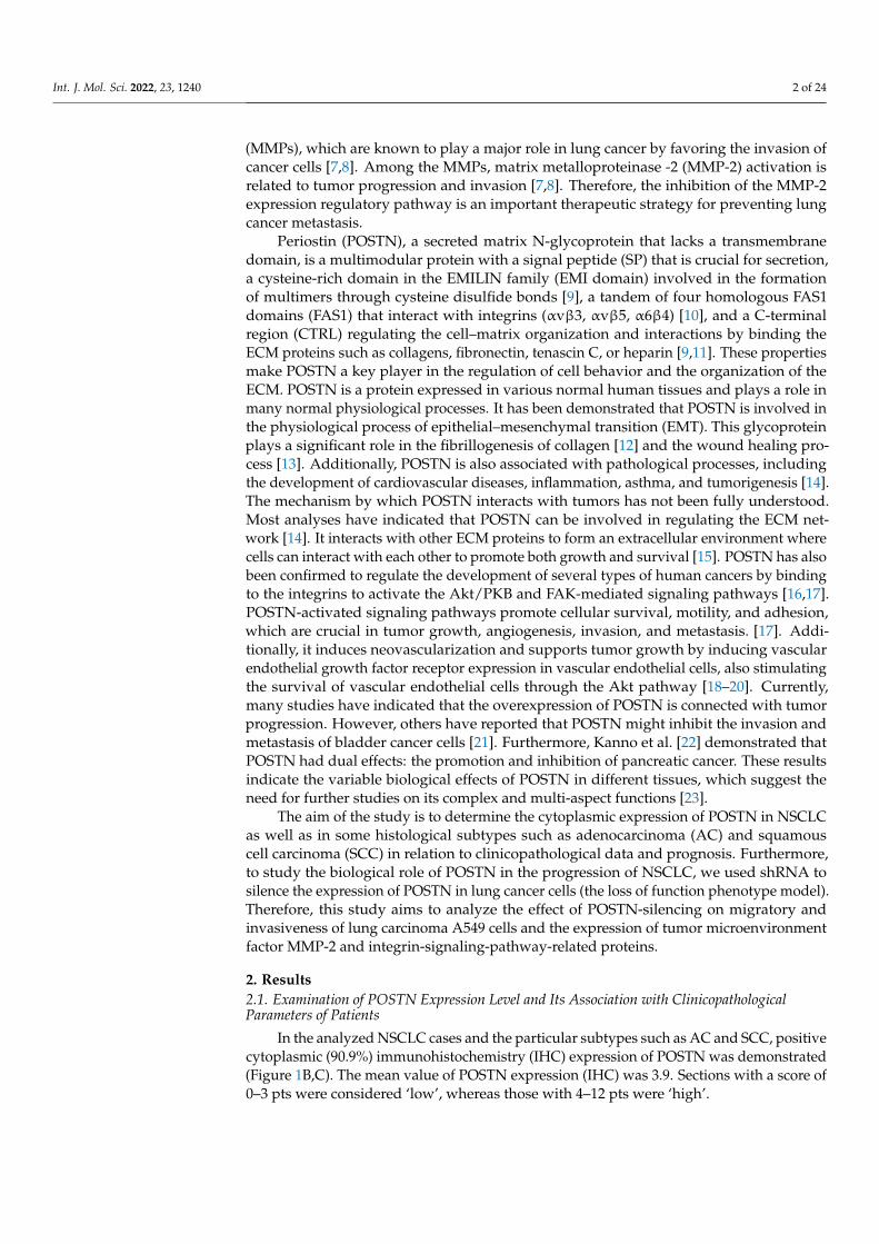
In the analyzed NSCLC cases and the particular subtypes such as AC and SCC, positivecytoplasmic (90.9%) immunohistochemistry (IHC) expression of POSTN was demonstrated(Figure 1B,C). The mean value of POSTN expression (IHC) was 3.9. Sections with a score of0–3 pts were considered ‘low’, whereas those with 4–12 pts were ‘high’.
Int. J. Mol. Sci. 2022, 23, 1240 3 of 24
Int. J. Mol. Sci. 2022, 23, 1240 3 of 26
2. Results 2.1. Examination of POSTN Expression Level and Its Association with Clinicopathological Parameters of Patients
In the analyzed NSCLC cases and the particular subtypes such as AC and SCC, positive cytoplasmic (90.9%) immunohistochemistry (IHC) expression of POSTN was demonstrated (Figure 1B,C). The mean value of POSTN expression (IHC) was 3.9. Sections with a score of 0–3 pts were considered ‘low’, whereas those with 4–12 pts were ‘high’.
Figure 1. Representative immunohistochemical images with cytoplasmic periostin (POSTN) expression in (A) non-malignant lung tissue (NMLT) and non-small cell lung carcinoma (NSCLC) subtypes: (B) adenocarcinoma (AC) and (C) squamous cell carcinoma (SCC). (D) negative control (NC). Magnification 50×, 200×. Bar =200 µm and 50 µm, respectively.
Figure 1. Representative immunohistochemical images with cytoplasmic periostin (POSTN) ex-pression in (A) non-malignant lung tissue (NMLT) and non-small cell lung carcinoma (NSCLC)subtypes: (B) adenocarcinoma (AC) and (C) squamous cell carcinoma (SCC). (D) negative control(NC). Magnification 50×, 200×. Bar = 200 µm and 50 µm, respectively.
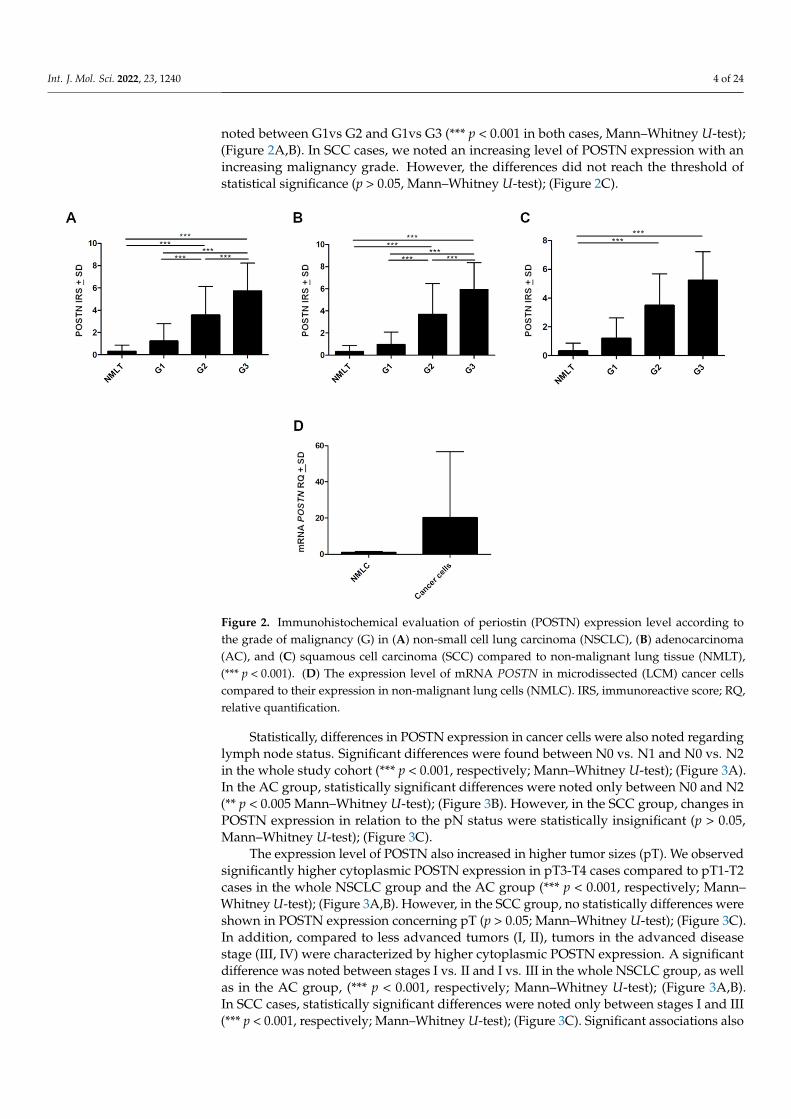
Cytoplasmic POSTN expressions were significantly higher in NSCLC as well as inSCC and AC subtypes compared to non-malignant lung tissue (NMLT) (**** p < 0.0001,respectively; Mann–Whitney U-test); (Figure 1A–C and Figure 2A–C). These results werealso confirmed by molecular studies using laser capture microdissection (LCM). We noticeda markedly higher level of POSTN mRNA expression in microdissected cancer cells com-pared to non-malignant lung cells (NMLC) (Figure 2D). Furthermore, statistical analysisdemonstrated an increasing level of POSTN (IHC) with an increasing malignancy grade(G) of the tumor in the whole cohort, as well as in AC cases. A significant difference was
Int. J. Mol. Sci. 2022, 23, 1240 4 of 24
noted between G1vs G2 and G1vs G3 (*** p < 0.001 in both cases, Mann–Whitney U-test);(Figure 2A,B). In SCC cases, we noted an increasing level of POSTN expression with anincreasing malignancy grade. However, the differences did not reach the threshold ofstatistical significance (p > 0.05, Mann–Whitney U-test); (Figure 2C).
Int. J. Mol. Sci. 2022, 23, 1240 4 of 26
Cytoplasmic POSTN expressions were significantly higher in NSCLC as well as in SCC and AC subtypes compared to non-malignant lung tissue (NMLT) (**** p < 0.0001, respectively; Mann–Whitney U-test); (Figures 1A–C and 2A–C). These results were also confirmed by molecular studies using laser capture microdissection (LCM). We noticed a markedly higher level of POSTN mRNA expression in microdissected cancer cells compared to non-malignant lung cells (NMLC) (Figure 2D). Furthermore, statistical analysis demonstrated an increasing level of POSTN (IHC) with an increasing malignancy grade (G) of the tumor in the whole cohort, as well as in AC cases. A significant difference was noted between G1vs G2 and G1vs G3 (*** p < 0.001 in both cases, Mann–Whitney U-test); (Figure 2A,B). In SCC cases, we noted an increasing level of POSTN expression with an increasing malignancy grade. However, the differences did not reach the threshold of statistical significance (p > 0.05, Mann–Whitney U-test); (Figure 2C).
Figure 2. Immunohistochemical evaluation of periostin (POSTN) expression level according to the grade of malignancy (G) in (A) non-small cell lung carcinoma (NSCLC), (B) adenocarcinoma (AC), and (C) squamous cell carcinoma (SCC) compared to non-malignant lung tissue (NMLT), (*** p < 0.001). (D) The expression level of mRNA POSTN in microdissected (LCM) cancer cells compared to their expression in non-malignant lung cells (NMLC). IRS, immunoreactive score; RQ, relative quantification.
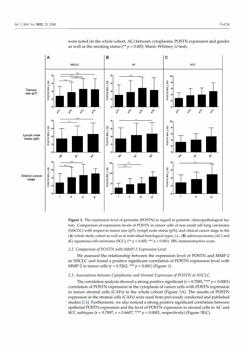
Statistically, differences in POSTN expression in cancer cells were also noted regarding lymph node status. Significant differences were found between N0 vs. N1 and N0 vs. N2 in the whole study cohort (*** p < 0.001, respectively; Mann–Whitney U-test); (Figure 3A). In the AC group, statistically significant differences were noted only between N0 and N2 (** p < 0.005 Mann–Whitney U-test); (Figure 3B). However, in the SCC group, changes in POSTN expression in relation to the pN status were statistically insignificant (p > 0.05, Mann–Whitney U-test); (Figure 3C).
Figure 2. Immunohistochemical evaluation of periostin (POSTN) expression level according tothe grade of malignancy (G) in (A) non-small cell lung carcinoma (NSCLC), (B) adenocarcinoma(AC), and (C) squamous cell carcinoma (SCC) compared to non-malignant lung tissue (NMLT),(*** p < 0.001). (D) The expression level of mRNA POSTN in microdissected (LCM) cancer cellscompared to their expression in non-malignant lung cells (NMLC). IRS, immunoreactive score; RQ,relative quantification.
Statistically, differences in POSTN expression in cancer cells were also noted regardinglymph node status. Significant differences were found between N0 vs. N1 and N0 vs. N2in the whole study cohort (*** p < 0.001, respectively; Mann–Whitney U-test); (Figure 3A).In the AC group, statistically significant differences were noted only between N0 and N2(** p < 0.005 Mann–Whitney U-test); (Figure 3B). However, in the SCC group, changes inPOSTN expression in relation to the pN status were statistically insignificant (p > 0.05,Mann–Whitney U-test); (Figure 3C).
The expression level of POSTN also increased in higher tumor sizes (pT). We observedsignificantly higher cytoplasmic POSTN expression in pT3-T4 cases compared to pT1-T2cases in the whole NSCLC group and the AC group (*** p < 0.001, respectively; Mann–Whitney U-test); (Figure 3A,B). However, in the SCC group, no statistically differences wereshown in POSTN expression concerning pT (p > 0.05; Mann–Whitney U-test); (Figure 3C).In addition, compared to less advanced tumors (I, II), tumors in the advanced diseasestage (III, IV) were characterized by higher cytoplasmic POSTN expression. A significantdifference was noted between stages I vs. II and I vs. III in the whole NSCLC group, as wellas in the AC group, (*** p < 0.001, respectively; Mann–Whitney U-test); (Figure 3A,B).In SCC cases, statistically significant differences were noted only between stages I and III(*** p < 0.001, respectively; Mann–Whitney U-test); (Figure 3C). Significant associations also
Int. J. Mol. Sci. 2022, 23, 1240 5 of 24
were noted (in the whole cohort, AC) between cytoplasmic POSTN expression and genderas well as the smoking status (** p < 0.005; Mann–Whitney U-test).
Int. J. Mol. Sci. 2022, 23, 1240 5 of 26
Figure 3. The expression level of periostin (POSTN) in regard to patients’ clinicopathological factors. Comparison of expression levels of POSTN in cancer cells of non-small cell lung carcinoma (NSCLC) with respect to tumor size (pT), lymph node status (pN), and clinical cancer stage in the (A) whole study cohort as well as in individual histological types, i.e., (B) adenocarcinoma (AC) and (C) squamous cell carcinoma (SCC); (** p < 0.005, *** p < 0.001). IRS, immunoreactive score.
The expression level of POSTN also increased in higher tumor sizes (pT). We observed significantly higher cytoplasmic POSTN expression in pT3-T4 cases compared to pT1-T2 cases in the whole NSCLC group and the AC group (*** p < 0.001, respectively; Mann–Whitney U-test); (Figure 3A,B). However, in the SCC group, no statistically differences were shown in POSTN expression concerning pT (p > 0.05; Mann–Whitney U-test); (Figure 3C). In addition, compared to less advanced tumors (I, II), tumors in the advanced disease stage (III, IV) were characterized by higher cytoplasmic POSTN expression. A significant difference was noted between stages I vs. II and I vs. III in the whole NSCLC group, as well as in the AC group, (*** p < 0.001, respectively; Mann–Whitney U-test); (Figure 3A,B). In SCC cases, statistically significant differences were noted only between stages I and III (*** P < 0.001, respectively; Mann–Whitney U-test); (Figure 3C). Significant associations also were noted (in the whole cohort, AC) between cytoplasmic POSTN expression and gender as well as the smoking status (** p < 0.005; Mann–Whitney U-test).
2.2. Comparison of POSTN with MMP-2 Expression Level
Figure 3. The expression level of periostin (POSTN) in regard to patients’ clinicopathological fac-tors. Comparison of expression levels of POSTN in cancer cells of non-small cell lung carcinoma(NSCLC) with respect to tumor size (pT), lymph node status (pN), and clinical cancer stage in the(A) whole study cohort as well as in individual histological types, i.e., (B) adenocarcinoma (AC) and(C) squamous cell carcinoma (SCC); (** p < 0.005, *** p < 0.001). IRS, immunoreactive score.
2.2. Comparison of POSTN with MMP-2 Expression Level
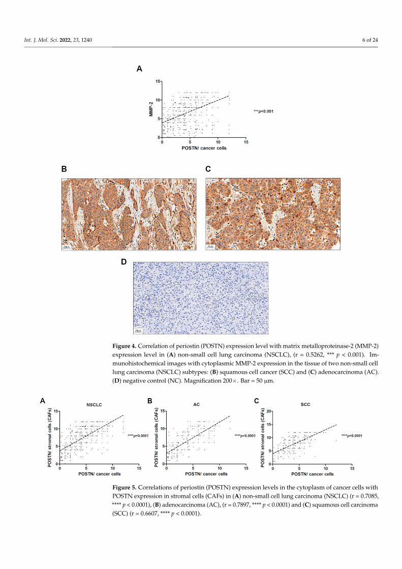
We assessed the relationship between the expression level of POSTN and MMP-2in NSCLC and found a positive significant correlation of POSTN expression level withMMP-2 in tumor cells (r = 0.5262, *** p < 0.001) (Figure 4).
2.3. Associations between Cytoplasmic and Stromal Expression of POSTN in NSCLC
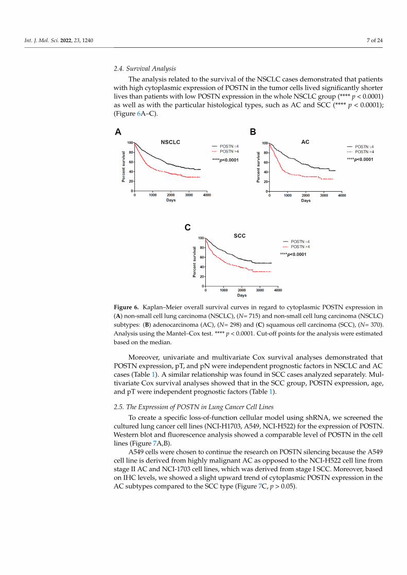
The correlation analysis showed a strong positive significant (r = 0.7085, **** p < 0.0001)correlation of POSTN expression in the cytoplasm of cancer cells with POSTN expressionin tumor stromal cells (CAFs) in the whole cohort (Figure 5A). The results of POSTNexpression in the stromal cells (CAFs) were used from previously conducted and publishedstudies [24]. Furthermore, we also noticed a strong positive significant correlation betweenepithelial POSTN expression and the level of POSTN expression in stromal cells in AC andSCC subtypes (r = 0.7897, r = 0.6607, **** p < 0.0001, respectively) (Figure 5B,C).
Int. J. Mol. Sci. 2022, 23, 1240 6 of 24
Int. J. Mol. Sci. 2022, 23, 1240 6 of 26
We assessed the relationship between the expression level of POSTN and MMP-2 in NSCLC and found a positive significant correlation of POSTN expression level with MMP-2 in tumor cells (r = 0.5262, *** p < 0.001) (Figure 4).
Figure 4. Correlation of periostin (POSTN) expression level with matrix metalloproteinase-2 (MMP-2) expression level in (A) non-small cell lung carcinoma (NSCLC), (r = 0.5262, *** p < 0.001). Immunohistochemical images with cytoplasmic MMP-2 expression in the tissue of two non-small cell lung carcinoma (NSCLC) subtypes: (B) squamous cell cancer (SCC) and (C) adenocarcinoma (AC). (D) negative control (NC). Magnification 200×. Bar =50 µm.
2.3. Associations between Cytoplasmic and Stromal Expression of POSTN in NSCLC The correlation analysis showed a strong positive significant (r = 0.7085, **** p <
0.0001) correlation of POSTN expression in the cytoplasm of cancer cells with POSTN expression in tumor stromal cells (CAFs) in the whole cohort (Figure 5A). The results of POSTN expression in the stromal cells (CAFs) were used from previously conducted and published studies [24]. Furthermore, we also noticed a strong positive significant correlation between epithelial POSTN expression and the level of POSTN expression in stromal cells in AC and SCC subtypes (r = 0.7897, r = 0.6607, **** p < 0.0001, respectively) (Figure 5B,C).
Figure 4. Correlation of periostin (POSTN) expression level with matrix metalloproteinase-2 (MMP-2)expression level in (A) non-small cell lung carcinoma (NSCLC), (r = 0.5262, *** p < 0.001). Im-munohistochemical images with cytoplasmic MMP-2 expression in the tissue of two non-small celllung carcinoma (NSCLC) subtypes: (B) squamous cell cancer (SCC) and (C) adenocarcinoma (AC).(D) negative control (NC). Magnification 200×. Bar = 50 µm.
Int. J. Mol. Sci. 2022, 23, 1240 7 of 26
Figure 5. Correlations of periostin (POSTN) expression levels in the cytoplasm of cancer cells with POSTN expression in stromal cells (CAFs) in (A) non-small cell lung carcinoma (NSCLC) (r = 0.7085, **** p < 0.0001), (B) adenocarcinoma (AC), (r = 0.7897, **** p < 0.0001) and (C) squamous cell carcinoma (SCC) (r = 0.6607, **** p < 0.0001).
2.4. Survival Analysis The analysis related to the survival of the NSCLC cases demonstrated that patients
with high cytoplasmic expression of POSTN in the tumor cells lived significantly shorter lives than patients with low POSTN expression in the whole NSCLC group (**** p < 0.0001) as well as with the particular histological types, such as AC and SCC (**** p < 0.0001); (Figure 6A–C).
Figure 6. Kaplan–Meier overall survival curves in regard to cytoplasmic POSTN expression in (A) non-small cell lung carcinoma (NSCLC), (N= 715) and non-small cell lung carcinoma (NSCLC) subtypes: (B) adenocarcinoma (AC), (N= 298) and (C) squamous cell carcinoma (SCC), (N= 370). Analysis using the Mantel–Cox test. **** p < 0.0001. Cut-off points for the analysis were estimated based on the median.
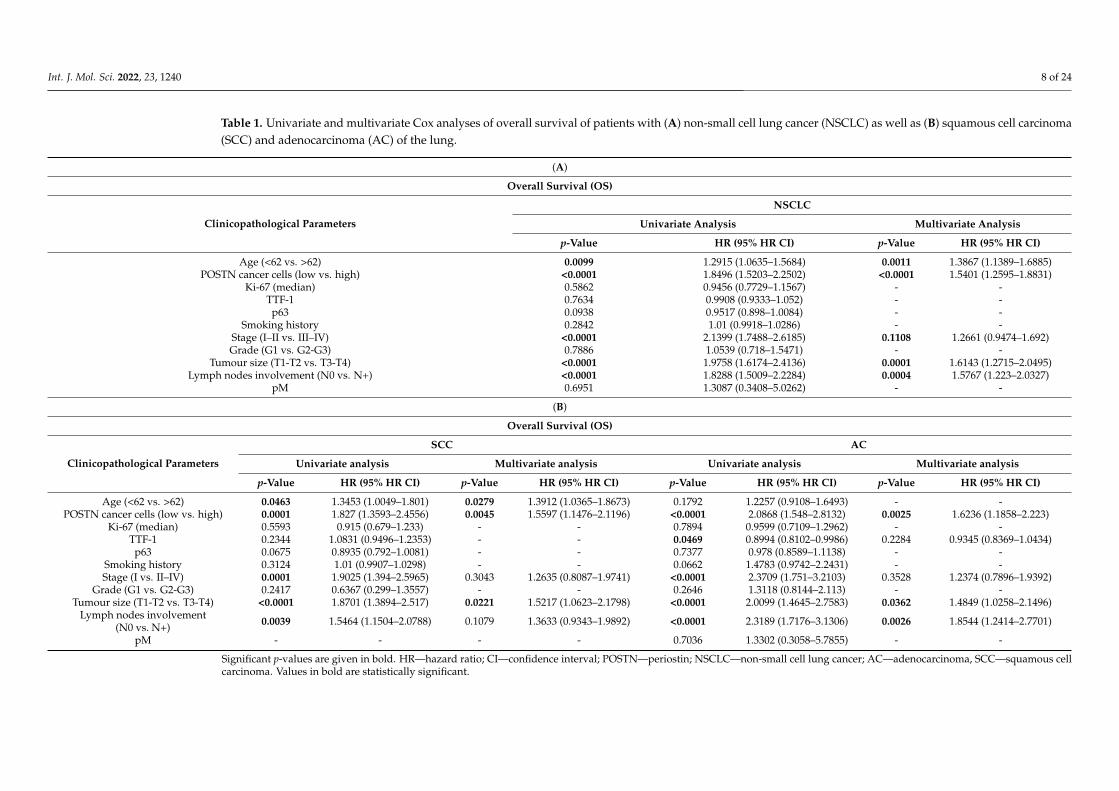
Moreover, univariate and multivariate Cox survival analyses demonstrated that POSTN expression, pT, and pN were independent prognostic factors in NSCLC and AC cases (Table 1). A similar relationship was found in SCC cases analyzed separately. Multivariate Cox survival analyses showed that in the SCC group, POSTN expression, age, and pT were independent prognostic factors (Table 1).
Figure 5. Correlations of periostin (POSTN) expression levels in the cytoplasm of cancer cells withPOSTN expression in stromal cells (CAFs) in (A) non-small cell lung carcinoma (NSCLC) (r = 0.7085,**** p < 0.0001), (B) adenocarcinoma (AC), (r = 0.7897, **** p < 0.0001) and (C) squamous cell carcinoma(SCC) (r = 0.6607, **** p < 0.0001).
Int. J. Mol. Sci. 2022, 23, 1240 7 of 24
2.4. Survival Analysis
The analysis related to the survival of the NSCLC cases demonstrated that patientswith high cytoplasmic expression of POSTN in the tumor cells lived significantly shorterlives than patients with low POSTN expression in the whole NSCLC group (**** p < 0.0001)as well as with the particular histological types, such as AC and SCC (**** p < 0.0001);(Figure 6A–C).
Int. J. Mol. Sci. 2022, 23, 1240 7 of 26
Figure 5. Correlations of periostin (POSTN) expression levels in the cytoplasm of cancer cells with POSTN expression in stromal cells (CAFs) in (A) non-small cell lung carcinoma (NSCLC) (r = 0.7085, **** p < 0.0001), (B) adenocarcinoma (AC), (r = 0.7897, **** p < 0.0001) and (C) squamous cell carcinoma (SCC) (r = 0.6607, **** p < 0.0001).
2.4. Survival Analysis The analysis related to the survival of the NSCLC cases demonstrated that patients
with high cytoplasmic expression of POSTN in the tumor cells lived significantly shorter lives than patients with low POSTN expression in the whole NSCLC group (**** p < 0.0001) as well as with the particular histological types, such as AC and SCC (**** p < 0.0001); (Figure 6A–C).
Figure 6. Kaplan–Meier overall survival curves in regard to cytoplasmic POSTN expression in (A) non-small cell lung carcinoma (NSCLC), (N= 715) and non-small cell lung carcinoma (NSCLC) subtypes: (B) adenocarcinoma (AC), (N= 298) and (C) squamous cell carcinoma (SCC), (N= 370). Analysis using the Mantel–Cox test. **** p < 0.0001. Cut-off points for the analysis were estimated based on the median.
Moreover, univariate and multivariate Cox survival analyses demonstrated that POSTN expression, pT, and pN were independent prognostic factors in NSCLC and AC cases (Table 1). A similar relationship was found in SCC cases analyzed separately. Multivariate Cox survival analyses showed that in the SCC group, POSTN expression, age, and pT were independent prognostic factors (Table 1).
Figure 6. Kaplan–Meier overall survival curves in regard to cytoplasmic POSTN expression in(A) non-small cell lung carcinoma (NSCLC), (N= 715) and non-small cell lung carcinoma (NSCLC)subtypes: (B) adenocarcinoma (AC), (N= 298) and (C) squamous cell carcinoma (SCC), (N= 370).Analysis using the Mantel–Cox test. **** p < 0.0001. Cut-off points for the analysis were estimatedbased on the median.
Moreover, univariate and multivariate Cox survival analyses demonstrated thatPOSTN expression, pT, and pN were independent prognostic factors in NSCLC and ACcases (Table 1). A similar relationship was found in SCC cases analyzed separately. Mul-tivariate Cox survival analyses showed that in the SCC group, POSTN expression, age,and pT were independent prognostic factors (Table 1).
2.5. The Expression of POSTN in Lung Cancer Cell Lines
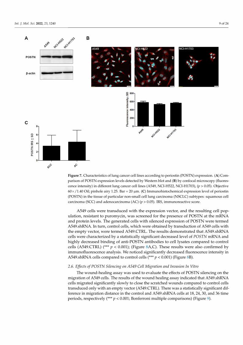
To create a specific loss-of-function cellular model using shRNA, we screened thecultured lung cancer cell lines (NCI-H1703, A549, NCI-H522) for the expression of POSTN.Western blot and fluorescence analysis showed a comparable level of POSTN in the celllines (Figure 7A,B).
A549 cells were chosen to continue the research on POSTN silencing because the A549cell line is derived from highly malignant AC as opposed to the NCI-H522 cell line fromstage II AC and NCI-1703 cell lines, which was derived from stage I SCC. Moreover, basedon IHC levels, we showed a slight upward trend of cytoplasmic POSTN expression in theAC subtypes compared to the SCC type (Figure 7C, p > 0.05).
Int. J. Mol. Sci. 2022, 23, 1240 8 of 24
Table 1. Univariate and multivariate Cox analyses of overall survival of patients with (A) non-small cell lung cancer (NSCLC) as well as (B) squamous cell carcinoma(SCC) and adenocarcinoma (AC) of the lung.
(A)
Overall Survival (OS)
Clinicopathological Parameters
NSCLC
Univariate Analysis Multivariate Analysis
p-Value HR (95% HR CI) p-Value HR (95% HR CI)
Age (<62 vs. >62) 0.0099 1.2915 (1.0635–1.5684) 0.0011 1.3867 (1.1389–1.6885)POSTN cancer cells (low vs. high) <0.0001 1.8496 (1.5203–2.2502) <0.0001 1.5401 (1.2595–1.8831)
Ki-67 (median) 0.5862 0.9456 (0.7729–1.1567) - -TTF-1 0.7634 0.9908 (0.9333–1.052) - -
p63 0.0938 0.9517 (0.898–1.0084) - -Smoking history 0.2842 1.01 (0.9918–1.0286) - -
Stage (I–II vs. III–IV) <0.0001 2.1399 (1.7488–2.6185) 0.1108 1.2661 (0.9474–1.692)Grade (G1 vs. G2-G3) 0.7886 1.0539 (0.718–1.5471) - -
Tumour size (T1-T2 vs. T3-T4) <0.0001 1.9758 (1.6174–2.4136) 0.0001 1.6143 (1.2715–2.0495)Lymph nodes involvement (N0 vs. N+) <0.0001 1.8288 (1.5009–2.2284) 0.0004 1.5767 (1.223–2.0327)
pM 0.6951 1.3087 (0.3408–5.0262) - -
(B)
Overall Survival (OS)
Clinicopathological Parameters
SCC AC
Univariate analysis Multivariate analysis Univariate analysis Multivariate analysis
p-Value HR (95% HR CI) p-Value HR (95% HR CI) p-Value HR (95% HR CI) p-Value HR (95% HR CI)
Age (<62 vs. >62) 0.0463 1.3453 (1.0049–1.801) 0.0279 1.3912 (1.0365–1.8673) 0.1792 1.2257 (0.9108–1.6493) - -POSTN cancer cells (low vs. high) 0.0001 1.827 (1.3593–2.4556) 0.0045 1.5597 (1.1476–2.1196) <0.0001 2.0868 (1.548–2.8132) 0.0025 1.6236 (1.1858–2.223)
Ki-67 (median) 0.5593 0.915 (0.679–1.233) - - 0.7894 0.9599 (0.7109–1.2962) - -TTF-1 0.2344 1.0831 (0.9496–1.2353) - - 0.0469 0.8994 (0.8102–0.9986) 0.2284 0.9345 (0.8369–1.0434)
p63 0.0675 0.8935 (0.792–1.0081) - - 0.7377 0.978 (0.8589–1.1138) - -Smoking history 0.3124 1.01 (0.9907–1.0298) - - 0.0662 1.4783 (0.9742–2.2431) - -Stage (I vs. II–IV) 0.0001 1.9025 (1.394–2.5965) 0.3043 1.2635 (0.8087–1.9741) <0.0001 2.3709 (1.751–3.2103) 0.3528 1.2374 (0.7896–1.9392)
Grade (G1 vs. G2-G3) 0.2417 0.6367 (0.299–1.3557) - - 0.2646 1.3118 (0.8144–2.113) - -Tumour size (T1-T2 vs. T3-T4) <0.0001 1.8701 (1.3894–2.517) 0.0221 1.5217 (1.0623–2.1798) <0.0001 2.0099 (1.4645–2.7583) 0.0362 1.4849 (1.0258–2.1496)
Lymph nodes involvement(N0 vs. N+) 0.0039 1.5464 (1.1504–2.0788) 0.1079 1.3633 (0.9343–1.9892) <0.0001 2.3189 (1.7176–3.1306) 0.0026 1.8544 (1.2414–2.7701)
pM - - - - 0.7036 1.3302 (0.3058–5.7855) - -
Significant p-values are given in bold. HR—hazard ratio; CI—confidence interval; POSTN—periostin; NSCLC—non-small cell lung cancer; AC—adenocarcinoma, SCC—squamous cellcarcinoma. Values in bold are statistically significant.
Int. J. Mol. Sci. 2022, 23, 1240 9 of 24
Int. J. Mol. Sci. 2022, 23, 1240 10 of 26
2.5. The Expression of POSTN in Lung Cancer Cell Lines To create a specific loss-of-function cellular model using shRNA, we screened the
cultured lung cancer cell lines (NCI-H1703, A549, NCI-H522) for the expression of POSTN. Western blot and fluorescence analysis showed a comparable level of POSTN in the cell lines (Figure 7A,B).
Figure 7. Characteristics of lung cancer cell lines according to periostin (POSTN) expression. (A) Comparison of POSTN expression levels detected by Western blot and (B) by confocal microscopy (fluorescence intensity) in different lung cancer cell lines (A549, NCI-H522, NCI-H1703), (p > 0.05). Objective 60×/1.40 Oil; pinhole airy 1.25. Bar = 20 µm. (C) Immunohistochemical expression level of periostin (POSTN) in the tissue of particular non-small cell lung carcinoma (NSCLC) subtypes: squamous cell carcinoma (SCC) and adenocarcinoma (AC) (p > 0.05). IRS, immunoreactive score.
A549 cells were chosen to continue the research on POSTN silencing because the A549 cell line is derived from highly malignant AC as opposed to the NCI-H522 cell line from stage II AC and NCI-1703 cell lines, which was derived from stage I SCC. Moreover, based on IHC levels, we showed a slight upward trend of cytoplasmic POSTN expression in the AC subtypes compared to the SCC type (Figure 7C, p > 0.05).
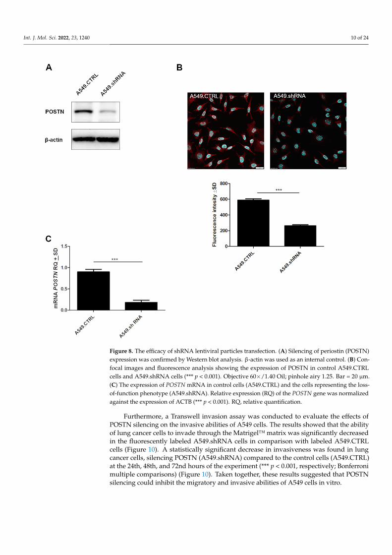
A549 cells were transduced with the expression vector, and the resulting cell population, resistant to puromycin, was screened for the presence of POSTN at the mRNA and protein levels. The generated cells with silenced expression of POSTN were termed A549.shRNA. In turn, control cells, which were obtained by transduction of A549 cells with the empty vector, were termed A549.CTRL. The results demonstrated that A549.shRNA cells were characterized by a statistically significant decreased level of POSTN mRNA and highly decreased binding of anti-POSTN antibodies to cell lysates compared to control cells (A549.CTRL) (*** p < 0.001); (Figure 8A,C). These results were
Figure 7. Characteristics of lung cancer cell lines according to periostin (POSTN) expression. (A) Com-parison of POSTN expression levels detected by Western blot and (B) by confocal microscopy (fluores-cence intensity) in different lung cancer cell lines (A549, NCI-H522, NCI-H1703), (p > 0.05). Objective60×/1.40 Oil; pinhole airy 1.25. Bar = 20 µm. (C) Immunohistochemical expression level of periostin(POSTN) in the tissue of particular non-small cell lung carcinoma (NSCLC) subtypes: squamous cellcarcinoma (SCC) and adenocarcinoma (AC) (p > 0.05). IRS, immunoreactive score.
A549 cells were transduced with the expression vector, and the resulting cell pop-ulation, resistant to puromycin, was screened for the presence of POSTN at the mRNAand protein levels. The generated cells with silenced expression of POSTN were termedA549.shRNA. In turn, control cells, which were obtained by transduction of A549 cells withthe empty vector, were termed A549.CTRL. The results demonstrated that A549.shRNAcells were characterized by a statistically significant decreased level of POSTN mRNA andhighly decreased binding of anti-POSTN antibodies to cell lysates compared to controlcells (A549.CTRL) (*** p < 0.001); (Figure 8A,C). These results were also confirmed byimmunofluorescence analysis. We noticed significantly decreased fluorescence intensity inA549.shRNA cells compared to control cells (*** p < 0.001) (Figure 8B).
2.6. Effects of POSTN Silencing on A549 Cell Migration and Invasion In Vitro
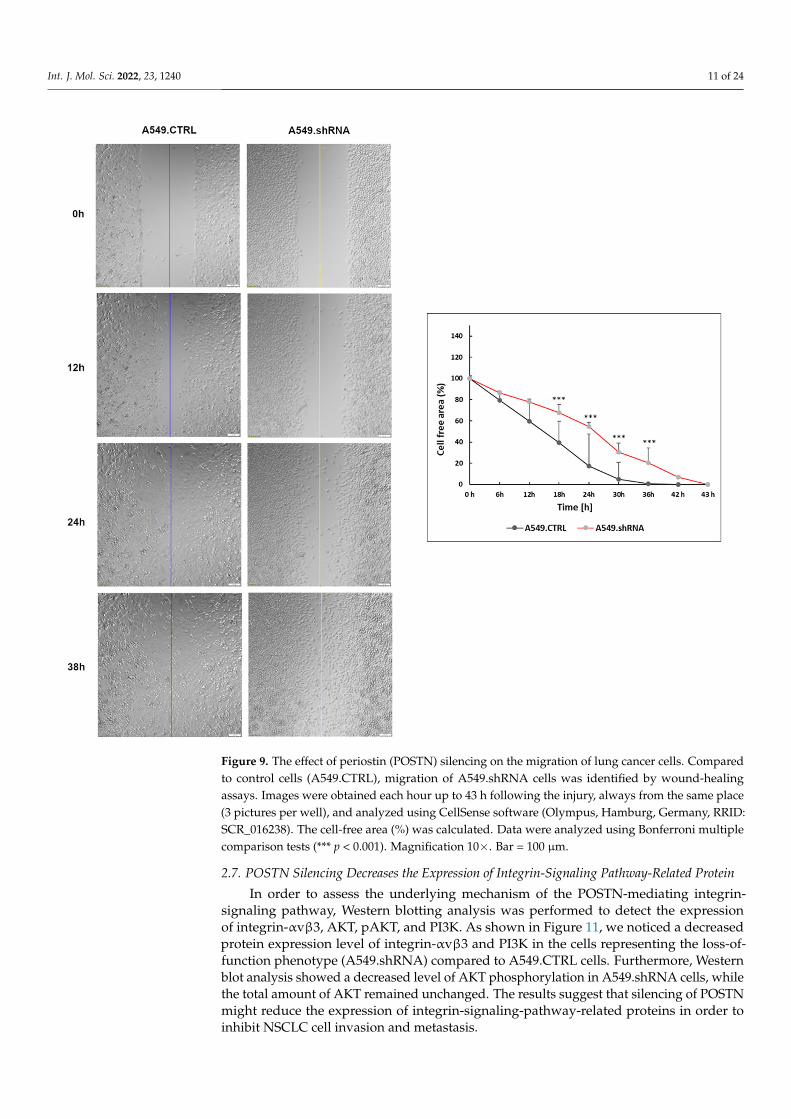
The wound-healing assay was used to evaluate the effects of POSTN silencing on themigration of A549 cells. The results of the wound healing assay indicated that A549.shRNAcells migrated significantly slowly to close the scratched wounds compared to control cellstransduced only with an empty vector (A549.CTRL). There was a statistically significant dif-ference in migration distance in the control and A549.shRNA cells at 18, 24, 30, and 36 timeperiods, respectively (*** p < 0.001; Bonferroni multiple comparisons) (Figure 9).
Int. J. Mol. Sci. 2022, 23, 1240 10 of 24
Int. J. Mol. Sci. 2022, 23, 1240 11 of 26
also confirmed by immunofluorescence analysis. We noticed significantly decreased flu-orescence intensity in A549.shRNA cells compared to control cells (*** p < 0.001) (Figure 8B).
Figure 8. The efficacy of shRNA lentiviral particles transfection. (A) Silencing of periostin (POSTN) expression was confirmed by Western blot analysis. β-actin was used as an internal control. (B) Confocal images and fluorescence analysis showing the expression of POSTN in control A549.CTRL cells and A549.shRNA cells (*** p < 0.001). Objective 60×/1.40 Oil; pinhole airy 1.25. Bar = 20 µm. (C) The expression of POSTN mRNA in control cells (A549.CTRL) and the cells representing the loss-of-function phenotype (A549.shRNA). Relative expression (RQ) of the POSTN gene was normalized against the expression of ACTB (*** p < 0.001). RQ, relative quantification.
2.6. Effects of POSTN Silencing on A549 Cell Migration and Invasion In Vitro The wound-healing assay was used to evaluate the effects of POSTN silencing on the
migration of A549 cells. The results of the wound healing assay indicated that A549.shRNA cells migrated significantly slowly to close the scratched wounds compared to control cells transduced only with an empty vector (A549.CTRL). There was a statisti-cally significant difference in migration distance in the control and A549.shRNA cells at 18, 24, 30, and 36 time periods, respectively (*** p < 0.001; Bonferroni multiple compari-sons) (Figure 9).
Figure 8. The efficacy of shRNA lentiviral particles transfection. (A) Silencing of periostin (POSTN)expression was confirmed by Western blot analysis. β-actin was used as an internal control. (B) Con-focal images and fluorescence analysis showing the expression of POSTN in control A549.CTRLcells and A549.shRNA cells (*** p < 0.001). Objective 60×/1.40 Oil; pinhole airy 1.25. Bar = 20 µm.(C) The expression of POSTN mRNA in control cells (A549.CTRL) and the cells representing the loss-of-function phenotype (A549.shRNA). Relative expression (RQ) of the POSTN gene was normalizedagainst the expression of ACTB (*** p < 0.001). RQ, relative quantification.
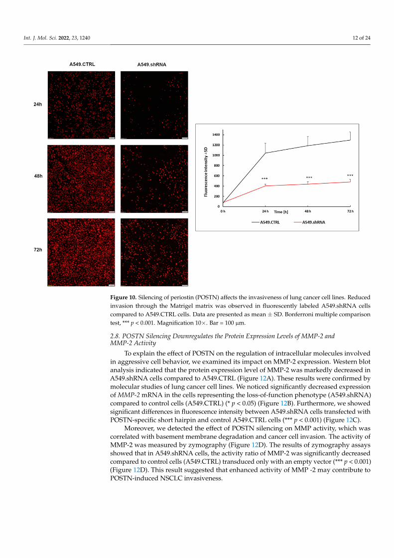
Furthermore, a Transwell invasion assay was conducted to evaluate the effects ofPOSTN silencing on the invasive abilities of A549 cells. The results showed that the abilityof lung cancer cells to invade through the Matrigel™ matrix was significantly decreasedin the fluorescently labeled A549.shRNA cells in comparison with labeled A549.CTRLcells (Figure 10). A statistically significant decrease in invasiveness was found in lungcancer cells, silencing POSTN (A549.shRNA) compared to the control cells (A549.CTRL)at the 24th, 48th, and 72nd hours of the experiment (*** p < 0.001, respectively; Bonferronimultiple comparisons) (Figure 10). Taken together, these results suggested that POSTNsilencing could inhibit the migratory and invasive abilities of A549 cells in vitro.
Int. J. Mol. Sci. 2022, 23, 1240 11 of 24Int. J. Mol. Sci. 2022, 23, 1240 12 of 26
Figure 9. The effect of periostin (POSTN) silencing on the migration of lung cancer cells. Compared to control cells (A549.CTRL), migration of A549.shRNA cells was identified by wound-healing as-says. Images were obtained each hour up to 43 h following the injury, always from the same place (3 pictures per well), and analyzed using CellSense software (Olympus, Hamburg, Germany, RRID: SCR_016238). The cell-free area (%) was calculated. Data were analyzed using Bonferroni multiple comparison tests (*** p < 0.001). Magnification 10×. Bar = 100 µm.
Furthermore, a Transwell invasion assay was conducted to evaluate the effects of POSTN silencing on the invasive abilities of A549 cells. The results showed that the ability of lung cancer cells to invade through the Matrigel™ matrix was significantly decreased in the fluorescently labeled A549.shRNA cells in comparison with labeled A549.CTRL cells (Figure 10). A statistically significant decrease in invasiveness was found in lung can-cer cells, silencing POSTN (A549.shRNA) compared to the control cells (A549.CTRL) at the 24th, 48th, and 72nd hours of the experiment (*** p < 0.001, respectively; Bonferroni multiple comparisons) (Figure 10). Taken together, these results suggested that POSTN silencing could inhibit the migratory and invasive abilities of A549 cells in vitro.
Figure 9. The effect of periostin (POSTN) silencing on the migration of lung cancer cells. Comparedto control cells (A549.CTRL), migration of A549.shRNA cells was identified by wound-healingassays. Images were obtained each hour up to 43 h following the injury, always from the same place(3 pictures per well), and analyzed using CellSense software (Olympus, Hamburg, Germany, RRID:SCR_016238). The cell-free area (%) was calculated. Data were analyzed using Bonferroni multiplecomparison tests (*** p < 0.001). Magnification 10×. Bar = 100 µm.
2.7. POSTN Silencing Decreases the Expression of Integrin-Signaling Pathway-Related Protein
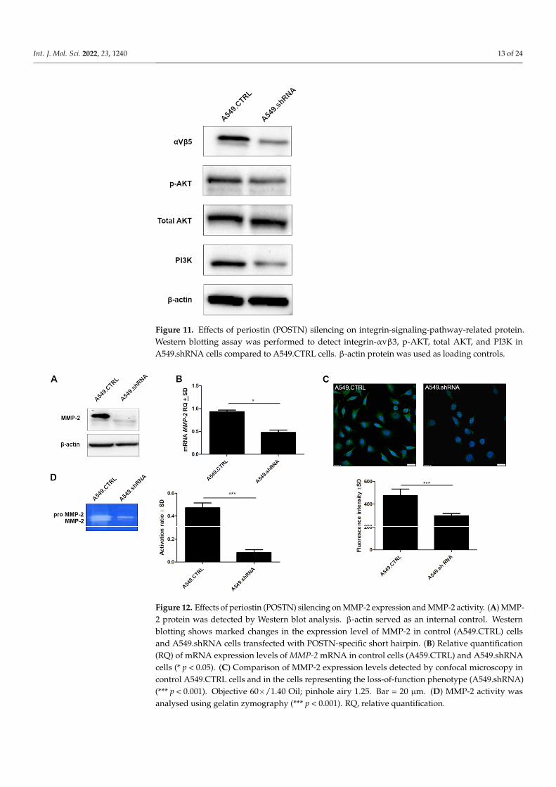
In order to assess the underlying mechanism of the POSTN-mediating integrin-signaling pathway, Western blotting analysis was performed to detect the expressionof integrin-αvβ3, AKT, pAKT, and PI3K. As shown in Figure 11, we noticed a decreasedprotein expression level of integrin-αvβ3 and PI3K in the cells representing the loss-of-function phenotype (A549.shRNA) compared to A549.CTRL cells. Furthermore, Westernblot analysis showed a decreased level of AKT phosphorylation in A549.shRNA cells, whilethe total amount of AKT remained unchanged. The results suggest that silencing of POSTNmight reduce the expression of integrin-signaling-pathway-related proteins in order toinhibit NSCLC cell invasion and metastasis.
Int. J. Mol. Sci. 2022, 23, 1240 12 of 24Int. J. Mol. Sci. 2022, 23, 1240 13 of 26
Figure 10. Silencing of periostin (POSTN) affects the invasiveness of lung cancer cell lines. Reduced invasion through the Matrigel matrix was observed in fluorescently labeled A549.shRNA cells com-pared to A549.CTRL cells. Data are presented as mean ± SD. Bonferroni multiple comparison test, *** p < 0.001. Magnification 10×. Bar = 100 µm.
2.7. POSTN Silencing Decreases the Expression of Integrin-Signaling Pathway-Related Protein In order to assess the underlying mechanism of the POSTN-mediating integrin-sig-
naling pathway, Western blotting analysis was performed to detect the expression of in-tegrin-αvβ3, AKT, pAKT, and PI3K. As shown in Figure 11, we noticed a decreased pro-tein expression level of integrin-αvβ3 and PI3K in the cells representing the loss-of-func-tion phenotype (A549.shRNA) compared to A549.CTRL cells. Furthermore, Western blot analysis showed a decreased level of AKT phosphorylation in A549.shRNA cells, while the total amount of AKT remained unchanged. The results suggest that silencing of POSTN might reduce the expression of integrin-signaling-pathway-related proteins in or-der to inhibit NSCLC cell invasion and metastasis.
Figure 10. Silencing of periostin (POSTN) affects the invasiveness of lung cancer cell lines. Reducedinvasion through the Matrigel matrix was observed in fluorescently labeled A549.shRNA cellscompared to A549.CTRL cells. Data are presented as mean ± SD. Bonferroni multiple comparisontest, *** p < 0.001. Magnification 10×. Bar = 100 µm.
2.8. POSTN Silencing Downregulates the Protein Expression Levels of MMP-2 andMMP-2 Activity
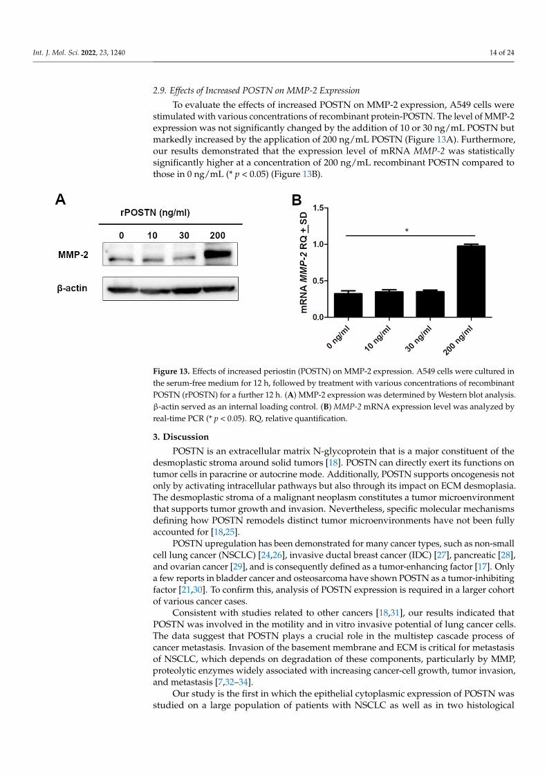
To explain the effect of POSTN on the regulation of intracellular molecules involvedin aggressive cell behavior, we examined its impact on MMP-2 expression. Western blotanalysis indicated that the protein expression level of MMP-2 was markedly decreased inA549.shRNA cells compared to A549.CTRL (Figure 12A). These results were confirmed bymolecular studies of lung cancer cell lines. We noticed significantly decreased expressionof MMP-2 mRNA in the cells representing the loss-of-function phenotype (A549.shRNA)compared to control cells (A549.CTRL) (* p < 0.05) (Figure 12B). Furthermore, we showedsignificant differences in fluorescence intensity between A549.shRNA cells transfected withPOSTN-specific short hairpin and control A549.CTRL cells (*** p < 0.001) (Figure 12C).
Moreover, we detected the effect of POSTN silencing on MMP activity, which wascorrelated with basement membrane degradation and cancer cell invasion. The activity ofMMP-2 was measured by zymography (Figure 12D). The results of zymography assaysshowed that in A549.shRNA cells, the activity ratio of MMP-2 was significantly decreasedcompared to control cells (A549.CTRL) transduced only with an empty vector (*** p < 0.001)(Figure 12D). This result suggested that enhanced activity of MMP -2 may contribute toPOSTN-induced NSCLC invasiveness.
Int. J. Mol. Sci. 2022, 23, 1240 13 of 24
Int. J. Mol. Sci. 2022, 23, 1240 14 of 26
Figure 11. Effects of periostin (POSTN) silencing on integrin-signaling-pathway-related protein. Western blotting assay was performed to detect integrin-αvβ3, p-AKT, total AKT, and PI3K in A549.shRNA cells compared to A549.CTRL cells. β-actin protein was used as loading controls.
2.8. POSTN Silencing Downregulates the Protein Expression Levels of MMP-2 and MMP-2 Activity
To explain the effect of POSTN on the regulation of intracellular molecules involved in aggressive cell behavior, we examined its impact on MMP-2 expression. Western blot analysis indicated that the protein expression level of MMP-2 was markedly decreased in A549.shRNA cells compared to A549.CTRL (Figure 12A). These results were confirmed by molecular studies of lung cancer cell lines. We noticed significantly decreased expres-sion of MMP-2 mRNA in the cells representing the loss-of-function phenotype (A549.shRNA) compared to control cells (A549.CTRL) (* p < 0.05) (Figure 12B). Further-more, we showed significant differences in fluorescence intensity between A549.shRNA cells transfected with POSTN-specific short hairpin and control A549.CTRL cells (*** p < 0.001) (Figure 12C).
Figure 11. Effects of periostin (POSTN) silencing on integrin-signaling-pathway-related protein.Western blotting assay was performed to detect integrin-αvβ3, p-AKT, total AKT, and PI3K inA549.shRNA cells compared to A549.CTRL cells. β-actin protein was used as loading controls.
Int. J. Mol. Sci. 2022, 23, 1240 15 of 26
Figure 12. Effects of periostin (POSTN) silencing on MMP-2 expression and MMP-2 activity. (A) MMP-2 protein was detected by Western blot analysis. β-actin served as an internal control. Western blotting shows marked changes in the expression level of MMP-2 in control (A549.CTRL) cells and A549.shRNA cells transfected with POSTN-specific short hairpin. (B) Relative quantification (RQ) of mRNA expression levels of MMP-2 mRNA in control cells (A459.CTRL) and A549.shRNA cells (* p < 0.05). (C) Comparison of MMP-2 expression levels detected by confocal microscopy in control A549.CTRL cells and in the cells representing the loss-of-function phenotype (A549.shRNA) (*** p < 0.001). Objective 60×/1.40 Oil; pinhole airy 1.25. Bar = 20 µm. (D) MMP-2 activity was analysed using gelatin zymography (*** p < 0.001). RQ, relative quantification.
Moreover, we detected the effect of POSTN silencing on MMP activity, which was correlated with basement membrane degradation and cancer cell invasion. The activity of MMP-2 was measured by zymography (Figure 12D). The results of zymography assays showed that in A549.shRNA cells, the activity ratio of MMP-2 was significantly decreased compared to control cells (A549.CTRL) transduced only with an empty vector (*** p < 0.001) (Figure 12D). This result suggested that enhanced activity of MMP -2 may contrib-ute to POSTN-induced NSCLC invasiveness.
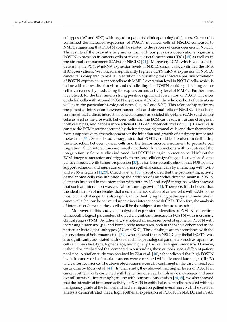
2.9. Effects of Increased POSTN on MMP-2 Expression To evaluate the effects of increased POSTN on MMP-2 expression, A549 cells were
stimulated with various concentrations of recombinant protein-POSTN. The level of MMP-2 expression was not significantly changed by the addition of 10 or 30 ng/mL POSTN but markedly increased by the application of 200 ng/mL POSTN (Figure 13A). Furthermore, our results demonstrated that the expression level of mRNA MMP-2 was statistically significantly higher at a concentration of 200 ng/mL recombinant POSTN com-pared to those in 0 ng/mL (* p < 0.05) (Figure 13B).
Figure 12. Effects of periostin (POSTN) silencing on MMP-2 expression and MMP-2 activity. (A) MMP-2 protein was detected by Western blot analysis. β-actin served as an internal control. Westernblotting shows marked changes in the expression level of MMP-2 in control (A549.CTRL) cellsand A549.shRNA cells transfected with POSTN-specific short hairpin. (B) Relative quantification(RQ) of mRNA expression levels of MMP-2 mRNA in control cells (A459.CTRL) and A549.shRNAcells (* p < 0.05). (C) Comparison of MMP-2 expression levels detected by confocal microscopy incontrol A549.CTRL cells and in the cells representing the loss-of-function phenotype (A549.shRNA)(*** p < 0.001). Objective 60×/1.40 Oil; pinhole airy 1.25. Bar = 20 µm. (D) MMP-2 activity wasanalysed using gelatin zymography (*** p < 0.001). RQ, relative quantification.
Int. J. Mol. Sci. 2022, 23, 1240 14 of 24
2.9. Effects of Increased POSTN on MMP-2 Expression
To evaluate the effects of increased POSTN on MMP-2 expression, A549 cells werestimulated with various concentrations of recombinant protein-POSTN. The level of MMP-2expression was not significantly changed by the addition of 10 or 30 ng/mL POSTN butmarkedly increased by the application of 200 ng/mL POSTN (Figure 13A). Furthermore,our results demonstrated that the expression level of mRNA MMP-2 was statisticallysignificantly higher at a concentration of 200 ng/mL recombinant POSTN compared tothose in 0 ng/mL (* p < 0.05) (Figure 13B).
Int. J. Mol. Sci. 2022, 23, 1240 16 of 26
Figure 13. Effects of increased periostin (POSTN) on MMP-2 expression. A549 cells were cultured in the serum-free medium for 12 h, followed by treatment with various concentrations of recombi-nant POSTN (rPOSTN) for a further 12 h. (A) MMP-2 expression was determined by Western blot analysis. β-actin served as an internal loading control. (B) MMP-2 mRNA expression level was an-alyzed by real-time PCR (* p < 0.05). RQ, relative quantification.
3. Discussion POSTN is an extracellular matrix N-glycoprotein that is a major constituent of the
desmoplastic stroma around solid tumors [18]. POSTN can directly exert its functions on tumor cells in paracrine or autocrine mode. Additionally, POSTN supports oncogenesis not only by activating intracellular pathways but also through its impact on ECM desmoplasia. The desmoplastic stroma of a malignant neoplasm constitutes a tumor mi-croenvironment that supports tumor growth and invasion. Nevertheless, specific molec-ular mechanisms defining how POSTN remodels distinct tumor microenvironments have not been fully accounted for [18,25].
POSTN upregulation has been demonstrated for many cancer types, such as non-small cell lung cancer (NSCLC) [24,26], invasive ductal breast cancer (IDC)[27], pancreatic [28], and ovarian cancer [29], and is consequently defined as a tumor-enhancing factor [17]. Only a few reports in bladder cancer and osteosarcoma have shown POSTN as a tumor-inhibiting factor [21,30]. To confirm this, analysis of POSTN expression is required in a larger cohort of various cancer cases.
Consistent with studies related to other cancers [18,31], our results indicated that POSTN was involved in the motility and in vitro invasive potential of lung cancer cells. The data suggest that POSTN plays a crucial role in the multistep cascade process of can-cer metastasis. Invasion of the basement membrane and ECM is critical for metastasis of NSCLC, which depends on degradation of these components, particularly by MMP, pro-teolytic enzymes widely associated with increasing cancer-cell growth, tumor invasion, and metastasis [7,32–34].
Our study is the first in which the epithelial cytoplasmic expression of POSTN was studied on a large population of patients with NSCLC as well as in two histological sub-types (AC and SCC) with regard to patients’ clinicopathological factors. Our results con-firmed the increased expression of POSTN in cancer cells of NSCLC compared to NMLT, suggesting that POSTN could be related to the process of carcinogenesis in NSCLC. The results of the present study are in line with our previous observations regarding POSTN expression in cancers cells of invasive ductal carcinoma (IDC) [35] as well as in the stromal compartment (CAFs) of NSCLC [24]. Moreover, LCM, which was used to determine the POSTN mRNA expression levels in NSCLC cancer cells, confirmed the TMA IHC obser-vations. We noticed a significantly higher POSTN mRNA expression in NSCLC cancer
Figure 13. Effects of increased periostin (POSTN) on MMP-2 expression. A549 cells were cultured inthe serum-free medium for 12 h, followed by treatment with various concentrations of recombinantPOSTN (rPOSTN) for a further 12 h. (A) MMP-2 expression was determined by Western blot analysis.β-actin served as an internal loading control. (B) MMP-2 mRNA expression level was analyzed byreal-time PCR (* p < 0.05). RQ, relative quantification.
3. Discussion
POSTN is an extracellular matrix N-glycoprotein that is a major constituent of thedesmoplastic stroma around solid tumors [18]. POSTN can directly exert its functions ontumor cells in paracrine or autocrine mode. Additionally, POSTN supports oncogenesis notonly by activating intracellular pathways but also through its impact on ECM desmoplasia.The desmoplastic stroma of a malignant neoplasm constitutes a tumor microenvironmentthat supports tumor growth and invasion. Nevertheless, specific molecular mechanismsdefining how POSTN remodels distinct tumor microenvironments have not been fullyaccounted for [18,25].
POSTN upregulation has been demonstrated for many cancer types, such as non-smallcell lung cancer (NSCLC) [24,26], invasive ductal breast cancer (IDC) [27], pancreatic [28],and ovarian cancer [29], and is consequently defined as a tumor-enhancing factor [17]. Onlya few reports in bladder cancer and osteosarcoma have shown POSTN as a tumor-inhibitingfactor [21,30]. To confirm this, analysis of POSTN expression is required in a larger cohortof various cancer cases.
Consistent with studies related to other cancers [18,31], our results indicated thatPOSTN was involved in the motility and in vitro invasive potential of lung cancer cells.The data suggest that POSTN plays a crucial role in the multistep cascade process ofcancer metastasis. Invasion of the basement membrane and ECM is critical for metastasisof NSCLC, which depends on degradation of these components, particularly by MMP,proteolytic enzymes widely associated with increasing cancer-cell growth, tumor invasion,and metastasis [7,32–34].
Our study is the first in which the epithelial cytoplasmic expression of POSTN wasstudied on a large population of patients with NSCLC as well as in two histological
Int. J. Mol. Sci. 2022, 23, 1240 15 of 24
subtypes (AC and SCC) with regard to patients’ clinicopathological factors. Our resultsconfirmed the increased expression of POSTN in cancer cells of NSCLC compared toNMLT, suggesting that POSTN could be related to the process of carcinogenesis in NSCLC.The results of the present study are in line with our previous observations regardingPOSTN expression in cancers cells of invasive ductal carcinoma (IDC) [35] as well as inthe stromal compartment (CAFs) of NSCLC [24]. Moreover, LCM, which was used todetermine the POSTN mRNA expression levels in NSCLC cancer cells, confirmed the TMAIHC observations. We noticed a significantly higher POSTN mRNA expression in NSCLCcancer cells compared to NMLT. In addition, in our study, we showed a positive correlationof POSTN expression in cancer cells with MMP-2 expression level in NSCLC cells, which isin line with our results of in vitro studies indicating that POSTN could regulate lung cancercell invasiveness by modulating the expression and activity level of MMP-2. Furthermore,we noticed, for the first time, a strong positive significant correlation of POSTN in cancerepithelial cells with stromal POSTN expression (CAFs) in the whole cohort of patients aswell as in the particular histological types (i.e., AC and SCC). This relationship indicatesthe potential interaction between cancer cells and stromal cells of NSCLC. It has beenconfirmed that a direct interaction between cancer-associated fibroblasts (CAFs) and cancercells as well as the cross-talk between cells and the ECM can result in further changes inboth cell types, and hence a more efficient CAF-led cancer cell invasion [11]. Cancer cellscan use the ECM proteins secreted by their neighboring stromal cells, and they themselvesform a supportive microenvironment for the initiation and growth of a primary tumor andmetastasis [36]. Several studies suggested that POSTN could be involved in facilitatingthe interaction between cancer cells and the tumor microenvironment to promote cellmigration. Such interactions are mostly mediated by interactions with receptors of theintegrin family. Some studies indicated that POSTN-integrin interaction could inhibit theECM–integrin interaction and trigger both the intracellular signaling and activation of somegenes connected with tumor progression [37]. It has been recently shown that POSTN maysupport adhesion and migration of ovarian epithelial cancer cells by interacting with αvβ3and αvβ5 integrins [11,29]. Orecchia et al. [38] also showed that the proliferating activityof melanoma cells was inhibited by the addition of antibodies directed against POSTNelements involved in the interaction with both αvβ3 and αvβ5 integrins, which showedthat such an interaction was crucial for tumor growth [11]. Therefore, it is believed thatthe identification of molecules that mediate the association of cancer cells with CAFs is themost crucial challenge. It is also significant to identify signaling pathways and molecules incancer cells that can be activated upon direct interaction with CAFs. Therefore, the analysisof interactions between these cells will be the subject of our future research.
Moreover, in this study, an analysis of expression intensities of POSTN concerningclinicopathological parameters showed a significant increase in POSTN with increasingclinical stages (TNM). Additionally, we noticed an increased level of epithelial POSTN withincreasing tumor size (pT) and lymph node metastases, both in the whole cohort and in theparticular histological subtypes (AC and SCC). These findings are in accordance with theobservations of Soltermann et al. [39], who showed that in NSCLC, epithelial POSTN wasalso significantly associated with several clinicopathological parameters such as squamouscell carcinoma histotype, higher stage, and higher pT as well as larger tumor size. However,it should be emphasized that compared to our studies, those authors used a different patientpool size. A similar study was obtained by Zhu et al. [40], who indicated that high POSTNlevels in cancer cells of ovarian cancers were correlated with advanced late stages (III/IV)and cancer recurrence. The above observations were also confirmed in the case of renal cellcarcinoma by Morra et al. [41]. In their study, they showed that higher levels of POSTN incancer epithelial cells correlated with higher tumor stage, lymph node metastases, and pooroverall survival. Interestingly, in line with our previous studies [24,35], we also showedthat the intensity of immunoreactivity of POSTN in epithelial cancer cells increased with themalignancy grade of the tumors and had an impact on patient overall survival. The survivalanalysis demonstrated that a high epithelial expression of POSTN in NSCLC and in AC
Int. J. Mol. Sci. 2022, 23, 1240 16 of 24
and SCC subtypes was associated with poor patient outcomes. This indicates the influenceof POSTN in cancer development. Moreover, a multivariate analysis showed that POSTNexpression in epithelial cancer cells can be an independent positive prognostic factor in thewhole NSCLC patient cohort as well as in AC and SCC groups. Similarly, Ben et al. [42]demonstrated that in pancreatic ductal adenocarcinoma (PDAC), high POSTN expressionin cancer epithelial cells was indicative of poor prognosis compared to the adjacent tissue.Furthermore, studies by Riener et al. [43], related to liver tumors, indicated that POSTNexpression in cancer epithelial cells was associated with reduced overall survival andcorrelated with tumor grade. A similar trend was found in our study. Similar results werealso reported in oesophageal squamous cell carcinoma by Wang et al. [44], who found thathigh POSTN expression correlated with poor prognosis and shorter overall survival, whichis in line with the tendency demonstrated in our study.
These experimental results, obtained using clinical material, were further supportedby in vitro studies. Our results indicated that POSTN silencing using shRNA (short-hairpinRNA) significantly inhibited the migratory and invasive capabilities of lung cancer cells(A549.shRNA) compared to control A549.CTRL cells. We conducted additional experimentsto consolidate the mechanistic study of the effect of POSTN on migration and invasion.Our study findings indicated that protein expressions of integrin-αvβ3 and PI3K/pAKTwere reduced in the cells representing the loss-of-function phenotype (A549.shRNA) com-pared to control cells, indicating the silencing of POSTN in the integrin-signaling pathwayduring the course of NSCLC. POSTN is capable of binding to integrins, including -αvβ3,-αvβ5, and -α6β4, thereby promoting activation of specific integrin-mediated signalingpathways such as Akt/PI3K signaling pathways, which leads to increased cell survival, an-giogenesis, invasion, and metastasis. Consequently, we may assume that POSTN silencingmay inhibit NSCLC progression by blocking the αvβ3 integrin/ PI3K/AKT signaling path-way. Interestingly, in the present study, we also found that POSTN silencing statisticallysignificantly decreased the protein expression level of MMP-2 as well as the enzyme activityof MMP-2 in A549.shRNA cells, which likely contributes to decreasing the migratory andinvasive ability of lung cancer cells. Thus, the results of our research indicate that POSTNsilencing might modulate the tumor microenvironment by affecting MMP-2, a proteinacting as a metastasis-associated factor of the tumor microenvironment, which serves animportant role in the degradation of the basement membrane and the invasion of cancercells [6–8]. Therefore, our results suggest that POSTN promotes the invasive ability oflung cancer cells, at least partly via tumor microenvironment factor MMP-2, highlightingMMP-2 as an effector of POSTN signaling in lung cancer cells [45]. Furthermore, to thebest of our knowledge, this is the first study to show a possible relationship betweenthe knockdown of POSTN expression in lung cancer cells in vitro and the downregula-tion of MMP-2. The αvβ integrin/ERK signaling pathway is one of the mechanisms bywhich MMP-2 expression is upregulated in tumor cells [46]. As a result, POSTN couldalso induce the upregulation of MMP-2 expression via the αvβ3 integrin/ERK pathway.Such a mechanism was demonstrated by Watanabe et al. [47]. The functional analyses inhuman periodontal ligament cells revealed that POSTN regulated MMP-2 expression viathe αvβ3 integrin/ERK signaling pathway. Moreover, Yan et al. [48] found that POSTNoverexpressing 293T cells showed increased MMP-9 activation. In turn, overexpression ofPOSTN in the bronchial epithelial cell line BEAS-2B promoted the epithelial expression ofMMP-2 and MMP-9 in a TGF-β-dependent manner [49]. Similar results were also reportedin glioma by Wang et al. [45]. In their study, they showed that POSTN promoted gliomacell invasiveness in vitro, accompanied by MMP-9 expression. A recent study of renal cellcarcinoma cells [33] also demonstrated that POSTN overexpression increased the activityof MMP-2 and MMP-9. Furthermore, they noticed that FAK knockdown attenuated MMPlevels, cell migration, and invasion, which were all enhanced by POSTN, suggesting thatPOSTN plays a critical role in the multistep cascade process of cancer metastasis [33].Similar conclusions were drawn by Ouanouki et al. [50], who indicated that silencing of
Int. J. Mol. Sci. 2022, 23, 1240 17 of 24
POSTN inhibited U-87 glioblastoma cell migration and invasive potential, which is in linewith the tendency demonstrated in our study.
In view of the above facts, we also examined the impact of the recombinant POSTNprotein on the expression of MMP-2 in the A549 lung cancer cell line. It was found thatrecombinant POSTN enhanced MMP-2 expression in a concentration-dependant manner,which confirms the results obtained earlier on A549 lung cancer cells transfected withPOSTN-specific short hairpin (A549.shRNA). We thus suggest that POSTN increases theinvasive ability of lung cancer cells by increasing their migratory properties, thus affectingthe expression of matrix metalloproteinases such as MMP-2, an endopeptidase playinga crucial role in the carcinogenesis of lung cancer, with functions in cell proliferation,tumor invasion, and metastasis. Our findings, indicating that exogenous POSTN increasedMMP-2 expression, correspond to previous data from Burgess et al. [49], who reportedthat POSTN induced the expression of MMP-9 in differentiated primary epithelial cells.Similarly, Kanno et al. [22] revealed that a high concentration of recombinant POSTNpromoted cell migration in pancreatic cancer cells, which is also in line with the tendencyobserved in our studies.
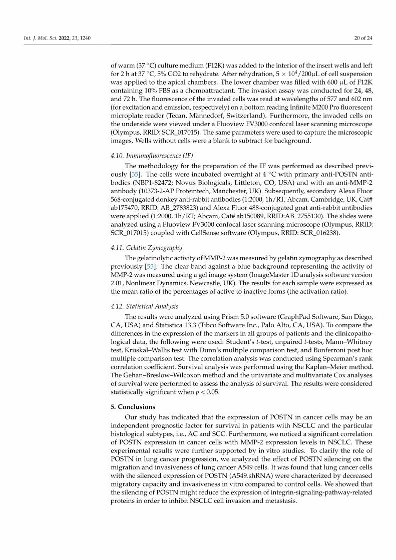
In summary, the results of this study demonstrate the important functional and molecularmechanisms of POSTN in tumor invasion. Silencing of POSTN could inhibit the migrationand in vitro invasive potential of lung cancer cells, most probably via the downregulation ofMMP-2 expression and activity as well as integrin-signaling related proteins. Furthermore, ourdata indicated that epithelial POSTN expression could be an independent negative prognosticfactor in NSCLC and could represent a potential future therapeutic target.
4. Materials and Methods4.1. Patient Cohort
In total, 715 NSCLC and 110 adjacent NMLT samples were collected from patientstreated in the Department of Thoracic Surgery of Wroclaw Medical University between2007 and 2017. The paraffin-embedded specimens included 110 NMLT, 298 AC, 370 SCC,and 47 large cell carcinomas (LCC). The histological tumor type was evaluated based on theWorld Health Organization Classification [51] by two independent pathologists and wasconfirmed by immunohistochemical staining for the marker proteins TTF-1 (AC marker)and p63 (LSCC marker). The pTNM classification was made in accordance with the recom-mendations of the International Association for the Study of Lung Cancer (IASLC) [52].
The experiment was conducted with the ethical standards and the approval of theBioethics Committee of the Wroclaw Medical University.
Laser microdissection was performed on 10 frozen NSCLC fragments and 6 NMLTs asthe control. Clinicopathological patient characteristics are given in Table 2.
4.2. Construction of Tissue Microarray (TMA) and Immunohistochemistry (IHC)
TMAs were constructed as described previously [24]. Briefly, three morphologically rep-resentative tumor cores from the center of the tumor, with a 1.5 mm core size, were assembledinto the TMA using the TMA Grand Master (3DHistech) automatic tissue microarrayer.
To investigate POSTN, MMP-2, TTF-1, and p63 protein expression in TMAs wereanalyzed by IHC, as described before [24]. POSTN was detected using a primary rabbitanti-human polyclonal antibody (NBP1-82472; Novus Biologicals) diluted 1:200, incu-bated for 20 min at RT. MMP-2 was detected using rabbit anti-human MMP-2 antibody(10373-2-AP Proteintech, Manchester, UK) diluted at 1:200, incubated for 20 min at RT.Furthermore, in order to detect TTF-1 and p63 expression, specific primary antibodies wereused: anti-TTF-1(mouse, Dako, Cat# IR056, RRID:AB_2755006; dilution: 1:50, 20 min RT)and anti-p63 (1:300 dilution; Dako, Glostrup, Denmark/Santa Clara, CA, USA, Cat# IR622,RRID:AB_2755007, 20 min RT). IHC reactions were performed using Dako AutostainerLink48 (Dako, Glostrup, Denmark). The visualization of the reactions was carried outusing EnVision™ FLEX High pH (Link) reagents (Dako), in accordance with the manufac-turer’s protocols.
Int. J. Mol. Sci. 2022, 23, 1240 18 of 24
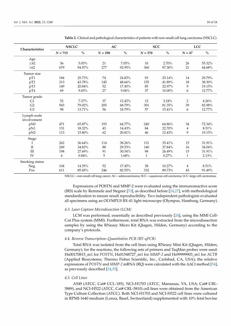
Table 2. Clinical and pathological characteristics of patients with non-small cell lung carcinoma (NSCLC).
CharacteristicsNSCLC AC SCC LCC
N = 715 % N = 298 % N = 370 % N = 47 %
Age≤62 36 5.03% 21 7.05% 10 2.70% 26 55.32%>62 679 94.97% 277 92.95% 360 97.30% 21 44.68%
Tumor sizepT1 184 25.73% 74 24.83% 93 25.14% 14 29.79%pT2 313 43.78% 145 48.66% 155 41.89% 18 38.30%pT3 149 20.84% 52 17.45% 85 22.97% 9 19.15%pT4 69 9.65% 27 9.06% 37 10.00% 6 12.77%
Tumor gradeG1 52 7.27% 37 12.42% 12 3.24% 2 4.26%G2 565 79.02% 205 68.79% 301 81.35% 39 82.98%G3 98 13.71% 56 18.79% 57 15.41% 6 12.77%
Lymph nodeinvolvement
pN0 471 65.87% 193 64.77% 240 64.86% 34 72.34%pN1 131 18.32% 43 14.43% 84 22.70% 4 8.51%pN2 113 15.80% 62 20.81% 46 12.43% 9 19.15%
StageI 262 36.64% 114 38.26% 131 35.41% 15 31.91%II 249 34.83% 88 29.53% 140 37.84% 16 34.04%III 198 27.69% 91 30.54% 98 26.49% 15 31.91%IV 6 0.84% 5 1.68% 1 0.27% 1 2.13%
Smoking statusNeg. 104 14.55% 52 17.45% 38 10.27% 4 8.51%Pos. 611 85.45% 246 82.55% 332 89.73% 43 91.49%
NSCLC—non-small cell lung cancer; AC—adenocarcinoma; SCC—squamous cell carcinoma; LCC–large cell carcinomas.
Expressions of POSTN and MMP-2 were evaluated using the immunoreactive score(IRS) scale by Remmele and Stegner [53], as described before [24,27], with methodologicalstandardization to ensure result reproducibility. Two independent pathologists evaluatedall specimens using an OLYMPUS BX-41 light microscope (Olympus, Hamburg, Germany).
4.3. Laser Capture Microdissection (LCM)
LCM was performed, essentially as described previously [24], using the MMI Cell-Cut Plus system (MMI). Furthermore, total RNA was extracted from the microdissectionsamples by using the RNeasy Micro Kit (Qiagen, Hilden, Germany) according to thecompany’s protocols.
4.4. Reverse Transcription–Quantitative PCR (RT–qPCR)
Total RNA was isolated from the cell lines using RNeasy Mini Kit (Qiagen, Hilden,Germany); for the reactions, the following sets of primers and TaqMan probes were used:Hs00170815_m1 for POSTN, Hs01548727_m1 for MMP-2 and Hs99999903_m1 for ACTB(Applied Biosystems; Thermo Fisher Scientific, Inc., Carlsbad, CA, USA); the relativeexpressions of POSTN and MMP-2 mRNA (RQ) were calculated with the ∆∆Ct method [54],as previously described [24,35].
4.5. Cell Lines
A549 (ATCC, Cat# CCL-185), NCI-H1703 (ATCC, Manassas, VA, USA; Cat# CRL-5889), and NCI-H522 (ATCC, Cat# CRL-5810) cell lines were obtained from the AmericanType Culture Collection (ATCC). Both NCI-H1703 and NCI-H522 cell lines were culturedin RPMI-1640 medium (Lonza, Basel, Switzerland) supplemented with 10% fetal bovine
Int. J. Mol. Sci. 2022, 23, 1240 19 of 24
serum (FBS) (Sigma-Aldrich, Saint Louis, MO, USA) and 2 mM L-glutamine. In turn, A549cells were routinely cultured in F12K supplemented (to final concentration) with 10% FBS,and 2 mM L-glutamine (Lonza, Basel, Switzerland).
LentiX 293T cells were purchased from Clontech Laboratories (Terra Bella Avenue,Mountain View, CA, USA) and cultured in αMEM supplemented with 10% FCS (Invitrogen),2 mM L-glutamine, 100 U/mL streptomycins, and 0.1 mg/mL penicillin (complete αMEM).The conditions of cell culture were as follows: temperature 37 ◦C and 5% CO2 concentration.
4.6. Virus Production, Transductions and Cell Maintenance
For lentivirus production packaging LentiX 293T cells were co-transfected at 50–60%confluence with 20 µg of POSTN MISSION shRNA (TRCN0000123055) expression vector orSHC016 PLKO.1-PURO NON-TARGET (SHC016-1EA) control vector (MISSION®shRNAPlasmid DNA system, Sigma-Aldrich, Saint Louis, MO, USA), 10 µg pMDL-g/p-RRE, 5 µgpRSV-REV, and 5 µg pMk-VSVG (Addgene, Watertown, MA, USA) using polyethyleneimine(Sigma-Aldrich, Saint Louis, MO, USA) at a concentration of 1 mg/mL. Culture super-natants containing virus particles were collected 48 h after transfection and clarified througha 0.45 µm pore size filter (Millipore, Billerica, MA, USA).
The virus-containing supernatant was concentrated 100× on an Amicon Ultra-15K:100.000(Millipore). A549 cells (2 × 10⁴) were transduced with the concentrated virus stock bycentrifuging (2460× g) at 23 ◦C for 2 h. The cells were selected for puromycin resistance(1 µg/mL) for 1 week and maintained in a medium containing 1 µg/mL puromycin.
4.7. Protein Isolation, SDS-PAGE, and Western Blotting
Protein preparation and Western blotting were performed as described previously [24].The following antibodies were used: primary rabbit anti-human POSTN polyclonal anti-body (NBP1-82472; Novus Biologicals, Littleton, CO, USA) diluted at 1:500, rabbit anti-human MMP-2 antibody (10373-2-AP Proteintech, Manchester, UK) diluted at 1:1000, rabbitanti-human αVβ3 antibody (NBP2-67557; Novus Biologicals) diluted at 1:1000, rabbit anti-human pAKT antibody (#4058 Cell Signaling Technology, Danvers, MA, USA), diluted at1:1000, rabbit anti-human AKT antibody (#9272 Cell Signaling Technology) diluted at 1:1000,and mouse anti-human PI3K antibody (MA5-17149, Thermo Fisher Scientific, Carlsbad,CA, USA) diluted at 1:1000. The results were documented in a Chemi-Doc XRS MolecularImager apparatus (Bio-Rad, RRID: SCR_014210). β-actin (Abcam ab8229, Cambridge, UK)was used as a loading control.
4.8. Wound-Healing Assay
We used the ibidi Culture-Insert in µ-Dish35mm, high system (ibidi GmbH, Munich,Germany) with two chambers to separate different cell types in order to measure themigratory properties of A549.shRNA and A549.CTRL cells. For the wound-healing assay,cells were serum-starved for 24 h for cell synchronization. Then, 5.0 × 10⁵ cells wereseeded into each chamber and grown to confluence in F12K medium containing 2% FBS for24 h. After incubation for an additional 48 h, cell migration was analyzed in six differentmicroscopic fields and calculated as the percentage of wound healing. Images of theinitial wound and the movement of cells into the scratched area were captured using aFluoview FV3000 confocal laser scanning microscope (Olympus, Hamburg, Germany, RRID:SCR_017015) coupled with CellSens software version 3.2 (Olympus, RRID: SCR_016238).
4.9. Transwell Invasion Assay
Invasion assays were performed using a BioCoat MatrigelTM invasion chamber (poresize of 8 µm) and an HTS FluoroBlok membrane insert (pore size of 8 µm) for a 24-well plate(Corning, MA, USA), respectively. For the invasion assay, cells were firstly starved usingserum-free media for 24 h. A549.shRNA and A549.CTRL cells were collected by trypsiniza-tion, resuspended in 1 mL PBS at a density of 106/mL, and labeled with CellTracker™RedCMTPX (Invitrogen, Carlsbad, CA, USA) at 37 ◦C for 30 min. For the invasion assay, 200 µL
Int. J. Mol. Sci. 2022, 23, 1240 20 of 24
of warm (37 ◦C) culture medium (F12K) was added to the interior of the insert wells and leftfor 2 h at 37 ◦C, 5% CO2 to rehydrate. After rehydration, 5 × 10⁴/200µL of cell suspensionwas applied to the apical chambers. The lower chamber was filled with 600 µL of F12Kcontaining 10% FBS as a chemoattractant. The invasion assay was conducted for 24, 48,and 72 h. The fluorescence of the invaded cells was read at wavelengths of 577 and 602 nm(for excitation and emission, respectively) on a bottom reading Infinite M200 Pro fluorescentmicroplate reader (Tecan, Männedorf, Switzerland). Furthermore, the invaded cells onthe underside were viewed under a Fluoview FV3000 confocal laser scanning microscope(Olympus, RRID: SCR_017015). The same parameters were used to capture the microscopicimages. Wells without cells were a blank to subtract for background.
4.10. Immunofluorescence (IF)
The methodology for the preparation of the IF was performed as described previ-ously [35]. The cells were incubated overnight at 4 ◦C with primary anti-POSTN anti-bodies (NBP1-82472; Novus Biologicals, Littleton, CO, USA) and with an anti-MMP-2antibody (10373-2-AP Proteintech, Manchester, UK). Subsequently, secondary Alexa Fluor568-conjugated donkey anti-rabbit antibodies (1:2000, 1h/RT; Abcam, Cambridge, UK, Cat#ab175470, RRID: AB_2783823) and Alexa Fluor 488-conjugated goat anti-rabbit antibodieswere applied (1:2000, 1h/RT; Abcam, Cat# ab150089, RRID:AB_2755130). The slides wereanalyzed using a Fluoview FV3000 confocal laser scanning microscope (Olympus, RRID:SCR_017015) coupled with CellSense software (Olympus, RRID: SCR_016238).
4.11. Gelatin Zymography
The gelatinolytic activity of MMP-2 was measured by gelatin zymography as describedpreviously [55]. The clear band against a blue background representing the activity ofMMP-2 was measured using a gel image system (ImageMaster 1D analysis software version2.01, Nonlinear Dynamics, Newcastle, UK). The results for each sample were expressed asthe mean ratio of the percentages of active to inactive forms (the activation ratio).
4.12. Statistical Analysis
The results were analyzed using Prism 5.0 software (GraphPad Software, San Diego,CA, USA) and Statistica 13.3 (Tibco Software Inc., Palo Alto, CA, USA). To compare thedifferences in the expression of the markers in all groups of patients and the clinicopatho-logical data, the following were used: Student’s t-test, unpaired t-tests, Mann–Whitneytest, Kruskal–Wallis test with Dunn’s multiple comparison test, and Bonferroni post hocmultiple comparison test. The correlation analysis was conducted using Spearman’s rankcorrelation coefficient. Survival analysis was performed using the Kaplan–Meier method.The Gehan–Breslow–Wilcoxon method and the univariate and multivariate Cox analysesof survival were performed to assess the analysis of survival. The results were consideredstatistically significant when p < 0.05.
5. Conclusions
Our study has indicated that the expression of POSTN in cancer cells may be anindependent prognostic factor for survival in patients with NSCLC and the particularhistological subtypes, i.e., AC and SCC. Furthermore, we noticed a significant correlationof POSTN expression in cancer cells with MMP-2 expression levels in NSCLC. Theseexperimental results were further supported by in vitro studies. To clarify the role ofPOSTN in lung cancer progression, we analyzed the effect of POSTN silencing on themigration and invasiveness of lung cancer A549 cells. It was found that lung cancer cellswith the silenced expression of POSTN (A549.shRNA) were characterized by decreasedmigratory capacity and invasiveness in vitro compared to control cells. We showed thatthe silencing of POSTN might reduce the expression of integrin-signaling-pathway-relatedproteins in order to inhibit NSCLC cell invasion and metastasis.
Int. J. Mol. Sci. 2022, 23, 1240 21 of 24
Interestingly, our results showed for the first time that POSTN might regulate lungcancer cell invasiveness by modulating the expression and activity level of MMP-2, an en-zyme that degrades the basement membrane and components of the ECM, allowing for theinvasion of tumor cells and proliferation in the metastatic environment (Figure 14). Thesefindings may indicate the potential role of POSTN in the pathogenesis and progressionof NSCLC.
Int. J. Mol. Sci. 2022, 23, 1240 23 of 26
expression of POSTN (A549.shRNA) were characterized by decreased migratory capacity and invasiveness in vitro compared to control cells. We showed that the silencing of POSTN might reduce the expression of integrin-signaling-pathway-related proteins in or-der to inhibit NSCLC cell invasion and metastasis.
Interestingly, our results showed for the first time that POSTN might regulate lung cancer cell invasiveness by modulating the expression and activity level of MMP-2, an enzyme that degrades the basement membrane and components of the ECM, allowing for the invasion of tumor cells and proliferation in the metastatic environment (Figure 14). These findings may indicate the potential role of POSTN in the pathogenesis and progres-sion of NSCLC.
Figure 14. Schema of the signaling pathway involved in the invasion of lung cancer. POSTN might regulate lung cancer cell invasiveness by modulating the expression and activity level of MMP-2, a protein acting as a metastasis-associated factor of the tumor microenvironment, which serves an important role in the degradation of the basement membrane and the invasion of cancer cells. The figure is based on Cui et al. [56], modified.
Author Contributions: Conceptualization, K.R.-W. and P.D.; formal analysis, K.R.-W.; investiga-tion, K.R.-W. and A.K.; methodology, K.R.-W., A.K. and P.D.; validation, K.R.-W. and A.K.; writ-ing—original draft, K.R.-W.; writing—review and editing P.D. All authors have read and agreed to the published version of the manuscript.
Funding: This research received no external funding.
Institutional Review Board Statement: The study was conducted in accordance with the Declara-tion of Helsinki, and approved by the Bioethics Committee of Wroclaw Medical University (decision no. KB-100/2020, 11th February 2020).
Informed Consent Statement: All patients provided written informed consent for the use of the material samples for scientific research.
Data Availability Statement: The raw data and the analytic methods will be made available to other researchers upon reasonable request for the purpose of reproducing the results in their own labor-atories. To access the protocols or datasets, contact [email protected].
Figure 14. Schema of the signaling pathway involved in the invasion of lung cancer. POSTN mightregulate lung cancer cell invasiveness by modulating the expression and activity level of MMP-2,a protein acting as a metastasis-associated factor of the tumor microenvironment, which servesan important role in the degradation of the basement membrane and the invasion of cancer cells.The figure is based on Cui et al. [56], modified.
Author Contributions: Conceptualization, K.R.-W. and P.D.; formal analysis, K.R.-W.; investigation,K.R.-W. and A.K.; methodology, K.R.-W., A.K. and P.D.; validation, K.R.-W. and A.K.; writing—original draft, K.R.-W.; writing—review and editing P.D. All authors have read and agreed to thepublished version of the manuscript.
Funding: This research received no external funding.
Institutional Review Board Statement: The study was conducted in accordance with the Declarationof Helsinki, and approved by the Bioethics Committee of Wroclaw Medical University (decision no.KB-100/2020, 11th February 2020).
Informed Consent Statement: All patients provided written informed consent for the use of thematerial samples for scientific research.
Data Availability Statement: The raw data and the analytic methods will be made available toother researchers upon reasonable request for the purpose of reproducing the results in their ownlaboratories. To access the protocols or datasets, contact [email protected].
Acknowledgments: We gratefully acknowledge our colleagues and collaborators, as follows, for con-tributing to the present work: Aleksandra Piotrowska, Jedrzej Grzegrzołka, Agnieszka Gomulkiewicz,Adrian Wasik of the Division of Histology and Embryology, Department of Human Morphology andEmbryology, Wroclaw Medical University, and Konrad Pawelczyk of the Department of ThoracicSurgery, Wroclaw Medical University.
Int. J. Mol. Sci. 2022, 23, 1240 22 of 24
Conflicts of Interest: The authors declare no conflict of interest.
References1. Hirsch, F.R.; Scagliotti, G.V.; Mulshine, J.L.; Kwon, R.; Curran, W.J.; Wu, Y.L.; Paz-Ares, L. Lung cancer: Current therapies and
new targeted treatments. Lancet 2017, 389, 299–311. [CrossRef]2. Wadowska, K.; Bil-Lula, I.; Trembecki, Ł.; Sliwinska-Mosson, M. Genetic markers in lung cancer diagnosis: A review. Int. J. Mol.
Sci. 2020, 21, 4569. [CrossRef] [PubMed]3. Bray, F.; Ferlay, J.; Soerjomataram, I.; Siegel, R.L.; Torre, L.A.; Jemal, A. Global cancer statistics 2018: GLOBOCAN estimates of
incidence and mortality worldwide for 36 cancers in 185 countries. CA Cancer J. Clin. 2018, 68, 394–424. [CrossRef]4. Łaczmanska, I.; Debicka, I.; Gil, J.; Michałowska, D.; Pawlak, I.; Sasiadek, M.M. Personalised medicine in lung cancer. Nowotwory
2021, 71, 122–128. [CrossRef]5. Cooper, W.A.; Lam, D.C.L.; O’Toole, S.A.; Minna, J.D. Molecular biology of lung cancer. J. Thorac. Dis. 2013, 5. [CrossRef]6. Wang, Y.; Wang, H.; Pan, T.; Li, L.; Li, J.; Yang, H. STIM1 silencing inhibits the migration and invasion of A549 cells. Mol. Med.
Rep. 2017, 16, 3283–3289. [CrossRef]7. Han, L.; Sheng, B.; Zeng, Q.; Yao, W.; Jiang, Q. Correlation between MMP2 expression in lung cancer tissues and clinical
parameters: A retrospective clinical analysis. BMC Pulm. Med. 2020, 20, 1–9. [CrossRef]8. Hadler-Olsen, E.; Winberg, J.O.; Uhlin-Hansen, L. Matrix metalloproteinases in cancer: Their value as diagnostic and prognostic
markers and therapeutic targets. Tumor Biol. 2013, 34, 2041–2051. [CrossRef]9. Kii, I.; Nishiyama, T.; Li, M.; Matsumoto, K.; Saito, M.; Amizuka, N.; Kudo, A. Incorporation of Tenascin-C into the Extracellular
Matrix by Periostin Underlies an Extracellular Meshwork Architecture. J. Biol. Chem. 2010, 285, 2028–2039. [CrossRef]10. Hoersch, S.; Andrade-Navarro, M.A. Periostin shows increased evolutionary plasticity in its alternatively spliced region. BMC
Evol. Biol. 2010, 10, 1–19. [CrossRef]11. González-González, L.; Alonso, J. Periostin: A matricellular protein with multiple functions in cancer development and
progression. Front. Oncol. 2018, 8, 1–15. [CrossRef] [PubMed]12. Norris, R.A.; Damon, B.; Mironov, V.; Kasyanov, V.; Ramamurthi, A.; Moreno-Rodriguez, R.; Trusk, T.; Potts, J.D.; Goodwin, R.L.;
Davis, J.; et al. Periostin regulates collagen fibrillogenesis and the biomechanical properties of connective tissues. J. Cell. Biochem.2007, 101, 695–711. [CrossRef] [PubMed]
13. Hamilton, D.W. Functional role of periostin in development and wound repair: Implications for connective tissue disease. J. CellCommun. Signal. 2008, 2, 9–17. [CrossRef] [PubMed]
14. Ruan, K.; Bao, S.; Ouyang, G. The multifaceted role of periostin in tumorigenesis. Cell. Mol. Life Sci. 2009, 66, 2219–2230.[CrossRef]
15. Zhou, Y.; Yang, W.; Ao, M.; Höti, N.; Gabrielson, E.; Chan, D.W.; Zhang, H.; Li, Q.K. Proteomic Analysis of the Air-Way Fluid inLung Cancer. Detection of Periostin in Bronchoalveolar Lavage (BAL). Front. Oncol. 2020, 10, 1072. [CrossRef]
16. Che, J.; Shen, W.Z.; Deng, Y.; Dai, Y.H.; Liao, Y.-D.; Yuan, X.L.; Zhang, P. Effects of lentivirus-mediated silencing of Periostinon tumor microenvironment and bone metastasis via the integrin-signaling pathway in lung cancer. Life Sci. 2017, 182, 10–21.[CrossRef]
17. Morra, L.; Moch, H. Periostin expression and epithelial-mesenchymal transition in cancer: A review and an update. VirchowsArch. 2011, 459, 465–475. [CrossRef]
18. Murakami, D.; Takamori, S.; Kawahara, A.; Mitsuoka, M.; Kashihara, M.; Yoshiyama, K.; Matsumoto, R.; Yokoyama, S.; Fujimoto,K.; Kawaguchi, A.; et al. Periostin expression in non-small cell lung cancer: Clinical significance. Kurume Med. J. 2017, 64, 13–20.[CrossRef]
19. Bao, S.; Ouyang, G.; Bai, X.; Huang, Z.; Ma, C.; Liu, M.; Shao, R.; Anderson, R.M.; Rich, J.N.; Wang, X.F. Periostin potentlypromotes metastatic growth of colon cancer by augmenting cell survival via the Akt/PKB pathway. Cancer Cell 2004, 5, 329–339.[CrossRef]
20. Shao, R.; Bao, S.; Bai, X.; Blanchette, C.; Anderson, R.M.; Dang, T.; Gishizky, M.L.; Marks, J.R.; Wang, X.-F. Acquired Expressionof Periostin by Human Breast Cancers Promotes Tumor Angiogenesis through Up-Regulation of Vascular Endothelial GrowthFactor Receptor 2 Expression. Mol. Cell. Biol. 2004, 24, 3992–4003. [CrossRef]
21. Kim, C.J.; Yoshioka, N.; Tambe, Y.; Kushima, R.; Okada, Y.; Inoue, H. Periostin is down-regulated in high grade human bladdercancers and suppresses in vitro cell invasiveness and in vivo metastasis of cancer cells. Int. J. Cancer 2005, 117, 51–58. [CrossRef][PubMed]
22. Kanno, A.; Satoh, K.; Masamune, A.; Hirota, M.; Kimura, K.; Umino, J.; Hamada, S.; Satoh, A.; Egawa, S.; Motoi, F.; et al. Periostin,secreted from stromal cells, has biphasic effect on cell migration and correlates with the epithelial to mesenchymal transition ofhuman pancreatic cancer cells. Int. J. Cancer 2008, 122, 2707–2718. [CrossRef] [PubMed]
23. Ye, D.; Zhou, C.; Wang, S.; Deng, H.; Shen, Z. Tumor suppression effect of targeting periostin with siRNA in a nude mouse modelof human laryngeal squamous cell carcinoma. J. Clin. Lab. Anal. 2019, 33, e22622. [CrossRef] [PubMed]
24. Ratajczak-Wielgomas, K.; Kmiecik, A.; Grzegrzołka, J.; Piotrowska, A.; Gomulkiewicz, A.; Partynska, A.; Pawelczyk, K.;Nowinska, K.; Podhorska-Okolow, M.; Dziegiel, P. Prognostic significance of stromal periostin expression in non-small cell lungcancer. Int. J. Mol. Sci. 2020, 21, 7025. [CrossRef]
Int. J. Mol. Sci. 2022, 23, 1240 23 of 24
25. Liu, Y.; Huang, Z.; Cui, D.; Ouyang, G. The Multiaspect Functions of Periostin in Tumor Progression. In Advances in ExperimentalMedicine and Biology; Springer: New York, NY, USA, 2019; Volume 1132, pp. 125–136.
26. Hong, L.; Sun, H.; Lv, X.; Yang, D.; Zhang, J.; Shi, Y. Expression of periostin in the serum of NSCLC and its function on proliferationand migration of human lung adenocarcinoma cell line (A549) in vitro. Mol. Biol. Rep. 2010, 37, 2285–2293. [CrossRef]
27. Ratajczak-Wielgomas, K.; Grzegrzolka, J.; Piotrowska, A.; Gomulkiewicz, A.; Witkiewicz, W.; Dziegiel, P. Periostin expression incancer-associated fibroblasts of invasive ductal breast carcinoma. Oncol. Rep. 2016, 36, 2745–2754. [CrossRef]
28. Baril, P.; Gangeswaran, R.; Mahon, P.C.; Caulee, K.; Kocher, H.M.; Harada, T.; Zhu, M.; Kalthoff, H.; Crnogorac-Jurcevic, T.;Lemoine, N.R. Periostin promotes invasiveness and resistance of pancreatic cancer cells to hypoxia-induced cell death: Role ofthe β4 integrin and the PI3k pathway. Oncogene 2007, 26, 2082–2094. [CrossRef]
29. Gillan, L.; Matei, D.; Fishman, D.A.; Gerbin, C.S.; Karlan, B.Y.; Chang, D.D. Periostin Secreted by Epithelial Ovarian Carcinoma Isa Ligand for V 3 and V 5 Integrins and Promotes Cell Motility. Cancer Res. 2002, 62, 5358–5364.
30. Yoshioka, N.; Fuji, S.; Shimakage, M.; Kodama, K.; Hakura, A.; Yutsudo, M.; Inoue, H.; Nojima, H. Suppression of anchorage-independent growth of human cancer cell lines by the TRIF52/periostin/OSF-2 gene. Exp. Cell Res. 2002, 279, 91–99. [CrossRef]
31. Sun, C.; Zhao, X.; Xu, K.; Gong, J.; Liu, W.; Ding, W.; Gou, Y.; Xia, G.; Ding, Q. Periostin: A promising target of therapeuticalintervention for prostate cancer. J. Transl. Med. 2011, 9, 1–10. [CrossRef]
32. Blanco-Prieto, S.; Barcia-Castro, L.; Páez de la Cadena, M.; Rodríguez-Berrocal, F.J.; Vázquez-Iglesias, L.; Botana-Rial, M.I.;Fernández-Villar, A.; De Chiara, L. Relevance of matrix metalloproteases in non-small cell lung cancer diagnosis. BMC Cancer2017, 17, 1–8. [CrossRef] [PubMed]
33. Chuanyu, S.; Yuqing, Z.; Chong, X.; Guowei, X.; Xiaojun, Z. Periostin promotes migration and invasion of renal cell carcinomathrough the integrin/focal adhesion kinase/c-Jun N-terminal kinase pathway. Tumor Biol. 2017, 39, 1010428317694549. [CrossRef]
34. Egeblad, M.; Werb, Z. New functions for the matrix metalloproteinases in cancer progression. Nat. Rev. Cancer 2002, 2, 161–174.[CrossRef] [PubMed]
35. Ratajczak-Wielgomas, K.; Grzegrzolka, J.; Piotrowska, A.; Matkowski, R.; Wojnar, A.; Rys, J.; Ugorski, M.; Dziegiel, P. Expressionof periostin in breast cancer cells. Int. J. Oncol. 2017, 51, 1300–1310. [CrossRef]
36. Liu, A.Y.; Zheng, H.; Ouyang, G. Periostin, a multifunctional matricellular protein in inflammatory and tumor microenvironments.Matrix Biol. 2014, 37, 150–156. [CrossRef]
37. Kudo, Y.; Siriwardena, B.S.M.S.; Hatano, H.; Ogawa, I.; Takata, T. Periostin: Novel diagnostic and therapeutic target for cancer.Histol. Histopathol. 2007, 22, 1167–1174. [CrossRef]
38. Orecchia, P.; Conte, R.; Balza, E.; Castellani, P.; Borsi, L.; Zardi, L.; Mingari, M.C.; Carnemolla, B. Identification of a novel cellbinding site of periostin involved in tumour growth. Eur. J. Cancer 2011, 47, 2221–2229. [CrossRef]
39. Soltermann, A.; Tischler, V.; Arbogast, S.; Braun, J.; Probst-Hensch, N.; Weder, W.; Moch, H.; Kristiansen, G. Prognostic Significanceof Epithelial-Mesenchymal and Mesenchymal-Epithelial Transition Protein Expression in Non-Small Cell Lung Cancer. Clin.Cancer Res. 2008, 14, 7430–7437. [CrossRef]
40. Zhu, M.; Fejzo, M.S.; Anderson, L.; Dering, J.; Ginther, C.; Ramos, L.; Gasson, J.C.; Karlan, B.Y.; Slamon, D.J. Periostin promotesovarian cancer angiogenesis and metastasis. Gynecol. Oncol. 2010, 119, 337–344. [CrossRef]
41. Morra, L.; Rechsteiner, M.; Casagrande, S.; Duc Luu, V.; Santimaria, R.; Diener, P.A.; Sulser, T.; Kristiansen, G.; Schraml, P.; Moch,H.; et al. Relevance of periostin splice variants in renal cell carcinoma. Am. J. Pathol. 2011, 179, 1513–1521. [CrossRef]
42. Ben, Q.W.; Jin, X.L.; Jun, L.; Cai, X.; Yuan, F.; Yuan, Y.Z. Periostin, a matrix specific protein, is associated with proliferation andinvasion of pancreatic cancer. Oncol. Rep. 2011, 25, 709–716. [CrossRef] [PubMed]
43. Riener, M.-O.; Fritzsche, F.R.; Soll, C.; Pestalozzi, B.C.; Probst-Hensch, N.; Clavien, P.-A.; Jochum, W.; Soltermann, A.; Moch, H.;Kristiansen, G. Expression of the extracellular matrix protein periostin in liver tumours and bile duct carcinomas. Histopathology2010, 56, 600–606. [CrossRef] [PubMed]
44. Wang, W.; Sun, Q.K.; He, Y.F.; Ma, D.C.; Xie, M.R.; Ji, C.S.; Hu, B. Overexpression of periostin is significantly correlated to thetumor angiogenesis and poor prognosis in patients with esophageal squamous cell carcinoma. Int. J. Clin. Exp. Pathol. 2014, 7,593–601. [PubMed]
45. Wang, H.; Wang, Y.; Jiang, C. Stromal protein periostin identified as a progression associated and prognostic biomarker in gliomavia inducing an invasive and proliferative phenotype. Int. J. Oncol. 2013, 42, 1716–1724. [CrossRef] [PubMed]
46. Shih, Y.W.; Chien, S.T.; Chen, P.S.; Lee, J.H.; Wu, S.H.; Yin, L. Te α-Mangostin Suppresses Phorbol 12-myristate 13-acetate-inducedMMP-2/MMP-9 Expressions via αvβ3 Integrin/FAK/ERK and NF-κB Signaling Pathway in Human Lung AdenocarcinomaA549 Cells. Cell Biochem. Biophys. 2010, 58, 31–44. [CrossRef] [PubMed]
47. Watanabe, T.; Yasue, A.; Fujihara, S.; Tanaka, E. PERIOSTIN regulates MMP-2 expression via the αvβ3 integrin/ERK pathway inhuman periodontal ligament cells. Arch. Oral Biol. 2012, 57, 52–59. [CrossRef]
48. Yan, W.; Shao, R. Transduction of a mesenchyme-specific gene periostin into 293T cells induces cell invasive activity throughepithelial-mesenchymal transformation. J. Biol. Chem. 2006, 281, 19700–19709. [CrossRef]
49. Burgess, J.K.; Jonker, M.R.; Berg, M.; Hacken, N.T.H.; Meyer, K.B.; van den Berge, M.; Nawijn, M.C.; Heijink, I.H. Early ViewOriginal article Periostin: Contributor to abnormal airway epithelial function in asthma? Eur. Respir. J. 2021, 57, 2001286.[CrossRef]
50. Ouanouki, A.; Lamy, S.; Annabi, B. Periostin, a signal transduction intermediate in TGF-β-induced EMT in U-87MG humanglioblastoma cells, and its inhibition by anthocyanidins. Oncotarget 2018, 9, 22023–22037. [CrossRef]

Int. J. Mol. Sci. 2022, 23, 1240 24 of 24
51. Travis, W.D.; Brambilla, E.; Nicholson, A.G.; Yatabe, Y.; Austin, J.H.M.; Beasley, M.B.; Chirieac, L.R.; Dacic, S.; Duhig, E.;Flieder, D.B.; et al. The 2015 World Health Organization Classification of Lung Tumors: Impact of Genetic, Clinical and RadiologicAdvances since the 2004 Classification. J. Thorac. Oncol. 2015, 10, 1243–1260. [CrossRef]
52. Detterbeck, F.C.; Boffa, D.J.; Kim, A.W.; Tanoue, L.T. The Eighth Edition Lung Cancer Stage Classification. Chest 2017, 151, 193–203.[CrossRef]
53. Remmele, W. Recommendation for uniform definition of an immunoreactive score (IRS) for immunohistochemical estrogenreceptor detection (ER-ICA) in breast cancer tissue. Pathologe 1987, 8, 138–140. [PubMed]
54. Livak, K.J.; Schmittgen, T.D. Analysis of relative gene expression data using real-time quantitative PCR and the 2(-Delta DeltaC(T)) Method. Methods 2001, 25, 402–408. [CrossRef] [PubMed]
55. Ratajczak-Wielgomas, K.; Gosk, J.; Rabczynski, J.; Augoff, K.; Podhorska-Okołów, M.; Gamian, A.; Rutowski, R. Expression ofMMP-2, TIMP-2, TGF-β1, and Decorin in Dupuytren’s Contracture. Connect. Tissue Res. 2012, 53, 469–477. [CrossRef] [PubMed]
56. Cui, J.; Dean, D.; Hornicek, F.J.; Chen, Z.; Duan, Z. The role of extracelluar matrix in osteosarcoma progression and metastasis. J.Exp. Clin. Cancer Res. 2020, 39, 1–11. [CrossRef] [PubMed]
Related Documents