September 2011 Vol. 7 No. 3 Int J Biomed Sci www.ijbs.org 230 INTERNATIONAL JOURNAL of BIOMEDICAL SCIENCE Gene Encoding Chitinase 3-Like 1 Protein (CHI3L1) is a Putative Oncogene Vadym M. Kavsan 1 , Vladimir P. Baklaushev 2 , Olena V. Balynska 1 , Anton V. Iershov 1 , Pavlo O. Areshkov 1 , Gaukhar M. Yusubalieva 2 , Nadezhda Ph. Grinenko 2 , Ilya V. Victorov 2 , Vadym I. Rymar 1 , Marc Sanson 4 , Vladimir P. Chekhonin 2, 3 1 Department of Biosynthesis of Nucleic Acids, Institute of Molecular Biology and Genetics, National Academy of Sciences of Ukraine, 150 Zabolotnogo Street, Kyiv, Ukraine; 2 Laboratory of Immunochemistry, V.P. Serbsky National Research Centre for Social and Forensic Psychiatry, RUSA Ministry of Health, Moscow, Russia; 3 Chair of Medical Nanobiotechnology N.I. Pirogov Russian State Medical University, Moscow, Russia; 4 AP-HP, Groupe Hospitalier Pitié-Salpêtrière, Service de Neurologie Mazarin and UMR 975 INSERM-UPMC, Paris, France ABSTRACT An important task in understanding oncogenesis is the identification of those genes whose copy number and expression increase during tumorigenesis. Previously, in an effort to identify genes which could be used as molecular markers for glial tumors, we compared gene expression in glioblastoma to the normal brain cells. Among the genes with the most pronounced increased expression in tumors there was CHI3L1, encod- ing the secreted chitinase 3-like 1 protein (also known as HC gp-39 or YKL-40). Expression of CHI3L1 was found increased significantly in various tumors in comparison with corresponding normal tissues. Here we show that CHI3L1 can decrease the doubling time of 293 cells. We have also demonstrated that CHI3L1 allows the anchorage-independent growth in soft agar and, in addition, stable CHI3L1 expression made 293 cells tumorigenic: these cells stimulate the initiation of tumors after their xenograft transplantation into the Wistar rat brains. Thus, the overexpression of CHI3L1 is likely to be critical in the development of some tumors and when we gain more information about mechanisms of CHI3L1 oncogenicity, it could be used as one of the potential targets for anticancer therapy. (Int J Biomed Sci 2011; 7 (3): 230-237) Keywords: glioblastoma; chitinase 3-like 1 protein (CHI3L1); oncogene Corresponding author: Vadym Kavsan, Department of Biosynthesis of Nucleic Acids, Institute of Molecular Biology and Genetics, National Academy of Sciences of Ukraine, 150 Zabolotnogo Street, Kyiv, Ukraine. E-mail: [email protected]. Received May 3, 2011; Accepted May 30, 2011 INTRODUCTION Previously, in an effort to identify genes which could be used as molecular markers for glial tumors, we com- pared gene expression in glioblastoma, the most aggres- sive human brain tumor, to the normal brain cells and revealed 44 genes with more than 5-fold higher expres- sion level in tumors. One of such genes overexpressed in glioblastoma was CHI3L1 , encoding the secreted chitinase 3-like 1 protein (1-3). The expression of CHI3L1 gene was found in synovial cells, articular cartilage chondrocytes and increased significantly in various tumors, and cell lines derived from such tumors, including tumors of the bone, brain, breast, lung, and ovary (4). CHI3L1 is a member of the 18 glycosyl hydrolase fam- ily mapped at 1q32 genomic region, which is frequently amplified in different human cancers; amplifications were ORIGINAL ARTICLE

Welcome message from author
This document is posted to help you gain knowledge. Please leave a comment to let me know what you think about it! Share it to your friends and learn new things together.
Transcript
September 2011 Vol. 7 No. 3 Int J Biomed Sci www.ijbs.org 230
InternatIonal journal of BIomedIcal scIence
Gene Encoding Chitinase 3-Like 1 Protein (CHI3L1) is a Putative Oncogene
Vadym M. Kavsan1, Vladimir P. Baklaushev2, Olena V. Balynska1, Anton V. Iershov1, Pavlo O. Areshkov1, Gaukhar M. Yusubalieva2, Nadezhda Ph. Grinenko2, Ilya V. Victorov2,
Vadym I. Rymar1, Marc Sanson4, Vladimir P. Chekhonin2, 3
1Department of Biosynthesis of Nucleic Acids, Institute of Molecular Biology and Genetics, National Academy of Sciences of Ukraine, 150 Zabolotnogo Street, Kyiv, Ukraine; 2Laboratory of Immunochemistry, V.P. Serbsky National Research Centre for Social and Forensic Psychiatry, RUSA Ministry of Health, Moscow, Russia; 3Chair of Medical Nanobiotechnology N.I. Pirogov
Russian State Medical University, Moscow, Russia; 4AP-HP, Groupe Hospitalier Pitié-Salpêtrière, Service de Neurologie Mazarin and UMR 975 INSERM-UPMC, Paris, France
AbstrAct
An important task in understanding oncogenesis is the identification of those genes whose copy number and expression increase during tumorigenesis. Previously, in an effort to identify genes which could be used as molecular markers for glial tumors, we compared gene expression in glioblastoma to the normal brain cells. Among the genes with the most pronounced increased expression in tumors there was CHI3L1, encod-ing the secreted chitinase 3-like 1 protein (also known as HC gp-39 or YKL-40). Expression of CHI3L1 was found increased significantly in various tumors in comparison with corresponding normal tissues. Here we show that CHI3L1 can decrease the doubling time of 293 cells. We have also demonstrated that CHI3L1 allows the anchorage-independent growth in soft agar and, in addition, stable CHI3L1 expression made 293 cells tumorigenic: these cells stimulate the initiation of tumors after their xenograft transplantation into the Wistar rat brains. Thus, the overexpression of CHI3L1 is likely to be critical in the development of some tumors and when we gain more information about mechanisms of CHI3L1 oncogenicity, it could be used as one of the potential targets for anticancer therapy. (Int J Biomed Sci 2011; 7 (3): 230-237)
Keywords: glioblastoma; chitinase 3-like 1 protein (CHI3L1); oncogene
Corresponding author: Vadym Kavsan, Department of Biosynthesis of Nucleic Acids, Institute of Molecular Biology and Genetics, National Academy of Sciences of Ukraine, 150 Zabolotnogo Street, Kyiv, Ukraine. E-mail: [email protected]. Received May 3, 2011; Accepted May 30, 2011
INTRODUCTION
Previously, in an effort to identify genes which could be used as molecular markers for glial tumors, we com-pared gene expression in glioblastoma, the most aggres-
sive human brain tumor, to the normal brain cells and revealed 44 genes with more than 5-fold higher expres-sion level in tumors. One of such genes overexpressed in glioblastoma was CHI3L1, encoding the secreted chitinase 3-like 1 protein (1-3). The expression of CHI3L1 gene was found in synovial cells, articular cartilage chondrocytes and increased significantly in various tumors, and cell lines derived from such tumors, including tumors of the bone, brain, breast, lung, and ovary (4).
CHI3L1 is a member of the 18 glycosyl hydrolase fam-ily mapped at 1q32 genomic region, which is frequently amplified in different human cancers; amplifications were
ORIGINAL ARTICLE

Gene encodInG chItInase 3-lIke 1 proteIn (chI3l1) Is a putatIve oncoGene
www.ijbs.org Int J Biomed Sci Vol. 7 No. 3 September 2011 231
documented for glioma, esophageal squamous cell carci-noma, retinoblastoma (5-7). 1q32 locus contains about 160 protein-coding genes (http://www.ncbi.nlm.nih.gov/gene). Among them, mouse double minute-4 p53 binding homolog (MDM4) and contactin 2 (CNTN2) are amplified in a subset of malignant gliomas without TP53 mutation or MDM2 am-plification (5), SMYD2 is amplified in esophageal squamous cell carcinoma (6), and KIF14 is amplified in breast cancer (7). However, copy number profiling for human glioma does not show CHI3L1 gene amplification (8). It was reported that polymorphism in the promoter region of CHI3L1 dis-rupts MYC/MAX binding site (9), which may lead to de-creased expression. However, our recent studies revealed no correlation between this polymorphism and gene expres-sion in glioblastoma (10). Thus, it is possible that CHI3L1 overexpression is explained by some other mechanisms, for example, by presence of distant regulators affected by am-plification in this or other types of tumors.
It was demonstrated that CHI3L1 has growth promot-ing activity and can initiate mitogen-activated kinase (MAPK) and phosphoinositide 3-kinase (PI-3K) signaling cascades in human connective tissue cells, leading to the phosphorylation of extracellular signal-regulated kinase 1 and 2 (ERK1/2) and AKT, respectively (11). In another study, the stimulation of human articular chondrocytes or skin fibroblasts with interleukin-1 or tumor necrosis factor alpha (TNFα) in the presence of CHI3L1 protein reduced the phosphorylation of both p38 and JNK1/2 (12). CHI3L1 expression had a positive association with the expression of phosphorylation of ERK1/2, which was strongly cor-related with a poor response to radiotherapy and a nega-tive clinical outcome (13). It was detected an obvious de-crease in phospho-ERK1/2 in CHI3L1 siRNA-transfected U87 cells accompanied by reduced cell proliferation after CHI3L1 silencing (14).
In the current study we demonstrate the oncogenic properties of CHI3L1 gene: its stable expression allows anchorage-independent cell growth in soft agar, decreases the doubling time of 293 cells and makes 293 cells tumori-genic being xenografted into the Wistar rat brains.
METHODS
cells293 cells (Human Embryonic Kidney 293 cells, also
often referred to HEK 293, or less precisely as HEK cells) were kindly provided by Prof. V. Filonenko; 293 cells, sta-bly transfected with pcDNA3.1 and pcDNA3.1/GFP were kindly provided by Dr. V. Grishko (Department of Cell
Signaling, Institute of Molecular Biology and Genetics, Kyiv, Ukraine). Cells were grown in Dulbecco’s modified Eagle’s medium (DMEM) (Sigma, San Diego, CA, USA) supplemented with 10% fetal bovine serum (FBS) (Sigma) and 100 µg/ml penicillin/100 u/ml streptomycin (Sigma) in an environment of 95% air/5% CO2.
Gene expression in gliomaGene expression data for 225 glioblastoma and 74 nor-
mal brain samples were obtained from the Gene Expres-sion Omnibus (GEO) repository (http://www.ncbi.nlm.nih.gov/geo). To compare data from different experiments we used normalization method proposed for real-time poly-merase chain reaction (PCR) (15) and normalized all data by dividing expression level of every gene on geometric average of three housekeeping genes: β-actin (ACTB), glyceraldehyde-3-phosphate dehydrogenase (GAPDH), and TATA-box binding protein (TBP). Comparison be-tween two groups was done by using the independent samples t test. A value of P<0.05 was considered as statis-tically significant.
Proteins and antibodiesNative CHI3L1 was purified from supernatant of con-
ditioned MG-63 cell medium as described by Harvey et al. (16).
Goat polyclonal antibodies against human CHI3L1 (S-18) were purchased from Santa Cruz Biotechnology (Santa Cruz, CA, USA), horse-radish peroxidase anti-goat im-munoglobulin conjugates used in Western blot, goat anti-rabbit Alexa Fluor 488 and rabbit anti-goat Alexa Fluor 488 antibodies used in immunofluorescence were from Invit-rogen (Carlsbad, CA, USA).
Immunofluorescence and confocal microscopy293 cells were seeded on coverslips and allowed to grow
to near-confluence. The cells were washed in cold phos-phate-buffered saline (PBS), fixed with 3.7% paraformal-dehyde, washed three times for 5 min each with PBS and blocked with 5% horse serum (Sigma) in PBS (blocking buffer) for 30 min at room temperature. Incubations were performed at room temperature with antibodies diluted in blocking buffer. Slides were mounted using PVA-DABCO (Sigma) and images were captured with a Zeiss LSM 510 Meta confocal microscope (Hamburg, Germany).
Cell proliferation assay of CHI3L1-treated 293 cells Cell count was performed to assay the cell growth
curve in CHI3L1-treated 293 cells. Briefly, an equal num-
Gene encodInG chItInase 3-lIke 1 proteIn (chI3l1) Is a putatIve oncoGene
September 2011 Vol. 7 No. 3 Int J Biomed Sci www.ijbs.org 232
ber of cells (5 × 10⁴) were seeded in 6-well cultural plates in DMEM containing 10% FBS, and then starved for 1 day, before being treated with CHI3L1 (100 ng/ml). The cells were then washed and trypsinized, and the number of cells was assessed using a hemocytometer (Neubauer Improved, Marienfeld, Germany). The data are expressed
as the mean ± SEM for three independent experiments.
Generation of 293 cell line, stably expressing CHI3L1The 1.0-kb Hind III/Bam HI fragment of CHI3L1
cDNA was ligated to the Bam HI/Hind III site of the pcDNA3.1(+)/neomycin mammalian expression vector (Invitrogen). To check the transfection effectiveness, af-ter 1 day of seeding, 293 cells were transfected by 5 µg of pcDNA3.1_CHI3L1 using 8 μl jetPEI (Polyplus, New York, NY, USA) per 60-mm dish. CHI3L1 levels were vi-sualized by Western blot and immunofluorescense.
To obtain a cell line stably producing CHI3L1 protein, 293 cells were transfected by 5 µg pcDNA3.1_CHI3L1 and 8 μl jetPEI per 60-mm dish. 0.8 mg/ml geneticin G418 sul-phate (Sigma) was used to select transfectants after 3 weeks.
Cell proliferation assay of 293 cells, stably expressing CHI3L1
293 cells stably expressing CHI3L1, and 293 cells, sta-bly transfected with pcDNA3.1 were seeded in quadru-plicates in 96 well plate at density 1 × 10³ cells/well and grown in DMEM, supplemented with 10% FBS, geneticin G418 sulphate and 100 μg/ml penicillin/100 u/ml strepto-mycin for 7 days. Cell proliferation was measured using MTT (Sigma) at days 1, 2, 3, 5, 6 and 7 of the seeding.
Soft agar colony formation assayFor soft agar assay, we placed 1 × 10⁴ 293 cells, sta-
bly expressing CHI3L1, 293 cells, stably transfected with pcDNA3.1 or 293 cells, stably expressing GFP in 2 ml of 0.35% low gelling temperature agarose (Gibco, New York, NY, USA) in DMEM supplemented with 10% FBS and overlaid them on 2 ml of 0.8% agarose/10%FBS/DMEM in a 35-mm diameter dish. We grew cells at 37ºC for 21 days to allow colony formation. Colonies were visualized by staining with 0.005 % crystal violet. The experiment was performed three times.
Xenograft modelsAll procedures were conducted in accordance with in-
stitutional regulations for use of laboratory animals. Adult female Wistar rats (200-220 g, n=10) were used as recipients for 293 cells, stably transfected with pcDNA3.1 or 293 cells
stably expressing CHI3L1. 4 × 10⁵ cells were injected stereo-tactically under ketamine anesthesia (Narishige Stereotactic Apparatus, Japan) into the caudoputamen at the following stereotactic coordinates: Ap −1, L 3.0, V 4.5, and TBS −2.4 mm according to the Rat Brain Atlas. The cells were injected with a Hamilton microsyringe connected to an infusomat at the rate of 3 μl/min (5-10 μl). Animals were killed at 21 day postinjection. Tumor volume (V) was estimated from the length (l) and width (w) of the tumor using the formula: V = (π/6) × ((l + w)/2)3. Tumors were fixed in formalin overnight at 4°C and embedded in paraffin. Tissue sections were de-waxed and stained with hematoxylin and eosin.
RESULTS
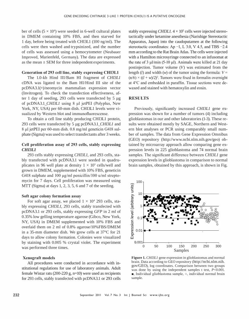
Previously, significantly increased CHI3L1 gene ex-pression was shown for a number of tumors (4) including glioblastomas in our and other laboratories (1-3). These re-sults were obtained mostly by SAGE, Northern and West-ern blot analyses or PCR using comparably small num-ber of samples. The data from Gene Expression Omnibus (GEO) repository (http://www.ncbi.nlm.nih.gov/geo) ob-tained by microarray approach allow comparing gene ex-pression levels in 225 glioblastoma and 74 normal brain samples. The significant difference between CHI3L1 gene expression levels in glioblastoma in comparison to normal brain samples, obtained by this approach, is shown in Fig.
Expr
essi
on le
vel (
arbi
trary
uni
ts)
Samples
0.01
0.1
1
10
100
0.0010 50 100 150 200 250 300
Figure 1. CHI3L1 gene expression in glioblastomas and normal brain. Data according to GEO repository (http://ncbi.nlm.nih.gov/GEO), log coordinates. Comparison between two groups was done by using the independent samples t test, P<0.001. ■, Individual glioblastoma sample, ○, individual normal brain sample.
Gene encodInG chItInase 3-lIke 1 proteIn (chI3l1) Is a putatIve oncoGene
www.ijbs.org Int J Biomed Sci Vol. 7 No. 3 September 2011 233
1. In the same time, it is possible to notice the significant overlap in the levels in individual samples of tumor and normal brain. As it was shown recently, CHI3L1 expres-sion is absent in subset of glioblastomas termed “proneu-ral”, which comprise up to 30% of all glioblastomas, due to promoter hypermethylation (17).
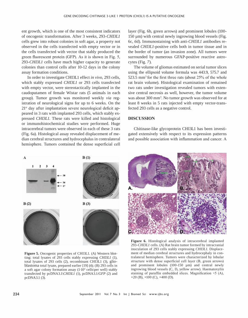
Proliferative response of cells on treatment by CHI3L1 was studied with 293 cell line (Human Embryonic Kidney 293 cells, also often referred to as HEK 293 cells), as these cells do not produce CHI3L1 under any culture conditions. Obtained results are shown in Fig. 2, demonstrating in-creased cell number in culture maintained in the presence of CHI3L1 compared with cells maintained in unsupple-mented medium.
Given that CHI3L1 is overexpressed in many hu-man cancer cells, we addressed the question of whether CHI3L1 possesses transforming properties on 293 cells, one of the most common cell types used in molecular bi-ology research. To this end, we used the expression con-struct pcDNA3.1/CHI3L1 under the control of cytomegal-ovirus (CMV) promoter to establish the 293-CHI3L1 cell line from parental 293 cells. Overexpression of CHI3L1 on the protein level in this cell line was confirmed by immu-nofluorescence and Western blot analysis.
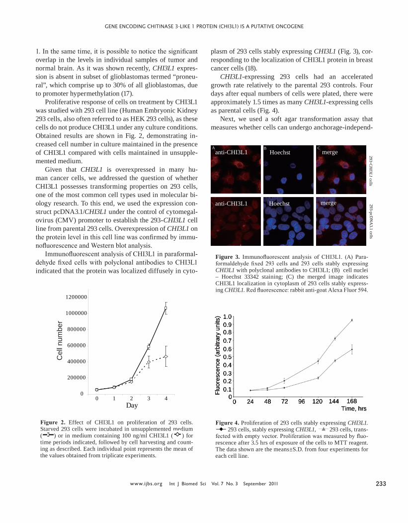
Immunofluorescent analysis of CHI3L1 in paraformal-dehyde fixed cells with polyclonal antibodies to CHI3L1 indicated that the protein was localized diffusely in cyto-
plasm of 293 cells stably expressing CHI3L1 (Fig. 3), cor-responding to the localization of CHI3L1 protein in breast cancer cells (18).
CHI3L1-expressing 293 cells had an accelerated growth rate relatively to the parental 293 controls. Four days after equal numbers of cells were plated, there were approximately 1.5 times as many CHI3L1-expressing cells as parental cells (Fig. 4).
Next, we used a soft agar transformation assay that measures whether cells can undergo anchorage-independ-
0
200000
400000
600000
800000
1000000
1200000
0 1 2 3 4Day
Cel
l num
ber
Figure 2. Effect of CHI3L1 on proliferation of 293 cells. Starved 293 cells were incubated in unsupplemented medium ( ) or in medium containing 100 ng/ml CHI3L1 ( ) for time periods indicated, followed by cell harvesting and count-ing as described. Each individual point represents the mean of the values obtained from triplicate experiments.
293 -CH
I3L1 cells
Hoechst
Hoechst
merge
merge 293-pcDN
A3.1 cells
A B C anti-CHI3L1
anti-CHI3L1
Figure 3. Immunofluorescent analysis of CHI3L1. (A) Para-formaldehyde fixed 293 cells and 293 cells stably expressing CHI3L1 with polyclonal antibodies to CHI3L1; (B) cell nuclei – Hoechst 33342 staining; (C) the merged image indicates CHI3L1 localization in cytoplasm of 293 cells stably express-ing CHI3L1. Red fluorescence: rabbit anti-goat Alexa Fluor 594.
1.00.90.80.70.60.50.40.30.20.1
00 24 48 72 96 120 144 168
Time, hrs
Fluo
resc
ence
(arb
itrar
y un
its)
Figure 4. Proliferation of 293 cells stably expressing CHI3L1. 293 cells, stably expressing CHI3L1, 293 cells, trans-
fected with empty vector. Proliferation was measured by fluo-rescence after 3.5 hrs of exposure of the cells to MTT reagent. The data shown are the means±S.D. from four experiments for each cell line.
Gene encodInG chItInase 3-lIke 1 proteIn (chI3l1) Is a putatIve oncoGene
September 2011 Vol. 7 No. 3 Int J Biomed Sci www.ijbs.org 234
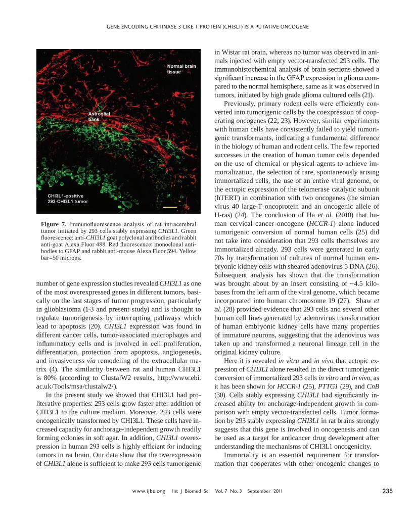
ent growth, which is one of the most consistent indicators of oncogenic transformation. After 3 weeks, 293-CHI3L1 cells grew into robust colonies in soft agar, a property not observed in the cells transfected with empty vector or in the cells transfected with vector that stably produced the green fluorescent protein (GFP). As it is shown in Fig. 5, 293-CHI3L1 cells have much higher capacity to generate colonies than control cells after 10-12 days in the colony assay formation conditions.
In order to investigate CHI3L1 effect in vivo, 293 cells, which stably expressed CHI3L1 or 293 cells transfected with empty vector, were stereotactically implanted in the caudoputamen of female Wistar rats (5 animals in each group). Tumor growth was monitored weekly via reg-istration of neurological signs for up to 6 weeks. On the 21st day after implantation severe neurological deficit ap-peared in 3 rats with implanted 293 cells, which stably ex-pressed CHI3L1. These rats were killed and histological or immunohistochemical studies were performed. Huge intracerebral tumors were observed in each of these 3 rats (Fig. 6a). Histological assay revealed displacement of me-dian cerebral structures and hydrocephalus in contralateral hemisphere. Tumors contained the dense superficial cell
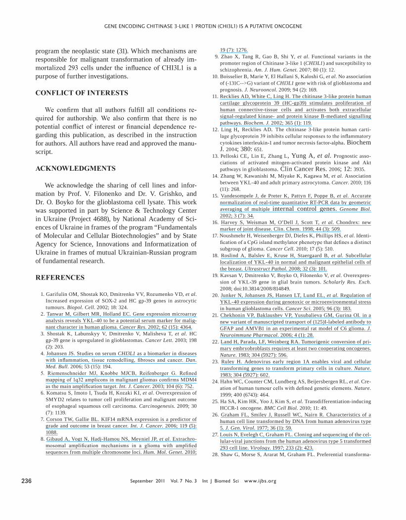
layer (Fig. 6b, green arrows) and prominent lobules (100-150 µm) with central newly ingrowing blood vessels (Fig. 6c, 6d). Immunostaining with anti-CHI3L1 antibodies re-vealed CHI3L1-positive cells both in tumor tissue and in the border of tumor (an invasion zone). All tumors were surrounded by numerous GFAP-positive reactive astro-cytes (Fig. 7).
The volume of gliomas estimated on serial tumor slices using the ellipsoid volume formula was 443.9, 575.7 and 523.5 mm3 for the first three rats (about 25% of the whole rat brain volume). Histological examination of remained two rats under investigation revealed tumors with exten-sive central necrosis as well, however, the tumor volume was about 300 mm3. No tumor growth was observed for at least 8 weeks in 5 rats injected with empty vector-trans-fected 293 cells as a negative control.
DISCUSSION
Chitinase-like glycoprotein CHI3L1 has been investi-gated extensively with respect to its expression patterns and possible association with inflammation and cancer. A
Figure 5. Oncogenic properties of CHI3L1. (A) Western blot-ting: total lysates of 293 cells stably expressing CHI3L1 (1), total lysates of 293 cells (2), recombinant CHI3L1 (3), glio-blastoma total lysate, prepared earlier [19] (4); (B) 293 cells in a soft agar colony formation assay (1·104 cells\per well) stably transfected by pcDNA3.1\CHI3L1 (1), pcDNA3.1/GFP (2) and pcDNA3.1 (3).
Figure 6. Histological analysis of intracerebral implanted 293-CHI3L1 cells. (A) Rat brain tumor formed by intracranial inoculation of 293 cells stably expressing CHI3L1. Displace-ment of median cerebral structures and hydrocephaly in con-tralateral hemisphere. Tumors were characterized by lobular structure with dense superficial cell layer (B, green arrows) and prominent lobules (100-150 µm) and central newly ingrowing blood vessels (C, D, yellow arrow). Haematoxylin staining of paraffin embedded slices. Magnification ×5 (A), ×20 (B), ×100 (C), ×400 (D).
Gene encodInG chItInase 3-lIke 1 proteIn (chI3l1) Is a putatIve oncoGene
www.ijbs.org Int J Biomed Sci Vol. 7 No. 3 September 2011 235
number of gene expression studies revealed CHI3L1 as one of the most overexpressed genes in different tumors, basi-cally on the last stages of tumor progression, particularly in glioblastoma (1-3 and present study) and is thought to regulate tumorigenesis by interrupting pathways which lead to apoptosis (20). CHI3L1 expression was found in different cancer cells, tumor-associated macrophages and inflammatory cells and is involved in cell proliferation, differentiation, protection from apoptosis, angiogenesis, and invasiveness via remodeling of the extracellular ma-trix (4). The similarity between rat and human CHI3L1 is 80% (according to ClustalW2 results, http://www.ebi.ac.uk/Tools/msa/clustalw2/).
In the present study we showed that CHI3L1 had pro-literative properties: 293 cells grow faster after addition of CHI3L1 to the culture medium. Moreover, 293 cells were oncogenically transformed by CHI3L1. These cells have in-creased capacity for anchorage-independent growth readily forming colonies in soft agar. In addition, CHI3L1 overex-pression in human 293 cells is highly efficient for inducing tumors in rat brain. Our data show that the overexpression of CHI3L1 alone is sufficient to make 293 cells tumorigenic
in Wistar rat brain, whereas no tumor was observed in ani-mals injected with empty vector-transfected 293 cells. The immunohistochemical analysis of brain sections showed a significant increase in the GFAP expression in glioma com-pared to the normal hemisphere, same as it was observed in tumors, initiated by high grade glioma cultured cells (21).
Previously, primary rodent cells were efficiently con-verted into tumorigenic cells by the coexpression of coop-erating oncogenes (22, 23). However, similar experiments with human cells have consistently failed to yield tumori-genic transformants, indicating a fundamental difference in the biology of human and rodent cells. The few reported successes in the creation of human tumor cells depended on the use of chemical or physical agents to achieve im-mortalization, the selection of rare, spontaneously arising immortalized cells, the use of an entire viral genome, or the ectopic expression of the telomerase catalytic subunit (hTERT) in combination with two oncogenes (the simian virus 40 large-T oncoprotein and an oncogenic allele of H-ras) (24). The conclusion of Ha et al. (2010) that hu-man cervical cancer oncogene (HCCR-1) alone induced tumorigenic conversion of normal human cells (25) did not take into consideration that 293 cells themselves are immortalized already. 293 cells were generated in early 70s by transformation of cultures of normal human em-bryonic kidney cells with sheared adenovirus 5 DNA (26). Subsequent analysis has shown that the transformation was brought about by an insert consisting of ~4.5 kilo-bases from the left arm of the viral genome, which became incorporated into human chromosome 19 (27). Shaw et al. (28) provided evidence that 293 cells and several other human cell lines generated by adenovirus transformation of human embryonic kidney cells have many properties of immature neurons, suggesting that the adenovirus was taken up and transformed a neuronal lineage cell in the original kidney culture.
Here it is revealed in vitro and in vivo that ectopic ex-pression of CHI3L1 alone resulted in the direct tumorigenic conversion of immortalized 293 cells in vitro and in vivo, as it has been shown for HCCR-1 (25), PTTG1 (29), and CnB (30). Cells stably expressing CHI3L1 had significantly in-creased ability for anchorage-independent growth in com-parison with empty vector-transfected cells. Tumor forma-tion by 293 stably expressing CHI3L1 in rat brains strongly suggests that this gene is involved in oncogenesis and can be used as a target for anticancer drug development after understanding the mechanisms of CHI3L1 oncogenicity.
Immortality is an essential requirement for transfor-mation that cooperates with other oncogenic changes to
Figure 7. Immunofluorescence analysis of rat intracerebral tumor initiated by 293 cells stably expressing CHI3L1. Green fluorescence: anti-CHI3L1 goat polyclonal antibodies and rabbit anti-goat Alexa Fluor 488. Red fluorescence: monoclonal anti-bodies to GFAP and rabbit anti-mouse Alexa Fluor 594. Yellow bar=50 microns.
Gene encodInG chItInase 3-lIke 1 proteIn (chI3l1) Is a putatIve oncoGene
September 2011 Vol. 7 No. 3 Int J Biomed Sci www.ijbs.org 236
program the neoplastic state (31). Which mechanisms are responsible for malignant transformation of already im-mortalized 293 cells under the influence of CHI3L1 is a purpose of further investigations.
CONFLICT OF INTERESTS
We confirm that all authors fulfill all conditions re-quired for authorship. We also confirm that there is no potential conflict of interest or financial dependence re-garding this publication, as described in the instruction for authors. All authors have read and approved the manu-script.
ACKNOWLEDGMENTS
We acknowledge the sharing of cell lines and infor-mation by Prof. V. Filonenko and Dr. V. Grishko, and Dr. O. Boyko for the glioblastoma cell lysate. This work was supported in part by Science & Technology Center in Ukraine (Project 4688), by National Academy of Sci-ences of Ukraine in frames of the program “Fundamentals of Molecular and Cellular Biotechnologies” and by State Agency for Science, Innovations and Informatization of Ukraine in frames of mutual Ukrainian-Russian program of fundamental research.
REFERENCES
1. Garifulin OM, Shostak KO, Dmitrenko VV, Rozumenko VD, et al. Increased expression of SOX-2 and HC gp-39 genes in astrocytic tumours. Biopol. Cell. 2002; 18: 324.
2. Tanwar M, Gilbert MR, Holland EC. Gene expression microarray analysis reveals YKL-40 to be a potential serum marker for malig-nant character in human glioma. Cancer Res. 2002; 62 (15): 4364.
3. Shostak K, Labunskyy V, Dmitrenko V, Malisheva T, et al. HC gp-39 gene is upregulated in glioblastomas. Cancer Lett. 2003; 198 (2): 203.
4. Johansen JS. Studies on serum CHI3L1 as a biomarker in diseases with inflammation, tissue remodelling, fibroses and cancer. Dan. Med. Bull. 2006; 53 (15): 194.
5. Riemenschneider MJ, Knobbe MJCB, Reifenberger G. Refined mapping of 1q32 amplicons in malignant gliomas confirms MDM4 as the main amplification target. Int. J. Cancer. 2003; 104 (6): 752.
6. Komatsu S, Imoto I, Tsuda H, Kozaki KI, et al. Overexpression of SMYD2 relates to tumor cell proliferation and malignant outcome of esophageal squamous cell carcinoma. Carcinogenesis. 2009; 30 (7): 1139.
7. Corson TW, Gallie BL. KIF14 mRNA expression is a predictor of grade and outcome in breast cancer. Int. J. Cancer. 2006; 119 (5): 1088.
8. Gibaud A, Vogt N, Hadj-Hamou NS, Meyniel JP, et al. Extrachro-mosomal amplification mechanisms in a glioma with amplified sequences from multiple chromosome loci. Hum. Mol. Genet. 2010;
19 (7): 1276.9. Zhao X, Tang R, Gao B, Shi Y, et al. Functional variants in the
promoter region of Chitinase 3-like 1 (CHI3L1) and susceptibility to schizophrenia. Am. J. Hum. Genet. 2007; 80 (1): 12.
10. Boisselier B, Marie Y, El Hallani S, Kaloshi G, et al. No association of (-131C-->G) variant of CHI3L1 gene with risk of glioblastoma and prognosis. J. Neurooncol. 2009; 94 (2): 169.
11. Recklies AD, White C, Ling H. The chitinase 3-like protein human cartilage glycoprotein 39 (HC-gp39) stimulates proliferation of human connective-tissue cells and activates both extracellular signal-regulated kinase- and protein kinase B-mediated signalling pathways. Biochem. J. 2002; 365 (1): 119.
12. Ling H, Recklies AD. The chitinase 3-like protein human carti-lage glycoprotein 39 inhibits cellular responses to the inflammatory cytokines interleukin-1 and tumor necrosis factor-alpha. Biochem J. 2004; 380: 651.
13. Pelloski CE, Lin E, Zhang L, Yung A, et al. Prognostic asso-ciations of activated mitogen-activated protein kinase and Akt pathways in glioblastoma. Clin Cancer Res. 2006; 12: 3935.
14. Zhang W, Kawanishi M, Miyake K, Kagawa M, et al. Association between YKL-40 and adult primary astrocytoma. Cancer. 2010; 116 (11): 268.
15. Vandesompele J, de Preter K, Pattyn F, Poppe B, et al. Accurate normalization of real-time quantitative RT-PCR data by geometric averaging of multiple internal control genes. Genome Biol. 2002; 3 (7): 34.
16. Harvey S, Weisman M, O’Dell J, Scott T, et al. Chondrex: new marker of joint disease. Clin. Chem. 1998; 44 (3): 509.
17. Noushmehr H, Weisenberger DJ, Diefes K, Phillips HS, et al. Identi-fication of a CpG island methylator phenotype that defines a distinct subgroup of glioma. Cancer Cell. 2010; 17 (5): 510.
18. Roslind A, Balslev E, Kruse H, Staergaard B, et al. Subcellular localization of YKL-40 in normal and malignant epithelial cells of the breast. Ultrastruct Pathol. 2008; 32 (3): 101.
19. Kavsan V, Dmitrenko V, Boyko O, Filonenko V, et al. Overexpres-sion of YKL-39 gene in glial brain tumors. Scholarly Res. Exch. 2008; doi:10.3814/2008/814849.
20. Junker N, Johansen JS, Hansen LT, Lund EL, et al. Regulation of YKL-40 expression during genotoxic or microenvironmental stress in human glioblastoma cells. Cancer Sci. 2005; 96 (3): 183.
21. Chekhonin VP, Baklaushev VP, Yusubalieva GM, Gurina OI. in a new variant of manuscripted transport of (125)I-labeled antibody to GFAP and AMVB1 in an experimental rat model of C6 glioma. J. Neuroimmune Pharmacol. 2006; 4 (1): 28.
22. Land H, Parada, LF, Weinberg RA. Tumorigenic conversion of pri-mary embryobroblasts requires at least two cooperating oncogenes. Nature. 1983; 304 (5927): 596.
23. Ruley H. Adenovirus early region 1A enables viral and cellular transforming genes to transform primary cells in culture. Nature. 1983; 304 (5927): 602.
24. Hahn WC, Counter CM, Lundberg AS, Beijersbergen RL, et al. Cre-ation of human tumour cells with defined genetic elements. Nature. 1999; 400 (6743): 464.
25. Ha SA, Kim HK, Yoo J, Kim S, et al. Transdifferentiation-inducing HCCR-1 oncogene. BMC Cell Biol. 2010; 11: 49.
26. Graham FL, Smiley J, Russell WC, Nairn R. Characteristics of a human cell line transformed by DNA from human adenovirus type 5. J. Gen. Virol. 1977; 36 (1): 59.
27. Louis N, Evelegh C, Graham FL. Cloning and sequencing of the cel-lular-viral junctions from the human adenovirus type 5 transformed 293 cell line. Virology. 1997; 233 (2): 423.
28. Shaw G, Morse S, Ararat M, Graham FL. Preferential transforma-
Gene encodInG chItInase 3-lIke 1 proteIn (chI3l1) Is a putatIve oncoGene
www.ijbs.org Int J Biomed Sci Vol. 7 No. 3 September 2011 237
tion of human neuronal cells by human adenoviruses and the origin of HEK 293 cells. FASEB J. 2002; 16 (8): 869.
29. Hamid T, Malik MT, Kakar SS. Ectopic expression of PTTG1/securin promotes tumorigenesis in human embryonic kidney cells. Mol. Cancer. 2005; 4 (1): 3.
30. Wang YL, Wang Y, Tong L, Wei Q. Overexpression of calcineurin B subunit (CnB) enhances the oncogenic potential of HEK293 cells. Cancer Sci. 2008; 99 (6): 1100.
31. Hahn WC. Immortalization and transformation of human cells. Mol. Cells. 2002; 13 (3): 351.
Related Documents