ORIGINAL RESEARCH ARTICLE published: 25 December 2013 doi: 10.3389/fphys.2013.00392 Exploring the role of CHI3L1 in “pre-metastatic” lungs of mammary tumor-bearing mice Stephania Libreros 1 , Ramon Garcia-Areas 1 , Patricia Keating 2 , Roberto Carrio 3 and Vijaya L. Iragavarapu-Charyulu 1 * 1 Department of Biomedical Sciences, College of Medicine, Florida Atlantic University, Boca Raton, FL, USA 2 Department of Biological Sciences, Florida Atlantic University, Boca Raton, FL, USA 3 Tumor Immunology Group, Department of Microbiology and Immunology, University of Miami Miller School of Medicine, Miami, FL, USA Edited by: Michal A. Rahat, Technion - Israel Institute for Technology, Israel Reviewed by: Uthayashanker Ezekiel, Saint Louis University, USA Zvi Granot, Hebrew University, Israel *Correspondence: Vijaya L. Iragavarapu-Charyulu, Department of Biomedical Sciences, C.E.S. College of Medicine, Florida Atlantic University, 777 Glades Road, Boca Raton, FL 33431, USA e-mail: [email protected] Elevated levels of chitinase-3-like-1 (CHI3L1) are associated with poor prognosis, shorter recurrence-free intervals and low survival in breast cancer patients. Breast cancer often metastasizes to the lung. We hypothesized that molecules expressed in the “pre-metastatic” lung microenvironment could support the newly immigrant tumor cells by providing growth and angiogenic factors. Macrophages are known to play an important role in tumor growth by releasing pro-angiogenic molecules. Using mouse mammary tumor models, we have previously shown that during neoplastic progression both the mammary tumor cells and splenic macrophages from tumor-bearing mice express higher levels of CHI3L1 compared to normal control mice. However, the role of CHI3L1 in inducing angiogenesis by macrophages at the pulmonary microenvironment to support newly arriving breast cancer cells is not yet known. In this study, we determined the expression of CHI3L1 in bronchoalveolar lavage macrophages and interstitial macrophages in regulating angiogenesis that could support the growth of newly immigrant mammary tumor cells into the lung. Here we show that in vitro treatment of pulmonary macrophages with recombinant murine CHI3L1 resulted in enhanced expression of pro-angiogenic molecules including CCL2, CXCL2, and MMP-9. We and others have previously shown that inhibition of CHI3L1 decreases the production of angiogenic molecules. In this study, we explored if in vivo administration of chitin microparticles has an effect on the expression of CHI3L1 and pro-angiogenic molecules in the lungs of mammary tumor-bearing mice. We show that treatment with chitin microparticles decreases the expression of CHI3L1 and pro-angiogenic molecules in the “metastatic” lung. These studies suggest that targeting CHI3L1 may serve as a potential therapeutic agent to inhibit angiogenesis and thus possibly tumor growth and metastasis. Keywords: CHI3L1, tumor growth, angiogenesis, macrophages, chitin microparticles, pulmonary environment INTRODUCTION Metastasis is the major cause of death in breast cancer patients. It is well established that breast cancer often metastasizes to the lung. Preferential colonization of specific tissues by breast cancer cells could be partially determined by the nature of microenvi- ronment within the target organ (Steeg, 2006). Recent studies have implicated specific cellular elements in the lungs that con- tribute to tumor growth. These include airway epithelial cells and immune cells, such as interstitial and alveolar macrophages, among others. Studies have demonstrated functional, morpho- logical, and phenotypic differences between these interstitial and alveolar macrophages (Sebring and Lehnert, 1992; Prokhorova et al., 1994; Johansson et al., 1997). However, there are lim- ited studies on interstitial macrophages in human lungs com- pared to alveolar macrophages, which can be easily obtained by bronchoalveolar lavage (BAL). The role of either interstitial or alveolar macrophages in the pre-metastatic lung in breast can- cer metastasis has not yet been elucidated. We hypothesize that interstitial macrophages, alveolar macrophages, or both, may alter the pre-metastatic landscape of the lung. Pulmonary macrophages may exert anti-tumor effects to sup- press the growth of newly-immigrated breast cancer cells, or alternatively exert pro-tumor effects by producing growth factors that support their establishment in the lung. By release of pro- teases, growth factors and cytokines, activated macrophages have the potential to influence each phase of the angiogenic process. This includes stimulating remodeling of the local extracellular matrix, inducing endothelial cells to migrate or proliferate, and inhibiting formation of differentiated capillaries. We and others have recently shown that a glycoprotein known as chitinase-3-like-1 protein is produced by macrophages from tumor-bearing hosts (Kawada et al., 2012; Libreros et al., 2012). Chitinase-3-like-1 glycoprotein (aka BRP-39, YKL-40) is a secreted protein that is upregulated in various types of can- cers, including breast (Johansen et al., 2003). This molecule is synthesized under inflammatory conditions, including bronchial www.frontiersin.org December 2013 | Volume 4 | Article 392 | 1

Welcome message from author
This document is posted to help you gain knowledge. Please leave a comment to let me know what you think about it! Share it to your friends and learn new things together.
Transcript
ORIGINAL RESEARCH ARTICLEpublished: 25 December 2013
doi: 10.3389/fphys.2013.00392
Exploring the role of CHI3L1 in “pre-metastatic” lungs ofmammary tumor-bearing miceStephania Libreros1, Ramon Garcia-Areas1, Patricia Keating2, Roberto Carrio3 and
Vijaya L. Iragavarapu-Charyulu1*
1 Department of Biomedical Sciences, College of Medicine, Florida Atlantic University, Boca Raton, FL, USA2 Department of Biological Sciences, Florida Atlantic University, Boca Raton, FL, USA3 Tumor Immunology Group, Department of Microbiology and Immunology, University of Miami Miller School of Medicine, Miami, FL, USA
Edited by:
Michal A. Rahat, Technion - IsraelInstitute for Technology, Israel
Reviewed by:
Uthayashanker Ezekiel, Saint LouisUniversity, USAZvi Granot, Hebrew University, Israel
*Correspondence:
Vijaya L. Iragavarapu-Charyulu,Department of Biomedical Sciences,C.E.S. College of Medicine, FloridaAtlantic University, 777 GladesRoad, Boca Raton, FL 33431, USAe-mail: [email protected]
Elevated levels of chitinase-3-like-1 (CHI3L1) are associated with poor prognosis, shorterrecurrence-free intervals and low survival in breast cancer patients. Breast canceroften metastasizes to the lung. We hypothesized that molecules expressed in the“pre-metastatic” lung microenvironment could support the newly immigrant tumor cellsby providing growth and angiogenic factors. Macrophages are known to play an importantrole in tumor growth by releasing pro-angiogenic molecules. Using mouse mammarytumor models, we have previously shown that during neoplastic progression both themammary tumor cells and splenic macrophages from tumor-bearing mice express higherlevels of CHI3L1 compared to normal control mice. However, the role of CHI3L1 ininducing angiogenesis by macrophages at the pulmonary microenvironment to supportnewly arriving breast cancer cells is not yet known. In this study, we determined theexpression of CHI3L1 in bronchoalveolar lavage macrophages and interstitial macrophagesin regulating angiogenesis that could support the growth of newly immigrant mammarytumor cells into the lung. Here we show that in vitro treatment of pulmonary macrophageswith recombinant murine CHI3L1 resulted in enhanced expression of pro-angiogenicmolecules including CCL2, CXCL2, and MMP-9. We and others have previously shown thatinhibition of CHI3L1 decreases the production of angiogenic molecules. In this study, weexplored if in vivo administration of chitin microparticles has an effect on the expression ofCHI3L1 and pro-angiogenic molecules in the lungs of mammary tumor-bearing mice. Weshow that treatment with chitin microparticles decreases the expression of CHI3L1 andpro-angiogenic molecules in the “metastatic” lung. These studies suggest that targetingCHI3L1 may serve as a potential therapeutic agent to inhibit angiogenesis and thuspossibly tumor growth and metastasis.
Keywords: CHI3L1, tumor growth, angiogenesis, macrophages, chitin microparticles, pulmonary environment
INTRODUCTIONMetastasis is the major cause of death in breast cancer patients.It is well established that breast cancer often metastasizes to thelung. Preferential colonization of specific tissues by breast cancercells could be partially determined by the nature of microenvi-ronment within the target organ (Steeg, 2006). Recent studieshave implicated specific cellular elements in the lungs that con-tribute to tumor growth. These include airway epithelial cellsand immune cells, such as interstitial and alveolar macrophages,among others. Studies have demonstrated functional, morpho-logical, and phenotypic differences between these interstitial andalveolar macrophages (Sebring and Lehnert, 1992; Prokhorovaet al., 1994; Johansson et al., 1997). However, there are lim-ited studies on interstitial macrophages in human lungs com-pared to alveolar macrophages, which can be easily obtained bybronchoalveolar lavage (BAL). The role of either interstitial oralveolar macrophages in the pre-metastatic lung in breast can-cer metastasis has not yet been elucidated. We hypothesize that
interstitial macrophages, alveolar macrophages, or both, may alterthe pre-metastatic landscape of the lung.
Pulmonary macrophages may exert anti-tumor effects to sup-press the growth of newly-immigrated breast cancer cells, oralternatively exert pro-tumor effects by producing growth factorsthat support their establishment in the lung. By release of pro-teases, growth factors and cytokines, activated macrophages havethe potential to influence each phase of the angiogenic process.This includes stimulating remodeling of the local extracellularmatrix, inducing endothelial cells to migrate or proliferate, andinhibiting formation of differentiated capillaries.
We and others have recently shown that a glycoproteinknown as chitinase-3-like-1 protein is produced by macrophagesfrom tumor-bearing hosts (Kawada et al., 2012; Libreros et al.,2012). Chitinase-3-like-1 glycoprotein (aka BRP-39, YKL-40) isa secreted protein that is upregulated in various types of can-cers, including breast (Johansen et al., 2003). This molecule issynthesized under inflammatory conditions, including bronchial
www.frontiersin.org December 2013 | Volume 4 | Article 392 | 1
Libreros et al. CHI3L1 expression in pre-mestastatic “lung macrophages”
asthma, inflammatory bowel disease, and cancer, but is nothighly expressed under physiological conditions (Johansen et al.,2006; Mizoguchi, 2006; Chupp et al., 2007; Coffman, 2008;Lee et al., 2011; Libreros et al., 2013). CHI3L1 is a chitin-binding glycoprotein that belongs to the family of chitinase-likeproteins, but is lacking in enzymatic activity (Henrissat andBairoch, 1993). This glycoprotein is expressed and secreted by avariety of cell types including articular chondrocytes, synovio-cytes, osteoblasts, macrophages, neutrophils, and epithelial cells(Johansen et al., 2001; Rehli et al., 2003; Mizoguchi, 2006; Rathckeand Vestergaard, 2006; Lee et al., 2009). In examining the functionof CHI3L1, we and others have found that CHI3L1 stimulatesthe production of pro-angiogenic molecules (Shao et al., 2009;Kawada et al., 2012; Libreros et al., 2012). Conversely, (Shaoet al., 2009) and (Libreros et al., 2012), have shown that inhibit-ing CHI3L1 with neutralizing antibodies or administration ofchitin microparticles, decreases the expression of pro-angiogenicmolecules (Shao et al., 2009; Libreros et al., 2012).
Currently there is little known regarding mechanistic linksbetween CHI3L1 expression by macrophages in the “pre-metatastic” lung, and tumor-related angiogenesis. We thereforeexamined the possibility that macrophage-derived CHI3L1 in thelung, upregulated by exposure to circulating CHI3L1 producedby tumor cells, “conditions” this organ to favor establishmentof newly-arrived, metastasizing cancer cells. Thus, in this studywe tested the role of CHI3L1 expression by bronchoalveolarand interstitial macrophages in regulating angiogenesis to pro-mote the growth of new mammary tumor cells in the lung.We demonstrate here that: (1) CHI3L1 is secreted by intersti-tial and alveolar macrophages from mammary tumor bearers;(2) CHI3L1 induces expression of pro-angiogenic molecules ininterstitial and alveolar macrophages; (3) in vivo treatment withchitin microparticles reduces angiogenesis; and (4) this treatmentalso decreases expression of CHI3L1, CCL2, CXCL2 and MMP-9.Our findings suggest that CHI3L1 may play a role in prepar-ing the “soil” in the “pre-metastatic” lung. Therefore, CHI3L1may be an attractive therapeutic target to inhibit breast cancermetastasis.
MATERIALS AND METHODSMICE AND CELL LINESFemale BALB/c mice were used in all studies (Charles RiverLaboratories, 8–12 week-olds), and were housed and used accord-ing to the National Institutes of Health guidelines, under proto-cols approved by Florida Atlantic University Institutional AnimalCare and Use Committee. Mammary tumor cells (4T1-luc-A4;Caliper Life Sciences, Hopkinton, MA) were maintained in RPMIwith 10% FCS (Kim et al., 2010). Tumor cells were implantedin mice by subcutaneous injection of 1 × 1054T1 tumor cellsin the lower right ventral quadrant. These tumors metastasizeto the lung ∼4–5 weeks post-tumor cell implantation. Normalmice and tumor bearers were assessed at ∼2 weeks- (“pre-metastatic”) and 5 weeks post-tumor implantation (“metastatic”)for the expression of cytokines. Tumor bearers treated with chitinmicroparticles (see below) were assessed at 5 weeks post-tumorcell implantation for metastasis, cytokine expression and tumorangiogenesis using AngioSense probe.
ISOLATION OF EPITHELIAL CELLS AND MACROPHAGES FROM THELUNGSBronchoalveolar macrophage isolationTracheas of euthanized mice were cannulated and lavaged with1 mL of saline. The bronchoalveolar lavage fluid (BALF) wasrecovered to obtain alveolar macrophages as described by Kogisoet al. (2011). Plastic-adherent macrophages were then enrichedby incubating BALF cells (106 cells/mL) in complete media(RPMI 1640 with 10% FBS, 10 units/mL penicillin and 10 μg/mLstreptomycin) for 1 h in a 5% CO2 incubator at 37◦C. Non-adherent cells were removed by washing with media, andthe BALF macrophages were then isolated by labeling withCD11b magnetic microbeads (Miltenyi Biotec, Cambridge, MA).CD11b+ cells were purified by positive selection using AutoMACs(Miltenyi). Alveolar CD11b+macrophages isolated from all ani-mals revealed >90% purity as determined by flow cytometricanalysis (FACS Calibur).
Isolation of alveolar epithelial cells and interstitial macrophagesImmediately after collecting BALF specimens, pulmonary epithe-lial cells and interstitial macrophages were isolated as previouslydescribed, with modifications [35]. Media (1 mL, RPMI 1640with 2% FBS and 1 mg/mL dispase (Invitrogen, Grand Island,NY)) was perfused into the airways and alveoli of the lungs. Thelungs were then removed and incubated at 37◦C for 1 h in thesame media with 2 mg/mL collagenase type II (Invitrogen) added,followed by tissue mincing. Cells were filtered through Teflonmesh of 40 μM pores and washed with the media with no cen-trifugation at 0◦C. Macrophages were isolated by plastic adher-ence, and were then purified by magnetic beads as described. Thepurity of the cells was >90% for CD11b expression as determinedby flow cytometric analysis. Non-adherent cells (alveolar epithe-lial cells) were labeled with CD146 microbeads (MiltenyiBiotec)and purified by AutoMACs (MiltenyiBiotec). The purity wasconfirmed to be >90% as assessed by flow cytometric analysis.
Cell culturePurified alveolar epithelial cells, and alveolar and interstitialmacrophages, were cultured at 2 × 10⁶ cells/mL for 18 h in com-plete media as described previously (Owen et al., 2005). All cellswere stimulated with either 500 ng/mL LPS (Sigma Chemical Co.,St. Louis, MO), or in combination with 1 ng/mL or 5 ng/mLof endotoxin-free rmCHI3L1 (R&D systems, Minneapolis, MN).Growth media (GM) was included as a control condition for someof the cultures. Cell-free supernatants were then collected andstored at −80◦C.
FLOW CYTOMETRYTotal lung homogenates and BALF cells were isolated and resus-pended in FACS buffer (PBS with 1% BSA and 0.1% sodiumazide) and stained for 30 min at 4◦C with FITC-conjugatedantibodies against CD11b (BDBiosciences, San Jose, CA), APC-conjugated Ly6C, PerCP-conjugated Ly6G, PerCPF4/80 (all fromBDBiosciences) or CD146 (Miltenyi Biotec). Isotype-matchedIgG was used for internal controls. For intracellular cytokinestaining, BD cytofix/cytoperm + fixation/permeabilization kitwere used according to manufacturer’s instructions and this was
Frontiers in Physiology | Vascular Physiology December 2013 | Volume 4 | Article 392 | 2
Libreros et al. CHI3L1 expression in pre-mestastatic “lung macrophages”
followed by labeling for CHI3L1. CHI3L1 antibody (generouslyprovided by Dr. Alison Humbles, MedImmune, MD) was conju-gated to a fluorescent dye using Lightning-Link PE conjugationkit (Novus, Littleton, CO) following manufacturer’s instructions.Samples were acquired in a FACSCalibur flow cytometer (BDBiosciences) and analyzed by Flow Jo software (Tree Star, Inc.,Ashland, OR).
CYTOKINE ELISABALF samples and culture supernatants from cells obtained fromcontrol and mammary tumor bearers were analyzed for CHI3L1,CCL2, CXCL2 and MMP-9 levels by ELISA (all from R&DSystems) according to manufacturer’s instructions. Absorbance at450 nm with wavelength correction at 570 nm was measured witha Tecan SLT Rainbow Reader (Lab Instruments, Research TrianglePark, NC) and optical density (OD) values of samples were con-verted to picograms against a standard curve plotted from knownquantities of recombinant murine cytokines.
WESTERN BLOT ANALYSISCells from total lung homogenates were lysed with sample buffer(20 mM dithiothreitol, 6% SDS, 0.25 M Tris, pH 6.8, 10% glyc-erol, 10 mM NaF and bromophenyl blue) and used to extracttotal protein. Total protein (20 μg) was resolved on 4–20% Mini-Protean SDS-PAGE gradient gels (BioRad Life Sciences, Hercules,CA) and transferred to PVDF membrane (Pierce) using a semi-dry transfer transblotter (BioRad). The membranes were blockedovernight at 4◦C in SeaBlock (Calbiochem), and subsequentlyincubated at room temperature for 1 h with anti-mouse CHI3L1polyclonal antibody (1 μg/mL) (Quidel, San Diego, CA) andanti-mouse beta actin polyclonal antibody (0.25 μg/mL) (Li-CorBiosciences, Lincoln, NE). Immunoblots were washed with 0.5%Tween-PBS followed by 1 h incubation at room temperature withappropriate infrared dye-conjugated secondary antibodies (Li-Cor Biosciences). Blots were washed again with 0.5% Tween-PBSand then dried at 37◦C for 20 min. The membranes then wereimaged and bands quantified using the Li-Cor Odyssey imag-ing system. Levels of proteins of interest were normalized tobeta-actin.
CONFOCAL MICROSCOPYLungs from control mice and 2-week mammary tumor bear-ers were perfused with a mixture of Optimal Tissue Cutting(OCT) embedding compound and PBS (70%/30% respectively).The lungs were then removed and snap frozen on dry ice.5 μM cryostat sections were mounted on SuperFrost Plus slides(Fisher Scientific, Fair Lawn, NJ), fixed in 4% paraformalde-hyde, and labeled with goat anti-mouse CC10 (1:100, SantaCruz Biotech, Santa Cruz, CA) and rabbit anti-mouse CHI3L1(1 μg/μL, Quidel) as described below. Alveolar and interstitialmacrophages were plated (0.5 x 10⁶ cells) on coverslips, fixed in4% paraformaldehyde, and labeled with goat anti-mouse CD68(1:100, macrophage marker) (Santa Cruz Biotech) and rabbitanti-mouse CHI3L1 (Quidel). Both cryostat sections and cellswere blocked in 10% normal horse serum in PBS prior tostaining with primary antibodies overnight at 4◦C. After wash-ing in PBS, cells and sections were incubated in the following
secondary antibodies: FITC-donkey anti-rabbit and PE-donkeyanti-goat (1:2000, both from Invitrogen, Life Technologies). Tovisualize nuclei, material was mounted with Vectashield contain-ing DAPI (Vector Laboratories, Burlingame, CA), and examinedby confocal microscopy (Carl Zeiss LSM 700, Microimaging,Thornwood, NY).
In vivo TREATMENT WITH CHITIN MICROPARTICLESChitin microparticles (1–10 μM, kindly provided by Dr. YoshimiShibata, Florida Atlantic University, FL) were prepared asdescribed previously (Shibata et al., 1997; Strong et al., 2002;Nishiyama et al., 2006). Tumor-bearing mice were treated byintraperitoneal injection with chitin microparticles (1 mg/mouse)starting 3 days post-tumor implantation and continuing everythird day for 5 weeks (Libreros et al., 2012).
ANGIOGENESIS DETERMINATIONTo assess vascularization in vivo, the near-infrared blood poolagent AngioSense 680 probe (2 nmol/mouse in 150 μL volume;Perkin Elmer, Waltham, MA) was injected via tail vein 24 h beforeimaging. Mice were imaged using a bioluminescence opticalimager (IVIS Lumina LTE, Perkin Elmer). Maximal near infraredsignals were quantified using Living Image 2.5 image analysis soft-ware (Xenogen, Perkin Elmer). Infrared signals are reported asphotons/sec.
STATISTICAL ANALYSESResults are expressed as group means ± SD. Statistical analy-ses were performed using GraphPad Prism 3 software (LaJolla,CA). Statistical comparisons of paired groups were determined byStudent’s t tests. Values of p < 0.05 were considered statisticallysignificant.
RESULTSCHI3L1 EXPRESSION IS INCREASED IN BALF AND TOTAL LUNG FROMMAMMARY TUMOR-BEARING MICEIncreased levels of CHI3L1 in the sera of breast cancer patientsare associated with poor prognosis (Johansen et al., 2003). Wehave previously reported higher circulating levels of CHI3L1 in4T1 mammary tumor-bearing mice compared with normal mice(Libreros et al., 2012). This tumor model shares similar char-acteristics with human breast cancer patients as mice bearing4T1 mammary tumors exhibit spontaneous tumor cell metasta-sis to the lung. The levels of CHI3L1 in the lungs are increasedduring pulmonary inflammation, and inflammation is known tocontribute to tumor growth and metastasis. Since it is knownthat breast tumor cells metastasize to the lung, we determined ifCHI3L1 expression is specifically altered in lungs of mammarytumor bearers compared to control mice.
The “pre-metastatic” and “metastatic” stages were describedby Yan et al. using the 4T1 mammary tumor model, withthe pre-metastatic stage occurring at 14 days post-tumor cellinoculation, and the metastatic stage at 4 weeks (Yan et al.,2010). We therefore assessed CHI3L1 expression in the lungsof mice inoculated with 4T1 mammary tumor cells at 2 weekspost-cell implantation, a time point at which no visible micro-metastasis is observed in the lungs (data not shown), and at
www.frontiersin.org December 2013 | Volume 4 | Article 392 | 3
Libreros et al. CHI3L1 expression in pre-mestastatic “lung macrophages”
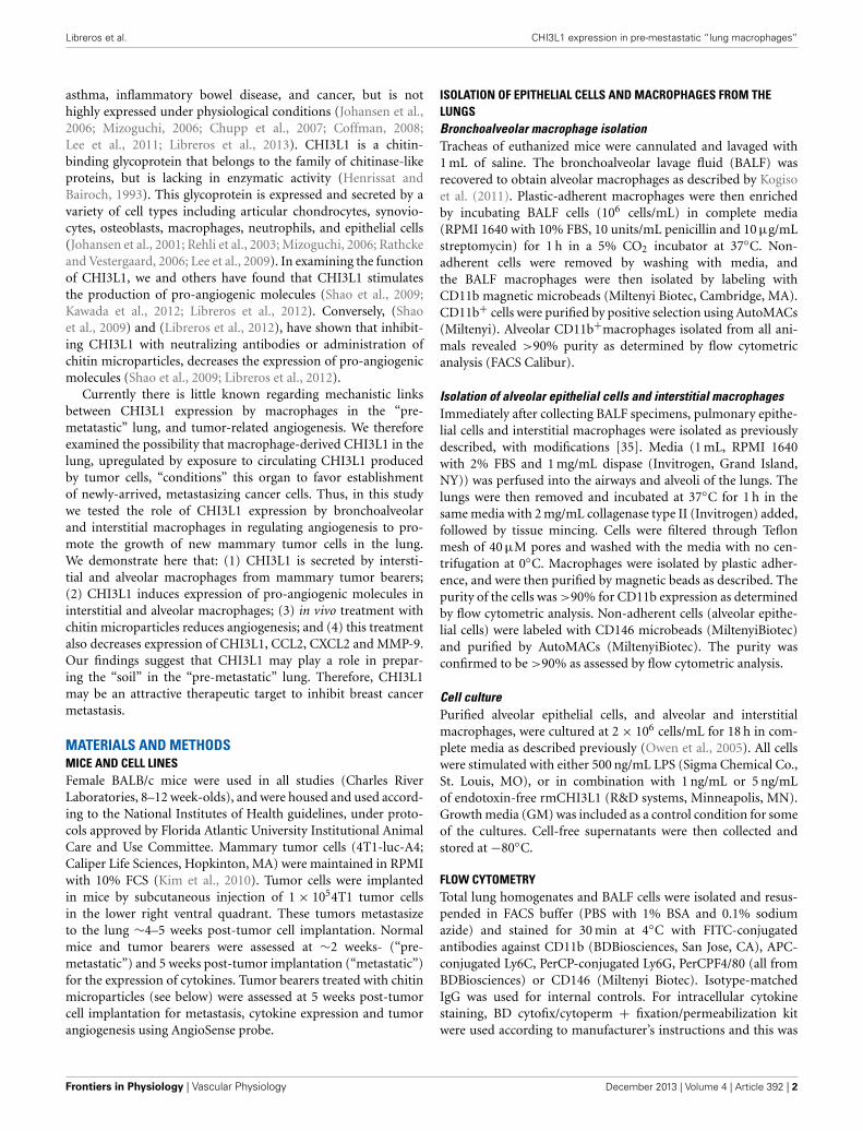
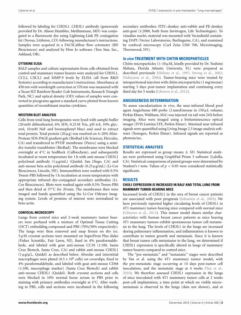
5 weeks when metastasis of 4T1 cells is known to be well-established (Yan et al., 2010; Libreros et al., 2012). We firstmeasured circulating levels of CHI3L1, which increased from25 × 103 ng/mL at 2 weeks, to 125 × 103 ng/mL at 5 weeks(Figure 1A). ELISA measurements demonstrated that signifi-cantly higher levels of CHI3L1 were also present in both BALFsamples (Figure 1B) and total lung homogenates (Figure 1C) at5 weeks post-tumor cell implantation, compared to the 2-weektime point. These higher levels of CHI3L1 could be due toexpression by the pulmonary tissue itself and/or the tumorcells that have infiltrated by 5 weeks (Libreros et al., 2013).Samples from the “pre-metastatic” stage would help differentiatebetween these possibilities, as tumor cells have not yet infil-trated, and we performed additional analyses at this stage. At2 weeks post-inoculation, significantly higher levels of CHI3L1were measured by ELISA in BALF samples from tumor bear-ers compared to control mice (Figure 2A). Western blot analysisof whole lungs from pre-metastatic tumor-bearers confirmedhigher levels of pulmonary CHI3L1 (Figure 2B), and ELISAassays of total lung homogenates quantified this increase at 2weeks (Figure 2C). CHI3L1 is secreted by a variety of cell types,including macrophages, neutrophils, colonic epithelial cells, andchondrocytes (Nyirkos and Golds, 1990; Hakala et al., 1993;Renkema et al., 1998; Volck et al., 1998; Mizoguchi, 2006), andrecent studies by Lee et al. (2009) have shown that CHI3L1 (akaBRP-39) is upregulated in inflamed airway epithelium, and thatit plays an active role in pulmonary inflammation (Lee et al.,2009). We therefore determined if CHI3L1 expression is specifi-cally altered in lung epithelial cells isolated from mammary tumorbearers at 2 weeks post-inoculation, compared to those fromcontrol mice. Production of CHI3L1 was increased more than5-fold in pulmonary epithelial cells from tumor bearers, as mea-sured by ELISA at 18 h post-plating (Figure 2D). To promote“inflammatory” conditions, cultures were treated with LPS tostimulate cytokine production, which exacerbated the increase inCHI3L1 levels displayed by cells from tumor bearers (Figure 2D).Localization of CHI3L1 in lung tissue samples by immunofluo-rescence showed that CHI3L1 was expressed by lung epithelialcells (CC10+ cells), and that this expression was increased in theairways of mammary tumor bearing mice compared to controls(Figure 2E).
CHI3L1 EXPRESSION IS INCREASED IN CD11B+GR1+ CELLS OFMAMMARY TUMOR BEARERSMyeloid-derived cells have been shown to be important inpromoting tumor growth, metastasis, and angiogenesis (vanKempen and Coussens, 2002; Yang et al., 2004). The lungs of4T1 mammary tumor bearers show infiltration by myeloid-derived suppressor cells, and in particular by CD11b+Gr1+ cellsthat establish a pre-metastatic niche by secreting proinflam-matory mediators (Yan et al., 2010; Younos et al., 2011). Wehave previously shown that splenic myeloid cells from mam-mary tumor-bearing mice express CHI3L1 (Libreros et al., 2012).To clearly delineate myeloid populations of cells in pulmonarytissue that could contribute to CHI3L1 expression, single cellsuspensions prepared from total lungs of normal and 2-weekmammary tumor-bearers were analyzed by flow cytometry. As
FIGURE 1 | CHI3L1 is expressed in serum and pulmonary tissue of
mammary tumor-bearing mice at 2 weeks (“pre-metastatic”) and 5
weeks (metastatic) post-tumor cell implantation. (A) Serum (B) BALFand (C) Total lung homogenates from 2 week and 5 week mammary tumorbearers were analyzed for CHI3L1 expression by ELISA. N = 10mice/group; ∗∗p ≤ 0.001.
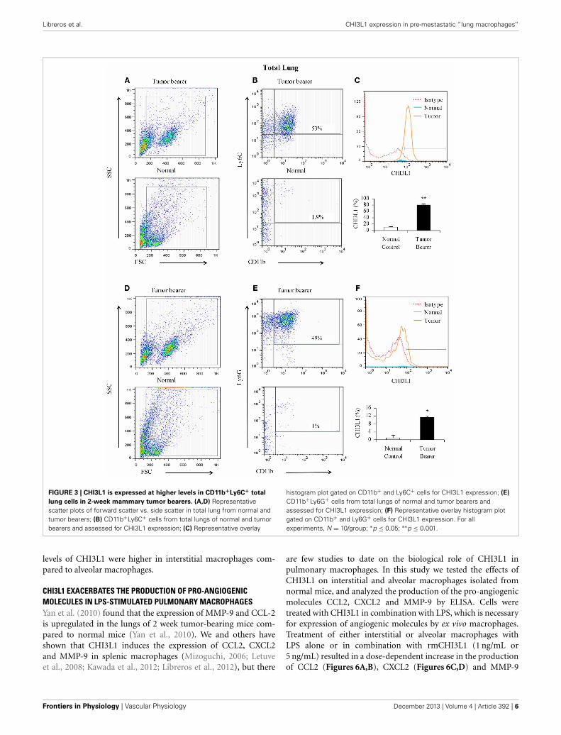
shown in Figures 3A–C (one representative assay out of five),CD11b+Ly6C+ cells from mammary tumor bearers expressCHI3L1 at higher levels compared to normals. CD11b+Ly6G+cells from tumor bearers express CHI3L1 but these levels arelower compared to the levels observed in CD11b+Ly6C+ cells(Figures 3D–F).
Since BALF was shown to contain elevated levels of CHI3L1,we determined which cell populations in the lavage contributeto the expression of CHI3L1 at 2 weeks post-tumor cell inocula-tion. Toward this we assessed CD11b+Ly6C+ and CD11b+Ly6G+cells in the lavage. CD11b+Ly6C+ cells from mammary tumorbearers express higher levels of CHI3L1 compared to normals(Figures 4A–C). Similar to what was observed in total lunghomogenates, CD11b+Ly6G+ cells from tumor bearers expressCHI3L1 at lower levels compared to the CD11b+Ly6C+ cells(Figures 4D–F).
Frontiers in Physiology | Vascular Physiology December 2013 | Volume 4 | Article 392 | 4
Libreros et al. CHI3L1 expression in pre-mestastatic “lung macrophages”
FIGURE 2 | CHI3L1 is expressed at higher levels in the pulmonary tissue
of 2-week 4T1 mammary tumor bearers. (A) CHI3L1 expression inbronchoalveolar lavage fluid by ELISA; (B) Western blot analysis of total lunghomogenates for CHI3L1 expression; (C,D) CHI3L1 expression by ELISA of
total lung homogenates (C) and lung epithelial cells (D). (E) Cellularco-localization of CHI3L1 with CC10, an airway-epithelial cell marker, incryostat sections visualized by confocal microscopy. For all experiments,N = 10/group; ∗p ≤ 0.05; ∗∗p ≤ 0.001.
CHI3L1 EXPRESSION IS INCREASED IN MACROPHAGES FROM THELUNGS OF MAMMARY TUMOR-BEARING MICEWe have previously shown that CHI3L1 is expressed at higherlevels in splenic macrophages of mammary tumor-bearing mice(Libreros et al., 2012). In this study, we determined the expres-sion levels of CHI3L1 in macrophages from the “pre-metastatic”lungs. Two broad subsets of macrophages are found in thelungs of mice and humans, i.e., alveolar macrophages whichline the surface of alveoli, and interstitial macrophages that arelocalized in the space between alveolar epithelium and vascularendothelium (Schneberger et al., 2011). Thus, alveolar and inter-stitial macrophages from normal and 2-week tumor bearers werepurified as described in the Methods section, and cultured ineither the absence or presence of LPS. Interstitial macrophages
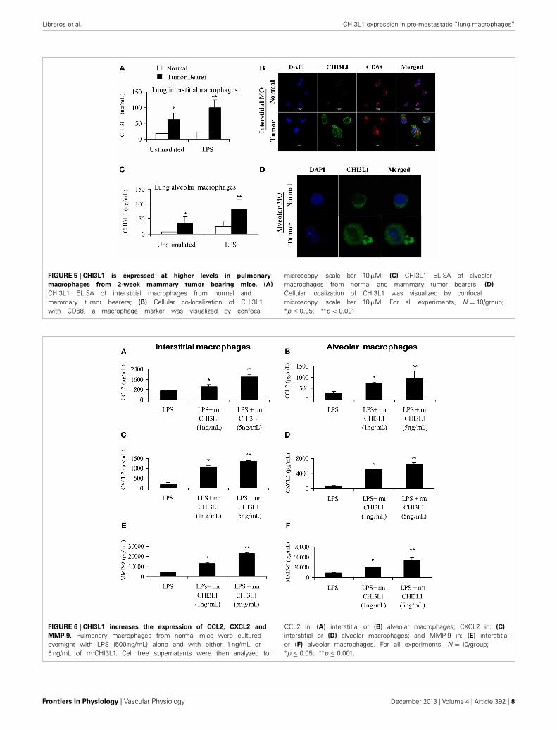
from mammary tumor-bearing mice secrete CHI3L1 and theselevels were further increased by stimulation with LPS as deter-mined by ELISA (Figure 5A). Localization of CHI3L1 in inter-stitial macrophages was then confirmed by immunofluorescentlabeling. Confocal images revealed higher intensity of CHI3L1expression in CD68+interstitial macrophages from tumor bear-ers, relative to normal mice (Figure 5B). Purified alveolarmacrophages were also analyzed. Similar to what was observedin the interstitial macrophage population, there were higher thannormal levels of CHI3L1 present in culture supernatants of alve-olar macrophages from 2 week tumor-bearing mice (Figure 5C).Intensity of CHI3L1 staining in alveolar macrophages similarlywas greater in tumor bearers’ macrophages, as determined byconfocal microscopy (Figure 5D). Interestingly, the expression
www.frontiersin.org December 2013 | Volume 4 | Article 392 | 5
Libreros et al. CHI3L1 expression in pre-mestastatic “lung macrophages”
FIGURE 3 | CHI3L1 is expressed at higher levels in CD11b+Ly6C+ total
lung cells in 2-week mammary tumor bearers. (A,D) Representativescatter plots of forward scatter vs. side scatter in total lung from normal andtumor bearers; (B) CD11b+Ly6C+ cells from total lungs of normal and tumorbearers and assessed for CHI3L1 expression; (C) Representative overlay
histogram plot gated on CD11b+ and Ly6C+ cells for CHI3L1 expression; (E)
CD11b+Ly6G+ cells from total lungs of normal and tumor bearers andassessed for CHI3L1 expression; (F) Representative overlay histogram plotgated on CD11b+ and Ly6G+ cells for CHI3L1 expression. For allexperiments, N = 10/group; ∗p ≤ 0.05; ∗∗p ≤ 0.001.
levels of CHI3L1 were higher in interstitial macrophages com-pared to alveolar macrophages.
CHI3L1 EXACERBATES THE PRODUCTION OF PRO-ANGIOGENICMOLECULES IN LPS-STIMULATED PULMONARY MACROPHAGESYan et al. (2010) found that the expression of MMP-9 and CCL-2is upregulated in the lungs of 2 week tumor-bearing mice com-pared to normal mice (Yan et al., 2010). We and others haveshown that CHI3L1 induces the expression of CCL2, CXCL2and MMP-9 in splenic macrophages (Mizoguchi, 2006; Letuveet al., 2008; Kawada et al., 2012; Libreros et al., 2012), but there
are few studies to date on the biological role of CHI3L1 inpulmonary macrophages. In this study we tested the effects ofCHI3L1 on interstitial and alveolar macrophages isolated fromnormal mice, and analyzed the production of the pro-angiogenicmolecules CCL2, CXCL2 and MMP-9 by ELISA. Cells weretreated with CHI3L1 in combination with LPS, which is necessaryfor expression of angiogenic molecules by ex vivo macrophages.Treatment of either interstitial or alveolar macrophages withLPS alone or in combination with rmCHI3L1 (1 ng/mL or5 ng/mL) resulted in a dose-dependent increase in the productionof CCL2 (Figures 6A,B), CXCL2 (Figures 6C,D) and MMP-9
Frontiers in Physiology | Vascular Physiology December 2013 | Volume 4 | Article 392 | 6

Libreros et al. CHI3L1 expression in pre-mestastatic “lung macrophages”
FIGURE 4 | CHI3L1 is expressed at higher levels in CD11b+Ly6C+bronchoalveolar lavage cells in 2-week mammary tumor bearers. (A,D)
Representative scatter plots of forward scatter vs. side scatter in alveolarlavage from normal and tumor bearers; (B) CD11b+Ly6C+ cells from alveolarlavage of normal and tumor bearers and assessed for CHI3L1 expression; (C)
Representative overlay histogram plot gated on CD11b+ Ly6C+ cells forCHI3L1 expression; (E) CD11b+Ly6G+ cells from alveolar lavage of normaland tumor bearers and assessed for CHI3L1 expression; (F) Representativeoverlay histogram plot gated on CD11b+Ly6G+ cells for CHI3L1 expression.For all experiments, N = 10/group; ∗p ≤ 0.05; ∗∗p ≤ 0.001.
(Figures 6E,F) in both interstitial and alveolar macrophages.Culturing with rmCHI3L1 alone in the absence of LPS revealeda similar trend to the one observed in cultures containing bothLPS and rmCHI3L1, but the levels of the proinflammatory medi-ators secreted were lower (data not shown). The combined effectsof LPS and rmCHI3L1 produced the greatest increase in theexpression of pro-angiogenic molecules.
In vivo TREATMENT WITH CHITIN MICROPARTICLES DECREASESANGIOGENESISIntraperitoneal treatment of mammary tumor bearers with chitinmicroparticles, a substrate for CHI3L1, results in decreased
tumor growth and pulmonary metastasis (Libreros et al., 2012).We and others established that 4T1 mammary tumors beginto infiltrate the lungs by 3 weeks post-tumor cell implanta-tion, and observable metastatic foci are seen in the lungs at 5weeks post-tumor cell implantation (Yan et al., 2010; Libreroset al., 2012). Therefore, we used 5 week tumor bearers to assessthe effects of in vivo chitin microparticle treatment. To testif early treatment with chitin microparticles affects angiogene-sis and tumor growth, an in vivo AngioSense probe was used.Mice treated with chitin microparticles after tumor cell inocula-tion had significantly reduced angiogenic fluorescent signals inin vivo imaged tumors compared to the tumors from untreated
www.frontiersin.org December 2013 | Volume 4 | Article 392 | 7
Libreros et al. CHI3L1 expression in pre-mestastatic “lung macrophages”
FIGURE 5 | CHI3L1 is expressed at higher levels in pulmonary
macrophages from 2-week mammary tumor bearing mice. (A)
CHI3L1 ELISA of interstitial macrophages from normal andmammary tumor bearers; (B) Cellular co-localization of CHI3L1with CD68, a macrophage marker was visualized by confocal
microscopy, scale bar 10 μM; (C) CHI3L1 ELISA of alveolarmacrophages from normal and mammary tumor bearers; (D)
Cellular localization of CHI3L1 was visualized by confocalmicroscopy, scale bar 10 μM. For all experiments, N = 10/group;∗p ≤ 0.05; ∗∗p < 0.001.
FIGURE 6 | CHI3L1 increases the expression of CCL2, CXCL2 and
MMP-9. Pulmonary macrophages from normal mice were culturedovernight with LPS (500 ng/mL) alone and with either 1 ng/mL or5 ng/mL of rmCHI3L1. Cell free supernatants were then analyzed for
CCL2 in: (A) interstitial or (B) alveolar macrophages; CXCL2 in: (C)
interstitial or (D) alveolar macrophages; and MMP-9 in: (E) interstitialor (F) alveolar macrophages. For all experiments, N = 10/group;∗p ≤ 0.05; ∗∗p ≤ 0.001.
Frontiers in Physiology | Vascular Physiology December 2013 | Volume 4 | Article 392 | 8
Libreros et al. CHI3L1 expression in pre-mestastatic “lung macrophages”
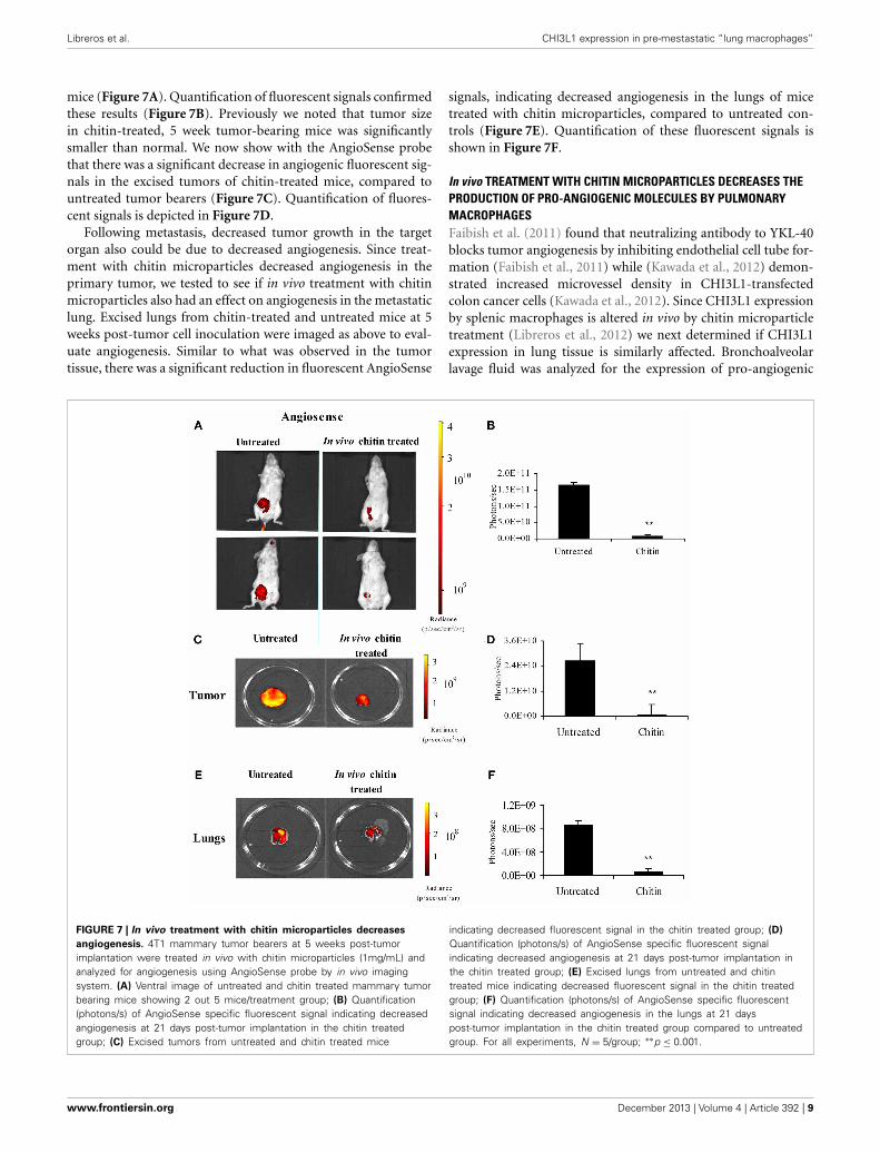
mice (Figure 7A). Quantification of fluorescent signals confirmedthese results (Figure 7B). Previously we noted that tumor sizein chitin-treated, 5 week tumor-bearing mice was significantlysmaller than normal. We now show with the AngioSense probethat there was a significant decrease in angiogenic fluorescent sig-nals in the excised tumors of chitin-treated mice, compared tountreated tumor bearers (Figure 7C). Quantification of fluores-cent signals is depicted in Figure 7D.
Following metastasis, decreased tumor growth in the targetorgan also could be due to decreased angiogenesis. Since treat-ment with chitin microparticles decreased angiogenesis in theprimary tumor, we tested to see if in vivo treatment with chitinmicroparticles also had an effect on angiogenesis in the metastaticlung. Excised lungs from chitin-treated and untreated mice at 5weeks post-tumor cell inoculation were imaged as above to eval-uate angiogenesis. Similar to what was observed in the tumortissue, there was a significant reduction in fluorescent AngioSense
signals, indicating decreased angiogenesis in the lungs of micetreated with chitin microparticles, compared to untreated con-trols (Figure 7E). Quantification of these fluorescent signals isshown in Figure 7F.
In vivo TREATMENT WITH CHITIN MICROPARTICLES DECREASES THEPRODUCTION OF PRO-ANGIOGENIC MOLECULES BY PULMONARYMACROPHAGESFaibish et al. (2011) found that neutralizing antibody to YKL-40blocks tumor angiogenesis by inhibiting endothelial cell tube for-mation (Faibish et al., 2011) while (Kawada et al., 2012) demon-strated increased microvessel density in CHI3L1-transfectedcolon cancer cells (Kawada et al., 2012). Since CHI3L1 expressionby splenic macrophages is altered in vivo by chitin microparticletreatment (Libreros et al., 2012) we next determined if CHI3L1expression in lung tissue is similarly affected. Bronchoalveolarlavage fluid was analyzed for the expression of pro-angiogenic
FIGURE 7 | In vivo treatment with chitin microparticles decreases
angiogenesis. 4T1 mammary tumor bearers at 5 weeks post-tumorimplantation were treated in vivo with chitin microparticles (1mg/mL) andanalyzed for angiogenesis using AngioSense probe by in vivo imagingsystem. (A) Ventral image of untreated and chitin treated mammary tumorbearing mice showing 2 out 5 mice/treatment group; (B) Quantification(photons/s) of AngioSense specific fluorescent signal indicating decreasedangiogenesis at 21 days post-tumor implantation in the chitin treatedgroup; (C) Excised tumors from untreated and chitin treated mice
indicating decreased fluorescent signal in the chitin treated group; (D)
Quantification (photons/s) of AngioSense specific fluorescent signalindicating decreased angiogenesis at 21 days post-tumor implantation inthe chitin treated group; (E) Excised lungs from untreated and chitintreated mice indicating decreased fluorescent signal in the chitin treatedgroup; (F) Quantification (photons/s) of AngioSense specific fluorescentsignal indicating decreased angiogenesis in the lungs at 21 dayspost-tumor implantation in the chitin treated group compared to untreatedgroup. For all experiments, N = 5/group; ∗∗p ≤ 0.001.
www.frontiersin.org December 2013 | Volume 4 | Article 392 | 9
Libreros et al. CHI3L1 expression in pre-mestastatic “lung macrophages”
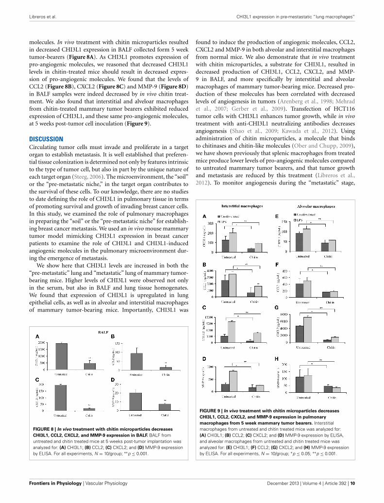
molecules. In vivo treatment with chitin microparticles resultedin decreased CHI3L1 expression in BALF collected form 5 weektumor-bearers (Figure 8A). As CHI3L1 promotes expression ofpro-angiogenic molecules, we reasoned that decreased CHI3L1levels in chitin-treated mice should result in decreased expres-sion of pro-angiogenic molecules. We found that the levels ofCCL2 (Figure 8B), CXCL2 (Figure 8C) and MMP-9 (Figure 8D)in BALF samples were indeed decreased by in vivo chitin treat-ment. We also found that interstitial and alveloar macrophagesfrom chitin-treated mammary tumor bearers exhibited reducedexpression of CHI3L1, and these same pro-angiogenic molecules,at 5 weeks post-tumor cell inoculation (Figure 9).
DISCUSSIONCirculating tumor cells must invade and proliferate in a targetorgan to establish metastasis. It is well established that preferen-tial tissue colonization is determined not only by features intrinsicto the type of tumor cell, but also in part by the unique nature ofeach target organ (Steeg, 2006). The microenvironment, the “soil”or the “pre-metastatic niche,” in the target organ contributes tothe survival of these cells. To our knowledge, there are no studiesto date defining the role of CHI3L1 in pulmonary tissue in termsof promoting survival and growth of invading breast cancer cells.In this study, we examined the role of pulmonary macrophagesin preparing the “soil” or the “pre-metastatic niche” for establish-ing breast cancer metastasis. We used an in vivo mouse mammarytumor model mimicking CHI3L1 expression in breast cancerpatients to examine the role of CHI3L1 and CHI3L1-inducedangiogenic molecules in the pulmonary microenvironment dur-ing the emergence of metastasis.
We show here that CHI3L1 levels are increased in both the“pre-metastatic” lung and “metastatic” lung of mammary tumor-bearing mice. Higher levels of CHI3L1 were observed not onlyin the serum, but also in BALF and lung tissue homogenates.We found that expression of CHI3L1 is upregulated in lungepithelial cells, as well as in alveolar and interstitial macrophagesof mammary tumor-bearing mice. Importantly, CHI3L1 was
FIGURE 8 | In vivo treatment with chitin microparticles decreases
CHI3L1, CCL2, CXCL2, and MMP-9 expression in BALF. BALF fromuntreated and chitin treated mice at 5 weeks post-tumor implantation wasanalyzed for: (A) CHI3L1; (B) CCL2; (C) CXCL2; and (D) MMP-9 expressionby ELISA. For all experiments, N = 10/group; ∗∗p ≤ 0.001.
found to induce the production of angiogenic molecules, CCL2,CXCL2 and MMP-9 in both alveolar and interstitial macrophagesfrom normal mice. We also demonstrate that in vivo treatmentwith chitin microparticles, a substrate for CHI3L1, resulted indecreased production of CHI3L1, CCL2, CXCL2, and MMP-9 in BALF, and more specifically by interstitial and alveolarmacrophages of mammary tumor-bearing mice. Decreased pro-duction of these molecules has been correlated with decreasedlevels of angiogenesis in tumors (Arenberg et al., 1998; Mehradet al., 2007; Gerber et al., 2009). Transfection of HCT116tumor cells with CHI3L1 enhances tumor growth, while in vivotreatment with anti-CHI3L1 neutralizing antibodies decreasesangiogenesis (Shao et al., 2009; Kawada et al., 2012). Usingadministration of chitin microparticles, a molecule that bindsto chitinases and chitin-like molecules (Ober and Chupp, 2009),we have shown previously that splenic macrophages from treatedmice produce lower levels of pro-angiogenic molecules comparedto untreated mammary tumor bearers, and that tumor growthand metastasis are reduced by this treatment (Libreros et al.,2012). To monitor angiogenesis during the “metastatic” stage,
FIGURE 9 | In vivo treatment with chitin microparticles decreases
CHI3L1, CCL2, CXCL2, and MMP-9 expression in pulmonary
macrophages from 5 week mammary tumor bearers. Interstitialmacrophages from untreated and chitin treated mice was analyzed for:(A) CHI3L1; (B) CCL2; (C) CXCL2; and (D) MMP-9 expression by ELISA,and alveolar macrophages from untreated and chitin treated mice wasanalyzed for: (E) CHI3L1; (F) CCL2; (G) CXCL2; and (H) MMP-9 expressionby ELISA. For all experiments, N = 10/group; ∗p ≤ 0.05; ∗∗p ≤ 0.001.
Frontiers in Physiology | Vascular Physiology December 2013 | Volume 4 | Article 392 | 10
Libreros et al. CHI3L1 expression in pre-mestastatic “lung macrophages”
in this study we used the AngioSense 680 probe, a marker forblood vessel density, to directly assess in vivo tumor angiogen-esis. In vivo imaging clearly demonstrated the effectiveness ofchitin microparticle treatment on angiogenesis. Chitin-treatedmice had lower fluorescent signals compared to the untreatedcontrols, and more importantly, excised tumors, as well as lungsfrom treated mice, had lower levels of AngioSense signals com-pared to the untreated group. It is well-established that tumorsdo not grow >1 mm3 in size without an adequate blood supply(Folkman, 1971), and our results indicate that decreased angio-genesis in mammary tumors correlates well with the smaller sizeof these tumors in chitin-treated mice. In this study, tumor sizeas determined by assessing for luciferase signals revealed <0.5 ×10⁶ photons in treated group vs. 3 × 10⁶ in untreated group (p <
0.005). Reduced angiogenesis in the lungs also correlates well withthe lower levels of pro-angiogenic molecules expressed by alveolarand interstitial macrophages in the chitin-treated group.
Macrophages have been described to be one of the key play-ers in many types of cancers by producing a variety of factorsthat can either promote or inhibit tumor growth and metasta-sis (Mantovani et al., 2002). Numerous studies have reported onthe role of tumor-infiltrating macrophages (TAMs) on tumorgrowth. There are very few reports on the role of macrophagesat a metastatic site in terms of supporting the growth of infil-trating tumor cells. As the metastatic site offers new challengesfor circulating tumor cells in terms of their survival, our focushas been to characterize macrophages in the lung microenvi-ronment of mammary tumor bearers in terms of how they maysupport invading breast cancer cells. Although tumor cells andactivated splenic macrophages express CHI3L1, a molecule asso-ciated with poor prognosis in breast cancer patients (Lal et al.,1999; Lau et al., 2006; Coffman, 2008; Libreros et al., 2012),few studies have analyzed CHI3L1 expression by alveolar andinterstitial macrophages in tumor bearing models. In compar-ing the different cell types present in the lung, i.e., epithelial cellsand macrophages, we found that both interstitial and alveolarmacrophages from 2 week mammary tumor-bearing mice expressmuch higher levels of CHI3L1 compared to epithelial cells. Priorwork by Chupp et al., reported higher levels of CHI3L1 expressionin biopsied lung tissue from patients with severe asthma, com-pared to those with a milder form, and that it localizes to thesubepithelium of pulmonary tissue (Chupp et al., 2007). Usingova-sensitized and challenged mice, Lee et al. (2009) showedthat CHI3L1 is expressed by airway epithelial cells and F4/80-positive macrophages during antigen-induced inflammation (Leeet al., 2009). Our results suggest that at early, pre-metastaticstages, CHI3L1 expression by either the airway epithelium orby “activated” lung macrophages may be induced by circulat-ing tumor-derived factors including CHI3L1, and that this inturn promotes conditions that favor the later establishment ofinfiltrating tumor cells.
The biological roles of CHI3L1 have been recently character-ized in terms of cell proliferation, angiogenesis, chemotaxis, andcell adhesion (Coffman, 2008; Shao et al., 2009; Kawada et al.,2012). We have previously shown that splenic macrophages frommammary tumor-bearing mice secrete higher levels of the pro-angiogenic molecules CCL2, CXCL2, and MMP-9 (compared to
non-tumor bearers) and that CHI3L1 stimulates this increasedproduction (Libreros et al., 2012). There are only few studies todate that have compared the production of angiogenic moleculesby alveolar and interstitial macrophages in response to CHI3L1 inthe context of inflammation. Letuve et al., showed that smokerswith chronic obstructive pulmonary disease (COPD) had ele-vated serum levels of CHI3L1, and BALF samples contained agreater proportion of alveolar macrophages expressing CHI3L1than smokers without COPD or non-smokers (Letuve et al.,2008). Inflammation associated with pulmonary sarcoid granulo-mas is also accompanied by expression of CHI3L1protein by bothmononuclear cells/macrophages and giant cells of the granuloma(Johansen et al., 2005). Expression of CHI3L1 in the inflamedpulmonary environment may affect the function of local lungmacrophages, and thereby favor the production of pro-angiogenicsubstances that promote tumor establishment and growth. Ourevidence suggests that expression of CCL2, CXCL2, and MMP-9by LPS-treated interstitial and alveolar macrophages from normalmice is enhanced by rmCHI3L1. These results are in agreementwith those of Letuve et al., and Kawada et al., in that CHI3L1stimulates macrophage production of IL-8 (homolog of mouseCXCL2), MCP-1 (CCL2) and MMP-9 (Letuve et al., 2008; Kawadaet al., 2012). In addition to its angiogenic function, CCL2 actsas a chemoattractant molecule that recruits not only tumor cells,but also leukocytes that provide growth factors for the immigrantpopulation of tumor cells (Carr et al., 1994; Craig and Loberg,2006). We have previously shown that T lymphocytes from mam-mary tumor-bearing mice produce CCL2 and that T cell-derivedCCL2 could also contribute to tumor growth directly via its pro-angiogenic activity and indirectly by attracting monocytes thatsecrete growth-promoting factors (Owen et al., 2005). Decreasedlevels of CCL2 therefore may have growth inhibitory activityon tumor cells. Additionally MMP-9, through its extracellularremodeling activities, may facilitate the immigration of tumorcells into the pulmonary environment (Coussens and Werb, 1996;Werb et al., 1999). Prior studies demonstrating that CHI3L1promotes both macrophage recruitment and angiogenesis in col-orectal cancer (Kawada et al., 2012) lend support to the idea thatCHI3L1 expressed by interstitial and alveolar lung macrophagesin the mammary tumor-bearing mice may likewise promote themigration and growth of metastasizing tumor cells.
At metastatic sites, specific populations of myeloid cells, i.e.,CD11b+Gr1+ cells, have been found to promote tumor cellextravasation, seeding and persistent growth (Qian et al., 2009,2011; Yan et al., 2010). The effect of a primary tumor affect-ing a distant organ such as the lung was previously investigatedby (Yan et al., 2010), and it was found that CD11b+Gr1+ cellsare increased in number in the “pre-metastatic” lungs of micewith mammary tumors (Yan et al., 2010). We have previouslyshown that splenic CD11b+Gr1 cells express CHI3L1. Since Gr1marker is a composite epitope between Ly6C and LyG anti-gens, we assessed the expression on CHI3L1 in CD11b+Ly6C+vs. CD11b+Ly6G+ populations of cells from total lung andthe lavage. We demonstrate that CD11b+Ly6C+ populationsin the lungs of tumor bearers produce high levels of CHI3L1.In the study by Yang et al. (2004) CD11b+Gr1 cells in thepre-metastatic lung down-regulated IFN-γ, contributing to the
www.frontiersin.org December 2013 | Volume 4 | Article 392 | 11
Libreros et al. CHI3L1 expression in pre-mestastatic “lung macrophages”
immunosuppressive stage, and we have shown previously thatCHI3L1 downregulates IFN-γ expression by splenic T cells(Libreros et al., 2012). Additionally, use of CHI3L1 knockout micein an allergic pulmonary model was shown to increase IFN-γin comparison to levels in wild type mice with normal CHI3L1expression (Lee et al., 2009). It may be speculated that CHI3L1in the pulmonary tissue may have similar effects on local IFN-γ expression, and thus contribute toward the establishment ofmetastases in our mammary tumor bearers. Chitin microparti-cle suppression of CHI3L1 could counteract this, and our priorfindings of increased IFN-γ levels in chitin microparticle-treatedtumor-bearers is consistent with this hypothesis, as these miceshow decreased mammary tumor growth (Libreros et al., 2012).Decreased tumor burden is known to affect the success of tumorcell metastasis to peripheral organs. Binding of chitin to CHI3L1may neutralize the adverse effects of CHI3L1 on tumor growthand metastasis both by decreasing angiogenesis and increas-ing IFN-γ expression. Thus, understanding how molecules likeCHI3L1, expressed in the target organ at “pre-metastatic” stages,can promote the establishment of cancer cells at these targetsites, may provide insights about how to disrupt these mecha-nisms therapeutically. Chitin microparticles may represent onepossible method to neutralize the adverse effects of endogenousCHI3L1 on cancer cell growth, particularly in an inflammatorytissue environment.
ACKNOWLEDGMENTSThe authors thank Drs. Yoshimi Shibata and Kathleen Guthrie forcritical review of the manuscript. Support was provided by grantsfrom the NIH/National Cancer Institute to V. I-C. (R15CA135513and R15CA135513-S1) and contribution from Mr. Jack Laub.
REFERENCESArenberg, D. A., Keane, M. P., DiGiovine, B., Kunkel, S. L., Morris, S. B., Xue, Y. Y.,
et al. (1998). Epithelial-neutrophil activating peptide (ENA-78) is an importantangiogenic factor in non-small cell lung cancer. J. Clin. Invest. 102, 465–472. doi:10.1172/JCI3145
Carr, M. W., Roth, S. J., Luther, E., Rose, S. S., and Springer, T. A. (1994). Monocytechemoattractant protein 1 acts as a T-lymphocyte chemoattractant. Proc. Natl.Acad. Sci. U.S.A. 91, 3652–3656. doi: 10.1073/pnas.91.9.3652
Chupp, G. L., Lee, C. G., Jarjour, N., Shim, Y. M., Holm, C. T., He, S., et al. (2007). Achitinase-like protein in the lung and circulation of patients with severe asthma.N. Engl. J. Med. 357, 2016–2027. doi: 10.1056/NEJMoa073600
Coffman, F. D. (2008). Chitinase 3-Like-1 (CHI3L1): a putative disease marker atthe interface of proteomics and glycomics. Crit. Rev. Clin. Lab. Sci. 45, 531–562.doi: 10.1080/10408360802334743
Coussens, L. M., and Werb, Z. (1996). Matrix metalloproteinases and the develop-ment of cancer. Chem. Biol. 3, 895–904. doi: 10.1016/S1074-5521(96)90178-7
Craig, M. J., and Loberg, R. D. (2006). CCL2 (Monocyte ChemoattractantProtein-1) in cancer bone metastases. Cancer Metastasis Rev. 25, 611–619. doi:10.1007/s10555-006-9027-x
Faibish, M., Francescone, R., Bentley, B., Yan, W., and Shao, R. (2011). A YKL-40-neutralizing antibody blocks tumor angiogenesis and progression: a potentialtherapeutic agent in cancers. Mol. Cancer Ther. 10, 742–751. doi: 10.1158/1535-7163.MCT-10-0868
Folkman, J. (1971). Tumor angiogenesis: therapeutic implications. N. Engl. J. Med.285, 1182–1186. doi: 10.1056/NEJM197111182852108
Gerber, P. A., Hippe, A., Buhren, B. A., Muller, A., and Homey, B. (2009).Chemokines in tumor-associated angiogenesis. Biol. Chem. 390, 1213–1223.doi: 10.1515/BC.2009.144
Hakala, B. E., White, C., and Recklies, A. D. (1993). Human cartilage gp-39, a majorsecretory product of articular chondrocytes and synovial cells, is a mammalianmember of a chitinase protein family. J. Biol. Chem. 268, 25803–25810.
Henrissat, B., and Bairoch, A. (1993). New families in the classification of glycosylhydrolases based on amino acid sequence similarities. Biochem. J. 293(Pt 3),781–788.
Johansen, J. S., Christensen, I. J., Riisbro, R., Greenall, M., Han, C., Price, P. A.,et al. (2003). High serum YKL-40 levels in patients with primary breast canceris related to short recurrence free survival. Breast Cancer Res. Treat 80, 15–21.doi: 10.1023/A:1024431000710
Johansen, J. S., Jensen, B. V., Roslind, A., Nielsen, D., and Price, P. A.(2006). Serum YKL-40, a new prognostic biomarker in cancer patients?Cancer Epidemiol. Biomarkers Prev. 15, 194–202. doi: 10.1158/1055-9965.EPI-05-0011
Johansen, J. S., Milman, N., Hansen, M., Garbarsch, C., Price, P. A., andGraudal, N. (2005). Increased serum YKL-40 in patients with pulmonarysarcoidosis–a potential marker of disease activity? Respir. Med. 99, 396–402. doi:10.1016/j.rmed.2004.09.016
Johansen, J. S., Olee, T., Price, P. A., Hashimoto, S., Ochs, R. L., and Lotz, M. (2001).Regulation of YKL-40 production by human articular chondrocytes. ArthritisRheum. 44, 826–837. doi: 10.1002/1529-0131(200104)44:4<826::AID-ANR139>3.0.CO;2-U</ArticleId>
Johansson, A., Lundborg, M., Skold, C. M., Lundahl, J., Tornling, G., Eklund, A.,et al. (1997). Functional, morphological, and phenotypical differences betweenrat alveolar and interstitial macrophages. Am. J. Respir. Cell Mol. Biol. 16,582–588. doi: 10.1165/ajrcmb.16.5.9160840
Kawada, M., Seno, H., Kanda, K., Nakanishi, Y., Akitake, R., Komekado, H., et al.(2012). Chitinase 3-like 1 promotes macrophage recruitment and angiogenesisin colorectal cancer. Oncogene 31, 3111–3123. doi: 10.1038/onc.2011.498
Kim, J. B., Urban, K., Cochran, E., Lee, S., Ang, A., Rice, B., et al. (2010). Non-invasive detection of a small number of bioluminescent cancer cells in vivo.PLoS ONE 5:e9364. doi: 10.1371/journal.pone.0009364
Kogiso, M., Nishiyama, A., Shinohara, T., Nakamura, M., Mizoguchi, E.,Misawa, Y., et al. (2011). Chitin particles induce size-dependent butcarbohydrate-independent innate eosinophilia. J. Leukoc. Biol. 90, 167–176. doi:10.1189/jlb.1110624
Lal, A., Lash, A. E., Altschul, S. F., Velculescu, V., Zhang, L., McLendon, R. E., et al.(1999). A public database for gene expression in human cancers. Cancer Res. 59,5403–5407.
Lau, S. H., Sham, J. S., Xie, D., Tzang, C. H., Tang, D., Ma, N., et al. (2006). Clusterinplays an important role in hepatocellular carcinoma metastasis. Oncogene 25,1242–1250. doi: 10.1038/sj.onc.1209141
Lee, C. G., Da Silva, C. A., Dela Cruz, C. S., Ahangari, F., Ma, B., Kang, M. J.,et al. (2011). Role of chitin and chitinase/chitinase-like proteins in inflam-mation, tissue remodeling, and injury. Annu. Rev. Physiol. 73, 479–501. doi:10.1146/annurev-physiol-012110-142250
Lee, C. G., Hartl, D., Lee, G. R., Koller, B., Matsuura, H., Da Silva, C. A., et al.(2009). Role of breast regression protein 39 (BRP-39)/chitinase 3-like-1 in Th2and IL-13-induced tissue responses and apoptosis. J. Exp. Med. 206, 1149–1166.doi: 10.1084/jem.20081271
Letuve, S., Kozhich, A., Arouche, N., Grandsaigne, M., Reed, J., Dombret,M. C., et al. (2008). YKL-40 is elevated in patients with chronic obstruc-tive pulmonary disease and activates alveolar macrophages. J. Immunol. 181,5167–5173.
Libreros, S., Garcia-Areas, R., and Iragavarapu-Charyulu, V. (2013). CHI3L1plays a role in cancer through enhanced production of pro-inflammatory/pro-tumorigenic and angiogenic factors. Immunol. Res. 57, 99–105. doi:10.1007/s12026-013-8459-y
Libreros, S., Garcia-Areas, R., Shibata, Y., Carrio, R., Torroella-Kouri, M.,and Iragavarapu-Charyulu, V. (2012). Induction of proinflammatory medi-ators by CHI3L1 is reduced by chitin treatment: decreased tumor metas-tasis in a breast cancer model. Int. J. Cancer, 131, 377–386. doi: 10.1002/ijc.26379
Mantovani, A., Sozzani, S., Locati, M., Allavena, P., and Sica, A. (2002) Macrophagepolarization: tumor-associated macrophages as a paradigm for polarized M2mononuclear phagocytes. Trends Immunol. 23, 549–555. doi: 10.1016/S1471-4906(02)02302-5
Mehrad, B., Keane, M. P., and Strieter, R. M. (2007). Chemokines as mediators ofangiogenesis. Thromb. Haemost. 97, 755–762. doi: 10.1160/TH07-01-0040
Mizoguchi, E. (2006). Chitinase 3-like-1 exacerbates intestinal inflammationby enhancing bacterial adhesion and invasion in colonic epithelial cells.Gastroenterology 130, 398–411. doi: 10.1053/j.gastro.2005.12.007
Frontiers in Physiology | Vascular Physiology December 2013 | Volume 4 | Article 392 | 12
Libreros et al. CHI3L1 expression in pre-mestastatic “lung macrophages”
Nishiyama, A., Tsuji, S., Yamashita, M., Henriksen, R. A., Myrvik, Q. N.,and Shibata, Y. (2006) Phagocytosis of N-acetyl-D-glucosamine particles,a Th1 adjuvant, by RAW 264.7 cells results in MAPK activation andTNF-alpha, but not IL-10, production. Cell Immunol. 239, 103–112. doi:10.1016/j.cellimm.2006.04.003
Nyirkos, P., and Golds, E. E. (1990). Human synovial cells secrete a 39 kDa pro-tein similar to a bovine mammary protein expressed during the non-lactatingperiod. Biochem. J. 269, 265–268.
Ober, C., and Chupp, G. L. (2009). The chitinase and chitinase-like pro-teins: a review of genetic and functional studies in asthma and immune-mediated diseases. Curr. Opin. Allergy Clin. Immunol. 9, 401–408. doi:10.1097/ACI.0b013e3283306533
Owen, J. L., Lopez, D. M., Grosso, J. F., Guthrie, K. M., Herbert, L. M., Torroella-Kouri, M. et al. (2005). The expression of CCL2 by T lymphocytes of mammarytumor bearers: role of tumor-derived factors. Cell Immunol. 235, 122–135. doi:10.1016/j.cellimm.2005.08.032
Prokhorova, S., Lavnikova, N., and Laskin, D. L. (1994). Functional characteriza-tion of interstitial macrophages and subpopulations of alveolar macrophagesfrom rat lung. J. Leukoc. Biol. 55, 141–146.
Qian, B., Deng, Y., Im, J. H., Muschel, R. J., Zou, Y., Li, J., et al. (2009).A distinct macrophage population mediates metastatic breast cancer cellextravasation, establishment and growth. PLoS ONE 4:e6562. doi: 10.1038/nature10138
Qian, B. Z., Li, J., Zhang, H., Kitamura, T., Zhang, J., Campion, L. R.,et al. (2011). CCL2 recruits inflammatory monocytes to facilitate breast-tumour metastasis. Nature 475, 222–225. doi: 10.1146/annurev-physiol-012110-142250
Rathcke, C. N., and Vestergaard, H. (2006). YKL-40, a new inflammatory markerwith relation to insulin resistance and with a role in endothelial dysfunc-tion and atherosclerosis. Inflamm. Res. 55, 221–227. doi: 10.1007/s00011-006-0076-y
Rehli, M., Niller, H. H., Ammon, C., Langmann, S., Schwarzfischer, L., Andreesen,R., et al. (2003). Transcriptional regulation of CHI3L1, a marker gene for latestages of macrophage differentiation. J. Biol. Chem. 278, 44058–44067. doi:10.1074/jbc.M306792200
Renkema, G. H., Boot, R. G., Au, F. L., Donker-Koopman, W. E., Strijland, A.,Muijsers, A. O., et al. (1998). Chitotriosidase, a chitinase, and the 39-kDahuman cartilage glycoprotein, a chitin-binding lectin, are homologues of family18 glycosyl hydrolases secreted by human macrophages. Eur. J. Biochem. 251,504–509. doi: 10.1046/j.1432-1327.1998.2510504.x
Schneberger, D., Aharonson-Raz, K., and Singh, B. (2011). Monocyte andmacrophage heterogeneity and Toll-like receptors in the lung. Cell Tissue Res.343, 97–106. doi: 10.1007/s00441-010-1032-2
Sebring, R. J., and Lehnert, B. E. (1992). Morphometric comparisonsof rat alveolar macrophages, pulmonary interstitial macrophages, andblood monocytes. Exp. Lung Res. 18, 479–496. doi: 10.3109/01902149209064341
Shao, R., Hamel, K., Petersen, L., Cao, Q. J., Arenas, R. B., Bigelow, C., et al. (2009).YKL-40, a secreted glycoprotein, promotes tumor angiogenesis. Oncogene 28,4456–4468. doi: 10.1038/onc.2009.292
Shibata, Y., Metzger, W. J., and Myrvik, Q. N. (1997). Chitin particle-inducedcell-mediated immunity is inhibited by soluble mannan: mannose receptor-mediated phagocytosis initiates IL-12 production. J. Immunol. 159, 2462–2467.
Steeg, P. S. (2006). Tumor metastasis: mechanistic insights and clinical challenges.Nat. Med. 12, 895–904. doi: 10.1038/nm1469
Strong, P., Clark, H., and Reid, K. (2002). Intranasal application of chitinmicroparticles down-regulates symptoms of allergic hypersensitivity toDermatophagoides pteronyssinus and Aspergillus fumigatus in murine mod-els of allergy. Clin. Exp. Allergy 32, 1794–1800. doi: 10.1046/j.1365-2222.2002.01551.x
van Kempen, L. C., and Coussens, L. M. (2002). MMP9 potentiates pul-monary metastasis formation. Cancer Cell 2, 251–252. doi: 10.1016/S1535-6108(02)00157-5
Volck, B., Price, P. A., Johansen, J. S., Sorensen, O., Benfield, T. L., Nielsen, H. J.,et al. (1998). YKL-40, a mammalian member of the chitinase family, is a matrixprotein of specific granules in human neutrophils. Proc. Assoc. Am. Physicians110, 351–360.
Werb, Z., Vu, T. H., Rinkenberger, J. L., and Coussens, L. M. (1999). Matrix-degrading proteases and angiogenesis during development and tumor forma-tion. APMIS 107, 11–18. doi: 10.1111/j.1699-0463.1999.tb01521.x
Yan, H. H., Pickup, M., Pang, Y., Gorska, A. E., Li, Z., Chytil, A., et al.(2010). Gr-1+CD11b+ myeloid cells tip the balance of immune protection totumor promotion in the premetastatic lung. Cancer Res. 70, 6139–6149. doi:10.1158/0008-5472.CAN-10-0706
Yang, L., DeBusk, L. M., Fukuda, K., Fingleton, B., Green-Jarvis, B., Shyr, Y., et al.(2004). Expansion of myeloid immune suppressor Gr+CD11b+ cells in tumor-bearing host directly promotes tumor angiogenesis. Cancer Cell 6, 409–421. doi:10.1016/j.ccr.2004.08.031
Younos, I., Donkor, M., Hoke, T., Dafferner, A., Samson, H., Westphal, S., et al.(2011). Tumor- and organ-dependent infiltration by myeloid-derived suppres-sor cells. Int. Immunopharmacol. 11, 816–826. doi: 10.1016/j.intimp.2011.02.021
Conflict of Interest Statement: The authors declare that the research was con-ducted in the absence of any commercial or financial relationships that could beconstrued as a potential conflict of interest.
Received: 11 July 2013; accepted: 12 December 2013; published online: 25 December2013.Citation: Libreros S, Garcia-Areas R, Keating P, Carrio R and Iragavarapu-CharyuluVL (2013) Exploring the role of CHI3L1 in “pre-metastatic” lungs of mammarytumor-bearing mice. Front. Physiol. 4:392. doi: 10.3389/fphys.2013.00392This article was submitted to Vascular Physiology, a section of the journal Frontiers inPhysiology.Copyright © 2013 Libreros, Garcia-Areas, Keating, Carrio andIragavarapu-Charyulu. This is an open-access article distributed under the termsof the Creative Commons Attribution License (CC BY). The use, distribution orreproduction in other forums is permitted, provided the original author(s) or licensorare credited and that the original publication in this journal is cited, in accordancewith accepted academic practice. No use, distribution or reproduction is permittedwhich does not comply with these terms.
www.frontiersin.org December 2013 | Volume 4 | Article 392 | 13
Related Documents