Annals of Anatomy 196 (2014) 296–302 Contents lists available at ScienceDirect Annals of Anatomy j ourna l h omepage: www.elsevier.de/aanat Research article GDNF and GFR co-receptor family in the developing feline gut L. Maruccio ∗ , L. D’Angelo, P. de Girolamo, C. Lucini, L. Castaldo Department of Veterinary Medicine and Animal Productions, University of Naples, Federico II, Naples, Italy a r t i c l e i n f o Article history: Received 6 August 2013 Received in revised form 11 March 2014 Accepted 12 March 2014 Keywords: Neurotrophic factor Intestine Stomach Enteric nervous system Endocrine cells a b s t r a c t Glial cell-line derived neurotrophic factor (GDNF) and the GFR co-receptors play a role in the developing enteric nervous system. The co-receptors elicit their action by binding receptor tyrosine kinase RET. This immunohistochemical study reports the presence of GDNF and its specific co-receptor GFR1 in the cat gastrointestinal apparatus during development, from stage 9 to 22. At stage 9 and 11, immunore- activity (IR) to GDNF was observed in the cells of mesenchyme of the anterior gut. From stage 14 to 22, GDNF IR was detected in nervous plexuses; moreover, GDNF and GFR1 IR appeared localized in gas- trointestinal endocrine cells. The presence of GDNF in the enteric nervous system and in the endocrine cells suggests an involvement of this neurotrophic factor in the gastrointestinal development. Moreover, the presence of the co-receptor GFR1 in endocrine cells and its absence in the enteric nervous system seems to indicate a different mode of transduction of GDNF signal. GFR2 and GFR3 co-receptors were not detected. © 2014 Elsevier GmbH. All rights reserved. 1. Introduction The GDNF family of ligands (GFLs) consists of four mem- bers: glial cell-line derived neurotrophic factor (GDNF), neurturin (NRTN), artemin (ARTN) and persephin (PSPN) (Baloh et al., 2000). They activate a receptor complex formed by RET and co-receptor (GFR) which conveys specificity of the transduction signal. GDNF binds to GFR1, NRTN to GFR2, ARTN to GFR3 and PSPN to GFR4 co-receptors. Some cross-talks were described between GDNF/GFR2 and ARTN/GFR1 (for a review see Sariola and Saarma, 2003). GFLs family factors and their specific co-receptors play an important role in regulating a wide range of processes in adult and developing animals. In adulthood, previous studies have doc- umented differences in neurotrophic factor presence in endocrine cells and the nervous system of the gut in adult higher vertebrates (Lucini et al., 2002), and lower as, e.g., the frog (Maruccio et al., 2004), and the fish (Lucini et al., 1999, 2003, 2005; de Girolamo et al., 1999; Hannestad et al., 2000). During development, GDNF is a trophic factor for many neuronal populations, specifically involved in enteric neurogenesis (Chalazonitis et al., 1998; Hearn et al., 1998; Heucheroth et al., 1998; Taraviras et al., 1999; Worley et al., 2000). Also, GFR1, GFR2 and GFR3 play a role in the developing enteric nervous system (Marcos-Gutierrez et al., 1997; Widenfalk et al., 1998; Golden et al., 1999; Shepherd et al., 2004). ∗ Corresponding author. Tel.: +39 0812536120; fax: +39 0812536097. E-mail address: [email protected] (L. Maruccio). GDNF knockout mice, GFR1 knockout mice and/or RET knock- out mice display an absence of enteric neurons (Schuchardt et al., 1994; Pichel et al., 1996; Cacalano et al., 1998; Enomoto et al., 1998; Tomac et al., 2000) leading to aganglionosis, reminiscent of Hirsch- prung’s disease (Uesaka et al., 2007). In contrast, studies addressing the involvement of GFLs in the development of endocrine cells have not been reported. The occurrence of GDNF and its specific receptor in gut develop- ment has been studied mainly in animal models such as the rat and mouse (Golden et al., 1999), chick embryo (Homma et al., 2000) and zebrafish (Shepherd et al., 2004). Studies investigating the presence of other co-receptors are few (Dolatshad and Saffrey, 2007; Yu et al., 1998). In order to extend studies to other animal models, we chose to study the cat due to its fairly short period of gestation. Furthermore, we have previously used this species to study the presence of RET in the gut (Lucini et al., 2013). Therefore, the aim of this study has been the investigation of the presence and distribution of GDNF and GFR co-receptors family in the developing gut of the domestic cat, using samples at different stages. 2. Materials and methods 2.1. Animals The cat embryos were removed from ovariohysterectomized pregnant queens. The queens, at 54 min before surgery were premedicated with atropine sulphate (ATI) 0.025 mg/kg SC and http://dx.doi.org/10.1016/j.aanat.2014.03.001 0940-9602/© 2014 Elsevier GmbH. All rights reserved.

Welcome message from author
This document is posted to help you gain knowledge. Please leave a comment to let me know what you think about it! Share it to your friends and learn new things together.
Transcript

R
G
LD
a
ARRA
KNISEE
1
b(T(bGGS
iauc(2etiHAn1
h0
Annals of Anatomy 196 (2014) 296–302
Contents lists available at ScienceDirect
Annals of Anatomy
j ourna l h omepage: www.elsev ier .de /aanat
esearch article
DNF and GFR� co-receptor family in the developing feline gut
. Maruccio ∗, L. D’Angelo, P. de Girolamo, C. Lucini, L. Castaldoepartment of Veterinary Medicine and Animal Productions, University of Naples, Federico II, Naples, Italy
r t i c l e i n f o
rticle history:eceived 6 August 2013eceived in revised form 11 March 2014ccepted 12 March 2014
eywords:eurotrophic factor
a b s t r a c t
Glial cell-line derived neurotrophic factor (GDNF) and the GFR� co-receptors play a role in the developingenteric nervous system. The co-receptors elicit their action by binding receptor tyrosine kinase RET.
This immunohistochemical study reports the presence of GDNF and its specific co-receptor GFR�1 inthe cat gastrointestinal apparatus during development, from stage 9 to 22. At stage 9 and 11, immunore-activity (IR) to GDNF was observed in the cells of mesenchyme of the anterior gut. From stage 14 to 22,GDNF IR was detected in nervous plexuses; moreover, GDNF and GFR�1 IR appeared localized in gas-
ntestinetomachnteric nervous systemndocrine cells
trointestinal endocrine cells. The presence of GDNF in the enteric nervous system and in the endocrinecells suggests an involvement of this neurotrophic factor in the gastrointestinal development. Moreover,the presence of the co-receptor GFR�1 in endocrine cells and its absence in the enteric nervous systemseems to indicate a different mode of transduction of GDNF signal. GFR�2 and GFR�3 co-receptors werenot detected.
. Introduction
The GDNF family of ligands (GFLs) consists of four mem-ers: glial cell-line derived neurotrophic factor (GDNF), neurturinNRTN), artemin (ARTN) and persephin (PSPN) (Baloh et al., 2000).hey activate a receptor complex formed by RET and co-receptorGFR�) which conveys specificity of the transduction signal. GDNFinds to GFR�1, NRTN to GFR�2, ARTN to GFR�3 and PSPN toFR�4 co-receptors. Some cross-talks were described betweenDNF/GFR�2 and ARTN/GFR�1 (for a review see Sariola andaarma, 2003).
GFLs family factors and their specific co-receptors play anmportant role in regulating a wide range of processes in adultnd developing animals. In adulthood, previous studies have doc-mented differences in neurotrophic factor presence in endocrineells and the nervous system of the gut in adult higher vertebratesLucini et al., 2002), and lower as, e.g., the frog (Maruccio et al.,004), and the fish (Lucini et al., 1999, 2003, 2005; de Girolamot al., 1999; Hannestad et al., 2000). During development, GDNF is arophic factor for many neuronal populations, specifically involvedn enteric neurogenesis (Chalazonitis et al., 1998; Hearn et al., 1998;eucheroth et al., 1998; Taraviras et al., 1999; Worley et al., 2000).
lso, GFR�1, GFR�2 and GFR�3 play a role in the developing entericervous system (Marcos-Gutierrez et al., 1997; Widenfalk et al.,998; Golden et al., 1999; Shepherd et al., 2004).∗ Corresponding author. Tel.: +39 0812536120; fax: +39 0812536097.E-mail address: [email protected] (L. Maruccio).
ttp://dx.doi.org/10.1016/j.aanat.2014.03.001940-9602/© 2014 Elsevier GmbH. All rights reserved.
© 2014 Elsevier GmbH. All rights reserved.
GDNF knockout mice, GFR�1 knockout mice and/or RET knock-out mice display an absence of enteric neurons (Schuchardt et al.,1994; Pichel et al., 1996; Cacalano et al., 1998; Enomoto et al., 1998;Tomac et al., 2000) leading to aganglionosis, reminiscent of Hirsch-prung’s disease (Uesaka et al., 2007). In contrast, studies addressingthe involvement of GFLs in the development of endocrine cells havenot been reported.
The occurrence of GDNF and its specific receptor in gut develop-ment has been studied mainly in animal models such as the rat andmouse (Golden et al., 1999), chick embryo (Homma et al., 2000) andzebrafish (Shepherd et al., 2004). Studies investigating the presenceof other co-receptors are few (Dolatshad and Saffrey, 2007; Yu et al.,1998).
In order to extend studies to other animal models, we chose tostudy the cat due to its fairly short period of gestation. Furthermore,we have previously used this species to study the presence of RETin the gut (Lucini et al., 2013). Therefore, the aim of this study hasbeen the investigation of the presence and distribution of GDNF andGFR� co-receptors family in the developing gut of the domestic cat,using samples at different stages.
2. Materials and methods
2.1. Animals
The cat embryos were removed from ovariohysterectomizedpregnant queens. The queens, at 54 min before surgery werepremedicated with atropine sulphate (ATI) 0.025 mg/kg SC and

L. Maruccio et al. / Annals of Anatomy 196 (2014) 296–302 297
Table 1Antisera used for IHC experiments.
Antisera Source Host Characteristics Dilution for lightmicroscopy
Dilution for fluorescencemicroscopy
GDNF (D-20) sc-328 Santa Cruz Biotechnology Rabbit C-terminus of GDNF human origin 1/400 1/40GFR alpha 1 (H-70) sc-10716 Santa Cruz Biotechnology Rabbit C-terminus of GFR alpha1 human origin 1/300 1/30
nus of GFR alpha2 human origin 1/200 1/20nus of GFR alpha 3 mouse origin 1/200 1/20Bovine antigen 1/1000 1/100
RtPFwa1
(wwabTi
2
wwu1wn(a1wJgcT
bItwRudp
2
stFmwDPwni
GFR alpha 2 (H-89) sc-28953 Santa Cruz Biotechnology Rabbit C-termiGFR alpha 3 (M-210) sc-28954 Santa Cruz Biotechnology Rabbit N-termiChromogranin 642 DiaSorin Minn, USA Rabbit against
imadyl (caprofen, Pfizer Inc., New York, NY, USA) 2 mg/kg. Then,hey were sedated with Domitor (medetomide Hydrochloride,fizer Inc.) 0.05 ml/kg IM and Altadol (tramadol chlorohydrateormevet Animal Health) 2 mg/kg IM. Anaesthesia was inducedith Rapinovet 0.4 ml/kg (propofol 4 mg/kg, Schering-Ploug Spa)
nd maintained after tracheal intubation with 1.5% isoflurane in l/min of oxygen.
The embryos identified according to Knospe (2002) as stage 915–17 days), 11 (17–18 days), 14 (21–23 days) and 16 (25–28 days)ere fixed in toto, whereas the foetuses at stage 19 (38–44 days)ere fixed after decapitation. From foetuses at stage 22 (55 days
pproximately) the gut was removed and fixed. Fixation was madey immersion in Bouin’s fluid for 12–24 h at room temperature (RT).he specimens were dehydrated in an ethanol series and embeddedn paraffin wax, then cut in 7 �-thick sections.
.2. Single immunohistochemical staining
Peroxidase-antiperoxidase (PAP) method (Sternberger, 1986)as used for immunohistochemical staining. Dewaxed sectionsere hydrated and subjected to microwave oven treatment tonmask the antigens (0.01 M sodium citrate buffer, pH 6.0, for0 min at 750 W) (Reynolds et al., 1994), then rinsed in distilledater and treated with 3% H2O2 for 20 min to block endoge-ous peroxidase activity. After rinsing in phosphate-buffered salinePBS), pH 7.4, containing 0.2% triton X-100 and 0.1% bovine serumlbumin (BSA), the sections were incubated for 30 min at RT with:5 normal serum of the species in which the primary antiserumas raised (rabbit serum: 011-000-120, goat serum: 005-000-121
ackson Immuno Research, Baltimore Pike, PA, USA) to block back-round. The sections were then incubated for 24 h at 4 ◦C in a humidhamber with each of the respective primary antibodies used (seeable 1).
After incubation, the sections washed in PBS, were further incu-ated for 30 min at RT with antiserum raised in goat against rabbitgG (GAR, 1:50, 111-035-003 Jackson Immuno Research). Thenhe sections were washed in PBS and incubated for 30 min at RTith rabbit PAP complex (1:100; 123-005-025 Jackson Immunoesearch). After rinsing, the immunoreactive sites were visualizedsing a fresh solution of 10 �g of 3,3′-diaminobenzidine tetrahy-rochloride (DAB, Sigma Chemical Co.) in 15 ml of Tris buffer 0.5 M,H 7.6, containing 1.5 ml of 0.03% H2O2.
.3. Double immunohistochemical staining
Double immunohistochemical staining using two primary anti-era raised in the same species; i.e. rabbit, was performed accordingo Wessel and McClay (1986), who used fluorochrome-conjugatedab fragments to avoid cross-talk of subsequent antisera. Pri-ary antisera used for double immunohistochemical stainingsere GDNF/GFR�1, GDNF/Chromogranin, GFR�1/Chromogranin.ewaxed, rehydrated, blocked sections were rinsed in 0.01 M
BS (pH 7.4) containing 0.2% Triton X-100 and 0.1% BSA. Sectionsere then incubated with primary antiserum (see Table 1) in 1:5ormal donkey serum for 48 h at RT. The sections were washedn PBS and incubated with GAR-Fab fragment conjugated to FITC
Fig. 1. Cat embryo stage 9. (a) EE staining. Overview of section showing neuraltube (nt), neural crest cells (arrows), somites (s), dorsal aort (da), and mesenchyme(arrowhead) around foregut (g). (b) GDNF IR in cells of neural crest (arrow); (c) GDNFIR in some mesenchymal cells around foregut (arrow). Scale bars = 200 �: (a); 50 �:(b) and (c).

298 L. Maruccio et al. / Annals of Anatomy 196 (2014) 296–302
Fig. 2. Cat embryo stage 14. (a) EE staining of section showing pancreas (p) and stomach (s). (b) GDNF IR in neurons and fibers of the myenteric (triangle) and submucous( of (bs ngle)H elium
flTo1wflFd
asterisk) plexuses and endocrine cell of stomach. (b′) and (c) Higher magnificationhowing small intestine (i). (f) GDNF IR in neurons and fibers of the myenteric (triaigher magnification of (f). (h) GFR�1 IR in endocrine cells of the anterior gut epith
uorochrome, diluted 1:30, (111-097-007, Jackson), for 2 h at RT.hereafter the sections were rinsed in PBS and incubated with thether primary antiserum (see Table 1), with normal donkey serum:5 for 48 h at RT. After rinsing in PBS, the sections were treated
ith affinity-pure donkey anti-rabbit IgG conjugated to TRITCuorochrome (111-025-152, Jackson), diluted 1:50 for 2 h at RT.inally the sections were washed with PBS, mounted with glyceriniluted with PBS 1:1.). (d) GFR�1 IR in endocrine cell of stomach epithelium; (e) EE staining of section and submucous (asterisk) plexuses and endocrine cell of anterior gut. (f′) and (g):. Scale bars= 200 �: (a), (b), (e) and (f); 50 �: (b′), (c), (f′) and (g); 100 �: (d) and (h).
2.4. Microscopical photographs
Immunohistochemical stainings were photographed using aLeica microscope DM RA2 (Leica Camera AG, Solms, Germany) con-
nected to a Leica DC 300F camera for light microscopy and to a LeicaDC camera for fluorescence and stored in a Leica IM 1000 archive.Photomicrograph processing and lettering was carried out by usingAdobe Photoshop CS5 (Adobe Systems, San Jose, CA). Color balance,
f Anatomy 196 (2014) 296–302 299
ce
2
stAe
Fsi
2
fibw
2
tw0s
3
idad
iatw1
Gmsi
cw
ifiInwocGtsITt
an
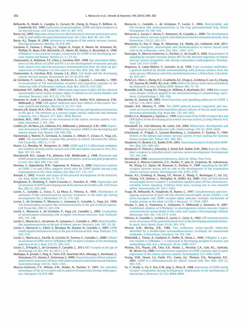
Fig. 3. Cat embryo stage 16. (a) GDNF IR in round endocrine cells of the gastricepithelium (b) GDNF IR in neurons and fibers of myenteric and submucous plexusesof posterior intestine; (c) GFR alpha 1 IR in round endocrine cells of the gastric
L. Maruccio et al. / Annals o
ontrast, and brightness of the images were adjusted to a variablextend and most of the color images were converted to gray-scale.
.5. Controls
Single immunohistochemical staining specificity was tested byuccessive substitution of the primary or secondary antisera orhe PAP complex with normal serum or PBS in repeated trials.dsorption controls were also made by using homologous and het-rologous antigens (Lucini et al., 2008).
The specificity of the immunoreactivity in double labeling withab fragments was tested by omitting the primary antibody in theecond staining, and by the two assays described below and accord-ng to Negoescu et al. (1994).
.5.1. Test 1To demonstrate the monovalence of the Fab fragment, after the
rst staining, sections were incubated for 48 h at RT with a PAP rab-it complex (A200/V, diluted:100, UCB, Braine-l’Alleud, Belgium)hich serves as the second-staining primary antibody.
.5.2. Test 2To demonstrate the complete Fab saturation of rabbit IgG epi-
opes, after the first staining, sections were incubated for 24 h at 4 ◦Cith a peroxidase-coupled donkey anti-rabbit Fab fragment (711-
36-132, diluted 1:30, Jackson) which serves as the second-stainingecondary antibody.
. Results
This study reports the presence of GDNF and its high affin-ty receptor GFR�1 in the cat gastrointestinal apparatus duringevelopment from 15–17 to 55 days of gestation, using samplest different stages. No immunopositivity to GFR�2 and GFR�3 wasetected in any developmental stages analyzed.
No reaction was observed in controls performed by substitut-ng the primary antibodies with PBS, normal serum or antibodiesdsorbed by their homologous antigens. On the contrary, the reac-ion was not modified by the substitution of primary antibodiesith antibodies adsorbed by heterologous antigens. Moreover, test
and test 2 controls were negative.In the embryos at stage 9 (Fig. 1a), immunoreactivity (IR) to
DNF was observed in few cells of neural crest extending in theesenchyme latero-ventrally to the neural tube (Fig. 1b) and in
cattered or grouped cells in the mesenchyme around the develop-ng foregut (Fig. 1c). No IR to GFR�1 was observed.
In the embryos at stage 11, GDNF IR was observed in small cellluster in the mesenchyme around the anterior gut. No IR to GFR�1as detected.
In the embryos at stage 14, in the gastric (Fig. 2a) and in anteriorntestinal region (Fig. 2e) GDNF IR was seen in neurons and nervousbers of myenteric and submucous plexuses (Fig. 2b, c, f and g).
n the posterior intestinal wall, GDNF IR was mainly detected inervous fibers and few neurons of the myenteric plexus. GDNF IRas also detected in the perinuclear cytoplasm of scattered cells
f the gastric (Fig. 2b′) and anterior gut epithelium (Fig. 2f). Theseells were round in shape and located at the base of the epithelium.FR�1 IR was observed in scattered cells in the basal epithelium of
he gastric wall (Fig. 2d). These cells appear mainly round or ovoid inhape, with positive staining in the perinuclear cytoplasm. GFR�1R was also observed in some cells of the anterior intestine (Fig. 2h).hey appeared mainly oval or elongated in shape with positivity in
he basal and apical cytoplasm.In the embryos at stage 16, GDNF IR was observed in neuronsnd fibers of the both nervous plexuses in stomach and all intesti-al tracts (Fig. 3a). In addition, IR to GDNF was frequently observed
epithelium. Scale bars = 200 �: (a); 50 �: (b) and (c).
in the perinuclear cytoplasm of scattered cells localized in the basalepithelium of the gastric wall (Fig. 3b) and more rarely in theintestinal epithelium. The positive cells were mainly round in thestomach, and elongated in the gut. Similarly, GFR�1 IR was detectedin scattered epithelial cells of the gastric (Fig. 3c) and intestinal wall.Also, these cells were mainly round in gastric and elongated in thegut epithelium.
In the foetuses at stages 19 (Fig. 4a) and 22, intense GDNFIR was observed in neurons and nervous fibers of the myentericand submucous gastric and intestinal (Fig. 4b) plexuses. GDNFimmunopositive fibers were also observed in circular muscle layer,connecting the myenteric and submucous plexuses. In addition,immunopositivity to GDNF was detected in some epithelial cellsthroughout the whole gastroenteric tract (Figs. 4c and 5d and f).In the gastric mucosa, positive cells, with IR localized in the peri-nuclear cytoplasm, were distributed in the basal epithelial region,and were round in shape. In the intestinal epithelium, GDNF IR was
detected in scattered cells, elongated or ovoid in shape. The posi-tivity was detected in the perinuclear cytoplasm, and sometimes inthe apical cytoplasmic portion.
300 L. Maruccio et al. / Annals of Anatomy 196 (2014) 296–302
Fig. 4. Cat foetus stage 19. (a) EE staining of section showing anterior and posterior intestine; (b) GDNF IR in neurons and fibers of the myenteric and submucous plexuses ofa ine ceo d (d)
(aa
Ge
dida
4
cdrs
etmt2sr
pIpGttcb
nterior intestine; (b′) Higher magnification of (b). (c) GDNF IR in elongated endocrf the anterior intestinal epithelium. Scale bars = 200 �: (a) and (b); 50 �: (b′), (c) an
GFR�1 IR was detected in few and round cells of the stomachFig. 5c) and in elongated cells of the intestinal epithelium (Figs. 4dnd 5e). The positivity was localized in the perinuclear cytoplasmnd sometimes in the apical cytoplasmic portion.
To characterize the cell epithelial population showing IR toDNF and the co-receptor, antiserum against chromogranin, anndocrine marker, was employed.
At all analyzed stages, some chromogranin positive cells (Fig. 5a)etected in the epithelium of gastrointestinal apparatus were pos-
tive to GDNF (Fig. 5b) and GFR�1. Moreover, the endocrine cellsisplayed co-presence of GFR�1 and GDNF both in gastric (Fig. 5cnd d) and intestinal tract (Fig. 5e and f).
. Discussion
This study reports the presence of GDNF and its specifico-receptor GFR�1 in the cat gastrointestinal apparatus duringevelopment. GFR�2 and GFR�3 co-receptors are not detected. Theesults obtained with positive and negative controls confirm thepecificity of the reactions.
GDNF positive cells observed at stages 9 and 11 may be consid-red as neuroblast migrating toward the gut. Indeed, it is knownhat neurons of the vertebrate ENS arise from vagal crest cells,
igrate through branchial arches in the foregut mesenchyme andhen in the gut wall (for review see Burns, 2005; Burns and Thapar,006). GDNF distribution in the cat, at stage 11, corresponds to thetart of colonization of the foregut by enteric neural crest cells inodents (Golden et al., 1999).
From stage 14 onward, GDNF is detected first in the nervouslexuses of gastric region, and then in those of intestinal tracts.
n addition, in each region, GDNF appears first in the myentericlexus, and then in the submucous plexus, Thus, the distribution ofDNF IR progresses cranio-caudally, from the stomach to different
racts of the gut, and centripetally, in an outer–inner progressionhrough the wall of the gut. These findings reflect the vagal crestell progression along the developing gut and their positioning onoth sides of the circular muscle layer in regions corresponding to
lls of the anterior intestinal epithelium; (d) GFR�1 IR in elongated endocrine cells.
the presumptive myenteric and submucousal ganglia (for reviewsee Burns, 2005; Burns and Thapar, 2006). Thus, the localizationof GDNF parallels the development of ENS in cat as reported inother mammals (Gershon, 1997; Golden et al., 1999; Schafer andMestres, 1999; Mallappa et al., 2008), and birds (Homma et al.,2000). Interestingly, other trophic factors (Maruccio et al., 2008)and neurotransmitters (Vittoria et al., 1992; Lucini et al., 1993)show similar distribution in the developing gut of duck. In addi-tion, in the cat, GDNF displays a similar distribution to RET (Luciniet al., 2013) confirming that they are both involved in the migra-tion of vagal crest cells in the gut (Young et al., 2001; Natarajanet al., 2002), and in the proliferation and neuronal differentiationof ENS precursors (Chalazonitis et al., 1998, 2012; Hearn et al., 1998;Heucheroth et al., 1998; Mwizerwa et al., 2011).
GFR�1 is completely lacking in ENS of all gastrointestinalregions of developing cat.
GFR�1 mRNA was detected in mouse (Golden et al., 1999) andzebrafish (Shepherd et al., 2004). These data could suggest thatGFR�1 mRNA might not be stored in the cytoplasm, and/or storedat a level below the detection sensibility of our demonstrationmethod. However, in the chick, GFR�1mRNA was lacking (Hommaet al., 2000) and GFR�1 protein was detected (Schiltz et al., 1999).
The lack of GFR�1 in the cat ENS could suggest that GDNF mayoperate through RET receptor that binds GFR�1, supplied in a sol-uble or trans form (Trupp et al., 1997) or by alternative binding toother co-receptor (Homma et al., 2000).
GDNF is also distributed in epithelial cells of stomach first, andintestine later, from stage 14 onward. Positive cell distribution ishigher in the gastric region, and lowers in the caudal tracts of gut.This pattern follows the cranio-caudal progression of gut develop-ment. From stage 14 onward, GFR�1 is also detected in cells of thegastric and gut epithelium, with similar localization and distribu-tion to GDNF.
Interestingly, GDNF is co-localized with GFR�1 in epithelialcells. In addition, GDNF-GFR�1 cells are also chromogranin pos-itive, and thus endocrine. These results are in agreement withdata reported in a previous study on cat pancreas (Lucini et al.,

L. Maruccio et al. / Annals of Anatomy 196 (2014) 296–302 301
Fig. 5. Cat foetus stage 22. (a) and (b) double immunostaining chromogranin/GDNF in intestinal epithelium: chromogranin (green), GDNF (red); (c) and (d) double immu-n and (G eferena
2coso
b2(r
ea
sd(2
sdet
ostaining GFR�1/GDNF in stomach epithelium: GFR�1 (green), GDNF (red); (e)
DNF(red). Scale bars = 200 �: (a) and (b); 50 �: (c)–(f). (For interpretation of the rrticle.)
008) showing GFR�1 in GDNF/chromogranin immunoreactiveells. Furthermore, the co-presence of GDNF and GFR�1 in cellsf gastroenteric epithelium strengthens the hypothesis that GDNFignal could be transduced by co-receptor GFR�1, independentlyf RET receptor (Sariola and Saarma, 2003; Airaksinen et al., 2006).
The presence of endocrine cells containing GDNF is supportedy previous findings on GDNF in adult gut of teleost (Lucini et al.,005), and other neurotrophic factors in different vertebrate gutLucini et al., 2002, 2003; Maruccio et al., 2004), and suggests otheroles aside from ENS development for neurotrophic factors.
Moreover the presence of neurotrophic factors in the gastro-ntero-pancreatic endocrine cells may help to define the intestines a kind of sensory organ as postulated by Furness et al. (1999).
In addition, the presence of GDNF and GFR�1 in endocrine cellsuggests an autocrine role for GDNF as reported in cat pancreasevelopment (Lucini et al., 2008) and in other mammalian tissuesKrieglstein et al., 1996; Belluardo et al., 1999; Lucini et al., 2003,005).
The onset of GDNF IR first in precursors of developing ENS and
ubsequently in gut endocrine cells seems to indicate a possibleouble origin of this neurotrophic factor in vagal crest cells and inpithelial gut cells. GDNF immunoreactive neurons and fibers inhe ENS may arise from vagal crest cells, whereas GDNF positivef) double immunostaining GFR�1/GDNF in intestinal epithelium: GFR�1 (green),ces to color in this figure legend, the reader is referred to the web version of this
endocrine cells may be of endodermic origin (Andrew et al., 1983,for review see Schonhoff et al., 2004).
Finally, these data complete the previous study on the catgut development (Lucini et al., 2008, 2013), confirming theinvolvement of GDNF in ENS biology during gut development inthis mammalian species also, as reported in other vertebrates(Shepherd et al., 2001).
Appendix A. Supplementary data
Supplementary data associated with this article can befound, in the online version, at http://dx.doi.org/10.1016/j.aanat.2014.03.001.
References
Airaksinen, M.S., Holm, L., Hätinen, T., 2006. Evolution of the GDNF family ligandsand receptors. Brain Behav. Evol. 68 (3), 181–190.
Andrew, A., Kramer, B., Rawdon, B.B., 1983. Gut and pancreatic amine precursor
uptake and decarboxylation cells are not neural crest derivatives. Gastroenter-ology 84 (2), 429–431.Baloh, R.H., Enomoto, H., Johnson Jr., E.M., Milbrandt, J., 2000. The GDNF family lig-ands and receptors—implications for neural development. Curr. Opin. Neurobiol.10, 103–110.
3 f Ana
B
B
B
C
C
C
d
D
E
F
G
G
H
H
H
H
K
K
L
L
L
L
L
L
L
M
M
02 L. Maruccio et al. / Annals o
elluardo, N., Mudò, G., Caniglia, G., Corsaro, M., Cheng, Q., Frasca, F., Belfiore, A.,Condorelli, D.F., 1999. Expression of neurotrophins. GDNF and their receptors inrat thyroid tissue. Cell Tissue Res. 295 (3), 467–475.
urns, A.J., 2005. Migration of neural crest-derived enteric nervous system precursorcells to and within the gastrointestinal tract. Int. J. Dev. Biol. 49, 143–150.
urns, A.J., Thapar, N., 2006. Advances in ontogeny of the enteric nervous system.Neurogastroenterol. Motility 18, 876–887.
acalano, G., Farinas, I., Wang, L.C., Hagler, K., Forgie, A., Moore, M., Armanini, M.,Phillips, H., Ryan, A.M., Reichardt, L.F., Hynes, M., Davies, A., Rosenthal, A., 1998.GFRalpha1 is an essential receptor component for GDNF in the developing ner-vous system and kidney. Neuron 21 (1), 53–62.
halazonitis, A., Rothman, T.P., Chen, J., Gershon, M.D., 1998. Age-dependent differ-ences in the effects of GDNF and NT-3 on the development of neurons and gliafrom neural crest-derived precursors immunoselected from the fetal rat gut:expression of GFRalpha-1 in vitro and in vivo. Dev. Biol. 204, 385–406.
halazonitis, A., Gershon, M.D., Greene, L.A., 2012. Cell death and the developingenteric nervous system. Neurochem. Int. 61 (6), 839–847.
e Girolamo, P., Lucini, C., Vega, J.A., Andreozzi, G., Coppola, L., Castaldo, L., 1999.Co-localization of Trk neurotrophin receptors and regulatory peptides in theendocrine cells of teleostean stomach. Anat. Rec. 256 (3), 219–226.
olatshad, N.F., Saffrey, M.J., 2007. Differential expression of glial cell line-derivedneurotrophic factor family receptor alpha-2 isoforms in rat urinary bladder andintestine. Neurosci. Lett. 415 (3), 215–218.
nomoto, H., Araki, T., Jackman, A., Heuckeroth, R.O., Snider, W.D., Johnson Jr., E.M.,Milbrandt, J., 1998. GFR alpha1-deficient mice have deficits in the enteric ner-vous system and kidneys. Neuron 21 (2), 317–324.
urness, J.B., Kunze, W.A., Clerc, N., 1999. Nutrient tasting and signaling mechanismsin the gut II. The intestine as a sensory organ: neural, endocrine and immuneresponses. Am. J. Physiol. 277, 4922–4928, Review.
ershon, M.D., 1997. Genes in the formation of the enteric nervous system. Curr.Opin. Neurobiol. 7, 101–109.
olden, J.P., De Maro, J.A., Osborne, P.A., Milbrandt, J., Johnson Jr., E.M., 1999. Expres-sion of neurturin, GDNF and GDNF family-receptor mRNA in the developing andmature mouse. Exp. Neurol. 158, 504–528.
annestad, J., Marino, F., Germanà, A., Catania, S., Abbate, F., Ciriaco, E., Vega, J.A.,2000. Trk neurotrophin receptor-like proteins in teleost Dicentrarchus labrax.Cell Tissue Res. 300 (1), 1–19.
earn, C.J., Murphy, M., Newgreen, D., 1998. GDNF and ET-3 differential modulatethe numbers of avian enteric neural crest cells and enteric neurons in vitro. Dev.Biol. 197, 93–106.
eucheroth, R.O., Lampe, P.A., Johnson, E.M., Mildbrandt, J., 1998. Neurturin andGDNF promote proliferation and survival of enteric neuron and glial progenitorsin vitro. Dev. Biol. 200, 116–129.
omma, S., Oppenheim, R.W., Yaginuma, H., Kimura, S., 2000. Expression patternof GDNF, c-ret and GFR�s suggests novel roles for GDNF ligands during earlyorganogenesis in the chick embryo. Dev. Biol. 217, 121–137.
nospe, C., 2002. Periods and stages of the prenatal development of the domesticcat. Anat. Histol. Embryol. 31, 37–51.
rieglstein, K., Deimling, F., Suter-Crazzola, C., Unsicker, K., 1996. Expression andlocalization of GDNF in developing and adult adrenal chromaffin cells. Cell TissueRes. 286 (2), 263–268.
ucini, C., Castaldo, L., Cocca, T., La Mura, E., Vittoria, A., 1993. Distribution ofsubstance P-like immunoreactive nervous structures in the duck gut duringdevelopment. Eur. J. Histochem. 37 (2), 173–182.
ucini, C., de Girolamo, P., Maruccio, L., Lamanna, C., Castaldo, L., Vega, J.A., 1999.Trk-neurotrophin receptor-like immunoreactivity in the gut of teleost species.Cell Tissue Res. 296 (2), 323–330.
ucini, C., Maruccio, L., de Girolamo, P., Vega, J.A., Castaldo, L., 2002. Localizationof neurotrophin-containing cells in higher vertebrate intestine. Anat. Embryol.205, 135–140.
ucini, C., Maruccio, L., Arcamone, N., Lamanna, C., Castaldo, L., 2003. Neurotrophin-like immunoreactivity in the gut of teleost species. Neurosci. Lett. 345, 33–36.
ucini, C., Maruccio, L., Tafuri, S., Bevaqua, M., Staiano, N., Castaldo, L., 2005. GDNFfamily ligand immunoreactivity in the gut of teleostean fish. Anat. Embryol. 210,265–274.
ucini, C., Maruccio, L., Facello, B., Cocchia, N., Tortora, G., Castaldo, L., 2008. Cellularlocalization of GDNF and its GFRalpha1/RET receptor complex in the developingpancreas of cat. J. Anat. 213 (5), 565–572.
ucini, C., D’Angelo, L., de Girolamo, P., Castaldo, L., 2013. RET receptor in the gut ofdeveloping cat. Res. Vet. Sci. 94 (1), 1–4.
allappa, A., Joseph, I., Ding, X., Torre, E.R., Sawchuk, M.A., Mwangi, S., Hochman, S.,Sitaraman, S.V., Anania, F., Srinivasan, S., 2008. Characterization of fetal and post-
natal enteric neuronal cell lines with improvement in intestinal neural function.Gastroenterology 134 (5), 1424–1435.arcos-Gutierrez, C.V., Wilson, S.W., Holder, N., Pachins, V., 1997. The zebrafishhomologue of the ret receptor and its pattern of expression during embryogen-esis. Oncogene 14, 879–889.
tomy 196 (2014) 296–302
Maruccio, L., Castaldo, L., de Girolamo, P., Lucini, C., 2004. Neurotrophin andTrk-receptor like immunoreactivity in the frog gastrointestinal frog. Histol.Histopathol. 19, 349–356.
Maruccio, L., Lucini, C., Russo, F., Antonucci, R., Castaldo, L., 2008. The developmentof avian enteric nervous system: distribution of artemin immunoreactivity. ActaHistochem. 110 (2), 163–171.
Mwizerwa, O., Das, P., Nagy, N., Akbareian, S.E., Mably, J.D., Goldstein, A.M., 2011.GDNF is mitogenic, neurotrophic and chemoattractive to enteric neural crestcells in the embryonic colon. Dev. Dyn., 1402–1411.
Natarajan, D., Marcos-Gutierrez, C., Pachnis, V., de Graaff, E., 2002. Requirement ofsignaling by receptor tyrosine kinase RET for the directed migration of entericnervous system progenitor cells during mammalian embryogenesis. Develop-ment 129, 5151–5160.
Negoescu, A., Labat-Moleur, F., Lorimier, et al., 1994. F(ab) secondary antibodies:a general method for double immunolabeling with primary antisera from thesame species. Efficiency control by chemiluminescence. J. Histochem. Cytochem.42, 433–437.
Pichel, J.G., Shen, L., Sheng, H.Z., Granholm, A.C., Drago, J., Grinberg, A., Lee, E.J., Huang,S.P., Saarma, M., Hoffer, B.J., et al., 1996. Defects in enteric innervation and kidneydevelopment in mice lacking GDNF. Nature 382, 73–76.
Reynolds, G.M., Young, K.I., Young, J.A., William, A., Rowlands, D.C., 1994. Microwaveoven antigen retrieval applied to the immunostaining of cytopathology speci-mens. Cytopathology 5, 345–358.
Sariola, H., Saarma, M., 2003. Novel functions and signalling pathways for GDNF. J.Cell Sci. 116, 3855–3862.
Schafer, K.H., Mestres, P., 1999. The GDNF-induced neurite outgrowth and neu-ronal survival in dissociated myenteric plexus cultures of the rat small intestinedecreases postnatally. Exp. Brain Res. 125, 447–452.
Schiltz, G.A., Benjamin, J., Epstein, L., 1999. Expression of the GDNF receptors Ret andGFR alpha1 in the developing avian enteric nervous system. J. Comp. Neurol. 414,193–211.
Schonhoff, S.E., Giel-Moloney, M., Leiter, A.B., 2004. Minireview: development anddifferentiation of gut endocrine cells. Endocrinology 145 (6), 2639–2644.
Schuchardt, A., D’Agati, V., Larsson-Blomberg, L., Costantini, F., Pachins, V., 1994.Defects in the kidney and enteric nervous system of mice lacking the tyrosinekinase receptor Ret. Nature 367, 380–383.
Shepherd, I.T., Beattie, C.E., Raible, D.W., 2001. Functional analysis of zebrafish GDNF.Dev. Biol. 231, 420–435.
Shepherd, I.T., Pietsch, J., Elworthy, S., Kelsh, R.N., Raible, D.W., 2004. Roles for GFRal-pha1 receptors in zebrafish enteric nervous system development. Development131, 241–249.
Sternberger, 1986. Immunocytochemistry, third ed. Wiley, New York.Taraviras, S., Marcos-Gutierrez, C.V., Durbec, P., Jani, H., Grigoriou, M., Sukumaran,
M., Wang, L.C., Hynes, M., Raisman, G., Pachins, V., 1999. Signalling by the RETreceptor tyrosine kinase and its role in the development of the mammalianenteric nervous system. Development 126, 2785–2797.
Tomac, A.C., Grinberg, A., Huang, S.P., Nosrat, C., Wang, Y., Borlongan, C., Lin, S.Z.,Chiang, Y.H., Holson, L., Westphal, H., Hoffer, B.J., 2000. Glial cell line-derivedneurotrophic factor receptor (1 availability regulates glial cell line-derived neu-rotrophic factor signaling: evidence from mice carrying one or two mutatedalleles. Neuroscience 95, 1011–1023.
Trupp, M., Belluardo, N., Funakoshi, H., Ibanez, C., 1997. Complementary and over-lapping expression of glial cell line-derived neurotrophic factor (GDNF), c-retproto-oncogene and GDNF receptor-alpha indicates multiple mechanism oftrophic actions in the adult rat CNS. J. Neurosci. 17, 3554–3567.
Uesaka, T., Jain, S., Yonemura, S., Uchiyama, Y., Milbrandt, J., Enomoto, H., 2007.Conditional ablation of GFRallpha1 in postmigratory enteric neurons triggersunconventional neural death in the colon and causes a Hirschsprung’s diseasephenotype. Dev. Dis. 134, 2171–2181.
Vittoria, A., Castaldo, L., La Mura, E., Lucini, C., Cecio, A., 1992. VIP-immunoreactivenerve structures of the gastrointestinal tract in the developing and adult domes-tic duck. Arch. Histol. Cytol. 55 (4), 361–374.
Wessel, G.M., McClay, D.R., 1986. Two embryonic tissue-specific moleculesidentified by a double-label immunofluorescence technique for monoclonalantibodies. J. Histochem. Cytochem. 34, 703–706.
Widenfalk, J., Tomac, A., Lindqvist, E., Hoffer, B., Olson, L., 1998. GFRalpha-3, a pro-tein related to GFRalpha-1, is expressed in developing peripheral neurons andensheathing cells. Eur. J. Neurosci. 10 (4), 1508–1517.
Worley, D.S., Pisano, J.M., Choi, E.D., Walus, L., Hession, C.A., Cate, R.L., Sanicola,M., Birren, S.J., 2000. Developmental regulation of GDNF response and receptorexpression in the enteric nervous system. Development 127, 4383–4393.
Young, H.M., Hearn, C.J., Farlie, P.G., Canty, A.J., Thomas, P.Q., Newgreen, D.F.,
2001. GDNF is a chemoattractant for enteric neural cells. Dev. Biol. 229 (2),503–516.Yu, T., Scully, S., Yu, Y., Fox, G.M., Jing, S., Zhou, R., 1998. Expression of GDNF familyreceptor components during development: implications in the mechanisms ofinteraction. J. Neurosci. 18 (12), 4684–4696.
Related Documents