Gating electron flow in electron bifurcating enzymes Journal: ChemComm Manuscript ID CC-FEA-07-2019-005611.R1 Article Type: Feature Article ChemComm

Welcome message from author
This document is posted to help you gain knowledge. Please leave a comment to let me know what you think about it! Share it to your friends and learn new things together.
Transcript
Gating electron flow in electron bifurcating enzymes
Journal: ChemComm
Manuscript ID CC-FEA-07-2019-005611.R1
Article Type: Feature Article
ChemComm
Journal Name
ARTICLE
This journal is © The Royal Society of Chemistry 20xx J. Name., 2013, 00, 1-3 | 1
Please do not adjust margins
Please do not adjust margins
Received 00th January 20xx,Accepted 00th January 20xx
DOI: 10.1039/x0xx00000x
www.rsc.org/
Electron Bifurcation: Progress and Grand ChallengesJonathon L. Yulya+, Carolyn E. Lubnerb+*, Peng Zhangc, David N. Beratana,c,d*, and John W. Peterse*
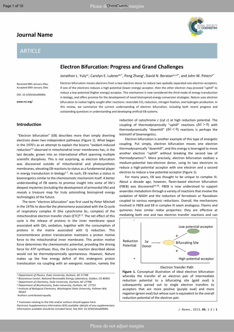
Electron bifurcation moves electrons from a two-electron donor to reduce two spatially separated one-electron acceptors. If one of the electrons reduces a high-potential (lower energy) acceptor, then the other electron may proceed “uphill” to reduce a low-potential (higher energy) acceptor. This mechanism is now considered the third mode of energy transduction in biology, and offers promise for the development of novel bioinspired energy conversion strategies. Nature uses electron bifurcation to realize highly sought-after reactions: reversible CO2 reduction, nitrogen fixation, and hydrogen production. In this review, we summarize the current understanding of electron bifurcation, including both recent progress and outstanding questions in understanding and developing artificial EB systems.
Introduction
“Electron bifurcation” (EB) describes more than simply diverting electrons down two independent pathways (Figure 1). What began in the 1970’s as an attempt to explain the bizarre “oxidant-induced reduction”3 observed in mitochondrial inner membranes has, in the last decade, grown into an international effort spanning multiple scientific disciplines. This is not surprising, as electron bifurcation was discovered outside of mitochondrial and photosynthetic membranes, elevating EB reactions to status as a fundamental player in energy transduction in biology5-7. As such, EB reaches a status in bioenergetics similar to the chemiosmotic mechanism itself. A better understanding of EB seems to promise insight into some of life’s deepest mysteries (including the development of primordial life) and reveals a treasure map for truly astonishing bioinspired energy technologies of the future.
The term “electron bifurcation” was first used by Peter Mitchell in the 1970s to describe the phenomena associated with the Q-cycle of respiratory complex III (the cytochrome bc1 complex) of the mitochondrial electron transfer chain (ETC)8, 9. The net effect of this cycle is the release of protons in the inner membrane space associated with QH2 oxidation, together with the consumption of protons in the matrix associated with Q reduction. This transmembrane proton translocation maintains a proton motive force to the mitochondrial inner membrane. This proton motive force determines the chemiosmotic potential, providing the driving force for ATP synthase; thus, the Q-cycle reaction described above would not be thermodynamically spontaneous. However, Nature makes up the free energy deficit of this endergonic proton translocation via coupling with an exergonic reaction, namely the
reduction of cytochrome c (cyt c) at high reduction potential. The coupling of thermodynamically “uphill” reactions ( ) with ∆𝐺 > 0thermodynamically “downhill” ( ) reactions is perhaps the ∆𝐺 < 0leitmotif of bioenergetics.
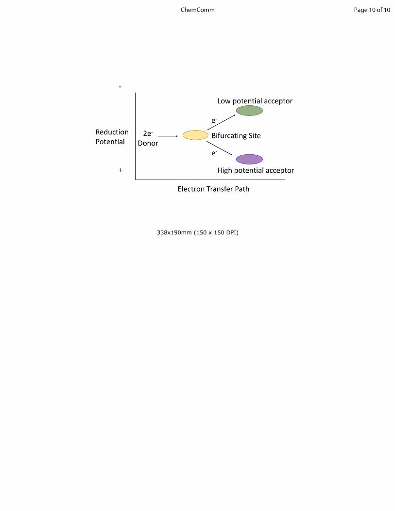
Electron bifurcation is another example of this type of energetic coupling. Put simply, electron bifurcation moves one electron thermodynamically “downhill”, and this energy is leveraged to move another electron “uphill” without breaking the second law of thermodynamics12. More precisely, electron bifurcation oxidizes a medium-potential two-electron donor, using its two electrons to reduce a high-potential acceptor with one electron and a second electron to reduce a low potential acceptor (Figure 1).
For many years, EB was thought to be unique to complex III. About a decade ago, however, flavin-based electron bifurcation (FBEB) was discovered14-18. FBEB is now understood to support anaerobic metabolism through a variety of reactions that involve the oxidation of NADH and the reduction of ferredoxin or flavodoxin coupled to various exergonic reductions. Overall, the mechanisms involved in FBEB and EB in complex III seem analogous. Flavins and quinones have similar redox properties: they are effective at mediating both one and two electron transfer reactions and can
a.Department of Physics, Duke University, Durham, NC 27708b.Biosciences Center, National Renewable Energy Laboratory, Golden, CO 80401c. Department of Chemistry, Duke University, Durham, NC 27708d.Department of Biochemistry, Duke University, Durham, NC 27710e. Institute of Biological Chemistry, Washington State University, Pullman WA,
99163 +Authors contributed equally
† Footnotes relating to the title and/or authors should appear here. Electronic Supplementary Information (ESI) available: [details of any supplementary information available should be included here]. See DOI: 10.1039/x0xx00000x
Figure 1. Conceptual illustration of ideal electron bifurcation whereby the transfer of an electron pair of intermediate reduction potential to a bifurcating site (gold oval) is subsequently parsed out to single electron transfers to acceptors that are more positive (purple oval) and more negative (green oval) but whose sum is equivalent to the overall reduction potential of the electron pair.
Page 1 of 10 ChemComm
ARTICLE Journal Name
2 | J. Name., 2012, 00, 1-3 This journal is © The Royal Society of Chemistry 20xx
Please do not adjust margins
Please do not adjust margins
function by employing inverted reduction potentials (we use the term “inverted” rather than the newer term “crossed” in this review to remind the reader of an older and separate literature on the subject19-23) with highly reducing intermediates.
Electron bifurcation is now understood to drive some of the most valued reactions in all of chemistry. For instance, nature uses the high energy electrons generated by EB to drive CO2 reduction24, nitrogen fixation2, 25, and hydrogen production26, 27 at biological temperatures and pressures, in addition to its role in the Q-cycle where it contributes to the chemiosmotic potential, that in turn drives ATP synthase. Furthermore, it is claimed that many electron bifurcases are reversible14, 16, 28-31, many (but not all14) in vivo. This suggests that it is possible for the energy interconversion associated with these reactions to be accomplished with near 100% efficiency18. This is not science fiction; these EB reactions occur in nature.
Because of the use of EB in living systems, we believe that astonishing insights for bioinspired energy technologies may lurk within electron bifurcases. The low-potential electron produced by EB could in principle be guided to a targeted acceptor (as long as that acceptor has a reduction potential similar to or higher than the reduction potential of the low potential pathway). So EB might be used to drive many reactions of vital importance to the energy and environmental sciences. Indeed, there seems no reason to doubt that, in principle, any low potential acceptor could be reduced in an EB reaction.
Unfortunately, the simplicity and elegance of biological EB masks the extreme difficulty of deploying EB in artificial systems. If all the redox species to be used in electron bifurcation were added to a mixture and electrons fed from an electrode, they would not perform electron bifurcation. Instead, most electrons would flow to the high-potential acceptors, as this configuration achieves the lowest free energy. Thus, to achieve electron bifurcation, the electrons must be “gated” towards the proper acceptors, despite a large driving force for “short circuit” reactions12, 30. Biology must therefore exert precise microscopic control to realize electron bifurcation. Understanding this control is a major motivation behind the study of biological EB.
The discoveries of EB outside of complex III have been a gold mine for mechanistic studies on the microscopic control wielded by biological EB. These reactions are performed in a variety of chemical and physical contexts, including in hyperthermophiles. For instance, many examples of biological EB do not require membrane-bound proteins and seem to play roles quite different from those of complex III. These EB systems also use a variety of schemes to channel electron flow down the endergonic and exergonic reaction paths. Thus, biology not only achieves astonishing microscopic control to accomplish EB, but also appears to have evolved multiple mechanistic routes to achieve this control. This multiplicity of options for control suggests that EB may constitute some of the most fertile ground for inspiring biomimetic energy technologies.
Structural and mechanistic studies on the cytochrome bc1 complex32-35, and more recently on FBEB1, 2, 15, 27, 36-41, reveal two basic requirements that are paramount in EB catalysis. The first major requirement is the use of an energetic (highly reducing) species capable of driving the reduction of the endergonic (compared to the mean reduction potential of the two-electron donor) half reaction. This is accomplished in the cytochrome bc1 complex and FBEB through the formation of a metastable SQ
intermediate. The presence of a metastable low-potential intermediate is a signature of inverted (or “crossed”) reduction potentials1, 6, 9, 12. A variety of different multiple electron redox cofactors may exhibit inverted potentials, including metal containing species20-23. The second requirement is an effective mechanism to gate electron flow, to direct the energetic electron toward the low-potential pathway with high fidelity.
In our view, much understanding of the bifurcating steps in these enzymatic reactions has been gained. Indeed, flavin-based EB has been reviewed many times7, 14-18, 24, 31, 42-45. However, there remain many unanswered questions surrounding the EB mechanisms and the biology of EB. The answers to these questions could enable significant advances in bioenergetics and energy science more broadly. We outline (in the second half of this review) several important conceptual issues that prevent a complete understanding of the molecular mechanisms behind EB, and that prevent the bioinspired inventor from taking further advantage of EB reactions. The opportunities and unresolved issues discussed here reflect the authors’ tastes, and other productive directions for exploration exist as well. To summarize, this Feature Article elaborates the recent progress in understanding biological electron gating mechanisms used by EB enzymes, in the context of the parameters defined by electron transfer theory, discusses unresolved questions in this field, and explains why we find these questions compelling: a “new era”17 for electron bifurcation truly lies ahead.
Gating electron flow
Gating electron flow in EB is vital, directing electron flow from one donor to two spatially separate acceptors, and preventing both electrons from proceeding to the overall high-potential (lower energy) acceptor. This allows the EB enzyme to minimize the free energy lost through the electron flow. Rates of electron-transfer events are well approximated using the Marcus theory of electron transfer46 and are influenced by distance, thermodynamic driving force, and dielectric environment. For instance, the nonadiabatic (tunneling) electron transfer rate in the small electronic coupling regime is given by
𝑘𝐸𝑇 =2𝜋ℏ ⟨|𝐻𝐷𝐴|2⟩𝐹𝐶. (1)
is the Frank-Condon factor, which in the high temperature limit 𝐹𝐶is given by47
𝐹𝐶 =1
4𝜋𝜆𝑘𝑇𝑒𝑥𝑝[ ― (∆𝐺 ∘ + 𝜆)2/(4𝜆𝑘𝑇)], (2)
The factor is the temperature averaged electronic coupling, ⟨|𝐻𝐷𝐴|2⟩often falling exponentially with electron donor/acceptor edge-to-edge distance48, is the reorganization energy, and is the 𝜆 ∆𝐺0
standard reaction free energy of the electron transfer.
Page 2 of 10ChemComm
Journal Name ARTICLE
This journal is © The Royal Society of Chemistry 20xx J. Name., 2013, 00, 1-3 | 3
Please do not adjust margins
Please do not adjust margins
Presumably, during the turnover of an electron bifurcase, electron gating mechanisms are achieved by modulating parameters that determine electron transfer rates, above. For example, the distance between the bifurcating donor and one of the acceptors might change during EB to allow the bifurcating site to deliver individual electrons to the two spatially separated acceptors in a highly selective manner. Side reactions that would disrupt the equal portioning of electrons into the high and low potential acceptors are known as “short circuits”30.
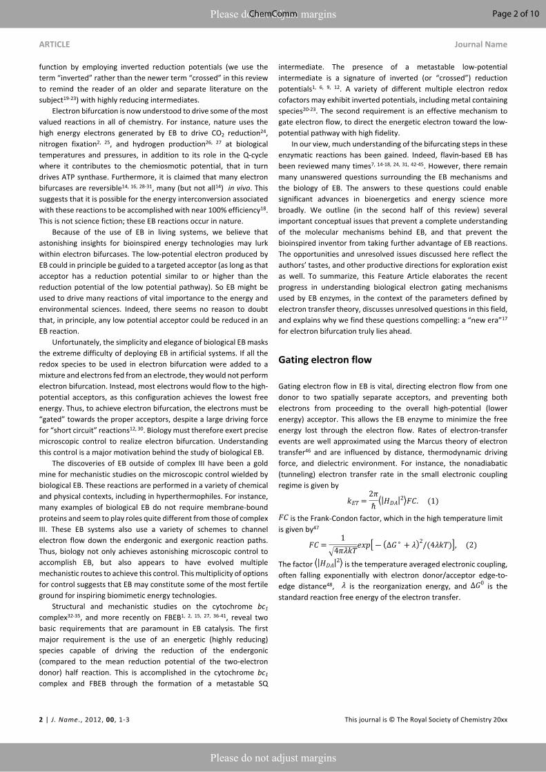
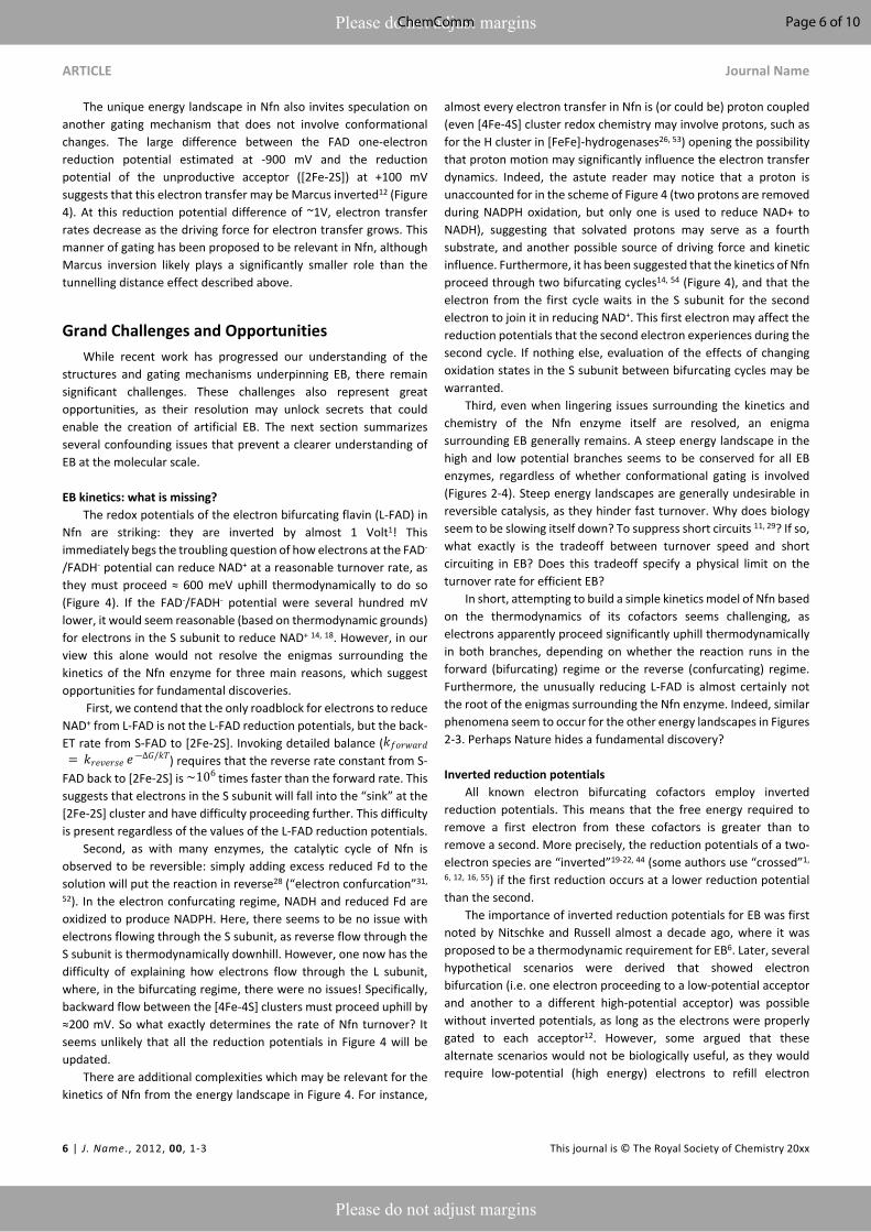
Conformational electron gatingElectron bifurcation is employed within the Q-cycle, coupling the oxidation of quinol (QH2) to quinone (Q) at the Qo site with reduction of the low-potential heme and the high-potential Rieske-type iron-sulfur (FeS) cluster. The reduction of the low-potential heme generates a driving force for quinone to become reduced again at the Qi site49, accumulating over two bifurcating cycles (Figure 2). The proton-coupled oxidation of QH2 and reduction of Q occur against a transmembrane proton motive force. However, this uphill process is coupled to the reduction of the high potential cytochrome c which
allows the Q-cycle to occur spontaneously. The net result of the Q-cycle is that half of the electrons that travel through the ETC are briefly detoured through the Q-cycle to generate additional proton motive force.
A first oxidation of QH2 results in the production of a metastable SQ state with low oxidation potential. The oxidation of QH2 to SQ
occurs at a potential of almost +500 mV4. The reduction of the Rieske FeS at a reduction potential of ~+275 mV, and subsequent reduction of cyt c1 at ~+250 mV, occur against an electrochemical potential gradient, so they are kinetically limited but feasible at the appropriate equilibrium conditions49. Since the overall reduction potential of QH2 to Q is about +100 mV and represents the average of the QH2 to SQ and the SQ to Q couples, this translates to a SQ to Q transition at a reduction potential more negative than -300 mV4, 10,
11, which is more than ample to drive the reduction of Q in a stepwise manner that is mediated by the cytochrome b (cyt b) hemes bL and bH. The reduction of the Rieske FeS cluster, assuming that it has transferred an electron to cyt c1, is more favored and should occur at a higher rate if the protein conformation and the relative distances of the Rieske FeS cluster and the nearest cyt b site remain unchanged. The complex, however, undergoes a conformational
Figure 2. Energy landscape (left) and depiction of electron transfers and conformational changes (right) in the cytochrome bc1 complex (complex III). Cofactors with low (more negative) reduction potentials are shown in green, and the cofactors with high (more positive) potential in purple. Quinone at the Qi site is shown in grey, and the fully oxidized Q at the Qo site is shown in yellow. A first (1-3) and second (4-6) electron bifurcating cycle complete one enzymatic turnover of complex III, resulting in one (net) QH2 oxidation, two reduced cytochrome c (cyt c), and two protons transduced across the membrane. In (1), quinone is oxidized at the Qo site, reducing the high-potential Rieske FeS cluster. Then (2) the complex undergoes a conformational change, so the Rieske center is in position to reduce cyt c1, and the low-potential semiquinone Q•- is no longer in range to reduce the Rieske center, instead reducing the low potential heme bL. Another QH2 from the Q pool (3) replaces oxidized Q at the Qi site, and (4-6) the process repeats again, allowing Q to be fully reduced to QH2 at the Qi site. Cofactor reduction potentials from Bergdoll et al.4 from measurements of submitochondrial particles from R. sphaereoides in the absence of the membrane potential. The Q/SQ and SQ/QH2 couples were estimated to be roughly as shown in this figure by Zhang et al.10 and later by Crofts et al.11 Cytochrome c redox properties were reviewed by Battistuzzi et al.13
Page 3 of 10 ChemComm
ARTICLE Journal Name
4 | J. Name., 2012, 00, 1-3 This journal is © The Royal Society of Chemistry 20xx
Please do not adjust margins
Please do not adjust margins
change that increases the distance between the SQ oxidation site and the Rieske center and, as a result, the balance is tipped to kinetically favor reduction at the cyt b site32, 34, 35 (Figure 2). This conformational change, however, is not sufficient to suppress the rate constants of all possible short-circuits29.
Mechanistically, the cytochrome bc1 complex has manipulated the energy landscapes of the two half reactions (the exergonic reduction of cyt c by QH2 and the endergonic coupling of the oxidation of QH2 in the inner membrane space to the reduction of Q in the matrix) so that the electron flux through the b hemes to reduce Q occurs against an electrochemical potential gradient. Specifically, the first electron transfer from QH2 to the Rieske cluster is rate limiting, and the downhill flow of electrons from the semiquinone Q•- to the bH occurs quickly, suppressing semiquinone (Q•- ) population at the Qo site11. However, It is important to note that this gating mechanism is not sufficient to suppress the rate constants for all “short circuit” reactions in complex III, but perhaps in combination with at least one other gating mechanism30. The other short-circuits arise as a result of the physiological conditions (near equilibrium) leading to the reversibility of the Q-cycle29.
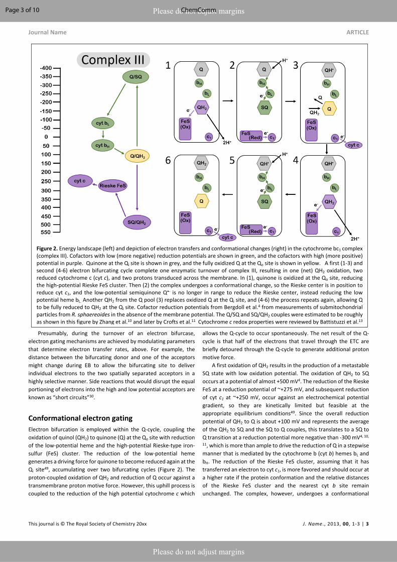
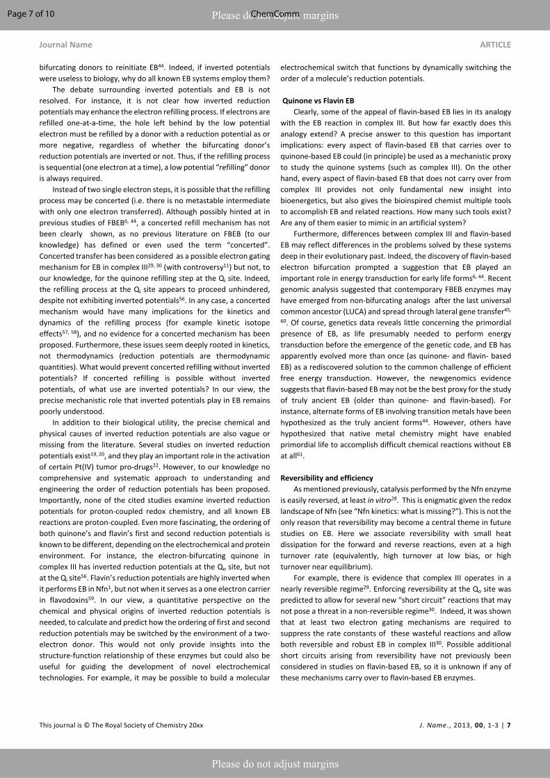
It has been proposed that the electron bifurcating flavoproteins operate in a manner that invokes conformational changes similar to those found in the cytochrome bc1 complex38, 39. For example, the FixABCX enzyme (Figure 3), which produces reducing equivalents for nitrogen fixation, couples the oxidation of nicotinamide adenine dinucleotide (NADH) to the reduction of quinone and to the reduction of the low potential electron carriers ferredoxin (Fd) or flavodoxin (Fld)14, 16, 50. Biochemical studies have revealed that the energy landscapes are likely to be qualitatively similar for the EB flavoproteins and for the cytochrome bc1 complex2, in the sense that the first electron transfer step from the EB cofactor is endergonic (along the overall exergonic pathway), and that it is the second electron that proceeds through the overall low-potential pathway.
The oxidation of NADH is coupled to the reduction of the bifurcating flavin. The oxidation of the hydroquinone form of the flavin (FADH-) is coupled to the reduction of a flavin site along the path toward Q2. This occurs at a relatively high (positive) reduction potential and produces a metastable flavin ASQ intermediate (FAD-) at the bifurcating site with sufficient driving force to affect the reduction of Fd or Fld with reduction potentials of ~-500 mV.
It was proposed that conformational changes analogous to those in the cytochrome bc1 complex may be invoked for FBEB. Structural and biochemical results on the enzyme butyryl CoA reductase39 and caffeyl-CoA dehydrogenase38 support a model in which the distance between the proximal electron acceptor along the high potential pathway is increased after the first electron transfer, resulting in a decrease in the rate of a second electron transfer down the same (high potential) pathway and favoring the transfer of the second electron down the low potential pathway toward ferredoxin reduction. It is presumed that the homologous FixABCX/Electron transfer flavoprotein (EtfABCX) complexes2, 41, 50 undergo similar conformational changes in which the relative distances of the nearest acceptors along the low and high potential pathways are modulated in a manner that causes the reduction of the low potential acceptor to be kinetically favored. This conformational change could hypothetically be realized by increasing the distance to the nearest high potential acceptor and/or decreasing the distance to the nearest low potential acceptor. The FixABCX (Figure 3) enzyme couples the oxidation of FAD HQ to the reduction of flavodoxin for use in nitrogenase catalysis2, and the reduction of quinone. EtfABCX couples the reduction of FAD HQ with the reduction of quinone and ferredoxin41. Both FixABCX and EtfABCX utilize NADH to initially reduce the bifurcating FAD site.
Gating electron transfer by cofactor placement
Figure 3. Energy landscape (left) and depiction of proposed electron transfers and conformational changes (right) in the FixABCX complex. Cofactors with low (more negative) reduction potentials are shown in green, and the cofactors with high (more positive) reduction potentials in purple. Fully oxidized FAD is shown in yellow. Analogous to complex III (Figure 2), the turnover of FixABCX proceeds via two electron bifurcating cycles (1-3) and (4-6). The net result is the two oxidations of NADH to NAD+, two reductions of ferredoxin (or flavodoxin), and one reduction of Q to QH2. The (1) oxidation of FADH- by one electron and transfer to the B flavin triggers (2) a conformational change increasing the distance between B flavin and/or decreasing the distance between the FixX FeS cluster and the bifurcating flavin. This makes the (3) rate of electron transfer from the A flavin along the low potential path to the reduction of Fld via FixX FeS cluster faster than the rate of transfer for the second electron from the A flavin to the B and C flavins. After reduction of the A flavin by NADH, the B flavin (4) returns to its original position within electron transfer range of the bifurcating FAD, and dissociation of reduced Fld initiates the second round of electron bifurcation (4-6). Energy landscape proposed by Ledbetter et al2.
Page 4 of 10ChemComm
Journal Name ARTICLE
This journal is © The Royal Society of Chemistry 20xx J. Name., 2013, 00, 1-3 | 5
Please do not adjust margins
Please do not adjust margins
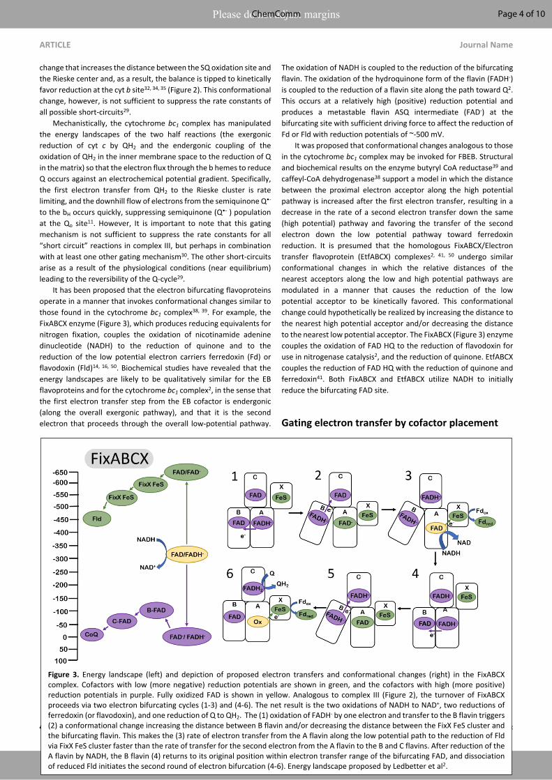
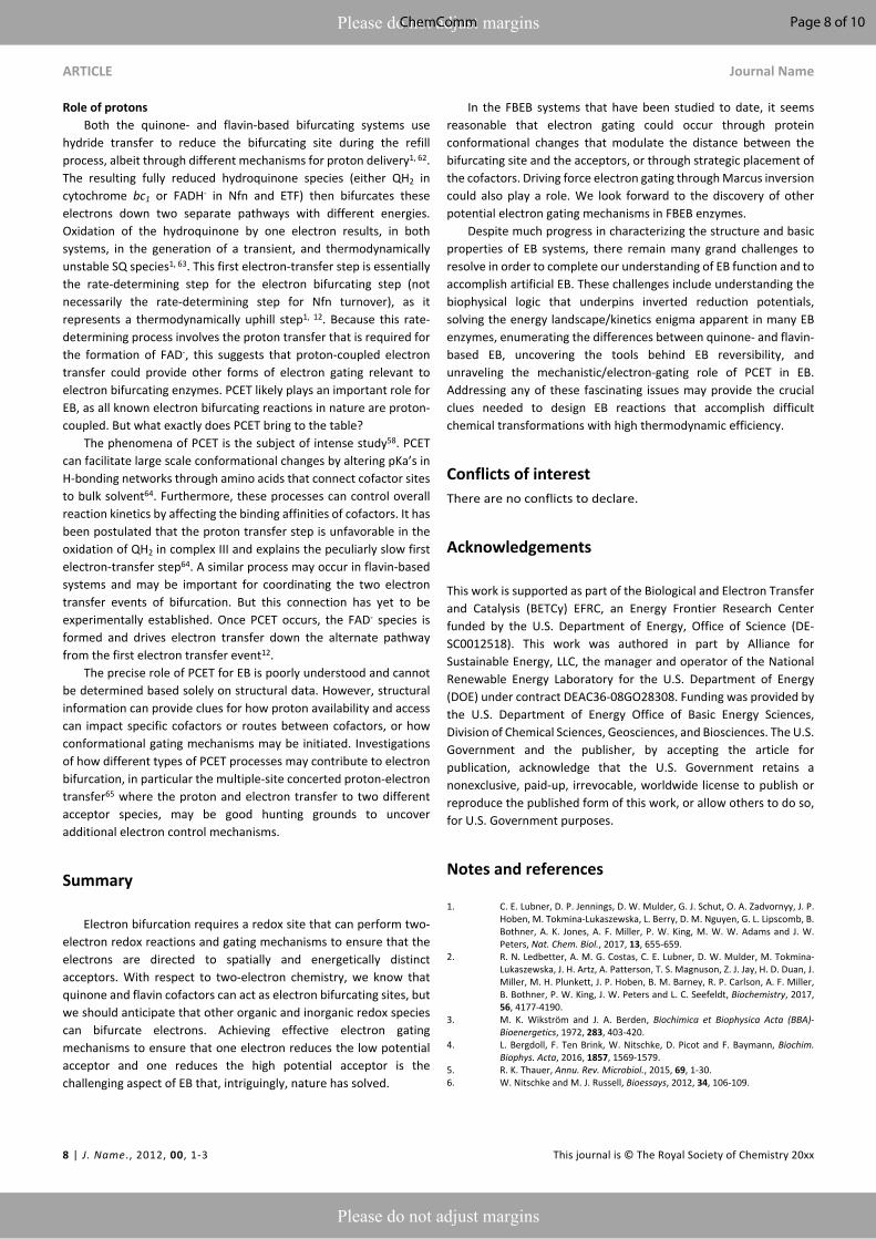
The NADH-dependent ferredoxin-NADP+ oxidoreductase (Nfn) is another well studied FBEB enzyme. The enzyme couples the oxidation of NADPH to the exergonic reduction of NAD+ and the endergonic reduction of Fd. In contrast to the cytochrome bc1 complex and bifurcating Fix/Etf enzymes, Nfn is a simple dimeric complex with a large and small subunit1, 51 (Figure 4). The bifurcating flavin is in the large (L) subunit which also harbours two low potential [4Fe-4S] clusters and the site for Fd reduction. The small (S) subunit harbours a single [2Fe-2S] cluster and an additional flavin near the site of NAD+ reduction. The clusters in the dimer are arranged such that the bifurcating flavin is central, near the L and S subunit interface.
The energy landscape of Nfn based on biochemical and spectroscopic studies is qualitatively similar yet quantitatively inflated compared with the energy landscape of the cytochrome bc1 complex and the Fix/Etf enzymes, encompassing a much larger range of reduction potentials. During catalysis, the bifurcating site is reduced by NADPH and the bifurcating flavin transfers the first electron to a [2Fe-2S] cluster on the path to NAD+ reduction (overall exergonic pathway). In a similar manner as in the cytochrome bc1 complex and Fix, transfer of the first electron occurs at a relatively
high (positive) reduction potential, leading to a highly energetic low (negative) potential intermediate. The measured reduction potentials of the [4Fe-4S] clusters along the endergonic pathway to Fd reduction are very low, with the cluster proximal to the bifurcating site estimated at ~-700 mV. This cluster is within 8 Å of the
bifurcating site, and the rate of electron transfer from the bifurcating flavin to this cluster was determined experimentally to be very fast, in the range of 10 ps1. These parameters imply that the reduction potential of the energetic FAD- is more negative than -900 mV, allowing an estimate of the complete redox energy landscape (Figure 4). The two electron FAD FADH- reduction potential of ~-300 mV, and the one-electron FAD FAD- couple estimated at -900 mV, places the FAD- → FADH- redox couple at ~+350 mV, resulting in an energy landscape that spans more than 1V in electrochemical potential, about 2-fold larger than the span of electrochemical potentials accessed by cytochrome bc1 and the bifurcating ETFs. Also unique to Nfn is the lack of experimental evidence indicating large scale conformational changes that could serve to gate electrons as in cytochrome bc1 and bifurcating ETFs, indicating a need for a different mechanism for electron gating to avoid short circuits (i.e. both electrons flowing down the exergonic path).
The gating mechanism that is likely employed by Nfn arises from the electron acceptor placement near the electron bifurcating L-FAD12. The first electron proceeds from L-FAD to reduce the [2Fe-2S] cluster, as the alternative reduction of the [4Fe-4S] cluster would be significantly uphill. Once this first electron has transferred, the highly
reducing FAD- can reduce the [4Fe-4S] cluster. Why does the second electron not follow the first to reduce [2Fe2S]? It may, but the rate of [4Fe-4S] reduction has been estimated to be orders of magnitude faster12, as the tunneling distance to [4Fe-4S] is about 7 Å closer to L-FAD than to [2Fe-2S]1.
Figure 4. Energy landscape (left) and depiction of proposed electron transfers and conformational changes (right) in the NADH-dependent ferredoxin NADP+ oxidoreductase (Nfn). Cofactors with low (more negative) reduction potentials are shown in green, and the cofactors with high (more positive) reduction potentials in purple. The oxidized form of the electron bifurcating flavin (FAD) is shown in yellow. The oxidation of FADH2 by one electron (1) results in the formation of FAD- with an extremely negative reduction potential. Since [4Fe-4S] is much closer to the electron bifurcating flavin than [2Fe-2S], this makes the (2) rate of electron transfer along the low potential path to the reduction of Fd via FeS clusters faster than the short circuit transfer from FAD- to [2Fe-2S] in the S subunit. (3) A second reduction of the bifurcating FAD and dissociation of reduced Fd initiates (4-6) the second round of electron transfer steps analogous to (1-3) resulting in the formation of an additional reduced Fd and completing the reduction of NAD+ to NADH. Cofactor reduction potentials shown from Lubner et al.1
Page 5 of 10 ChemComm
ARTICLE Journal Name
6 | J. Name., 2012, 00, 1-3 This journal is © The Royal Society of Chemistry 20xx
Please do not adjust margins
Please do not adjust margins
The unique energy landscape in Nfn also invites speculation on another gating mechanism that does not involve conformational changes. The large difference between the FAD one-electron reduction potential estimated at -900 mV and the reduction potential of the unproductive acceptor ([2Fe-2S]) at +100 mV suggests that this electron transfer may be Marcus inverted12 (Figure 4). At this reduction potential difference of ~1V, electron transfer rates decrease as the driving force for electron transfer grows. This manner of gating has been proposed to be relevant in Nfn, although Marcus inversion likely plays a significantly smaller role than the tunnelling distance effect described above.
Grand Challenges and OpportunitiesWhile recent work has progressed our understanding of the
structures and gating mechanisms underpinning EB, there remain significant challenges. These challenges also represent great opportunities, as their resolution may unlock secrets that could enable the creation of artificial EB. The next section summarizes several confounding issues that prevent a clearer understanding of EB at the molecular scale.
EB kinetics: what is missing?The redox potentials of the electron bifurcating flavin (L-FAD) in
Nfn are striking: they are inverted by almost 1 Volt1! This immediately begs the troubling question of how electrons at the FAD-
/FADH- potential can reduce NAD+ at a reasonable turnover rate, as they must proceed ≈ 600 meV uphill thermodynamically to do so (Figure 4). If the FAD-/FADH- potential were several hundred mV lower, it would seem reasonable (based on thermodynamic grounds) for electrons in the S subunit to reduce NAD+ 14, 18. However, in our view this alone would not resolve the enigmas surrounding the kinetics of the Nfn enzyme for three main reasons, which suggest opportunities for fundamental discoveries.
First, we contend that the only roadblock for electrons to reduce NAD+ from L-FAD is not the L-FAD reduction potentials, but the back-ET rate from S-FAD to [2Fe-2S]. Invoking detailed balance (𝑘𝑓𝑜𝑟𝑤𝑎𝑟𝑑
) requires that the reverse rate constant from S- = 𝑘𝑟𝑒𝑣𝑒𝑟𝑠𝑒 𝑒 ―∆𝐺/𝑘𝑇
FAD back to [2Fe-2S] is times faster than the forward rate. This ~106
suggests that electrons in the S subunit will fall into the “sink” at the [2Fe-2S] cluster and have difficulty proceeding further. This difficulty is present regardless of the values of the L-FAD reduction potentials.
Second, as with many enzymes, the catalytic cycle of Nfn is observed to be reversible: simply adding excess reduced Fd to the solution will put the reaction in reverse28 (“electron confurcation”31,
52). In the electron confurcating regime, NADH and reduced Fd are oxidized to produce NADPH. Here, there seems to be no issue with electrons flowing through the S subunit, as reverse flow through the S subunit is thermodynamically downhill. However, one now has the difficulty of explaining how electrons flow through the L subunit, where, in the bifurcating regime, there were no issues! Specifically, backward flow between the [4Fe-4S] clusters must proceed uphill by ≈200 mV. So what exactly determines the rate of Nfn turnover? It seems unlikely that all the reduction potentials in Figure 4 will be updated.
There are additional complexities which may be relevant for the kinetics of Nfn from the energy landscape in Figure 4. For instance,
almost every electron transfer in Nfn is (or could be) proton coupled (even [4Fe-4S] cluster redox chemistry may involve protons, such as for the H cluster in [FeFe]-hydrogenases26, 53) opening the possibility that proton motion may significantly influence the electron transfer dynamics. Indeed, the astute reader may notice that a proton is unaccounted for in the scheme of Figure 4 (two protons are removed during NADPH oxidation, but only one is used to reduce NAD+ to NADH), suggesting that solvated protons may serve as a fourth substrate, and another possible source of driving force and kinetic influence. Furthermore, it has been suggested that the kinetics of Nfn proceed through two bifurcating cycles14, 54 (Figure 4), and that the electron from the first cycle waits in the S subunit for the second electron to join it in reducing NAD+. This first electron may affect the reduction potentials that the second electron experiences during the second cycle. If nothing else, evaluation of the effects of changing oxidation states in the S subunit between bifurcating cycles may be warranted.
Third, even when lingering issues surrounding the kinetics and chemistry of the Nfn enzyme itself are resolved, an enigma surrounding EB generally remains. A steep energy landscape in the high and low potential branches seems to be conserved for all EB enzymes, regardless of whether conformational gating is involved (Figures 2-4). Steep energy landscapes are generally undesirable in reversible catalysis, as they hinder fast turnover. Why does biology seem to be slowing itself down? To suppress short circuits 11, 29? If so, what exactly is the tradeoff between turnover speed and short circuiting in EB? Does this tradeoff specify a physical limit on the turnover rate for efficient EB?
In short, attempting to build a simple kinetics model of Nfn based on the thermodynamics of its cofactors seems challenging, as electrons apparently proceed significantly uphill thermodynamically in both branches, depending on whether the reaction runs in the forward (bifurcating) regime or the reverse (confurcating) regime. Furthermore, the unusually reducing L-FAD is almost certainly not the root of the enigmas surrounding the Nfn enzyme. Indeed, similar phenomena seem to occur for the other energy landscapes in Figures 2-3. Perhaps Nature hides a fundamental discovery?
Inverted reduction potentialsAll known electron bifurcating cofactors employ inverted
reduction potentials. This means that the free energy required to remove a first electron from these cofactors is greater than to remove a second. More precisely, the reduction potentials of a two-electron species are “inverted”19-22, 44 (some authors use “crossed”1,
6, 12, 16, 55) if the first reduction occurs at a lower reduction potential than the second.
The importance of inverted reduction potentials for EB was first noted by Nitschke and Russell almost a decade ago, where it was proposed to be a thermodynamic requirement for EB6. Later, several hypothetical scenarios were derived that showed electron bifurcation (i.e. one electron proceeding to a low-potential acceptor and another to a different high-potential acceptor) was possible without inverted potentials, as long as the electrons were properly gated to each acceptor12. However, some argued that these alternate scenarios would not be biologically useful, as they would require low-potential (high energy) electrons to refill electron
Page 6 of 10ChemComm
Journal Name ARTICLE
This journal is © The Royal Society of Chemistry 20xx J. Name., 2013, 00, 1-3 | 7
Please do not adjust margins
Please do not adjust margins
bifurcating donors to reinitiate EB44. Indeed, if inverted potentials were useless to biology, why do all known EB systems employ them?
The debate surrounding inverted potentials and EB is not resolved. For instance, it is not clear how inverted reduction potentials may enhance the electron refilling process. If electrons are refilled one-at-a-time, the hole left behind by the low potential electron must be refilled by a donor with a reduction potential as or more negative, regardless of whether the bifurcating donor’s reduction potentials are inverted or not. Thus, if the refilling process is sequential (one electron at a time), a low potential “refilling” donor is always required.
Instead of two single electron steps, it is possible that the refilling process may be concerted (i.e. there is no metastable intermediate with only one electron transferred). Although possibly hinted at in previous studies of FBEB6, 44, a concerted refill mechanism has not been clearly shown, as no previous literature on FBEB (to our knowledge) has defined or even used the term “concerted”. Concerted transfer has been considered as a possible electron gating mechanism for EB in complex III29, 30 (with controversy11) but not, to our knowledge, for the quinone refilling step at the Qi site. Indeed, the refilling process at the Qi site appears to proceed unhindered, despite not exhibiting inverted potentials56. In any case, a concerted mechanism would have many implications for the kinetics and dynamics of the refilling process (for example kinetic isotope effects57, 58), and no evidence for a concerted mechanism has been proposed. Furthermore, these issues seem deeply rooted in kinetics, not thermodynamics (reduction potentials are thermodynamic quantities). What would prevent concerted refilling without inverted potentials? If concerted refilling is possible without inverted potentials, of what use are inverted potentials? In our view, the precise mechanistic role that inverted potentials play in EB remains poorly understood.
In addition to their biological utility, the precise chemical and physical causes of inverted reduction potentials are also vague or missing from the literature. Several studies on inverted reduction potentials exist19, 20, and they play an important role in the activation of certain Pt(IV) tumor pro-drugs22. However, to our knowledge no comprehensive and systematic approach to understanding and engineering the order of reduction potentials has been proposed. Importantly, none of the cited studies examine inverted reduction potentials for proton-coupled redox chemistry, and all known EB reactions are proton-coupled. Even more fascinating, the ordering of both quinone’s and flavin’s first and second reduction potentials is known to be different, depending on the electrochemical and protein environment. For instance, the electron-bifurcating quinone in complex III has inverted reduction potentials at the Qo site, but not at the Qi site56. Flavin’s reduction potentials are highly inverted when it performs EB in Nfn1, but not when it serves as a one electron carrier in flavodoxins59. In our view, a quantitative perspective on the chemical and physical origins of inverted reduction potentials is needed, to calculate and predict how the ordering of first and second reduction potentials may be switched by the environment of a two-electron donor. This would not only provide insights into the structure-function relationship of these enzymes but could also be useful for guiding the development of novel electrochemical technologies. For example, it may be possible to build a molecular
electrochemical switch that functions by dynamically switching the order of a molecule’s reduction potentials.
Quinone vs Flavin EBClearly, some of the appeal of flavin-based EB lies in its analogy
with the EB reaction in complex III. But how far exactly does this analogy extend? A precise answer to this question has important implications: every aspect of flavin-based EB that carries over to quinone-based EB could (in principle) be used as a mechanistic proxy to study the quinone systems (such as complex III). On the other hand, every aspect of flavin-based EB that does not carry over from complex III provides not only fundamental new insight into bioenergetics, but also gives the bioinspired chemist multiple tools to accomplish EB and related reactions. How many such tools exist? Are any of them easier to mimic in an artificial system?
Furthermore, differences between complex III and flavin-based EB may reflect differences in the problems solved by these systems deep in their evolutionary past. Indeed, the discovery of flavin-based electron bifurcation prompted a suggestion that EB played an important role in energy transduction for early life forms6, 44. Recent genomic analysis suggested that contemporary FBEB enzymes may have emerged from non-bifurcating analogs after the last universal common ancestor (LUCA) and spread through lateral gene transfer45,
60. Of course, genetics data reveals little concerning the primordial presence of EB, as life presumably needed to perform energy transduction before the emergence of the genetic code, and EB has apparently evolved more than once (as quinone- and flavin- based EB) as a rediscovered solution to the common challenge of efficient free energy transduction. However, the newgenomics evidence suggests that flavin-based EB may not be the best proxy for the study of truly ancient EB (older than quinone- and flavin-based). For instance, alternate forms of EB involving transition metals have been hypothesized as the truly ancient forms44. However, others have hypothesized that native metal chemistry might have enabled primordial life to accomplish difficult chemical reactions without EB at all61.
Reversibility and efficiencyAs mentioned previously, catalysis performed by the Nfn enzyme
is easily reversed, at least in vitro28. This is enigmatic given the redox landscape of Nfn (see “Nfn kinetics: what is missing?”). This is not the only reason that reversibility may become a central theme in future studies on EB. Here we associate reversibility with small heat dissipation for the forward and reverse reactions, even at a high turnover rate (equivalently, high turnover at low bias, or high turnover near equilibrium).
For example, there is evidence that complex III operates in a nearly reversible regime29. Enforcing reversibility at the Qo site was predicted to allow for several new “short circuit” reactions that may not pose a threat in a non-reversible regime30. Indeed, it was shown that at least two electron gating mechanisms are required to suppress the rate constants of these wasteful reactions and allow both reversible and robust EB in complex III30. Possible additional short circuits arising from reversibility have not previously been considered in studies on flavin-based EB, so it is unknown if any of these mechanisms carry over to flavin-based EB enzymes.
Page 7 of 10 ChemComm
ARTICLE Journal Name
8 | J. Name., 2012, 00, 1-3 This journal is © The Royal Society of Chemistry 20xx
Please do not adjust margins
Please do not adjust margins
Role of protonsBoth the quinone- and flavin-based bifurcating systems use
hydride transfer to reduce the bifurcating site during the refill process, albeit through different mechanisms for proton delivery1, 62. The resulting fully reduced hydroquinone species (either QH2 in cytochrome bc1 or FADH- in Nfn and ETF) then bifurcates these electrons down two separate pathways with different energies. Oxidation of the hydroquinone by one electron results, in both systems, in the generation of a transient, and thermodynamically unstable SQ species1, 63. This first electron-transfer step is essentially the rate-determining step for the electron bifurcating step (not necessarily the rate-determining step for Nfn turnover), as it represents a thermodynamically uphill step1, 12. Because this rate-determining process involves the proton transfer that is required for the formation of FAD-, this suggests that proton-coupled electron transfer could provide other forms of electron gating relevant to electron bifurcating enzymes. PCET likely plays an important role for EB, as all known electron bifurcating reactions in nature are proton-coupled. But what exactly does PCET bring to the table?
The phenomena of PCET is the subject of intense study58. PCET can facilitate large scale conformational changes by altering pKa’s in H-bonding networks through amino acids that connect cofactor sites to bulk solvent64. Furthermore, these processes can control overall reaction kinetics by affecting the binding affinities of cofactors. It has been postulated that the proton transfer step is unfavorable in the oxidation of QH2 in complex III and explains the peculiarly slow first electron-transfer step64. A similar process may occur in flavin-based systems and may be important for coordinating the two electron transfer events of bifurcation. But this connection has yet to be experimentally established. Once PCET occurs, the FAD- species is formed and drives electron transfer down the alternate pathway from the first electron transfer event12.
The precise role of PCET for EB is poorly understood and cannot be determined based solely on structural data. However, structural information can provide clues for how proton availability and access can impact specific cofactors or routes between cofactors, or how conformational gating mechanisms may be initiated. Investigations of how different types of PCET processes may contribute to electron bifurcation, in particular the multiple-site concerted proton-electron transfer65 where the proton and electron transfer to two different acceptor species, may be good hunting grounds to uncover additional electron control mechanisms.
Summary
Electron bifurcation requires a redox site that can perform two-electron redox reactions and gating mechanisms to ensure that the electrons are directed to spatially and energetically distinct acceptors. With respect to two-electron chemistry, we know that quinone and flavin cofactors can act as electron bifurcating sites, but we should anticipate that other organic and inorganic redox species can bifurcate electrons. Achieving effective electron gating mechanisms to ensure that one electron reduces the low potential acceptor and one reduces the high potential acceptor is the challenging aspect of EB that, intriguingly, nature has solved.
In the FBEB systems that have been studied to date, it seems reasonable that electron gating could occur through protein conformational changes that modulate the distance between the bifurcating site and the acceptors, or through strategic placement of the cofactors. Driving force electron gating through Marcus inversion could also play a role. We look forward to the discovery of other potential electron gating mechanisms in FBEB enzymes.
Despite much progress in characterizing the structure and basic properties of EB systems, there remain many grand challenges to resolve in order to complete our understanding of EB function and to accomplish artificial EB. These challenges include understanding the biophysical logic that underpins inverted reduction potentials, solving the energy landscape/kinetics enigma apparent in many EB enzymes, enumerating the differences between quinone- and flavin-based EB, uncovering the tools behind EB reversibility, and unraveling the mechanistic/electron-gating role of PCET in EB. Addressing any of these fascinating issues may provide the crucial clues needed to design EB reactions that accomplish difficult chemical transformations with high thermodynamic efficiency.
Conflicts of interestThere are no conflicts to declare.
Acknowledgements
This work is supported as part of the Biological and Electron Transfer and Catalysis (BETCy) EFRC, an Energy Frontier Research Center funded by the U.S. Department of Energy, Office of Science (DE-SC0012518). This work was authored in part by Alliance for Sustainable Energy, LLC, the manager and operator of the National Renewable Energy Laboratory for the U.S. Department of Energy (DOE) under contract DEAC36-08GO28308. Funding was provided by the U.S. Department of Energy Office of Basic Energy Sciences, Division of Chemical Sciences, Geosciences, and Biosciences. The U.S. Government and the publisher, by accepting the article for publication, acknowledge that the U.S. Government retains a nonexclusive, paid-up, irrevocable, worldwide license to publish or reproduce the published form of this work, or allow others to do so, for U.S. Government purposes.
Notes and references
1. C. E. Lubner, D. P. Jennings, D. W. Mulder, G. J. Schut, O. A. Zadvornyy, J. P. Hoben, M. Tokmina-Lukaszewska, L. Berry, D. M. Nguyen, G. L. Lipscomb, B. Bothner, A. K. Jones, A. F. Miller, P. W. King, M. W. W. Adams and J. W. Peters, Nat. Chem. Biol., 2017, 13, 655-659.
2. R. N. Ledbetter, A. M. G. Costas, C. E. Lubner, D. W. Mulder, M. Tokmina-Lukaszewska, J. H. Artz, A. Patterson, T. S. Magnuson, Z. J. Jay, H. D. Duan, J. Miller, M. H. Plunkett, J. P. Hoben, B. M. Barney, R. P. Carlson, A. F. Miller, B. Bothner, P. W. King, J. W. Peters and L. C. Seefeldt, Biochemistry, 2017, 56, 4177-4190.
3. M. K. Wikström and J. A. Berden, Biochimica et Biophysica Acta (BBA)-Bioenergetics, 1972, 283, 403-420.
4. L. Bergdoll, F. Ten Brink, W. Nitschke, D. Picot and F. Baymann, Biochim. Biophys. Acta, 2016, 1857, 1569-1579.
5. R. K. Thauer, Annu. Rev. Microbiol., 2015, 69, 1-30.6. W. Nitschke and M. J. Russell, Bioessays, 2012, 34, 106-109.
Page 8 of 10ChemComm
Journal Name ARTICLE
This journal is © The Royal Society of Chemistry 20xx J. Name., 2013, 00, 1-3 | 9
Please do not adjust margins
Please do not adjust margins
7. J. W. Peters, A.-F. Miller, A. K. Jones, P. W. King and M. W. Adams, Curr. Opin. Chem. Biol., 2016, 31, 146-152.
8. P. Mitchell, Febs Lett, 1975, 59, 137-139.9. P. Mitchell, J. Theor. Biol., 1976, 62, 327-367.10. H. Zhang, A. Osyczka, P. L. Dutton and C. C. Moser, Biochim. Biophys. Acta,
2007, 1767, 883-887.11. A. R. Crofts, S. Hong, C. Wilson, R. Burton, D. Victoria, C. Harrison and K.
Schulten, Biochimica et Biophysica Acta (BBA)-Bioenergetics, 2013, 1827, 1362-1377.
12. P. Zhang, J. L. Yuly, C. E. Lubner, D. W. Mulder, P. W. King, J. W. Peters and D. N. Beratan, Acc. Chem. Res., 2017, 50, 2410-2417.
13. G. Battistuzzi, M. Borsari and M. Sola, Antioxid. Redox Signal., 2001, 3, 279-291.
14. W. Buckel and R. K. Thauer, Chem Rev, 2018, 118, 3862-3886.15. G. Herrmann, E. Jayamani, G. Mai and W. Buckel, J Bacteriol, 2008, 190, 784-
791.16. V. Muller, N. P. Chowdhury and M. Basen, Annual Review of Microbiology,
Vol 72, 2018, 72, 331-353.17. J. W. Peters, D. N. Beratan, B. Bothner, R. B. Dyer, C. S. Harwood, Z. M.
Heiden, R. Hille, A. K. Jones, P. W. King, Y. Lu, C. E. Lubner, S. D. Minteer, D. W. Mulder, S. Raugei, G. J. Schut, L. C. Seefeldt, M. Tokmina-Lukaszewska, O. A. Zadvornyy, P. Zhang and M. W. W. Adams, Curr. Opin. Chem. Biol., 2018, 47, 32-38.
18. W. Buckel and R. K. Thauer, Front. Microbiol., 2018, 9.19. D. H. Evans and K. Hu, J. Chem. Soc., Faraday Trans., 1996, 92, 3983-3990.20. D. H. Evans, Chem. Rev., 2008, 108, 2113-2144.21. R. L. Lord, C. K. Schauer, F. A. Schultz and M.-H. Baik, J. Am. Chem. Soc., 2011,
133, 18234-18242.22. M. C. McCormick, K. Keijzer, A. Polavarapu, F. A. Schultz and M.-H. Baik, J.
Am. Chem. Soc., 2014, 136, 8992-9000.23. D. H. Evans and M. W. Lehmann, Acta Chem. Scand., 1999, 53, 765-774.24. C. E. Lubner and J. W. Peters, ChemBioChem, 2017, 18, 2295-2297.25. R. N. Ledbetter, A. M. Garcia Costas, C. E. Lubner, D. W. Mulder, M.
Tokmina-Lukaszewska, J. H. Artz, A. Patterson, T. S. Magnuson, Z. J. Jay and H. D. Duan, Biochemistry, 2017, 56, 4177-4190.
26. C. Sommer, A. Adamska-Venkatesh, K. Pawlak, J. A. Birrell, O. Rüdiger, E. J. Reijerse and W. Lubitz, J. Am. Chem. Soc., 2017, 139, 1440-1443.
27. G. J. Schut and M. W. W. Adams, J. Bacteriol., 2009, 191, 4451-4457.28. G. Schut, personal communication.29. A. Osyczka, C. C. Moser, F. Daldal and P. L. Dutton, Nature, 2004, 427, 607-
612.30. A. Osyczka, C. C. Moser and P. L. Dutton, Trends Biochem. Sci., 2005, 30, 176-
182.31. Z. Yan and J. G. Ferry, Front. Microbiol., 2018, 9, 1322.32. A. R. Crofts, Photosynth. Res., 2004, 80, 223-243.33. A. R. Crofts and E. A. Berry, Curr. Opin. Struct. Biol., 1998, 8, 501-509.34. A. R. Crofts, S. W. Rose, R. L. Burton, A. V. Desai, P. J. A. Kenis and S. A.
Dikanov, J. Phys. Chem. B, 2017, 121, 3701-3717.35. A. Osyczka, C. C. Moser, F. Daldal and P. L. Dutton, Nature, 2004, 427, 607-
612.36. J. Bertsch, A. Parthasarathy, W. Buckel and V. Muller, J. Biol. Chem., 2013,
288, 11304-11311.37. N. P. Chowdhury, J. Kahnt and W. Buckel, FEBS J., 2015, 282, 3149-3160.38. J. K. Demmer, J. Bertsch, C. Oppinger, H. Wohlers, K. Kayastha, U. Demmer,
U. Ermler and V. Muller, FEBS Lett., 2018, 592, 332-342.39. J. K. Demmer, N. P. Chowdhury, T. Selmer, U. Ermler and W. Buckel, Nat
Commun, 2017, 8, 1577.40. A. K. Kaster, J. Moll, K. Parey and R. K. Thauer, Proc. Natl. Acad. Sci. U. S. A.,
2011, 108, 2981-2986.41. G. J. Schut, N. Mohamed-Raseek, M. Tokmina-Lukaszewska, D. W. Mulder,
D. M. N. Nguyen, G. L. Lipscomb, J. P. Hoben, A. Patterson, C. E. Lubner, P. W. King, J. W. Peters, B. Bothner, A. F. Miller and M. W. W. Adams, J. Biol. Chem., 2019, 294, 3271-3283.
42. W. Buckel and R. K. Thauer, Biochim. Biophys. Acta, 2013, 1827, 94-113.43. J. W. Peters, D. N. Beratan, G. J. Schut and M. W. Adams, Chem. Commun.,
2018, 54, 4091-4099.44. F. Baymann, B. Schoepp-Cothenet, S. Duval, M. Guiral, M. Brugna, C. Baffert,
M. J. Russell and W. Nitschke, Front. Microbiol., 2018, 9.45. J. Liang, H. Huang and S. Wang, Front. Microbiol., 2019, 10.46. R. A. Marcus, J Chem Phys, 1956, 24, 966.47. W. Schmickler and W. Vielstich, Electrochimica Acta, 1973, 18, 883-888.48. H. B. Gray and J. R. Winkler, Q. Rev. Biophys., 2003, 36, 341-372.49. C. C. Moser, T. A. Farid, S. E. Chobot and P. L. Dutton, Bba-Bioenergetics,
2006, 1757, 1096-1109.50. A. M. G. Costas, S. Poudel, A. F. Miller, G. J. Schut, R. N. Ledbetter, K. R. Fixen,
L. C. Seefeldt, M. W. W. Adams, C. S. Harwood, E. S. Boyd and J. W. Peters, J. Bacteriol., 2017, 199.
51. J. K. Demmer, H. Huang, S. N. Wang, U. Demmer, R. K. Thauer and U. Ermler, J. Biol. Chem., 2015, 290, 21985-21995.
52. V. Müller, N. P. Chowdhury and M. Basen, Annu. Rev. Microbiol., 2018, 72, 331-353.
53. D. W. Mulder, M. W. Ratzloff, M. Bruschi, C. Greco, E. Koonce, J. W. Peters and P. W. King, J. Am. Chem. Soc., 2014, 136, 15394-15402.
54. S. Wang, H. Huang, J. Moll and R. K. Thauer, J. Bacteriol., 2010, 192, 5115-5123.
55. J. K. Demmer, N. P. Chowdhury, T. Selmer, U. Ermler and W. Buckel, Nature communications, 2017, 8, 1577.
56. H. Zhang, S. E. Chobot, A. Osyczka, C. A. Wraight, P. L. Dutton and C. C. Moser, J. Bioenerg. Biomembr., 2008, 40, 493-499.
57. S. Hammes-Schiffer and A. A. Stuchebrukhov, Chem. Rev., 2010, 110, 6939-6960.
58. A. Migliore, N. F. Polizzi, M. J. Therien and D. N. Beratan, Chem. Rev., 2014, 114, 3381-3465.
59. S. Alagaratnam, G. van Pouderoyen, T. Pijning, B. W. Dijkstra, D. Cavazzini, G. L. Rossi, W. M. Van Dongen, C. P. van Mierlo, W. J. van Berkel and G. W. Canters, Protein Sci., 2005, 14, 2284-2295.
60. S. Poudel, E. C. Dunham, M. R. Lindsay, M. J. Amenabar, E. M. Fones, D. R. Colman and E. S. Boyd, Front. Microbiol., 2018, 9.
61. F. L. Sousa, M. Preiner and W. F. Martin, Curr. Opin. Microbiol., 2018, 43, 77-83.
62. V. R. I. Kaila, J R Soc Interface, 2018, 15.63. P. R. Vennam, N. Fisher, M. D. Krzyaniak, D. M. Kramer and M. K. Bowman,
ChemBioChem, 2013, 14, 1745-1753.64. A. R. Crofts, Bba-Bioenergetics, 2004, 1655, 77-92.65. J. W. Darcy, B. Koronkiewicz, G. A. Parada and J. M. Mayer, Acc. Chem. Res.,
2018, 51, 2391-2399.
Page 9 of 10 ChemComm
338x190mm (150 x 150 DPI)
Page 10 of 10ChemComm
Related Documents