International Journal of Molecular Sciences Article GABA A Receptor/STEP61 Signaling Pathway May Be Involved in Emulsified Isoflurane Anesthesia in Rats Xingkai Zhao † , Guangjun Chang † , Yan Cheng and Zhenlei Zhou * Department of Veterinary Clinical Science, College of Veterinary Medicine, Nanjing Agricultural University, Nanjing 210095, China; [email protected] (X.Z.); [email protected] (G.C.); [email protected] (Y.C.) * Correspondence: [email protected]; Tel.: +86-25-8439-5505; Fax: +86-25-8439-8669 † These authors contributed equally to this work. Received: 17 May 2020; Accepted: 5 June 2020; Published: 7 June 2020 Abstract: (1) Background: Emulsified isoflurane (EISO) is a type of intravenous anesthetic. How emulsified isoflurane works in the brain is still unclear. The aim of this study was to explore whether epigenetic mechanisms affect anesthesia and to evaluate the anesthetic effects of emulsified isoflurane in rats. (2) Methods: Rats were randomly divided into four groups (n = 8/group): The tail vein was injected with normal saline 0.1 mL·kg -1 ·min -1 for the control (Con) group, with intralipid for the fat emulsion (FE) group, with EISO at 60 mg·kg -1 ·min -1 for the high-concentration (HD) group, and 45 mg·kg -1 ·min -1 for the low-concentration (LD) group. The consciousness state, motor function of limbs, and response to nociceptive stimulus were observed after drug administration. (3) Results: Using real-time polymerase chain reaction (PCR) to assess the promoter methylation of ion channel proteins in the cerebral cortex of rats anesthetized by EISO, we demonstrated that the change in the promoters’ methylation of the coding genes for gamma-aminobutyric acid A receptor α1 subunit (GABA Aα1 ), N-methyl-D-aspartate receptor subunit 1 (NMDAR1), and mu opioid receptor 1 (OPRM1) was accompanied by the change in messenger ribonucleic acid (mRNA) and protein expression by these genes. (4) Conclusion: These data suggest that the epigenetic factors’ modulation might offer a novel approach to explore the anesthetic mechanism of EISO. Keywords: DNA methylation; emulsified isoflurane (EISO); epigenetic; general anesthesia; ion channel protein 1. Introduction The earliest attempt at a non-inhalation method of administering volatile anesthetics was intravenous administration. Currently, most volatile anesthetics are purely liquid preparations. Clinical experiments have shown that volatile liquid anesthetics can be injected intravenously. However, this intravenous injection causes serious tissue and organ damage, and presents a variety of other complications [1]. Therefore, if volatile anesthetics are used to produce intravenous anesthetic effects, it is necessary to change the delivery method of the volatile liquid drugs. The development and wide application of fat emulsion products and the successful development of other low-water-soluble intravenous anesthetic emulsions, such as propofol, provide a new area for the use of intravenous anesthetics. Since the 1990s, anesthesiologists have studied the fat emulsion preparation of volatile anesthetics in depth, and successfully developed a stable isoflurane injection preparation, emulsified isoflurane (EISO), with a 30% intralipid emulsion as the solvent. There are many advantages of delivering volatile anesthetics via intravenous injection compared to inhalational delivery. Intravenous injection of volatile anesthetics acts rapidly for the induction of anesthesia, by circumventing the anesthetic circuitry and the lung’s functional residual capacity. Int. J. Mol. Sci. 2020, 21, 4078; doi:10.3390/ijms21114078 www.mdpi.com/journal/ijms

Welcome message from author
This document is posted to help you gain knowledge. Please leave a comment to let me know what you think about it! Share it to your friends and learn new things together.
Transcript
International Journal of
Molecular Sciences
Article
GABAA Receptor/STEP61 Signaling Pathway May BeInvolved in Emulsified Isoflurane Anesthesia in Rats
Xingkai Zhao † , Guangjun Chang †, Yan Cheng and Zhenlei Zhou *
Department of Veterinary Clinical Science, College of Veterinary Medicine, Nanjing Agricultural University,Nanjing 210095, China; [email protected] (X.Z.); [email protected] (G.C.);[email protected] (Y.C.)* Correspondence: [email protected]; Tel.: +86-25-8439-5505; Fax: +86-25-8439-8669† These authors contributed equally to this work.
Received: 17 May 2020; Accepted: 5 June 2020; Published: 7 June 2020�����������������
Abstract: (1) Background: Emulsified isoflurane (EISO) is a type of intravenous anesthetic.How emulsified isoflurane works in the brain is still unclear. The aim of this study was to explorewhether epigenetic mechanisms affect anesthesia and to evaluate the anesthetic effects of emulsifiedisoflurane in rats. (2) Methods: Rats were randomly divided into four groups (n = 8/group): The tailvein was injected with normal saline 0.1 mL·kg−1
·min−1 for the control (Con) group, with intralipid forthe fat emulsion (FE) group, with EISO at 60 mg·kg−1
·min−1 for the high-concentration (HD) group,and 45 mg·kg−1
·min−1 for the low-concentration (LD) group. The consciousness state, motor functionof limbs, and response to nociceptive stimulus were observed after drug administration. (3) Results:Using real-time polymerase chain reaction (PCR) to assess the promoter methylation of ion channelproteins in the cerebral cortex of rats anesthetized by EISO, we demonstrated that the change in thepromoters’ methylation of the coding genes for gamma-aminobutyric acid A receptor α1 subunit(GABAAα1), N-methyl-D-aspartate receptor subunit 1 (NMDAR1), and mu opioid receptor 1 (OPRM1)was accompanied by the change in messenger ribonucleic acid (mRNA) and protein expression bythese genes. (4) Conclusion: These data suggest that the epigenetic factors’ modulation might offer anovel approach to explore the anesthetic mechanism of EISO.
Keywords: DNA methylation; emulsified isoflurane (EISO); epigenetic; general anesthesia;ion channel protein
1. Introduction
The earliest attempt at a non-inhalation method of administering volatile anesthetics wasintravenous administration. Currently, most volatile anesthetics are purely liquid preparations. Clinicalexperiments have shown that volatile liquid anesthetics can be injected intravenously. However,this intravenous injection causes serious tissue and organ damage, and presents a variety of othercomplications [1]. Therefore, if volatile anesthetics are used to produce intravenous anesthetic effects,it is necessary to change the delivery method of the volatile liquid drugs. The development andwide application of fat emulsion products and the successful development of other low-water-solubleintravenous anesthetic emulsions, such as propofol, provide a new area for the use of intravenousanesthetics. Since the 1990s, anesthesiologists have studied the fat emulsion preparation of volatileanesthetics in depth, and successfully developed a stable isoflurane injection preparation, emulsifiedisoflurane (EISO), with a 30% intralipid emulsion as the solvent.
There are many advantages of delivering volatile anesthetics via intravenous injection comparedto inhalational delivery. Intravenous injection of volatile anesthetics acts rapidly for the inductionof anesthesia, by circumventing the anesthetic circuitry and the lung’s functional residual capacity.
Int. J. Mol. Sci. 2020, 21, 4078; doi:10.3390/ijms21114078 www.mdpi.com/journal/ijms

Int. J. Mol. Sci. 2020, 21, 4078 2 of 12
The injection of liquid volatile anesthetics is typically lethal to humans [2,3] and animals [4]. Adverseeffects from the use of EISO were not observed in animals in previous studies [3,5]. EISO wasused as an intravenous regional anesthesia in rat tails [6]. How the central nervous system wasultimately anesthetized by the intravenous injection of EISO, resulting in the loss of movement, pain,memory, and motor reflexes, is still unclear. DNA methylation is the transfer of a methyl group onS-adenosyl-L-methionine to a cytosine in a 5′-cytosine-phosphate-guanine-3′ (CpG) dinucleotide, withcovalent linkages. DNA sequences containing a large number of CpG sites are often referred to as CpGislands, and there are many such CpG islands in mammalian genomic DNA, most of which are locatedin the promoter region. The methylated DNA can recruit methyl-CpG-bindingprotein-2 (MeCP2),which recognizes methylated cytosine in CpG dinucleotides, causing chromatin compression and genesilencing [7–9].
In recent years, many in vitro studies have shown that the mechanism of anesthesia may involve cellmembranes, multiple receptors, ion channels, and neurotransmitters [10–12]. The N-methyl-D-aspartate(NMDA) receptor is an important excitatory amino acid receptor in the central nervous system.The gamma-aminobutyric acid A (GABAA) receptor is the most important inhibitory transmitter in thecentral nervous system, and the µ-opioid receptor (OPRM) is another important analgesic receptorin the central nervous system. The aim of our study was to explore whether epigenetic mechanismsare involved in regulating the activation of these receptors and their expression in the cerebral cortex,thereby affecting and regulating the expression of related ion channel proteins.
2. Results
2.1. Changes in Physiological Parameters after Emulsified Isoflurane Administration
In general, EISO injection significantly reduced the body temperature and reduced the respiratoryrate (Supplemental Table S4). In addition, there was no significant difference in the body temperature orrespiratory rate between the control (Con) group and fat emulsion (FE) group, or the low-concentration(LD) group and high-concentration (HD) group. The general monitoring results for anesthesia areshown in Table 1. The palpebral reflex (PR) results in the Con and FE groups were lower than those inthe LD and HD groups. The corneal reflex (CR) results in the Con and FE groups were significantlylower than those in the LD and HD groups, and the CR result in the LD group was significantly lowerthan that in the HD group. Regarding the perianal reflex (PeR), the levels of PeR in the Con andFE groups were lower than those in the LD and HD groups. Regarding the reflective effects scores,as shown in Table 2, the sedation scores (SS), analgesic scores (AS), muscle relaxation scores (MRS),posture scores (PS), auditory response scores (AR), and total scores (TS) in the HD and LD groups weresignificantly higher than those in the Con and FE groups. In addition, the AS in the HD group washigher than that in the LD group. In general, the total score was more than 11 points, which indicatesthat a good anesthetic effect was achieved.
Table 1. The general monitoring results of Sprague Dawley (SD) rats anesthetized by emulsifiedisoflurane injection.
Item Con FE LD HD SEp Value
Treated Time Tr × T
PR 1.00 a 1.00 a 2.85 b 2.93 b 0.04 <0.01 <0.01 <0.01CR 1.00 a 1.00 a 2.88 b 2.95 c 0.03 <0.01 <0.01 <0.01PeR 1.00 a 1.00 a 2.93 b 2.93 b 0.04 <0.01 <0.01 <0.01
Con, Control; FE, Fat Emulsion; LD, Low Dose; HD, High Dose; PR, Palpebral reflex; CR, Corneal reflex; PeR,Perianal reflex; Tr × T, Treated × Time; SE, standard error; Means in the same row with different Roman letters (a, b,and c) represent significant differences.

Int. J. Mol. Sci. 2020, 21, 4078 3 of 12
Table 2. Scores of the reflective effects of emulsified isoflurane administrated by intravenous infusionin SD rats.
Item Con FE LD HD SEp Value
Treated Time Tr × T
SS 0.00 a 0.00 a 2.80 b 2.80 b 0.05 <0.01 <0.01 <0.01AS 0.00 a 0.00 a 2.90 b 2.93 c 0.03 <0.01 <0.01 <0.01MRS 0.00 a 0.00 a 2.93 b 2.93 b 0.03 <0.01 <0.01 <0.01PS 0.00 a 0.00 a 3.00 b 3.00 b 0.03 <0.01 <0.01 <0.01AR 0.00 a 0.00 a 2.93 b 2.93 b 0.03 <0.01 <0.01 <0.01TS 0.00 a 0.00 a 14.55 b 14.58 b 0.08 <0.01 <0.01 <0.01
Con, Control; FE, Fat Emulsion; LD, Low Dose; HD, High Dose; PR, Palpebral reflex; CR, Corneal reflex; PeR,Perianal reflex; Tr × T, Treated × Time; SE, standard error; Means in the same row with different Roman letters (aand b) represent significant differences.
2.2. Messenger RNA (mRNA) Expressions in the Tissue of the Parietal Lobe of the Cerebral Cortex
Regarding the expression of the receptor-encoding gene, the mRNA expressions of GABAAα1and Oprm1 from the cerebral cortex in the HD and LD groups were higher than those in the Con andFE groups, while N-methyl-D-aspartate receptor subunit 1 (NMDAR1) and N-methyl-D-aspartatereceptor subunit 2B (NR2B) showed the opposite (Figure 1A). There was no significant differencebetween the FE and Con groups.
Int. J. Mol. Sci. 2020, 21, x FOR PEER REVIEW 4 of 13
Figure 1. The messenger RNA (mRNA) expression of related genes encoding ion channels and receptor related proteins (A), coding transcription factor-related genes (B), and other cytokines (C). Different colors are used to indicate the different processing methods. β-actin was used as the reference gene for normalization, and the difference was calculated with STATISTICAL ANALYSIS SYSTEM (SAS). Superscripts with different letters indicate significant differences (p < 0.05).
2.3. Protein Results of the Candidate Receptor
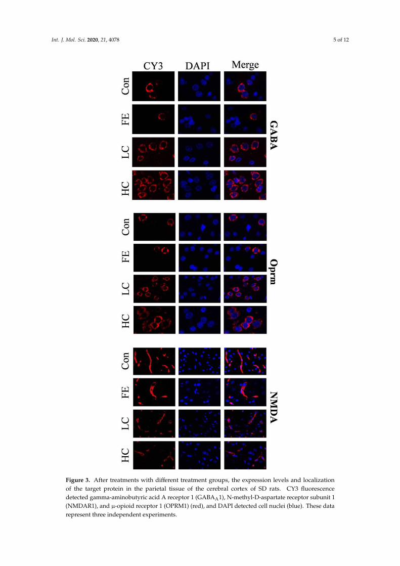
Our results showed that the administration of EISO remarkably upregulated the protein expressions of GABAAα1 and OPRM1 in the HD and LD groups compared to the Con and FE groups. However, the NMDAR1 expression was significantly downregulated in the HD and LD groups compared to the Con and FE groups (Figure 2). There was no expression difference in those protein expressions between the HD and LD groups. This indicates that EISO induced anesthesia by regulating the expression of ligand-gated channel-related proteins. We used immunofluorescence to observe the location of the target proteins. ESIO administration enhanced the expressions of GABAAα1 and OPRM1, but weakened the expression of NMDA in the HD and LD groups compared to the Con and FE groups (Figure 3).
Figure 1. The messenger RNA (mRNA) expression of related genes encoding ion channels and receptorrelated proteins (A), coding transcription factor-related genes (B), and other cytokines (C). Differentcolors are used to indicate the different processing methods. β-actin was used as the reference genefor normalization, and the difference was calculated with STATISTICAL ANALYSIS SYSTEM (SAS).Superscripts with different letters indicate significant differences (p < 0.05).
Regarding transcription factors, such as extracellular signal-regulated kinase 1/2(ERK1/2), c-Jun Nterminal kinase (JNK), protein 38 (P38), and nuclear factor kappa-B (NF-κB), the mRNA expressionsof the factors in the HD group were significantly higher than those in the Con group (Figure 1B).The expressions of ERK and p38 mRNA in the LD group were significantly higher than those in the

Int. J. Mol. Sci. 2020, 21, 4078 4 of 12
Con group. In addition, the expressions of JNK and NF-κB mRNA in the HD group were significantlyhigher than those in the FE groups. There was no significant difference in the expressions of thosefactors between the FE and Con groups.
As for the neurological function factors, the expressions of striatal-enriched protein tyrosinephosphatase 61 (STEP61) and Notch mRNA in the LD and HD groups were significantly higherthan those in the Con group, and the tyrosine kinase FYN expression was the opposite (Figure 1C).The expression of Notch mRNA in the FE group was significantly higher than that in the Con group.The expression level of Notch in the HD group was significantly higher than that in the FE group.
2.3. Protein Results of the Candidate Receptor
Our results showed that the administration of EISO remarkably upregulated the protein expressionsof GABAAα1 and OPRM1 in the HD and LD groups compared to the Con and FE groups. However,the NMDAR1 expression was significantly downregulated in the HD and LD groups compared tothe Con and FE groups (Figure 2). There was no expression difference in those protein expressionsbetween the HD and LD groups. This indicates that EISO induced anesthesia by regulating theexpression of ligand-gated channel-related proteins. We used immunofluorescence to observe thelocation of the target proteins. ESIO administration enhanced the expressions of GABAAα1 andOPRM1, but weakened the expression of NMDA in the HD and LD groups compared to the Con andFE groups (Figure 3).
Int. J. Mol. Sci. 2020, 21, x FOR PEER REVIEW 5 of 13
Figure 2. The determination of protein expression in the parietal lobe of Sprague Dawley (SD) rats injected with different drugs. The control (Con) group is indicated by a blank bar, the fat emulsion (FE) group is indicated by a black bar, the low-concentration (LD) group is indicated by a blue bar, and the high-concentration (HD) group is indicated by a red bar. Compared to the protein expression of the Con group and FE group, the LD group and HD group show significant changes. Superscripts with different letters indicate significant differences (p < 0.05). The left-to-right bands of each protein represent the corresponding protein expression levels in the parietal cortex samples of SD rats injected with different drugs and concentrations.
Figure 2. The determination of protein expression in the parietal lobe of Sprague Dawley (SD) ratsinjected with different drugs. The control (Con) group is indicated by a blank bar, the fat emulsion (FE)group is indicated by a black bar, the low-concentration (LD) group is indicated by a blue bar, andthe high-concentration (HD) group is indicated by a red bar. Compared to the protein expression ofthe Con group and FE group, the LD group and HD group show significant changes. Superscriptswith different letters indicate significant differences (p < 0.05). The left-to-right bands of each proteinrepresent the corresponding protein expression levels in the parietal cortex samples of SD rats injectedwith different drugs and concentrations.
Int. J. Mol. Sci. 2020, 21, 4078 5 of 12Int. J. Mol. Sci. 2020, 21, x FOR PEER REVIEW 6 of 13
Figure 3. After treatments with different treatment groups, the expression levels and localization of the target protein in the parietal tissue of the cerebral cortex of SD rats. CY3 fluorescence detected gamma-aminobutyric acid A receptor 1 (GABAA1), N-methyl-D-aspartate receptor subunit 1 (NMDAR1), and μ-opioid receptor 1 (OPRM1) (red), and DAPI detected cell nuclei (blue). These data represent three independent experiments.
Figure 3. After treatments with different treatment groups, the expression levels and localizationof the target protein in the parietal tissue of the cerebral cortex of SD rats. CY3 fluorescencedetected gamma-aminobutyric acid A receptor 1 (GABAA1), N-methyl-D-aspartate receptor subunit 1(NMDAR1), and µ-opioid receptor 1 (OPRM1) (red), and DAPI detected cell nuclei (blue). These datarepresent three independent experiments.

Int. J. Mol. Sci. 2020, 21, 4078 6 of 12
2.4. Methylation Analysis in the Promoter Region of Candidate Genes
The promoter region of the GABAAα1, Oprm1, and NMDAR1 genes was found by comparing themRNA sequence with the genome sequence. Then, according to the analysis results of the transcriptionfactor binding sites in the promoter region by the Matinspector in the Genomatix online tool, the corepromoter structure of GABAAα1, Oprm1, and NMDAR1 was determined (Figure 4A). The promotersequences of those genes were found in National Center for Biotechnology Information (NCBI).Subsequently, the potential binding sites of the relevant transcription factors and DNA methylationsites in the promoter regions of these genes were analyzed to confirm the core promoter sequencesdetermined by methylation. The methylation rates of the promoter regions of GABAAα1 and OPRM1in the HD and LD groups were lower than those in the Con and FE groups, whereas, NMDAR1demonstrated the opposite (Figure 4B).
Int. J. Mol. Sci. 2020, 21, x FOR PEER REVIEW 7 of 13
2.4. Methylation Analysis in the Promoter Region of Candidate Genes
The promoter region of the GABAAα1, Oprm1, and NMDAR1 genes was found by comparing the mRNA sequence with the genome sequence. Then, according to the analysis results of the transcription factor binding sites in the promoter region by the Matinspector in the Genomatix online tool, the core promoter structure of GABAAα1, Oprm1, and NMDAR1 was determined (Figure 4A). The promoter sequences of those genes were found in National Center for Biotechnology Information (NCBI). Subsequently, the potential binding sites of the relevant transcription factors and DNA methylation sites in the promoter regions of these genes were analyzed to confirm the core promoter sequences determined by methylation. The methylation rates of the promoter regions of GABAAα1 and OPRM1 in the HD and LD groups were lower than those in the Con and FE groups, whereas, NMDAR1 demonstrated the opposite (Figure 4B).
Figure 4. DNA methylation detection in the promoter area of GABAAα1, NMDAR1, and OPRM1 in the parietal lobe of cerebral cortex samples from rats injected with different medicine. The structure of the primary transcription factor binding sites and DNA methylation positions in the promoter area of the candidate genes. Different symbols are used to indicate the positions of the transcriptional factors and restriction enzymes. The primer pairs used for the methylation assay are denoted by gray arrows (A). The detection results of the methylation rates of the GABAAα1, NMDAR1, and OPRM1 promoter regions. Different colors are used to indicate the different processing methods. The difference was calculated with SAS. The results are expressed as the mean ± SE. Superscripts with different letters indicate significant differences (p < 0.05) (B).
3. Discussion
The circulation and respiratory system are two systems that are affected by anesthetic drugs [13]. The study of intravenous emulsified isoflurane indicated that there were no adverse reactions found except for caudal local skin ulcers. [14] In our study, rats were injected with 60 mg·kg−1·min−1 and 45 mg·kg−1·min−1 emulsified isoflurane through the tail vein. The body temperature and respiratory frequency showed a downward trend after injection. These indicated that EISO had a significant effect on the rat circulation and respiratory system during anesthesia. In the detection of the eyelid reflex, there was no observed phenomenon of dodging, closed eyes, or blinking, which proved that the rats were in the clinical anesthesia stage period. The rats had no eyeball contractions, tremors, or blinking in the corneal reflex test and no obvious anal contraction in the anal reflex test.
The results proved that the rats were in a safe and appropriate state of anesthesia. In the monitoring of the analgesic effects, the rats in the acupuncture and clamp detection sites did not have any pain reaction, or sedative or muscle relaxation effects. During the period, this state met the requirements for clinical surgery. Liu found that the minimum alveolar concentration (MAC) of EISO by intravenous injection in beagle dogs was lower than that during isoflurane inhalation anesthesia [15]. We speculate that the difference in the concentration gradient of anesthetics in the blood and
Figure 4. DNA methylation detection in the promoter area of GABAAα1, NMDAR1, and OPRM1 inthe parietal lobe of cerebral cortex samples from rats injected with different medicine. The structure ofthe primary transcription factor binding sites and DNA methylation positions in the promoter area ofthe candidate genes. Different symbols are used to indicate the positions of the transcriptional factorsand restriction enzymes. The primer pairs used for the methylation assay are denoted by gray arrows(A). The detection results of the methylation rates of the GABAAα1, NMDAR1, and OPRM1 promoterregions. Different colors are used to indicate the different processing methods. The difference wascalculated with SAS. The results are expressed as the mean ± SE. Superscripts with different lettersindicate significant differences (p < 0.05) (B).
3. Discussion
The circulation and respiratory system are two systems that are affected by anesthetic drugs [13].The study of intravenous emulsified isoflurane indicated that there were no adverse reactions foundexcept for caudal local skin ulcers. [14] In our study, rats were injected with 60 mg·kg−1
·min−1 and45 mg·kg−1
·min−1 emulsified isoflurane through the tail vein. The body temperature and respiratoryfrequency showed a downward trend after injection. These indicated that EISO had a significant effecton the rat circulation and respiratory system during anesthesia. In the detection of the eyelid reflex,there was no observed phenomenon of dodging, closed eyes, or blinking, which proved that the ratswere in the clinical anesthesia stage period. The rats had no eyeball contractions, tremors, or blinkingin the corneal reflex test and no obvious anal contraction in the anal reflex test.
The results proved that the rats were in a safe and appropriate state of anesthesia. In the monitoringof the analgesic effects, the rats in the acupuncture and clamp detection sites did not have any painreaction, or sedative or muscle relaxation effects. During the period, this state met the requirements forclinical surgery. Liu found that the minimum alveolar concentration (MAC) of EISO by intravenousinjection in beagle dogs was lower than that during isoflurane inhalation anesthesia [15]. We speculate

Int. J. Mol. Sci. 2020, 21, 4078 7 of 12
that the difference in the concentration gradient of anesthetics in the blood and alveoli as caused bydifferent routes of administration is related to the balance time, not the change in anesthetic efficacy.
Regarding the tactile pain-related perceptions in the parietal cortex, we selected this tissue tostudy the anesthetic mechanism of emulsified isoflurane at the molecular level. Recent studies provedthat ion channel modulation is the basis for general anesthesia [16]. The GABAA receptor is an ionchannel sensitive to volatile anesthetics with clinically available concentrations, and this receptorbelongs to the superfamily of cysteine ring neurotransmitter receptors. It is also the most abundantinhibitory neurotransmitter receptor in the brain [17]. Studies demonstrated that isoflurane has a directstereoselectivity for the GABAA receptor, which indicates that isoflurane may directly combine with asubunit on the GABAA receptor to produce a physiological effect [18]. Previous studies indicated thattwo different α subunits or two different β subunits were present in at least some GABAA receptorsand that 98% of α1 receptors contained either γ1, γ2, γ3, or δ subunits [19]; hence, GABAAα1 was thetarget of our study.
We also found that both GABAAα1 protein expression and mRNA expression increasedsignificantly after EISO injection. STEP61, a tyrosine phosphatase specifically expressed in neurons,was also a downstream molecule of the GABAA receptor and increased significantly after EISOinjection. The increased expression of STEP61 reduced the molecular binding between ERK and FYN.Many studies have shown that ERK also plays a key regulatory role for NMDA receptors, and theincreased expression of ERK can promote the activation of NMDA receptor phosphorylation [20].However, in our study, we found that ERK expression increased with the decrease in NR2B expression.We speculated that the intralipid influenced the expression of ERK. NR1 and NR2B were both importantsubstrates of STEP61, and FYN-mediated phosphorylation of NR2B-Tyrl472 could promote NR2Baggregation to synapses, which was negatively regulated by STEP61 [21]. This indicated that EISOfurther affects the expression of NR1 and NR2B subunits by activating the GABAA/STEP pathway,thereby downregulating the expression of the NMDA receptors and participating in the transmissionand regulation of pain information in the cerebral cortex.
To further explore the anesthetic mechanism of EISO, we explored DNA methylation detection,which is an epigenetic event and plays a key role in the regulation of gene expression [22]. We found thatthe methylation degree of the promoter regions of the coding genes of GABAAα1, NMDAR1, and OPRM1subunits changed significantly after EISO injection, and this methylation regulated the expression ofthese three subunits. However, it was not clear how EISO regulated the methylation of the GABAAα1,NMDAR1, and OPRM1 promoters. The methylation of the promoters of these coding genes altered thedegree of DNA compression, thereby affecting the binding rate of transcription factors and altering theexpression of the proteins encoded by these genes. We also found that NF-κB and cAMP-responseelement binding protein (CREB), as downstream molecules of P38, were important transcription factorsof these three target genes, which were also significantly upregulated in the experiment. However,the expression of NMDAR1 was decreased after the injection of EISO. This indicated that DNAmethylation altered gene expression more than transcription factor expression altered gene expressionin our study. Our protein experiments also confirmed this hypothesis.
A previous study showed that isoflurane produced both experimental neuroprotective andneurotoxic effects on the developing brain, depending on the duration and level of exposure, and EISO,as an intravenous anesthetic, may have protective and injurious effects on rats [23]. In both in vivoand in vitro ischemia models, mitogen-activated protein kinase (MAPK) was demonstrated to playa crucial role in regulating brain cell death and survival after ischemia [24]. In previous studies,OPRM was found to be directly regulated by MAPK. In our study, we found that EISO affected theexpression of the transcription factors CREB and NF-κB through the MAPK signaling pathway, furtheraffecting the expression of three target genes [25–27]. In future studies, we will continue to investigatewhich transcription factors play key roles while observing the degree of chromatin loosening after thetranscription factor binds to the coding gene to better explore the epigenetic mechanisms by whichEISO causes anesthesia.

Int. J. Mol. Sci. 2020, 21, 4078 8 of 12
In conclusion, we found that after the anesthesia of rats with EISO, the expression of relatedfactors in the GABAA receptor/STEP61 signaling pathway changed, the expression of the GABAA
receptor increased, and the expression of the NMDA receptor decreased, accompanied by a changein the promoter methylation. The MAPK signaling pathway might also be involved in regulation.More work is needed in the future to determine the sequence and causality of downstream factors ofthe GABAA receptor/STEP61 signaling pathway.
4. Methods
Experimental Sprague Dawley (SD) rats were housed in individual cages in the Centre ofExperimental Animals at Nanjing Agricultural University (Nanjing, China) in accordance withthe guidelines of Animal Ethics Committee at Nanjing Agricultural University in compliancewith the Regulations for the Administration of Affairs Concerning Experimental Animals (TheState Science and Technology Commission of People’s Republic of China, 1988). All experimentalprotocols were approved by the animal Care and Use Committee of Nanjing Agricultural University,1999(#NJAU-Rattus-2018061305, approved on 13 June 2018).
4.1. Animals
We used 32 male SD rats, 220–230 g, 2–3 months old for this study. All rats were housed separatelyunder standard laboratory conditions with a 12:12 h light/dark cycle, 22 ◦C, and 50% humidity.The rats had free access to water and standard mouse chow. Prior to the investigations, the rats wereallowed to habituate to their new surroundings for one week after having been transferred from thebreeder. Our study was approved by the Nanjing Agricultural University animal ethics committee.The following experiments were conducted.
4.2. Drugs
Isoflurane emulsions of eight percent by volume were prepared by adding pure isoflurane to 30%intralipid at 20 ◦C. The solution was then vortexed at 50 Hz every five minutes for one minute, lastingtwo hours, stored at 4 ◦C between injections, and warmed to 20 ◦C.
4.3. Experimental Design
The rats were fasted for 12 h before the experiment but free to drink water. The rats wererandomly separated into four groups (n = 8): Con, FE, HD, and LD. The caudal vein was injectedwith saline solution (0.1 mL·kg−1
·min−1), 30% fat intralipid (0.1 mL·kg−1·min−1), or 8% EISO (60 or
45 mg·kg−1·min−1), with a constant flow syringe pump to maintain anesthesia for 25 min, and the
anesthetic conditions of the rats were assessed. In the experiment, the doses of EISO used formaintaining anesthesia and for inducing anesthesia were the same. The HD group used EISOat 60 mg·kg−1
·min−1, the LD group was at 45 mg·kg−1·min−1, and the process of anesthesia was
relatively stable. Tissue samples from the parietal lobe of the cerebral cortex were taken from rats afterdecapitation and preserved in liquid nitrogen.
4.4. Physiological Indicator Test
After the start of administration, the body temperature and respiratory rate of the rats weremeasured in five-minute intervals.
4.5. Monitoring of Biological Reflection
Observing and monitoring reflexes, such as the corneal reflex (CR), and the anal reflex bluntness,and disappearance, have been used as one of the main methods to evaluate the depth of anesthesia.The corneal reflex mainly stimulates the cornea through a hard lane to observe whether there areeye contractions, tremors, eye movements, and other phenomena. The anal reflex (AR) refers to the
Int. J. Mol. Sci. 2020, 21, 4078 9 of 12
contraction of the anal sphincter when the anus is suddenly stimulated. The detailed scoring criteria isdescribed in Supplemental Table S1.
4.6. Monitoring of Braking and Analgesic Effects
The scoring criteria used to investigate the brake and analgesic effects included sedation, analgesia,muscle relaxation, posture, and hearing sensitivity. Sedation was scored based on the position ofthe eyeball and the sensitivity to the external environment. Analgesia was judged by the animalreaction to clamping the animal’s toes with a hemostat. Muscle relaxation was judged by the tensionof the masseter muscle and leg muscles. The posture was scored by observing the lying postureof the small rat. Hearing induction was evaluated by clapping hands near the ear of the rat andobserving the reaction. The detailed scoring criteria was described in previous studies and are shownin Supplemental Table S2. Evaluations of the brake and analgesic effects were made in five-minuteintervals after the beginning of drug injection and the resulting scores were recorded.
4.7. Quantitative Real-Time Polymerase Chain Reaction (PCR)
The total ribonucleic acid (RNA) was extracted from the tissue with the RNA iso Plus™ (cat.9108, Takara, Dalian, Liaoning Province, China) reagent according to the manufacturer’s instructions.The complementary deoxyribonucleic acid (cDNA) was prepared using a reverse transcription kit(cat. RR036A, Takara, Dalian, Liaoning Province, China) according to the manufacturer’s instruction.The cDNA was subjected to quantitative PCR amplification for the transcription of candidate genes.The primers are shown in Supplemental Table S3. Each RNA sample was processed in two copiesusing an amount of DNA equivalent to the total RNA, starting at 250 ng. The target sequences ofthe candidate genes were amplified with specific primer pairs using the SYBR Premix Ex Taq Kit(cat. DRR420A, Takara, Dalian, Liaoning Province, China). Amplifying PCR and monitoring of thefluorescent emission in real time were performed using the ABI Prism 7300 Sequence Detection System(Applied Biosystems, Inc., Foster City, CA, USA).
4.8. Genomic DNA Methylation Status Monitoring
We obtained the genomic DNA of the brain cortex using a DNA extraction kit (KG203, TIANGENBIOTECH Co., Ltd., Shanghai, China). The target fragment was subjected to restriction analysis usingDNAMAN software (Lynnon Biosoft, San Ramon, CA, USA), and the genomic DNA was pre-cutby restriction enzymes that did not cleave the target fragment. This study used EcoRI (cat. 1040S,Takara, Dalian, Liaoning Province, China) to pre-cut the genomic DNA. We used MspI and HpaII(cat. 1150A, Takara, Dalian, Liaoning Province, China) to process the mass of each sample equalizedafter digestion, and each sample was made with three parallels, MspI, HpaII, and a blank controltube. Quantification of the absolute copy number of the genomic DNA was performed after enzymetreatment with RT-PCR and the calculation of the methylation of sample’s promoter. The primers usedfor methylation detection are listed in Supplemental Table S3.
4.9. Western Blotting
The proteins in the tissues were separated by sodium dodecyl sulfate-polyacrylamide gelelectrophoresis using a 10% acrylamide gel and transferred to a polyvinylidene fluoride membraneby electroelution. The membranes were blocked with 5% non-fat milk powder in 0.01 M PBS (pH7.4) and 0.1% Tween-20 at room temperature for 2 h. Substitute blots for members were incubatedwith primary antibodies against GABAAα1 (1:200; sc-7348, Santa Cruz, Dallas, TX, USA), NMDAR1(1:200; sc-1468, Santa Cruz, Dallas, TX, USA), Oprm1 (1:200; sc-247994, Santa Cruz, Dallas, TX,USA), and glyceraldehyde-phosphate dehydrogenase (GAPDH) (ab8245, Abcam, Cambridge, MA,USA) overnight at 4 ◦C by gentle shaking, followed by the addition of the appropriate horseradishperoxidase-conjugated secondary antibodies (1:2000; A0208, Beyotime Biotechnology, Shanghai, China)and incubation at room temperature for 4 h. The expression levels of the proteins were quantitatively

Int. J. Mol. Sci. 2020, 21, 4078 10 of 12
analyzed through comparison with GAPDH as an internal control. We used the Bio-Rad Gel Doc 2000system analysis software (Bio-Rad, Hercules, CA, USA) to conduct the grey level of the bands.
4.10. Immunofluorescence
Paraffin sections were made of the cerebral cortex. The following primary antibodies were usedfor dual fluorescent labeling, GABAAα1, NMDAR1, and OPRM1. Subsequently, we observed thesections under a fluorescence microscope with different laser wavelengths.
4.11. Statistical Analysis
The data from the assessments of biological reflections and braking and analgesic effects wereanalyzed as repeated measures using the MIXED procedures of SAS (SAS version 9.4, SAS InstituteInc., San Diego, CA, USA). The effects of fat emulsions and emulsified isoflurane were considered fixedand the effect of the scoring time was analyzed as a repeated measure. The effect of the experimentalrates was considered a random effect. In addition, the data of mRNA and protein expression, and thepercentage of promoter methylation, were analyzed using the ANOVA package of SAS. All data wererepresented as the mean ± standard error (SE). The effects were considered significant at p < 0.05 andhighly significant at p < 0.01. Trends were discussed at p < 0.10.
5. Conclusions
Emulsified isoflurane is a good anesthetic for intravenous injection. After producing anesthesia inrats with EISO, the expression of related factors in the GABAA receptor/STEP61 signaling pathwaychanged, the expression of the GABAA receptor increased, and the expression of the NMDA receptordecreased, accompanied by a change in the promoter methylation. The MAPK signaling pathway mayalso be involved in regulation. More work is still needed in the future to determine the relationshipbetween the downstream molecules of this signaling pathway and to explore the relevant mechanismsby which EISO affects promoter methylation.
Supplementary Materials: Supplementary materials can be found at http://www.mdpi.com/1422-0067/21/11/4078/s1.
Author Contributions: In our study, different members were responsible for different tasks. X.Z.: This authorhelped with the experimental design and thesis writing. G.C.: This author helped with the data analysis. Y.C.:This author helped with the animal feeding. Z.Z.: This author is the correspondence author. All authors have readand agreed to the published version of the manuscript.
Funding: The current study was supported by the National Key R&D Program of China (Project No.2017YFD0502205), the National Natural Science Foundation of China (Grants31572579 and 31702301), and thePriority Academic Program Development of Jiangsu Higher Education Institutions (PAPD).
Conflicts of Interest: The authors declare no conflict of interest.
Abbreviations
EISO emulsified isofluraneCon control groupFE intralipid groupHD high-concentration groupLD low-concentration groupPCR polymerase chain reactionGABAAα1 gamma-aminobutyric acid A receptor α1 subunitNMDAR1 N-methyl-D-aspartate receptor subunit 1OPRM1 µ opioid receptor 1GABAA gamma-aminobutyric acid A receptorNMDA N-methyl-D-aspartate receptorOPRM µ opioid receptorCpG 5′-cytosine-phosphate-guanine-3′
mRNA messenger ribonucleic acid

Int. J. Mol. Sci. 2020, 21, 4078 11 of 12
SD Sprague DawleyDNA deoxyribonucleic acidRNA ribonucleic acidMeCP2 methyl-CpG-bindingprotein-2STEP61 Striatal-enriched protein tyrosine phosphatase 61cDNA complementary deoxyribonucleic acidGAPDH glyceraldehyde-phosphate dehydrogenasePeR perianal reflexCR corneal reflexAR anal reflexSS the sedation scoresAS analgesic scoresMRS muscle relaxation scoresPS posture scoresAR auditory response scoresTS total scoresNR2B N-methyl-D-aspartate receptor subunit 2BERK1/2 extracellular signal-regulated kinase 1/2JNK c-Jun N terminal kinasep38 protein 38NF-κB nuclear factor kappa-BFYN tyrosine kinase FynMAC minimum alveolar concentrationMAPK mitogen-activated protein kinasesY1472 tyrosine 1472SAS STATISTICAL ANALYSIS SYSTEMCREB cAMP-response element binding protein
References
1. Spadavecchia, C.; Haga, H.A.; Ranheim, B. Concentration-dependent isoflurane effects on withdrawalreflexes in pigs and the role of the stimulation paradigm. Vet. J. 2012, 194, 375–379. [CrossRef] [PubMed]
2. Cascorbi, H.F.; Helrich, M.; Krantz, J.C.; BAKER, L.R.; Rozman, R.S.; Rudo, F.G. Hazards of methoxyfluraneemulsions in man. Anesth. Analg. 1968, 47, 557–559. [CrossRef] [PubMed]
3. Stemp, L.I. Intravenous injection of liquid halothane. Anesth. Analg. 1990, 70, 250–255. [CrossRef] [PubMed]4. Kawamoto, M.; Suzuki, N.; Takasaki, M. Acute pulmonary edema after intravenous liquid halothane in dogs.
Anesth. Analg. 1992, 74, 747–752. [CrossRef]5. Biber, B.; Martner, J.; Werner, O. Halothane by the I.V. route in experimental animals. Acta Anaesthesiol. Scand.
2010, 26, 658–659. [CrossRef]6. Zhou, C.; Gan, J.; Liu, J.; Luo, W.J.; Zhang, W.S.; Chai, Y.F. The Interaction Between Emulsified Isoflurane and
Lidocaine Is Synergism in Intravenous Regional Anesthesia in Rats. Anesth. Analg. 2011, 113, 245. [CrossRef]7. Illingworth, R.S.; Bird, A.P. CpG islands—‘A rough guide’. FEBS Lett. 2009, 583, 1713–1720. [CrossRef]8. Mccabe, M.T.; Brandes, J.C.; Vertino, P.M. Cancer DNA methylation: Molecular mechanisms and clinical
implications. Clin. Cancer Res. Off. J. Am. Assoc. Cancer Res. 2009, 15, 3927. [CrossRef]9. Weber, M.; Schübeler, D. Genomic patterns of DNA methylation: Targets and function of an epigenetic mark.
Curr. Opin. Cell Biol. 2007, 19, 273–280. [CrossRef]10. Franks, N.P. Molecular targets underlying general anaesthesia. Br. J. Pharmacol. 2010, 147, S72–S81. [CrossRef]11. Kitamura, A.; Satoh, R.; Nagano, T.; Matsuda, H.; Shimizu, T.; Sakamoto, A.; Ogawa, R. Halothane modulates
NMDA and non-NMDA excitatory synaptic transmission in rat cortical neurons. J. Anesth. 2005, 19, 66–72.[CrossRef] [PubMed]
12. Urban, B.W.; Markus, B.; Martin, B. Interactions of anesthetics with their targets: Non-specific, specific orboth? Pharmacol. Ther. 2006, 111, 729–770. [CrossRef] [PubMed]
13. Steffey, E.P.; Mama, K.R.; Brosnan, R.J. Inhalation anesthetics. Lumb Jones’ Vet. Anesth. 2015, 3, 297–323.

Int. J. Mol. Sci. 2020, 21, 4078 12 of 12
14. Eger, R.P.; MacLeod, B.A. Anaesthesia by intravenous emulsified isoflurane in mice. Can. J. Anaesth. 1995,42, 173–176. [CrossRef]
15. Liu, J.; Yang, X.; Ma, H.; Yang, Z.; Zhang, W. Comparison of MAC between intravenous injected isofluranelipid emulsion and inhaled isoflurane in dogs. Anesthesiology 2004, 101, A127.
16. Narahashi, T.; Aistrup, G.L.; Lindstrom, J.M.; Marszalec, W.; Nagata, K.; Wang, F.; Yeh, J.Z. Ion channelmodulation as the basis for general anesthesia. Toxicol. Lett. 1998, 100–101, 185–191. [CrossRef]
17. Campagna, J.A.; Miller, K.W.; Forman, S.A. Mechanisms of actions of inhaled anesthetics. N. Engl. J. Med.2003, 348, 909–910. [CrossRef]
18. Hall, A.C.; Lieb, W.R.; Franks, N.P. Stereoselective and non-stereoselective actions of isoflurane on theGABAA receptor. Br. J. Pharmacol. 1994, 112, 906–910. [CrossRef]
19. Sieghart, W.; Fuchs, K.; Tretter, V.; Ebert, V.; Jechlinger, M.; Höger, H.; Adamiker, D. Structure and subunitcomposition of GABA A receptors. Neurochem. Int. 1999, 34, 379–385. [CrossRef]
20. Petrenko, A.B.; Yamakura, T.; Baba, H.; Shimoji, K. The role of N-methyl-D-aspartate (NMDA) receptors inpain: A review. Anesth. Analg. 2003, 97, 1108–1116. [CrossRef]
21. Goebel-Goody, S.M.; Baum, M.; Paspalas, C.D.; Fernandez, S.M.; Carty, N.C.; Kurup, P.; Lombroso, P.J.Therapeutic Implications for Striatal-Enriched Protein Tyrosine Phosphatase (STEP) in NeuropsychiatricDisorders. Pharmacol. Rev. 2011, 64, 65. [CrossRef] [PubMed]
22. Chang, G.; Liu, X.; Ma, N.; Yan, J.; Dai, H.; Roy, A.C.; Shen, X. Dietary Addition of Sodium ButyrateContributes toAttenuated Feeding-Induced Hepatocyte Apoptosis in Dairy Goats. J. Agric. Food Chem. 2018,66, 9995–10002. [CrossRef] [PubMed]
23. Zhao, X.; Yang, Z.; Liang, G.; Wu, Z.; Peng, Y.; Joseph, D.J.; Inan, S.; Wei, H. Dual effects of isoflurane onproliferation, differentiation, and survival in human neuroprogenitor cells. Anesthesiology 2013, 118, 537–549.[CrossRef] [PubMed]
24. Nozaki, K.; Nishimura, M.; Hashimoto, N. Mitogen-activated protein kinases and cerebral ischemia.Mol. Neurobiol. 2001, 23, 1–19. [CrossRef]
25. Al-Hasani, R.; Bruchas, M.R. Molecular mechanisms of opioid receptor-dependent signaling and behavior.Anesthesiology 2011, 115, 1363–1381. [CrossRef]
26. Bickler, P.E.; Fahlman, C.S. The inhaled anesthetic, isoflurane, enhances Ca2+-dependent survival signalingin cortical neurons and modulates MAP kinases, apoptosis proteins and transcription factors during hypoxia.Anesth. Analg. 2006, 103, 419. [CrossRef]
27. Zheng, S.; Zuo, Z. Isoflurane preconditioning induces neuroprotection against ischemia via activation of P38mitogen-activated protein kinases. Mol. Pharmacol. 2004, 65, 1172–1180. [CrossRef]
© 2020 by the authors. Licensee MDPI, Basel, Switzerland. This article is an open accessarticle distributed under the terms and conditions of the Creative Commons Attribution(CC BY) license (http://creativecommons.org/licenses/by/4.0/).
Related Documents