c-Tubulin 2 Nucleates Microtubules and Is Downregulated in Mouse Early Embryogenesis Stanislav Vinopal 1 , Marke ´ta C ˇ ernohorska ´ 1 , Vadym Sulimenko 1 , Tetyana Sulimenko 1 , Ve ˇ ra Vosecka ´ 1 , Matya ´s ˇ Flemr 2 , Eduarda Dra ´ berova ´ 1 , Pavel Dra ´ ber 1 * 1 Department of Biology of Cytoskeleton, Institute of Molecular Genetics, Academy of Sciences of the Czech Republic, Prague, Czech Republic, 2 Department of Epigenetic Regulations, Institute of Molecular Genetics, Academy of Sciences of the Czech Republic, Prague, Czech Republic Abstract c-Tubulin is the key protein for microtubule nucleation. Duplication of the c-tubulin gene occurred several times during evolution, and in mammals c-tubulin genes encode proteins which share ,97% sequence identity. Previous analysis of Tubg1 and Tubg2 knock-out mice has suggested that c-tubulins are not functionally equivalent. Tubg1 knock-out mice died at the blastocyst stage, whereas Tubg2 knock-out mice developed normally and were fertile. It was proposed that c-tubulin 1 represents ubiquitous c-tubulin, while c-tubulin 2 may have some specific functions and cannot substitute for c-tubulin 1 deficiency in blastocysts. The molecular basis of the suggested functional difference between c-tubulins remains unknown. Here we show that exogenous c-tubulin 2 is targeted to centrosomes and interacts with c-tubulin complex proteins 2 and 4. Depletion of c-tubulin 1 by RNAi in U2OS cells causes impaired microtubule nucleation and metaphase arrest. Wild-type phenotype in c-tubulin 1-depleted cells is restored by expression of exogenous mouse or human c-tubulin 2. Further, we show at both mRNA and protein levels using RT-qPCR and 2D-PAGE, respectively, that in contrast to Tubg1, the Tubg2 expression is dramatically reduced in mouse blastocysts. This indicates that c-tubulin 2 cannot rescue c-tubulin 1 deficiency in knock-out blastocysts, owing to its very low amount. The combined data suggest that c-tubulin 2 is able to nucleate microtubules and substitute for c-tubulin 1. We propose that mammalian c-tubulins are functionally redundant with respect to the nucleation activity. Citation: Vinopal S, C ˇ ernohorska ´ M, Sulimenko V, Sulimenko T, Vosecka ´ V, et al. (2012) c-Tubulin 2 Nucleates Microtubules and Is Downregulated in Mouse Early Embryogenesis. PLoS ONE 7(1): e29919. doi:10.1371/journal.pone.0029919 Editor: Claude Prigent, Institut de Ge ´ne ´tique et De ´veloppement de Rennes, France Received August 23, 2011; Accepted December 6, 2011; Published January 3, 2012 Copyright: ß 2012 Vinopal et al. This is an open-access article distributed under the terms of the Creative Commons Attribution License, which permits unrestricted use, distribution, and reproduction in any medium, provided the original author and source are credited. Funding: This work was supported in part by Grants 204/09/H084 (SV, MC ˇ ), 204/09/1777 (PD), and P302/10/1759 (ED) from the Grant Agency of the Czech Republic, Grants LC545 (TS) and 1M6837805001 (VV) from Ministry of Education, Youth and Sports of the Czech Republic, KAN200520701 (VS) from the Grant Agency of the Czech Academy of Sciences, Grant 11109 from the Grant Agency of Charles University (SV), and by the Institutional Research Support Grant AVOZ50520514 (PD). The funders had no role in study design, data collection and analysis, decision to publish, or preparation of the manuscript. Competing Interests: The authors have declared that no competing interests exist. * E-mail: [email protected] Introduction c-Tubulin is a highly conserved member of the tubulin superfamily essential for microtubule nucleation in all eukaryotes [1–3]. It assembles together with other proteins, named Gamma- tubulin Complex Proteins (GCPs) in human, into two main c- Tubulin Complexes (cTuCs): the c-Tubulin Small Complex (cTuSC) and the c-Tubulin Ring Complex (cTuRC). The cTuSC, a vital component of microtubule nucleation machinery in all eukaryotes, is composed of two molecules of c-tubulin and one copy each of GCP2 and GCP3. The cTuRCs are found only in metazoa and consist of seven cTuSCs and additional GCPs, including GCP4-6 [4,5]. The cTuRC is a ring structure with an arrangement of c-tubulin molecules that matches the 13-fold symmetry of a microtubule. It serves as a template for microtubule polymerization [6]. It has recently been shown that the budding yeast cTuSCs alone form in vitro ring structures similar to cTuRCs [7]; it supports the general template model of microtubule nucleation [6]. cTuCs are concentrated at Microtubule Organizing Centers (MTOCs) such as centrosomes and basal bodies in animals or spindle pole bodies in fungi. They are also found on nuclear membranes in acentrosomal plants and on Golgi membranes, condensed mitotic chromosomes, midbodies and along microtu- bules in mitotic spindles [8]. We have recently reported nucleolar localization of c-tubulin [9]. However, the majority of cTuCs exist in cytoplasm in soluble form [10]. In addition to its function in microtubule nucleation, c-tubulin is also involved in centriole biogenesis [11,12], regulation of microtubule (+) end dynamics [13–15], regulation of the anaphase-promoting complex/cyclo- some during interphase in Aspergillus [16] or regulation of bipolar spindle assembly in fission yeast [17]. Many organisms including Arabidopsis [18], Paramecium [19], Euplotes [20], Drosophila [21] and mammals [22–24] possess two genes encoding c-tubulin. Nevertheless, phylogenetic analyses revealed that c-tubulin gene duplication in mammals occurred independent of the others [23,24]. Mammalian c-tubulin genes are located on the same chromosome in tandem, and their coding sequences share very high sequence similarity (.94% in human)[22]. Although it was initially assumed that c-tubulin genes are functionally redundant [22], gene knock-out analysis of Tubg1 and Tubg2 in mice suggested that they might have different functions [23]. While Tubg1 was expressed ubiquitously, Tubg2 was primarily detected in brain and also in blastocysts. Tubg1 -/- embryos stopped their development at the morula/blastocyst stage because of severe mitotic defects. Tubg2 -/- mice developed PLoS ONE | www.plosone.org 1 January 2012 | Volume 7 | Issue 1 | e29919

Welcome message from author
This document is posted to help you gain knowledge. Please leave a comment to let me know what you think about it! Share it to your friends and learn new things together.
Transcript

c-Tubulin 2 Nucleates Microtubules and IsDownregulated in Mouse Early EmbryogenesisStanislav Vinopal1, Marketa Cernohorska1, Vadym Sulimenko1, Tetyana Sulimenko1, Vera Vosecka1,
Matyas Flemr2, Eduarda Draberova1, Pavel Draber1*
1 Department of Biology of Cytoskeleton, Institute of Molecular Genetics, Academy of Sciences of the Czech Republic, Prague, Czech Republic, 2 Department of Epigenetic
Regulations, Institute of Molecular Genetics, Academy of Sciences of the Czech Republic, Prague, Czech Republic
Abstract
c-Tubulin is the key protein for microtubule nucleation. Duplication of the c-tubulin gene occurred several times duringevolution, and in mammals c-tubulin genes encode proteins which share ,97% sequence identity. Previous analysis ofTubg1 and Tubg2 knock-out mice has suggested that c-tubulins are not functionally equivalent. Tubg1 knock-out mice diedat the blastocyst stage, whereas Tubg2 knock-out mice developed normally and were fertile. It was proposed that c-tubulin1 represents ubiquitous c-tubulin, while c-tubulin 2 may have some specific functions and cannot substitute for c-tubulin 1deficiency in blastocysts. The molecular basis of the suggested functional difference between c-tubulins remains unknown.Here we show that exogenous c-tubulin 2 is targeted to centrosomes and interacts with c-tubulin complex proteins 2 and 4.Depletion of c-tubulin 1 by RNAi in U2OS cells causes impaired microtubule nucleation and metaphase arrest. Wild-typephenotype in c-tubulin 1-depleted cells is restored by expression of exogenous mouse or human c-tubulin 2. Further, weshow at both mRNA and protein levels using RT-qPCR and 2D-PAGE, respectively, that in contrast to Tubg1, the Tubg2expression is dramatically reduced in mouse blastocysts. This indicates that c-tubulin 2 cannot rescue c-tubulin 1 deficiencyin knock-out blastocysts, owing to its very low amount. The combined data suggest that c-tubulin 2 is able to nucleatemicrotubules and substitute for c-tubulin 1. We propose that mammalian c-tubulins are functionally redundant with respectto the nucleation activity.
Citation: Vinopal S, Cernohorska M, Sulimenko V, Sulimenko T, Vosecka V, et al. (2012) c-Tubulin 2 Nucleates Microtubules and Is Downregulated in Mouse EarlyEmbryogenesis. PLoS ONE 7(1): e29919. doi:10.1371/journal.pone.0029919
Editor: Claude Prigent, Institut de Genetique et Developpement de Rennes, France
Received August 23, 2011; Accepted December 6, 2011; Published January 3, 2012
Copyright: � 2012 Vinopal et al. This is an open-access article distributed under the terms of the Creative Commons Attribution License, which permitsunrestricted use, distribution, and reproduction in any medium, provided the original author and source are credited.
Funding: This work was supported in part by Grants 204/09/H084 (SV, MC), 204/09/1777 (PD), and P302/10/1759 (ED) from the Grant Agency of the CzechRepublic, Grants LC545 (TS) and 1M6837805001 (VV) from Ministry of Education, Youth and Sports of the Czech Republic, KAN200520701 (VS) from the GrantAgency of the Czech Academy of Sciences, Grant 11109 from the Grant Agency of Charles University (SV), and by the Institutional Research Support GrantAVOZ50520514 (PD). The funders had no role in study design, data collection and analysis, decision to publish, or preparation of the manuscript.
Competing Interests: The authors have declared that no competing interests exist.
* E-mail: [email protected]
Introduction
c-Tubulin is a highly conserved member of the tubulin
superfamily essential for microtubule nucleation in all eukaryotes
[1–3]. It assembles together with other proteins, named Gamma-
tubulin Complex Proteins (GCPs) in human, into two main c-
Tubulin Complexes (cTuCs): the c-Tubulin Small Complex
(cTuSC) and the c-Tubulin Ring Complex (cTuRC). The cTuSC,
a vital component of microtubule nucleation machinery in all
eukaryotes, is composed of two molecules of c-tubulin and one
copy each of GCP2 and GCP3. The cTuRCs are found only in
metazoa and consist of seven cTuSCs and additional GCPs,
including GCP4-6 [4,5]. The cTuRC is a ring structure with an
arrangement of c-tubulin molecules that matches the 13-fold
symmetry of a microtubule. It serves as a template for microtubule
polymerization [6]. It has recently been shown that the budding
yeast cTuSCs alone form in vitro ring structures similar to cTuRCs
[7]; it supports the general template model of microtubule
nucleation [6].
cTuCs are concentrated at Microtubule Organizing Centers
(MTOCs) such as centrosomes and basal bodies in animals or
spindle pole bodies in fungi. They are also found on nuclear
membranes in acentrosomal plants and on Golgi membranes,
condensed mitotic chromosomes, midbodies and along microtu-
bules in mitotic spindles [8]. We have recently reported nucleolar
localization of c-tubulin [9]. However, the majority of cTuCs exist
in cytoplasm in soluble form [10]. In addition to its function in
microtubule nucleation, c-tubulin is also involved in centriole
biogenesis [11,12], regulation of microtubule (+) end dynamics
[13–15], regulation of the anaphase-promoting complex/cyclo-
some during interphase in Aspergillus [16] or regulation of bipolar
spindle assembly in fission yeast [17].
Many organisms including Arabidopsis [18], Paramecium [19],
Euplotes [20], Drosophila [21] and mammals [22–24] possess two
genes encoding c-tubulin. Nevertheless, phylogenetic analyses
revealed that c-tubulin gene duplication in mammals occurred
independent of the others [23,24]. Mammalian c-tubulin genes
are located on the same chromosome in tandem, and their coding
sequences share very high sequence similarity (.94% in
human)[22]. Although it was initially assumed that c-tubulin
genes are functionally redundant [22], gene knock-out analysis of
Tubg1 and Tubg2 in mice suggested that they might have different
functions [23]. While Tubg1 was expressed ubiquitously, Tubg2 was
primarily detected in brain and also in blastocysts. Tubg1-/-
embryos stopped their development at the morula/blastocyst stage
because of severe mitotic defects. Tubg2-/- mice developed
PLoS ONE | www.plosone.org 1 January 2012 | Volume 7 | Issue 1 | e29919

normally and produced fertile offspring. However, adults exhibited
some behavioral changes including abnormalities in circadian
rhythm and different reaction to painful stimulations. These
findings led to a conclusion that c-tubulin 1 is the conventional c-
tubulin, whereas c-tubulin 2, which lacks the capability to rescue
the consequences of c-tubulin 1 deficiency, might have specific
function(s) in the brain [23]. Nevertheless, the molecular basis of
suggested functional differences between c-tubulin 1 and c-tubulin
2 is unknown.
To gain a deeper insight into the potential functional differences
of mammalian c-tubulins, we have examined subcellular distribu-
tion of c-tubulin 2 in cultured cells, its interactions with GCPs,
capability to nucleate microtubules and substitute for c-tubulin 1.
We have also analyzed c-tubulin 2 expression in the course of
mouse preimplantation development. Our results indicate that
even though c-tubulins are differentially expressed during mouse
early embryogenesis and in adult tissues, they are functionally
redundant with respect to their nucleation activity.
Results
c-Tubulin 2 is indistinguishable from c-tubulin 1 insubcellular localization and interactions with GCPs
To decide whether or not c-tubulin 2 differs from c-tubulin 1 in
subcellular localization, we examined U2OS cells expressing
FLAG-tagged mouse or human c-tubulin 2 (Tubg2-FLAG,
TUBG2-FLAG) by immunofluorescence microscopy with anti-
FLAG antibody. Centrosomes were marked with antibody to
pericentrin. U2OS cells expressing FLAG-tagged mouse and
human c-tubulin 1 (Tubg1-FLAG, TUBG1-FLAG) served as
controls. As expected, exogenous mouse (Fig. 1A, a) and human
(Fig. 1A, c) c-tubulin 1 localized to the centrosomes in both
interphase and mitotic cells. FLAG-tagged c-tubulin 1 was also
found along mitotic spindle and diffusely in cytoplasm. The same
staining pattern was detected in cells expressing exogenous mouse
(Fig. 1A, b) and human (Fig. 1A, d) c-tubulin 2. Fully displayed
immunofluorescence of Fig. 1A appears in Fig. S1.
Next we checked by coimmunoprecipitation the ability of c-
tubulin 2 to interact with GCP2 (cTuSC marker) and GCP4
(cTuRC marker). FLAG-tagged mouse c-tubulin 1, c-tubulin 2 or
Fyn kinase (negative control) were immunoprecipitated from HEK
293FT cells with rabbit anti-FLAG antibody. Immunoblot analysis
revealed that both FLAG-tagged c-tubulins interacted with GCP2
and GCP4, yet no coimmunoprecipitation was observed in case of
FLAG-tagged Fyn kinase (Fig. 1B, upper panel). Negative control
rabbit antibody failed to coimmunoprecipitate GCP proteins (not
shown). In addition, the reciprocal precipitation with antibody to
GCP2 (IgG2b), confirmed the interaction of FLAG-tagged c-
tubulins with GCP2 (Fig. 1B, lower panel). Again, negative control
antibody (IgG2b) did not coimmunoprecipitate FLAG-tagged c-
tubulins (not shown). We obtained the same results when lysates
from HEK 293FT cells expressing FLAG-tagged human c-tubulin
1 and c-tubulin 2 were used for immunoprecipitation with anti-
FLAG and anti-GCP2 antibodies (Fig. S2). Altogether the data
indicate that mammalian c-tubulin 2 is indiscernible from
c-tubulin 1 as far as the subcellular distribution and interactions
Figure 1. Exogenous c-tubulin 2 locates to centrosomes andinteracts with GCPs. (A) Localization of FLAG-tagged c-tubulins.Human U2OS cells expressing mouse c-tubulin 1 (a, Tubg1-FLAG),mouse c-tubulin 2 (b, Tubg2-FLAG), human c-tubulin 1 (c, TUBG1-FLAG)and human c-tubulin 2 (d, TUBG2-FLAG) were stained for FLAG (red)and pericentrin (green). DNA was stained with DAPI (blue). Arrowsdenote positions of MTOCs where FLAG-tagged c-tubulins co-localizewith pericentrin. Final images were made by maximum intensityprojection of 3 deconvolved z-sections spaced at 0.25 mm. Scale bar10 mm. (B) Coimmunoprecipitation of mouse c-tubulins with GCP2 andGCP4 proteins. Extracts from HEK cells expressing FLAG-tagged c-
tubulin 1 (Tubg1-FLAG), c-tubulin 2 (Tubg2-FLAG) or control mouse Fyn(Fyn-FLAG) were immunoprecipitated with antibodies to FLAG or GCP2,and blots were probed with antibodies to FLAG, GCP2, GCP4 and c-tubulin (c-Tb). Extracts (1), immunoprecipitated proteins (2), protein Awithout antibodies incubated with extracts (3), immobilized antibodiesnot incubated with extracts (4). Arrowheads indicate the positions ofexogenous c-tubulins.doi:10.1371/journal.pone.0029919.g001
c-Tubulin 2 and Microtubule Nucleation
PLoS ONE | www.plosone.org 2 January 2012 | Volume 7 | Issue 1 | e29919
with components of small and large c-tubulin complexes are
concerned.
c-Tubulin 2 rescues mitotic progression in c-tubulin 1-depleted cells
To find out whether or not c-tubulin 2 is able to take the place
of c-tubulin 1, we performed phenotypic rescue experiments in
U2OS cells depleted of c-tubulin 1 by RNAi. As demonstrated by
immunoblotting, transfection of TUBG1-specific siRNAs (KD1
and KD2) led to a substantial reduction of total c-tubulin content
when compared to negative control cells (Fig. S3A). Noticeably, it
means that c-tubulin 1 is the dominant c-tubulin in U2OS cells,
because the specificity of both KD1 and KD2 siRNAs was verified
in silico (NCBI BLAST) and by means of RT-qPCR (data not
shown). Since KD2 siRNA proved to be more efficient, it was used
in further experiments. Effective c-tubulin depletion by KD2
siRNA was further confirmed by immunofluorescence microscopy
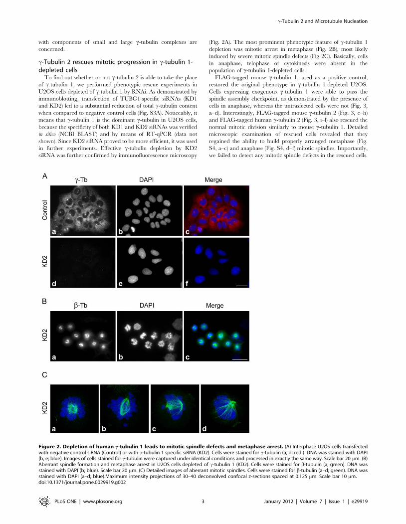
(Fig. 2A). The most prominent phenotypic feature of c-tubulin 1
depletion was mitotic arrest in metaphase (Fig. 2B), most likely
induced by severe mitotic spindle defects (Fig 2C). Basically, cells
in anaphase, telophase or cytokinesis were absent in the
population of c-tubulin 1-depleted cells.
FLAG-tagged mouse c-tubulin 1, used as a positive control,
restored the original phenotype in c-tubulin 1-depleted U2OS.
Cells expressing exogenous c-tubulin 1 were able to pass the
spindle assembly checkpoint, as demonstrated by the presence of
cells in anaphase, whereas the untrasfected cells were not (Fig. 3,
a–d). Interestingly, FLAG-tagged mouse c-tubulin 2 (Fig. 3, e–h)
and FLAG-tagged human c-tubulin 2 (Fig. 3, i–l) also rescued the
normal mitotic division similarly to mouse c-tubulin 1. Detailed
microscopic examination of rescued cells revealed that they
regained the ability to build properly arranged metaphase (Fig.
S4, a–c) and anaphase (Fig. S4, d–f) mitotic spindles. Importantly,
we failed to detect any mitotic spindle defects in the rescued cells.
Figure 2. Depletion of human c-tubulin 1 leads to mitotic spindle defects and metaphase arrest. (A) Interphase U2OS cells transfectedwith negative control siRNA (Control) or with c-tubulin 1 specific siRNA (KD2). Cells were stained for c-tubulin (a, d; red ). DNA was stained with DAPI(b, e; blue). Images of cells stained for c-tubulin were captured under identical conditions and processed in exactly the same way. Scale bar 20 mm. (B)Aberrant spindle formation and metaphase arrest in U2OS cells depleted of c-tubulin 1 (KD2). Cells were stained for b-tubulin (a; green). DNA wasstained with DAPI (b; blue). Scale bar 20 mm. (C) Detailed images of aberrant mitotic spindles. Cells were stained for b-tubulin (a–d; green). DNA wasstained with DAPI (a–d; blue).Maximum intensity projections of 30–40 deconvolved confocal z-sections spaced at 0.125 mm. Scale bar 10 mm.doi:10.1371/journal.pone.0029919.g002
c-Tubulin 2 and Microtubule Nucleation
PLoS ONE | www.plosone.org 3 January 2012 | Volume 7 | Issue 1 | e29919

Immunoblot tests in performed rescue experiments confirmed an
effective expression of FLAG-tagged c-tubulins in c-tubulin 1-
depleted cells (Fig. S3B). These findings suggest that c-tubulin 2 is
capable of replacing c-tubulin 1 during mitosis.
c-Tubulin 2 nucleates microtubulesTaking advantage of the above described phenotypic rescue
experimental set-up, we further investigated the microtubule
nucleating capability of c-tubulin 2 in microtubule regrowth
experiments. The amount of c-tubulin on prophase/metaphase
centrosomes is significantly higher than that in interphase due to
the process called centrosome maturation [25,26]. We therefore
first focused on mitotic centrosomes, where one could expect a
prominent effect of c-tubulin depletion on microtubule nucle-
ation. Microtubules were depolymerized by nocodazole, washed
by ice-cold PBS, and allowed to regrow before fixation and
staining for b-tubulin. Mitotic cells became more abundant in the
course of nocodazole treatment. While the regrowth of
microtubules from centrosomes was easily observable in cells
transfected with negative control siRNA (Fig. 4A, a–d), it was
substantially delayed and/or impaired in c-tubulin 1-depleted
cells (Fig. 4A, e–h). Clearly recognizable microtubule asters were
seen in 97% (n = 369) of negative control mitotic cells. In c-
tubulin 1-depleted cells, however, microtubule asters were
indistinct and formed in only 18% (n = 274) of mitotic cells. As
expected, FLAG-tagged mouse c-tubulin 1 (positive control)
rescued the microtubule aster formation in c-tubulin 1-depleted
cells (Fig. 4B, a–d). In accordance with our previous results, both
FLAG-tagged mouse c-tubulin 2 (Fig. 4B, e–h) and FLAG-tagged
human c-tubulin 2 (Fig. 4B, i–l) also rescued aster formation.
Clear microtubule regrowth was observed in all c-tubulin 1-
depleted cells expressing exogenous c-tubulin 2; it indicates that
c-tubulin 2 is capable of centrosomal microtubule nucleation in
mitotic cells.
In order to strengthen the evidence of microtubule nucleation
capability of c-tubulin 2, we quantified microtubule formation in
vivo by the tracking microtubule (+) ends marked by EB1-GFP in
interphase U2OS cells (U2OS-EB1). For live cell imaging we used
the shRNA system based on pLKO.1 vectors. Puromycin selection
for 6 days made it possible to analyze only c-tubulin-depleted cells.
We constructed TUBG1-specific shRNA expressing vectors based
on siRNAs (KD1 and KD2), and tested their effectivity by
immunoblotting (Fig S5A). Since KD2 shRNA was found more
efficient, further experiments were limited to that. Substantial c-
tubulin depletion by KD2 shRNA was confirmed by immunoflu-
orescence microscopy (Fig. S5B). Additionally, we prepared
TagRFP-tagged mouse c-tubulin 1 (pmTubg1-TagRFP) and
TagRFP-tagged human c-tubulin 2 (phTUBG2-TagRFP) for
phenotypic rescue experiments. TagRFP (pCI-TagRFP) served
as control.
Following puromycin selection, transfected U2OS-EB1 cells
were subjected to live cell imaging; time-lapse sequences of EB1-
GFP dynamics were acquired only from cells coexpressing
TagRFP or TagRFP-tagged proteins. Immunoblotting confirmed
an effective expression of tagged c-tubulins in c-tubulin 1-depleted
cells (Fig. S6). Results of typical experiments are presented in
Fig. 5, where single-frame (Fig. 5, a–d) as well as 60-frame
projections (Fig. 5, e–h) of time-lapse sequences are shown. While
TagRFP was found in both cytoplasm and nuclei (Fig 5, a–b),
TagRFP-tagged c-tubulins were concentrated to MTOC (Fig. 5,
c–d). This is more distinctly demonstrated in Fig. S7, where green
and red channels are depicted separately. The density of
microtubule (+) end tracks, reconstructed by maximum intensity
projection of time-lapse sequences, was markedly reduced in c-
Figure 3. c-Tubulin 2 restores normal mitotic spindle functioning in c-tubulin 1-depleted cells. U2OS cells depleted of c-tubulin 1 andexpressing FLAG-tagged mouse c-tubulin 1 (a-d, Tubg1-FLAG), mouse c-tubulin 2 (e-h, Tubg2-FLAG) or human c-tubulin 2 (i–l, TUBG2-FLAG) werestained for FLAG (a, e, i; red) and b-tubulin (b, f, j; green). DNA was stained with DAPI (c, g, k; blue). Scale bar 20 mm.doi:10.1371/journal.pone.0029919.g003
c-Tubulin 2 and Microtubule Nucleation
PLoS ONE | www.plosone.org 4 January 2012 | Volume 7 | Issue 1 | e29919

tubulin 1-depleted cells (Fig. 5, f) when compared with negative
control cells (Fig. 5, e). This most likely reflects an impaired
microtubule nucleation. In contrast, the density of EB1 tracks in
cells rescued by exogenous mouse c-tubulin 1 (Fig. 5, g) resembled
that seen in negative controls cells (Fig. 5, e). Clear phenotypic
rescue was also observed in cells expressing exogenous human
c-tubulin 2 (Fig. 5, h). These findings were confirmed by
evaluation of statistical data as documented in histograms of the
microtubule growth rates, where the number of EB1 tracks was
normalized by the cell area and tracking time (Fig. 6). To compare
whole populations of EB1 tracks in analyzed cells, we applied
Bonferroni correction of p-values to velocity histograms (Fig. 6).
Figure 4. c-Tubulin 2 rescues centrosomal microtubule nucleation in c-tubulin 1-depleted mitotic cells. A) U2OS cells transfected withnegative control siRNA (Control) or with c-tubulin 1 specific siRNA (KD2) were treated with 10 mM nocodazole for 6 h and fixed after 3 min incubationin medium without nocodazole. Cells were stained for c-tubulin (a, e; red ) and b-tubulin (b, f; green). DNA was stained with DAPI (c, g; blue).Fluorescence images of cells stained for c-tubulin were captured under identical conditions and processed in exactly the same way. Scale bar 10 mm.(B) U2OS cells depleted of c-tubulin 1 and expressing FLAG-tagged mouse c-tubulin 1 (a–d, Tubg1-FLAG), mouse c-tubulin 2 (e–h, Tubg2-FLAG) orhuman c-tubulin 2 (i–l, TUBG2-FLAG) were treated with 10 mM nocodazole for 6 h and fixed after 3 min incubation in medium without nocodazole.Cells were stained for FLAG (a, e, i; red) and b-tubulin (b, f, j; green). DNA was stained with DAPI (c, g, k; blue). Scale bar 10 mm.doi:10.1371/journal.pone.0029919.g004
c-Tubulin 2 and Microtubule Nucleation
PLoS ONE | www.plosone.org 5 January 2012 | Volume 7 | Issue 1 | e29919

Calculated p-values for differences among individual growth
velocity groups were multiplied by the number of all growth
velocity groups in the histogram (n = 13). Based on this correction,
the number of EB1 tracks was significantly reduced in c-tubulin 1-
depleted cells when compared with negative control cells
(p,0.0001, Fig. 6A). Conversely, the number of EB1 tracks was
significantly higher in cells rescued by exogenous mouse c-tubulin
1 (p,1.1026, Fig. 6B) or human c-tubulin 2 (p,1.1025, Fig. 6C)
than in c-tubulin 1-depleted cells. Differences between negative
control (blue columns in Fig. 6A) and c-tubulin 2 expressing cells
(blue columns in Fig. 6C) were statistically insignificant. Interest-
ingly, the number of EB1 tracks in cells expressing exogenous
mouse c-tubulin 1 (blue columns in Fig. 6B) exceeded that seen in
negative control (p,0.05; blue columns in Fig. 6A) or in cells
expressing exogenous c-tubulin 2 (p,0.05; blue columns in
Fig. 6C). Taken collectively, our experimental data demonstrate
that mammalian c-tubulin 2 is able to nucleate microtubules and
substitute for c-tubulin 1 even in interphase cells.
Tubg2 is downregulated in mouse preimplantationdevelopment
Since c-tubulin 2 was capable to substitute for c-tubulin 1 in
cultured cells, its inability to do so in blastocysts [23] is intriguing.
We therefore quantified by RT-qPCR the mRNA levels of Tubg1
and Tubg2 in mouse oocytes, 2-cell stage embryos, 8-cell stage
embryos and blastocysts. Adult mouse liver and brain tissues
served as controls, because Tubg2 expression is high in brain and
low in liver [23,24]. Geometric mean of mouse peptidylprolyl
isomerase A (Ppia) and mouse glyceraldehyde-3-phosphate dehy-
drogenase (Gapdh) mRNA levels were used for normalization.
Tubg1 mRNA level decreased 17 times, when 2-cell stage embryos
were compared with blastocysts, and was almost equal in liver and
brain (Fig 7A). In contrast, Tubg2 mRNA level decreased
dramatically by almost three orders of magnitude (815 times),
when these two developmental stages were compared. Tubg2
expression in blastocysts was comparable to that in liver and was
38 times lower than in brain (Fig. 7B). For comparison, mRNA
levels were also ascertained for Tubgcp2 and Tubgcp5 that encode,
respectively, GCP2 and GCP5 proteins. While Tubgcp2 mRNA
level remained relatively stable (Fig. 7C), that of Tubgcp5 decreased
9 times when comparing the 2-cell stage embryos and blastocysts
(Fig. 7D). Notably, the highest mRNA levels of tested genes were
detected in oocytes, which probably reflects the high content of
stored maternal mRNA [27]. Taken together, our data clearly
show that Tubg2 mRNA level is appreciably decreasing during
mouse preimplantation development.
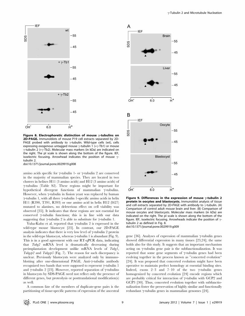
RT-qPCR analysis disclosed that blastocyst contains a very low
amount of Tubg2 mRNA. However, c-tubulin 2 protein might still
be present. To analyze the expression of Tubg2 at the protein level,
we first identified the positions of mouse c-tubulin 1 and c-tubulin
2 in samples separated by 2D-PAGE. Different antibodies reacting
with both c-tubulins were used for immunoblotting. The exact
positions of c-tubulin 1 and c-tubulin 2 were determined by
overexpression of, respectively, untagged mouse c-tubulin 1 and c-
tubulin 2 in P19 cells, where Tubg2 was undetectable by RT-qPCR
(Fig. S8). Immunoblotting of untransfected and transfected cells
with anti-c-tubulin antibodies revealed that the signal of main c-
tubulin isoforms in P19 cells (Fig. 8, wt) was enhanced in cells
overexpressing the c-tubulin 1 (Fig. 8, +c-Tb1). In cells
overexpressing c-tubulin 2, a new signal appeared in a more
basic position compared to c-tubulin1 isoforms (Fig. 8, +c-Tb2).
This was in agreement with theoretical isoelectric points for c-
tubulin 1 (5.66) and c-tubulin 2 (5.80). These experiments
demonstrate that mouse c-tubulins can be easily discriminated
on 2D-PAGE.
To rule out the possibility that the isoelectric point of exogenous
c-tubulin 2 expressed in P19 cells substantially differs from that in
mouse tissues, we compared the expression of c-tubulins in mouse
Figure 5. c-Tubulin 2 rescues microtubule formation in c-tubulin 1-depleted cells during interphase. Time-lapse imaging of U2OS-EB1cells for quantitative evaluation of microtubule (+) end dynamics. Cells with depleted c-tubulin 1 (KD2) expressing either TagRFP (pCI-TagRFP), mousec-tubulin 1 (pmTubg1-TagRFP) or human c-tubulin 2 (phTUBG2-TagRFP). Cells with empty vector (pLKO.1) expressing TagRFP (pCI-TagRFP) served asnegative control. (a–d) Still images of typical cells selected for evaluation. Only cells expressing both EB1-GFP (green) and TagRFP (red) or c-tubulin-TagRFP fusions (red) were evaluated. In contrast to freely diffusible TagRFP (a, b), c-tubulin-TagRFP fusions properly localized to MTOCs (c, d) markedby white arrows. (e–f) Maximum intensity projections of 60 consecutive time-frames from acquired time-lapse sequences. Note the markedly lowerdensity of microtubule tracks in cell with depleted human c-tubulin 1 (f). Microtubule track density is rescued in cells expressing exogenous mouse c-tubulin 1 (g) or exogenous human c-tubulin 2 (h). Scale bar 10 mm.doi:10.1371/journal.pone.0029919.g005
c-Tubulin 2 and Microtubule Nucleation
PLoS ONE | www.plosone.org 6 January 2012 | Volume 7 | Issue 1 | e29919

brain and mouse liver, where Tubg2 expression is high and low,
respectively [23,24]. For this, we used immunoblotting after
2D-PAGE separation of samples containing similar total protein
amounts. c-Tubulin 1 was clearly detectable in both brain and
liver. In contrast, a strong signal in the position of c-tubulin 2 was
detected merely in brain, whereas it was undetectable in liver
(Fig. 9A). Again, these results correlated with data obtained in RT-
qPCR experiments (Fig. 7B). The performed experiments
confirmed that c-tubulin 2 can be discriminated by 2D-PAGE
also in mouse tissues. Using the same approach, we compared the
expression of c-tubulin 1 and c-tubulin 2 in mouse oocytes and
blastocysts. Samples were prepared from 150 fully grown oocytes
at the GV stage and from 197 early blastocysts to ensure that the
total protein amount in blastocyst sample was not underestimated.
A fully grown oocyte (from adult animals) at GV stage contains
approximately 30 ng of protein. Zona pellucida contributes to this
amount some 4–5 ng [28]. An early blastocyst contains approx-
imately 25 ng of protein [29]. c-Tubulin 1 was clearly detectable
in both oocytes and blastocysts. On the other hand, while there
was a strong signal detectable in the position of c-tubulin 2 in
oocytes, the relevant signal in this position was dramatically
reduced in blastocysts (Fig. 9B). Expression of c-tubulin 2 at the
protein level in oocytes and blastocysts thus correlated with its
mRNA level (Fig. 7B). Collectively taken, these data strongly
indicate that a very low amount of c-tubulin 2 is present in wild-
type blastocysts due to its transcriptional downregulation.
Discussion
Mammalian c-tubulins are encoded by two closely related genes
[22,24], and specific functions have been attributed to them. [23].
The molecular basis of suggested functional differences between c-
tubulins is however unknown. In this study we document that
mammalian c-tubulin 2 is able to nucleate microtubules and
substitute for c-tubulin 1. In addition, we show that Tubg1 and Tubg2
are differentially transcribed during mouse early embryogenesis,
with Tubg2 transcription being progressively downregulated.
In general, c-tubulins are highly conserved proteins in all
eukaryotes. At the amino acid sequence level, human c-tubulin 1
and c-tubulin 2, respectively, show 98.9% and 97.6% identity with
the corresponding mouse isoforms (Table S1) [23]. To study the
subcellular localization and function of human and mouse c-
tubulin 2, we have chosen human osteosarcoma cells U2OS.
Because of their flat shape, they are excellent for immunofluores-
cence analysis and are easily transfectable. Moreover, the the
selection of U2OS made it possible to answer the question whether
or not the mouse c-tubulin 2 is capable of replacing human c-
tubulin 1. We have used exogenously expressed FLAG-tagged
mouse and human c-tubulins to evaluate the subcellular
localization of c-tubulin 2 proteins and their interactions with
GCPs. It was reported previously that exogenous mouse c-tubulin
2 located to interphase and mitotic centrosomes in mouse Eph4
epithelial cells [23]. Our data corroborate this finding by showing
that both human and mouse c-tubulin 2 are recruited to
interphase and mitotic centrosomes in human U2OS cells. By
immunoprecipitation experiments we found that c-tubulin 2
interacted with GCP2, an integral component of cTuSCs.
Reciprocal coimmunoprecipitations of c-tubulin 2 and GCP4
(T. Sulimenko, unpublished data) indicated that c-tubulin 2
normally also incorporated in cTuRCs. We found no differences
between c-tubulin 1 and c-tubulin 2 with regard to their
localization and interactions. Intriguingly, antibody to GCP2
coimmunoprecipitated more endogenous than exogenous c-
tubulins (Fig. 1B, Fig. S2). A similar result was obtained with
Figure 6. Quantitative evaluation of microtubule formation inphenotypic rescue experiments. Microtubule (+) end dynamics inU2OS-EB1 cells presented as velocity histograms. Cells with depleted c-tubulin 1 (KD2) or negative control cells (pLKO.1), expressing eitherTagRFP (pCI-TagRFP), mouse c-tubulin 1 (pmTubg1-TagRFP) or humanc-tubulin 2 (phTUBG2-TagRFP). (A) Comparison of negative control cells(pLKO.1+pCI-TagRFP; n = 19) with c-tubulin 1 depleted cells (KD2+pCI-TagRFP; n = 15). (B) Comparison of cells rescued with mouse c-tubulin 1(positive control; KD2+pmTubg1-TagRFP; n = 18) with c-tubulin 1depleted cells. (C) Comparison of cells rescued with human c-tubulin2 (KD2+hTubg2-TagRFP; n = 19) with c-tubulin 1 depleted cells. Data arefrom 3 independent experiments. Bars represent means6SD. Asterisksrepresent the p-values (p) of two-sided unpaired t-test (****, p,0.00001; ***, p,0.0001; **, p,0.001; *, p,0.01).doi:10.1371/journal.pone.0029919.g006
c-Tubulin 2 and Microtubule Nucleation
PLoS ONE | www.plosone.org 7 January 2012 | Volume 7 | Issue 1 | e29919

antibody to GCP4 (T. Sulimenko, unpublished data). This fact
might indicate a slow turnover of cTuCs, because precipitations
were performed 48 hours after transfection. Alternatively, FLAG
tags might interfere with interaction of c-tubulin with GCPs.
However, this seems unlikely as FLAG tags were fused to the C-
termini of c-tubulins, which probably is not involved in the
interaction with GCP2 and GCP3 [30]. Moreover, FLAG-tagged
c-tubulins rescued normal mitotic progression in c-tubulin 1-
depleted cells (Fig. 3).
The most remarkable phenotypic sign of c-tubulin 1-depleted
U2OS cells was arrest in metaphase caused by mitotic spindle defects
such as monopolar or collapsed spindles (Fig. 2C), previously
described in mammalian cells depleted of c-tubulin [11,23,31].
Similar defects were detected in cells in which c-tubulin localization
to centrosomes, mitotic spindle and mitotic chromatin was damaged
by depletion of cTuRC recruitment factors like GCP-WD/NEDD1
[11,31] or components of augmin complex [32,33]. As expected, the
observed phenotype was reverted by expression of mouse c-tubulin
1. Both human and mouse c-tubulin 2 likewise rescued the normal
mitotic progression in c-tubulin 1-depleted cells, indicating that
mammalian c-tubulin 2 is able to substitute for c-tubulin 1 in vivo
(Fig. 3). Consistent with these findings are the results of microtubule
regrowth experiments on mitotic cells which reveal that c-tubulin 2
does have microtubule nucleating capability (Fig. 4). We used only
KD2 siRNA and corresponding shRNA for phenotypic rescue
experiments, because it was more efficient than KD1 (Fig. S3A, Fig.
S5A) and its specificity was verified in an independent study [34].
Rescue experiments also ruled out potential off-target RNAi effects.
When testing the microtubule (+) end dynamics in c-tubulin 1-
depleted cells, we observed a significant reduction in the number
of EB1 tracks in interphase cells (Fig. 5, f; Fig. 6A), a sign of
impaired microtubule nucleation. Alternatively, reduction in the
EB1 track number might be explained by changes in microtubule
dynamics; the nucleation is not affected but the fraction of growing
microtubules relative to pausing or depolymerizing microtubules is
diminished. Although one cannot exclude a potential contribution
of impaired microtubule (+) ends dynamics to the observed
phenotype, we consider this possibility much less probable because
it has been previously demonstrated by regrowth experiments that
microtubule nucleation is impaired and/or delayed in interphase
cells depleted of c-tubulin [31]. We therefore conclude that c-
tubulin 2 is able to nucleate microtubules also in interphase cells.
Interestingly, a higher number of EB1 tracks was in c-tubulin 1-
depleted cells expressing exogenous c-tubulin 1 than in cells
expressing exogenous c-tubulin 2 (Fig. 6). It might imply that for
interphase cells c-tubulin 1 is a more potent nucleator of
microtubules than c-tubulin 2. However, no corresponding
differences in microtubule regrowth were observed in mitotic cells
(Fig. 4), where centrosomes are highly enriched with cTuCs
[25,26], and where consequently the potential differences in
nucleation capability ought to be stronger. In addition, statistical
significance (p,0.05) of this difference is relatively low. We
therefore do not think that c-tubulin 1 and c-tubulin 2
substantially differ in nucleation activity.
Functional redundancy of mammalian c-tubulins was expected
because of their high sequence similarity [22]. Importantly, only 6
Figure 7. Tubg2 mRNA level is decreasing during mouse preimplantation development. mRNA levels of Tubg1 (A), Tubg2 (B), Tubgcp2 (C)and Tubgcp5 (D) in mouse oocyte, 2-cell stage embryo, 8-cell stage embryo, blastocyst and liver relative to the level found in brain. Data arepresented as mean fold change (columns) with individual samples displayed (diamonds). Three biological replicates were measured twice underidentical conditions. Note that the Y-axis is in the logarithmic scale.doi:10.1371/journal.pone.0029919.g007
c-Tubulin 2 and Microtubule Nucleation
PLoS ONE | www.plosone.org 8 January 2012 | Volume 7 | Issue 1 | e29919
amino acids specific for c-tubulin 1- or c-tubulin 2 are conserved
in the majority of mammalian species. They are located in two
clusters in helixes H11 (3 amino acids) and H12 (3 amino acids) of
c-tubulins (Table S2). These regions might be important for
hypothetical divergent functions of mammalian c-tubulins.
However, when c-tubulin in fission yeast was replaced by human
c-tubulin 1, with all three c-tubulin 1-specific amino acids in helix
H11 (R390, T391, R393) or one amino acid in helix H12 (I427)
mutated to alanines, no deleterious effect on cell viability was
observed [35]. It indicates that these regions are not essential for
conserved c-tubulin functions; this is in line with our data
suggesting that c-tubulin 2 is able to substitute for c-tubulin 1.
Yuba-Kubo et al. reported that c-tubulin 2 is expressed in the
wild-type mouse blastocyst [23]. In contrast, our 2D-PAGE
analysis indicates that there is very low level of c-tubulin 2 protein
in the wild-type blastocyst, whereas c-tubulin 1 is abundant (Fig. 9).
This is in a good agreement with our RT-qPCR data, indicating
that Tubg2 mRNA level is dramatically decreasing during
preimplantation development unlike mRNA levels of Tubg1,
Tubgcp2 and Tubgcp5 (Fig. 7). The reason for such discrepancy is
unclear. Previously blastocysts were analyzed only by immuno-
blotting after one-dimensional PAGE. Anti-c-tubulin antibody
recognized two bands that were supposed to represent c-tubulin 1
and c-tubulin 2 [23]. However, reported separation of c-tubulins
in blastocysts by SDS-PAGE need not reflect only the presence of
different genes, but proteolysis or posttranslational modification(s)
as well.
A common fate of the members of duplicate-gene pairs is the
partitioning of tissue-specific patterns of expression of the ancestral
gene [36]. Analyses of expression of mammalian c-tubulin genes
showed differential expression in many tissues [23,24]; the same
holds also for this study. It suggests that an important mechanism
acting on c-tubulin gene pair is the subfunctionalization. It was
reported that some gene segments of c-tubulin genes had been
evolving together in the process known as ’’concerted evolution‘‘
[24]. It was proposed that concerted evolution might have been
operative to maintain perfect homology at essential binding sites.
Indeed, exons 2–3 and 7–10 of the two c-tubulin genes
homogenized by concerted evolution [24] encode regions which
are probably critical for interaction of c-tubulin with GCP2 and
GCP3 [30]. Thus, concerted evolution together with subfunctio-
nalization foster the preservation of highly similar and functionally
redundant c-tubulin genes in mammalian genomes [24].
Figure 8. Electrophoretic distinction of mouse c-tubulins on2D-PAGE. Immunoblots of mouse P19 cell extracts separated by 2D-PAGE probed with antibody to c-tubulin. Wild-type cells (wt), cellsexpressing exogenous untagged mouse c-tubulin 1 (+c-Tb1) or mousec-tubulin 2 (+c-Tb2). Molecular mass markers (in kDa) are indicated onthe right. The pI scale is shown along the bottom of the figure. IEF,isoelectric focusing. Arrowhead indicates the position of mouse c-tubulin 2.doi:10.1371/journal.pone.0029919.g008
Figure 9. Differences in the expression of mouse c-tubulin 2protein in oocytes and blastocysts. Immunoblot analysis of tissueand cell extracts separated by 2D-PAGE with antibody to c-tubulin. (A)Comparison of control adult mouse brain and liver. (B) Comparison ofmouse oocytes and blastocysts. Molecular mass markers (in kDa) areindicated on the right. The pI scale is shown along the bottom of thefigure. IEF, isoelectric focusing. Arrowheads indicate the position of c-tubulin 2 as defined in Fig. 8.doi:10.1371/journal.pone.0029919.g009
c-Tubulin 2 and Microtubule Nucleation
PLoS ONE | www.plosone.org 9 January 2012 | Volume 7 | Issue 1 | e29919

Our data allow an alternative interpretation of Tubg1-/- and
Tubg2-/- phenotypes previously described in mice [23]. Endoge-
nous c-tubulin 2 cannot rescue c-tubulin 1 deficiency in Tubg1-/-
blastocyst, even though it can nucleate microtubules, because it is
not present in a sufficient amount. It was previously reported that
knock out of single gene resulted in overexpression of related genes
[37–39]. Our data do not strictly exclude the possibility that c-
tubulin 2 expression could be up-regulated in Tubg1-/- blastocysts,
however, c-tubulin 2 may be insufficient to fully replacer the
lacking c-tubulin 1. On the other hand, whole-mount immuno-
staining with anti-c-tubulin antibody in Tubg1-/- blastocyst cells
did not identify any c-tubulin 2-positive foci, even though one
pericentrin-positive focus occurred in each cell [23]. It was
suggested that c-tubulin 1 was necessary for recruitment of c-
tubulin 2 to blastocyst centrosomes [23]. We propose that such
observation can be alternatively explained by the absence of c-
tubulin 2 at the blastocyst stage both both in wild type and
Tubg1-/- embryos. Behavioral abnormalities of Tubg2 -/- mice do
not necessarily imply unknown function(s) of c-tubulin 2. They
might also reflect a reduction of total c-tubulin in brain of Tubg2-/-
mice, since Tubg2 is highly expressed in the brain [23,24] as
demonstrated also in this study. Yet, we cannot exclude the
possibility that brain c-tubulin 2 has some additional still unknown
function(s). Thorough phenotype analysis of Tubg2-/- mice could
shed more light on c-tubulin 2 function(s) in brain and its
development. Further, elucidation of transcriptional regulation of
c-tubulin genes would by very important not only from the
developmental point of view but also with respect to tumorigen-
esis. Significantly higher expression of c-tubulin was found in high-
versus low-grade gliomas, common brain cancers [40,41].
In conclusion, the findings indicate that mammalian c-tubulin 2
is able to nucleate microtubules and substitute for c-tubulin 1.
Although c-tubulins are differentially expressed during mouse
early embryogenesis and in adult tissues, they are functionally
redundant with respect to their nucleation activity.
Materials and Methods
Ethics statementAll mice were maintained in accordance with the Institute of
Molecular Genetics Guidelines. Experiments were approved by
the Committee on the Ethics of Animal Experiments of the
Institute of Molecular Genetics (permit number 18/2009).
Cell cultures and transfectionsHuman osteogenic sarcoma cells U2OS, human glioblastoma
cell line T98G, mouse embryonal carcinoma cells P19, mouse
neuroblastoma Neuro-2a and mouse embryonal fibroblasts NIH
3T3 were obtained from the American Type Culture Collection.
Human kidney embryonal cells HEK293-FT (HEK) were from
Promega Biotec. Mouse bone marrow-derived mast cell line
(BMMC) was kindly provided by M. Hibbs (Ludwig Institute for
Cancer Research, Melbourne, Australia). Cells were cultured in
Dulbecco’s modified Eagle’s medium (DMEM) containing 10%
fetal bovine serum, penicillin (100 units/ml), and streptomycin
(0.1 mg/ml). Cells were grown at 37uC in 5% CO2 in air, and
passaged every 2 or 3 days using 0.25% trypsin/0.01% EDTA in
PBS. BMMC were cultured in RPMI 1640 medium supplemented
with serum, antibiotics and interleukin 3 (PeproTech) as described
previously [42].
U2OS cells were transfected with 2.5 mg (single plasmid) or 4 mg
(cotransfection of 2 plasmids) DNA/well in a 6-well plate using
Lipofectamine LTX reagent (Invitrogen) (DNA[mg]: LTX [ml]
ratio was 1:2.5) and Opti MEM medium (Gibco) according to the
manufacturer’s instruction. After 12 h, the transfection mixture
was replaced with fresh complete medium, and cells were
incubated for 48 h. HEK cells were transfected with 17 mg DNA
per 9-cm tissue culture dish using 51 mg polyethylenimine
(Polysciences) and serum-free DMEM. After 24 h, the transfection
mixture was replaced with fresh medium supplemented with
serum, and cells were incubated for additional 24 h.
Some of the U2OS cells, growing on coverslips, were treated
with 10 mM nocodazole (Sigma) for 6 h. Afterwards, cells were
washed 5 times in ice-cold PBS, transferred to new medium and
incubated for 3 min at 28uC before fixation.
Mouse oocytes, embryos and tissuesOocytes and embryos were obtained from 6–8 week old
C57BL/6 mice. Fully grown germinal vesicle (GV) oocytes were
liberated from ovaries by puncturing the antral follicles with
syringe needle and collected in M2 medium (Sigma) containing
0.2 mM isobutylmethylxanthine (IBMX; Sigma). To obtain
preimplantation embryos, mice were superovulated with 5 IU of
Folligon (pregnant mare serum gonadotropin; PMSG) (Intervet)
followed by stimulation with 5 IU of human chorionic gonado-
trophin (hCG, Sigma) 47 hours post-PMSG. The stimulated mice
were mated with 8–10 week old C57BL/6 males immediately after
hCG injection. Two-cell and eight-cell stage embryos were
collected 48 and 68 hours post-hCG, respectively, by flushing
the oviducts with M2 medium. Blastocyst stage embryos were
collected 96 hours post-hCG by flushing the uteri with M2
medium. Oocytes and embryos were washed 5 times in PBS prior
to transfer into TRI reagent (Ambion) for RNA isolation or into
buffer for 2D-PAGE. Liver and brain were dissected from 6–8
week old female C57BL/6 mice.
DNA constructsTotal RNA from BALB/c adult mouse brain or from human
cell line T98G was isolated by the RNeasy Mini kit (QIAGEN)
according to the manufacturer’s directions. The purity and
integrity of the RNA preparations were checked using Experion
automated electrophoresis system for microfluidic chip-based
analysis and RNA StdSens analysis kit (Bio-Rad Laboratories).
The quantity of RNA was checked by Nanodrop spectrophotom-
eter (NanoDrop Technologies). Reverse transcription was per-
formed with oligo(dT) primers and SuperScript III Reverse
Transcriptase kit (Invitrogen).
The full length human c-tubulin 2 (TUBG2, Refseq ID:
NM_016437) was amplified by PCR using forward 59-
GCCCACGTCTGAAGAGCGATGC-39 and reverse 59-CTG-
GAGATGAACCAAGAAGGGTTG-39 primers and T98G cell
cDNA as template. The full length mouse c-tubulin 1 (Tubg1,
Refseq ID: NM_134024) and mouse c-tubulin 2 (Tubg2, Refseq
ID: NM_134028) were amplified by PCR using the following
specific primers - Tubg1: forward 59-GAGAGACTGCAACGCC-
GATGTCTG-39 and reverse 59-TTGTGAGGTCCCTGATC-
TGTGCTC-39; Tubg2, forward 59- GGCAGGAGTTCCTCT-
CAGTCGTGAC-39 and reverse 59-TGTGAGGCGAAGTTGG-
GTCAGAG-39 - and mouse brain cDNA as template.
PCR products were ligated into pCR 3.1 vector (Invitrogen) by
TA-cloning method. Sequencing revealed that the M413V variant
of human c-tubulin 2 was isolated (refSNP ID: rs1046097). The
constructed vectors (pCR-hTUBG2, pCR-mTubg1, pCR-
mTubg2) served as templates for additional PCRs with following
specific forward and reverse primers carrying EcoRI/SalI restric-
tion sites (underlined): human TUBG2, forward 59-CTGAATTC-
CACGTCTGAAGAGCGATGC-39 and reverse 59-TCAAGTC-
GACCTGCTCCTGGGTGCC-39; mouse Tubg1: forward
c-Tubulin 2 and Microtubule Nucleation
PLoS ONE | www.plosone.org 10 January 2012 | Volume 7 | Issue 1 | e29919

59-ACGAATTCTGCCTGAGGAGCGATGC-39 and reverse 59-
TCAAGTCGACCTGCTCCTGGGTGCC-39; mouse Tubg2:
forward 59- CTGAATTCGGTCTGATCGGCGATGC-39 and
reverse 59-TCAAGTCGACCTGCTCCTGGGTGCC-39. PCR
products without stop codon were digested with EcoRI/SalI
restriction enzymes and inserted into pFLAG-CMV5a vector
(Sigma) resulting in C-terminally FLAG-tagged human c-tubulin 2
(phTUBG2-FLAG), mouse c-tubulin 1 (pmTubg1-FLAG) and
mouse c-tubulin 2 (pmTubg2-FLAG). Constructs encoding C-
terminally FLAG-tagged human c-tubulin 1 (phTUBG1-FLAG)
or mouse Fyn (pFyn-FLAG) were described previously [9].
Complete coding sequences of mouse Tubg1 and Tubg2 were cut
out from pCR-mTubg1 and pCR-mTubg2, respectively, by EcoRI
and inserted into pCI-NEO (Promega) to create vectors expressing
untagged mouse c-tubulin 1 (pCI-mTubg1) and c-tubulin 2 (pCI-
mTubg2).
Coding sequence of monomeric red fluorescent protein
TagRFP-T (GenBank: EU582019.1) was amplified from
pcDNA3.1-TagRFP (kind gift of Dr. R.Y. Tsien, HHMI at the
University of California, San Diego, USA) by PCR with the
following specific forward and reverse primers carrying SalI and
NotI restriction sites (underlined): forward 59-AGTCGACG-
GAGGTGGTGGAGGTATGGTGTCTAAGGGCGAAGA-39 (add-
ed 5x glycine coding motif in italic) and reverse 59- TGC-
GGCCGCTTACTTGTACAGCTCGTCCATGCCA -39. PCR
products were ligated into pCR 2.1 vector (Invitrogen) by TA-
cloning method. TagRFP coding sequence was digested from this
vector using SalI/NotI restriction enzymes and inserted into pCI-
Neo (Promega) resulting in a vector encoding TagRFP (pCI-
TagRFP) and allowing construction of C-terminally TagRFP-
tagged fusion proteins. Coding sequences of mouse Tubg1 and
human TUBG2 without stop codon were cut out from pmTubg1-
FLAG and phTUBG2-FLAG, respectively, by EcoRI/SalI restric-
tion enzymes and ligated into pCI-TagRFP resulting in vectors
encoding TagRFP-tagged mouse c-tubulin 1 (pmTubg1-TagRFP)
and human c-tubulin 2 (phTUBG2-TagRFP). All constructs were
verified by sequencing.
U2OS cells stably expressing EB1-GFP (U2OS-EB1) were
obtained by transfection of cells with pEB1-GFP, obtained from
Dr. Y. Mimori-Kiyosue [43], and selection in 1.1 mg/ml geneticin
(G418, Sigma) for 2 weeks. Cells were then diluted to one cell/well
on 96-well plate and allowed to grow for 2 weeks. Homogeneous
colonies expressing EB1-GFP were propagated.
AntibodiesThe following anti-peptide antibodies prepared to human c-
tubulin were used: mouse monoclonal antibodies TU-30 (IgG1)
and TU-32 (IgG1) to the sequence 434–449 [44]; monoclonal
antibody GTU 88 (IgG1; Sigma, T6657) and rabbit antibody
(Sigma, T5192) to the sequence 38–53. The anti-c-tubulin
antibodies react with both c-tubulin 1 and c-tubulin 2 in mouse
and human. b-Tubulin was detected with monoclonal antibody
TUB 2.1 conjugated with FITC (IgG1; Sigma F2043) and
pericentrin with rabbit antibody (Covance PRB-432C). Rabbit
antibodies to GAPDH (G9545) and FLAG peptide (F7425) as well
as monoclonal antibody M2 (IgG1) to FLAG peptide (F1804) were
from Sigma. Monoclonal antibodies to GCP2 protein, GCP2-01
(IgG2b) and GCP2-02 (IgG1), were described previously [9].
Monoclonal antibody to GCP4 (IgG1) was from Santa Cruz (sc-
271876). Monoclonal antibody NF-09 (IgG2b) to neurofilament
NF-M protein [45] and rabbit antibody to non-muscle myosin BT-
561 (Biomed Techn. Inc) served as controls.
The Cy3-conjugated anti-mouse and anti-rabbit antibodies
were from Jackson Immunoresearch Laboratories. Anti-rabbit
antibody conjugated with Alexa 488 was from Invitrogen.
Secondary horseradish peroxidase-conjugated antibodies were
from Promega.
RNAiU2OS cells in 6-well plates were transfected with siRNAs (final
concentration 20 nM) using Lipofectamine RNAi MAX (Invitro-
gen) according to the manufacturer’s instruction. Five siRNAs
(Ambion/Applied Biosystems) that target the regions present in
human c-tubulin 1, namely, (59-GGGAGAAAAGATCCAT-
GAG-39; siRNA ID #9227), (59-CGCATCTCTTTCTCA-
TATA-39; siRNA ID #120194), (59-GGACATTTTTGACAT-
CATA -39; siRNA ID #9317), (59-GAACCTGTCGCC-
AGTATGA-39; siRNA ID #120784), (59-GGTATCCTAA-
GAAGCTGGT-39; siRNA ID #9396) were tested. Maximal
depletion was reached by transfecting the siRNA twice with a 72-h
time interval and harvesting cells 72 h after the second
transfection. Negative control siRNA was from Ambion/Applied
Biosystems (Silencer Negative Control #1 siRNA).
Immunoblotting and immunofluorescence analyses revealed
that the highest reduction of c-tubulin was obtained with siRNA
ID#9396 (KD1) and siRNA ID#120194 (KD2; results not
shown). These siRNAs were used for some phenotypic rescue
experiments. In that case siRNA was mixed with the plasmid of
interest and transfected into cells which already underwent the first
round of 72 h-long siRNA treatment, using Lipofectamine LTX.
Cells were analyzed 72 h after the second transfection.
The selected siRNAs were also used for construction of shRNA
vectors based on pLKO.1 (Addgene, #8453) that enabled
puromycin selection. Corresponding sense and antisense oligonu-
cleotides were synthetized by Sigma-Aldrich: 9396sh-sense 59-
CCGGGGTATCCTAAGAAGCTGGTTTCTCGAGACCAGC-
TTCTTAGGATACCTGTTTTTG-39, 9396-antisense 59-AAT-
TCAAAAACAGGTATCCTAAGAAGCTGGTCTCGAGAA-
ACCAGCTTCTTAGGATACC-39; 120194sh-sense 59- CCGGC-
GCATCTCTTTCTCATATATTCTCGAGTATATGAGAAAG-
AGATGCGTGTTTTTG-39, 120194sh-antisense 59- AATT-
CAAAAACACGCATCTCTTTCTCATATACTCGAGAATAT-
ATGAGAAAGAGATGCG-39. Sense and antisense oligonucleo-
tides, at final concentration 45 mM of each, were annealed in 1x
NEB 2 buffer (New England Biolabs) by initial warming up to
95uC followed by slow (3 h) cooling down to the room tem-
perature. Annealed oligonucleotides were inserted into pLKO.1
previously linearized with AgeI/EcoRI resulting in human TUBG1
shRNA expressing vectors p9396sh (KD1) and p120194sh (KD2).
Correct sequences of all vectors were verified by sequencing.
shRNA vectors were transfected in the same way as other
plasmids used in the study. To select shRNA expressing cells, the
transfection mixture was replaced with fresh complete medium
12 h later. Puromycin (Sigma) at final concentration 2.5 mg/ml
was added after 12 h incubation, and cells were selected in
puromycin for 6 days before analysis.
Reverse transcription quantitative real-time PCR (RT-qPCR) analysis
Total cellular RNA was extracted in three independent
isolations from 20 mouse oocytes and 5–10 embryos using TRI
reagent (Ambion) according to manufacturer’s instructions. Three
independent isolations of total cellular RNA were also made from
four mouse cell lines Neuro2a, P19, BMMC and 3T3 using
RNeasy Mini kit (QIAGEN). In three independent experiments
mouse livers and brains were frozen in liquid nitrogen,
homogenized under liquid nitrogen using mortar and pestle, and
total RNA was extracted from 10–15 mg of homogenized tissue
c-Tubulin 2 and Microtubule Nucleation
PLoS ONE | www.plosone.org 11 January 2012 | Volume 7 | Issue 1 | e29919

using RNeasy Minikit (QIAGEN). Concentration of the purified
RNA was determined with spectrophotometer NanoDrop
(Thermo Scientific). The quality of RNA was checked on Agilent
2100 Bioanalyzer. All RNA samples were of good qPCR quality
(RNA integrity number [RIN] $7.6 for all tissue samples;
RIN$9.8 for all cell lines samples). Purified RNA was stored at
270uC. Purified RNAs from oocytes and embryos were converted
to cDNA immediately after isolation. RNA from each sample was
converted to cDNA using the ImProm-II RT kit (Promega). For
tissues and cell lines, each reaction sample (20 ml) contained 1 mg
RNA, random hexamer primers (25 ng/ ml), ImPROM-II reac-
tion buffer, 5.6 mM MgCl2, dNTP mix (0.5 mM each dNTP),
0.5 ml RNasin and 1 ml ImProm-II reverse transcriptase. For
oocytes and embryos, all isolated RNA was used for reverse
transcription.
Quantitative PCR was performed with gene-specific primers for
mouse c-tubulin 1 (Tubg1, NM_134024), mouse c-tubulin 2
(Tubg2, NM_134028), mouse GCP2 (Tubgcp2, NM_133755),
mouse GCP5 (Tubgcp5, NM_146190), mouse peptidylprolyl
isomerase A (Ppia, NM_008907) and mouse glyceraldehyde-3-
phosphate dehydrogenase (Gapdh, NM_008084). All primers were
tested in silico by NCBI BLAST to amplify specific targets. Primer
sequences are summarized in Table S3. Oligonucleotides were
from East Port (Prague, Czech Republic).
Quantitative PCRs were carried out on LightCycler 480 System
(Roche). Each reaction (5 ml) consisted of 2.5 ml LightCyclerH 480
SYBR Green I Master (Roche), 0.5 ml mixed gene-specific forward
and reverse primers (5 mM each) and 2 ml diluted cDNA. cDNA
samples from brain, liver and cell lines were diluted 1:50. In case
of oocyte and embryonal cDNA samples, the used amounts of
cDNA per reaction corresponded to 1/4 of oocyte, 1/2 of 2-cell
stage embryo, 1/10 of 8-cell stage embryo and 1/10 of blastocyst.
Calibration curves for tested genes were made by serial dilutions
(dilution factor 4) of brain cDNA. Each sample was run in
duplicate. Thermocycling parametres are described in Text S1.
Identity of PCR products was verified by sequencing.
Preparation of cell extractsWhole-cell extracts for SDS-PAGE were prepared by rinsing
the cells twice in Hepes buffer (50 mM Hepes adjusted to pH 7.6
with NaOH, 75 mM NaCl, 1 mM MgCl2 and 1 mM EGTA),
scraping them into Hepes buffer supplemented with protease
(Roche; Complete EDTA-free protease mixture) and phosphatase
(1 mM Na3VO4 and 1 mM NaF) inhibitors, and solubilizing in
hot SDS-sample buffer [46] without bromphenol blue and boiling
for 5 min.
When preparing the extracts for immunoprecipitation, cells
were rinsed twice in cold Hepes buffer and extracted for 10 min at
4uC with Hepes buffer supplemented with protease and phospha-
tase inhibitors and 1% Nonidet P-40. The suspension was then
spun down (20,000 g, 15 min, 4uC).
For preparation of samples for 2D-PAGE, oocytes and
blastocysts were directly lysed in 2D-PAGE sample buffer [47].
Brain and liver were mixed with cold Hepes buffer supplemented
with protease and phosphatase inhibitors in tissue/buffer ratio
1:10 and homogenized in teflon/glass grinder. The suspension was
then spun down (20,000 g, 15 min, 4uC) and 10–15 ml aliquots of
supernatant were mixed with 200 ml of sample buffer. Similarly,
20 ml aliquots of supernatants from 1% NP-40 extracts of P19 cells
were mixed with 200 ml of sample buffer.
Protein quantifications in lysates and SDS-PAGE-samples were
performed, respectively, with bicinchoninic acid assay and silver
dot assay [48].
Immunoprecipitation, gel electrophoresis andimmunoblotting
Immunoprecipitation from 1% NP-40 extracts was performed as
described [49]. Cell extracts were incubated with beads of protein A
(Pierce, Rockford, IL) saturated with: (I) rabbit antibody to FLAG,
(II) monoclonal antibody GCP2-01 (IgG2b) to GCP2 (III) rabbit
antibody to non-muscle myosin (negative control), (IV) monoclonal
antibody NF-09 (IgG2a; negative control) or with (V) immobilized
protein A alone. Gel electrophoresis and immunoblotting were
performed using standard protocols. Two-dimensional electropho-
resis (2D-PAGE) was performed as described [47] using for the first
dimension 7 cm long Immobiline DryStrip gels with a linear pH 4-
7 gradient (Amersham Biosciences). Comparable protein amounts
were loaded in case of P19, liver and brain extracts (,25 mg).
For immunoblotting, rabbit antibodies to GAPDH, c-tubulin
(Sigma T5192) and FLAG peptide (Sigma F7425) were diluted
1:20,000, 1:5,000 and 1:2,000, respectively. Monoclonal antibod-
ies to c-tubulin (GTU88) and GCP4 were diluted 1:10,000 and
1:2,000, respectively. Monoclonal antibodies to c-tubulin (TU-32)
and GCP2 (GCP2-02), in the form of spent culture supernatants,
were diluted 1:10. Peroxidase-conjugated secondary antibodies
were diluted 1:10,000. Bound antibodies were detected by
SuperSignal WestPico Chemiluminescent reagents (Pierce).
ImmunofluorescenceImmunofluorescence staining was performed as previously
described [50]. Samples were fixed in methanol at 220uC, air-
dried and washed in PBS. Rabbit antibodies to FLAG peptide and
pericentrin were diluted 1:1000 and 1:750, respectively. Mono-
clonal antibodies to FLAG peptide and b-tubulin were diluted
1:1,000 and 1:100, respectively. Monoclonal antibody TU-30 to c-
tubulin was used as spent culture medium diluted 1:50. Cy3-
conjugated anti-mouse and ant-rabbit antibodies were diluted
1:1,000, Alexa 488-conjugated anti-rabbit antibody was diluted
1:200. For double-label immunofluorescence, coverslips were
incubated separately with the primary antibodies, and simulta-
neously with the secondary conjugated antibodies. The prepara-
tions were mounted in MOWIOL 4–88 (Calbiochem) supple-
mented with 4,6-diamidino-2-phenylindole (DAPI, Sigma) to label
nuclei, and examined on Delta Vision Core system (Applied
Precision) equipped with 60x/1.42 NA oil-immersion objective.
Optical z-sections were acquired in 0.25–0.30 mm steps. Z-stacks
were deconvolved by a built-in deconvolution program (Softworx)
using default parameters. Alternatively, some preparations were
examined on Olympus AX-70 equipped with 40x/1.0 NA water
objective. Some preparations were also examined on confocal
microscope Leica SP5 with 60x/1.4 NA oil objective. Optical z-
sections were acquired at 0.125 mm. Z-stacks were deconvolved by
Huygens Professional software (SVI, The Netherlands). All
presented Maximum Intensity Projections (MIP) of deconvolved
z-stacks were prepared in ImageJ (NIH/USA). Conjugates alone
gave no significant staining.
Time-lapse imagingFor time-lapse imaging, U2OS cells expressing EB1-GFP were
grown on glass-bottom-dishes (MatTek) and transfected with
different sets of plasmids, as specified in Result section. Before
imaging, DMEM was replaced with medium for live cell imaging
(DMEM without phenol red, riboflavin, folic acid, pyridoxal,
Fe[NO3]3 and puromycin). Only cells expressing TagRFP or
TagRPF-fusion proteins were selected for time-lapse imaging.
Time-lapse sequences of EB1-GFP dynamics were collected for
2 min at 1 sec intervals on Delta Vision Core system (Applied
c-Tubulin 2 and Microtubule Nucleation
PLoS ONE | www.plosone.org 12 January 2012 | Volume 7 | Issue 1 | e29919

Precision) equipped with 60x/1.42 NA oil-immersion objective.
The focus plane was near the coverslip where the best resolution of
EB1 comets was observed. Time-lapse sequences were adjusted in
ImageJ (NIH, USA) by manual cropping of individual cells,
enhancement of brightness and contrast, and converting sequences
to a depth of 8 bits. Adjusted time-lapse sequences of individual
cells were analyzed by in-house-written particle tracking plug-in
implemented in Ellipse program version 2.07 (ViDiTo, Systems,
Kosice Slovakia; http://www.ellipse.sk) as described [42]. The
particle speed was calculated as the ratio of particle trajectory
length and trajectory duration. Statistical analysis was performed
with the Student’s two-tailed unpaired t-test using Microsoft Excel.
Supporting Information
Figure S1 Exogenous c-tubulin 2 locates to centro-somes. Human U2OS cells expressing FLAG-tagged mouse c-
tubulin 1 (a–d, Tubg1-FLAG), mouse c-tubulin 2 (e–h, Tubg2-
FLAG), human c-tubulin 1 (i–l, TUBG1-FLAG) and human c-
tubulin 2 (m–p, TUBG2-FLAG) were stained for FLAG (red) and
pericentrin (green). DNA was stained with DAPI (blue). Final
images were made by maximum intensity projection of 3
deconvolved z-sections spaced at 0.25 mm. Scale bar 10 mm.
(TIF)
Figure S2 Coimmunoprecipitation of human c-tubulinswith GCP2 and GCP4 proteins. Extracts from HEK cells
expressing FLAG-tagged human c-tubulin 1 (TUBG1-FLAG),
human c-tubulin 2 (TUBG2-FLAG) or control mouse Fyn (Fyn-
FLAG) were immunoprecipitated with antibodies to FLAG or
GCP2, and blots were probed with antibodies to FLAG, GCP2,
GCP4 and c-tubulin (c-Tb). Extracts (1), immunoprecipitated
proteins (2), protein A without antibodies incubated with extracts
(3), immobilized antibodies not incubated with extracts (4).
Arrowheads indicate the positions of exogenous c-tubulins.
(TIF)
Figure S3 Immunoblot analysis of U2OS cells inphenotypic rescue experiments with FLAG-tagged c-tubulins. (A) Immunoblot analysis of whole cell extracts from
cells transfected with negative control (Control) or c-tubulin
specific siRNAs (KD1 and KD2). Staining with antibodies to c-
tubulin (c-Tb) and GAPDH. (B) Cells with depleted c-tubulin 1
(KD2), expressing FLAG-tagged mouse c-tubulin 1 (Tubg1-
FLAG), mouse c-tubulin 2 (Tubg2-FLAG) or human c-tubulin 2
(TUBG2-FLAG). Immunoblots of whole cell lysates probed with
antibodies to c-tubulin (c-Tb), FLAG and GAPDH (loading
control). Arrowhead indicates the position of endogenous c-
tubulin.
(TIF)
Figure S4 c-Tubulin 2 rescues mitotic spindle organi-zation and function in c-tubulin 1-depleted cells. U2OS
cells depleted of c-tubulin 1 and expressing FLAG-tagged mouse
c-tubulin 1 (a, d; Tubg1-FLAG), mouse c-tubulin 2 (b, e; Tubg2-
FLAG) or human c-tubulin 2 (c, f; TUBG2-FLAG). Cells were
stained for FLAG (red) and b-tubulin (green). DNA was stained
with DAPI (blue). Final images were made by maximum intensity
projection of 30–40 deconvolved confocal z-sections spaced at
0.125 mm. Scale bars 5 mm.
(TIF)
Figure S5 Depletion of c-tubulin 1 in U2OS cells byshRNA. Cells transfected with empty pLKO.1 vector (Control),
TUBG1 shRNA expressing vectors p9396sh (KD1) or p120194sh
(KD2). (A) Immunoblots of whole cell lysates probed with
antibodies to c-tubulin (c-Tb) and GAPDH (loading control). (B)
Immunofluorescence staining with antibody to c-tubulin (red) and
with DAPI (blue). Fluorescence images of cells stained for c-
tubulin were captured under identical conditions and processed in
exactly the same manner. Scale bar 20 mm.
(TIF)
Figure S6 Immunoblot analysis of U2OS cells inphenotypic rescue experiments with TagRFP-tagged c-tubulins. U2OS-EB1 cells with depleted c-tubulin 1 (KD2;
shRNA) or negative control cells (NC; pLKO.1), expressing
TagRFP, tagged mouse c-tubulin 1 (Tubg1-TagRFP) or tagged
human c-tubulin 2 (TUBG2-TagRFP). Immunoblots of whole cell
lysates probed with antibodies to c-tubulin (c-Tb) and GAPDH
(loading control). Arrowhead indicates the position of endogenous
c-tubulin.
(TIF)
Figure S7 c-Tubulin 2 rescues microtubule formation inc-tubulin 1-depleted cells during interphase. Time-lapse
imaging of U2OS-EB1 cells for quantitative evaluation of
microtubule (+) end dynamics. Cells with depleted c-tubulin 1
(KD2) expressing either mouse c-tubulin 1 (pmTubg1-TagRFP) or
human c-tubulin 2 (phTUBG2-TagRFP). Single frame coloured
images Fig 5c and Fig. 5d were separated to red and green
channels for a better evaluation of c-tubulin-TagRFP fusions (red)
and EB1-GFP (green). White arrows mark MTOCs.
(TIF)
Figure S8 Comparison of c-tubulin 2 expression inmouse brain and cell lines. Expression of gene for c-tubulin
2 (Tubg2) in neuroblastoma (Neuro2a), bone marrow mast cells
(BMMC), embryonal fibroblasts (3T3) and embryonic carcinoma
cells (P19) relative to the level in brain. Data are presented as
mean fold change (columns) with individual samples displayed
(diamonds). Three biological replicates were quantified twice
under identical conditions. *, undetectable level in P19 cells.
(TIF)
Table S1 Sequence alignments of human and mouse c-tubulins.(PDF)
Table S2 Multiple sequence alignment of carboxy-terminal domains of mammalian c-tubulins.(PDF)
Table S3 Sequences of primers used for RT-qPCRanalysis of mouse genes.(PDF)
Text S1 Thermocycling parameters at quantitativePCR.(PDF)
Acknowledgments
We thank Dr. Y. Mimori-Kiyosue (KAN Research Institute, Kyoto, Japan)
for EB1-GFP construct, Dr. R.Y. Tsien (HHMI at the University of
California, San Diego, USA) for pcDNA3.1-TagRFP construct and Dr. M.
Hibbs (Ludwig Institute for Cancer Research, Melbourne, Australia) for
BMMC cells. We thank Dr. Petr Svoboda (Institute of Molecular Genetics
AS CR, Prague, Czech Republic) for critical reading of the manuscript. S.
Vinopal was supported in part by the Department of Cell Biology, Faculty
of Science, Charles University, Prague, Czech Republic.
Author Contributions
Conceived and designed the experiments: SV ED PD. Performed the
experiments: SV MC VS TS ED VV MF. Analyzed the data: SV VS TS
ED PD. Wrote the paper: SV PD.
c-Tubulin 2 and Microtubule Nucleation
PLoS ONE | www.plosone.org 13 January 2012 | Volume 7 | Issue 1 | e29919
References
1. Oakley BR, Oakley CE, Yoon Y, Jung M (1990) c-Tubulin is a component ofthe spindle pole body that is essential for microtubule function in Aspergillus
nidulans. Cell 61: 1289–1301.2. Stearns T, Evans L, Kirschner M (1991) c-Tubulin is highly conserved
component of the centrosome. Cell 65: 825–836.3. Joshi HC, Palacios MJ, McNamara L, Cleveland DW (1992) c-Tubulin is a
centrosomal protein required for cell cycle-dependent microtubule nucleation.
Nature 356: 80–83.4. Wiese C, Zheng Y (2006) Microtubule nucleation: gamma-tubulin and beyond.
J Cell Sci 119: 4143–4153.5. Raynaud-Messina B, Merdes A (2007) c-Tubulin complexes and microtubule
organization. Curr Opin Cell Biol 19: 24–30.
6. Moritz M, Braunfeld MB, Guenebaut V, Heuser J, Agard DA (2000) Structureof the c-tubulin ring complex: a template for microtubule nucleation. Nat Cell
Biol 2: 365–370.7. Kollman JM, Polka JK, Zelter A, Davis TN, Agard DA (2010) Microtubule
nucleating c-TuSC assembles structures with 13-fold microtubule-like symmetry.
Nature 466: 879–882.8. Luders J, Stearns T (2007) Microtubule-organizing centres: a re-evaluation. Nat
Rev Mol Cell Biol 8: 161–167.9. Horejsı B, Vinopal S, Sladkova V, Draberova E, Sulimenko V, et al. (2011)
Nuclear c-tubulin associates with nucleoli and interacts with tumor suppressorprotein C53. J Cell Physiol 227: 367–382.
10. Moudjou M, Bordes N, Paintrand M, Bornens M (1996) c-Tubulin in
mammalian cells: the centrosomal and the cytosolic forms. J Cell Sci 109:875–887.
11. Haren L, Remy MH, Bazin I, Callebaut I, Wright M, et al. (2006) NEDD1-dependent recruitment of the c-tubulin ring complex to the centrosome is
necessary for centriole duplication and spindle assembly. J Cell Biol 172:
505–515.12. Dammermann A, Maddox PS, Desai A, Oegema K (2008) SAS-4 is recruited to
a dynamic structure in newly forming centrioles that is stabilized by the gamma-tubulin-mediated addition of centriolar microtubules. J Cell Biol 180: 771–785.
13. Zimmerman S, Chang F (2005) Effects of c-tubulin complex proteins onmicrotubule nucleation and catastrophe in fission yeast. Mol Biol Cell 16:
2719–2733.
14. Cuschieri L, Miller R, Vogel J (2006) Gamma-tubulin is required for properrecruitment and assembly of Kar9-Bim1 complexes in budding yeast. Mol Biol
Cell 17: 4420–4434.15. Bouissou A, Verollet C, Sousa A, Sampaio P, Wright M, et al. (2009) c-Tubulin
ring complexes regulate microtubule plus end dynamics. J Cell Biol 187:
327–334.16. Nayak T, Edgerton-Morgan H, Horio T, Xiong Y, De Souza CP, et al. (2010)
Gamma-tubulin regulates the anaphase-promoting complex/cyclosome duringinterphase. J Cell Biol 190: 317–330.
17. Rodriguez AS, Batac J, Killilea AN, Filopei J, Simeonov DR, et al. (2008)Protein complexes at the microtubule organizing center regulate bipolar spindle
assembly. Cell Cycle 7: 1246–1253.
18. Liu B, Joshi HC, Wilson TJ, Silflow CD, Palevitz BA, et al. (1994) c-Tubulin inArabidopsis: gene sequence, immunoblot, and immunofluorescence studies.
Plant Cell 6: 303–314.19. Ruiz F, Beisson J, Rossier J, Dupuis-Williams P (1999) Basal body duplication in
Paramecium requires gamma-tubulin. Current Biology 9: 43–46.
20. Tan M, Heckmann K (1998) The two gamma-tubulin-encoding genes of theciliate Euplotes crassus differ in their sequences, codon usage, transcription
initiation sites and poly(A) addition sites. Gene 210: 53–60.21. Wilson PG, Zheng Y, Oakley CE, Oakley BR, Borisy GG, et al. (1997)
Differential expression of two c-tubulin isoforms during gametogenesis anddevelopment in Drosophila. Dev Biol 184: 207–221.
22. Wise DO, Krahe R, Oakley BR (2000) The c-tubulin gene family in humans.
Genomics 67: 164–170.23. Yuba-Kubo A, Kubo A, Hata M, Tsukita S (2005) Gene knockout analysis of
two c-tubulin isoforms in mice. Develop Biol 282: 361–373.24. Carson AR, Scherer SW (2009) Identifying concerted evolution and gene
conversion in mammalian gene pairs lasting over 100 million years. BMC Evol
Biol 9: 156.25. Khodjakov A, Rieder CL (1999) The sudden recruitment of c-tubulin to the
centrosome at the onset of mitosis and its dynamic exchange throughout the cellcycle, do not require microtubules. J Cell Biol 146: 585–596.
26. Piehl M, Tulu US, Wadsworth P, Cassimeris L (2004) Centrosome maturation:
Measurement of microtubule nucleation throughout the cell cycle by using GFP-tagged EB1. Proc Natl Acad Sci USA 101: 1584–1588.
27. Piko L, Clegg KB (1982) Quantitative changes in total RNA, total poly(A), andribosomes in early mouse embryos. Dev Biol 89: 362–378.
28. Schultz RM, Wassarman PM (1977) Biochemical studies of mammalianoogenesis: Protein synthesis during oocyte growth and meiotic maturation in
the mouse. J Cell Sci 24: 167–194.29. Sellens MH, Stein S, Sherman MI (1981) Protein and free amino acid content in
preimplantation mouse embryos and in blastocysts under various culture
conditions. J Reprod Fertil 61: 307–315.30. Guillet V, Knibiehler M, Gregory-Pauron L, Remy MH, Chemin C, et al.
(2011) Crystal structure of gamma-tubulin complex protein GCP4 providesinsight into microtubule nucleation. Nat Struct Mol Biol 18: 915–919.
31. Luders J, Patel UK, Stearns T (2006) GCP-WD is a c-tubulin targeting factor
required for centrosomal and chromatin mediated microtubule nucleation.Nature Cell Biol 8: 137–147.
32. Uehara R, Nozawa RS, Tomioka A, Petry S, Vale RD, et al. (2009) The augmincomplex plays a critical role in spindle microtubule generation for mitotic
progression and cytokinesis in human cells. Proc Natl Acad Sci U S A 106:
6998–7003.33. Uehara R, Goshima G (2010) Functional central spindle assembly requires de
novo microtubule generation in the interchromosomal region during anaphase.J Cell Biol 191: 259–267.
34. Hutchins JR, Toyoda Y, Hegemann B, Poser I, Heriche JK, et al. (2010)Systematic analysis of human protein complexes identifies chromosome
segregation proteins. Science 328: 593–599.
35. Hendrickson TW, Yao J, Bhadury S, Corbett AH, Joshi HC (2001) Conditionalmutations in gamma-tubulin reveal its involvement in chromosome segregation
and cytokinesis. Mol Biol Cell 12: 2469–2481.36. Lynch M, Force A (2000) The probability of duplicate gene preservation by
subfunctionalization. Genetics 154: 459–473.
37. Lam EW, Glassford J, Banerji L, Thomas NS, Sicinski P, et al. (2000) Cyclin D3compensates for loss of cyclin D2 in mouse B-lymphocytes activated via the
antigen receptor and CD40. J Biol Chem 275: 3479–3484.38. Kafri R, Levy M, Pilpel Y (2006) The regulatory utilization of genetic
redundancy through responsive backup circuits. Proc Natl Acad Sci U S A 103:11653–11658.
39. DeLuna A, Springer M, KIrschner MW, Kishony R (2010) Need-based up-
regulation of protein levels in response to deletion of their duplicate genes. PLoSBiol 8: e1000347.
40. Katsetos CD, Reddy G, Draberova E, Smejkalova B, Del Valle L, et al. (2006)Altered cellular distribution and subcellular sorting of c-tubulin in diffuse
astrocytic gliomas and human glioblastoma cell lines. J Neuropathol Exp Neurol
65: 465–477.41. Loh JK, Lieu AS, Chou CH, Lin FY, Wu CH, et al. (2010) Differential
expression of centrosomal proteins at different stages of human glioma. BmcCancer 10: 268.
42. Hajkova Z, Bugajev V, Draberova E, Vinopal S, Draberova L, et al. (2011)STIM1-directed reorganization of microtubules in activated cells. J Immunol
186: 913–923.
43. Mimori-Kiyosue Y, Shiina N, Tsukita S (2004) The dynamic behavior of theAPC-binding protein EB1 on the distal ends of microtubules. Curr Biol 10:
865–868.44. Novakova M, Draberova E, Schurmann W, Czihak G, Viklicky V, et al. (1996)
c-Tubulin redistribution in taxol-treated mitotic cells probed by monoclonal
antibodies. Cell Motil Cytoskel 33: 38–51.45. Draberova E, Sulimenko V, Kukharskyy V, Draber P (1999) Monoclonal
antibody NF-09 specific for neurofilament protein NF-M. Folia Biol (Prague) 45:163–165.
46. Laemmli UK (1970) Cleavage of structural proteins during the assembly of thehead of bacteriophage T4. Nature 227: 680–685.
47. Sulimenko V, Sulimenko T, Poznanovic S, Nechiporuk-Zloy V, Bohm JK, et al.
(2002) Association of brain c-tubulins with ab-tubulin dimers. Biochem J 365:889–895.
48. Draber P (1991) Quantitation of proteins in sample buffer for sodium dodecylsulfate-polyacrylamide gel electrophoresis using colloidal silver. Electrophoresis
12: 453–456.
49. Kukharskyy V, Sulimenko V, Macurek L, Sulimenko T, Draberova E, et al.(2004) Complexes of c-tubulin with non-receptor protein tyrosine kinases Src
and Fyn in differentiating P19 embryonal carcinoma cells. Exp Cell Res 298:218–228.
50. Draberova E, Draber P (1993) A microtubule-interacting protein involved in
coalignment of vimentin intermediate filaments with microtubules. J Cell Sci106: 1263–1273.
c-Tubulin 2 and Microtubule Nucleation
PLoS ONE | www.plosone.org 14 January 2012 | Volume 7 | Issue 1 | e29919

Table S1 Alignments of human and mouse γ-tubulins γ-Tubulin 1 H.s. MPREIITLQLGQCGNQIGFEFWKQLCAEHGISPEGIVEEFATEGTDRKDVFFYQADDEHYIPRAVLLDLEPRVIHSILNSPYAKLYNPENIYLSEHGGGAGNNWASGFSQGEKIHEDIFD 120 M.m. MPREIITLQLGQCGNQIGFEFWKQLCAEHGISPEGIVEEFATEGTDRKDVFFYQADDEHYIPRAVLLDLEPRVIHSILNSSYAKLYNPENIYLSEHGGGAGNNWASGFSQGEKIHEDIFD 120 ********************************************************************************.*************************************** H.s. IIDREADGSDSLEGFVLCHSIAGGTGSGLGSYLLERLNDRYPKKLVQTYSVFPNQDEMSDVVVQPYNSLLTLKRLTQNADCVVVLDNTALNRIATDRLHIQNPSFSQINQLVSTIMSAST 240 M.m. IIDREADGSDSLEGFVLCHSIAGGTGSGLGSYLLERLNDRYPKKLVQTYSVFPNQDEMSDVVVQPYNSLLTLKRLTQNADCVVVLDNTALNLIATDRLHIQNPSFSQINQLVSTIMSAST 240 ******************************************************************************************* **************************** H.s. TTLRYPGYMNNDLIGLIASLIPTPRLHFLMTGYTPLTTDQSVASVRKTTVLDVMRRLLQPKNVMVSTGRDRQTNHCYIAILNIIQGEVDPTQVHKSLQRIRERKLANFIPWGPASIQVAL 360 M.m. TTLRYPGYMNNDLIGLIASLIPTPRLHFLMTGYTPLTTDQSVASVRKTTVLDVMRRLLQPKNVMVSTGRDRQTNHCYIAILNIIQGEVDPTQVHKSLQRIRERKLANFIPWGPASIQVAL 360 ************************************************************************************************************************ H.s. SRKSPYLPSAHRVSGLMMANHTSISSLFERTCRQYDKLRKREAFLEQFRKEDMFKDNFDEMDTSREIVQQLIDEYHAATRPDYISWGTQEQ 451 M.m. SRKSPYLPSAHRVSGLMMANHTSISSLFERTCRQFDKLRKREAFMEQFRKEDIFKDNFDEMDTSREIVQQLIDEYHAATRPDYISWGTQEQ 451 **********************************:*********:*******:************************************** γ-Tubulin 2 H.s. MPREIITLQLGQCGNQIGFEFWKQLCAEHGISPEGIVEEFATEGTDRKDVFFYQADDEHYIPRAVLLDLEPRVIHSILNSPYAKLYNPENIYLSEHGGGAGNNWASGFSQGEKIHEDIFD 120 M.m. MPREIITLQLGQCGNQIGFEFWKQLCAEHGISPEGIVEEFATEGTDRKDVFFYQADDEHYIPRAVLLDLEPRVIHSILNSSYAKLYNPENIYLSEHGGGAGNNWGRGFSQGEKIHEDIFD 120 ********************************************************************************.***********************. ************** H.s. IIDREADGSDSLEGFVLCHSIAGGTGSGLGSYLLERLNDRYPKKLVQTYSVFPYQDEMSDVVVQPYNSLLTLKRLTQNADCVVVLDNTALNRIATDRLHIQNPSFSQINQLVSTIMSAST 240 M.m. IIDREADGSDSLEGFVLCHSIAGGTGSGLGSYLLERLNDRYPKKLVQTYSVFPNQDEMSDVVVQPYNSLLTLKRLTQNADCVVVLDNTALNRIATDRLHIQNPSFSQINQLVSTIMSAST 240 ***************************************************** ****************************************************************** H.s. TTLRYPGYMNNDLIGLIASLIPTPRLHFLMTGYTPLTTDQSVASVRKTTVLDVMRRLLQPKNVMVSTGRDRQTNHCYIAILNIIQGEVDPTQVHKSLQRIRERKLANFIPWGPASIQVAL 360 M.m. TTLRYPGYMNNDLIGLIASLIPTPRLHFLMTGYTPLTTDQSVASVRKTTVLDVMRRLLQPKNVMVSTGRDRQTNHCYIAILNIIQGEVDPTQVHKSLQRIRERKLANFIPWGPASIQVAL 360 ************************************************************************************************************************ H.s. SRKSPYLPSAHRVSGLMMANHTSISSLFESSCQQFDKLRKRDAFLEQFRKEDMFKDNFDEMDRSREVVQELIDEYHAATQPDYISWGTQEQ 451 M.m. SRKSPYLPSAHRVSGLMMANHTSISSLFESSCQQYDKLWKRGAFLEQFRKEDIFKDNFEEMHRSREVVQELIDEYHAATRPDYISWGTQEQ 451 **********************************:*** **.**********:*****:**.*****************:*********** __________________________________________________________________________________________________________________________________________________________ H.s., human (Homo sapiens); M. m., mouse (Mus musculus). Accesions numbers (NCBI database): human γ-tubulin 1, NP_001061.2; mouse γ-tubulin 1, NP_598785.1; human γ-tubulin 2, NP_057521.1; mouse γ-tubulin 2, NP_598789.1. An asterisk indicates positions which have a single, fully conserved residue. A colon indicates conservation among groups of strongly similar properties. A period indicates conservation among groups of weakly similar properties. Different amino acids are highlighted.

Table S2. Multiple sequence alignment of carboxy-terminal domains of mammalian γ-tubulins Species Part of helix H11 Part of helix H12 Accesion number
γ-tubulin 2 Human (Homo sapiens) (386) SLFESSCQQFDK (419) DEMDRSREVVQELIDE NP_057521.1 Chimpanzee (Pan troglodytes) (386) SLFESSCQQFDK (419) DEMDRSREVVQELIDE XP_003315538 Mouse (Mus musculus) (386) SLFESSCQQYDK (419) EEMHRSREVVQELIDE NP_598789.1 Rat (Rattus norvegicus) (411) SLFESSCQQYDK (444) EEMDRSREVVQELIDE NP_001178004.1 Horse (Equus caballus) (386) SLFESSCQQYDK (419) DELDRSREVVQELIDE XP_001493749.1 Bat (Myotis lucifugus) (386) SLFESSCQQYDK (419) DELDRSREVVQELIDE ENSMLUP00000007140 (Ensembl) Bovine (Bos taurus) (386) SLFESSCQQYDK (419) DELDRSREVVQELIDE NP_001032704.1 Dog (Canis familiaris) (386) SLFESSCQQYDK (419) DELDTSREVVQELIDE XP_548085.2 γ-tubulin 1 Human (Homo sapiens) (386) SLFERTCRQYDK (419) DEMDTSREIVQQLIDE NP_001061.2 Chimpanzee (Pan troglodytes) (386) SLFERTCRQYDK (419) DEMDTSREIVQQLIDE XP_001162243 Mouse (Mus musculus) (386) SLFERTCRQFDK (419) DEMDTSREIVQQLIDE NP_598785.1 Rat (Rattus norvegicus) (386) SLFERTCRQFDK (419) DEMDTSREIVQQLIDE NP_665721.1 Horse (Equus caballus) (386) SLFERTCRQYDK (419) DELDTSREIVQQLIDE XP_001493897.2 Bat (Myotis lucifugus) (386) SLFERTCRQYDK (419) DELDTSREIVQQLIDE ENSMLUP00000015217 (Ensembl) Bovine (Bos taurus) (386) SLFERTCRQYDK (419) DELDTSREIVQQLIDE NP_001069723.1 Dog (Canis familiaris) (386) SLFERTCRQYDK (419) DELDTSREVVHQLIDE NP_001003105.1 **** :*:*:** :*:. ***:*::****
Protein sequences of indicated mammalian γ-tubulins were aligned by online version of ClustalW2 (www.ebi.ac.uk/Tools/msa/clustalw2) using default parametres. Only clusters of conserved amino acids located on helix H11 and helix H12 of γ-tubulin are shown. Numbers in brackets indicate the position of the first amino acid in the whole polypeptide chain. Accesion numbers to NCBI or Ensembl databases are provided. An asterisk indicates positions which have one single, fully conserved residue. A colon indicates conservation among groups of strongly similar properties. A period indicates conservation among groups of weakly similar properties. Conserved γ-tubulin 2-specific amino acids are highlighted.
Table S3. Sequences of primers used for RT-qPCR analysis of mouse genes
Name Sequence Amplicon length ___________________________________________________________________________ Tubg1, fwd 5’-GCCACACGGCCAGACTATAT-3’ 131 bp
Tubg1, rev 5’-TCCCTGATCTGTGCTCCGAG-3’
Tubg2, fwd 5’-GAGGAGATGCACAGATCGAGG-3’ 140 bp
Tubg2, rev 5’-GGACTGTGCTTCTTGTCCAGG-3’
Tubgcp2, fwd 5’-CGGGCTTGAGCTCAGACACAGTTT-3’ 220 bp
Tubgcp2, rev 5’-GGCGGCCACAGGGAGGATTC-3’
Tubgcp5, fwd 5’-TGTCGTCCGGGCATCTCACCT-3’ 185 bp
Tubgcp5, rev 5’-TCCCTTGCGCCGTCCCATAG-3’
Ppia, fwd 5’-CGCGTCTCCTTCGAGCTGTTTG-3’ 150 bp
Ppia, rev 5’-TGTAAAGTCACCACCCTGGCACAT-3’
Gadph, fwd 5’-AACTTTGGCATTGTGGAAGG-3’ 68 bp
Gapdh, rev 5’-ATCCACAGTCTTCTGGGTGG-3’ ___________________________________________________________________________

Text S1 Thermocycling parameters at quantitative PCR
Data presented in Fig. 7 and Fig. S7 were obtained in the LightCycler® 480 (Roche) using the
following thermocycling parameters: initial denaturation - 95°C/5 min; cycling - 45 cycles.
Target
temperature
Fluorescence
acquisition mode
Hold Ramp Rate
95°C None 10 s 4.8°C/s
60°C None 20 s 2.5°C/s
72°C Single 20 s 4.8°C/s
Melting curve
Target
temperature
Fluorescence
acquisition mode
Hold Ramp Rate Acquisitions #
95°C None 10 s 4.8°C/s X
60°C None 20 s 2.5°C/s X
72°C Continuous X 0.06°C/s 10/1°C
40°C None 40 s 2.5°C/s X
Cp values of all samples were determined in LightCycler® 480 Software, release 1.5.0, by the
module “Abs quant/2nd Derivative Max”. Melting curves were analyzed in the module
”Melting curve genotyping”. Only samples with the correct melting and amplification curves
were further evaluated. PCR efficiencies (E) for probed genes were calculated from
calibration curves by the LightCycler® 480 Software. Calculation of the normalized relative
quantity (NRQ) of evaluated transcripts was based on the following formula [1]:
NRQ =Egoi
ΔCp,goi
Eref0ΔCp,ref0
0
f
∏f
taking into account different PCR efficiencies of the gene of interest (goi) and multiple
reference genes (ref) used for normalization.

Expression of mouse hypoxanthine guanine phosphoribosyl transferase (Hprt,
NM_013556), mouse peptidylprolyl isomerase A (Ppia, NM_008907) and mouse
glyceraldehyde-3-phosphate dehydrogenase (Gapdh, NM_008084) was determined and their
average expression stability (M) calculated as described previously [2]. Hprt and Ppia are
considered to be suitable house-keeping genes during mouse early embryogenesis [3].
However, Hprt was excluded from internal control gene set, because it was the least stable
gene in the analyzed sample collection. Thus, geometric mean of Ppia and Gapdh expression
was used for normalization.
References
1. Hellemans J, Mortier G, De Paepe A, Speleman F, Vandesompele J (2007) qBase relative quantification framework and software for management and automated analysis of real-time quantitative PCR data. Genome Biol 8: R19.
2. Vandesompele J, De Preter K, Pattyn F, Poppe B, Van Roy N et al. (2002) Accurate normalization of real-time quantitative RT-PCR data by geometric averaging of multiple internal control genes. Genome Biol 3: RESEARCH0034.
3. Mamo S, Gal AB, Bodo S, Dinnyes A (2007) Quantitative evaluation and selection of reference genes in mouse oocytes and embryos cultured in vivo and in vitro. BMC Dev Biol 7: 14.


Related Documents