Journal of Physiology (1990). 428. pp. 639.--652 639 W'ith 1 figure and 2 plates Printed in Great Britain MUSCLE INJURY, CROSS-SECTIONAL AREA AND FIBRE TYPE DISTRIBUTION IN MOUSE SOLEUS AFTER INTERMITTENT WHEEL-RUNNING BY A. WERNIG, A. IRINTCHEV AND P. WEISSHAUPT From the Department of Physiology II, UTniversity of Bonn, Wilhelmstrasse 31, D-5300 Bonn 1, FRG (Received 23 October 1989) SUMMARY 1. It was previously noticed that mouse soleus, but not extensor digitorum longus (EDL) muscles, suffer fibre damage at the onset of voluntary wheel-running without further injuries thereafter. 2. In CBA/J mice trained continuously for 5 months and rested for periods of 1, 2, 3, 4 and 5 weeks acute muscle damage was found in soleus 7 days after the resumption of wheel-running. On single cross-sections damage was present on average in 87 +±3-5% (mean+ S.D., n = 15) of the fibres, but only in 047±+021 % (n = 9) and 1-3 +11 % (n = 4) in control animals rested for 0-6 weeks after continuous running or in untrained controls. 3. Repeated muscle damage occurred when mice exercised for 4 days at intervals of 21-25 days, and after thirteen running episodes within 12 months marked changes in soleus, but not EDL muscles, were present. In cross-sections the total number of muscle fibre profiles was significantly larger in soleus of intermittent runners (768+68, n = 6; P < 0-05), compared to continuous runners (676+54, n = 3) and sedentary animals (683 + 33, n = 4). This is probably due to incomplete repair which results in 'split fibres'. 4. At the same time total muscle fibre cross-sectional area was significantly elevated in intermittent runners (P < 0 05), mainly due to increase in fibre diameters. Net cross-sectional areas were 0-59 + 0-069 mm2 (n = 6) in intermittent, 0-53+0-076 mm2 (n = 3) in continuous runners and 0-46+0-031 mm2 (n = 3) in sedentary controls. 5. Tetanic and twitch force were also significantly elevated in soleus of intermittent runners while the ratio force/area remained the same. 6. There was an increase in the proportion of type I fibres in soleus from 75 + 0 9 % (n = 4) in untrained controls to 90 + 4.4 % (n = 6; P < 0-05) in intermittent runners and 81+5-6% (n = 3; n.s.) in continuous runners. 7. Resistance to block of synaptic transmission in soleus was significantly higher in intermittent runners for two levels of curare, indicating enhanced safety margins. 8. EDL muscles in intermittent runners were not different from sedentary controls in any of the parameters studied. In particular, muscle fibres with signs of previous .NIS 8030

Welcome message from author
This document is posted to help you gain knowledge. Please leave a comment to let me know what you think about it! Share it to your friends and learn new things together.
Transcript

Journal of Physiology (1990). 428. pp. 639.--652 639W'ith 1 figure and 2 plates
Printed in Great Britain
MUSCLE INJURY, CROSS-SECTIONAL AREA AND FIBRE TYPEDISTRIBUTION IN MOUSE SOLEUS AFTER INTERMITTENT
WHEEL-RUNNING
BY A. WERNIG, A. IRINTCHEV AND P. WEISSHAUPTFrom the Department of Physiology II, UTniversity of Bonn, Wilhelmstrasse 31,
D-5300 Bonn 1, FRG
(Received 23 October 1989)
SUMMARY
1. It was previously noticed that mouse soleus, but not extensor digitorum longus(EDL) muscles, suffer fibre damage at the onset of voluntary wheel-running withoutfurther injuries thereafter.
2. In CBA/J mice trained continuously for 5 months and rested for periods of1, 2, 3, 4 and 5 weeks acute muscle damage was found in soleus 7 days after theresumption of wheel-running. On single cross-sections damage was present onaverage in 87 +±3-5% (mean+ S.D., n = 15) of the fibres, but only in 047±+021 %(n = 9) and 1-3 +11 % (n = 4) in control animals rested for 0-6 weeks aftercontinuous running or in untrained controls.
3. Repeated muscle damage occurred when mice exercised for 4 days at intervalsof 21-25 days, and after thirteen running episodes within 12 months marked changesin soleus, but not EDL muscles, were present. In cross-sections the total number ofmuscle fibre profiles was significantly larger in soleus of intermittent runners(768+68, n = 6; P < 0-05), compared to continuous runners (676+54, n = 3) andsedentary animals (683 + 33, n = 4). This is probably due to incomplete repair whichresults in 'split fibres'.
4. At the same time total muscle fibre cross-sectional area was significantlyelevated in intermittent runners (P < 0 05), mainly due to increase in fibre diameters.Net cross-sectional areas were 0-59 + 0-069 mm2 (n = 6) in intermittent,0-53+0-076 mm2 (n = 3) in continuous runners and 0-46+0-031 mm2 (n = 3) insedentary controls.
5. Tetanic and twitch force were also significantly elevated in soleus ofintermittent runners while the ratio force/area remained the same.
6. There was an increase in the proportion of type I fibres in soleus from 75 + 0 9 %(n = 4) in untrained controls to 90 + 4.4% (n = 6; P < 0-05) in intermittent runnersand 81+5-6% (n = 3; n.s.) in continuous runners.
7. Resistance to block of synaptic transmission in soleus was significantly higherin intermittent runners for two levels of curare, indicating enhanced safety margins.
8. EDL muscles in intermittent runners were not different from sedentary controlsin any of the parameters studied. In particular, muscle fibres with signs of previous
.NIS 8030
6 A. WVERNIG. A. IRINTCHEV AND P W'EISSHA UPT
damage (split fibres, central nuclei) were rare (on average 05-0X6 %) and equallyfrequent in all experimental groups.
9. However, in continuously trained compared to sedentary animals, EDLmuscles showed typical signs of endurance training, i.e. lower tetanic force, elevatedtwitch time-to-peak values, and increase in fast oxidative (type JIB) fibres. A slightincrease in the proportion of type I fibres was also noticed: 3X1 + 1P8% (n = 3) versus12+0-3% (n = 3, P < 005).
10. It is concluded that intermittent wheel-running causes an increase in cross-sectional area and muscle force in soleus in spite of repeated muscle damage and theoccurrence of split fibres. It is speculated that the vulnerability of soleus in wheel-running might depend on the adaptability of the spinal motor patterns. Collateralaxonal sprouting is discussed as a possible mechanism underlying the simultaneousincrease in the proportion of type I fibres.
INTTRODUCTION
It is well documented that motor activity can cause muscle fibre damage in bothman and animals (for recent publications see Armstrong, Ogilvie & Schwane, 1983;Hikida, Staron, Hagerman, Sherman & Costill, 1983; Kuipers, Drukker, Frederik,Geurten & van Kranenburg, 1983; Friden, 1984; Jones, Newham, Round & Tolfree,1986; Irintchev & Wernig, 1987). Under normal conditions such injuries, with andwithout muscle fibre necrosis, seem to be easily compensated for by muscle fibreregeneration and cell repair, since after recovery from acute muscle weakness andsoreness, muscle function is not apparently further impaired (Byrnes, Clarkson,White, Hsieh, Frykman & Maughan, 1985). However, little is known about theconsequences of repeated exercise-induced muscle damage in man occurring duringa lifespan and controlled animal experiments have not, to our knowledge, beenperformed.
It was observed recently that soleus and tibialis anterior, but not extensordigitorum longus (EDL), muscles of mice suffer focal muscle fibre damage at theonset of voluntary wheel-running; with continuation of exercise, muscle fibre repairoccurs within days and no further muscle damage can be detected although somesplit muscle fibres remain (Irintchev & Wernig, 1987). The present experiments wereperformed to study in detail first, whether and after what duration of rest musclescan suffer fresh running-induced damage and second, to study the effects of repeatedshort running periods, and thus repeated damage, versus continuous running overseveral months, on muscle isometric contractile properties, fibre type compositionand muscle structure.
Preliminary results of this work have been published previously (Wernig &Irintchev, 1989).
METHODS
Animals and training procedureA total of forty-two male and female CBA/J mice purchased at an age of 10-12 weeks from
Charles River Laboratories (Germany) were used in this study. One group of male mice was trainedfor a period of 5 months continuously, then exercise was discontinued for periods of 1, 2. 3. 4 and
640

INJURY-INDUCED MUSCLE CHANGES
5 weeks and resumed for 7 days (fifteen animals) or investigated immediately after rest periods of0, 2 and 4-6 weeks (ten animals). Age-matched non-exercised animals served as controls (fouranimals). Another group consisted of female animals divided into three subgroups. One subgroupwas trained continuously for 12 months (runners, n = 4), one served as untrained control (non-runners, n = 4) and one performed thirteen training periods each of 4 days followed by a rest periodof 21-25 days over 12 months (intermittent runners, n = 6). Intermittent runners were investigated3-5 weeks after the last running period; runners exercised until the acute experiment.Wheel-running in this study was unforced. During training periods each cage was supplied with
a running wheel to which the animal had free access; mice usually covered several kilometres pernight with little or no activity during day periods. Oxygen consumption during the 12 h darkperiod was elevated by 25 and 54% in two runners compared to non-runners (Badke, 1988). Sincerunning was unforced the daily amount did not remain constant. It typically ranged from 2-9 km,with little changes in some animals but substantial changes during training periods in others.
Trained animals and male control animals were housed individually in lucite cages; femalecontrol animals were kept together in a single standard cage. They had access to standard dry food(Altromin) and water ad libitum. Running activity in wheels was continuously recorded and thenumber of turns usually read daily. Animal laboratories were kept at constant temperature(23 + 2 °C) with 12 h dark-light cycles including 15 min dim light at each change.
Isometric tension measurementsUnder pentobarbitone anaesthesia (40 mg/kg) nerve-muscle preparations of soleus and EDL
muscles were removed before animals were killed by dislocation of the cervical spinal column.Isolated preparations were mounted in chambers and superfused with aerated (95 % 02 5% C02)Tyrode solution (125 mM-NaCl, 24 mM-NaHCO3, 5-37 mm-KCl, 1 mM-MgCl2, 1-8 mM-CaCI2 and 5%glucose) kept at a constant temperature of 25 + 0-5 °C (cf. Segal & Faulkner, 1985). Direct musclestimulation was performed via bath electrodes (silver wires) with pulses of 20-25 V and 0-5 msduration. Prior to measurements, muscle length was adjusted to give maximal responses to singlepulses. Tetanic stimulation was performed with trains of square pulses of 50 Hz for soleus (T50) and100 Hz for EDL (Tloo) lasting for 2 s. Indirect muscle stimulation was via nerve suction electrodeswith pulses of 3 x threshold amplitude (usually 3-5 V, 0-1 ms). To estimate the safety margin oftransmission, nerves were stimulated in normal Tyrode solution and solution containing 0-6 or1-2 ,tM-d-tubocurarine. Nerve index is defined as the relative force (in per cent) produced in theblocking solution compared to normal Tyrode solution. Other experimental details were asdescribed previously (Badke, Irintchev & Wernig, 1989).
Histochemical and morphometric evaluationsImmediately after removal from the animal or after tension recording (see above) isolated
muscles were frozen onto a piece of liver (fixed in formalin and thoroughly washed with runningtap water followed by Tyrode solution) in isopentane pre-cooled with liquid nitrogen.
Frozen cross-sections (10 /aM) from three positions in the endplate region, 300 /m apart, were cutin series and stained with 1 % Toluidine Blue/i % Borax or tested for the following enzymeactivities: succinate dehydrogenase (SDH; Nachlas, Tsou, DeSouza, Cheng & Seligman, 1957),glycogen phosphorylase (GP; Lojda, Gossrau & Schiebler, 1976), myofibrillar ATPase after acid(pH 4.5, ac-mATPase) and alkali (pH 10-3, alk-mATPase) pre-incubation (modified by Butler &Cosmos, 1981; Guth & Samaha, 1970) and acid phosphatase (AcPase; Lojda et al. 1976; withNaphthol AS-TR phosphate and para-rosaniline).
Acute muscle fibre damageThis was defined as the presence of pathological criteria in any of the levels of sectioning in at
least two of the staining procedures performed. The following criteria were considered aspathological (Carpenter & Karpati, 1984; Irintchev & Wernig, 1987).
Toluidine Blue: pale fibres with featureless 'glass' appearance (necrosis, Plate 1A); small,diffusely basophilic muscle fibres, often containing central nuclei and appearing singly (Plate 1 C)or in clusters (Plate 1 A) at the place of necrotized fibre segments (regeneration); prominentperipheral basophilia (ribosomal proliferation after sublethal injury, Plate 1 A and C); coarseinhomogeneous myofibrillar pattern (destruction of myofibrils, Plate 1 C and E).Enzyme reactions for SDH, GP, mATPase: complete or patchy disappearance of reaction21 PHY 428
641
A. WERNIG, A. IRINTCHEV AND P. WEISSHAUPT
product (disturbed metabolism due to necrosis or sublethal injury, Plate 1B, see also Carlson &Faulkner, 1983; Maki, Korthals & Prockop, 1986); redistribution of SDH staining pattern(regeneration, Plate 1D).
Acid phosphatase: intracellularly localized enzyme-positive cells (phagocytosis); presence ofAcPase-positive granules (lysosomes) in muscle fibres (Plate I F). Normal muscle fibres arecompletely negative for AcPase with the Naphthol AS-TR phosphate/para-rosaniline method butinjured fibres are often positive as a result of increased lysosomal activity (Vihko, Rantamaki &Salminen, 1978; Salminen & Vihko, 1980).For quantification of acute muscle damage, one section stained for GP-activity was photographed
at a primary magnification of x 600 using a video camera. A series of overlapping videoprints wasmade to reconstruct the whole muscle cross-section. This composite picture was used as a reference'hard copy' on which the pathological alterations at all three levels were marked.
Chronic fibre damageThis was defined from the incidence of central muscle fibre nuclei and 'split fibres' in the absence
of any of the above-mentioned characteristics for acute damage (Plate 2A). However, only isolated'split fibres' could easily be recognized in a single section on the basis of complementarity offragments, equal staining intensity of all profiles in a split fibre (e.g. mATPase), and size and shapeapproximating that of adjacent normal fibres (in Plate 2A, 'split fibre' marked by three arrows);in the case of adjacent split fibres (central part in Plate 2A) arbitrary borders have to be taken.This way, ignoring the possibility of new fibre formation, each small profile was considered as partof a split fibre. In a separate evaluation the total number of individual profiles on a cross-sectionwas determined.
Number of fibresFibres in the soleus muscle were counted on videoprint montages of a cross-section through the
endplate region stained for mATPase activity (acid pre-incubation, pH 4-5; magnificationx 230-370). Where applicable, videoprints from adjacent sections were compared to ensure thatdamaged fibres or folded parts of sections were properly evaluated. Two separate counts wereperformed: in one, split fibres were defined as single fibres (see above), in the other each individualprofile was counted separately. Unless defined differently, all calculations in this work are based oncounting fibres, not profiles. No attempt was made in EDL to obtain a total fibre count.
Mu,scle fibre diametersThese were measured from videoprints of Toluidine Blue-stained sections using a digitizing
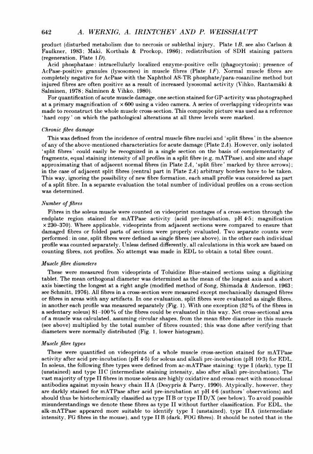
tablet. The mean orthogonal diameter was determined as the mean of the longest axis and a shortaxis bisecting the longest at a right angle (modified method of Song, Shimada & Anderson, 1963;see Schmitt, 1976). All fibres in a cross-section were measured except mechanically damaged fibresor fibres in areas with any artifacts. In one evaluation, split fibres were evaluated as single fibres.in another each profile was measured separately (Fig. 1). With one exception (52% of the fibres ina sedentary soleus) 81-100% of the fibres could be evaluated in this way. Net cross-sectional areaof a muscle was calculated, assuming circular shapes, from the mean fibre diameter in this muscle(see above) multiplied by the total number of fibres counted; this was done after verifying thatdiameters were normally distributed (Fig. 1, lower histogram).
Muscle fibre typesThese were quantified on videoprints of a whole muscle cross-section stained for mATPase
activity after acid pre-incubation (pH 4-5) for soleus and alkali pre-incubation (pH 10-3) for EDL.In soleus, the following fibre types were defined from ac-mATPase staining: type I (dark), type II(unstained) and type JIC (intermediate staining intensity, also after alkali pre-incubation). Thevast majority of type II fibres in mouse soleus are highly oxidative and cross-react with monoclonalantibodies against myosin heavy chain IIA (Desypris & Parry, 1990). Atypically, however, theyare darkly stained for mATPase after acid pre-incubation at pH 4-6 (authors' observations) andshould thus be histochemically classified as type IIB or type IID/X (see below). To avoid possiblemisunderstandings we denote these fibres as type II without further classification. For EDL, thealk-mATPase appeared more suitable to identify type I (unstained), type IIA (intermediateintensity, FG fibres in the mouse), and type JIB (dark, FOG fibres). It should be noted that in the
642
IANJlUR Y-IXDUCED MUSCLE CHANGES
0)
a)
01)U-
643
10 20 30 40 50Mean orthogonal diameter (pm)
Fig. 1. Normalized frequency distributions of average orthogonal muscle fibre diametersobtained from frozen cross-sections of soleus muscles of intermittent (@), continuous (0)and non-runners (A). The numbers of muscles evaluated were six, three and three. Upperhistogram: each muscle fibre profile was measured and counted individually. Lowerhistogram: all small profiles were assumed to be segments of a split fibre. Presumed splitfibres were counted and measured as single muscle fibres. Expressed in this way thenumber of counts declined from 4491 to 3375 in intermittent runners: in continuousrunners and non-runners numbers remained similar (declined from 2039 to 2002 and 1575to 1563, respectively).
mouse, a new type of fast-oxidative fibre was recently described (type II X. Schiaffino, Gorza.Satore, Saggin, Ansoni. Vianello, Gundersen & L6mo, 1989) which was, however, not consideredhere (see also Termin. Staron & Pette, 1989; for type IID fibres).
RESULTS
Exercise-induced damage following short rest periodsIt was noticed previously that soleus, but not EDL muscles, suffer focal muscle
fibre damage including necrosis at the onset of wheel-running, but no further damageoccurs with continuation of running for several months (Irintchev & Wernig, 1987).
21-2

A. WlrERNSIG. A. IRINTCHE' AND P. 1IVEISSHAUPT
When running in wheels was stopped for periods of 1-5 weeks following 5 months ofcontinuous exercise. and then resumed for 1 week. fresh damage occurred in soleusmuscles: average 8-7 + 3 5% (mean + S.D.) of the muscle fibres (Table 1). Plate I A-Fshows different signs of acute damage detectable. The amount of damage is notobviously correlated with the duration of the rest period (Table 1) or the amount ofrunning during the following week of activity (total amount, or activity during thefirst or last day; data not shown). Control animals studied 0. 2 and 4-6 weeks aftertermination of a period of 5 months of continuous exercise, and age-matchedunexercised animals had about 1% muscle fibres displaying signs of acute damage(Table 1). These results indicate that animals adapted to wheel-running becomevulnerable to acute muscle damage after short rest periods (see Discussion).
Split muscle fibres and central nuclei without signs of acute damage (Plate 2A)were previously observed several months after damage and repair of muscle fibres(Irintchev & Wernig, 1987). Split fibres probably originate from incomplete fusion ofproliferated satellite cells after focal fibre damage at the very beginning of exerciseor to a small degree, during normal activity in cages. Internalization of nuclei occursin sublethally damaged or regenerated fibres and apparently persists for long periodsthereafter (e.g. in regenerated fibres in the mdx-mutant mouse; Anderson. Bressler& Ovalle, 1988). Both split fibres and central nuclei were present in continuouslytrained muscles (1S5+073 and 2-4+1-0%, Table 1) but to a negligible extent inunexercised animals (0-6+ 0-2 %, P < 0-05. t test, Table 1).
Repeated short periods versus continuous wheel-runningIt was shown above (and Table 1) that exercised animals are vulnerable to fresh
damage in soleus upon onset of running after short rest periods. Another group ofanimals was therefore trained intermittently over a period of 12 months with 4 dayswheel-running followed by 21-25 days of rest, to test if repeated muscle damagewould alter muscle properties. The occurrence of repeated damage was confirmed inthree animals which showed various degrees of acute damage following two. threeand four running episodes (data not shown).
Histological and contractile properties of soleus musclesNot unexpectedly, the percentage of muscle fibres showing signs of previous
damage (split fibres, central nuclei) is highly elevated after 12 months of intermittentrunning (Plate 2B and C). Accordingly, a considerable proportion of small diameterprofiles is present, raising the total numbers of profiles in complete muscle cross-sections to 768+68 (mean+ S.D.; n = 6) compared to 676+54 (n = 3) in continuousrunners and 683 + 33 (n = 4) in non-runners (P < 005, Table 2). Only when splitfibres are counted as single muscle fibres, i.e. small fragments are considered asunfused parts of regenerated fibres, are their numbers similar to those of the othergroups (Table 2) and the frequency distribution of diameters then becomes bell-shaped (Fig. 1). Thus the estimated occurrence of altered fibres was 12+49%(n = 6, Table 2) with twice as many split fibres as muscle fibres which only had centralnuclei. Altered fibres were slightly but significantly more frequent in runners(3-4+0-12 %; n = 3) than in non-runners (0-53+0-17%; n = 4; Table 2). Despite theunusual muscle architecture, tetanic and single twitch forces were larger in solei ofintermittently trained animals than in unexercised animals (P < 0-05, Table 2). This
644
INJURY-INDUCED MUSCLE CHANGES
TABLE 1. Amount of damage (acute and chronic, for definitions see Methods) in soleus muscles after5 months of continuous running, a rest period of 1-5 weeks followed by 7 days of wheel-running(A), 5 months of continuous running and rest periods of 0, 2 and 4-6 weeks (B), together withunexercised age-matched controls (C)
Signs of damage
Acute
(A) Running after rest perio1 5-8 (3f5-88)2 13-0 (3-7-34)3 11-4 (00-23)4 8 6 (3-7-13)5 47 (17-76)
Average 8-7 + 3-5*(B) Weeks after exercise0 03 (00-07)2 04 (00-09)
4-6 07 (0{0-22)Average 0-47 + 0-21
(C) Unexercised controls1-3+1-1(00-24)
Chronicds of (weeks)2-7 (0-1-5 5)1-2 (00-44)1-2 (00-24)0-8 (0-0-1P9)1-6 (1P4-1P9)1-5+0-73
25 (1-1-41)34 (09-6)1 3 (0 6-2 8)2-4+ 10*
0-6+0-2(0-4-09)
Number of animals(muscles)
4 (4)4 (4)2 (2)3 (3)2 (2)
4 (5)2 (2)3 (3)
4(7)
Numbers represent percentage of fibres per complete cross-section through soleus muscle cut inthe endplate region; mean values and ranges are given for individual groups as well as S.D. for theoverall groups.
* Significant difference as compared to unexercised controls (P < 0 05), t test.
TABLE 2. Physiological and histological parameters of soleus muscles of sedentary, continuouslyand intermittently exercised CBA/J mice
Physiological parametersTetanic force P0 (T50, miN)Twitch force P, (mN)Twitch-tetanus ratioTwitch time-to-peak (ms)Twitch half-relaxation time (ms)Tetanic force per muscle cross-sectional area(N/cm2)§
Animal body weight (g)Number of animals (muscles)
Histological parametersProfiles per cross-section§Muscle fibres per cross-sectionNet cross-sectional area (mm2) (calculated)§Altered fibres (%) (range)
Sedentary126+ 2632+3-8
0-25+ 00354+5-485+ 11
278 + 3-6
Continuous Intermittentrunners runners
146+ 3234+4-8
0-23+0-0450+5 1t81+ 12
28-5+5-1
27-7+09 27 3+2-24 (8) 4 (8)
683 + 33674+ 340-46 + 0-0310-53+0-17(0-30-065)
676+ 54653+500 53 + 0-0763-4+0-12$(3*3-3*5)
160 + 29*40+6-6*
0-25+0-0256+66178 + 10
282 + 29
27-3 + 2-36 (12)
768 + 68*661 + 300-59+ 0-069*12 +4.9**(8-1-19)
Fibre types (%)type I 75+09 81+5-6 90+4.4**type II 24+1-1 19+65t 8-7+446*type IIC 0 9+0 4 0-6+0-4 0 9+09
Number of animals (muscles) 4 (7) 3 (5) 6 (11)Mean number +S.D. and some ranges are given. Symbols: *, ** = P < 005, < 001 compared
with sedentary animals (t test). t, =P < 005, <0-01 compared with intermittent runners(t test). § = 3, 3 and 6 muscles from as many animals.
645

A. W;VERNIG. A. IRIXTCHEV AND P. WEISSHA UPT
increase is obviously due to an increase in cross-sectional area (Table 2) since specificforce (tetanic force per cross-sectional muscle fibre area) remains unchanged. Times-to-peak of single twitches were shortest in continuously trained solei (P < 005,Table 2).When screening for correlates it appeared that intermittent runners with larger
body weight (and larger contractile force of soleus muscles) had more altered fibres;in the six animals studied, mean body weight was 29-7 + 21 g in the three animalswith the largest degree of damage and 25-3 + 1 1 g S.D. in the others. Strikingly, theamount of running during the 4-day periods was not correlated with the amount ofchronic damage (r = -05, n.s.).
Fibre-type distribution in soleus muscleThere is a clear increase in type I fibres in intermittently trained soleus muscles
over unexercised muscles (Plate 2B and C, Table 2) while continuous runners areintermediate. It was noted previously that in soleus muscles fibre-type groupingoccurs after wheel-running (Irintchev & Wernig, 1987). Thus it is possible that theincrease in type I fibres is related to muscle damage and repair (see Discussion). Nosignificant correlations were found, however, between the occurrence of type I fibresand the amount of running during the running episodes, the frequency of chronicallyaltered fibres or body weight.
EDL muscles in continuously and intermittently trained miceIn contrast to soleus (and tibialis anterior), EDL muscles do not suffer injury
during wheel-running (see Irintchev & Wernig, 1987); thus only changes consequenton the amount of exercise should be present. Typically, EDL is working against littleload, thus performing mainly endurance training; accordingly there is in fact a cleardecrease in contractile force and increase in time-to-peak values in EDL of
TABLE 3. Physiological and histological parameters of EDL muscles of sedentary, continuouslyand intermittently exercised CBA/J mice
Continuous IntermittentPhysiological parameters Sedentary runners
Tetanic force PO (T,00, mN) 333 +32 285 +32*Twitch force Pt (mN) 92 + 12 87 + 10Twitch time-to-peak (ms) 16 + 1-0 19 + 1-6**Twitch half-relaxation time (ms) 19 + 3-9 23 + 6-1Muscle weight (mg) 13 + 1-4 13 +1-7Number of animals (muscles) 4 (8) 4 (8)
Histological parametersAltered fibres (%) 05 +01 06 +0-2Fibre types (%)
type IIA 54+3-8 46+4-1*type JIB 45+40 51+3-9*type I 1-2+0-3 3-1+1-8*
Number of muscle fibres evaluated 745 +53 734+ 100Number of animals (muscles) 3 (5) 3 (5)
* P < 005; **P < 0-01 compared with sedentary animals (t test).
runners
323 + 4693+ 1818+1-721+3-812+1-36 (10)
05+02
55+3-544+3-31-1+02761 + 101
6 (7)
646

IVJURY-INDUCCED MUSCLE CHANGES
continuous runners while intermittent runners are not altered at all (Table 3). Fibre-type distributions in intermittent runners are strikingly similar to controls (Table 3).In continuous runners, however, a small but significant increase in type I and typeJIB fibres at the cost of type IIA fibres is present; this can be considered as anincrease in oxidative capacity in response to endurance exercise.
TABLE 4. Nerve block (nerve index) of soleus muscles of sedentary, continuously andintermittentlv trained mice for two concentrations of curare
Nerve index (%)
Continuous IntermittentSedentary runners runners
0-6,/m-d-tubocurarineSingle twitch 91-2 + 7, 89 7+ 6-0 93 9 + 7-4
(4) (8) (11)Tetanus, 2 s (50 Hz) 674+87 724+ 10-1 853 +97**t
(5) (8) (11)Tetanus, 02s (100 Hz) 84-8+11-1 873+49 948+27*t
(3) (6) (9)1-2,um-d-tubocurarine
Single twiteh 422 + 264 806+ 117 85 8+5 7**(3) (3) (5)
Tetanus. 2 s (50 Hz) 34-8 +40 7 5100+14 9 54-4 + 5-8(3) (3) (5)
Tetanus. 0-2 s (100 Hz) 24-6+ 12-1 44-3 + 14 5 64.4 + 10-0** t(2) (3) (5)
Number of muscles tested is in parentheses.* P < 0-05 compared to sedentary animals (t test). ** P < 0-01 compared to sedentary animals
(t test). t P < 0-05 compared to continuous runners (t test). + P < 0.01 compared to continuousrunners (t test).
Other observationsIt has been observed previously that prolonged elevated nerve activity causes
changes in transmitter release in vertebrate neuromuscular junctions (Dorlochter,Brinkers, Irintchev & Wernig, 1990; but see Hinz & Wernig, 1988). Resistanceto curare block, commonly used as a measure of safety margin of transmission, wastested here and significant differences are found. While there is merely a trendtowards higher resistance to block in continuously trained soleus muscle, the increaseis significant for intermittent runners (Table 4). Thus for soleus, the experimentalgroups with higher proportions of type I fibres have higher resistances to block. Notethat in mouse, resistance is generally higher in the predominantly slow soleus thanin the fast EDL muscle (M. Dorlochter, M. Brinkers, A. Irintchev and A. Wernig,unpublished).
DISCUSSION
Previous experiments revealed that in wheel-running, muscle fibre damage occursat the onset of exercise but not thereafter. Therefore it is surprising to find acutemuscle fibre damage in trained soleus muscles on resumption of running after restperiods as short as one week. In wheel-running, soleus and tibialis, but not EDL,
647
A. WERNIG, A. IRINTCHEV AND P. WEISSHA UPT
muscles may perform eccentric movements and show signs of damage while EDLdoes not (Irintchev & Wernig, 1987). Eccentric muscle contractions were found tocause muscle fibre damage in several species, including man (Armstrong et al. 1983;Friden, Skafianos & Hargens, 1986; Jones et al. 1986). It is therefore possible thateccentric contractions are the cause of damage in the present experiment. The natureof the adaptation that prevents further damage with continuation of wheel-runningis unknown but it evidently fails during intermittent exercise or after short restperiods. Improvements in the co-ordination of motor units during wheel-runningcould reduce the amount of eccentric contraction in muscle fibres of individual motorunits, and deadaptation might occur within short rest periods due to return to theco-ordination patterns used on flat ground. Muscle pain, which in humans is acomponent of muscle soreness, might in mice be the sensory feedback necessary foradaptations to occur at the CNS level. During intermittent training, adaptations towheel-running might not occur, since running periods are short (4 days) and followedby relatively long rest periods (21-25 days). The relatively long-lasting (severalweeks) protective effect of a single exercise-induced injury in man (Byrnes et al.1985)-contrasted with the vulnerability within a week after cessation of running inmice (present results)-might be due to the better learning and storage capacity ofthe human CNS.The increase in type I fibres in solei of intermittent runners clearly suggests that
changes also occur at muscle fibre level; it is not clear, however, whether these canbe considered as an adaptation or as a mere byproduct of other events. At presentit is not known whether type I fibres are less vulnerable to running-induced damagethan type II fibres. It is unlikely that elevated motoneurone activity caused anincrease in proportion of type I fibres in soleus, for example via fibre transformation(e.g. Salmons & Sreter, 1976); if this were the case, continuous runners should showlarger, not smaller, changes than intermittent runners. The increase in type I fibresis evidently related to the amount and frequency of muscle damage. In EDL muscles,which lack signs of damage, fibre type changes are absent in intermittent runnerswhereas typical effects of endurance training are present in continuous runners (seebelow).
It is likely, therefore, that the increase in type I fibres in soleus is a directconsequence of muscle damage and repair, and adaptations to prevent furtherdamage might occur at the CNS level.
Possible mechanisms for an increase in the proportion of type I fibres in repeatedlydamaged soleusThe increase in the proportion of type I fibres in soleus of intermittently running
mice appears to be related to muscle fibre damage. Two different mechanisms couldaccount for such an occurrence.
It is conceivable that regenerated cells predominantly express acid stable myosinATPase, i.e. type I fibre characteristics owing to incorporation of new nuclei fromfused satellite cells. In this case one might expect a mosaic distribution of differentATPase along the length of muscle fibres since fibre damage occurs focally (Irintchev& Wernig, 1987); this possibility was not ruled out in the present experiments andin other circumstances has indeed been observed (Staron & Pette, 1987). However,
648
INJUR Y-ILVDLTCED MlTSCLE CHANGES
split muscle fibres expressing type II fibre characteristics have also been observedfollowing muscle injury (Irintchev & Wernig, 1987), which renders this notion lesslikely.A more likely mechanism is axonal sprouting and new synapse formation. Recent
observations show axonal sprouting to be a physiological process which is enhancedin pathological situations, in particular when accompanied by muscle fibre inactivity(for a review see Wernig & Herrera, 1986). In the case of focal muscle fibre damageand repair, sprouting factors originating from proliferating satellite cells or musclefibre segments temporarily or permanently disconnected from the endplate-bearing,and thus action potential-generating, parts might cause enhanced collateral andterminal sprouting.The observed increase in type I fibres would suggest a higher sprouting capacity
of type I motoneurones, which might in addition overrule other activity patterns tocause transformation into type I fibres (for further discussions of this hypothesis seeSalmons & Sreter, 1976; Pette & Vrbovai, 1985; Wernig & Herrera, 1986).Interestingly, tonic nerve stimulation via implanted electrodes which causes anincrease in type I fibres seems to involve some muscle fibre necrosis and repair (seeMaier, Gambke & Pette, 1986). Also after enforced treadmill running (Muller, 1974)increase in type I fibres in soleus was accompanied by accumulation of split fibressuggesting muscle damage in the course of training. In contrast, during trans-formation of denervated muscle from slow to fast, embryonic myosin chains could bedetected only near the site of electrode implantation (Gorza, Gundersen, L6mo,Schiaffino & Westgaard, 1988) indicating that satellite cell proliferation was notinvolved in fibre transformation.
Muscle damage and contractile forceSoleus muscles of intermittently running animals contain considerable numbers of
'split muscle fibres'. This seeming disadvantage is obviously more than balanced bythe marked muscle hypertrophy. Since tetanic force was less in continuously runninganimals, although the actual duration of training was longer by a factor of 5-6, forceis not increased by more running. Possibly, therefore, multiple episodes of satellitecell proliferation which also take place in subtotally damaged fibres (Teriivainen,1970; Darr & Schultz, 1987) lead to increased cross-sectional area and thuscontractile forces.
There have been reports of muscle fibre hyperplasia as a consequence of elevatedmotor activity (e.g. Gonyea, Sale, Gonyea & Mikesky, 1986). Also in the presentexperiments, the number of individual muscle fibre profiles recognizable on singlecross-sections was markedly elevated; however, when adjacent fragments arecounted as single fibres, which admittedly includes some arbitrary decision andignores the possible existence of small fibre branches (Schmalbruch, 1984), thenumber of muscle fibres becomes similar to that in control animals.
Training effects in soleus and EDL muscles in continuous runnersIn EDL muscles which do not show running-induced damage, contractile force and
contraction times are not altered in intermittently trained animals, but are typicallychanged in continuous runners. A similar concomitant decline in force and increase
649

A. WIERNIG, A. IRLYTCHEV AND P. WEISSHA UPT
in time-to-peak of the single twitch observed here may be found after substantialelectrical stimulation of innervated fast muscle (Salmons & Vrbova', 1969; Kernell,Eerbeek, Verhey & Donselaar, 1987). At the same time a significant increase in fast-oxidative fibres (possibly including type lIX fibres, Schiaffino et al. 1989) wasnoticed which is the most typical outcome of endurance training (Edstrom &Grimby, 1986). The occurrence of additional type I fibres in EDL is possibly causedby a corresponding change in activity pattern of one or two type II motoneurones,or the type I motoneurone(s) present in the muscle extended their motor unit size e.g.by hyperinnervating muscle fibres (for further discussion see Wernig & Herrera,1986). The moderate increase in type I fibres in some of the continuously trained soleimight have similar origins to those discussed above for EDL but it is more likely tobe connected with fibre damage occurring at the very onset of running.
Emil Angelov took part in some of the morphometric evaluations and isometric tensionmeasurements reported here. We are thankful to Stefanie Briel and Elena Wiebe for excellenttechnical assistance, and we thank Drs Stanley Salmons (Liverpool) and Hans Hoppeler (Bern) forcarefully reading and criticizing an earlier version of the manuscript. This work was financiallysupported by a grant from the Deutsche Forschungsgemeinschaft (We 859).
REFERENCES
ANDERSON, J. E., BRESSLER, B. H. & OVALLE, W. K. (1988). Functional regeneration in thehindlimb skeletal muscle of the mdx mouse. Journal of Muscle Research and Cell Motility 9,499-515.
ARMSTRONG. R. B.. OGILVIE, R. W. & SCHWANE, J. A. (1983). Eccentric exercise-induced injury torat skeletal muscle. Journal of Applied Physiology 54, 80-93.
BADKE,. A. (1988). Der Einfluss der Laufaktivitat auf die Reinnervation des Muskulus soleus derMaus. Dissertation. Universitat Bonn.
BADKE, A., IRINTCHEV, A. & WVERNIG, A. (1989). Maturation of transmission in reinnervated mousesoleus muscle. Muscle and Nerve 12, 580-586.
BUTLER, J. & COSMOS, E. (1981). Enzymic markers to identify muscle-nerve formation duringembryogenesis: modified myosin ATPase and silver-cholinesterase histochemical reactions.Experimental Neurology 73, 831-836.
BYRNES. W. C.. CLARKSON, P. M., WHITE. J. S., HSIEH, S. S., FRYKMAN, P. N & MAUGHAN, R. J.(1985). Delayed onset muscle soreness following repeated bouts of downhill running. Journal ofApplied Physiology 59, 710-715.
CARLSON. B. M. & FAULKNER, J. A. (1983). The regeneration of skeletal muscle fibres followinginjury: a review. Medicine and Science in Sports and Exercise 15, 187-198.
CARPENTER, S. & KARPATI. G. (1984). Pathology of Skeletal Mlluscle. Churchill Livingstone, NewYork, Edinburgh.
DARR, K. C. & SCHULTZ, E. (1987). Exercise-induced satellite cell activation in growing and matureskeletal muscle. Journal of Applied Physiology 63, 1816-1821.
DESYPRIS, G. & PARRY, D. J. (1990). Relative efficacy of slow and fast motoneurons to reinnervatemouse soleus muscle. American Journal of Physiology 258, C62-70.
DORLOCHTER, M., BRINKERS, M., IRINTCHEV, A. & WERNIG, A. (1990). Enhanced running activitycauses increase in transmitter release in mouse EDL muscle. Pfuigers Archiv 415, suppl. 1, R78.
EDSTR6M, L. & GRIMBY, L. (1986). Effect of exercise on the motor unit. Muscle and Nerve 9,104-126.
FRIDIEN. J. (1984). Muscle soreness after exercise: implications of morphological changes.International Journal of Sports Medicine 5, 57-66.
FRIDEIN, J., SKAFIANOS, P. N. & HARGENS, A. R. (1986). Muscle soreness and intramuscular fluidpressure: comparison between eccentric and concentric load. Journal of Applied Physiology 61,2175-2179.
650
INJU"RY-INDUTCED MUTSCLE CHANGES
GONYEA, WV. J., SALE, D. G., GONYEA, F. B. & MIKESKY, A. (1986). Exercise-induced increases inmuscle fiber number. European Journal of Applied Physiology 55, 137-141.
GORZA, L., GUNDERSEN, K., L6mo, T., SCHIAFFINO, S. & WESTGAARD, R. H. (1988). Slow-to-fasttransformation of denervated soleus muscles by chronic high-frequency stimulation in the rat.Journal of Physiology 402, 627-649.
GUTH, L. & SAMAHA, F. J. (1970). Procedure for the histochemical demonstration of actomyosinATPase. Experimental Neurology 28, 365-367.
HIKIDA, R. S., STARON, R. S., HAGERMAN, F. C., SHERMAN, W. M. & COSTILL. D. L. (1983). Musclefiber necrosis associated with human marathon runners. Journal of Neurological Sciences 59,185-203.
HINZ, I. & WERNIG. A. (1988). Prolonged nerve stimulation causes changes in transmitter releaseat the frog neuromuscular junction. Journal of Physiology 401, 557-565.
IRINTCHEV, A. & WERNIG, A. (1987). Muscle damage and repair in voluntarily running mice: strainand muscle differences. Cell and Tissue Research 249. 509-521.
JONES, D. A., NEWHAM, D. J., ROuND, J. M. & TOLFREE, S. E. J. (1986). Experimental humanmuscle damage: morphological changes in relation to other indices of damage. Journal ofPhysiology 375, 435-448.
KERNELL, D., EERBEEK, 0., VERHEY, B. A. & DONSELAAR, Y. (1987). Effects of physiologicalamounts of high- and low-rate chronic stimulation on fast-twitch muscle of the cat hindlimb. I.Speed- and force-related properties. Journal of Neurophysiology 58, 598-613.
KUIPERS, H., DRUKKER, J., FREDERIK, P. M., GEURTEN, P. & VAN KRANENBURG, G. (1983). Muscledegeneration after exercise in rats. International Journal of Sports Medicine 4, 45-51.
LOJDA, Z., GoSSRAU, R. & SCHIEBLER, T. H. (1976). Enzymhistochemische Methoden. SpringerVerlag, Berlin, Heidelberg, New York.
MAIER, A., GAMBKE, B. & PETTE, D. (1986). Degeneration-regeneration as a mechanismcontributing to the fast to slow conversion of chronically stimulated fast-twitch rabbit muscle.Cell and Tissue Research 244, 635-643.
MAKI, T., KORTHALS, J. K. & PROCKOP, L. D. (1986). Distribution of muscle changes inexperimental ischemic myopathy. .Muscle and .Verve 9, 394-398.
Mi1LLER, W. (1974). Temporal progress of muscle adaptation to endurance training in hind limbmuscles of young rats. A histochemical and morphometrical study. Cell and Tissue Research 156,61-87.
NACHLAS, M. M., Tsou, K. C., DE SOUZA, E., CHENG. C. S. & SELIGMAN, A. M. (1957). Cytochemicaldemonstration of succinic dehydrogenase by the use of a new p-nitrophenyl substitutedditetrazole. Journal of Histochemistry and Cytochemistry 5, 420-436.
PETTE, D. & VRBOVA, G. (1985). Neural control of phenotypic expression in mammalian musclefibres. Muscle and Nerve 8, 676-689.
SALMINEN, A. & VIHKO, V. (1980). Acid proteolytic capacity in mouse cardiac and skeletal musclesafter prolonged submaximal exercise. Pfliugers Archiv 389, 17-20.
SALMONS, S. & SRE,TER, F. A. (1976). Significance of impulse activity in the transformation ofskeletal muscle type. Nature 263, 30-34.
SALMONS, S. & VRBOVAI, G. (1969). The influence of activity on some contractile characteristics ofmammalian fast and slow muscles. Journal of Physiology 201, 535-549.
SCHIAFFINO, S., GORZA, L., SATORE, S., SAGGIN, L., ANSONI, S., VIANELLO, M., GUNDERSEN, K. &LOMO, T. (1989). Three myosin heavy chain isoforms in type two skeletal muscle fibres. Journalof Muscle Research and Cell iotility 10, 197-205.
SCHMALBRUCH, H. (1984). Regenerated muscle fibres in Duchenne muscular dystrophy: a serialsection study. Neurology (Cleveland) 34, 60-65.
SCHMITT, H. P. (1976). Measurement of voluntary muscle fiber cross sections: a comparative studyof different possible methods. Microscopica Acta 77, 427-440.
SEGAL. S. S. & FAULKNER, J. A. (1985). Temperature-dependent physiological stability of ratskeletal muscle in vitro. American Journal of Physiology 248, C265-270.
SONG, S. K., SHIMADA, N. & ANDERSON. P. J. (1963). Orthogonal diameters in the analysis ofmuscle fibres' size and form. Nature 200, 1220-1221.
STARON, R. S. & PETTE, D. (1987). Non-uniform myosin expression along single fibres ofchronically stimulated and contralateral rabbit tibialis anterior muscles. Pftugers Archiv 409,67-73.
651

A. WER.NIG, A. IRINTCHEV AND P. WEISSHAUPT
TERXVXINEN, H. (1970). Satellite cells of striated muscle after compression injury so slight as notto cause degeneration of the muscle fibres. Zeitschrift fur Zellforschung 103, 320-327.
TERMIN, A., STARON, R. S. & PETTE, D. (1989). Myosin heavy chain isoforms in histochemicallydefined fiber types of rat muscle. Hi8tochemistry 92, 453-457.
VIHKO, V., RANTAMAKI. J. & SALMINEN, A. (1978). Exhaustive physical exercise and acid hydrolaseactivity in mouse skeletal muscle. Histochemistry 57, 237-249.
WERNIG, A. & HERRERA, A. A. (1986). Sprouting and remodelling at the nerve-muscle junction.Progress in Neurobiology 27, 251-291.
WERNIG, A. & IRINTCHEV, A. (1989). Intermittent voluntary running causes changes in musclefiber type composition and in contractile muscle force in the mouse. Pflugers Archiv 431, suppl. 1,R33.
EXPLANATION OF PLATES
PLATE 1
Signs of acute muscle damage in cryostat cross-sections (10 ,um) from short-term exercised (1 week)mouse soleus muscle. Toluidine Blue staining (A, C and E) reveals a necrotic pale fibre with 'glass'appearance (asterisk in A), single (asterisks in C) and clusters (arrow-heads in A) of small, darklystained regenerating fibres, inhomogeneous coarse myofibrillar pattern (small arrow-heads in C andE). prominent peripheral basophilia (fibres marked with B in A and C). Note numerous infiltratedcells surrounding muscle fibres in A. Reaction for glycogen phosphorylase (B) shows muscle fibreswith inhomogeneous loss of activity (arrow-heads in B) and normally stained fibres in the upperright corner. Sections stained for succinate dehydrogenase (D, section adjacent to C), regeneratingfibres (asterisks in C and D) appear lightly stained but clumped reaction product is often seen(upper fibre marked with asterisk); complete loss of activity is seen in one muscle fibre (largearrow-heads in C and D). Acid phosphatase activity, undetectable in normal fibres, is visualized insome affected fibres (arrow-heads in E and F) indicating enhanced lysosomal activity. Scale bar inF indicates 30 ,um for A and 50 ,um for B-F.
PLATE 2
Cross-sections from soleus muscle stained with Toluidine Blue (A) and for myofibrillar ATPaseactivity after acid pre-incubation, pH 4-5 (B and C). A, concomitant acute and chronic signs ofmuscle injury in a mouse exercised for 5 months, followed by 1 week rest and 1 week exercise. Inthe central part, several small fibre profiles without signs of acute damage, representing fragmentsof split muscle fibres, are seen. Isolated split fibres are easily recognized in a single section (arrows),but in the case of adjacent split fibres (central part of A) arbitrary borders have to be taken. Someotherwise normal fibres have central nuclei (asterisks). Most split fibres and fibres with centralnuclei do not display any sign of acute injury (compare Plate 1) and are a consequence of damageat the very onset of exercise. A regenerating fibre (small arrow-head) and a fibre with prominentperipheral basophilia and inhomogeneous coarse myofibrillar pattern (large arrow-head) provideevidence of acute damage after renewal of exercise. B and C, distribution of type I (dark) and typeII (white) fibres in a sedentary control (B) and an intermittently exercised (C) soleus muscle; theintermittently exercised soleus contains very few type II fibres and numerous split fibres (upperpart). In C several intermediately staining (type IIC) fibres can be seen. Scale bar indicates100 ,um for A and 300 /am for B and C.
652

I he Journal oJ I'hyswology, Vol. 428 plate 1
A~~ ~~~~~k
- .
*~~~~~~~~~~~
4-,
On,
Wt. ~ ~ xe;
.-~~..\....
A. WAERNIG and others
rn 7 T I A -F-% I . I -11 1. - -
tr singP1.9.b)

The Journal of Physiology, Vol. 428
A. WNERNIG and others
Plate 2
Related Documents