Neuropsycholoy~o. Vol. 33, No. 6, pp. 769-719. 1995 CopyrIght f:’ 1995 Elsewer Science Ltd Prmted in Great Britain. All rights reserved 0028-3932195 $9.50+0.00 002&3932(95)00018-6 FUNCTIONAL SIGNIFICANCE OF INDIVIDUAL VARIATIONS IN CALLOSAL AREA M. YANK1 YAZGAN,*+ BRUCE E. WEXLER,f MARCEL KINSBOURNE,$ BRADLEY PETERSON? and JAMES F. LECKMAN? tChiid Study Center and IDepartment of Psychiatry, Yale University School of Medicine, New Haven, Connecticut, U.S.A.: and 5Center for Cognitive Studies, Tufts University, Medford, Massachusetts. U.S.A. (Received 28 June 1994; accepted 17 January 1995) Abstract-We considered the hypothesis that the richness of callosal interhemispheric connections has a role in determining the degree of behaviouraf laterality and time-sharing ability in dual-task performance. Behavioural laterality as measured by dichotic word listening, line bisection and turning bias tests correlated inversely with the midsagittal cross-sectional area ofthe corpus callosum, as seen on MRI. The amount of dual task interference was strongly inversely correlated with the callosal area in both within-hemisphere and between-hemispheres conditions. These relationships between normal variations in callosal area, and outcomes on tests both of laterality and time-sharing capacity in normal adults suggest that the corpus callosum assumes a cross-excitatory role when subjects perform these tasks. Key Words: corpus callosum; laterality; line bisection; dual task; dichotic listening. INTRODUCTION The corpus callosum (CC) is the largest connecting pathway between the hemispheres. The CC consists of fibres of two types: those of larger diameter, which mediate sensory-motor coordination, and those, more numerous, of small diameter, which connect association areas. Normal variation in the CC cross-sectional area has been shown to reflect the callosal complement of small diametered fibre types, which may play a particular role in maintaining excitation-inhibition balance between hemispheres [l, 21. Because the size of the CC reflects its neural complement more than is the case for most cerebral structures, the CC lends itself to a test ofthe manner in which gross structure relates to function in the human cerebrum. Its fibres course uniformly in the coronal plane, and therefore, a sagittal MRI slice through the CC indexes the number of small diametered axons transmitted through it. Among functions proposed for the CC is mediation of interaction between the extensive territories of the hemispheres it links homotopically [S, 251. Behavioural lateralitp is implicated as related to such hemispheric interaction. When stimuli for the same task (e.g. identification of speech sounds) are presented separately to each hemisphere, the hemispheres differ systematically in the level of performance they generate. The discrep- ancy between correct identification of right and left sided stimuli is expressed as a lateralit~ *Address for correspondence: Marmara University Hospital. Altunizade. Istanbul. Turkey. 169

Welcome message from author
This document is posted to help you gain knowledge. Please leave a comment to let me know what you think about it! Share it to your friends and learn new things together.
Transcript
Neuropsycholoy~o. Vol. 33, No. 6, pp. 769-719. 1995 CopyrIght f:’ 1995 Elsewer Science Ltd
Prmted in Great Britain. All rights reserved 0028-3932195 $9.50+0.00
002&3932(95)00018-6
FUNCTIONAL SIGNIFICANCE OF INDIVIDUAL VARIATIONS IN CALLOSAL AREA
M. YANK1 YAZGAN,*+ BRUCE E. WEXLER,f MARCEL KINSBOURNE,$ BRADLEY PETERSON? and JAMES F. LECKMAN?
tChiid Study Center and IDepartment of Psychiatry, Yale University School of Medicine, New Haven, Connecticut, U.S.A.: and 5Center for Cognitive Studies, Tufts University, Medford, Massachusetts. U.S.A.
(Received 28 June 1994; accepted 17 January 1995)
Abstract-We considered the hypothesis that the richness of callosal interhemispheric connections has a role in determining the degree of behaviouraf laterality and time-sharing ability in dual-task performance. Behavioural laterality as measured by dichotic word listening, line bisection and turning bias tests correlated inversely with the midsagittal cross-sectional area ofthe corpus callosum, as seen on MRI. The amount of dual task interference was strongly inversely correlated with the callosal area in both within-hemisphere and between-hemispheres conditions. These relationships between normal variations in callosal area, and outcomes on tests both of laterality and time-sharing capacity in normal adults suggest that the corpus callosum assumes a cross-excitatory role when subjects perform these tasks.
Key Words: corpus callosum; laterality; line bisection; dual task; dichotic listening.
INTRODUCTION
The corpus callosum (CC) is the largest connecting pathway between the hemispheres. The CC consists of fibres of two types: those of larger diameter, which mediate sensory-motor coordination, and those, more numerous, of small diameter, which connect association areas. Normal variation in the CC cross-sectional area has been shown to reflect the callosal complement of small diametered fibre types, which may play a particular role in maintaining excitation-inhibition balance between hemispheres [l, 21. Because the size of the CC reflects its neural complement more than is the case for most cerebral structures, the CC lends itself to a test ofthe manner in which gross structure relates to function in the human cerebrum. Its fibres course uniformly in the coronal plane, and therefore, a sagittal MRI slice through the CC indexes the number of small diametered axons transmitted through it.
Among functions proposed for the CC is mediation of interaction between the extensive territories of the hemispheres it links homotopically [S, 251. Behavioural lateralitp is
implicated as related to such hemispheric interaction. When stimuli for the same task (e.g. identification of speech sounds) are presented separately to each hemisphere, the hemispheres differ systematically in the level of performance they generate. The discrep- ancy between correct identification of right and left sided stimuli is expressed as a lateralit~
*Address for correspondence: Marmara University Hospital. Altunizade. Istanbul. Turkey.
169
770 M. Y. YAZGAN et al
index, and the asymmetry expressed by this index is referred to as a laterality efect. In general, this index indicates a right ear and visual field advantage for verbal messages, and a left side advantage for messages conveying certain types of spatio-temporal information [9, 101.
Behavioural laterality is modifiable by unilateral cerebral disease, increasing or decreasing depending on the hemisphere involved. The data from neurological populations also indicate that callosal lesions, which impair interhemispheric structural connectivity, also diminish interhemispheric transfer of information and alter behavioural laterality measures [6, 21, 381. The degree of behavioural laterality (e.g. perceptual asymmetry as measured by dichotic tests) also varies among individuals with intact brains [lo]. Explanations proposed for this normal variability include the degree of hemispheric specialization [47], “strength’ of crossed and uncrossed central auditory connections [48], individual differences in cognitive strategies [lo], and relative hemisphere arousal levels c331.
In this study, we considered an additional option: That the richness of callosal interhemispheric connections has a role in determining the degree of behavioural laterality. Previous studies using in vivo MRI brain data have demonstrated relationships between size of the CC or its subregions and behavioural measures [11-13, 18, 36, 53, 551.
Two functions attributed to the CC that predict a relationship between interhemispheric callosal connectivity (as appears in the midsagittal cross-sectional area of the CC) and behavioural laterality are: (1) The CC transmits inhibition, i.e. when one hemisphere engages in a task for which it is specialized, it concurrently inhibits the other [14,15,35]. If laterality effects parallel the extent to which one hemisphere is more activated than the other, and if a larger CC transmits more inhibition, then callosal area should correlate positively with the degree of behavioural laterality; (2) The CC transmits excitation, i.e. the activated hemisphere generates cross-callosal activation, maintaining the other (unstimulated) hemisphere in a functionally activated state [S, 16, 25, 26, 321. If behavioural laterality reflects the degree of imbalance between the activation levels of two hemispheres [24,26], CC area should vary inversely with the degree of behavioural laterality.
Cross-callosal excitation may also serve to enlarge the amount of cortex that is available for neural computation. Larger CC area should bring a performance advantage for difficult and demanding tasks and allow for optimal integration of cortical activity [S], on the assumption that the relevant computations benefit from the availability of wider areas of cortex [34]. Previous studies have shown that resources are normally shared between hemispheres even for highly specialized tasks [SS]. In two recent reports, demanding tasks such as verbal fluency tests [ 181, and measures of intelligence [46] were shown to correlate positively with area of the splenium, a region of the corpus callosum. However, in another study, performance on difficult cognitive tasks was not correlated with CC area [l 1, 131. Indirect support for wider cortical involvement in complex tasks derives from two recent functional neuro-imaging studies in which a complex version of a motor task was associated with recruitment of additional cortical regions [44], and widespread meta- bolic activation during an unfamiliar and difficult task diminished in extent after practice c171.
At neurophysiological level, almost all callosal fibres are excitatory [20], however, their functional effects may be either excitatory or inhibitory depending on the nature of the interneurons. Excitation and inhibition in this text are used as implying functional effects of neural activity.
FUKTIONAL SIGNIFICANCE OF CALLOSAL AREA 771
HYPOTHESES
We studied whether midsagittal cross-sectional area of the CC (‘CC area’) in normal subjects relates to an auditoryyverbal test of perceptual asymmetry (fused dichotic words test), two tests of overt bias in lateral directionality (line bisection, whole body turning bias), and an attention demanding time-sharing test involving dual-task performance (verbal- manual interference). Since, in this study, we focused on inhibitory/excitatory functions of the CC that may be reflected in midsagittal cross-sectional area of the CC [l, 2, 133, we did not include in our hypotheses other functions of the CC such as transfer of sensory -motor information [54].
The balance of activation of the hemispheres contributes to the extent of many lateral biases in behavioural measures such as dichotic listening, line bisection, and turning bias tests. When one hemisphere is more activated than the other, attention is directed along the vector resultant of the interaction between opponent lateral orienting systems in the hemispheres each of which drives attention contralaterally [24,26]. We would predict an association between the CC and behavioural laterality to the extent to which the CC area influences the balance of either excitation or inhibition.
In the dichofic words listening test, subjects are asked to identify competing stimuli presented to separate channels (ears). A right ear advantage has been found for a large majority of right-handed subjects who have left hemisphere specialization for language [5 1, 52,561. According to the callosal inhibition model, an increase in CC area will be associated with increased callosal inhibition from the activated left to right hemisphere, and therefore larger REA. The callosal cross-excitation model predicts increased callosal cross-excitation from left hemisphere, which is activated by the verbal task, to the right hemisphere with larger CC area, resulting in smaller REA.
In the lirle bisection test, when subjects are asked to indicate the midpoint of a horizontal line, most normal subjects exhibit a slight but systematic bias to the left of the actual midpoint [7]. This bias is minimized or reversed with right brain disease, mostly with parietal locations [49]. The effect in normals, also known as ‘pseudo-neglect’, may reflect the greater activation of the right hemisphere which is specialized for visuo-spatial tasks. If CC mediates inhibitory influences between the hemispheres, then larger CC area will be associated with larger bias. lfcallosal influence if primarily excitatory, larger CC area leading to activated states in both hemispheres should be associated with smaller bias.
In the turning hius test, subjects who turn around at the end of a 3 m walk must choose to turn tither right or left. We have found a leftward bias for the adults we have studied until now. Using a different methodology, Bracha et al. [S] have found a similar bias, but limited to females. Assuming that turning bias reflects greater activation of the contralateral hemisphere, a larger bias for turning may be predicted with smaller CC area according to the cross-excitation model. and a reverse relationship with the cross-inhibition model.
The I.c~rh~r/-m~lnua[ inrcl+wnce (dual task) test is based upon the observation that when subjects arc required to perform two tasks at the same time, one involving each hemisphere, they do better than when these two tasks both rely on the same hemisphere [28, 291. If the CC mediates inhibition, then the larger CC area would result in overall less effective dual task performance, because it would transmit more interfering neural ‘cross-talk’. If CC mediates excitation, this would recruit more of the neural network at the disposal of the performance. enlarge the available functional cerebral space [29], and enhance the ability to time-share between tasks. This should be particularly true with larger CC areas.
772 M. Y. YAZGAN er al.
METHODS Subjects
The group consisted of 11 (nine men and two women) healthy, strongly right-handed (as assessed by Edinburgh Handedness Inventory [37]) individuals. Mean (k SD.) age was 34.2 (k 8.9)years. Socioeconomic status (1.6f0.5 on Hollingshead index [19]) andeducational level (16.3 +4 years ofeducation) were relatively high. WAIS-R [SO] information (12.2 k 1.9) and block design (12.6 If: 1.6) subtest scores were above average. Subject characteristics and instruments for their assessment have been described in detail elsewhere C39-411.
Neuropsychological procedures
Fused word dichotic listening test. Two words differing only in initial consonant (e.g. cake-take) were presented simultaneously, one to each ear. Because of their high degree of acoustic spectral overlap, the members of each dichotic pair fuse into a single auditory percept. As a result subjects are aware of hearing only one word from each pair. In individuals with left hemisphere language specialization, the word presented to the right ear is reported more often than the word presented to the left ear [Sl, 561. A laterality score reflects the magnitude of this perceptual asymmetry (REA=[words heard at the right ear-words heard at the left earl/sum of the two). Test&retest correlations for this test have ranged from 0.88 to 0.91 1521. Fused dichotic words data are not available for one of the subjects.
Line bisection. Subjects were asked to divide five lines of different lengths (16&240 mm) into equal halves with a pencil held in their right hands. The deviation in millimetres from the actual midpoint was divided by half the actual length of the line and multiplied by 100 to derive a laterality index of percentage shift 1431. Percentage shifts were averaged across five trials and a negative sign indicated a leftward bias. Test-retest reliability for seven subjects tested twice, with an average interval of 14.4 days, was =0.68 (PcO.05)
Turning bias. The direction in which subjects turned was noted, as they completed each one of what they were told was a series of five evaluations of gait, in a room symmetrically spacious and free of distracters. Percentage of leftward turns was used as the dependent variable. Test-retest reliability for nine subjects tested twice, with average intervals of 7.1 days, was r =0.80 (PcO.05).
Verbal-manual interference test (VMI). This procedure, based on the dual task paradigm [30], measures the impact oflateralized concurrent language activity on a timed unimanual task. For a baseline condition subjects were asked to complete a motor task for each hand (right and left). As an interference condition, they were asked to do the motor task concurrently with a language task. The order of first hand use was randomized to avoid transfer of learning effects. The motor task was the Trail Making Test-Part B [42]. In this test, subjects connect circles with alternating contents of sequential numbers and letters. The language task instruction was to respond vocally by conjugating into past tense a heard list of commonly used irregular verbs. The time interval between verbs was 2-3 sec. Subjects were instructed to do the best they can in performing both tasks at the same time, rather than focus on one of the tasks during the dual task condition. The time for completion of tasks under each condition was determined in seconds. The number oferrors in motor and in language performance were also recorded. Interference is expressed as proportional decrements in right and left hand performances [29]. The decrements are calculated as, X% = 100 x (time for dual task performance-time for single task performance)/time for single task performance, for each hand (R% and L%).
Scanning procedures and image analysis All subjects were imaged on a 1.5 Tesla GE Signa scanner. A sagittal series (TR 500, TE 20,5 mm slice thickness,
2.5 mm skip, 24 cm FOV, 256 x 192 matrix, 2 nex) was obtained for CC measurements. The sagittal series was forwarded to a DEC/VAXstation 3200 PIXAR image computer system for quantitative image analysis.
Sagittal MR images were magnified to twice their original 256 x 192 size. The midsagittal image was selected for analysis and an electronic bitpad (Summagraphics) was then used to circumscribe manually the CC in a clockwise direction, beginning at the rostrum and moving along the superior border of the genu, body and splenium, turning then to trace the inferior borders of those structures in the reverse direction to close the border again at the rostrum. Following this step, five CC subregional areas were defined by dividing the overall curvilinear length of the centreline into equal fifths, using the ‘object-centred’ method of Allen et al. [3]. To perform the subdivision, the CC centreline is constructed and then divided into five equal lengths which delineate the five regional areas. The extent of the intervening CC body is marked. The operator marks the two points each at both the rostrum and splenium of the CC where the curvature of those structure flattens out effectively marking the extent of the intervening CC body. The boundary segments defined by these two points are sent to a computerized algorithm that determines the centreline along the length of the CC body by first finding the tangents to the upper CC border, then constructing a perpendicular to each ofthese tangents to intersect with the inferior CC boundary, and then finding the midpoint of this perpendicular line segment joining the upper and lower CC boundaries. These many midpoints are then connected and smoothed, yielding the centreline. Finally, this line is extended to the entire length of the CC by manually marking with a point both the tip of the rostrum and the splenium; these two points are then joined with the nearest end of the computer-generated centreline by two additional straight line segments, all three curvilinear segments comprising the final centreline measurements. The overall curvilinear length of this centreline is then
FUNCTIONAL SIGNIFICANCE OF CALLOSAL AREA 713
divided by perpendiculars into equal fifths to define the five CC subregional areas. After these steps were completed, the overall midsagittal cross-sectional area and five subregional CC areas were automatically determined. Further details for scanning and image analysis procedures have been presented elsewhere [39,40].
RESULTS
Behavioural laterality effects on functional measures, in accordance with previous literature and our predictions, were significant by t-tests (0.001 < PcO.02). In the dichotic listening test, a substantial right ear advantage (REA) (mean =0.28, S.D. =0.23) was seen. In line bisection, a leftward shift was observed (mean = - 3.3%, S.D. = 2.2). In the turning bias test, a leftward bias was noted (mean = 74.6%, S.D. = 13).
An analysis of variance (ANOVA) on the VMI (dual task) data, with hand and task conditions as within-subject variables, demonstrated a significantly lateralized impact of concurrent language activity on right hand performance (F= 12.2, d.f.= 10, P=O.O06). Significant main effects of concurrent task condition (F= 21.6, d.f. = 10, P=O.OOl) and hand (F= 5.6, d.f. = 10, P=O.O4) were also observed. Subjects performed both verbal and manual tasks without errors, and the number of verbal responses were comparable between subjects.
Corrrlutions with CC urea
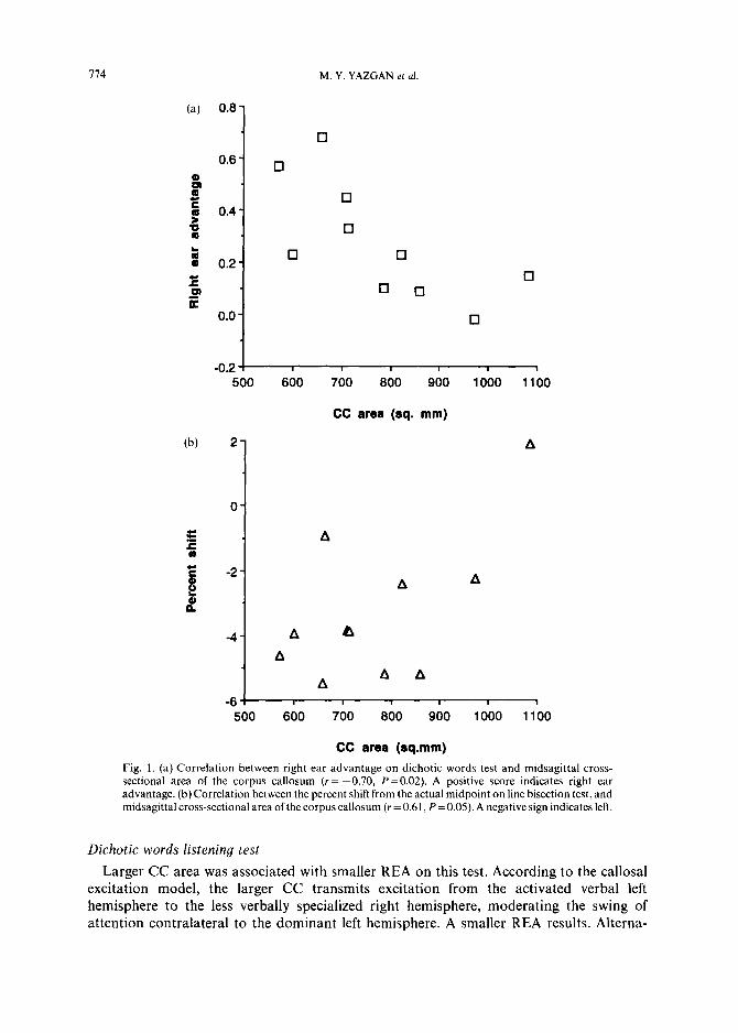
REA on the dichotic listening test decreased as the CC area became larger (r= -0.70, P=O.O2). With increased CC area, subjects showed smaller leftward deviation on line bisection (v=O.61, P=O.O5), and smaller leftward turning bias (r=-0.71, P=O.Ol) (Fig. la, b).
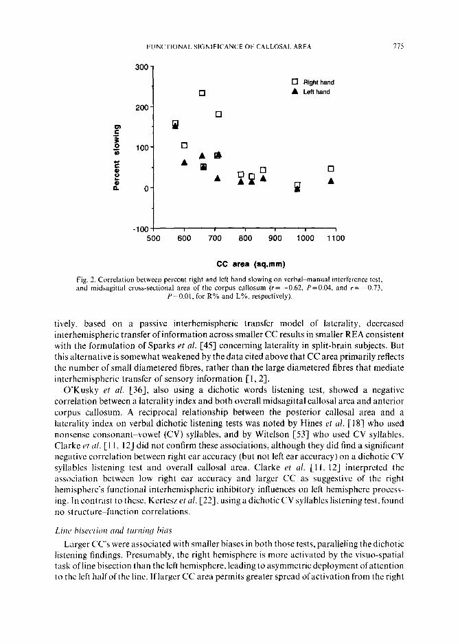
Right and left hand slowing (R% and L%) with the concurrent verbal task were both inversely correlated with the CC areas at a significant level (Y= -0.62, P=O.O4 and r= -0.73, P=O.Ol,for R% and L%,respectively) (Fig. 2). Thedegreeofasymmetry ofhand slowing did not correlate significantly with the CC area (r = -0.27).
The behavioural measures correlated significantly (Irl=O.67-0.78, P-cO.04) with the CC subregional areas, as might be predicted based on the previous finding that all subregional areas significantly correlated with the overall CC area (r = 0.884.94) [33]. The magnitude and direction of the correlations of the behavioural measures with each CC subregion were comparable to the correlations with the overall CC area. We could therefore not make inferences about regional specificity.
Other correlations
The asymmetry of hand slowing (R%-L%) on VMI was significantly correlated with REA (r=0.79, P=O.O05). The remaining behavioural laterality measures did not correlate significantly with each other. They also did not correlate with age, SES, and the scores on WAIS-R subscales.
DISCUSSION We found strong relationships between individual variations in callosal area and the
outcomes on three measures of laterality. Behavioural laterality measures (dichotic word listening, line bisection and turning bias) significantly correlated with the CC area, such that normal behavioural laterality increased as the CC became smaller. These relationships were supportive of an excitatory function for the CC in performing the tasks we used.
714 M. Y. YAZGAN et ul
(a) 0.8
0.8 al $ E g 0.4
z
5 0.2 E P K
0.0
cl
cl
cl
El
cl
Cl
0 0 cl
Cl
-v.c I
500 800 700 8;)O 900 lOLO l&O
(b) 2
0
E -2 t z P
-4
-8 500 800 700 800 900 1000 1100
CC area (sq.mm)
Fig. 1. (a) Correlation between right ear advantage on dichotic words test and midsagittal cross- sectional area of the corpus callosum (r= -0.70, P =0.02). A positive score indicates right ear advantage. (b) Correlation between the percent shift from the actual midpoint on line bisection test, and midsagittal cross-sectional area of the corpus callosum (r = 0.61, P=O.O5). A negative sign indicates left.
CC area (sq. mm)
A
A
A A
Dichotic words listening test
Larger CC area was associated with smaller REA on this test. According to the callosal excitation model, the larger CC transmits excitation from the activated verbal left hemisphere to the less verbally specialized right hemisphere, moderating the swing of attention contralateral to the dominant left hemisphere. A smaller REA results. Alterna-
FUNCTIONAL SIGNIFICANCE OF CALLOSAL AREA
300-
0 Right hand
0 A Lefl hand
200 - 0
P
loo- n
AB
A ?? cl 0
A %A O- P A
-100 1 I I I I I 500 600 700 800 900 1000 1100
CC area (sq.mm)
115
Fig. 2. Correlation between percent right and left hand slowing on verbalkmanual interference test, and midsagittal cross-sectional area of the corpus callosum (r= -0.62, P=O.O4, and r= -0.73.
P=O.Ol. for R% and L%, respectively).
tively, based on a passive interhemispheric transfer model of laterality, decreased interhemispheric transfer of information across smaller CC results in smaller REA consistent with the formulation of Sparks er al. [45] concerning laterality in split-brain subjects. But this alternative is somewhat weakened by the data cited above that CC area primarily reflects the number of small diametered fibres, rather than the large diametered fibres that mediate interhemispheric transfer of sensory information [ 1,2].
O’Kusky et ul. [36], also using a dichotic words listening test, showed a negative correlation between a laterality index and both overall midsagittal callosal area and anterior corpus callosum. A reciprocal relationship between the posterior callosal area and a laterality index on verbal dichotic listening tests was noted by Hines rt al. [18] who used nonsense consonant-vowel (CV) syllables, and by Witelson [53] who used CV syllables. Clarke et (I/. [ 1 I, 121 did not confirm these associations, although they did find a significant negative correlation between right ear accuracy (but not left ear accuracy) on a dichotic CV syllables listening test and overall callosal area. Clarke et al. [l 1, 121 interpreted the association between low right ear accuracy and larger CC as suggestive of the right hemisphere’s functional interhemispheric inhibitory influences on left hemisphere process- ing. In contrast to these, Kertesz et al. [22], using a dichotic CV syllables listening test. found no structure--function correlations.
Line hisrction unrl timing bias
Larger CC’s were associated with smaller biases in both those tests, paralleling the dichotic listening findings. Presumably, the right hemisphere is more activated by the visuo-spatial task of line bisection than the left hemisphere, leading to asymmetric deployment of attention to the left half of the line. If larger CC area permits greater spread of activation from the right
176 M. Y. YAZGAN et 01.
hemisphere to the left, then this should result in reducing imbalanced activation. Smaller CC area, in the same perspective, would lead to less moderation of the leftward (contralateral to the right hemisphere) attentional bias.
The findings from these three tests of behavioural laterality suggest that as one side of the brain assumes control of the behaviour in these tasks, a smaller CC favours increasing control by the specialized hemisphere, whereas the larger CC distributes this role more equitably between the two sides.
Verbal-manual interference
The magnitude of the resulting asymmetry, with better performance for the tasks lateralized to different hemispheres as expected [ZS, 303, did not correlate significantly with the CC area. However, the amount of dual task interference was strongly inversely correlated with the CC area in both within-hemisphere (right hand) and between-hemispheres (left hand) conditions. Left hand slowing was significantly higher than in previously reported results [30], reflecting the increased demands and complexity of the task we used. As the CC area became smaller, the left hand (right hemisphere) performance was more interfered with by the verbal (left hemisphere) activity. This between-hemispheres relationship might reflect activation of systems distributed through the whole cerebrum rather than activation of a single hemisphere with increased task demands [34].
The impact of callosal area on right hand performance appears to be paradoxical. The fact that the amount of within-hemisphere interference is moderated by the CC, which connects to the hemisphere that is not primarily involved in either task, can be explained by the cross- excitation model. A larger CC favours a larger and better integrated system by facilitating recruitment of additional sectors of the network in the other hemisphere, resulting in less interference. Section or congenital absence of the corpus callosum has been reported to be associated with both inter- and intra-hemispheric processing deficits [32]. Recent functional neuroimaging studies also support the view that cognitive functions are subserved by not only the specialized unilateral territories defined by neuropsychological lesion studies but also extensive additional areas in both hemispheres [23,44].
An alternative explanation for the relationship between CC area and left hand performance during dual task would be that transfer of sensory-motor information between hemispheres increase with increasing CC size in response to increased task demands. As indicated above, however, available neuroanatomic data suggest that the midsagittal cross- sectional area of the corpus callosum as imaged in MRI reflects the number of small diametered fibres rather than the larger diametered fibres which transmit sensory information [l, 21. This would also not explain the paradoxical relationship between CC area and right hand (intra-hemispheric) interference.
LIMITATIONS
The corpus callosum is composed of fibres with excitatory and inhibitory functional effects, the proportions and distributions of which are unknown in the CCs of these particular subjects. Whereas the tasks we used appear to tap the CC’s excitatory potential, other tasks might highlight its inhibitory role [11-l 3, 551. Although there is evidence for regional specificity of the CC in subserving different neuropsychological functions, structure-function correlations could not be assessed adequately because of high intercorrelations between CC subregion sizes. The limited variability in the gender [3],
FUNCTIONAL SIGNIFICANCE OF CALLOSAL AREA 711
educational and socioeconomic characteristics of the subject sample does not permit adequate correlational analyses for these variables. Most importantly, our small sample size warrants caution in interpreting the findings. Our preliminary findings of increased behavioural laterality on three different measures and decreased ability to time-share on a dual-task with smaller CC size therefore require replication in a larger group.
CONCLUSIONS
Subject to confirmation, these results show a strong relationship between midsagittal cross-sectional area of the corpus callosum and outcomes on tests both of laterality and time- sharing capacity in normal adults. They raise the possibility that individual differences in cognitive capacities more generally depend on neuroanatomical variation in the correspond- ingly specialized cerebral processors. The findings also emphasize that the brain’s working principles feature flexible recruitment of territories within multipurpose neural networks to the dominant computational activity of the moment.
A(,!,r~oM?ledgemrrzt.c-This study was supported in part by NIH grants MH44843, MH49351, HD03008, RR00125 (General Clinical Research Center), MH30929 (Mental Health Clinical Research Center). Dr Yazgan was partly supported by Marmara University Faculty of Medicine, Istanbul, Turkey.
I.
2.
3.
4.
5. 6.
7.
x.
9.
IO. II.
12.
13.
II 15.
Ih 17
REFERENCES Aboitiz, F., Scheibel, A. B., Fisher. R. S. and Zaidel, E. Fiber composition of the human corpus callosum. Bruin Res. 598, 1433153, 1992. Aboitiz, F., Scheibel, A. B., Fisher, R. S. and Zaidel, E. Individual differences in brain asymmetries and fiber composition in the human corpus callosum. Brain Res. 598, 154~161, 1992. Allen, L. S., Richey, M. F., Chai, Y. M. and Gorski, R. A. Sex differences in the corpus callosum of the living human being. /. Nrurosc~i. 11, 9333942, 1991. Berlucchi, G. Anatomical and physiological aspects of visual functions of corpus callosum. Bruin Res. 37, 371 392. 1972. Berlucchi. G. Two hemispheres but one brain. Brhar. Bruin Sci. 6, 17lll73, 1983. Bogen, J. E. The callosal syndromes. In ClinicnL Neuropsychologg. 3rd edn, K. M. Heilman and E. Valenstein (Editors), pp. 337 408. Oxford University Press, Oxford, 1993. Bowers, D. and Heilman, K. M. Etfects of hemispace on tactile line bisection task. Neurops~choloMitr 18, 491 498, 1980. Bracha, H. S.. Seitz, D. J., Otemaa, J. and Click. S. D. Rotational movement (circling) in normal humans: Sex differences and relationship to hand, foot, and eye preference. Brain Res. 411, 231 235, 1987. Bryden. M. P. An overview of the dichotic listening procedure and its relation to cerebral organization. In Htrndhook of’ Dichofic~ Listmin~g: Theory, Method, md Resrarch, K. Hugdahl (Editor). pp. 143. Wiley. Chichester. 1988. Bryden, M. P. Larerulity: Functional A.stmmrtrg in the lntnct Brain. Academic Press, New York, 1982. Clarke, J. M. Interhemispheric functions’in humans: Relationship between anatomical measures of the corpus callosum, behavioral laterality effects. and cognitive profiles. Doctoral dissertation. University ofCalifornia at Los Angeles, 1990. Clarke. J. M.. Lufkin, R. B. and Zaidel, E. Corpus callosum morphometry and dichotic listening performance: Individual differences in functional interhemispheric inhibition’? Nrurops~c,ho/oUiu 31, 547-557. 1993. Clarke, J. M. and Zaidel, E. Anatomical-behavioral relationships: Corpus callosum morphometry and hemispheric specialization. Behur. BrcG Rrs., in press. Cook. N. D.. Homotopic callosal inhibition. Brtrin Lmg. 23, I16 125, 1984. Denenberg, V. H. General systems theory. brain organization and early experience. Aar. J. Ph!xio/.: Rq~rl. /,rrqr. Cmp. Ph~~iol. 238, 2 133223, 1980. Ciuiard, Y. Cerebral hemispheres and selective attention. Acrtr P.s.~~c~/io/. 46, 41 ~~61, 1980. Haver. R. J.. Sieger. J. R.. MacLachlan, A.. Soderling. E.. Lottenberg, S. and Buchsbaum. M. Regional glucose metabolic changes after learning a complex visuospatial,‘motion task: A positron emission tomographic study. Hrrrirl Rcs. 570, 134 143. 1992.
IX Hines. M., Chiu. L.. McAdams. L.. Bentler, P. M. and Lipcamon. J. Cognition and thccorpus callosum: Verbal
778 M. Y. YAZGAN et al
fluency, visuospatial ability, and language lateralization related to the midsagittal surface areas of callosal subregions. Behau. Neurosci. 106, 3-14, 1992.
19. Hollingshead, A. B. Four-factor Index of Social Status. Yale University, New Haven, Connecticut, 1975. 20. Innocenti, G. M. The general organization of callosal connections in the cerebral cortex. In Cerebral Cortex,
E. G. Jones and A. A. Peters (Editors), pp. 291-354. Plenum Press, New York, 1986. 21. Kashiwagi, A., Kashiwagi, T., Nishikawa, T., Tanabe, H. and Okuda, J.-I. Hemispatial neglect in a patient with
callosal infarction. Brain 113, 100~1023, 1990. 22. Kertesz, A., Polk, M., Howell, J. and Black, S. E. Cerebral dominance, sex, and callosal size in MRI. Neurology
37, 1385-1388, 1987. 23. Kim, S-G., Ashe, J., Hendrich, K., Ellerman, J. M., Merkle, H., Ugurbil, K. and Georgopoulos, A. P.
Functional magnetic resonance imaging of motor cortex: Hemispheric asymmetry and handedness. Science 261, 615-617, 1993.
24. Kinsbourne, M. The cerebral basis of lateral asymmetries in attention. Acta Psycholog. 33, 193-201, 1970. 25. Kinsbourne, M. Mechanisms of hemispheric interaction in the brain. In Hemispheric Disconnection and
Cerebral Function, M. Kinsbourne and W. L. Smith (Editors), pp. 26c-285. Thomas, Springfield, III. 1974. 26. Kinsbourne, M. Hemineglect and hemisphere rivalry. Advances in Neurology l&4149, 1977. 27. Kinsbourne, M. Orientational bias model of unilateral neglect: Evidence from attentional gradients within
hemispace. In Unilateral Neglect: Clinical and Experimental Studies, I. H. Robertson and J. C. Marshall (Editors), pp. 63-81. Lawrence Erlbaum Associates, Hove, UK, 1993.
28. Kinsbourne, M. and Cook, J. Generalized and lateralized effects of concurrent verbalization on a unimanual skill. Q. J. exp. Psychol. 23, 341-345, 1971.
29. Kinsbourne, M. and Hicks, R. E. Functional cerebral space: A model for overflow, transfer and interference effects in human performance. In Attention and Performance VII, M. Requin (Editor). Lawrence Erlbaum, Hillsdale, New Jersey, 1978.
30. Kinsbourne, M. and Hiscock, M. Asymmetries of dual-task performance. In Cerebral Hemisphere Asymmetry: Method, Theory and Application, J. B. Hellige (Editor). Praeger, New York, 1983.
31. Kosaka, B., Hiscock, M., Strauss, E., Wada, A. J. and Purves, S. Dual task performance by patients with left or right speech dominance as determined by carotid amytal tests, Neuropsychologia 31, 127-136, 1993.
32. Lassonde, M. The facilitatory influence of the corpus callosum on intrahemispheric processing. In Two Hemispheres--One Brain: Functions of the Corpus Callosum, Centre de Recherche en Sciences Neurologique (Editor), pp. 385401. Liss, New York, 1986.
33. Levy, J., Heller, W., Banich, M. T. and Burton, L. A. Are variations among right-handed individuals in perceptual asymmetries caused by characteristic arousal differences between hemispheres? J. exp. fsychol.: Hum. Percept. Perform. 9, 329-359, 1983.
34. McFarland, K. A. and Ashton, R. The influence of concurrent task difficulty on manual performance. Neuropsychologia 16, 735-741, 1978.
35. Moscovitch, M. Development of lateralization oflanguage functions and its relations to cognitive and linguistic development: A review and some theoretical speculations. In Language Deoelopment and Neurological Theory, S. J. Segalowitz and F. A. Gruber (Editors), pp. 193-211. Academic Press, New York, 1977.
36. O’Kusky, J., Strauss, E., Kosaka, B., Wada, J., Li, D., Druhan, M and Petrie, J. The corpus callosum is larger with right-hemisphere cerebral speech dominance. Ann. Neural. 24, 379-383, 1988.
37. Oldfield, R. C. The assessment and analysis of handedness: The Edinburgh inventory. Neuropsychologia 9, 97-113,1971.
38. Pelletier, J., Habib, M., Lyon-Caen, O., Salamon, G., Poncet, M. and Khalil, R. Functional and magnetic resonance imaging correlates of callosal involvement in multiple sclerosis. Arch. Neural. 50, 1077 1082, 1993.
39. Peterson, B.S., Leckman, J. F., Wetzles, R., Duncan, J., Riddle, M. A., Hardin, M. T. and Cohen, D. J. Corpus callosum morphology from MR images in Tourette’s syndrome. Psychiat. Res.: Neuroimaging 55, 85-99.
40. Peterson, B. S., Riddle, M. A., Cohen, D. J., Katz, L. D., Smith, J. C. and Leckman, J. F. Human basal ganglia volume asymmetries on magnetic resonance images. Msg. Res. Imag. 11, 493498, 1993.
41. Peterson, B. S., Riddle, M. A., Cohen, D. J., Katz, L. D., Smith, J. C., Hardin, M. T. and Leckman, J. F. Reduced basal ganglia volumes in Tourette’s syndrome using three-dimensional reconstruction techniques from magnetic resonance images. Neurology 43,941-949, 1993.
42. Reitan, R. M. and Wolfson, D. The Halstead&Reitan Neuropsychological Test Battery. Neuropsychology Press, Tucson, Arizona, 1987.
43. Schenkenberg, T., Bradford, D. C. and Ajax, E. T. Line bisection and unilateral visual neglect in patients with neurological impairment. Neurology 30, 509-517, 1980.
44. Shibasaki, H., Sadato, N., Lyshkow, H., Yonekura, Y., Honda, M., Nagamine, T., Suwazono, S., Magata, Y.. Ikeda, A., Miyazaki, M., Fukuyama, H., Asato, R. and Konishi, J. Both primary motor cortex and supplementary motor area play an important role in complex finger movement. Brain 116, 1387-1398, 1993.
45. Sparks, R. and Geschwind, N. Dichoticlistening in man after section ofneocortical commisures. Cortrx4,3 16, 1968.
FUNCTIONAL SIGNIFICANCE OF CALLOSAL AREA 719
46. Strauss, E., Wada, J. and Hunter, M. Callosal morphology and performance on intelligence tests. J. c/in. rxp. Neuropsychol. 16, 79-83, 1994.
47. Studdert-Kennedy, M. and Shankweiler, D. Hemispheric specialization for language processes. Science 211, 960-961, 1981.
48. Teng, E. L. Dichotic ear differences is a poor index for the functional asymmetry between the cerebral hemispheres. Neuropsycho[ogia 19, 2355240, I98 I.
49. Vallar, G. The anatomical basis of spatial neglect. In Unilateral Neglect: Clinical and Experimental Studies, I. H. Robertson and J. C. Marshall (Editors), pp. 27-62. Lawrence Erlbaum Associates, Hove, U.K., 1993.
50. Wechsler, D. Manualfor the Wechsler Adult Zntelligence Scale-Revised. Psychological Corp., New York, 1981. 51. Wexler, B. E. Dichotic presentation as a method for single hemisphere stimulation studies. In Handbook of
Dichotic Listening, K. Hugdahl (Editor). Wiley, London, 1988. 52. Wexler, B. E. and Halwes, T. Increasing power of dichotic methods: The fused rhymed words test.
Neuropsychologia 21, 5946, 1983. 53. Witelson, S. Structural correlates of cognition in the human brain. In Neurobiology of’ Higher Cognitive
Function, A. B. Scheibel and A. F. Wechsler (Editors), pp. 1655183. Guilford, New York, 1990. 54. Zaidel, E. Disconnection syndrome as a model for laterality effects in the normal brain. In Cerebral Hemisphere
As~wnetry: Method, Theory and Application, J. Hellige (Editor), pp. 955151. Praeger, New York, 1983. 55. Zaidel, E., Clarke, J. M. and Suyenobu, B. Hemispheric independence: A paradigm case for cognitive
neuroscience. In Neurobiology of Higher Cogniriue Function, A. B. Scheibel and A. F. Wechsler (Editors). pp. 297 355, Guilford, New York, 1990.
56. Zatorre, R. J. Ear asymmetry on the dichotic fused words test and cerebral speech lateralization determined by the carotid sodium amytal test. Neuropsychologia 27, 1207-1219. 1989.
Related Documents