Research Report Frequency-dependent changes in synaptic plasticity and brain-derived neurotrophic factor (BDNF) expression in the CA1 to perirhinal cortex projection John Kealy, Sean Commins ⁎ Department of Psychology, National University of Ireland Maynooth, Maynooth, Co. Kildare, Ireland ARTICLE INFO ABSTRACT Article history: Accepted 22 February 2010 Available online 26 February 2010 The ability of a synapse to be modulated both positively and negatively may be considered as a plausible model for the formation of learning and memory. The CA1 to perirhinal cortex projection is one of the multiple hippocampal–neocortical projections considered to be crucially involved in memory consolidation. We and others have previously demonstrated the ability of this projection to undergo long-term potentiation (LTP), however it is currently unknown whether the CA1-perirhinal projection can also be modified negatively (i.e. demonstrate long-term depression (LTD)). Here we investigate whether the CA1 to perirhinal projection in vivo in the anaesthetised animal shows a frequency-dependent pattern of synaptic plasticity that is coupled with brain-derived neurotrophic factor (BDNF) expression. Five groups of animals were used and each group underwent one of five different stimulation protocols (1 Hz, 5 Hz, 10 Hz, 50 Hz or 100 Hz) followed by post- stimulation recordings at baseline stimulation intensity (0.05 Hz) for 1 h. Paired-pulse facilitation (PPF) recordings were taken both during baseline and 1 h post-stimulation across six inter-pulse intervals (IPIs). Following all experiments, tissue samples were taken from area CA1 and perirhinal cortex from both the unstimulated and stimulated hemispheres of each brain and analysed for BDNF. Results indicated that LTP was observed following 50 Hz and 100 Hz HFS but LTD was not observed following any low-frequency stimulation. Pre- and post-stimulation PPF recordings revealed no difference for any of the stimulation frequencies, suggesting that the plasticity observed may involve a post- rather than a presynaptic mechanism. Finally, changes in BDNF were positively correlated with stimulation frequency in the area CA1 but the same pattern was not observed in the perirhinal cortex. These findings suggest that the CA1 to perirhinal cortex projection is electrophysiologically excitatory in nature and that changes in BDNF levels in this projection may not be predictive of changes in synaptic plasticity. © 2010 Elsevier B.V. All rights reserved. Keywords: Long-term potentiation Long-term depression Paired-pulse facilitation Hippocampus Perirhinal cortex BDNF BRAIN RESEARCH 1326 (2010) 51 – 61 ⁎ Corresponding author. Department of Psychology, National University of Ireland Maynooth, Maynooth, Co. Kildare, Ireland. Fax: +353 1 708 4767. E-mail address: [email protected] (S. Commins). 0006-8993/$ – see front matter © 2010 Elsevier B.V. All rights reserved. doi:10.1016/j.brainres.2010.02.065 available at www.sciencedirect.com www.elsevier.com/locate/brainres

Welcome message from author
This document is posted to help you gain knowledge. Please leave a comment to let me know what you think about it! Share it to your friends and learn new things together.
Transcript
B R A I N R E S E A R C H 1 3 2 6 ( 2 0 1 0 ) 5 1 – 6 1
ava i l ab l e a t www.sc i enced i r ec t . com
www.e l sev i e r . com/ loca te /b ra i n res
Research Report
Frequency-dependent changes in synaptic plasticity andbrain-derived neurotrophic factor (BDNF) expressionin the CA1 to perirhinal cortex projection
John Kealy, Sean Commins⁎
Department of Psychology, National University of Ireland Maynooth, Maynooth, Co. Kildare, Ireland
A R T I C L E I N F O
⁎ Corresponding author. Department of Psych708 4767.
E-mail address: [email protected]
0006-8993/$ – see front matter © 2010 Elsevidoi:10.1016/j.brainres.2010.02.065
A B S T R A C T
Article history:Accepted 22 February 2010Available online 26 February 2010
The ability of a synapse to be modulated both positively and negatively may be consideredas a plausible model for the formation of learning andmemory. The CA1 to perirhinal cortexprojection is one of the multiple hippocampal–neocortical projections considered to becrucially involved in memory consolidation. We and others have previously demonstratedthe ability of this projection to undergo long-term potentiation (LTP), however it is currentlyunknown whether the CA1-perirhinal projection can also be modified negatively (i.e.demonstrate long-term depression (LTD)). Here we investigate whether the CA1 toperirhinal projection in vivo in the anaesthetised animal shows a frequency-dependentpattern of synaptic plasticity that is coupled with brain-derived neurotrophic factor (BDNF)expression. Five groups of animals were used and each group underwent one of fivedifferent stimulation protocols (1 Hz, 5 Hz, 10 Hz, 50 Hz or 100 Hz) followed by post-stimulation recordings at baseline stimulation intensity (0.05 Hz) for 1 h. Paired-pulsefacilitation (PPF) recordings were taken both during baseline and 1 h post-stimulation acrosssix inter-pulse intervals (IPIs). Following all experiments, tissue samples were taken fromarea CA1 and perirhinal cortex from both the unstimulated and stimulated hemispheres ofeach brain and analysed for BDNF. Results indicated that LTP was observed following 50 Hzand 100 Hz HFS but LTD was not observed following any low-frequency stimulation. Pre-and post-stimulation PPF recordings revealed no difference for any of the stimulationfrequencies, suggesting that the plasticity observed may involve a post- rather than apresynaptic mechanism. Finally, changes in BDNF were positively correlated withstimulation frequency in the area CA1 but the same pattern was not observed in theperirhinal cortex. These findings suggest that the CA1 to perirhinal cortex projection iselectrophysiologically excitatory in nature and that changes in BDNF levels in thisprojection may not be predictive of changes in synaptic plasticity.
© 2010 Elsevier B.V. All rights reserved.
Keywords:Long-term potentiationLong-term depressionPaired-pulse facilitationHippocampusPerirhinal cortexBDNF
ology, National University of Ireland Maynooth, Maynooth, Co. Kildare, Ireland. Fax: +353 1
(S. Commins).
er B.V. All rights reserved.

52 B R A I N R E S E A R C H 1 3 2 6 ( 2 0 1 0 ) 5 1 – 6 1
1. Introduction
The hippocampal formation (HF) is an important structure forlearning and memory (Scoville and Milner, 1957; O'Keefe andNadel, 1978; Squire, 1992). The HF is thought to play a particularrole in associating different forms of sensory information (Rolls,1996) required to form long-term memories. Although it is notthe site of storage for memories, the HF acts in an integrativerole to output sensory information to the neocortex for long-term storage (Squire et al., 1984; Squire, 1992; McClelland et al.,1995; Squire and Alvarez, 1995). Among the possible physiolog-ical mechanisms for memory formation and consolidation arechanges in synaptic plasticity (Martin et al., 2000).
Long-term potentiation (LTP) is just one aspect of synapticplasticity, representing a long-lasting increase in synapticstrength (Bliss and Lømo, 1973). Long-term depression (LTD)considered the converse process, is a long-lasting reduction insynaptic strength usually induced following low-frequencystimulation (LFS; Bramham and Srebro, 1987). This phenom-enon has also been described in the literature. The ability of asynapse to be modulated both positively and negatively bydifferent frequencies supports the case for changes insynaptic strength as a candidate for the physiological basisof learning andmemory (Bear et al., 1987; Thiels et al., 1996). Inparticular, this modifiable ability of the synapse supportsrecent suggestions that memories are dynamic rather thanstatic in nature, they can be updated, erased or impaired bysubsequent experiences and therefore, there is a need forsynapses to reflect this by also having the ability to changestrength, raise or lower synaptic thresholds (Abraham andWilliams, 2008) and to shift the synaptic modification range(Rioult-Pedotti et al., 2007) rather than simply being unmodifi-able and locked at a certain strength.
Previously, we have described both short- and long-termsynaptic plasticity in the CA1-perirhinal cortex projection. Wehave demonstrated that stimulation of area CA1 using a250 Hz high-frequency stimulation (HFS) protocol results inpaired-pulse facilitation (PPF) and LTP in the perirhinal cortex(Kealy and Commins, 2009). These findings agree withprevious research which has demonstrated that this projec-tion is indeed capable of sustaining long-term changes insynaptic plasticity (Cousens andOtto, 1998; Ivanco and Racine,2000). We now wish to extend our previous findings andexamine whether different stimulation frequencies havedifferential effects on synaptic plasticity in this pathway andwhether the CA1 to perirhinal cortex projection is also capableof sustaining LTD, thus conforming to the Bienenstock–Cooper–Munro (BCM) model of biphasic synaptic modification(Bienenstock et al., 1982).
In hippocampal area CA1, a number of studies have shownthat LTD can be induced by LFS (Thiels et al., 1994; Doyère et al.,1996; Citri et al., 2009; Hosseinmardi et al., 2009). NMDA-dependent homosynaptic LTD in area CA1 can be inducedwith LFS (1–3 Hz) of the Schaffer collateral (Dudek and Bear,1992). Dudek and Bear (1993) later showed that this synapticplasticitywasbidirectional, LTP andLTDcould be induced in thesame synapses following a series of HFS and LFS protocols andthese findingswere later reproduced in vivo (Heynen et al., 1996).Further, our laboratory has demonstrated that the hippocampal
output projections are also capable of being modified in anactivity-dependent fashion. For example, the CA1-subiculmprojection is capable of sustaining LTP (Commins et al., 1998b),while theCA1 to entorhinal cortexprojectionhasbeen shown tosustain LTP following 50 Hz, 100 Hz and 250 Hz HFS protocols(Craig and Commins, 2005, 2007) and LTD following 1 Hz, 5 Hzand 10 Hz LFS protocols (Craig and Commins, 2007). In theperirhinal cortex, activity-dependent LTD has been describedfollowing 1 Hz LFS that seems to be reliant on metabotropicglutamate (mGlu) receptors (Cho et al., 2000b, 2002). Recently,kainate glutamate receptor-dependent LTD has also beenidentified in the perirhinal cortex and this seems to be inducedvia a different level of activity compared to AMPA-dependentLTD (Park et al., 2006). Interestingly, a role for perirhinal LTD inobject recognition memory has been suggested as antagonismof L-type voltage-dependent calcium channels (VLDCCs) blocksobject recognitionmemory and the induction of LTD but not theinduction of LTP (Seoane et al., 2009). Furthermore, viralblockade of mGlu and AMPA receptors block perirhinal LTD invitro and also recognition memory (Griffiths et al., 2008). Theseexperiments suggest that LTD and LTP may play differentialroles in perirhinal-dependent recognition memory, with anLTD-likemechanismbeing suggested as the process underlyingthis process (Warburton et al., 2003; Barker et al., 2006).
There is increasing molecular evidence to suggest that LTPandLTDaredifferent extremesof the sameprocess (Bienenstocket al., 1982; Bear, 2003; Yu et al., 2008). There are a number ofmolecular processes found to be important in LTP that are alsoimplicated in LTD including Ca2+ (Mulkey and Malenka, 1992;Cummings et al., 1996), protein phosphatases (Mulkey et al.,1993, 1994; Dickinson et al., 2009), BDNF (Aicardi et al., 2004),extracellular signal-regulated kinase (ERK; Norman et al., 2000;Thiels et al., 2002; Gallagher et al., 2004), NMDA glutamatereceptors (Dudek and Bear, 1992; Kirkwood et al., 1993) andAMPA glutamate receptors (Lüscher et al., 1999; Wang andLinden, 2000). The most studied model of the LTP/LTD dichot-omy is the cycling of AMPA glutamate receptors to and from thepostsynaptic membrane. The insertion of AMPA receptors intothe postsynaptic membrane has been implicated in the induc-tion of LTP (Shi et al., 1999, 2001; Barry and Ziff, 2002) andconversely, the endocytosis of AMPA receptors has beensuggested to be the mechanism underlying LTD (Lüscher et al.,1999; Wang and Linden, 2000; Holman et al., 2007). This AMPAreceptor endocytosis has been shown to be Ca2+-dependent(Beattie et al., 2000) mGlu receptor-dependent (Snyder et al.,2001; Xiao et al., 2001), protein kinase C-dependent (Czarneckiet al., 2007) and Arc-dependent (Bramham et al., 2010).
The presence of BDNF can also alter the effect observed atdifferent stimulation frequencies, suggesting that neurotro-phins may play a role in modulating metaplasticity (priorsynaptic activity resulting in changes in the potential forsynaptic plasticity; Abraham and Bear, 1996). It has beensuggested that BDNF may shift the synaptic modulationthreshold, preventing the induction of LTD in vivo (Jiang et al.,2003). For example, at lower frequencies where LTP is normallynot observed, LTP can be induced in the presence of BDNF(Figurov et al., 1996; Huber et al., 1998). Huber et al. (1998) alsodemonstrated that at frequencies that would normally inducestrongLTD; there isanattenuationofdepression in thepresenceof BDNF. Other studies have shown that application of BDNF in
53B R A I N R E S E A R C H 1 3 2 6 ( 2 0 1 0 ) 5 1 – 6 1
visual cortex slices can completely block LTD (Akaneya et al.,1996; Kumura et al., 2000) and that this attenuation of LTD byBDNF is phospholipase C-dependent (Ikegaya et al., 2002).Furthermore, in the hippocampus there is an activity-dependenteffect for BDNF expression. LFS shows little or no changes inBDNF levels whereas the use of HFS results in significant in-creases inBDNF levels (Springer et al., 1994;Hartmannet al., 2001;Balkowiec and Katz, 2002; Gärtner and Staiger, 2002). In theperirhinal cortex, levels of HFS sufficient to induce LTP lead tosignificant increases in BDNF levels lasting 5–12 min andconversely levels of LFS sufficient to induce LTD lead to asignificant but transient decrease in BDNF levels (Aicardi et al.,2004). Similar to LTP, surface expression of the TrkB receptor alsohas also been shown to be activity-dependent with LFS resultinginno increase inTrkB surface expressionbut there is a significantincrease inTrkBsurfaceexpression followingHFS (Duetal., 2000).
In this study, we will examine the plastic properties of theCA1 to perirhinal cortex projection by using five differentstimulation frequency protocols (1 Hz, 5 Hz, 10 Hz, 50 Hz and100 Hz). We will investigate the effect of each stimulationfrequency on synaptic strength; we expect HFS protocols toinduce LTP and LFS protocols to induce LTD. Therefore weexpect to determine the BCM threshold for this projection, i.e.the frequency at which LTD becomes LTP. In addition, we willdetermine whether these changes in stimulation frequenciesaffect short-term plasticity in this projection by analysingpaired-pulse facilitation (PPF). Furthermore, we will determinewhether the locus of change in synaptic strength is pre- orpostsynaptic by comparing PPF levels before and after stimula-tion. Finally, we will analyse BDNF levels in the area CA1 andperirhinal cortex using ELISA. We hypothesise that following aHFS protocol, there will be significantly higher levels of BDNF inthe stimulated hemisphere compared to the unstimulatedhemisphere and that following a LFS protocol, there will besignificantly lower levels of BDNF in the stimulated hemispherecompared to the unstimulated hemisphere.
2. Results
2.1. Effect of different stimulation frequencies on synapticstrength
We initially recorded fEPSPs in all groups using a 0.05 Hzstimulation protocol for 10 min (−10–0min) before applyingone of five different stimulation protocols (1 Hz, 5 Hz, 10 Hz,50 Hz and 100 Hz). Following this, recordings were againconducted at 0.05 Hz for a further 60 min. To examine the effectsof the different stimulation frequencies on synaptic strength, a5×4 mixed factorial ANOVA was used with time analysed on 4levels (−10–0min, 0–10min, 20–30 min and 50–60 min) as awithin-groupsmeasure. Stimulation frequencywasanalysed on5 levels (1 Hz, 5 Hz, 10 Hz, 50 Hz and100 Hz) as a between-groupsmeasure. Overall, there was a significant effect of time(F=126.536; df=3, 135; p<0.001). Stimulation frequency wasalso shown to have a significant effect (F=5689.724; df=4, 45;p<0.001). In addition, an interaction effect between stimulationfrequency and time was shown (F=13.076; df=12, 135; p<0.001).
Each stimulation frequency group was analysed separatelyin order to determine the effect of the stimulation frequency on
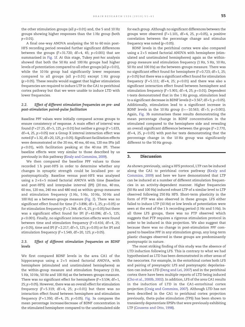
synaptic plasticity. Repeated-measures ANOVAs were ran foreach group with Bonferroni correction to examine the specificdifferences at four different times (−10–0 min, 0–10 min,20–30min and 50–60min) within each group. There was asignificant effect for time in the 1 Hz group (n=6, F=5.922; df=3,27; p<0.01, Fig. 1a). Post-hoc analysis showed that 1 Hz stimula-tion failed to induce post-tetanic potentiation (PTP) as therewasno significant difference in fEPSP slope size between baselineand the 0–10min interval (103.005%±1.606; p>0.05) or the20–30min interval (103.487%±2.622; p>0.05). However, fEPSPslopes during the 50–60 min interval (113.17%±5.75) weresignificantly higher compared to baseline levels (p<0.05).
In the 5 Hz group (n=6, Fig. 1b), there was a significanteffect found for time (F=90.090; df=3, 27; p<0.001). The 5 Hzstimulation protocol induced potentiation lasting approxi-mately 25 min before dropping to below 120% of baseline. ThefEPSPs recorded in the 0–10 min interval (135.614%±1.237)were significantly higher than baseline levels (p<0.001) andthey were also significantly higher than fEPSP averages at the20–30 min (119.173%±1.867; p<0.001) and 50–60 min intervals(110.494%±1.750; p<0.001). However, even though fEPSPs inthe 0–10 min interval were significantly higher than those inthe 20–30 min (p<0.001) and the 50–60 min intervals (p<0.001),this was not classified as PTP as the potentiation seen in thefirst 10 min remains at approximately the same level through-out that time, as opposed to PTP's characteristic large increasein fEPSP slope over the first 2–3 min post-HFS followed by asharp decline in fEPSP slope size.
A significant effect for time was also found in the 10 Hzgroup (n=6, F=26.725; df=3, 27; p<0.001, Fig. 1c). There was asignificant increase in fEPSP slope size compared to baselinelevels at the 0–10 min (123.392%±2.261; p<0.001) and20–30 min intervals (115.477%±2.090; p<0.05) but there wasno significant difference between baseline fEPSP slope valuesand those in the 50–60 min interval (102.857%±1.008; p>0.05).In addition, there was no significant difference between the0–10 min interval and the 20–30 min (p>0.05) which indicatesthat there was no PTP following a 10 Hz stimulation.
As with the three previous groups, there was a significanteffect for time in the 50 Hz group (n=6, F=40.712; df=3, 27;p<0.001; Fig. 1d). Unlike the other groups, a 50 Hz stimulationinduced PTP and LTP. PTP was observed in the first 3 min ofrecording before fEPSP slopes decreased in size. However,fEPSP slopes in the 0–10 min interval (156.365%±4.549)remained significantly higher compared to baseline(p<0.001). fEPSP slopes remained significantly higher com-pared to baseline levels in both the 20–30 min (138.416%±2.862; p<0.001) and the 50–60 min intervals (130.550%±2.069;p<0.001). The fEPSP slope values in the 0–10 min interval weresignificantly higher compared to the 50–60 min interval(p<0.01) which indicates that PTP had been induced.
Finally, there was also a significant effect for time in the100 Hz group (n=6, F=35.836; df=3, 27; p<0.001; Fig. 1e). Likethe 50 Hz group, the 100 Hz group also exhibited PTP and LTP.Again PTP was observed in the first 3 min of recording beforefEPSP slopes decreased in size. Overall, fEPSP slopes in the0–10 min interval (156.742%±6.381) remained significantlypotentiated compared to baseline levels (p<0.001). fEPSPslopes also remained significantly higher compared to base-line levels in the 20–30 min (140.941%±3.271; p<0.001) and the

Fig. 1 – Plot of fEPSP slopes before and after (a) 1 Hz, (b) 5 Hz, (c) 10 Hz, (d) 50 Hz and (e) 100 Hz stimulation protocols in the CA1 toperirhinal cortex projection (n=6 in each group). Each point represents an average of three fEPSP slopes recorded over 1 min anddata is expressed as a percentage of baseline fEPSP slopes where baseline is approximately 100%. Insets are representativefEPSP traces from each group showing typical fEPSPs at baseline (dashed line) and 1 h post-HFS (solid line). (f) Comparison ofpotentiation levels for the five stimulation frequencies averaged over the final 10 min of recording expressed as a percentage ofbaseline fEPSP slopes.
54 B R A I N R E S E A R C H 1 3 2 6 ( 2 0 1 0 ) 5 1 – 6 1
50–60 min intervals (125.336%±2.895; p<0.001). Finally, thefEPSP slope values in the 0–10 min interval were significantlyhigher compared to the 50–60 min interval (p<0.01) indicatingthat PTP had been induced.
Finally we compared the differences between each group atthe four specified time points (−10–0 min, 0–10 min, 20–30 minand 50–60 min). Baseline levels (−10–0 min) showed nosignificant differences between the responses of the fivedifferent stimulation frequencies (F<0.001; df=4, 45; p>0.05)as would be expected. However, there were significantdifferences between the five stimulating frequencies imme-
diately after HFS (0 to 10 min post-HFS; F=36.953; df=4, 45;p<0.001). Tukey post-hoc analysis revealed that the 50 Hz and100 Hz groups had significantly higher responses compared tothe other three groups (all p<0.01), while the 5 Hz and 10 Hzgroups had significantly higher responses than 1 Hz only (bothp<0.01).
Looking at the middle time period after HFS (20 to 30 minpost-HFS) again showed a significant effect for stimulationfrequency (F=37.558; df=4, 45; p<0.001). Tukey post-hocanalysis revealing a similar pattern: the 50 Hz and 100 Hzgroups displayed significantly higher responses compared to
55B R A I N R E S E A R C H 1 3 2 6 ( 2 0 1 0 ) 5 1 – 6 1
the other stimulation groups (all p<0.01) and, the 5 and 10 Hzgroups showing higher responses than the 1 Hz group (bothp<0.01).
A final one-way ANOVA analysing the 50 to 60 min post-HFS recording period revealed further significant differencesbetween the groups (F=31.720; df=4, 45; p<0.001) that aresummarised in Fig. 1f. At this stage, Tukey post-hoc analysisshowed that both the 50 Hz and 100 Hz groups had higherlevels of potentiation compared to all other groups (all p<0.01),while the 10 Hz group had significantly lower responsescompared to all groups (all p<0.01) except 1 Hz group(p>0.05). These results would suggest that higher stimulationfrequencies are required to induce LTP in the CA1 to perirhinalcortex pathway but that we were unable to induce LTD withlower frequencies.
2.2. Effect of different stimulation frequencies on pre- andpost-stimulation paired-pulse facilitation
Baseline PPF values were initially compared across groups toensure consistency of response. A main effect of interval wasfound (F=27.25, df=5, 125, p<0.01) but neither a group (F=1.633,df=4, 25, p>0.05) nor a Group X interval interaction effect wasnoted (F=1.32, df=20, 125,p>0.05). Significant facilitationeffectswere demonstrated at the 20ms, 40ms, 60ms, 120ms IPIs (allp<0.01), with facilitation peaking at the 40 ms IPI. Thesebaseline effects were very similar to those described by uspreviously in this pathway (Kealy and Commins, 2009).
We then compared the baseline PPF values to thoserecorded 1 h post-HFS in order to determine whether anychanges in synaptic strength could be localised pre- orpostsynaptically. Baseline versus post-HFS was analysedusing a 2×6×5 mixed factorial ANOVA with time (baselineand post-HFS) and interpulse interval (IPI) (20 ms, 40 ms,60 ms, 120 ms, 240 ms and 480 ms) as within-group measuresand stimulation frequency (1 Hz, 5 Hz, 10 Hz, 50 Hz and100 Hz) as a between-groups measure (Fig. 2). There was nosignificant effect found for time (F=3.896; df=1, 25; p>0.05) orfor stimulation frequency (F=1.344; df=4, 25; p>0.05) but therewas a significant effect found for IPI (F=43.096; df=5, 125;p<0.001). Finally, no significant interaction effects were foundbetween time and stimulation frequency (F=0.416; df=4, 25;p>0.05), time and IPI (F=2.217; df=5, 125; p>0.05) or for IPI andstimulation frequency (F=1.549, df=20, 125; p>0.05).
2.3. Effect of different stimulation frequencies on BDNFlevels
We first compared BDNF levels in the area CA1 of thehippocampus using a 2×5 mixed factorial ANOVA, withhemisphere (stimulated and unstimulated hemispheres) asthe within-group measure and stimulation frequency (1 Hz,5 Hz, 10 Hz, 50 Hz and 100 Hz) as the between-groups measure.There was no significant effect for hemisphere (F=3.534; df=1,25; p>0.05). However, therewas an overall effect for stimulationfrequency (F=5.319; df=4, 25; p<0.01) but there was nointeraction effect found between hemisphere and stimulationfrequency (F=1.350; df=4, 25; p>0.05). Fig. 3a compares themean percentage increase/decrease of BDNF concentration inthe stimulated hemisphere compared to the unstimulated side
for each group. Although no significant differences between thegroups were observed (F=1.101, df=4, 25, p>0.05), a positivecorrelation between the percentage change and stimulusfrequency was noted (p<0.05).
BDNF levels in the perirhinal cortex were also comparedusing a 2×5 mixed factorial ANOVA with hemisphere (stim-ulated and unstimulated hemispheres) again as the within-group measure and stimulation frequency (1 Hz, 5 Hz, 10 Hz,50 Hz and 100 Hz) as the between-groups measure. There wasno significant effect found for hemisphere (F=0.723; df=1, 25;p>0.05) but there was a significant effect found for stimulationfrequency (F=5.111; df=4, 25; p<0.01) and there was also asignificant interaction effect found between hemisphere andstimulation frequency (F=5.901; df=4, 25; p<0.01). Dependentt-tests demonstrated that in the 1 Hz group, stimulation leadto a significant decrease in BDNF levels (t=3.567; df=5; p<0.05).Additionally, stimulation lead to a significant increase inBDNF levels in the 10 Hz group (t=−10.563; df=5; p<0.001).Again, Fig. 3b summarises these results demonstrating themean percentage change in BDNF concentration in thestimulated compared to the hemisphere side and revealingan overall significant difference between the groups (F=2.770,df=4, 25, p<0.05) with post-hoc tests demonstrating that thepercentage change in the 10 Hz group was significantlydifferent to the 50 Hz group.
3. Discussion
As shown previously, using a HFS protocol, LTP can be inducedalong the CA1 to perirhinal cortex pathway (Kealy andCommins, 2009) and here we have demonstrated that LTPcan be induced at a number of different stimulation frequen-cies in an activity-dependent manner. Higher frequencies(50 Hz and 100 Hz) induced robust LTP of a similar level to LTPobserved following 250 Hz HFS. Short-term plasticity in theform of PTP was also observed in these groups. LFS eitherfailed to induce LTP (10 Hz) or low levels of potentiation wereseen at the end of the 1 h recording period (1 Hz and 5 Hz). Inall three LFS groups, there was no PTP observed whichsuggests that PTP requires a rigorous stimulation protocol inorder to be induced in this projection. We also suggest thatbecause there was no change in post-stimulation PPF com-pared to baseline PPF in any stimulation group, any long-termplastic changes observed in these groups are predominantlypostsynaptic in nature.
The most striking finding of this study was the absence ofLTD induction following LFS. This is contrary to what we hadhypothesised as LTD has been demonstrated in other areas ofthe neocortex. For example, in the entorhinal cortex both LFSand pairing of presynaptic LFS and postsynaptic depolarisa-tion can induce LTD (Deng and Lei, 2007) and in the perirhinalcortex there have been multiple reports of LTD being induced(Cho et al., 2000b, 2002). In addition, LFS of the area CA1 resultsin the induction of LTD in the CA1-entorhinal cortexprojection (Craig and Commins, 2007). Although LTD has notbeen described in the CA1 to perirhinal cortex projectionpreviously, theta-pulse stimulation (TPS) has been shown totransiently depotentiate EPSPs that were previously exhibitingLTP (Cousens and Otto, 1998).
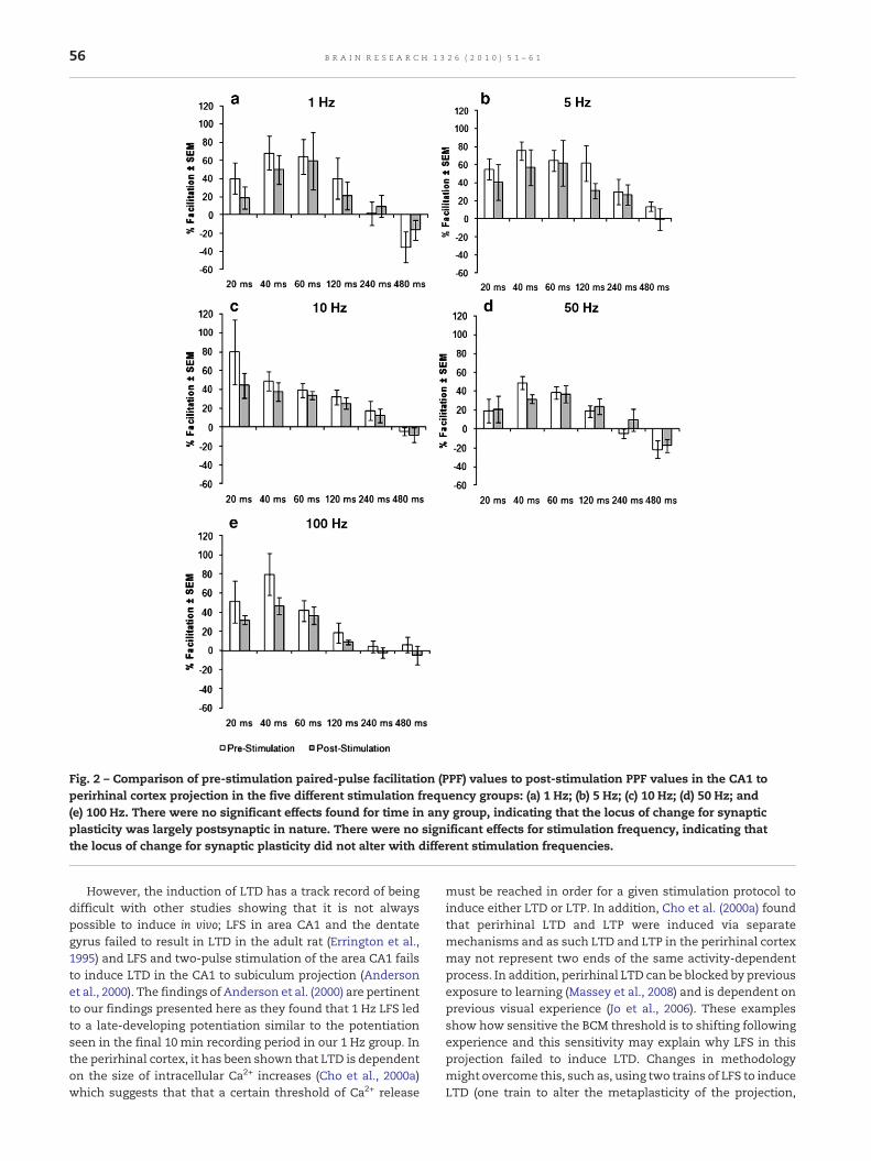
Fig. 2 – Comparison of pre-stimulation paired-pulse facilitation (PPF) values to post-stimulation PPF values in the CA1 toperirhinal cortex projection in the five different stimulation frequency groups: (a) 1 Hz; (b) 5 Hz; (c) 10 Hz; (d) 50 Hz; and(e) 100 Hz. There were no significant effects found for time in any group, indicating that the locus of change for synapticplasticity was largely postsynaptic in nature. There were no significant effects for stimulation frequency, indicating thatthe locus of change for synaptic plasticity did not alter with different stimulation frequencies.
56 B R A I N R E S E A R C H 1 3 2 6 ( 2 0 1 0 ) 5 1 – 6 1
However, the induction of LTD has a track record of beingdifficult with other studies showing that it is not alwayspossible to induce in vivo; LFS in area CA1 and the dentategyrus failed to result in LTD in the adult rat (Errington et al.,1995) and LFS and two-pulse stimulation of the area CA1 failsto induce LTD in the CA1 to subiculum projection (Andersonet al., 2000). The findings of Anderson et al. (2000) are pertinentto our findings presented here as they found that 1 Hz LFS ledto a late-developing potentiation similar to the potentiationseen in the final 10 min recording period in our 1 Hz group. Inthe perirhinal cortex, it has been shown that LTD is dependenton the size of intracellular Ca2+ increases (Cho et al., 2000a)which suggests that that a certain threshold of Ca2+ release
must be reached in order for a given stimulation protocol toinduce either LTD or LTP. In addition, Cho et al. (2000a) foundthat perirhinal LTD and LTP were induced via separatemechanisms and as such LTD and LTP in the perirhinal cortexmay not represent two ends of the same activity-dependentprocess. In addition, perirhinal LTD can be blocked by previousexposure to learning (Massey et al., 2008) and is dependent onprevious visual experience (Jo et al., 2006). These examplesshow how sensitive the BCM threshold is to shifting followingexperience and this sensitivity may explain why LFS in thisprojection failed to induce LTD. Changes in methodologymight overcome this, such as, using two trains of LFS to induceLTD (one train to alter the metaplasticity of the projection,
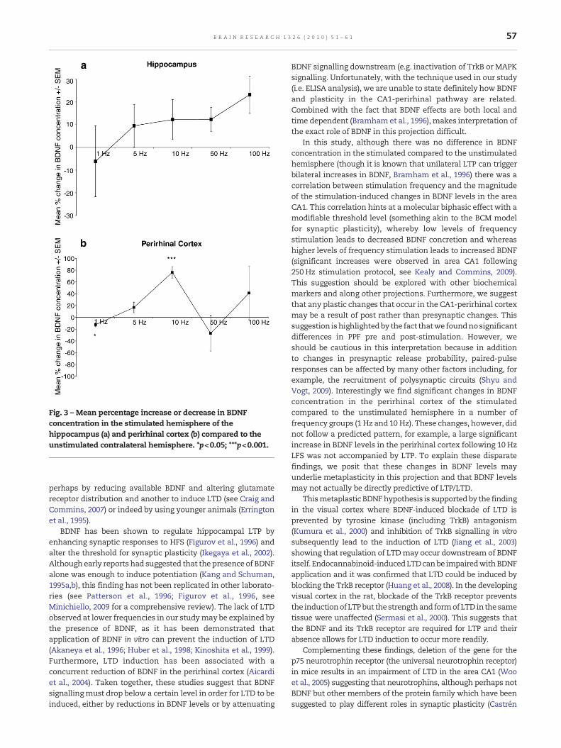
Fig. 3 – Mean percentage increase or decrease in BDNFconcentration in the stimulated hemisphere of thehippocampus (a) and perirhinal cortex (b) compared to theunstimulated contralateral hemisphere. *p<0.05; ***p<0.001.
57B R A I N R E S E A R C H 1 3 2 6 ( 2 0 1 0 ) 5 1 – 6 1
perhaps by reducing available BDNF and altering glutamatereceptor distribution and another to induce LTD (see Craig andCommins, 2007) or indeed by using younger animals (Erringtonet al., 1995).
BDNF has been shown to regulate hippocampal LTP byenhancing synaptic responses to HFS (Figurov et al., 1996) andalter the threshold for synaptic plasticity (Ikegaya et al., 2002).Although early reports had suggested that the presence of BDNFalone was enough to induce potentiation (Kang and Schuman,1995a,b), this finding has not been replicated in other laborato-ries (see Patterson et al., 1996; Figurov et al., 1996, seeMinichiello, 2009 for a comprehensive review). The lack of LTDobserved at lower frequencies in our studymay be explained bythe presence of BDNF, as it has been demonstrated thatapplication of BDNF in vitro can prevent the induction of LTD(Akaneya et al., 1996; Huber et al., 1998; Kinoshita et al., 1999).Furthermore, LTD induction has been associated with aconcurrent reduction of BDNF in the perirhinal cortex (Aicardiet al., 2004). Taken together, these studies suggest that BDNFsignallingmust drop below a certain level in order for LTD to beinduced, either by reductions in BDNF levels or by attenuating
BDNF signalling downstream (e.g. inactivation of TrkB or MAPKsignalling. Unfortunately, with the technique used in our study(i.e. ELISA analysis), we are unable to state definitely how BDNFand plasticity in the CA1-perirhinal pathway are related.Combined with the fact that BDNF effects are both local andtime dependent (Bramham et al., 1996), makes interpretation ofthe exact role of BDNF in this projection difficult.
In this study, although there was no difference in BDNFconcentration in the stimulated compared to the unstimulatedhemisphere (though it is known that unilateral LTP can triggerbilateral increases in BDNF, Bramham et al., 1996) there was acorrelation between stimulation frequency and the magnitudeof the stimulation-induced changes in BDNF levels in the areaCA1. This correlation hints at amolecular biphasic effect with amodifiable threshold level (something akin to the BCM modelfor synaptic plasticity), whereby low levels of frequencystimulation leads to decreased BDNF concretion and whereashigher levels of frequency stimulation leads to increased BDNF(significant increases were observed in area CA1 following250 Hz stimulation protocol, see Kealy and Commins, 2009).This suggestion should be explored with other biochemicalmarkers and along other projections. Furthermore, we suggestthat any plastic changes that occur in the CA1-perirhinal cortexmay be a result of post rather than presynaptic changes. Thissuggestion ishighlightedby the fact thatwe foundnosignificantdifferences in PPF pre and post-stimulation. However, weshould be cautious in this interpretation because in additionto changes in presynaptic release probability, paired-pulseresponses can be affected by many other factors including, forexample, the recruitment of polysynaptic circuits (Shyu andVogt, 2009). Interestingly we find significant changes in BDNFconcentration in the perirhinal cortex of the stimulatedcompared to the unstimulated hemisphere in a number offrequency groups (1 Hz and 10 Hz). These changes, however, didnot follow a predicted pattern, for example, a large significantincrease in BDNF levels in the perirhinal cortex following 10 HzLFS was not accompanied by LTP. To explain these disparatefindings, we posit that these changes in BDNF levels mayunderlie metaplasticity in this projection and that BDNF levelsmay not actually be directly predictive of LTP/LTD.
Thismetaplastic BDNFhypothesis is supportedby the findingin the visual cortex where BDNF-induced blockade of LTD isprevented by tyrosine kinase (including TrkB) antagonism(Kumura et al., 2000) and inhibition of TrkB signalling in vitrosubsequently lead to the induction of LTD (Jiang et al., 2003)showing that regulation of LTDmay occur downstream of BDNFitself. Endocannabinoid-inducedLTDcanbe impairedwithBDNFapplication and it was confirmed that LTD could be induced byblocking the TrkB receptor (Huang et al., 2008). In the developingvisual cortex in the rat, blockade of the TrkB receptor preventsthe inductionofLTPbut thestrengthand formofLTDinthesametissue were unaffected (Sermasi et al., 2000). This suggests thatthe BDNF and its TrkB receptor are required for LTP and theirabsence allows for LTD induction to occur more readily.
Complementing these findings, deletion of the gene for thep75 neurotrophin receptor (the universal neurotrophin receptor)in mice results in an impairment of LTD in the area CA1 (Wooet al., 2005) suggesting that neurotrophins, although perhaps notBDNF but other members of the protein family which have beensuggested to play different roles in synaptic plasticity (Castrén
58 B R A I N R E S E A R C H 1 3 2 6 ( 2 0 1 0 ) 5 1 – 6 1
et al., 1993), are involved in the induction of LTD through the p75receptor. As blockade of tyrosine kinase receptors in general stillallowed for LTD induction (Kumura et al., 2000), the case for p75playing a greater role in LTD is made stronger. Further to this,different forms of BDNFmay also play distinct roles in plasticity.BDNF is initially secreted as a precursor proBDNF before matureBDNF is generated. Mature BDNF binds the receptor TrkB,whereas proBDNF binds p75 (Lu, 2003). Through these tworeceptors pro- andmatureBDNFelicit different cellular responseswithproBDNF-p75 binding, for example, facilitatinghippocampalLTD (Woo et al., 2005). Future research in this projection couldlook at the roles played by different forms of BDNF and itsreceptors in modulating synaptic plasticity.
Lastly, it could be that this projection is electrophysiologicallyexcitatory in nature and that LTD is just difficult to induce.Evidence for this comes fromtheprojection's readiness to induceLTP (Cousens and Otto, 1998; Kealy and Commins, 2009) and thelack of LTD using LFS protocols shown previously to induce LTD.In the projections going from the hippocampus to the entorhinalcortex, the projections originating in the distal CA1 andproximalsubiculum, and terminating in the lateral entorhinal cortex showsimilar ‘excitability’ (ability to sustain LTP readily and withlimited LTD) whereas those originating in the proximal CA1 anddistal subiculum, and terminating in the medial entorhinalcortex showed a greater tendency for electrophysiologically‘inhibitory’ activity (greater tendency to show LTD, see CraigandCommins, 2007). As the lateral entorhinal cortex anatomicallyshows strong interconnectivity with the perirhinal cortex andthe medial entorhinal cortex receives input from the postrhinalcortex (Insausti et al., 1997; Burwell and Amaral, 1998) and thereseems to be a segregation of projections connecting thehippocampus and the entorhinal cortex (Witter et al., 2000), wesuggest that the CA1 to perirhinal cortex projection forms part ofthe CA1/subiculum to lateral entorhinal cortex circuit. Thereforea tendency towards potentiation over depression may beexpected. We predict that the postrhinal cortex is most likelyassociated with the hippocampus to medial entorhinal cortexcircuit andwehypothesise projections from the hippocampus tothe postrhinal cortex to exhibit depressionmore readily.While itseems clear that, in general, hippocampal–parahippocampalprojections can be segregated along two parallel pathwaysanatomically (Witter et al., 2000) and perhaps, as suggestedabove, electrophysiologically, it is difficult to know at presentwhether this segregation is also reflected at a functional level. Itis known that there is a functional difference along thelongitudinal axis of the hippocampus, with the dorsal hippo-campus involvedmore in informationprocessingand theventralhippocampus involved in emotion and stress (Fanselow andDong, 2010), more research is needed to examine functionaldifferences along the hippocampal–parahippocampal parallelprojections.
4. Experimental procedures
4.1. Surgery
Adult male Wistar rats (n=30; approximately 3months old;weight: 300–400 g; Biomedical Unit; University College Dublin)were anaesthetised using urethane (ethyl carbamate; 1.5 mg/kg;
i.p.; Sigma) andmounted on a stereotaxic frame. An incisionwasmade to expose the skull and burr holes were made to allow theelectrodes to be inserted into the correct coordinates (PaxinosandWatson, 2005).
A stainless bipolar stimulating electrodes (50 μm diameter),insulated apart from the tip, were aimed at the area CA1.Stainless wire recording electrodes (50 μm diameter) wereaimed at the perirhinal cortex. Coordinates for both electrodeswere based on previous studies on this projection (Cousens andOtto, 1998; Kealy andCommins, 2009); the stimulating electrode(CA1) was inserted 6.3 mm posterior to Bregma, 5.5 mm lateralto themidline and lowered to a depth of 2.2 mmmeasured fromthe surface of the brain. The recording electrode (perirhinalcortex)was inserted 5.2 mmposterior to Bregma, 4.3 mm lateralto themidline and lowered to a depth of 5.0 mmmeasured fromthe surface of the brain. The recording electrodewas inserted atan angle of 17° to increase the chances of hitting the correctpopulation of cells that are part of the monosynaptic CA1 toperirhinal projection described previously (Cousens and Otto,1998; Kealy and Commins, 2009).
4.2. Stimulation and data acquisition
Signals were filtered between 1 Hz and 1 kHz and then ampli-fied (DAM-50 differential amplifier, World Precision Instru-ments, Hertfordshire, UK). Recordings were digitised using a PCrunning Spike2 (version 5.02, CED, Cambridge, UK) connected toa CED-1401 interface (CED, Cambridge, UK). In analysing thefield excitatory postsynaptic potentials (fEPSPs), the slope of theresponse was measured (Gooney and Lynch, 2001). The slopewas calculated from the middle two-thirds of the downward-deflecting component of the response.
Field EPSPs were evoked by stimulating with a square-wave(constant current pulse of 0.1 ms duration at a frequency of0.05 Hz). AlthoughbothPopulationSpike and fEPSPsweremoni-tored, the incidences of the occurrence of population spikeremained low (less than 5% of cases). Bymeans of input/outputcurve determination the maximum fEPSP was found for eachindividual animal. The intensity of stimulus was then set at alevel that evoked a fEPSP slope of 50–60% of themaximum. Thislevel is similar to thatwehaveused before (Craig andCommins,2005, 2006) on the CA1 and subicular to EC pathway, CousensandOtto (1998) on the CA1–perirhinal pathway and to that usedby other authors in area CA1 (Wöhrl et al., 2007).
Baseline paired-pulse facilitation (PPF) effects were mea-sured after the electrodes were allowed to settle for 10 min.Pairs of stimuli were then delivered with various interpulseintervals (IPIs) of 20, 40, 60, 120, 240 and 480 ms. The PPF valuewas calculated by taking the value of six slopes values fromthe first stimuli out of the pairs (fEPSP1) for a given IPI andthen normalising the average of six slope values from thesecond stimuli from the pairs (fEPSP2) with the resulting valueexpressed as a percentage (Commins et al., 1998a). Followingthis, baseline measurements were induced at a rate of 0.05 Hzfor another 10 min.
Following baseline recording baseline responses to singlestimuli (0.05 Hz) over 10 min, each group (n=6 in each) wastreated using one of five possible stimulation protocols (1 Hz,5 Hz, 10 Hz, 50 Hz or 100 Hz). Each stimulation protocolcontained a constant number of stimuli (900) and varied only
59B R A I N R E S E A R C H 1 3 2 6 ( 2 0 1 0 ) 5 1 – 6 1
in the frequency by which the pulses were delivered.Following these stimulation protocols, recordings were madeat 0.05 Hz for 1 h. Following this hour of recording, post-HFSPPF recordings were made using the same protocol as the pre-HFS PPF recordings. The stimulus intensity for all experimen-tal interventions (PPF and the five stimulation frequencieslisted above) was set at baseline intensity. Changes in fEPSPslope were assessed by measuring the slope of the responsepost-HFS compared to baseline response.
4.3. Brain-derived neurotrophic factor enzyme-linkedimmunosorbent assay (ELISA)
After each experiment, all animals were sacrificed by decap-itation and their brains were immediately removed. Thebrains were then dissected on ice. An area of approximately3 mm3 was removed from both area CA1 and perirhinal cortexthat contained the final resting place of both the stimulatingand recording electrode respectively. Equivalent sized areas inthe same animal's unstimulated hemisphere were alsoremoved for subsequent analysis. These samples were frozenand stored in Krebs-CaCl2/dimethyl sulfoxide (DMSO; Krebssolution was made using pre-prepared formula; all fromSigma, Ireland). All samples were later washed in Krebs-CaCl2 and homogenised. 10 μl aliquots of each sample wereanalysed using a Bradford protein assay in order to determinetotal protein concentration. Utilising these results, the samplesolutionswere normalised to be equal in concentration and anenzyme-linked immunosorbent assay (ELISA) for BDNF wasperformed using a BDNF Emax ImmunoAssay System kit(Promega Corporation), following themanufacturer's protocol.
4.4. Statistical analysis
A series of dependent t-tests and repeated measures analysesof variance (ANOVAs) with the appropriate post-hoc test(Tukey at the 5% level of significance) were performed usingSPSS 15.0 for Windows (SPSS Software, Seattle, WA, USA). Astar-rated system was used where appropriate (*p<0.05;**p<0.01; ***p<0.001).
4.5. Ethical considerations
Laboratory procedures for the maintenance and experimen-tation of animals conformed to the Department of Health(Ireland) guidelines and the European directive 86/609/EC.Every effort was made to minimise the suffering and thenumber of animals used in this study.
Acknowledgment
This work has emanated from research conducted with thefinancial support of the Science Foundation of Ireland (SFI).
R E F E R E N C E S
Abraham, W.C., Bear, M.F., 1996. Metaplasticity: the plasticity ofsynaptic plasticity. Trends Neurosci. 19, 126–130.
Abraham, W.C., Williams, J.M., 2008. LTP maintenance and itsprotein synthesis-dependence. Neurobiol. Learn. Mem. 89,260–268.
Aicardi, G., Argilli, E., Capello, S., Santi, S., Riccio, M., Thoenend, H.,Canossa, M., 2004. Induction of long-term potentiation anddepression is reflected by corresponding changes in secretionof endogenous brain-derived neurotrophic factor. Proc. Natl.Acad. Sci. U. S. A. 101, 15788–15792.
Akaneya, Y., Tsumoto, T., Hatanaka, H., 1996. Brain-derivedneurotrophic factor blocks long-term depression in rat visualcortex. J. Neurophysiol. 76, 4198–4201.
Anderson, M., Commins, S., O'Mara, S.M., 2000. The effects of lowfrequency and two-pulse stimulation protocols on synaptictransmission in theCA1-subiculumpathway in theanaesthetizedrat. Neurosci. Lett. 279, 181–184.
Balkowiec, A., Katz, D.M., 2002. Cellular mechanisms regulatingactivity-dependent release of native brain-derived neurotrophicfactor from hippocampal neurons. J. Neurosci. 22, 10399–10407.
Barker, G.R.I., Warburton, E.C., Koder, T., Dolman, N.P., More, J.C.,Aggleton, J.P., Bashir, Z.I., Auberson, Y.P., Jane, D.E., Brown,M.W.,2006. The different effects on recognition memory of perirhinalkainate andNMDAglutamate receptor antagonism: implicationsfor underlying plasticitymechanisms. J. Neurosci. 26, 3561–3566.
Barry, M.F., Ziff, E.B., 2002. Receptor trafficking and the plasticity ofexcitatory synapses. Curr. Opin. Neurobiol. 12, 279–286.
Bear, M.F., 2003. Bidirectional synaptic plasticity: from theory toreality. In: Bliss, T., Collingridge, G.L., Morris, R. (Eds.), Long-termPotentiation: Enhancing Neuroscience for 30 Years. OxfordUniversity Press, New York, pp. 67–77.
Bear, M.F., Cooper, L.N., Ebner, F.F., 1987. A physiological basis for atheory of synapse modification. Science 237, 42–48.
Beattie, E.C., Carroll, R.C., Yu, X., Morishita, W., Yasuda, H., vonZastrow, M., Malenka, R.C., 2000. Regulation of AMPA receptorendocytosis by a signaling mechanism shared with LTD. Nat.Neurosci. 3, 1291–1300.
Bienenstock, E.L., Cooper, L.N., Munro, P.W., 1982. Theory for thedevelopment of neuron selectivity: orientation specificity andbinocular interaction in visual cortex. J. Neurosci. 2, 32–48.
Bliss, T.V.P., Lømo, T., 1973. Long-lasting potentiation of synaptictransmission in the dentate area of the anaesthetized rabbitfollowing stimulation of the perforant path. J. Physiol. 232,331–356.
Bramham,C.R.,Alme,M.N.,Bittins,M.,Kuipers,S.D.,Nair,R.R., Pai, B.,Panja, D., Schubert, M., Soule, J., Tiron, A., Wibrand, K., 2010. TheArc of synaptic memory. Exp Brain Res. 200 (2), 125–140.
Bramham,C.R., Southard,T., Sarvey, J.M.,Herkenham,M., Brady, L.S.,1996. Unilateral LTP triggers bilateral increases in hippocampalneurotrophin and trk receptor mRNA expression in behavingrats: evidence for interhemispheric communication. J. Comp.Neurol. 368, 371–382.
Bramham, C.R., Srebro, B., 1987. Induction of long-term depressionand potentiation by low- and high-frequency stimulation inthe dentate area of the anesthetized rat: magnitude, timecourse and EEG. Brain Res. 405, 100–107.
Burwell, R.D., Amaral, D.G., 1998. The perirhinal and postrhinalcortices of the rat: interconnectivity and connections with theentorhinal cortex. J. Comp. Neurol. 391, 293–321.
Castrén, E., Pitkänen, M., Sirviö, J., Parsadanian, A., Lindholm, D.,Thoenen, H., Riekinnen, P.J., 1993. The induction of LTPincreases BDNF and NGF mRNA but decreases NT-3 mRNA inthe dentate gyrus. NeuroReport 4, 895–898.
Cho, K., Aggleton, J.P., Brown, M.W., Bashir, Z.I., 2000a. Anexperimental test of the role of postsynaptic calcium levels indetermining synaptic strength using perirhinal cortex of rat.J. Physiol. 532, 459–466.
Cho, K., Brown, M.W., Bashir, Z.I., 2002. Mechanisms andphysiological role of enhancement of mGlu5 receptor functionby group II mGlu receptor activation in rat perirhinal cortex.J. Physiol. 540, 895–906.
60 B R A I N R E S E A R C H 1 3 2 6 ( 2 0 1 0 ) 5 1 – 6 1
Cho, K., Kemp, N., Noel, J., Aggleton, J.P., Brown, M.W., Bashir, Z.I.,2000b. A new form of long-term depression in the perirhinalcortex. Nat. Neurosci. 3, 150–156.
Citri, A., Soler-Llavina, G., Bhattacharyya, S., Malenka, R.C., 2009.N-methyl-D-aspartate receptor- and metabotropic glutamatereceptor-dependent long-term depression are differentiallyregulated by the ubiquitin-proteasome system. Eur. J. Neurosci.30 (8), 1443–1450.
Commins, S., Gigg, J., Anderson, M., O'Mara, S.M., 1998a. Interac-tion between paired-pulse facilitation and long-term potenti-ation in the projection from hippocampal area CA1 to thesubiculum. NeuroReport 9, 4109–4113.
Commins, S., Gigg, J., Anderson, M., O'Mara, S.M., 1998b. Theprojection from hippocampal area CA1 to the subiculumsustains long-term potentiation. NeuroReport 9 (5), 847–850.
Cousens, G., Otto, T.A., 1998. Induction and transient suppressionof long-term potentiation in the peri- and postrhinal corticesfollowing theta-related stimulation of hippocampal field CA1.Brain Res. 780, 95–101.
Craig, S., Commins, S., 2005. Interaction between paired-pulsefacilitation and long-term potentiation in the projection fromhippocampal area CA1 to the entorhinal cortex. Neurosci. Res.53, 140–146.
Craig, S., Commins, S., 2006. The subiculum to entorhinal cortexprojection is capable of sustaining both short- and long-termplastic changes. Behav. Brain Res. 174, 281–288.
Craig, S., Commins, S., 2007. Plastic andmetaplastic changes in theCA1 and subicular projections to the entorhinal cortex. BrainRes. 1147, 124–139.
Cummings, J.A., Mulkey, R.M., Nicoll, R.A., Malenka, R.C., 1996. Ca2+
signaling requirements for long-term depression in thehippocampus. Neuron 16, 825–833.
Czarnecki, A., Birtoli, B., Ulrich, D., 2007. Cellular mechanisms ofburst firing-mediated long-term depression in rat neocorticalpyramidal cells. J. Physiol. 578, 471–479.
Deng, P.Y., Lei, S., 2007. Long-term depression in identified stellateneurons of juvenile rat entorhinal cortex. J. Neurophysiol. 97,727–737.
Dickinson, B.A., Jo, J., Seok, H., Son, G.H.,Whitcomb, D.J., Davies, C.H.,Sheng, M., Collingridge, G.L., Cho, K., 2009. A novel mechanism ofhippocampal LTD involving muscarinic receptor-triggered inter-actions between AMPARs, GRIP and liprin-alpha. Mol. Brain 2, 18.
Doyère,V., Errington,M.L., Laroche,S., Bliss,T.V., 1996. Low-frequencytrains of paired stimuli induce long-term depression in area CA1but not in dentate gyrus of the intact rat. Hippocampus 6, 52–57.
Du, J., Feng, L., Yang, F., Lu, B., 2000. Activity- and Ca(2+)-dependent modulation of surface expression of brain-derivedneurotrophic factor receptors in hippocampal neurons. J. CellBiol. 150, 1423–1434.
Dudek, S.M., Bear,M.F., 1992.Homosynaptic long-termdepression inarea CA1 of hippocampus and effects of N-methyl-D-aspartatereceptor blockade. Proc. Natl. Acad. Sci. U. S. A. 89, 4363–4367.
Dudek, S.M., Bear,M.F., 1993. Bidirectional long-termmodificationofsynaptic effectiveness in the adult and immature hippocampus.J. Neurosci. 13, 2910–2918.
Errington, M.L., Bliss, T.V., Richter-Levin, G., Yenk, K., Doyère, V.,Laroche, S., 1995. Stimulation at 1–5 Hz does not producelong-term depression or depotentiation in the hippocampus ofthe adult rat in vivo. J. Neurophysiol. 74, 1793–1799.
Fanselow, M.S., Dong, H.W., 2010. Are the dorsal and ventralhippocampus functionally distinct structures? Neuron. 65, 7–19.
Figurov, A., Pozzo-Miller, L.D., Olafsson, P., Wang, T., Lu, B., 1996.Regulation of synaptic responses to high-frequency stimulationand LTP by neurotrophins in the hippocampus. Nature 381,706–709.
Gallagher, S.M., Daly, C.A., Bear,M.F., Huber, K.M., 2004. Extracellularsignal-regulated protein kinase activation is required formetabotropic glutamate receptor-dependent long-termdepression in hippocampal area CA1. J. Neurosci. 24, 4859–4864.
Gärtner, A., Staiger, V., 2002. Neurotrophin secretion fromhippocampalneuronsevokedby long-term-potentiation-inducingelectrical stimulation patterns. Proc. Natl. Acad. Sci. U. S. A. 99,6386–6391.
Gooney,M., Lynch,M.A., 2001. Long-termpotentiation in thedentategyrus of the rat hippocampus is accompanied by brain-derivedneurotrophic factor-induced activation of TrkB. J. Neurochem.77, 1198–1207.
Griffiths, S., Scott, H., Glover, C., Bienemann, A., Ghorbel, M.T., Uney,J., Brown, M.W., Warburton, E.C., Bashir, Z.I., 2008. Expression oflong-term depression underlies visual recognition memory.Neuron 58, 186–194.
Hartmann, M., Heumann, R., Lessmann, V., 2001. Synaptic secretionof BDNF after high-frequency stimulation of glutamatergicsynapses. EMBO J. 20, 5887–5897.
Heynen, A.J., Abraham, W.C., Bear, M.F., 1996. Bidirectionalmodification of CA1 synapses in the adult hippocampus invivo. Nature 381, 163–166.
Holman,D., Feligioni,M.,Henley, J.M., 2007.Differential redistributionof native AMPA receptor complexes following LTD induction inacute hippocampal slices. Neuropharmacology 52, 92–99.
Hosseinmardi, N., Fathollahi, Y., Naghdi, N., Javan, M., 2009. Thetapulsestimulation:anatural stimuluspatterncan trigger long-termdepressionbut fails to reverse long-termpotentiation inmorphinewithdrawn hippocampus area CA1. Brain Res. 1296, 1–14.
Huang, Y., Yasuda, H., Sarihi, A., Tsumoto, T., 2008. Roles ofendocannabinoids in heterosynaptic long-term depression ofexcitatory synaptic transmission in visual cortex of youngmice. J. Neurosci. 28, 7074–7083.
Huber,K.M., Sawtell,N.B., Bear,M.F., 1998. Brain-derivedneurotrophicfactor alters the synaptic modification threshold in visual cortex.Neuropharmacology 37, 571–579.
Ikegaya, Y., Ishizaka, Y., Matsuki, N., 2002. BDNF attenuateshippocampal LTD via activation of phospholipase C: implicationsfor a vertical shift in the frequency-response curve of synapticplasticity. Eur. J. NeuroSci. 16, 145–148.
Insausti, R., Herrero, M.T., Witter, M.P., 1997. Entorhinal cortex ofthe rat: cytoarchitectonic subdivisions and the origin anddistribution of cortical efferents. Hippocampus 7, 146–183.
Ivanco,T.L., Racine,R.J., 2000. Long-termpotentiation in the reciprocalcorticohippocampal and corticocortical pathways in the chroni-cally implanted, freely moving rat. Hippocampus 10, 143–152.
Jiang, B., Akaneya, Y., Hata, Y., Tsumoto, T., 2003. Long-termdepression is not induced by low-frequency stimulation in ratvisual cortex in vivo: a possible preventing role of endogenousbrain-derived neurotrophic factor. J. Neurosci. 23, 3761–3770.
Jo, J., Ball, S.M., Seok, H., Oh, S.B., Massey, P.V., Molnar, E., Bashir, Z.I.,Cho,K., 2006. Experience-dependentmodificationofmechanismsof long-term depression. Nat. Neurosci. 9, 170–172.
Kang, H.J., Schuman, E.M., 1995a. Long-lasting neurotrophin-inducedenhancementof synaptic transmission in theadulthippocampus.Science 267, 1658–1662.
Kang, H.J., Schuman, E.M., 1995b. Neurotrophin-inducedmodulationof synaptic transmission in the adult hippocampus. J. Physiol.(Paris) 89, 11–22.
Kealy, J., Commins, S., 2009. Antagonism of glutamate receptors inthe CA1 to perirhinal cortex projection prevents long-termpotentiation and attenuates levels of brain-derivedneurotrophicfactor. Brain Res. 1265, 53–64.
Kinoshita, S., Yasuda, H., Taniguchi, N., Katoh-Semba, R., Hatanaka,H., Tsumoto,T., 1999. Brain-derivedneurotrophic factor preventslow-frequency inputs from inducing long-termdepression in thedeveloping visual cortex. J. Neurosci. 19, 2122–2130.
Kirkwood, A., Dudek, S.M., Gold, J.T., Aizenman, C.D., Bear, M.F.,1993. Common forms of synaptic plasticity in the hippocampusand neocortex in vitro. Science 260, 1518–1521.
Kumura,E.,Kimura, F., Taniguchi,N.,Tsumoto,T., 2000.Brain-derivedneurotrophic factor blocks long-term depression in solitaryneurones cultured from rat visual cortex. J. Physiol. 524, 195–204.
61B R A I N R E S E A R C H 1 3 2 6 ( 2 0 1 0 ) 5 1 – 6 1
Lu, B., 2003. Pro-region of neurotrophins: role in synapticmodulation. Neuron. 39, 735–738.
Lüscher, C., Xia, H., Beattie, E.C., Carroll, R.C., von Zastrow, M.,Malenka, R.C., Nicoll, R.A., 1999. Role of AMPA receptor cyclingin synaptic transmission and plasticity. Neuron 24, 649–658.
Martin, S.J., Grimwood, P.D., Morris, R.G., 2000. Synaptic plasticityand memory: an evaluation of the hypothesis. Annu. Rev.Neurosci. 23, 649–711.
Massey,P.V., Phythian,D.,Narduzzo,K.,Warburton,E.C., Brown,M.W.,Bashir, Z.I., 2008. Learning-specific changes in long-termdepression in adult perirhinal cortex. J. Neurosci. 28, 7548–7554.
McClelland, J.L., McNaughton, B.L., O'Reilly, R.C., 1995. Why thereare complementary learning systems in the hippocampus andneocortex: insights from the successes and failures ofconnectionist models of learning and memory. Psychol. Rev.102, 419–457.
Minichiello, L., 2009. TrkB signalling pathways in LTP and learning.Nat. Rev. Neurosci. 10, 850–860.
Mulkey, R.M., Endo, S., Shenolikar, S., Malenka, R.C., 1994.Involvement of a calcineurin/inhibitor-1 phosphatase cascadein hippocampal long-term depression. Nature 369, 486–488.
Mulkey, R.M., Herron, C.E., Malenka, R.C., 1993. An essential role forprotein phosphatases in hippocampal long-term depression.Science 261, 1051–1055.
Mulkey,R.M.,Malenka,R.C., 1992.Mechanismsunderlying inductionof homosynaptic long-term depression in area CA1 of thehippocampus. Neuron 9, 967–975.
Norman, E.D., Thiels, E., Barrionuevo, G., Klann, E., 2000. Long-termdepression in the hippocampus in vivo is associated withprotein phosphatase-dependent alterations in extracellularsignal-regulated kinase. J. Neurochem. 74, 192–198.
O'Keefe, J., Nadel, L., 1978. The Hippocampus as a Cognitive Map.Oxford University Press, Great Britain.
Park, Y., Jo, J., Isaac, J.T., Cho, K., 2006. Long-term depression ofkainate receptor-mediated synaptic transmission. Neuron 49,95–106.
Patterson, S.L., Abel, T., Deuel, T.A., Martin, K.C., Rose, J.C., Kandel,E.R., 1996. Recombinant BDNF rescues deficits in basal synaptictransmission and hippocampal LTP in BDNF knockout mice.Neuron 16, 1137–1145.
Paxinos, G., Watson, C., 2005. The Rat Brain in StereotaxicCoordinates, 5th ed. Academic Press, San Diego.
Rioult-Pedotti,M.S., Donoghue, J.P., Dunaevsky, A., 2007. Plasticity ofthe synaptic modification range. J. Neurophysiol. 98, 3688–3695.
Rolls, E.T., 1996. A theory of hippocampal function in memory.Hippocampus 6, 601–620.
Scoville, W.B., Milner, B., 1957. Loss of recent memory afterbilateral hippocampal lesions. J. Neurol. Neurosurg. Psychiatr.20, 11–21.
Seoane, A., Massey, P.V., Keen, H., Bashir, Z.I., Brown, M.W., 2009.L-type voltage-dependent calcium channel antagonists impairperirhinal long-termrecognitionmemoryandplasticityprocesses.J. Neurosci. 29, 9534–9544.
Sermasi, E., Margotti, E., Cattaneo, A., Domenici, L., 2000. Trk Bsignalling controls LTP but not LTD expression in the developingrat visual cortex. Eur. J. NeuroSci. 12, 1411–1419.
Shi, S., Hayashi, Y., Esteban, J.A., Malinow, R., 2001. Subunit-specificrules governing AMPA receptor trafficking to synapses inhippocampal pyramidal neurons. Cell 105, 331–343.
Shi, S.H., Hayashi, Y., Petralia, R.S., Zaman, S.H., Wenthold, R.J.,Svoboda, K., Malinow, R., 1999. Rapid spine delivery and
redistribution of AMPA receptors after synaptic NMDA receptoractivation. Science 284, 1811–1816.
Shyu, B.C., Vogt, B.A., 2009. Short-term synaptic plasticity in thenociceptive thalamic-anterior cingulate pathway. Mol. Pain. 4,5–51.
Snyder, E.M., Philpot, B.D., Huber, K.M., Dong, X., Fallon, J.R., Bear,M.F., 2001. Internalization of ionotropic glutamate receptors inresponse to mGluR activation. Nat. Neurosci. 4, 1079–1085.
Springer, J.E., Gwag, B.J., Sessler, F.M., 1994. Neurotrophic factormRNA expression in dentate gyrus is increased following invivo stimulation of the angular bundle. Mol. Brain Res. 23,135–143.
Squire, L.R., 1992. Memory and the hippocampus: a synthesis fromfindings with rats, monkeys, and humans. Psychol. Rev. 99,195–231.
Squire, L.R., Alvarez, P., 1995. Retrograde amnesia and memoryconsolidation: a neurobiological perspective. Curr. Opin.Neurobiol. 5, 178–183.
Squire, L.R., Cohen, N.J., Nadel, L., 1984. The medial temporalregions and memory consolidation: a new hypothesis. In:Weingartner, H., Parker, E. (Eds.), Memory Consolidation.Erlbaum, Hillsdale, New Jersey, pp. 185–210.
Thiels, E., Barrionuevo, G., Berger, T.W., 1994. Excitatory stimulationduring postsynaptic inhibition induces long-term depression inhippocampus in vivo. J. Neurophysiol. 72, 3009–3016.
Thiels, E., Kanterewicz, B.I., Norman, E.D., Trzaskos, J.M., Klann, E.,2002. Long-term depression in the adult hippocampus in vivoinvolves activation of extracellular signal-regulated kinase andphosphorylation of Elk-1. J. Neurosci. 22, 2054–2062.
Thiels, E., Xie, X., Yeckel, M.F., Barrionuevo, G., Berger, T.W., 1996.NMDA receptor-dependent LTD in different subfields ofhippocampus in vivo and in vitro. Hippocampus 6, 43–51.
Wang, Y.T., Linden, D.J., 2000. Expression of cerebellar long-termdepression requirespostsynaptic clathrin-mediatedendocytosis.Neuron 25, 635–647.
Warburton, E.C., Koder, T., Cho, K., Massey, P.V., Duguid, G.,Barker, G.R., Aggleton, J.P., Bashir, Z.I., Brown, M.W., 2003.Cholinergic neurotransmission is essential for perirhinalcortical plasticity and recognition memory. Neuron 38,987–996.
Witter, M.P., Naber, P.A., van Haeften, T., Machielsen, W.C.,Rombouts, S.A., Barkhof, F., Scheltens, P., Lopes da Silva, F.H.,2000. Cortico-hippocampal communication by way of parallelparahippocampal-subicular pathways. Hippocampus. 10,398–410.
Woo, N.H., Teng, H.K., Siao, C.J., Chiaruttini, C., Pang, P.T., Milner,T.A., Hempstead, B.L., Lu, B., 2005. Activation of p75NTR byproBDNF facilitates hippocampal long-term depression. Nat.Neurosci. 8, 1069–1077.
Wöhrl, R., Eisenach, S., Manahan-Vaughan, D., Heinemann, U.,vonHaebler, D., 2007. Acute and long-term effects ofMK-801 ondirect cortical input evoked homosynaptic and heterosynapticplasticity in the CA1 region of the female rat. Eur. J. NeuroSci.26, 2873–2883.
Xiao, M.Y., Zhou, Q., Nicoll, R.A., 2001. Metabotropic glutamatereceptor activation causes a rapid redistribution of AMPAreceptors. Neuropharmacology 41, 664–671.
Yu, S.Y., Wu, D.C., Liu, L., Ge, Y., Wang, Y.T., 2008. Role of AMPAreceptor trafficking in NMDA receptor-dependent synapticplasticity in the rat lateral amygdala. J. Neurochem. 106,889–899.
Related Documents