Welcome message from author
This document is posted to help you gain knowledge. Please leave a comment to let me know what you think about it! Share it to your friends and learn new things together.
Transcript


In 1919, Anderson published a series of foot injuries sustained by aviators in World War I which he called Aviator's Astralagus.
He emphasized that the mechanism of injury was excessive dorsiflexion of the foot as pressure was applied to the rudder bar.

In 1952, Wildenauer gave a complete description of the blood supply, which allowed for a better understanding of the complications of talus fractures.
In 1970, Hawkins developed a classification of talus fractures, which provided guidelines for treatment as well as the prognosis of different fracture types
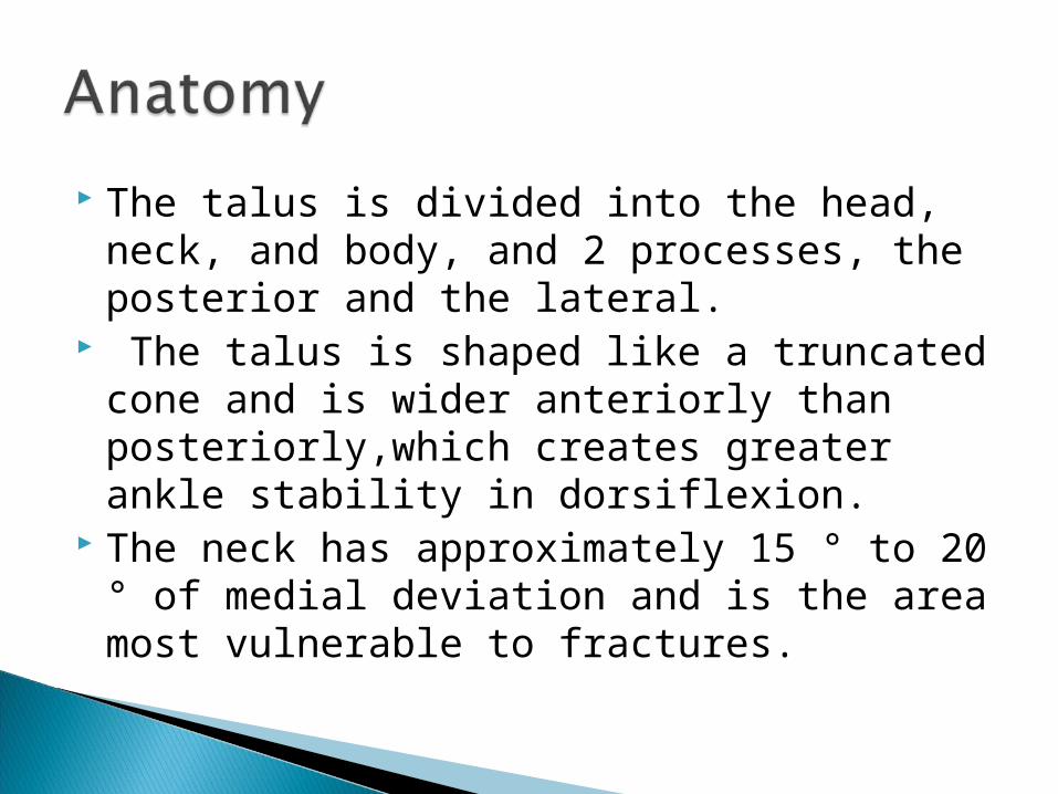
The talus is divided into the head, neck, and body, and 2 processes, the posterior and the lateral.
The talus is shaped like a truncated cone and is wider anteriorly than posteriorly,which creates greater ankle stability in dorsiflexion.
The neck has approximately 15 ° to 20 ° of medial deviation and is the area most vulnerable to fractures.

There are no musculotendinous attachments to the talus,the tendon of the flexor hallucis longus slides within the groove formed by the medial and lateral tubercles of the posterior process.
Nearly 70% of the entire surface area is articular surface, which gives the talus a total of 7 articular surfaces.
Because of the large amount of articular surface and the lack of any musculotendinous attachments, the talus is left with a tenuous blood supply.


Posterior tibial
Anterior tibial
Peroneal

calcaneal branches supplying the posterior process
The artery to the tarsal canal supplying the head and neck and
The deltoid branch supplying the neck and body
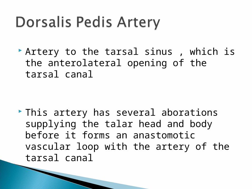
Artery to the tarsal sinus , which is the anterolateral opening of the tarsal canal
This artery has several aborations supplying the talar head and body before it forms an anastomotic vascular loop with the artery of the tarsal canal
Several small branches joining the calcaneal branches.
It also contributes to the anastomotic plexus of the tarsal sinus by way of the penetrating peroneal artery


Within the tarsal canal and the tarsal sinus the critical anastomosis perforates the inferior neck to form primary source of blood supply to the body of the talus.
In talar neck fractures with increasing displacement, the branches from the dorsalis pedis artery as well as artery of the tarsal canal and artery of the tarsal sinus can be disrupted.

So the rate of osteonecrosis depends upon the degree of fracture displacement.
Talar Neck fractures-Hyper dorsiflexion The neck of the talus impacts against the
leading edge of distal tibia. Fracture of talar body-Axial Load Lateral and posteromedial processes-Low
energy injuries. Can result from inversion and eversion
mechanism.


A high degree of suspicion is required for detection of the talar process fractures. These injuries can be difficult to appreciate on routine radiographs.
In high energy talar fractures significant compromise of the soft tissue envelop is common.

Association of dislocation with talar neck fracture is common.
Emergency reduction of the dislocated talus is one of the key principles in the management of the fractures of the talus.
An accurate assessment of the vascular and neurologic status of the foot is important.

Plain Radiographic views- Standard Ap,Lateral and Mortise views. Canale and Kelly view of the foot- A view of the talar neck achieved by placing
the foot plantigrade on the x ray film and angling the beam at 75degrees top the perpendicular.
Pronation and internal rotation of the foot helps to visualise medial aspect of the neck.


CT scans provoid better information than plain radiographs.
MRI is useful for diagnosis of osteonecrosis

Emergency reduction of the dislocated joints,
urgent anatomic fracture reduction and stabilization, maintaining an intact vascular supply and soft tissue envelope provide the best probability of regaining an excellent functional result.

Hawkins Classification-
Type I-Vertical Fractures without displacement.
Type II-Displaced fractures with subluxation/dislocation of the subtalar joint
Type III-displacement of body from both subtalar and ankle joint.
Type IV-Similar to type III with dislocation of talar head from the talonavicular joint.


Non operative Management- Only Type I fractures can be treated
nonoperatively. Conformation of the anatomically
maintained reduction should be done with CT scan.
Non weight bearing in short leg cast for 8-12 weeks.

Type II fractures-To flex the knee followed by plantar flexion of the foot.
Type III fractures-Plantar flexion and varus positioning of foot
Transverse steinman pin may be required to be passed through the calcaneus.
Direct pressure on the talar fragment may be required to reduced fragments.

Multiple attempts at closed reduction can increase the risk of complications
Residual displacement of as little as 2 mm alter the contact characteristics of the sub talar joint.
Displacement of the fragments can cause skin tenting and necrosis.
Anteromedial Approach
Lateral Approach
Posterior Approach
Combined Approaches

Most commonly used approach. Fascilitate medial malleolar osteotomy if
required. Medial malleolar osteotomy preserves the
deltoid ligament and thereby protect the blood supply.


This may lessen the chance of damage to the blood supply.
Howerver exposure of the lateral surface of the talus and sub talar joint requires extra caution to avoid injury to blood vessel of the sinus tarsi.
Facilitates visualization of subtalar joint. Facilitates placement of shoulder screw.


The screws directed from posterior to anterior may facilitate placement of screws perpendicular to the fracture line and achieves compression lag screw.
The risk of neurovascular compromise present.

Used when severe comminution present.
Caution to be taken to protect the tenuous blood supply to the talar body.

Anterior to posterior screw fixation
Posterior to anterior screw fixation
Direct plate fixation

Advantages
Direct visualisation of the fracture reduction
Avoidance of cartilage damage
Disadvantages
Difficult to insert perpendicular to fracture
Less strong as compared to posterior to anterior

Advantages Stronger than
anterior screw fixaion
Easley inserted perpendicular to fracture site
Disadantages Indirect visualisation
of the fracture Risk of iatrogenic
nerve damage.

Advantages Strong fixation Useful to buttress
comminuted fragments
Disadvantages
Extensive soft tissue dissection
Risk of hardware prominence

Can be used when closed reduction is successful
Percutaneuos fixation can be used to fascitate early ROM.

Infection and skin necrosis
Osteonecrosis
Malunion
Post- traumatic arthritis

Treatment is extremely challenging
Avascular body of the talus acts as large sequestrum.
Surgical debridement including talectomy may be required as treatment

Type I- 0-13% Type II- 20-50% Type III- 80%
Hawkin’s sign-Subchondral resorption of bone indication vascularity of the talar body.
Bone scan and MRI-3 weeks after the injury.

Patellar tendon bearing orthosis Primary triple arthrodeisis Total talectomy with tibia and calcaneal
fusion Subtalar fusion Plantar fusion. Blair fusion.


Dorsal displacement of the distal fragment and varus malunion are common.
Results in limitation of the dorsiflexion and painful gait.
46-69% Subtalar joint most commonly involved. Due to osteonecrosis, cartilage damage,
immobilisation and malalingement.
Treatment –Local analgesic infiltration, Arthrodesis of the involved
joints.

Sneppen classification-
Type I-Transcondral fractures Type II-sagittal,coronal and horizontal
shearing fractures Type III-Fractures of the posterior tubercle. Type IV-Fractures of the lateral process Type V-Crush fractures

ORIF with cortical screws. Excision of the small fragments. In highly selective cases primary artrodesis
can be done.
Weigh bearing is started after union of the fracture.

By axial load applied to the talar head through navicular bone
Principle of the treatment include maintenance of the alingemnt of the dorsomedial arch of the foot
Preventing talonavicular joint incongruity and instability.

Displaced fractures may be treated with ORIF with minifragment screws.
Talonavicular arthritis is a common complication which is treated with longitudinal arch support with increased arch rigidity.

Hawkin’s classification-
Nonarticular chip ractures Single large fragment involving the
talofibular and subtalar articulations Communited fracture involving both
articulations.

Undisplaced and reduced fractures are treated with short leg cast immobilisation for 4-6 weeks.
Dispalced fractures with large fragments are treated with ORIF with screw fixation.

This fracture is associated with subtalar dislocations
Excision of the ununited or mal united fragments seems to relive local irritation symptoms
Related Documents