* † * †

Welcome message from author
This document is posted to help you gain knowledge. Please leave a comment to let me know what you think about it! Share it to your friends and learn new things together.
Transcript
Fractal analysis of resting state functional
connectivity of the brain
Wonsang You∗1, Sophie Achard†2, Jörg Stadler1, Bernd Brückner1 and Udo Sei�ert3
1Leibniz Institute for Neurobiology, Magdeburg, Germany2GIPSA-lab, CNRS, UMR 5216, Grenoble, France
3Biosystems Engineering, Fraunhofer-Institute for Factory Operation andAutomation, Magdeburg, Germany
August 4, 2012
Abstract
A variety of resting state neuroimaging data tend to exhibit fractalbehavior where their power spectrums follow power-law scaling. Restingstate functional connectivity is signi�cantly in�uenced by fractal behav-ior which may not directly originate from neuronal population activitiesof the brain. To describe the fractal behavior, we adopted the fraction-ally integrated process (FIP) model instead of the fractional Gaussiannoise (FGN) since the FIP model covers more general aspects of fractal-ity than the FGN model. This model provides a theoretical basis for thedependence of resting state functional connectivity on fractal behavior.Inspired by this idea, we introduce a novel concept called the nonfractal
connectivity which is de�ned as the correlation of short memory indepen-dent of fractal behavior, and compared it with the fractal connectivitywhich is an asymptotic wavelet correlation. We propose several wavelet-based estimators of fractal connectivity and nonfractal connectivity for amultivariate fractionally integrated noise (mFIN). These estimators wereevaluated through simulation studies and applied to the analyses of restingstate fMRI data of the rat brain.
1 Introduction
The dynamics of endogenous neuronal activities has been an important issuein neuroscience since it is supposed to take control of most neuronal activitiesarising in the brain [1]. The huge default-mode functional network of the brainhas been usually investigated through resting state neuroimaging data suchas electroencephalography (EEG) and functional magnetic resonance imaging
∗[email protected]†[email protected]
1
(fMRI) [2�5]. One of the major goals in resting state neuroimaging research isthe reliable inference of functional dynamics of spontaneous neuronal populationactivities from resting state neuroimaging data. However, it is not straightfor-ward since resting state signals may be signi�cantly a�ected by non-neuronalphysiological factors. On the other hand, the response to stimulation in task-based experimental paradigm is prominently correlated with brain activitieseither directly or indirectly.
One of the non-neuronal obstacles in resting state neuroimaging studies isthe fractal behavior (or long-range dependence) where the power spectrum tendsto exhibit 1/fα power law scaling across low frequencies. This phenomenon hasbeen observed through a number of resting state neuroimaging studies [6�9].As such a phenomenon has been ubiquitously observed in nature, the fractalbehavior in neuroimaging data may also arise from various mediators such asrespiration [10, 11], cardiac �uctuations [12], system noise, hemodynamics (inthe case of fMRI) as well as neuronal activities [13�15].
The classical model of fractal behavior or long memory in baseline neu-roimaging signals has been the fractional Gaussian noise (FGN) which is de�nedas an increment process of fractional Brownian motion (FBM) and completelycharacterized by both Hurst exponent and variance [16]. The FGN model hasbeen adopted to various fractal-based analyses of fMRI data for a decade so asto account for scale-free dynamics of neuroimaging signals [17�21].
However, there is a controversy about whether the FGN is the most ap-propriate model for resting state neuroimaging signals among a variety of longmemory models. While the FGN model is de�ned just with two parametersunder mathematically strict conditions of self-similarity, a neuroimaging signalis produced from a nonlinear biological system which is controlled by numer-ous hidden parameters. In this reason, the fractionally integrated process (FIP)model, based on the concept of fractional di�erencing [22], is worth considera-tion as an alternative to the FGN model since it embraces diverse types of longmemory. Indeed, the FGN is regarded as a special type of the FIP model whichis more extensive than FGN.
In this paper, we adopted the fractionally integrated process (FIP) modelto e�ectively describe the fractal behavior of neuroimaging signals. In the FIPmodel, a neuroimaging signal is represented as the output of a long memory(LM) �lter whose input is a nonfractal signal (sometimes called short memory
as a notion corresponding to long memory). In other words, a nonfractal signalis transformed into a neuroimaging signal with fractal behavior through longmemory �ltering as shown in Fig. 1, which indicates that the fractal behavioris attributed not to the nonfractal input but to the LM �lter. The in�uenceof several factors on the fractal behavior can be well aggregated in terms of asequence of long memory �lters.
The FIP model sheds light on the in�uence of fractal behavior on functionalconnectivity. The correlation of resting state neuroimaging signals betweentwo brain regions may signi�cantly di�er from that of the nonfractal inputsignals according to the di�erence of memory parameters. Hence, the ordinarycorrelation of resting state neuroimaging data may not well re�ect the functional
2
Figure 1: The FIP model representation of resting state neuroimaing signals.
dynamics of spontaneous neuronal activities due to the fractal behavior.The theoretical expectation that functional connectivity may be in�uenced
by fractal behavior leads us to take into account the correlation of fractal-freeinput signals as a novel concept of resting state functional connectivity while thePearson correlation of neuroimaging signals has been the most popular de�ni-tion of functional connectivity. This particular correlation, which is independentof fractal behavior, is called the nonfractal connectivity. Its mathematical de-scription is provided in section 3. The nonfractal connectivity is not exactlyidentical to the correlation of spontaneous neuronal population activities due tothe nonlinearity of neurophysiological systems. However, it may give us betterinformation on correlation structure of spontaneous neuronal populations thanordinary correlation of neuroimaging data since it eliminates the distortion offunctional connectivity due to fractal behavior.
The nonfractal connectivity is comparable to fractal connectivity which was�rst proposed in [23] as the asymptotic value of wavelet correlations over lowfrequency scales. The wavelet correlations of two long memory processes areconverged on a speci�c value which is determined by the correlation of shortmemory parts as well as memory parameters. We show the theoretical relevanceof nonfractal connectivity to fractal connectivity.
In this paper, we propose three wavelet-based approaches to estimating bothnonfractal connectivity and fractal connectivity: (1) the SDF(Spectral densityfunction)-based method, (2) the covariance-based method, and (3) the linearity-based method. As prerequisite to estimating these connectivities, memory pa-rameters should be estimated a priori. We tested two wavelet-based univariateestimators of memory parameter: the wavelet-based least-mean-squares (LMS)method and the wavelet-based maximum likelihood (ML) method. The perfor-mance of all proposed estimators was veri�ed through simulation studies. Wealso show an example of applying these estimators to resting state fMRI datataken from anesthetized rat brains to estimate nonfractal connectivity.
This paper is organized as follows. In section 2, the FIP model of restingstate neuroimaging signals is brie�y introduced, and the concepts of nonfractalconnectivity and fractal connectivity are described in section 3. The wavelet-based estimators of both connectivities are proposed in section 4. The resultsof simulation studies and experiments in fMRI data are provided in section 5and 6.
3
2 Long memory model
Fractal properties of a time series can be modeled as long memory models such asFGN, ARFIMA, and GARCH processes. In this section, we especially introduceboth univariate and multivariate fractionally integrated processes (FIP) thatencompass several classes of long memory such as fractionally integrated noise(FIN), FGN, and ARFIMA [22,24].
2.1 Univariate case
Let x(t) be a real-valued discrete process of length N given by
u(t) = (1− L)dx(t) (1)
where d ∈ R, L denotes the back-shift operator, and u(t) (called short memory)is a stationary process whose spectral density Su (f) is a non-negative symmetricfunction bounded on (−π, π) and bounded away from zero at the origin. x(t)can be represented as the convolution of u(t) with the long memory (LM) �lterg(t) as follows
x(t) =
∞∑τ=0
g(τ)u(t− τ) (2)
where
g(t) :=dΓ(d+ t)
Γ(d+ 1)Γ(t+ 1). (3)
If −1/2 < d < 1/2, the spectral density of x(t) is given by
S(f) =∣∣1− e−if ∣∣−2d
Su(f). (4)
The fractal behavior is controlled by the memory parameter d. If 0 < d < 1/2,the process x(t) is said to be a stationary long memory process with memoryparameter d while x(t) is nonstationary if d > 0.5. If d = 0, the process becomesa white noise.
2.2 Multivariate case
The de�nition of the univariate long memory model can be extended to themultivariate case. Consider a real-valued q-vector process X(t) given by (1− L)d1 0
. . .
0 (1− L)dq
X1(t)
...Xq(t)
=
U1(t)...
Uq(t)
, (5)
where U(t) = (U1(t), ..., Uq(t)) is a multivariate stationary process whose spec-tral density S(f) = [Sm,n(f)] is bounded on (−π, π) and bounded away fromzero at the origin. For −1/2 < dk < 1/2, the spectral density of U is given by
S (f) = Φ (f) Su (f) Φ∗ (f) (6)
4
where
Φ (f) =
(1− eif )−d1 0. . .
0 (1− eif )−dq
. (7)
In the case of 0 < dk < 1/2 for 1 ≤ k ≤ q for 1 ≤ k ≤ q, X(t) is said to bea stationary long memory process with memory parameter d = (d1, · · · , dq). IfU(t) is a vector ARMA process, X(t) becomes a multivariate ARFIMA process.On the other hand, if U(t) is a vector i.i.d. random variable, i.e.
U(t)i.i.d.∼ N (0,Σu) , (8)
X(t) becomes a multivariate fractionally integrated noise (mFIN). In this case,the cross-spectral density of xm(t) and xn(t) is given by
Sm,n (f) = γm,n(1− eif
)−dm (1− e−if
)−dn(9)
where γm,n is identical to the (m,n)-th element of Σu.
3 Nonfractal and fractal connectivity
As discussed in the section 1, the most popular de�nition of functional connec-tivity has been the Pearson correlation. The multivariate long memory modelintroduced in the section 2 additionally provides two novel de�nitions of restingstate functional connectivity: fractal connectivity and nonfractal connectivity.While fractal connectivity is de�ned based on the asymptotics of wavelet cor-relation, nonfractal connectivity is de�ned based on the covariance of shortmemory.
3.1 Nonfractal connectivity
Let X(t) be an mFIN process with memory parameter d, and U(t) be a shortmemory function of X(t) given in (5). The nonfractal connectivity of xm(t) andxn(t) is de�ned as
Dm,n =γm,n√γm,mγn,n
(10)
where γm,n denotes the covariance of um(t) and un(t); that is, γm,n := E [um(1)un(1)].
3.2 Fractal connectivity
The variance of a discrete time series can be decomposed over several fre-quency bands (called scales) through the discrete wavelet transform (DWT).LetWi(j, k) be the wavelet coe�cient of the ith process xi(t) at scale j and timepoint k. The wavelet covariance is de�ned as νm,n(j) := cor (Wm(j, k),Wn(j, k))
5
at scale j. Since the wavelet coe�cients of an FIP at a scale j is covariance sta-tionary, νm,n(j) is independent of time t. LetHj(f) be the squared gain functionof the wavelet �lter such that
Hj(f) ≈{
2j for 2π/2j+1 ≤ |f | ≤ 2π/2j
0 otherwise. (11)
Then, the wavelet covariance of xm(t) and xn(t) at scale j is related to thecross-spectral density [25] as follows
νm,n(j) = 2π
∫ π
−πHj(f)SX(f)df. (12)
The wavelet correlation ρm,n(j) := cor (Wm(j, k),Wn(j, k)) is given by
ρm,n(j) =νm,n(j)√νm(j)νn(j)
. (13)
Theorem 1 (Asymptotic wavelet covariance). Suppose that X(t) is a multivari-ate FIN process which satis�es the condition (8). Then, the wavelet covarianceof xm(t) and xn(t) at scale j is approximated by
νm,n(j) ≈ γm,nβm,n2j(dm+dn) as j →∞ (14)
where
βm,n := 2 cos(π
2(dm − dn)
) 1− 2dm+dn−1
1− dm − dn(2π)−dm−dn . (15)
Proof. It is well known the following Taylor series
sin−dm−dn (f/2) ≈
(f/2)−dm−dn +
dm + dn6
(f
2
)2−(dm+dn)
. (16)
From (9) and (16),
Sm,n (f) = γm,n2−dm−dn
((f
2
)−dm−dn+
dm + dn6
(f
2
)2−(dm+dn)). (17)
Substituting Sm,n (f) in (12) with (17), we �nally get (14).
The asymptotic property of wavelet correlation 1 was also proved for moregeneral cases of short memory in [23]. From (13) and (14), the wavelet correla-tion of xm(t) and xn(t) asymptotically converges to
ρm,n(j)→ ρ∞m,n := Dm,nΥ(dm, dn) as j →∞ (18)
6
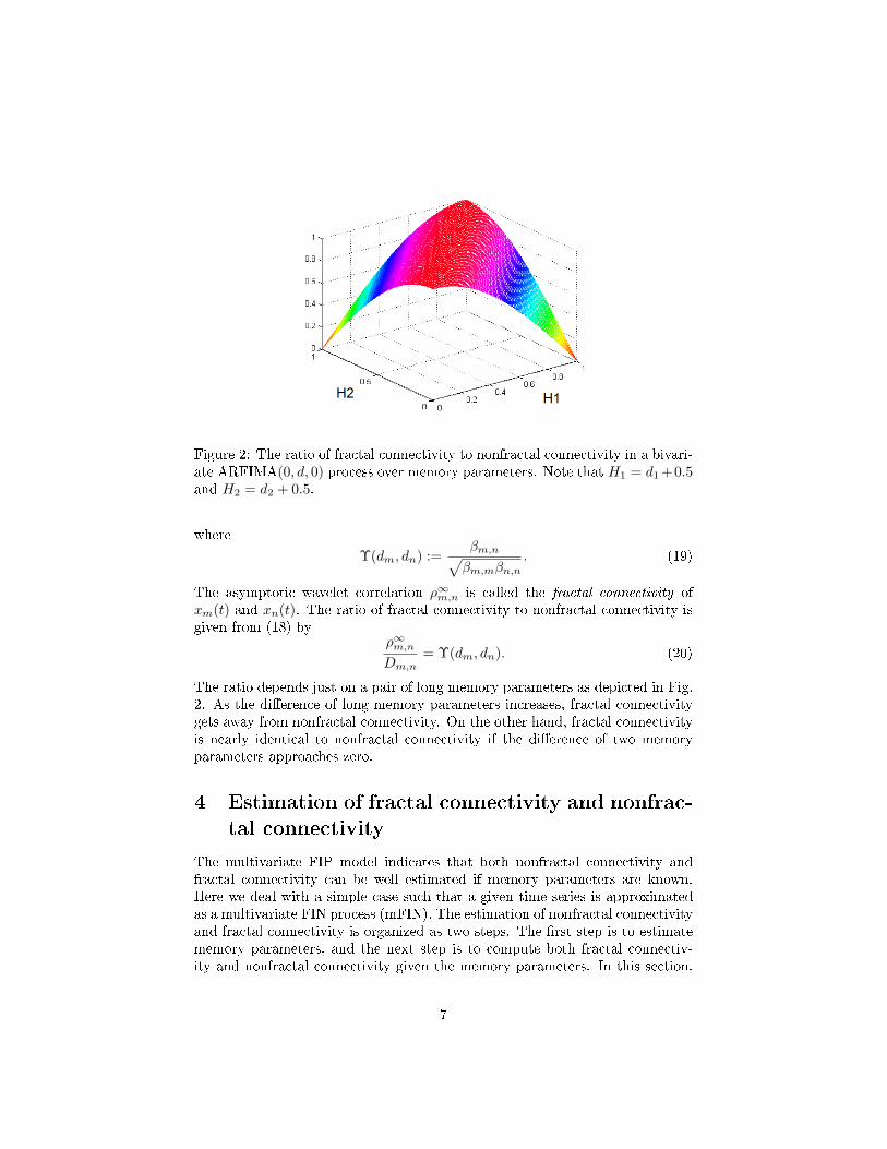
Figure 2: The ratio of fractal connectivity to nonfractal connectivity in a bivari-ate ARFIMA(0, d, 0) process over memory parameters. Note that H1 = d1 +0.5and H2 = d2 + 0.5.
where
Υ(dm, dn) :=βm,n√βm,mβn,n
. (19)
The asymptotic wavelet correlation ρ∞m,n is called the fractal connectivity ofxm(t) and xn(t). The ratio of fractal connectivity to nonfractal connectivity isgiven from (18) by
ρ∞m,nDm,n
= Υ(dm, dn). (20)
The ratio depends just on a pair of long memory parameters as depicted in Fig.2. As the di�erence of long memory parameters increases, fractal connectivitygets away from nonfractal connectivity. On the other hand, fractal connectivityis nearly identical to nonfractal connectivity if the di�erence of two memoryparameters approaches zero.
4 Estimation of fractal connectivity and nonfrac-
tal connectivity
The multivariate FIP model indicates that both nonfractal connectivity andfractal connectivity can be well estimated if memory parameters are known.Here we deal with a simple case such that a given time series is approximatedas a multivariate FIN process (mFIN). The estimation of nonfractal connectivityand fractal connectivity is organized as two steps. The �rst step is to estimatememory parameters, and the next step is to compute both fractal connectiv-ity and nonfractal connectivity given the memory parameters. In this section,
7
several wavelet-based techniques which can be exploited in each step are in-troduced. All of these methods are based on the wavelet transform which areoptimal to investigate the properties of long memory processes.
4.1 Estimation of memory parameters
4.1.1 Wavelet least-mean-squares method (LMS)
By taking logarithm to (14), we obtain the following linear relationship ofwavelet variance in the logarithm scale:
log2 [νm(j)] ≈ 2dmj + cm. (21)
It enables us to estimate the memory parameters dm by linear regression overa given scale interval J = jlow → jhigh on the basis of the biased estimator ofwavelet variance given by
νm(j) =1
nj2j
nj∑t=1
W 2m(j, t) (22)
where nj is the number of coe�cients in scale j except boundary coe�cients [25].In a similar manner with [23], the optimal scale interval Jopt for linear regressionin (21) is globally determined by
Jopt = argminJ⊂J
σ2LS(J ) (23)
where J is the space of all scale intervals, ∆J = jhigh − jlow + 1, and
σ2LS(J ) =
1
∆J
jhigh∑j=jlow
q∑m=1
{log2 [νm]− 2dmj − cm
}2
. (24)
4.1.2 Wavelet maximum-likelihood method (ML)
The likelihood function for memory parameter dm and asymptotic variance Gmis given by
L(dm, γm |xm(t)
):=
1
(2π)N/2 |Σm|1/2e−x
T Σ−1m x/2. (25)
The matrix Σm denotes the covariance matrix of xm(t), and can be replaced
by Σm := WTΛW where W is a wavelet transform matrix and Λ is a diagonalmatrix which has diagonal elements Λm(j) := νm(j) given in (14) for j =1, ..., J as an average value of spectral density function (SDF) over the band[1/2j+1, 1/2j
]. The reduced log likelihood function can be obtained on the
8
basis of the Brockwell and Davis's method [26]:
l(dm, γm |xm(t)
)(26)
= −2 logL(dm, γm |xm(t)
)−N log(2π)−N
= N log (γm,m) + log(Λm(J + 1)) +
J∑j=1
Nj log (Λm(j))
with N = 2J , Nj = N/2j , and
γm,n :=1
N
(V TmVn
Λm,n(J + 1)+
J∑j=1
1
Λm,n(j)
Nj−1∑t=0
Wm(j, t)Wn(j, t)
(27)
where Vm are the scaling coe�cients at scale J and Wj,t is the t-th element
of j-th level wavelet coe�cients. The optimal memory parameter dm can beestimated by minimizing (26) with respect to dm [25].
4.2 Estimation of short-memory covariance
4.2.1 The SDF-based method (SDF)
The estimator γm,n of short memory covariance can be semiparametrically com-puted by (27). Since Vm ≈ 0 in stationary long memory processes, the equationcan be approximated as
γm,n ≈1
N
J∑j=1
1
Λm,n(j)
Nj−1∑t=0
Wm(j, t)Wn(j, t). (28)
4.2.2 The covariance-based method (COV)
Alternatively, the short memory covariance can be estimated by exploiting theproperties such that the sum of wavelet covariances over all scales is identicalto the covariance of a time series; i.e.,
σ2m,n =
cov (Vm, Vn)
N+
J∑j=1
cov (Wm(j, t),Wn(j, t))
2j. (29)
Since cov (Vm, Vn) ≈ 0 for a FIN process, the estimator of short memory covari-ance can be obtained from (14) and (29) as follows
γm,n =σ2m,n
2Bm,n∑Jj=1 2(dm+dn−1)j
(2π)dm+dn . (30)
9

4.2.3 The linearity-based method (LIN)
The estimator of short memory covariance can be also obtained in the otherway based on the linearity of wavelet covariance over scales as follows.
γm,n =2cm,n−1
Bm,n cos(π2 (dm − dn)
) (2π)dm+dn (31)
where
cm,n =1
J
J∑j=1
[log2 νm,n(j)− (dm + dn)j] , (32)
Bm,n :=1− 2dm+dn−1
1− dm − dn. (33)
4.3 Estimation of fractal and nonfractal connectivity
After the estimators for memory parameters d and the short memory covarianceΓ are obtained, the nonfractal connectivity Dm,n can be estimated by using (10)as follows
Dm,n =γm,n√γm,mγn,n
. (34)
Likewise, fractal connectivity ρ∞m,n can be estimated from (18) and (34) as fol-lows
ρ∞m,n := Dm,nΥ(dm, dn). (35)
5 Simulation study
In this section, the performance of three wavelet-based estimators for nonfractalconnectivity was evaluated. We also analyzed the in�uence of short memorycondition, dimension, and length of time series on the estimation of nonfractalconnectivity. By combining a connectivity estimator with a memory parameterestimator, six pairs of estimator, such as LMS-LIN, LMS-COV, LMS-SDF, ML-LIN, ML-COV, ML-SDF methods, were �nally tested.
5.1 Setup
We simulated multivariate ARFIMA(p,d, 0) processes that belong to the FIPmodel. First, the short memory U(t) in (5) was given as an ARMA(p, 0) processas follows
U(t) = Φ−1p (L)Aε(t). (36)
In (36), εi(t) for i = 1, · · · , q is an i.i.d. random variable where
cov(εm(t), εn(t)) =
{1 if m = n
0 if m 6= n,(37)
10

Φp(L) =
∑pi=1 ϕ1,iL
i 0. . .
0∑pi=1 ϕq,iL
i
, (38)
and
A =
1 0 · · · · · · 00 1 a · · · a... a
. . ....
...... 1 a
0 a · · · a 1
. (39)
If we set
a =1±√
1− bρb
(40)
where b = ρ(q−2)−(q−3), the short memory correlation is forced to beDm,n = ρfor m,n > 1 and m 6= n. Afterwards, the memory parameters d were equallydistributed over d ∈ (−1/2, 1/2), and the multivariate ARMA(p, 0) process was�ltered by the LM �lter de�ned in (3).
5.2 E�ects of short memory condition
To study the e�ects of short memory conditions on the performance of esti-mators, we performed Monte Carlo simulations with 100 repetitions of four-dimensional ARFIMA(p, d, 0) processes under four di�erent types of short mem-ory condition in (38) and (39):
(1A) A = I and ϕk,i = 0
(1B) A = I, ϕk,1 = 0.9 and ϕk,i = 0 for i > 1
(2A) A = A0 and ϕk,i = 0
(2B) A = A0, ϕk,1 = 0.9 and ϕk,i = 0 for i > 1
where d = {0.2, 0.4, 0.6, 0.8} and A0 was set with ρ = 0.3 in (40). In theconditions (1A) and (1B), each short memory process is statistically independentof each other while the conditions (2A) and (2B) let short memory processesbe cross-correlated. On the other hand, the conditions (1B) and (2B) let eachprocess be autocorrelated.
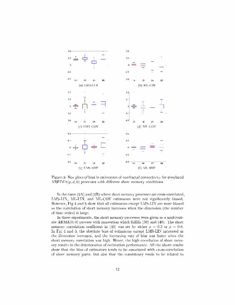
In Fig. 3, all methods were weak in the conditions (1B) and (2B) where shortmemory parts were more auto-correlated; the relative decrease in consistency fortwo cases was common to all six methods. The deteriorated performance in (1B)and (2B) is a foreseeable result since these short memory conditions no longerfollow the assumption of mFIN in (8) adopted for our proposed estimators.Hence, our proposed estimators were not e�cient when the set of short memorysignals cannot be approximated as a multivariate i.i.d. process.
11
(a) LMS-LIN (b) ML-LIN
(c) LMS-COV (d) ML-COV
(e) LMS-SDF (f) ML-SDF
Figure 3: Box plots of bias in estimation of nonfractal connectivity for simulatedARFIMA(p, d, 0) processes with di�erent short memory conditions.
In the cases (2A) and (2B) where short memory processes are cross-correlated,LMS-LIN, ML-LIN, and ML-COV estimators were not signi�cantly biased.However, Fig 4 and 5 show that all estimators except LMS-LIN are more biasedas the correlation of short memory increases when the dimension (the numberof time series) is large.
In these experiments, the short memory processes were given as a multivari-ate ARMA(0, 0) process with innovation which ful�lls (39) and (40). The shortmemory correlation coe�cient in (40) was set by either ρ = 0.2 or ρ = 0.8.In Fig 4 and 5, the absolute bias of estimators except LMS-LIN increased asthe dimension increases, and the increasing rate of bias was faster when theshort memory correlation was high. Hence, the high correlation of short mem-ory results in the deterioration of estimation performance. All the above resultsshow that the bias of estimators tends to be associated with cross-correlationof short memory parts, but also that the consistency tends to be related to
12
(a) LMS-LIN (b) ML-LIN
(c) LMS-COV (d) ML-COV
(e) LMS-SDF (f) ML-SDF
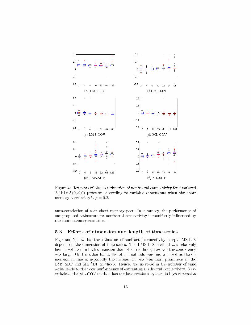
Figure 4: Box plots of bias in estimation of nonfractal connectivity for simulatedARFIMA(0, d, 0) processes according to variable dimensions when the shortmemory correlation is ρ = 0.2.
auto-correlation of each short memory part. In summary, the performance ofour proposed estimators for nonfractal connectivity is manifestly in�uenced bythe short memory conditions.
5.3 E�ects of dimension and length of time series
Fig 4 and 5 show that the estimators of nonfractal connectivity except LMS-LINdepend on the dimension of time series. The LMS-LIN method was relativelyless biased even in high dimension than other methods, however the consistencywas large. On the other hand, the other methods were more biased as the di-mension increases; especially the increase in bias was more prominent in theLMS-SDF and ML-SDF methods. Hence, the increase in the number of timeseries leads to the poor performance of estimating nonfractal connectivity. Nev-ertheless, the ML-COV method has the best consistency even in high dimension
13
(a) LMS-LIN (b) ML-LIN
(c) LMS-COV (d) ML-COV
(e) LMS-SDF (f) ML-SDF
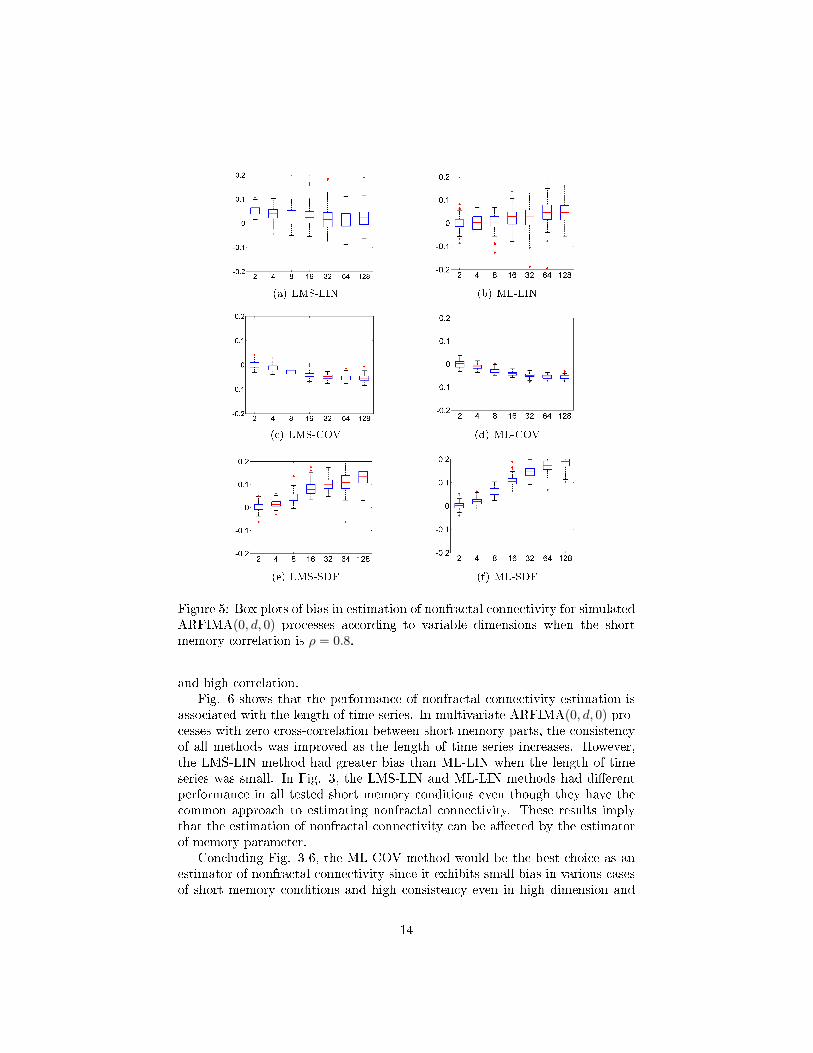
Figure 5: Box plots of bias in estimation of nonfractal connectivity for simulatedARFIMA(0, d, 0) processes according to variable dimensions when the shortmemory correlation is ρ = 0.8.
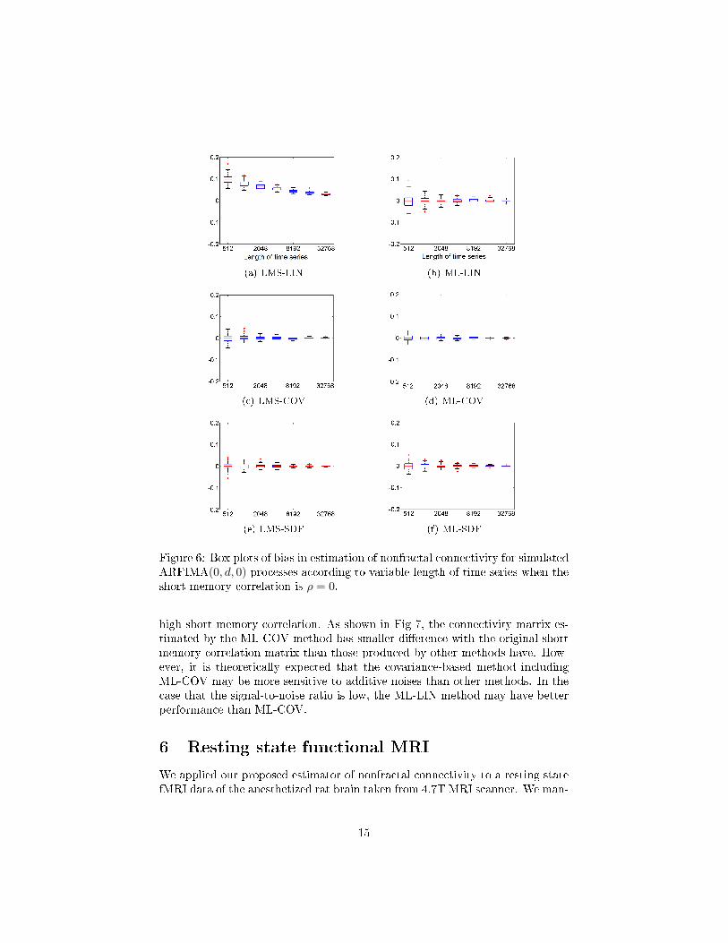
and high correlation.Fig. 6 shows that the performance of nonfractal connectivity estimation is
associated with the length of time series. In multivariate ARFIMA(0, d, 0) pro-cesses with zero cross-correlation between short memory parts, the consistencyof all methods was improved as the length of time series increases. However,the LMS-LIN method had greater bias than ML-LIN when the length of timeseries was small. In Fig. 3, the LMS-LIN and ML-LIN methods had di�erentperformance in all tested short memory conditions even though they have thecommon approach to estimating nonfractal connectivity. These results implythat the estimation of nonfractal connectivity can be a�ected by the estimatorof memory parameter.
Concluding Fig. 3-6, the ML-COV method would be the best choice as anestimator of nonfractal connectivity since it exhibits small bias in various casesof short memory conditions and high consistency even in high dimension and
14
(a) LMS-LIN (b) ML-LIN
(c) LMS-COV (d) ML-COV
(e) LMS-SDF (f) ML-SDF
Figure 6: Box plots of bias in estimation of nonfractal connectivity for simulatedARFIMA(0, d, 0) processes according to variable length of time series when theshort memory correlation is ρ = 0.
high short memory correlation. As shown in Fig 7, the connectivity matrix es-timated by the ML-COV method has smaller di�erence with the original shortmemory correlation matrix than those produced by other methods have. How-ever, it is theoretically expected that the covariance-based method includingML-COV may be more sensitive to additive noises than other methods. In thecase that the signal-to-noise ratio is low, the ML-LIN method may have betterperformance than ML-COV.
6 Resting state functional MRI
We applied our proposed estimator of nonfractal connectivity to a resting statefMRI data of the anesthetized rat brain taken from 4.7T MRI scanner. We man-
15
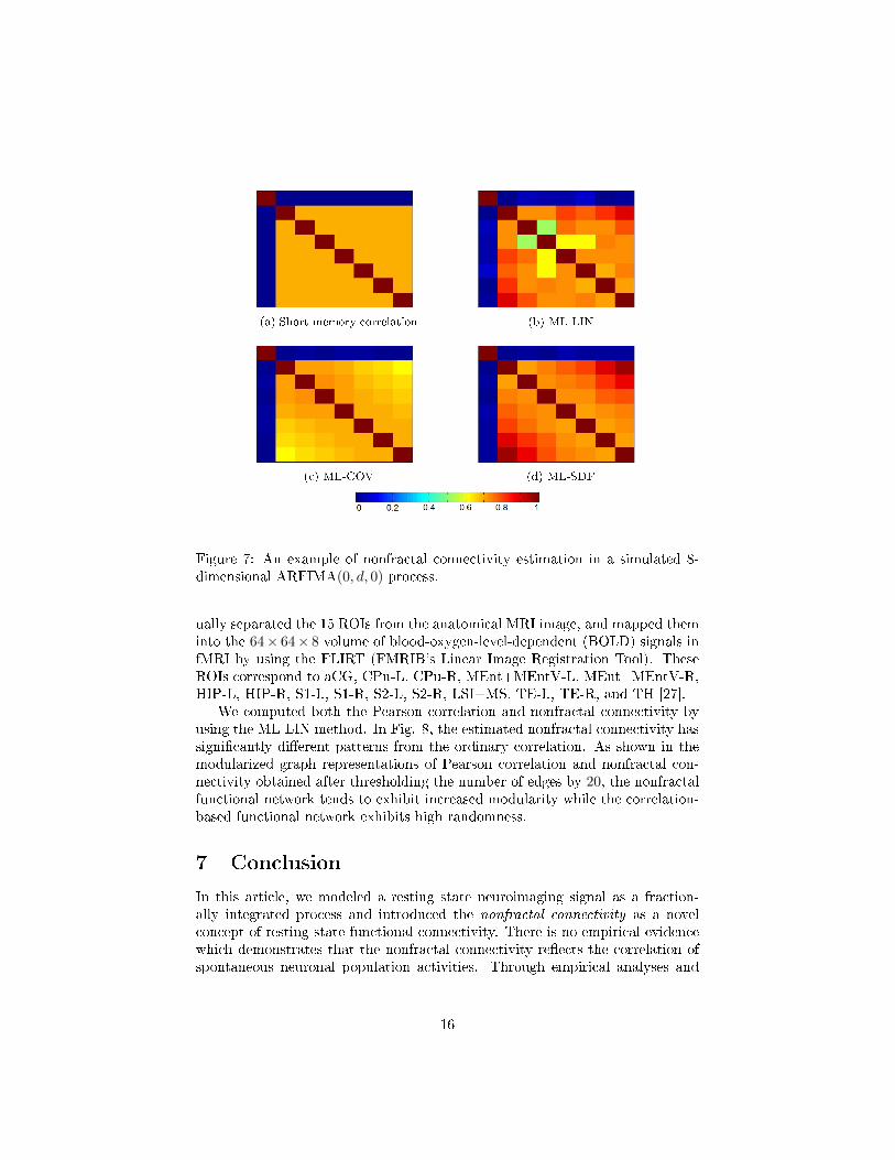
(a) Short memory correlation (b) ML-LIN
(c) ML-COV (d) ML-SDF
Figure 7: An example of nonfractal connectivity estimation in a simulated 8-dimensional ARFIMA(0, d, 0) process.
ually separated the 15 ROIs from the anatomical MRI image, and mapped theminto the 64× 64× 8 volume of blood-oxygen-level-dependent (BOLD) signals infMRI by using the FLIRT (FMRIB's Linear Image Registration Tool). TheseROIs correspond to aCG, CPu-L, CPu-R, MEnt+MEntV-L, MEnt+MEntV-R,HIP-L, HIP-R, S1-L, S1-R, S2-L, S2-R, LSI+MS, TE-L, TE-R, and TH [27].
We computed both the Pearson correlation and nonfractal connectivity byusing the ML-LIN method. In Fig. 8, the estimated nonfractal connectivity hassigni�cantly di�erent patterns from the ordinary correlation. As shown in themodularized graph representations of Pearson correlation and nonfractal con-nectivity obtained after thresholding the number of edges by 20, the nonfractalfunctional network tends to exhibit increased modularity while the correlation-based functional network exhibits high randomness.
7 Conclusion
In this article, we modeled a resting state neuroimaging signal as a fraction-ally integrated process and introduced the nonfractal connectivity as a novelconcept of resting state functional connectivity. There is no empirical evidencewhich demonstrates that the nonfractal connectivity re�ects the correlation ofspontaneous neuronal population activities. Through empirical analyses and
16
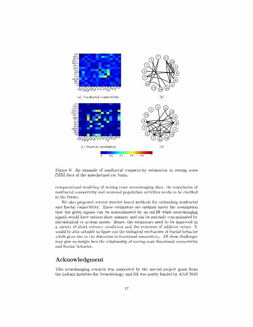
(a) Nonfractal connectivity (b)
(c) Pearson correlation (d)
Figure 8: An example of nonfractal connectivity estimation in resting statefMRI data of the anesthetized rat brain.
computational modeling of resting state neuroimaging data, the association ofnonfractal connectivity and neuronal population activities needs to be clari�edin the future.
We also proposed several wavelet-based methods for estimating nonfractaland fractal connectivity. These estimators are optimal under the assumptionthat the given signals can be approximated by an mFIN while neuroimagingsignals would have various short memory and can be seriously contaminated byphysiological or system noises. Hence, the estimators need to be improved ina variety of short memory conditions and the existence of additive noises. Itwould be also valuable to �gure out the biological mechanism of fractal behaviorwhich gives rise to the distortion in functional connectivity. All these challengesmay give us insight into the relationship of resting state functional connectivityand fractal behavior.
Acknowledgment
This neuroimaging research was supported by the special project grant fromthe Leibniz Institute for Neurobiology, and SA was partly funded by ANR 2010
17
JCJC 0302 01. We thank Frank Angenstein for fMRI data.
References
[1] Michael D Fox and Marcus E Raichle. Spontaneous �uctuations in brainactivity observed with functional magnetic resonance imaging. Nature re-
views. Neuroscience, 8(9):700�11, 2007.
[2] H Laufs, K Krakow, P Sterzer, E Eger, a Beyerle, a Salek-Haddadi, anda Kleinschmidt. Electroencephalographic signatures of attentional and cog-nitive default modes in spontaneous brain activity �uctuations at rest.Proceedings of the National Academy of Sciences of the United States of
America, 100(19):11053�8, September 2003.
[3] M De Luca, C F Beckmann, N De Stefano, P M Matthews, and S MSmith. fMRI resting state networks de�ne distinct modes of long-distanceinteractions in the human brain. NeuroImage, 29(4):1359�67, 2006.
[4] F Musso, J Brinkmeyer, a Mobascher, T Warbrick, and G Winterer. Spon-taneous brain activity and EEG microstates. A novel EEG/fMRI analysisapproach to explore resting-state networks. NeuroImage, 52(4):1149�61,October 2010.
[5] Gustavo Deco, Viktor K. Jirsa, and Anthony R. McIntosh. Emerging con-cepts for the dynamical organization of resting-state activity in the brain.Nature Reviews Neuroscience, 12(1):43�56, January 2011.
[6] E. Zarahn, GK Aguirre, and M. D'Esposito. Empirical analyses of BOLDfMRI statistics. NeuroImage, 5(3):179�197, 1997.
[7] C.J. Stam and E.A. de Bruin. Scale-free dynamics of global functionalconnectivity in the human brain. Human Brain Mapping, 22(2):97�109,June 2004.
[8] Dimitri Van de Ville, Juliane Britz, and Christoph M Michel. EEG mi-crostate sequences in healthy humans at rest reveal scale-free dynamics.Proceedings of the National Academy of Sciences of the United States of
America, 107(42):18179�84, October 2010.
[9] Paul Expert, Renaud Lambiotte, D.R. Chialvo, Kim Christensen, H.J.Jensen, D.J. Sharp, and Federico Turkheimer. Self-similar correlation func-tion in brain resting-state functional magnetic resonance imaging. Journalof The Royal Society Interface, 8(57):472, 2011.
[10] D Cordes, V.M. M Haughton, K Arfanakis, J.D. D Carew, P.A. a Turski,C.H. H Moritz, M.A. a Quigley, and M.E. E Meyerand. Frequencies con-tributing to functional connectivity in the cerebral cortex in" resting-state"data. American Journal of Neuroradiology, 22(7):1326, August 2001.
18
[11] Rasmus M Birn, Jason B Diamond, Monica A Smith, and Peter A Bandet-tini. Separating respiratory-variation-related �uctuations from neuronal-activity-related �uctuations in fMRI. NeuroImage, 31(4):1536�48, July2006.
[12] B West, R Zhang, A Sanders, S Miniyar, J Zuckerman, and B Levine.Fractal �uctuations in cardiac time series. Physica A: Statistical Mechanics
and its Applications, 270(3-4):552�566, August 1999.
[13] M C Teich, C Heneghan, S B Lowen, T Ozaki, and E Kaplan. Fractalcharacter of the neural spike train in the visual system of the cat. Journalof the Optical Society of America. A, Optics, image science, and vision,14(3):529�46, March 1997.
[14] Alberto Mazzoni, Frédéric D Broccard, Elizabeth Garcia-Perez, PaoloBonifazi, Maria Elisabetta Ruaro, and Vincent Torre. On the dynamicsof the spontaneous activity in neuronal networks. PloS one, 2(5):e439,January 2007.
[15] Paolo Allegrini, Danilo Menicucci, Remo Bedini, Leone Fronzoni, AngeloGemignani, Paolo Grigolini, Bruce J. West, and Paolo Paradisi. Spon-taneous brain activity as a source of ideal 1/f noise. Physical Review E,80(6):1�13, December 2009.
[16] Jan Beran. Statistics for long-memory processes. CRC Press, 1994.
[17] E Bullmore, C Long, J Suckling, J Fadili, G Calvert, F Zelaya, T a Carpen-ter, and M Brammer. Colored noise and computational inference in neuro-physiological (fMRI) time series analysis: resampling methods in time andwavelet domains. Human brain mapping, 12(2):61�78, February 2001.
[18] Ed Bullmore, Jalal Fadili, Voichita Maxim, Levent Sendur, BrandonWhitcher, John Suckling, Michael Brammer, and Michael Breakspear.Wavelets and functional magnetic resonance imaging of the human brain.NeuroImage, 23 Suppl 1:S234�49, 2004.
[19] Voichiµa Maxim, Levent Sendur, Jalal Fadili, John Suckling, RebeccaGould, Rob Howard, Ed T Bullmore, and L. �endur. Fractional Gaussiannoise, functional MRI and Alzheimer's disease. NeuroImage, 25(1):141�158,March 2005.
[20] Sophie Achard, Raymond Salvador, Brandon Whitcher, John Suckling, andE T Bullmore. A resilient, low-frequency, small-world human brain func-tional network with highly connected association cortical hubs. The Jour-nal of neuroscience : the o�cial journal of the Society for Neuroscience,26(1):63�72, 2006.
[21] Alle-Meije Wink, Ed Bullmore, Anna Barnes, Frederic Bernard, and JohnSuckling. Monofractal and multifractal dynamics of low frequency en-dogenous brain oscillations in functional MRI. Human Brain Mapping,29(7):791�801, July 2008.
19
[22] JRM J.R.M. Hosking. Fractional di�erencing. Biometrika, 68(1):165, 1981.
[23] Sophie Achard, Danielle S. Bassett, Andreas Meyer-Lindenberg, andEd Bullmore. Fractal connectivity of long-memory networks. Physical
Review E, 77(3):1�12, 2008.
[24] C.W.J. Granger and R. Joyeux. An introduction to long-memory timeseries models and fractional di�erencing. Journal of time series analysis,1(1):15�29, 1980.
[25] Donald B. Percival and Andrew T. Walden. Wavelet methods for time
series analysis. Cambridge University Press, 2000.
[26] P J Brockwell, R A Davis, J O Berger, S E Fienberg, J Gani, K Kricke-berg, I Olkin, and B Singer. Time Series: Theory and Methods. Series inStatistics. Springer, 1991.
[27] George Paxinos and Charles Watson. The rat brain in stereotaxic coordi-
nates. Academic Press, 2007.
20
Related Documents