REVIEW Folding funnels, binding funnels, and protein function CHUNG-JUNG TSAI, 1 SANDEEP KUMAR, 2 BUYONG MA, 1 and RUTH NUSSINOV 2,3 1 Laboratory of Experimental and Computational Biology, NCI-FCRDC, Bldg. 469, Room 151, Frederick, Maryland 21702 2 Intramural Research Support Program—SAIC, Laboratory of Experimental and Computational Biology, NCI-FCRDC, Bldg. 469, Room 151, Frederick, Maryland 21702 3 Sackler Institute of Molecular Medicine, Sackler Faculty of Medicine, Tel Aviv University, Tel Aviv 69978, Israel ~Received January 22, 1999; Accepted March 4, 1999! Abstract Folding funnels have been the focus of considerable attention during the last few years. These have mostly been discussed in the general context of the theory of protein folding. Here we extend the utility of the concept of folding funnels, relating them to biological mechanisms and function. In particular, here we describe the shape of the funnels in light of protein synthesis and folding; flexibility, conformational diversity, and binding mechanisms; and the asso- ciated binding funnels, illustrating the multiple routes and the range of complexed conformers. Specifically, the walls of the folding funnels, their crevices, and bumps are related to the complexity of protein folding, and hence to sequential vs. nonsequential folding. Whereas the former is more frequently observed in eukaryotic proteins, where the rate of protein synthesis is slower, the latter is more frequent in prokaryotes, with faster translation rates. The bottoms of the funnels reflect the extent of the flexibility of the proteins. Rugged floors imply a range of conformational isomers, which may be close on the energy landscape. Rather than undergoing an induced fit binding mechanism, the conformational ensembles around the rugged bottoms argue that the conformers, which are most complementary to the ligand, will bind to it with the equilibrium shifting in their favor. Furthermore, depending on the extent of the ruggedness, or of the smoothness with only a few minima, we may infer nonspecific, broad range vs. specific binding. In particular, folding and binding are similar processes, with similar underlying principles. Hence, the shape of the folding funnel of the monomer enables making reasonable guesses regarding the shape of the corresponding binding funnel. Proteins having a broad range of binding, such as proteolytic enzymes or relatively nonspecific endonucleases, may be expected to have not only rugged floors in their folding funnels, but their binding funnels will also behave similarly, with a range of complexed conformations. Hence, knowledge of the shape of the folding funnels is biologically very useful. The converse also holds: If kinetic and thermodynamic data are available, hints regarding the role of the protein and its binding selectivity may be obtained. Thus, the utility of the concept of the funnel carries over to the origin of the protein and to its function. Keywords: binding funnels; conformational ensembles; energy landscape; folding funnels; function; misfolding The concept of folding funnels, which has been conceived a num- ber of years ago ~Bryngelson & Wolynes, 1989; Karplus & Shakhno- vitch, 1992; Baldwin, 1994, 1995; Karplus et al., 1995; Onuchic et al., 1995; Wolynes et al., 1995; Dill & Chan, 1997; Karplus, 1997; Lazaridis & Karplus, 1997; Gruebele & Wolynes, 1998!, has revolutionized our understanding of protein folding. Its most im- portant point, namely, the stipulation that protein folding pro- gresses via multiple routes going downhill rather than through a single pathway, has immediately elegantly shown a way out of the long-standing baffling Levinthal paradox ~ Levinthal, 1969!. Fur- thermore, the funnel concept has illustrated how the down-gliding conformation may get trapped at some crevice along its way and, depending on the depth of its trap and the height of its barrier- bump, its gliding would be resumed. More recent work has shown that some modification of the multiple pathways descending down- hill may be in place ~Gruebele & Wolynes, 1998; Martinez et al., 1998!. While there are many conformations going through numer- ous paths, there still appear to be some obligatory steps in the folding reaction. Transition state ensembles may be well defined and conformationally restricted ~ Martinez et al., 1998!. The recent illustration that the different funnel energy landscapes can be cor- respondingly portrayed by disconnectivity graphs pictorially drawn as different types of rooted trees ~ Frauenfelder et al., 1991; Becker & Karplus, 1997; Frauenfelder & Leeson, 1998; Wales et al., 1998! has been very instrumental. These trees aid in understanding how, despite the significant traps that the down-marching conformations encounter, they still manage to reach the bottom of the funnel. Yet, Reprint requests to: R. Nussinov, NCI-FCRF Bldg. 469, Room 151, Frederick, Maryland 21702; e-mail: [email protected]. Protein Science ~1999!, 8:1181–1190. Cambridge University Press. Printed in the USA. Copyright © 1999 The Protein Society 1181

Welcome message from author
This document is posted to help you gain knowledge. Please leave a comment to let me know what you think about it! Share it to your friends and learn new things together.
Transcript
REVIEW
Folding funnels, binding funnels, and protein function
CHUNG-JUNG TSAI,1 SANDEEP KUMAR,2 BUYONG MA,1 and RUTH NUSSINOV2,3
1Laboratory of Experimental and Computational Biology, NCI-FCRDC, Bldg. 469, Room 151, Frederick, Maryland 217022Intramural Research Support Program—SAIC, Laboratory of Experimental and Computational Biology,NCI-FCRDC, Bldg. 469, Room 151, Frederick, Maryland 21702
3Sackler Institute of Molecular Medicine, Sackler Faculty of Medicine, Tel Aviv University, Tel Aviv 69978, Israel
~Received January 22, 1999;Accepted March 4, 1999!
Abstract
Folding funnels have been the focus of considerable attention during the last few years. These have mostly beendiscussed in the general context of the theory of protein folding. Here we extend the utility of the concept of foldingfunnels, relating them to biological mechanisms and function. In particular, here we describe the shape of the funnelsin light of protein synthesis and folding; flexibility, conformational diversity, and binding mechanisms; and the asso-ciated binding funnels, illustrating the multiple routes and the range of complexed conformers. Specifically, the wallsof the folding funnels, their crevices, and bumps are related to the complexity of protein folding, and hence to sequentialvs. nonsequential folding. Whereas the former is more frequently observed in eukaryotic proteins, where the rate ofprotein synthesis is slower, the latter is more frequent in prokaryotes, with faster translation rates. The bottoms of thefunnels reflect the extent of the flexibility of the proteins. Rugged floors imply a range of conformational isomers, whichmay be close on the energy landscape. Rather than undergoing aninduced fitbinding mechanism, the conformationalensembles around the rugged bottoms argue that the conformers, which are most complementary to the ligand, will bindto it with the equilibrium shifting in their favor. Furthermore, depending on the extent of the ruggedness, or of thesmoothness with only a few minima, we may infer nonspecific, broad range vs. specific binding. In particular, foldingand binding are similar processes, with similar underlying principles. Hence, the shape of the folding funnel of themonomer enables making reasonable guesses regarding the shape of the corresponding binding funnel. Proteins havinga broad range of binding, such as proteolytic enzymes or relatively nonspecific endonucleases, may be expected to havenot only rugged floors in their folding funnels, but their binding funnels will also behave similarly, with a range ofcomplexedconformations. Hence, knowledge of the shape of the folding funnels is biologically very useful. Theconverse also holds: If kinetic and thermodynamic data are available, hints regarding the role of the protein and itsbinding selectivity may be obtained. Thus, the utility of the concept of the funnel carries over to the origin of the proteinand to its function.
Keywords: binding funnels; conformational ensembles; energy landscape; folding funnels; function; misfolding
The concept of folding funnels, which has been conceived a num-ber of years ago~Bryngelson & Wolynes, 1989; Karplus & Shakhno-vitch, 1992; Baldwin, 1994, 1995; Karplus et al., 1995; Onuchicet al., 1995; Wolynes et al., 1995; Dill & Chan, 1997; Karplus,1997; Lazaridis & Karplus, 1997; Gruebele & Wolynes, 1998!, hasrevolutionized our understanding of protein folding. Its most im-portant point, namely, the stipulation that protein folding pro-gresses via multiple routes going downhill rather than through asingle pathway, has immediately elegantly shown a way out of thelong-standing baffling Levinthal paradox~Levinthal, 1969!. Fur-thermore, the funnel concept has illustrated how the down-gliding
conformation may get trapped at some crevice along its way and,depending on the depth of its trap and the height of its barrier-bump, its gliding would be resumed. More recent work has shownthat some modification of the multiple pathways descending down-hill may be in place~Gruebele & Wolynes, 1998; Martinez et al.,1998!. While there are many conformations going through numer-ous paths, there still appear to be some obligatory steps in thefolding reaction. Transition state ensembles may be well definedand conformationally restricted~Martinez et al., 1998!. The recentillustration that the different funnel energy landscapes can be cor-respondingly portrayed by disconnectivity graphs pictorially drawnas different types of rooted trees~Frauenfelder et al., 1991; Becker& Karplus, 1997; Frauenfelder & Leeson, 1998; Wales et al., 1998!has been very instrumental. These trees aid in understanding how,despite the significant traps that the down-marching conformationsencounter, they still manage to reach the bottom of the funnel. Yet,
Reprint requests to: R. Nussinov, NCI-FCRF Bldg. 469, Room 151,Frederick, Maryland 21702; e-mail: [email protected].
Protein Science~1999!, 8:1181–1190. Cambridge University Press. Printed in the USA.Copyright © 1999 The Protein Society
1181
in spite of the inspiring beauty of the folding funnel concept, itsimplications for protein function have not been considered. Pro-teins, like any other molecule in vivo or in vitro, function throughtheir binding. Hence, to understand protein function, we need toconsider their intra- and intermolecular associations.
Recently we have described the implications of folding funnelsto straightforwardly rationalize binding mechanisms. Considerableattention has been focused on the slopes of the folding funnels,their bumps and crevices, and their bottoms. Yet, the protein mol-ecule always functions through binding. And, when consideringbinding mechanisms, the unbound protein has been largely viewedas existing in a single~most stable! conformation, hence, the terms“lock-and-key” and “induced-fit,” “crystal effects,” etc. On theother hand, when viewed in the context of conformational isomersaround the bottom, binding mechanisms such as crystallization,induced-fit, specific vs. broad-range nonspecific binding, domainswapping~Bennett et al., 1994, 1995!, and misfolding are ex-plained simply by the extent of the ruggedness of the funnel aroundthe bottom, the narrow valleys, and the barrier heights. A rigidprotein, with a highly specific binding, can be viewed as having asmooth bottom with a single or very few minima. On the otherhand, a nonspecific protein, binding to a range of potential ligandscan be pictured as one having a very rugged bottom with ratherlow barriers separating the multiple minima valleys. We have there-fore argued that the ensemble of conformational isomers aroundthe bottom of the folding funnel implicitly replace long-held no-tions in binding, such as “lock-and-key”~Fischer, 1894! and“induced-fit” ~Koshland, 1958!, crystal packing effects, hinge bend-ing motions, domain swapping~Bennett et al., 1994, 1995!, andmisfolding. Depending on the ruggedness of the folding funnelaround its bottom, its hills and canyons, and their correspondingheights and depths, these can be directly understood. The moreflexible the protein, the larger the ensemble of conformers, and thelower the barriers between them. The conformer that binds theligand is the one that is complementary to it, with the equilibriumadjusting itself in favor of this conformer~Foote & Milstein, 1994!.
Previously, we have presented a general scheme for proteinfolding and binding~Tsai et al., 1996, 1997a, 1997b; Xu et al.,1997, 1998!. That scheme has evolved from a series of systematicinvestigations encompassing protein architecture, the hydrophobiceffect, hydrophilic bridges, compact hydrophobic folding units,and domain swapping~Xu et al., 1998!. The clear and uniformconclusion was that the sole difference between folding and bind-ing is the presence, or absence, of chain connectivity. Hence, byrecognizing the similarities, and the differences, between these twoprocesses, they can be utilized toward understanding, and hencetoward prediction of both.
Discussion
Macrostate, microstate, and microfunnel-likeenergy landscape
The new view of protein folding, embodied in the funnel-shapedenergy landscape, is a beautiful conceptual base capturing the re-alities of the folding process. Its attractiveness lies in its immediateutility: Already the bare concept of the funnel shape explains whyprotein folding is neither a random search nor does it follow asingle pathway toward its native conformation. In this back-to-back review series, the profound first review~Dill, 1999! adds anew statistical mechanics description to the funnel-shaped energy
landscape theory. In the second review, we provide a practicalfolding0binding model within the framework of the funnel con-cept, describing its usefulness for the mechanism of protein fold-ing, binding, and function.
The language used in the funnel-shaped energy landscape isimplicitly referred to as a macrostate, that is, an ensemble as de-scribed in statistical mechanics~Dill, 1999!. A single pathwayracing down to the bottom of a funnel, which can only be observedin a computer simulation, is a micropath. Now, in the precedingreview, Dill has filled the gap between the macrostate and themicrostate. Bridging between the microscopics and macroscopicsfurther validates the notion and the proposition that the energylandscape concept can be successfully used for describing foldingthermodynamics and kinetics~Chan & Dill, 1994, 1998!, as well asfor developing faster search algorithms~Dill et al., 1997!.
In this review, we start with a practical description of the mi-cropath, a single pathway rolling down toward the funnel bottom.In our model~Tsai et al., 1998, 1999!, protein folding is the out-come of a combinatorial assembly of a set of transient buildingblocks. The formation of any building block in a given sequencecan also already be described and guided by a microfunnel-likeenergy landsacpe. The mutual recognition between building blocksresembles a fusion of two microfunnel-like landscapes. At thebottom of a subfunnel-like landscape resides a compact, stablehydrophobic folding unit. Such a hydrophobic folding unit, in turn,serves as the basic unit in building a functional, multidomain pro-tein, an oligomer, or a functional complex.
Here we discuss and relate distinctive funnel shapes and surfaceruggedness to a variety of protein folding, binding, and functionalmechanisms. We focus on the shapes of the energy landscapeassociated with protein complexes and their implications. In bind-ing, as in folding, multiple conformations may race down thefunnel toward favorable, complexed structures. Nevertheless, dueto the fact that the number of atoms is larger in complexes ascompared to protein monomers, and due to the lack of the chainconnectivity, the potential number of conformations in binding issubstantially larger than in folding. Binding funnels can thereforebe very complicated.
Protein binding
Proteins, like any other molecule in vivo or in vitro, functionthrough their binding. The target molecules vary in size and com-plexity from a single metal cation to proteins or other macromol-ecules. Protein binding is comparable to protein folding only whenthe ligands are amino acids, peptides, or proteins. However, thereis no difference in the principles governing binding of differenttypes of molecules, such as amino acids or nucleotides. For thisreason, we do not specify the chemical nature of the ligands ex-plicitly, and our discussion is general in nature.
Protein binding may be classified utilizing different criteria.These may include the chemical nature of the ligands. Alterna-tively, it may be the size of the ligand, i.e., a small molecule ormacromolecular binding. Further, considering the outcome of theinteraction process, binding may be classified as “inert” or “reac-tive.” Inert binding implies simple physical binding with no chem-ical modification of either the protein or the ligand. On the otherhand, reactive binding implies that a chemical reaction takes placeas a result of the binding process, as in the case of an enzymecatalyzed reaction. Binding processes may further be classified bythe stability of the protein as a monomer. In a two-state binding
1182 C.-J. Tsai et al.
process, the protein has a single native state, which is the boundstate. In a three-state binding process, the protein is stable in bothmonomeric and bound states. Binding may further be classified bythe rigidity of the protein and of the ligand. Carrying out a carefulexamination of the energy landscapes of different binding pro-cesses and comparing them with folding funnels may thereforeprove beneficial. For example, as we will see later, rigidity andflexibility of either the free or the bound protein are closely relatedto protein function.
Folding funnels and binding funnels: Concept
Protein folding is a spontaneous process. It is executed in the cellat a constant temperature, below its transition temperature. Asprotein folding behaves like a first-order two-state phase transition,both states~the native conformation and the associated, unfoldedstate! have equal population times at the transition temperature.Below the transition temperature, the native folded protein is thermo-dynamically more favorable than the unfolded state, which is anensemble of denatured conformations. By utilizing a funnel shapeto portray the protein folding energy landscape as a function ofconformational space, one implies that protein folding is a self-driven process rather than a random search. Along the surface ofa funnel, there are many alternate paths flowing from the toptoward its bottom. Folding kinetics depends on how long an indi-vidual descending conformation will be trapped at some bumps onthe funnel surface. In a two-state transition, free energy barriers,which are the source of the bumps, are expected along the funnelsurface. Clearly, the higher the barrier, the longer it will take thedownhill folding process to reach the native conformation near thebottom of the funnel. The funnel theory provides particularly at-tractive guidelines for describing the protein folding process. Nev-ertheless, a “practical” folding model is still essential both to explainthe variability in the behavior of protein folding, with some mol-ecules folding rapidly, and others slowly, and some more prone tomisfolding than others, and to clarify the logical consequences forbinding.
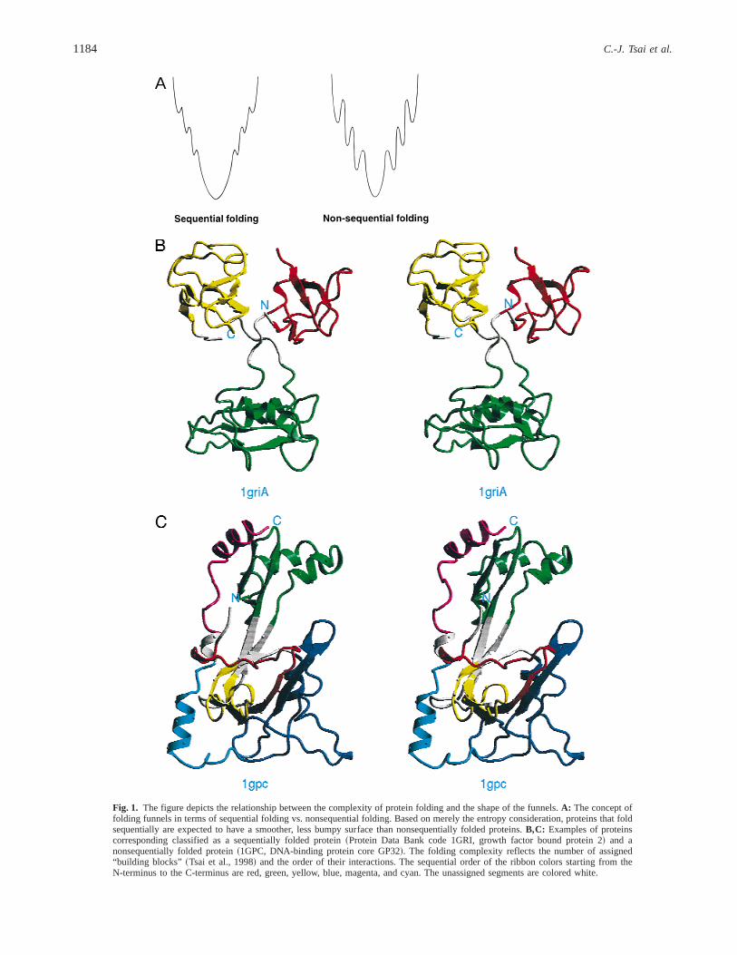
The process of protein folding can be either sequential or non-sequential~Tsai et al., 1999!. In a sequentially folding protein,sequentially covalently connected fragments are adjacent to oneanother in the three-dimensional~3D! structure. Such folding ismore often seen in eukaryotes and is thought to be the outcome ofslower translation rates~Netzer & Hartl, 1997!. In contrast, in anonsequentially folded protein, structurally adjacent fragments arenot connected sequentially. As a result, in such proteins the poly-peptide chain crosses back and forth between different domains~orhydrophobic folding units!. Nonsequential folding is more com-mon in prokaryotes typically having faster translation rates. Thelandscape of folding funnels can be smooth and simple. Simplerfunnels are likely to be observed in sequentially folding proteins.Nonsequentially folding proteins may translate to complicated fun-nels, with crevices and bumps along their paths. Such proteinshave a higher probability of being trapped in local minima, andhence misfold. On the other hand, sequentially folding proteins, inwhich consecutive fragments in the chain interact with each otherare less likely to misfold. Figure 1 depicts this relationship be-tween the complexity of protein folding and the shape of the funnels.
During evolution, proteins with nonrandom sequences, whichare able to fold rapidly, within biologically relevant time scales,have been selected. Folding funnels for such proteins have funnel-like energy landscapes. These landscapes consist of deep valley~s!
with well-defined local and global minima. On the other hand, aprotein with a random sequence will not be able to fold rapidly,due to the lack of stabilizing intramolecular hydrophobic and elec-trostatic interactions. Since for such a random sequence proteinthere is no energy difference between ordered and disordered states,this protein is expected to have a very shallow funnel~if it existsat all!, lacking any well-defined local or global minimum. As sucha protein is unlikely to exist in the living cell, we may argue thatfolding funnels are the result of evolutionary selection, acting inaccord with physical laws.
Clearly, if we use the unfolded state as a reference state and alldegrees of freedom as coordinates, a binding process involving analready folded protein can be depicted by a funnel-like shape.Further, there is also no doubt that the less frequent case of atwo-state binding process, in which binding and folding take placesimultaneously, also displays a funnel-like shape. However, as it ismore convenient to use the folded protein as a reference state, thequestion arises as to whether the binding process can still be por-trayed by a funnel under these circumstances. In the extreme caseof rigid protein binding, the degrees of freedom are reduced tothree translational and three rotational for each two-molecule as-sociation. However, even rigid docking requires the robust deter-mination of the global free energy minimum from the myriad of allpossible conformations. In this sense, problems of protein foldingand binding are quite similar, and thus one can extend Levinthal’sparadox to protein binding. However, as in the case of folding, itcan also be readily explained by the existence of the multiplepathways, here for protein–protein binding. Hence, the extensionof the funnel concept to binding processes should also be veryuseful ~Miller & Dill, 1997 !.
The difficulties involved in the prediction of docked configura-tions have been a subject of numerous studies~Katchalski-Katziret al., 1992; Cherfils & Janin, 1993; Norel et al., 1994, 1995; Fischeret al., 1995; Dunbrack et al., 1997!. The problem can be defined asfollows: given the structures of two molecules, predict their “cor-rect” bound configuration. If the structures to be docked are takenfrom their crystallized cocomplex, predicting their fitted associa-tion is relatively straightforward. On the other hand, if the struc-tures have been determined when the molecules are in their “free,”“unbound” state, the problem can be very difficult. Yet, even struc-tures presumably determined in the unbound state are actually alsobound; except in this case they are bound to their twin molecule inthe crystal. If the molecules are relatively rigid and have relativelysmooth funnels with a single or a few minima, their conformationaldiversity is limited, and hence there is a higher likelihood that theconformers binding in the “free,” “unbound” state will be those bind-ing to their ligand~s!. If, however, the protein is flexible, its foldingfunnel has rugged bottom consisting of several minima separatedby small energy barriers. Hence, its conformational diversity is larger,and the conformer binding to its cofactor, or ligand, may be dif-ferent from the one which is bound in the crystal. In such a case, ifin predicting the conformation of the complex one picks the struc-ture that is found in the crystal of the single molecule, achieving acorrect prediction of the bound conformation is much more diffi-cult. As Verkhivker et al.~1996! have pointed out, for robust ligand-protein docking, the binding energy landscape must have a funnelshape, leading to the global free energy minimum. This has recentlybeen elegantly illustrated by Zhang et al.~1999!, who have shownthat there are energy gradients, or funnels, near the binding sites.They have further used their finding to provide an explanation forthe rapid association rates.
Binding funnels and protein function 1183
A
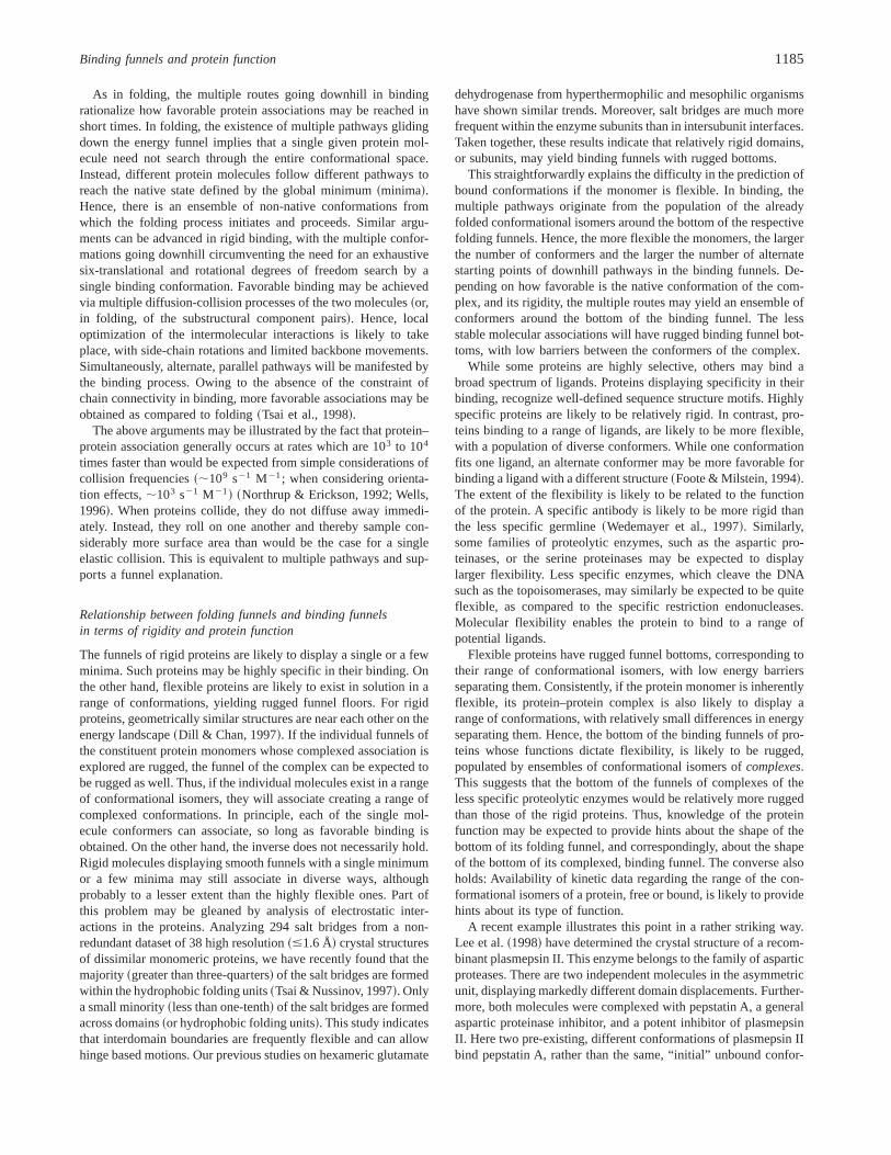
Fig. 1. The figure depicts the relationship between the complexity of protein folding and the shape of the funnels.A: The concept offolding funnels in terms of sequential folding vs. nonsequential folding. Based on merely the entropy consideration, proteins that foldsequentially are expected to have a smoother, less bumpy surface than nonsequentially folded proteins.B,C: Examples of proteinscorresponding classified as a sequentially folded protein~Protein Data Bank code 1GRI, growth factor bound protein 2! and anonsequentially folded protein~1GPC, DNA-binding protein core GP32!. The folding complexity reflects the number of assigned“building blocks” ~Tsai et al., 1998! and the order of their interactions. The sequential order of the ribbon colors starting from theN-terminus to the C-terminus are red, green, yellow, blue, magenta, and cyan. The unassigned segments are colored white.
1184 C.-J. Tsai et al.
As in folding, the multiple routes going downhill in bindingrationalize how favorable protein associations may be reached inshort times. In folding, the existence of multiple pathways glidingdown the energy funnel implies that a single given protein mol-ecule need not search through the entire conformational space.Instead, different protein molecules follow different pathways toreach the native state defined by the global minimum~minima!.Hence, there is an ensemble of non-native conformations fromwhich the folding process initiates and proceeds. Similar argu-ments can be advanced in rigid binding, with the multiple confor-mations going downhill circumventing the need for an exhaustivesix-translational and rotational degrees of freedom search by asingle binding conformation. Favorable binding may be achievedvia multiple diffusion-collision processes of the two molecules~or,in folding, of the substructural component pairs!. Hence, localoptimization of the intermolecular interactions is likely to takeplace, with side-chain rotations and limited backbone movements.Simultaneously, alternate, parallel pathways will be manifested bythe binding process. Owing to the absence of the constraint ofchain connectivity in binding, more favorable associations may beobtained as compared to folding~Tsai et al., 1998!.
The above arguments may be illustrated by the fact that protein–protein association generally occurs at rates which are 103 to 104
times faster than would be expected from simple considerations ofcollision frequencies~;109 s21 M21; when considering orienta-tion effects,;103 s21 M21! ~Northrup & Erickson, 1992; Wells,1996!. When proteins collide, they do not diffuse away immedi-ately. Instead, they roll on one another and thereby sample con-siderably more surface area than would be the case for a singleelastic collision. This is equivalent to multiple pathways and sup-ports a funnel explanation.
Relationship between folding funnels and binding funnelsin terms of rigidity and protein function
The funnels of rigid proteins are likely to display a single or a fewminima. Such proteins may be highly specific in their binding. Onthe other hand, flexible proteins are likely to exist in solution in arange of conformations, yielding rugged funnel floors. For rigidproteins, geometrically similar structures are near each other on theenergy landscape~Dill & Chan, 1997!. If the individual funnels ofthe constituent protein monomers whose complexed association isexplored are rugged, the funnel of the complex can be expected tobe rugged as well. Thus, if the individual molecules exist in a rangeof conformational isomers, they will associate creating a range ofcomplexed conformations. In principle, each of the single mol-ecule conformers can associate, so long as favorable binding isobtained. On the other hand, the inverse does not necessarily hold.Rigid molecules displaying smooth funnels with a single minimumor a few minima may still associate in diverse ways, althoughprobably to a lesser extent than the highly flexible ones. Part ofthis problem may be gleaned by analysis of electrostatic inter-actions in the proteins. Analyzing 294 salt bridges from a non-redundant dataset of 38 high resolution~#1.6 Å! crystal structuresof dissimilar monomeric proteins, we have recently found that themajority ~greater than three-quarters! of the salt bridges are formedwithin the hydrophobic folding units~Tsai & Nussinov, 1997!. Onlya small minority~less than one-tenth! of the salt bridges are formedacross domains~or hydrophobic folding units!. This study indicatesthat interdomain boundaries are frequently flexible and can allowhinge based motions. Our previous studies on hexameric glutamate
dehydrogenase from hyperthermophilic and mesophilic organismshave shown similar trends. Moreover, salt bridges are much morefrequent within the enzyme subunits than in intersubunit interfaces.Taken together, these results indicate that relatively rigid domains,or subunits, may yield binding funnels with rugged bottoms.
This straightforwardly explains the difficulty in the prediction ofbound conformations if the monomer is flexible. In binding, themultiple pathways originate from the population of the alreadyfolded conformational isomers around the bottom of the respectivefolding funnels. Hence, the more flexible the monomers, the largerthe number of conformers and the larger the number of alternatestarting points of downhill pathways in the binding funnels. De-pending on how favorable is the native conformation of the com-plex, and its rigidity, the multiple routes may yield an ensemble ofconformers around the bottom of the binding funnel. The lessstable molecular associations will have rugged binding funnel bot-toms, with low barriers between the conformers of the complex.
While some proteins are highly selective, others may bind abroad spectrum of ligands. Proteins displaying specificity in theirbinding, recognize well-defined sequence structure motifs. Highlyspecific proteins are likely to be relatively rigid. In contrast, pro-teins binding to a range of ligands, are likely to be more flexible,with a population of diverse conformers. While one conformationfits one ligand, an alternate conformer may be more favorable forbinding a ligand with a different structure~Foote & Milstein, 1994!.The extent of the flexibility is likely to be related to the functionof the protein. A specific antibody is likely to be more rigid thanthe less specific germline~Wedemayer et al., 1997!. Similarly,some families of proteolytic enzymes, such as the aspartic pro-teinases, or the serine proteinases may be expected to displaylarger flexibility. Less specific enzymes, which cleave the DNAsuch as the topoisomerases, may similarly be expected to be quiteflexible, as compared to the specific restriction endonucleases.Molecular flexibility enables the protein to bind to a range ofpotential ligands.
Flexible proteins have rugged funnel bottoms, corresponding totheir range of conformational isomers, with low energy barriersseparating them. Consistently, if the protein monomer is inherentlyflexible, its protein–protein complex is also likely to display arange of conformations, with relatively small differences in energyseparating them. Hence, the bottom of the binding funnels of pro-teins whose functions dictate flexibility, is likely to be rugged,populated by ensembles of conformational isomers ofcomplexes.This suggests that the bottom of the funnels of complexes of theless specific proteolytic enzymes would be relatively more ruggedthan those of the rigid proteins. Thus, knowledge of the proteinfunction may be expected to provide hints about the shape of thebottom of its folding funnel, and correspondingly, about the shapeof the bottom of its complexed, binding funnel. The converse alsoholds: Availability of kinetic data regarding the range of the con-formational isomers of a protein, free or bound, is likely to providehints about its type of function.
A recent example illustrates this point in a rather striking way.Lee et al.~1998! have determined the crystal structure of a recom-binant plasmepsin II. This enzyme belongs to the family of asparticproteases. There are two independent molecules in the asymmetricunit, displaying markedly different domain displacements. Further-more, both molecules were complexed with pepstatin A, a generalaspartic proteinase inhibitor, and a potent inhibitor of plasmepsinII. Here two pre-existing, different conformations of plasmepsin IIbind pepstatin A, rather than the same, “initial” unbound confor-
Binding funnels and protein function 1185
mation binding through an induced fit mechanism. Hence, in a sim-ilar vein, the bottom of the funnel of the complex is populated bydifferent complexed conformations. In the crystal, they bind to eachother as different conformers. We may reasonably assume that ad-ditional conformational isomers of the complex exist in solution.However, the conformers of the complex that cocrystallize are morecomplementary, and hence more favorable for binding to each other.The equilibrium of thecomplexedconformations around the bot-tom of the binding funnel results in a conformational shift favoringthe conformers of the complex, which bind to the growing crystal.
Previously, it has been shown that two molecules in the samecrystallographic asymmetry unit can differ in their relative domain–domain orientation. Muller et al.~1998! have crystallized the tissuefactor, a member of the cytokine receptor superfamily. Tissue fac-tor is an obligate cofactor of coagulation factor VIIa. Muller et al.have shown that there is a hinge rotation of 12.78 in the rabbittissue factor between the domains when the two molecules in theasymmetric unit are compared. This suggests that under crystalli-zation conditions at least one of these conformers was in a highlypopulated state. The other conformer is complementary to it, andthus binds and cocrystallizes with it, with the equilibrium shiftingin its direction. Hence, again, binding and folding behave simi-larly. In the plasmepsin case, the different conformers of the com-plex bind as a unit to the growing crystal. In the tissue factor case,it is different conformers of thesingle molecules, which bind toeach other in the asymmetric unit of the crystal.
The function of the proteolytic enzymes is to catalyze hydrolysisof the protein backbone via nucleophilic attack on the carbonylcarbon of the peptide bond. During this nucleophilic attack, thetrigonal carbonyl carbon of the peptide bond becomes tetragonal in
the intermediate, or transition state~Creighton, 1993!. Five differ-ent catalytic genera of proteolytic enzymes, in which serine, thre-onine, cysteine, aspartic or metallo groups provide a nucleophileduring catalysis, are known. These five genera can be subdividedinto innumerable clans and families~Barrett et al., 1998!. Forexample, there are about 40 families of serine- and threonine-proteinases alone~Perona & Craik, 1995; Barrett et al., 1998!.While being excellent examples of convergent evolution, theseenzymes also show extensive structural diversity. Actually, it isoften the case that enzymes belonging to different clans adoptdifferent overall folds. Consider, for example, the serine protein-ases. Trypsin, streptogrisin A, togavirin, IgA1-specific serine en-dopeptidase, hepatitis C polyprotein peptidase, etc. belong to clanSA. They all adopt a doubleb-barrel fold. On the other hand, thosebelonging to clan SC, namely, prolyl oligopeptidase, carboxypep-tidase C, etc., adopt ana0b hydrolase fold~Barrett et al., 1998!.The structural diversity of proteinases is equally matched by thediversity of the substrates they recognize. For a particular protein-ase, only a limited extent of substrate specificity is found. Trypsinprefers to cut the peptide bond following Arg or Lys residues,while chymotrypsin cuts the peptide bonds formed by Tyr, Phe,Trp, and Leu. However, it is evident that these enzymes are capa-ble of recognizing different substrate conformations. This indicatesrugged bottoms in the binding funnels of such enzyme substrate com-plexes, which may, in turn, arise from the flexibility of the enzymeand of the substrate molecules. Similarly, topoisomerases I and IIcleave DNA strands nonspecifically. In this case, the enzyme is alsoable to recognize different substrate conformations. Hence, topo-isomerases are also a good example of flexible binding with theenzyme-substrate complex having rugged bottom binding funnels.
A
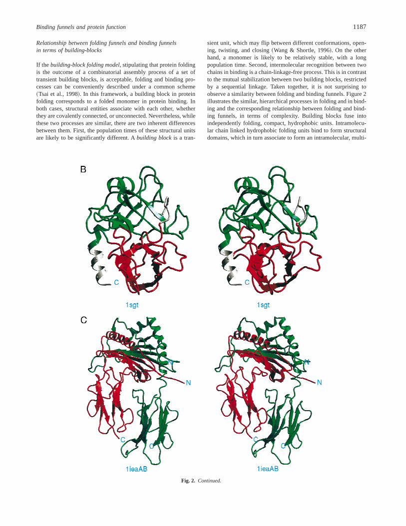
Fig. 2. The figure illustrates the hierarchical processes in folding and inbinding, and the corresponding relationship between folding funnels andbinding funnels, in terms ofcomplexity. A: The funnel complexity isillustrated by the increasing number of bumps from building blocks tohydrophobic folding units~HFU!, to domains, to protein fold, and finallyto the quaternary structure, the outcome of a bindings process. In general,the hierarchical processes act like~consecutively! fusing two individualfunnels into one.B: Two HFUs associate as a functional domain.C: Twomultidomain subunits bind to form a functional heterodimer. The protein inB is trypsin~1SGT! with each HFU in different color. The proteins inC isa MHC CLASS II complex with covalently bound HB peptide~1IEA!.~Figure continues on facing page.!
1186 C.-J. Tsai et al.
Relationship between folding funnels and binding funnelsin terms of building-blocks
If the building-block folding model, stipulating that protein foldingis the outcome of a combinatorial assembly process of a set oftransient building blocks, is acceptable, folding and binding pro-cesses can be conveniently described under a common scheme~Tsai et al., 1998!. In this framework, a building block in proteinfolding corresponds to a folded monomer in protein binding. Inboth cases, structural entities associate with each other, whetherthey are covalently connected, or unconnected. Nevertheless, whilethese two processes are similar, there are two inherent differencesbetween them. First, the population times of these structural unitsare likely to be significantly different. Abuilding blockis a tran-
sient unit, which may flip between different conformations, open-ing, twisting, and closing~Wang & Shortle, 1996!. On the otherhand, a monomer is likely to be relatively stable, with a longpopulation time. Second, intermolecular recognition between twochains in binding is a chain-linkage-free process. This is in contrastto the mutual stabilization between two building blocks, restrictedby a sequential linkage. Taken together, it is not surprising toobserve a similarity between folding and binding funnels. Figure 2illustrates the similar, hierarchical processes in folding and in bind-ing and the corresponding relationship between folding and bind-ing funnels, in terms of complexity. Building blocks fuse intoindependently folding, compact, hydrophobic units. Intramolecu-lar chain linked hydrophobic folding units bind to form structuraldomains, which in turn associate to form an intramolecular, multi-
Fig. 2. Continued.
Binding funnels and protein function 1187
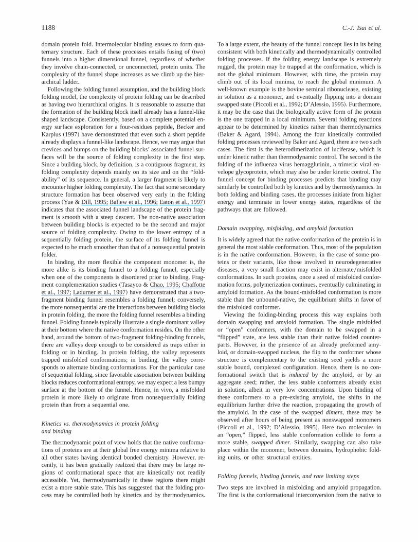
domain protein fold. Intermolecular binding ensues to form qua-ternary structure. Each of these processes entails fusing of~two!funnels into a higher dimensional funnel, regardless of whetherthey involve chain-connected, or unconnected, protein units. Thecomplexity of the funnel shape increases as we climb up the hier-archical ladder.
Following the folding funnel assumption, and the building blockfolding model, the complexity of protein folding can be describedas having two hierarchical origins. It is reasonable to assume thatthe formation of the building block itself already has a funnel-likeshaped landscape. Consistently, based on a complete potential en-ergy surface exploration for a four-residues peptide, Becker andKarplus~1997! have demonstrated that even such a short peptidealready displays a funnel-like landscape. Hence, we may argue thatcrevices and bumps on the building blocks’ associated funnel sur-faces will be the source of folding complexity in the first step.Since a building block, by definition, is a contiguous fragment, itsfolding complexity depends mainly on its size and on the “fold-ability” of its sequence. In general, a larger fragment is likely toencounter higher folding complexity. The fact that some secondarystructure formation has been observed very early in the foldingprocess~Yue & Dill, 1995; Ballew et al., 1996; Eaton et al., 1997!indicates that the associated funnel landscape of the protein frag-ment is smooth with a steep descent. The non-native associationbetween building blocks is expected to be the second and majorsource of folding complexity. Owing to the lower entropy of asequentially folding protein, the surface of its folding funnel isexpected to be much smoother than that of a nonsequential proteinfolder.
In binding, the more flexible the component monomer is, themore alike is its binding funnel to a folding funnel, especiallywhen one of the components is disordered prior to binding. Frag-ment complementation studies~Tasayco & Chao, 1995; Chaffotteet al., 1997; Ladurner et al., 1997! have demonstrated that a two-fragment binding funnel resembles a folding funnel; conversely,the more nonsequential are the interactions between building blocksin protein folding, the more the folding funnel resembles a bindingfunnel. Folding funnels typically illustrate a single dominant valleyat their bottom where the native conformation resides. On the otherhand, around the bottom of two-fragment folding-binding funnels,there are valleys deep enough to be considered as traps either infolding or in binding. In protein folding, the valley representstrapped misfolded conformations; in binding, the valley corre-sponds to alternate binding conformations. For the particular caseof sequential folding, since favorable association between buildingblocks reduces conformational entropy, we may expect a less bumpysurface at the bottom of the funnel. Hence, in vivo, a misfoldedprotein is more likely to originate from nonsequentially foldingprotein than from a sequential one.
Kinetics vs. thermodynamics in protein foldingand binding
The thermodynamic point of view holds that the native conforma-tions of proteins are at their global free energy minima relative toall other states having identical bonded chemistry. However, re-cently, it has been gradually realized that there may be large re-gions of conformational space that are kinetically not readilyaccessible. Yet, thermodynamically in these regions there mightexist a more stable state. This has suggested that the folding pro-cess may be controlled both by kinetics and by thermodynamics.
To a large extent, the beauty of the funnel concept lies in its beingconsistent with both kinetically and thermodynamically controlledfolding processes. If the folding energy landscape is extremelyrugged, the protein may be trapped at the conformation, which isnot the global minimum. However, with time, the protein mayclimb out of its local minima, to reach the global minimum. Awell-known example is the bovine seminal ribonuclease, existingin solution as a monomer, and eventually flipping into a domainswapped state~Piccoli et al., 1992; D’Alessio, 1995!. Furthermore,it may be the case that the biologically active form of the proteinis the one trapped in a local minimum. Several folding reactionsappear to be determined by kinetics rather than thermodynamics~Baker & Agard, 1994!. Among the four kinetically controlledfolding processes reviewed by Baker and Agard, there are two suchcases. The first is the heterodimerization of luciferase, which isunder kinetic rather than thermodynamic control. The second is thefolding of the influenza virus hemagglutinin, a trimeric viral en-velope glycoprotein, which may also be under kinetic control. Thefunnel concept for binding processes predicts that binding maysimilarly be controlled both by kinetics and by thermodynamics. Inboth folding and binding cases, the processes initiate from higherenergy and terminate in lower energy states, regardless of thepathways that are followed.
Domain swapping, misfolding, and amyloid formation
It is widely agreed that the native conformation of the protein is ingeneral the most stable conformation. Thus, most of the populationis in the native conformation. However, in the case of some pro-teins or their variants, like those involved in neurodegenerativediseases, a very small fraction may exist in alternate0misfoldedconformations. In such proteins, once a seed of misfolded confor-mation forms, polymerization continues, eventually culminating inamyloid formation. As the bound-misfolded conformation is morestable than the unbound-native, the equilibrium shifts in favor ofthe misfolded conformer.
Viewing the folding-binding process this way explains bothdomain swapping and amyloid formation. The single misfoldedor “open” conformers, with the domain to be swapped in a“flipped” state, are less stable than their native folded counter-parts. However, in the presence of an already preformed amy-loid, or domain-swapped nucleus, the flip to the conformer whosestructure is complementary to the existing seed yields a morestable bound, complexed configuration. Hence, there is no con-formational switch that isinduced by the amyloid, or by anaggregate seed; rather, the less stable conformers already existin solution, albeit in very low concentrations. Upon binding ofthese conformers to a pre-existing amyloid, the shifts in theequilibrium further drive the reaction, propagating the growth ofthe amyloid. In the case of the swappeddimers, these may beobserved after hours of being present as nonswapped monomers~Piccoli et al., 1992; D’Alessio, 1995!. Here two molecules inan “open,” flipped, less stable conformation collide to form amore stable,swapped dimer. Similarly, swapping can also takeplace within the monomer, between domains, hydrophobic fold-ing units, or other structural entities.
Folding funnels, binding funnels, and rate limiting steps
Two steps are involved in misfolding and amyloid propagation.The first is the conformational interconversion from the native to
1188 C.-J. Tsai et al.
the misfolded monomer, and the second is the binding of themisfolded conformer to the growing amyloid fiber. Can one predictthe rate limiting step from the shape of the funnel? If the barriersare low, such as in cases of highly flexible proteins existing in arange of conformations, the rate limiting steps are unlikely to bethe conformational interconversions. In such cases, the limitingsteps might be the binding. On the other hand, if the barriers arehigh, the conformational interconversion may be expected to serveas the bottle neck in amyloid growth~Prusiner, 1991; Jarrett &Lansbury, 1993!. However, amyloid formation is an extremelyslow process. Hence, it is quite likely that the limiting step isdetermined by the formation of the seed, that is, the initial amyloidfiber, which in turn is determined by the conformational intercon-version and binding taking place simultaneously. In the correspond-ing folding-binding funnel, there is an entropy barrier that cannotbe overcome without the presence of amyloid. This is similar to thecase of supercool water, which stays in a liquid state until a seedis dropped into it. The seed dramatically reduces the barrier heightbetween the liquid water state and the solid ice state.
Interestingly, nucleation is also found to be the rate limiting stepin protein folding. Experiment and theory are both in agreementregarding the importance of nucleation mechanisms in protein fold-ing. The seeds for protein binding~amyloid propagation! are clas-sical nuclei. These are well-formed elements of structure present inthe ground states~final amyloid!. However, the nucleation mech-anisms use diffuse, extended regions, which are observed in thetransition states~Fersht, 1997!.
The complexity of protein folding, binding mechanisms,and the funnel shape
Given the structure of the protein monomer, we may have a notionof the shape of its funnel. The larger the complexity of its structure,the more jagged the walls of the funnel are likely to be. Thestructural complexity is a function of the types of interactions inthe protein; namely, the complexity depends on whether the seg-ments of the chain, which interact with each other, are sequentiallyor nonsequentially connected~Baldwin & Rose, 1999!. In sequen-tial folding, the shape of the funnel is the outcome of joining manymicrofunnels, each corresponding to a transient “building block”of the protein structure. Sequential folding has been proposed tooccur more frequently in eukaryotic cells, having a slower rate oftranslation~Netzer & Hartl, 1997!. On the other hand, in prokary-otes, where the rate of protein synthesis is considerably faster,chances of nonsequential folding are higher. Since in nonsequen-tial folding the probability of misfolding is consequently substan-tially higher, there may be a more frequent need for the help ofmolecular chaperones. On the next hierarchical level~Tsai et al.,1998!, we observe binding funnels. These similarly reflect theconsequences of merging single-molecule funnels. In both fold-ing and binding processes, the same fundamental principles areinvolved.
While inspection of the funnel walls narrates the story of thecomplexity of the folding and its potential~sequential or non-sequential! mechanism, inspection of the funnel bottoms revealsthe binding mechanism. If around the bottom of the funnel of themonomer we observe a conformational ensemble, depicted as aseries of minima that are close to each other on the energy land-scape with low barriers separating them, we may deduce the ex-istence of a flexible protein. On the other hand, a smooth bottom,with well-defined, deep minimum, implies a rigid molecule. Flex-
ible molecules exhibit conformational diversity. Yet, they haveoften been viewed as displaying an “induced fit” binding mecha-nism, depending on which conformations have been crystallized inthe “free,” “unbound”~to its cognate ligand, though bound to itstwin molecule in the crystal! form. The conformations of suchmolecules have also sometimes been suggested to be the outcomeof “crystal effects,” again, depending on which conformers havebeen favorable for binding in the crystal. Furthermore, foldingfunnels illustrating bottoms with a deep minimum, with additionalminima relatively not too different in depth, and with the inter-vening barriers still not too high, may reflect metastable states. Thecorresponding proteins may be candidates for domain swapping, orfor forming amyloids.
Moreover, inspection of the extent of the ruggedness around thebottom of the folding funnels enables us to have a notion of theextent of the ruggedness around the bottom of the correspondingbinding funnels. In the case of a flexible monomer, a number ofconformational isomers may bind a given ligand. While the sta-bility of the resulting complexes may differ, if the single moleculeconformers are relatively close on the energy landscape, the com-plexed conformers may similarly be conformationally and ener-getically quite close. On the other hand, for a rigid molecule, witha well-defined minimum~or a few minima! and a smooth bottom,a consistently similarly shaped funnel floor may be observed in thebinding funnel. One may expect a considerably smaller number ofcomplexed, favorable, bound conformations. Hence, the shape ofthe folding funnel of the single molecule is very instructive. Theconverse also holds,if kinetic data exist. If available, we may beable to predict the mechanism of folding, of binding, and possiblyin some cases the rate limiting steps in the binding process. Weshall further be able infer its binding selectivity or its nonspeci-ficity. Thus, the concept of the funnel goes well beyond compre-hension of the multiple routes going downhill, and overcoming thepuzzling time scale paradox. Given thebiological considerationsof the rates of synthesis in eukaryotes and in prokaryotes, and ofthe function of the proteinvia its binding, the funnel concept isvery useful.
Acknowledgments
We thank Dr. Jacob Maizel for encouragement and for helpful discussions.The research of R. Nussinov in Israel has been supported in part by grantnumber 95-00208 from BSF, Israel, by a grant from the Israel ScienceFoundation administered by the Israel Academy of Sciences, by the Magnetgrant, by the Ministry of Science grant, and by the Tel Aviv UniversityBasic Research and Adams Brain Center grants. This project has beenfunded in whole or in part with Federal funds from the National CancerInstitute, National Institutes of Health, under contract number NO1-CO-56000. The content of this publication does not necessarily reflect the viewor policies of the Department of Health and Human Services, nor doesmention of trade names, commercial products, or organization imply en-dorsement by the U.S. government.
References
Baker D, Agard DA. 1994. Kinetics versus thermodynamics in protein folding.Biochemistry 33:7505–7509.
Baldwin RL. 1994. Matching speed and stability.Nature 369:183–184.Baldwin RL. 1995. The nature of protein folding pathways: The classical versus
the new view.J Biomol NMR 5:103–109.Baldwin RL, Rose GD. 1999. Is protein folding hierarchic? I. Local structure
and peptide folding.Trends Biochem Sci 24:26–33.Ballew RM, Sabelko J, Gruebele M. 1996. Direct observation of fast protein
folding: The initial collapse of apomyoglobin.Proc Natl Acad Sci USA93:5759–5764.
Binding funnels and protein function 1189
Barrett AJ, Rawlings ND, Woessner JF. 1998.Handbook of proteolytic enzymes.San Diego, California: Academic Press.
Becker OM, Karplus M. 1997. The topology of multidimensional potentialenergy surfaces: Theory and application to peptide structure and kinetics.J Chem Phys 106:1495–1517.
Bennett MJ, Choe S, Eisenberg D. 1994. Domain swapping: Entangling alli-ances between proteins.Proc Natl Acad Sci USA 91:3127–3131.
Bennett MJ, Schlunegger MP, Eisenberg D. 1995. 3D domain swapping: Amechanism for oligomer assembly.Protein Sci 4:2455-2468.
Bryngelson JD, Wolynes PG. 1989. Intermediates and barrier crossing in arandom energy model~with applications to protein folding!. J Phys Chem93:6902–6915.
Chaffotte AF, Li JH, Georgescu RE, Goldberg ME, Tasayco ML. 1997. Rec-ognition between disordered states: Kinetics of the self-assembly of thio-redoxin fragments.Biochemistry 36:16040–16048.
Chan HS, Dill KA. 1994. Transition states and folding dynamics of proteins andheteropolymers.J Chem Phys 100:9238–9257.
Chan HS, Dill KA. 1998. Protein folding in the landscape perspective: Chevronplots and non-Arrhenius kinetics.Proteins 30:2–33.
Cherfils J, Janin J. 1993. Protein docking algorithms: Simulating molecularrecognition.Curr Opin Struct Biol 3:265–269.
Creighton TE. 1993.Proteins: Structure and molecular properties, 2nd ed.NewYork: WH Freeman and Company.
D’Alessio G. 1995. Oligomer evolution in action.Nat Struct Biol 2:11–13.Dill KA. 1999. Polymer principles and protein folding.Protein Sci 8:1166–
1180.Dill KA, Chan HS. 1997. From Levinthal to pathways to funnels.Nat Struct
Biol 4:10–19.Dill KA, Phillips, AT, Rosen JB. 1997. Protein structure and energy landscape
dependence on sequence using a continuous energy function.J Comp Biol4:227–239.
Dunbrack RL, Gerloff DL, Bower M, Chen X, Lichtarge O, Cohen FE. 1997.Meeting review: The second meeting on the critical assessment of tech-niques for protein structure prediction~CASP2!, Asilomar, California, De-cember 13–16, 1996.Folding Design 2:R27–R42.
Eaton WA, Munoz V, Thompson PA, Chan CK, Hofrichter J. 1997. Submilli-second kinetics of protein folding.Curr Opin Struct Biol 7:10–14.
Fersht AR. 1997. Nucleation mechanisms in protein folding.Curr Opin StructBiol 7:3–9.
Fischer D, Lin SL, Wolfson HJ, Nussinov, R. 1995. A geometry-based suite ofmolecular docking processes.J Mol Biol 248:459–477.
Fischer E. 1894.Ber Dt Chem Ges 27:2985–2991.Foote J, Milstein C. 1994. Conformational isomerism and the diversity of anti-
bodies.Proc Natl Acad Sci USA 91:10370–10374.Frauenfelder H, Leeson DT. 1998. The energy landscape in non-biological
molecules.Nat Struct Biol 5:757–759.Frauenfelder H, Sligar SG, Wolynes PG. 1991. The energy landscapes and
motions of proteins.Science 254:1598–1603.Gruebele M, Wolynes P. 1998. Satisfying turns in folding transitions.Nat Struct
Biol 5:662–665.Jarrett JT, Lansbury PT Jr. 1993. Seeding “one-dimensional crystallization” of
amyloid: A pathogenic mechanism in Alzheimer’s disease and scrapie?Cell73:1055–1058.
Karplus M. 1997. The Levinthal paradox: yesterday and today.Folding Design2:S69–S75.
Karplus M, Sali A, Shakhnovitch E. 1995. Comment: Kinetics of protein fold-ing. Nature 373:664–665.
Karplus M, Shakhnovitch E. 1992. Protein folding: Theoretical studies of thermo-dynamics and dynamics. In: Creighton T, ed.Protein folding. New York:WH Freeman & Sons. pp 127–195.
Katchalski-Katzir E, Shariv I, Eisenstein M, Friesem AA, Aflalo C, Vakser IA.1992. Molecular surface recognition: Determination of geometric fit be-tween protein and their ligands by correlation techniques.Proc Natl AcadSci USA 89:2195–2199.
Koshland DE Jr. 1958. Application of a theory of enzyme specificity to proteinsynthesis.Proc Natl Acad Sci USA 44:98–123.
Ladurner AG, Itzhaki LS, Gay FDP, Fersht AR. 1997. Complementation ofpeptide fragments of the single domain protein chymotrypsin inhibitor 2.J Mol Biol 273:317–329.
Lazaridis T, Karplus M. 1997. “New view” of protein folding reconciled withthe old through multiple unfolding simulations.Science 278:1928–1931.
Lee AY, Gulnik SV, Erickson JW. 1998. Conformational switching in an asparticproteinase.Nat Struct Biol 5:866–871.
Levinthal C. 1969. How to fold graciously. In: Debrunner P, Tsibris JCM,
Munck E, eds.Mossbauer spectroscopy in biological systems~Proceedingsof a meeting held at Allerton House, Monticello, Illinois!. Urbana, Illinois:University of Illinois Press. p 22.
Martinez JC, Pisabarro MT, Serrano L. 1998. Obligatory steps in protein foldingand the conformational diversity of the transition state.Nat Struct Biol5:721–729.
Miller DW, Dill KA. 1997. Ligand binding to proteins: The binding landscapemodel.Protein Sci 6:2166–2179.
Muller Y, Kelley RF, De Vos AM. 1998. Hinge bending within the cytokinereceptor superfamily revealed by the 2.4 Å crystal structure of the extra-cellular domain of the rabbit tissue factor.Protein Sci 7:1106–1115.
Netzer WJ, Hartl FU. 1997. Recombination of protein domains facilitated byco-translational folding in eukaryotes.Nature 388:343–349.
Norel R, Lin SL, Wolfson H, Nussinov R. 1994. Shape complementarity atprotein–protein interfaces.Biopolymers 34:933–940.
Norel R, Lin SL, Wolfson H, Nussinov R. 1995. Molecular surface comple-mentarity at protein–protein interfaces: The critical role played by surfacenormals at well placed, sparse, points in docking.J Mol Biol 252:263–273.
Northrup SH, Erickson HP. 1992. Kinetics of protein–protein association ex-plained by Brownian dynamics computer simulation.Proc Natl Acad SciUSA 89:3338–3342.
Onuchic JN, Wolynes PG, Luthey-Schulten Z, Socci ND. 1995. Towards anoutline of the topography of a realistic protein folding funnel.Proc NatlAcad Sci USA 92:3626–3630.
Perona JJ, Craik CS. 1995. Structural basis of substrate specificity in the serineproteases.Protein Sci 4:337–360.
Piccoli R, Tamburrini M, Piccialli G, Di Donato A, Parente A, D’Alessio G.1992. The dual-mode quaternary structure of seminal rnase.Proc Natl AcadSci USA 89:1870–1874.
Prusiner SB. 1991. Molecular biology of prion diseases.Science 252:1515–1522.
Tasayco ML, Chao K. 1995. NMR study of the reconstitution of theb-sheet ofthioredoxin by fragment complementation.Proteins SFG 22:41–44.
Tsai CJ, Lin SL, Wolfson, H, Nussinov R. 1996. Protein–protein interfaces:Architectures and interactions in protein–protein interfaces and in proteincores. Their similarities and differences.Critic Rev Biochem Mol Biol 31:127–152.
Tsai CJ, Lin SL, Wolfson, H, Nussinov R. 1997a. Studies of protein–proteininterfaces: Statistical analysis of the hydrophobic effect.Protein Sci 6:53-64.
Tsai CJ, Maizel JV Jr, Nussinov R. 1999. Distinguishing between sequential andnon-sequentially folded protein: Implications for folding and misfolding.Protein Sci. In press.
Tsai CJ, Nussinov R. 1997. Hydrophobic folding units derived from dissimilarmonomer structures and their interactions.Protein Sci 6:24–42.
Tsai CJ, Xu D, Nussinov R. 1997b. Structural motifs at protein–protein inter-faces: Protein cores versus two-state and three-state model complexes.Pro-tein Sci 6:1793–1805.
Tsai CJ, Xu D, Nussinov R. 1998. Protein folding via binding, and vice versa.Folding Design 3:R71–R80.
Verkhivker GM, Rejto PA, Gehlhaar DK, Freer ST. 1996. Exploring the energylandscapes of molecular recognition by a genetic algorithm: Analysis of therequirements for robust docking of HIV-1 protease and FKBP-12 com-plexes.Proteins SFG 250:342–352.
Wales DJ, Miller MA, Walsh TR. 1998. Archetypal energy landscapes.Nature394:758–760.
Wang Y, Shortle D. 1996. A dynamic bundle of four adjacent hydrophobicsegments in the denatured state of staphylococcal nuclease.Protein Sci5:1898–1906.
Wedemayer GJ, Patten PA, Wang LH, Schultz PG, Stevens RC. 1997. Structuralinsights into the evolution of an antibody combining site.Nat Struct Biol276:1665–1669.
Wells JA. 1996. Binding in the growth hormone receptor complex.Proc NatlAcad Sci USA 93:1–6.
Wolynes PG, Onuchic JN, Thirumalai D. 1995. Navigating the folding routes.Science 267:1619–1620.
Yue K, Dill KA. 1995. Forces of tertiary structural organization in globularproteins.Proc Natl Acad Sci USA 92:146–150.
Xu D, Lin SL, Nussinov R. 1997. Protein binding versus protein folding: Therole of hydrophilic bridges in protein association.J Mol Biol 265:68–84.
Xu D, Tsai CJ, Nussinov R. 1998. Mechanism and evolution of protein dimer-ization.Protein Sci 7:533–544.
Zhang C, Chen J, DeLisi C. 1999. Protein–protein recognition: Exploring theenergy funnels near the binding sites.Proteins SFG 34:255–267.
1190 C.-J. Tsai et al.
Related Documents